
Submitted:
09 October 2024
Posted:
15 October 2024
You are already at the latest version
Abstract
Treating odontogenic infections of the head, neck and oral cavity requires the understanding of surgical and pharmacotherapeutic interventions as they apply to single and multispace infections. Three clinical cases are presented where Prevotella species were significant opportunistic pathogens. Since Prevotella can become virulent under conditions including post-extraction wound healing of an extraction site adjacent to an intact dentition, excessive plaque and calculus, nearby gingivitis or periodontitis, or an immunocompromised host, Metronidazole administration was deemed necessary in the cases described here. This intervention mitigated or resolved these odontogenic infections that were refractory to conservative surgical therapy that included source control. The involvement of Prevotella in odontogenic infections and pericoronitis, and the antibiotics used to treat common oral and maxillofacial infections with this bacterium are reviewed. Prevotella is susceptible to Metronidazole (Flagyl), and the antibiotic is effective in mitigating the disease process when basic principles of the treatment of odontogenic infections are employed.
Keywords:
Prevotella
; odontogenic infection
; metronidazole(flagyl)
; pericoronitis
Introduction
Prevotella species are inhabitants of the periodontal sulcus, living in the crevicular fluid and in the dental biofilm attached to hard tissue surfaces (teeth) and mucosa. These obligate anaerobic Gram-negative rods are part of the normal oral microbiome and are not opportunistic microorganisms in the immunocompetent host [1].
Mixed infections comprising anaerobic, facultative anaerobic, and aerobic bacteria colonize perialveolar and fascial spaces, contributing to odontogenic infections of the head and neck that can progress to life-threatening circumstances, including bacteremia, sepsis, airway compromise and death [2,3]. Pericoronitis, perialveolar infections (sometimes called subperiosteal infections), and multi-space odontogenic infections all show evidence of Prevotella species as isolates in the mixed oral flora.
Odontogenic infections are infections of tooth-related origins usually relating to endodontic or periodontal sources [4,5]. Possible etiologies can include necrotic pulp from carious or fractured teeth, pericoronitis, or deep periodontal pockets where bacteria cause infections [6]. Anaerobic and aerobic bacteria are implicated in 50–60% of odontogenic infections, with the most commonly isolated facultative anaerobic organisms being Viridans streptococci, and the most commonly isolated obligate anaerobic bacteria being Bacteroides spp, Prevotella spp, and Peptostretpococcus spp. [4].
Pericoronitis is an infection of the soft tissue around the crown of a partially erupted tooth and is usually caused by normal microorganisms that inhabit the oral cavity [4]. Bacterial plaque can accumulate underneath the soft tissue which may lead to inflammation [7]. The bacteria that are found at the highest levels in symptomatic pericoronitis include Actinomyces oris, Eikenella corrodens, Eubacterium nodatum, Fusobacterium nucleatum, Treponema denticola, Eubacterium saburreum, and various Streptococcus species [8]. With proper oral hygiene and strong host defenses, most patients can eliminate the bacteria that are trapped under the operculum without leading to further complications or infections. If the bacteria are not removed under the operculum, pericoronitis can lead to jaw inflammation and phlegmon [9]. Infection may spread, causing a multi-space infection of the head and neck regions.
Management of odontogenic infections in the oral cavity includes controlling the source of the infection by surgical removal, establishing drainage, and mobilizing the host defense system to fight the infection. The clinician must have fundamental knowledge of the anatomy of the head and neck region, specifically the fascial planes through which infections can spread. Odontogenic infections typically begin as vestibular space abscesses where it is localized and can be treated easily [4]. The infection, when localized, may present as an abscess, swelling, cellulitis, and trismus. If the infection persists and spreads, it will take the path of least resistance, potentially leading to life-threatening complications. Deep fascial space infections of the neck are most frequently odontogenic in origin and can be life-threatening if the abscess extends to the adjacent fascial space [10,11].
Principles of source control include (i) decreasing the bacterial load by incision, opening the wound to introduce air, and changing the environment from anaerobic to aerobic, thus eradicating obligate anaerobes; (ii) drainage that comprises decompressing the swelling, releasing the purulent exudate, and promoting the blood supply; (iii) irrigation, thereby washing out bacteria, bacterial toxins and inflammatory by-products; and (iv) tooth extraction [12]. Empiric antibiotics targeted to opportunistic Prevotella species are used in immunocompromised hosts, severe infections, and infections refractory to surgery.
Methods
Three clinical cases are presented below where Prevotella species were significant opportunistic pathogens in odontogenic infections, and surgery combined with metronidazole was used to eradicate the infections. For the narrative review, the PubMed database was searched using the following terms: “Prevotella AND Odontogenic Infection AND (OR Amoxicillin OR Clindamycin OR Metronidazole OR Flagyl).” We identified 46 articles from 1996 to 2024 of which 40 were published in English. Twenty-five additional references were used to develop the section on Prevotella pathogenesis, and other publications were cited within the paper. A total of 68 references were reviewed and cited. Observational/retrospective studies, literature reviews, and randomized controlled trials were reviewed; foreign language papers were excluded. The three cases discussed were correlated with the literature.
Case Review
CASE 1: Perialveolar Infection
A 25-year-old female presented for extraction of teeth numbers 1, 16, 17, and 32 (Figure 1). Her medical history was unremarkable. Surgery was performed with removal of bone, sectioning of teeth and irrigation with dilute Chlorhexidine 0.12% mouthwash and sterile saline. The surgical sites were closed with 4.0 chromic gut sutures. She returned to the office for a post-operative visit one week later that revealed pink healthy healing mucosa in four quadrants. There was no erythema, edema or exudate. The sockets were irrigated with sterile saline alone, and there was no evidence of food impaction. The sutures were removed at this visit. She was told to return to the office if she exhibited any subjective signs of pain or swelling (Figure 2).
Twenty-one days after extraction surgery, she returned to the office with concern of yellow fluid in the socket of tooth #17, swelling and mild pain. The floor of mouth and buccal space were not involved. The socket was gently opened with cotton pliers and irrigated with 20 ml of dilute Chlorhexidine 0.12% mouthwash mixed with sterile saline. A prescription was given for Amoxicillin 500 mg, 15 tablets, take one by mouth three times a day until completed. An empiric antibiotic approach with conservative irrigation and drainage was performed as in many perialveolar infections previously treated in our practice.
Twenty-nine days after extraction surgery, she returned to the office with concern of yellow fluid in the socket of tooth #32, swelling and mild pain. The floor of mouth and buccal space were not involved. The socket was opened gently with cotton pliers and irrigated with 20 ml of dilute Chlorhexidine 0.12% mouthwash and sterile saline. A prescription was given for Chlorhexidine 0.12% mouthwash and the patient was instructed to irrigate the socket twice a day with the mouthwash alone for one week. A new prescription was given for Clindamycin 300 mg, 20 tablets, “take one by mouth four times a day until gone.”
The patient returned thirty-six days after extraction surgery for follow-up and the #32 socket was irrigated with dilute Chlorhexidine 0.12% and sterile saline without evidence of purulent exudate.
Forty-five days after extraction surgery, the patient called the weekend on-call doctor and reported that there was exudate coming from the #32 socket again. The doctor explained to the patient that effective tissue levels of the antibiotic may not have been achieved, and prescribed Amoxicillin 500 mg, 40 tablets, take one by mouth three times a day until gone.
On day forty-seven after the extraction surgery, we saw the patient, concerned that there was a resistant bacterium colonizing the #32 space. The #17 space was without evidence of erythema, edema or exudate at this time. Incision and drainage were performed at this site and there was no evidence of purulent exudate. Samples were taken at the #32 site for culture of anaerobic and aerobic bacteria, Gram staining and antibiotic sensitivity testing. The socket of #32 revealed caseous material and shiny white collagen-like tissue. The area was irrigated with 60 ml of dilute Chlorhexidine 0.12% mouthwash and sterile saline. The patient continued taking the Amoxicillin.
Fifty days after the extraction surgery, after completing the Amoxicillin prescription, the patient called and reported that the #17 site was draining purulent exudate. The Gram-stain results for the #32 socket reported “No white blood cells. No organisms seen.” The aerobic culture reported at fifty-one days after the extraction surgery revealed, “Moderate growth of normal oral flora from the #32 extraction wound.” The anaerobic culture reported at day 53 after extraction surgery revealed, “Prevotella buccae isolated, with heavy growth” (Table 1).
The #17 site was opened with cotton pliers, irrigated with 20 ml dilute Chlorhexidine 0.12% and sterile saline, and the fluid was cultured again. Only one report was filed by the hospital microbiology department, “Moderate growth of mixed oropharyngeal flora. Final report.”
Based on the anaerobic culture and sensitivity results from the day 50 surgery, Metronidazole 500 mg, 40 tablets, take one by mouth four times a day until gone, was prescribed. The patient continued to irrigate the #17 socket at home with dilute Chlorhexidine 0.12% and water. The patient was followed weekly for improvement and there was no recurrence of the perialveolar infections at the #17 or #32 sockets.
We had never treated a refractory perialveolar infection like this in 23 years of clinical practice. Therefore, it is important to emphasize careful follow-up and repeated surgical interventions to mitigate these odontogenic infections.
CASE 2: Multi-space Odontogenic Infection
A 23-year-old male patient presented from the hospital emergency department to the lead author’s private office to be evaluated for an oral infection. His medical history was unremarkable. Six days prior, he was treated for a carious, impacted tooth #32 by a community dental provider and the tooth was removed. An Amoxicillin prescription was given upon discharge. He was given intravenous Clindamycin at the hospital.
Upon examination, his temperature was 99.4° F and he was trismatic to 1 cm of mouth opening (Figure 3, left image). The perialveolar, buccal and sublingual spaces exhibited moderate edema. There was a purulent exudate expressed from the socket of tooth #32 (Figure 3, right image) Nitrous oxide-Oxygen sedation was used to facilitate oral opening to approximately 3 cm. Incision and drainage surgery was performed with full thickness mucoperiosteal flap, and 5 ml of purulent exudate was removed, and the space was irrigated with dilute Chlorhexidine 0.12% mouthwash and sterile saline. The surgical site was left open for continued drainage. Cultures of the extraction socket of #32 were taken for Gram-stain, aerobic bacteria, and anaerobic bacteria and sent to the hospital. The patient returned to the hospital for a blood test and observation.
A Complete Blood Count with differential analysis was ordered and the results revealed “White blood cells 12.4K, 86% Neutrophils.” The Gram-stain revealed “Moderate white blood cells, few Gram-positive cocci, few Gram-negative rods, and rare Gram-positive rods.” The aerobic culture revealed “Light growth Eikenella corrodens, light growth normal oral flora, rare growth Streptococcus anginosus (S. milleri group).”
We recommended the hospitalist send him home with Metronidazole 500 mg, 40 tablets, “take one tablet every six hours until gone.”
The patient was followed in the private office the next day with improvement of trismus, swelling and pain. He was instructed to rinse with Chlorhexidine 0.12% diluted with water at home for one week. At day 3 post incision and drainage, the anaerobic culture was returned and revealed, “Mixed varieties of Prevotella species isolated. Light growth of mixed anaerobic flora including Prevotella buccae.” CLSI standards lead the microbiology laboratory to not do antibiotic sensitivity testing as no bacteria revealed itself in high quantities outside the expected mixed odontogenic normal flora.
The patient was seen again in the lead author’s office at post operative day 8 with resolution of his infection.
CASE 3: Severe Pericoronitis
A 42-year-old male presented to the office with acute pericoronitis, trismus and pain. The patient had an unremarkable medical history. His vital signs were BP 160/110 with a HR of 90. His BMI was 33. Upon oral evaluation, he exhibited a severe pericoronitis associated with tooth #17 that had extended into the left buccal space (Figure 4). There was no floor of mouth raise or purulent exudate present. He was trismatic to approximately 2 cm of oral opening. After he was examined in the private office, he was sent to the hospital for impending surgery and intravenous antibiotics.
He was advised to have nothing to eat or drink (NPO) until surgery in the operating room under general anesthesia. He was prescribed Unasyn (ampicillin-sulbactam) 3 g intravenously upon arrival at the hospital, due to trismus and the suspicion of an advancing polymicrobial infection.
Surgery was performed in the operating room under oral-endotracheal general anesthesia. A full thickness mucoperiosteal was elevated and tooth #17 was extracted with removal of alveolar bone, and the space was irrigated with Chlorhexidine 0.12% mouthwash diluted with sterile saline, approximately 60 ml. There was serosanguinous fluid in the buccal space without the presence of purulence indicative of cellulitis. The wound was left open to promote drainage. Cultures were taken for aerobic and anaerobic bacteria, and a Gram-stain was requested from the laboratory. The Gram-stain revealed, “Many white blood cells, moderate Gram-positive cocci, and few Gram-negative rods.” The aerobic culture revealed, “Moderate growth of mixed skin flora, light growth of Klebsiella oxytoca, light growth of Streptococcus anginosus (S. milleri group).” Three days later, the anaerobic culture revealed, “Heavy Fusobacterium, moderate growth Parvimonas micra (Peptostreptococcus micros), and moderate growth of mixed varieties of Prevotella species including Prevotella intermedia” (Figure 5).
The patient was discharged that day with a prescription for Metronidazole 500 mg, 28 tablets, take one tablet four times a day until gone; Augmentin 875 mg, 14 tablets, take one tablet twice a day until gone; Chlorhexidine 0.12% mouthwash, rinse ½ oz. twice a day and spit out, for one week (Table 2). We also prescribed an opioid-containing analgesic as needed for pain.
The patient was followed in the office the next day for improvement and his symptoms had subsided by the one-week post-operative examination.
Aerobic Organisms: Mixed Skin Flora, K. oxytoca, S. anginosus (25%)
Oral Microflora and Odontogenic Infections
The microbiome of the oral cavity is one of the most varied flora in the human body. The variety of bacteria in the oral cavity is a result of the many microenvironments, generated by the various surfaces of teeth, the gingival sulcus, buccal mucosa, and tongue [13]. Each organism has a unique set of conditions that allows the bacteria to survive in that region. Each location in the oral cavity has appropriate nutrients and oxygen tension or physical protection from unfavorable conditions [13]. Plum et al. have proposed that Streptococcus salivarius and Veillonella spp. often colonize the tongue and buccal surfaces; Actinomycetes and Streptococcus spp. often colonize the enamel; and Fusobacterium, Prevotella, Porphyromonas, and Spirochetes colonize gingival surfaces [14]. Streptoccus spp., Fusobacteria, and Prevotella are bacteria identified in the cases we have discussed above. When managing odontogenic infections, it is important to understand the nature of the flora and the environment – specifically analyzing the organism’s oxygen requirement, which will be crucial when identifying the possible bacteria and how to treat the infection. The most common causes of odontogenic infections include caries (65%), pericoronitis (36%) and periodontitis (21%) [15,16].
Prevotella Pathogenesis
The Prevotella species, P. buccae, P. intermedia, P. loescheii, P. melaninogenica, P. nigrescens are pleomorphic, strict anaerobes, and form black-pigmented colonies. They can convert glucose into acetic acid and succinic acid, and require vitamin K and hemin for growth. P. intermedia is [normally] associated with chronic periodontitis and endodontic infections. Its virulence factors include phospholipase A, IgA/IgG proteases, mercaptans, and hydrogen sulfide [17]. P. intermedia is one of the microorganisms in the “orange complex,” as decribed by Socransky et al. [18]. Increasing the colonization of periodontal plaque with microorganisms in this group, which also includes Fusobacterium nucleatum and Peptostreptococcus micros, leads to more sites being colonized by Tannerella forsythia, Porphyromonas gingivalis and Treponema denticola, which are, in turn, characterized as the “red complex” [17,18]. P. buccae was identified in Case 1 and Case 2. Case 3 identified P. intermedia and “other Prevotella spp.” that were not specified in the culture results returned from the hospital.
In 1990, the moderately saccharolytic and predominantly oral species, Bacteroides oralis and B. melaninogenicus, were reclassified in the genus Prevotella, [19] and the genus name Bacteroides was restricted to Bacteroides fragilis and closely related species, including B. thetaiotaomicron.
Several strains of P. intermedia were found to inhibit the mitogen-or antigen-induced proliferation of B-cells and T-cells, suggesting that immunosupression mediated by periodontal bacteria may contribute to the pathogenesis of periodontal disease [20]. P. intermedia can bind to the basement membrane protein, laminin, which is abundant in the periodontal pocket. It ferments glucose and sucrose, hydrolyzes starch, and produces various acids as metabolic end products, including acetic and succinic acids [21]. P. intermedia was identified in Case 3 reviewed above.
P. intermedia expresses a 65 kD molecule that acts as a receptor for the Fc region of human IgG, thereby shielding the bacterium from the humoral response of the host [22]. A 38 kD cysteine protease of P. intermedia completely degrades IgG, IgA and IgM within 24 h [23]. P. intermedia has hemolytic activity that enables it to acquire iron for its metabolism [24]. It also has hemoglobin receptors that can bind hemoglobin with a Kdiss of 2.5x10⁻⁸ M [25], and has hemagglutination activity associated with its fimbriae [25].
Another virulence factor of P. intermedia is interpain A, which degrades the C3 component of complement, synergistically with gingipains [26]. Interpain A can also activate the C1 complex in serum, which results in the deposition of C1q on inert and bacterial surfaces. Endodontic pathogens, particularly P. intermedia, can kill infiltrating neutrophils, which may be a mechanism by which endodontic infections get established [27]. P. intermedia has the highest DNA degradation ability among periodontopathogenic bacteria, which may explain its ability to escape neutrophil extracellular traps [28].
P. intermedia lipopolysaccharide (LPS) can induce IL-8 gene expression in human dental pulp fibroblasts. This ability appears to be specific to P. intermedia LPS, since synthetic LPS and Salmonella LPS do not increase IL-8 mRNA levels in the host cells [29]. P. intermedia LPS stimulates the production of tumor necrosis factor-alpha (TNF-a) in monocyte-derived macrophages by activating the three types of mitogen-activated protein kinases (MAPKs), ERK1/2, JNK1/2 and p38 [30]. The adhesion of P. intermedia to HEp-2, KB and HeLa cells and fibroblasts is inhibited by lactoferrin in a dose-dependent manner [31].
In patients with advanced periodontal disease, systemic administration of Metronidazole plus Amoxicillin inhibits the growth of Aggregatibacter actinomycetemcomitans, P. gingivalis and P. intermedia, and causes a reduction in the inflammatory lesion [32]. The MIC for Metronidazole against P. intermedia was found to be about 1 µg/mL [33].
Prevotella and Odontogenic Infections
In line with recent studies addressing the treatment of odontogenic abscesses, the most prevalent anaerobic bacteria were: Prevotella spp. Peptostreptococcus (Micromonas micros), Bacteroides, and different species of Actinomyces, F. nucleatum, comprising 53% of the anaerobes [13,15,39,40,41,42,43]. Along with Prevotella spp., mixed oropharyngeal flora (which generally includes Peptostreptococcus spp.) was identified in Case 1. The aerobic and facultative anaerobic bacteria found in high count include: Viridans streptococci, alpha-hemolytic streptococci, S. aureus, and E. coli [13,15,40,44,45]. Fungi have also been identified [41]. When addressing larger odontogenic infections, the bacteria involved are more pathogenic than those predominantly isolated from smaller odontogenic infections [46].
In the lead author’s clinical practice, Prevotella is often resistant to β-lactam antibiotics. Refractory infection is generally observed when Amoxicillin is prescribed to treat odontogenic infections, and these patients continue to be reinfected even after Cleocin is also given. Amoxicillin appears to have become popular in the prophylaxis of Viridans group streptococcal infective endocarditis for dental procedures because of its minimal side effects and its improved absorption from the gastrointestinal tract, providing higher and more sustained serum concentrations. Viridans group streptococci are part of the oral microbiome and have been shown to be antagonistic to periodontal pathogens such as Prevotella and other obligate anaerobes [47]. Khalil, et. al discussed opportunistic bacterial pathogens exhibiting resistance after just days of antibiotic administration used for both prophylaxis against surgical wound infections or for the prophylaxis against Viridans group streptococcal infective endocarditis, and a single dose of Amoxicillin induced a significant selection of resistant strains and caused ecological disturbance in the microflora of the oral cavity [48].
Infections by Prevotella, Eikenella, Fusobacterium and other anaerobic Gram-negative organisms—that frequently include Viridans streptococci, Neisseria, Klebsiella and other facultative anaerobes—are mitigated by surgery and common orally administered antibiotics used to treat odontogenetic infections, including Amoxicillin, Penicillin, Augmentin, and Clindamycin (Table 3). These organisms live as part of the normal oral microbiome and biofilm—with facultative anaerobic and aerobic organisms—but they can become virulent given the opportunity in odontogenic infections. Although any antibiotic can contribute to pseudomembranous colitis, the use of Clindamycin continues to decrease due to its contribution to this disease. It is important to emphasize here that Clindamycin is not always effective against all Prevotella spp.
We are surprised that Penicillin has been reported as 100% effective in eradicating Prevotella according to the CLSI (Table 3 and Table 4). Penicillin or the aminopenicillin Amoxicillin will not eradicate resistant Prevotella spp., as demonstrated in all three cases above. We suspect that we did not see Prevotella becoming an opportunistic pathogen, because of the old-fashioned "Penicillin plus Flagyl" regimens for odontogenic infections, as the Flagyl (Metronidazole) component was bactericidal for Prevotella. Metronidazole certainly fell out of fashion because of the Disulfiram-like reaction and its poor tolerance due to gastrointestinal upset. It is of interest to note that many antibiotics in the table are intravenous (IV) only. Community providers need a cost-effective and available oral (PO) medication.
The old “Penicillin plus Flagyl” regimen might be the first empiric line, if Prevotella is the suspected actor in a mixed flora multispace odontogentic infection or cellulitis. Surgery and Metronidazole alone seem adequate in treating mild to moderate odontogenic infections in the immunocompetent host.
The aerobic and facultative anaerobes found in high count include: Viridans streptococci, alpha-hemolytic streptococci, S. aureus, and E. coli [13,15,40,44,45]. Fungi have also been collected in small cultures [41]. When addressing larger odontogenic infections, the bacteria involved are more pathogenic than those predominantly isolated from smaller odontogenic infections [46]. These mixed flora odontogenic infections refractory to surgery alone require combination antibiotic therapy (Beta-lactams, or antibiotics covering facultative organisms in addition to Metronidazole) as Metronidazole only eradicates anaerobic bacteria as demonstrated in Case 3 above.
Metronidazole
Metronidazole is a commonly used antibiotic and antiprotozoal medication, given both orally and intravenously, belonging to the nitroimidazole class. It is synthetically derived from azomycin which was originally detected in Streptomyces spp. in the 1950s [49]. In 1962, it was used for the treatment of vaginitis due to Trichomonas vaginalis and has also been effective in treating patients with gingivitis. Metronidazole is the first choice in the treatment of Clostridium difficile [49].
Metronidazole exerts its antimicrobial effects through a stepwise mechanism. It is first taken into the microorganism by diffusion across the cell membranes of anaerobic pathogens [50]. It exerts its antimicrobial effects after the reduction of its nitro group through the transfer of an electron, activating the prodrug and creating reactive intermediates and radicals that are toxic to the microorganisms, leading to the inhibition of growth or outright killing by the breaking of DNA strands [49].
Metronidazole is absorbed rapidly by the gastrointestinal tract often bypassing the enterohepatic circulation, as observed in studies with mice and rats [49]. The liver breaks down Metronidazole into 5 metabolites. The hydroxy metabolite has a longer half-life than its parent compound. The majority of metronidazole and the metabolites are excreted in the subject’s urine and feces, with approximately 12% being excreted unchanged [51]. Metronidazole proved to be effective in contributing to the eradication of the odontogenic infections in all three cases presented above.
Metronidazole and The Disulfiram-Like Reaction
There is a commonly known association of a disulfiram-like reaction with the use of Metronidazole and alcohol. Disulfiram is a medication commonly used to treat alcohol dependence. Its mechanism of action is to inhibit the enzyme aldehyde dehydrogenase thus leading to the accumulation of acetaldehyde [52]. Metronidazole has a similar mechanism. While it does not inhibit aldehyde dehydrogenase, it does inhibit alcohol dehydrogenase. When alcohol is mixed with disulfiram or Metronidazole there are a range of unpleasant symptoms that accompany the build-up of acetaldehyde. Common symptoms include nausea, vomiting, abdominal pain, flushing of the face, tachycardia, throbbing headaches, and syncope. In more severe cases, the symptoms may include respiratory depression, cardiovascular collapse, arrhythmias, convulsions, and death [53].
While Prevotella has been shown to be highly susceptible to Metronidazole, the symptoms associated with the disulfiram-like reactions are a contributing factor for health care providers not wanting to prescribe this antibiotic.
Careful administration of Metronidazole with close patient follow-up, as presented in the cases above, proved to avoid the untoward outcome of a disulfiram-like reaction.
Odontogenic Infections and Antibiotic Susceptibility
Mixed flora bacterial isolates were sent to the laboratory for culture and sensitivity evaluation in all three cases presented above. Each case grew bacteria that was resistant to empiric antibiotic therapy. With the clinical laboratory evaluation and guidance, either Metronidazole or a combination antibiotic therapeutic approach was used to choose antibiotics mitigating or eradicating opportunistic organisms susceptible to drugs given at the appropriate MBC. Of the cases studied by Chunduri et. al., one-third of the patients were successfully treated solely with incision and drainage. Penicillin has been the antibiotic of choice for uncomplicated dental infections because of its activity against many facultative and anaerobic organisms recovered from these mixed infections [42]. Only in severe abscesses is penicillin prescribed following surgical incision and drainage [44]. The administration of the penicillin drug class is considered to be useful for the empirical management of odontogenic infections, as described by other studies published in Spain [41]. The advantages of such treatment are based upon the bacterial susceptibility to the antibiotic as well as the low cost [41,54,55,56]. However, some authors have found that penicillin-resistant organisms are increasingly isolated from abscesses of odontogenic origin because of the production of b-lactamase, which restricts the effectiveness of the penicillin [9,15,45,56,57,58]. The ability of species to synthesize b-lactamase should be determined before treating with b-lactam antibiotics to avoid potential antibiotic resistance [59]. Patel et al. found Prevotella to be the most prevalent in producing b-lactamase, followed by Capnocytophaga, Veillonella and Bacteroides [60]. The b-lactamase produced protects the bacteria and other surrounding bacteria in the polymicrobial niches against antibiotic activity [61]. There are several mechanisms through which microorganisms can gain resistance to b-lactam antibiotics. These include alteration in penicillin-binding proteins, barriers to target sites, and the ability to inactivate the antibiotic through production of b-lactamase. Prevotella’s resistance to b-lactam antibiotics results from the expression of b-lactamase genes. Kuriyama et al. have found Prevotella to have many b-lactamase-positive strains [56]. When considering penicillin use, it is important to ascertain whether the patient has allergies to this or other antibiotics.
Other antibiotics that have proven to be effective include penicillinase-resistant penicillin, Clindamycin, or a combination of Metronidazole plus Amoxicillin or a macrolide [42]. In Cases 1 and 2, P. buccae was isolated, and Clindamycin was deemed ineffective and not used as a second-line therapy. Metronidazole has been studied extensively and has been seen to have excellent activity (100%) against all the anaerobic isolates, but possesses little to no activity against aerobes [15,62]. Chan et al have found there to be a possible decrease in therapeutic effectiveness when Metronidazole is used alone [63]. Ciprofloxacin, Amoxicillin with a beta-lactam inhibitor like clavulanic acid, and Clindamycin were also effective [15,64]. Maestre et al. found that Prevotella isolates were susceptible to Amoxicillin-clavulanic acid, while Amoxicillin alone exhibited resistance rates ranging from 17.1% in P. buccae to 26.3% in P. denticola [65]. In all Prevotella species tested, resistance rates to Metronidazole was below 6%, and Clindamycin resistance ranged from 0% to 21.1% [65]. When testing P. buccae, P. denticola, and P. intermedia, resistance rates were found to range from 9.1% to 21% for Clindamycin, 0% to 5.9% for Metronidazole, and 5.9% to 36.4% for Tetracycline [65].
A study by Shakya et al. [15] examined the antibiotic susceptibility of Prevotella species and found that 0/8 were susceptible to penicillin, 6/8 to Clindamycin, 8/8 to Amoxicillin + clavulanic acid, and 8/8 to Metronidazole. For patients with penicillin allergies, Clindamycin, erythromycin, tetracyclines, and levofloxacin were found to be adequate alternative regimens [45]. The family of tetracycline antibiotics has been available and commonly prescribed since the mid-1950s [63]. Metronidazole was used in all of the cases presented above as it is a narrow-spectrum antibiotic targeting anaerobic organisms. Our objective was to avoid broader spectrum agents with either a narrow therapeutic index or more drug side effects. Because of its widespread use and emergence of drug-resistant microorganisms, a culture and sensitivity test should be performed prior to prescribing tetracycline or its derivatives to ensure its effectiveness.
There has been some discussion of the use of combination therapy. Alou et al. found eradication of b-lactamase producing strains to be achieved quicker by the use of combination therapy of Amoxicillin + clavulanic acid + tinidazole and by Clindamycin + tinidazole than by a b-lactam antibiotic alone [63]. We concluded that combination therapy was essential in Case 3 as “moderate growth of mixed varieties of Prevotella” was identified. Although P. intermedia was identified, we were concerned about the unidentified Prevotella spp. that could have been resistant to empiric therapy.
Based on literature researched and the clinical cases, when a specimen contains a single organism considered to be an opportunistic pathogen, or if there is a clear predominance of growth in a mixed culture, then susceptibility testing is performed on that organism. Occasionally, however, when known empiric therapies are found to be effective, such antibiotics will be prescribed without susceptibility testing. For example, a culture with Eikenella sp. and S. anginosus found in small quantities among other indigenous flora, no susceptibility testing would be performed. For Eikenella, oral therapy with Amoxicillin/clavulanate (Augmentin) is a well-accepted treatment. This would also cover much of the indigenous flora. (Dr. Steven Schapiro, Director of Microbiology, Kaiser Permanente-Northern California, personal communication)
Even for anaerobic infections that are often polymicrobial and do not require susceptibility testing, empiric therapy including surgery is usually curative [66]. If susceptibility testing is needed for anaerobic organisms such as Prevotella, Metronidazole is reliable as the first line of empiric treatment, as resistance in these organisms to Metronidazole is uncommon [67]. Meropenem is an intravenous broad-spectrum single use Carbapenem antibiotic reserved for the treatment of complicated infections. It is effective against anaerobic organisms such as P. buccae and would be appropriate if organisms resistant to empiric treatment were identified. (Table 4)
To aid in the antimicrobial effectiveness, determination of the breakpoint values is important in analyzing susceptibility data for different species of organisms. The breakpoint value is used to categorize bacteria as susceptible, intermediate, or resistant to that specific antibiotic. In the present study, the susceptibility breakpoints were determined by the NCCLS (National Committee for Clinical Laboratory Standards) criteria. Breakpoints are determined based on data concerning the clinical outcome, the pharmacology of the agents, which includes the tissue and serum concentrations, the degree of protein-binding, and the susceptibility of bacteria to antimicrobial agents. When antibiotics are administered, concentrations of antibiotics in oral and maxillofacial regions are much smaller than those found in serum samples. This could be a result of the various factors of pharmacokinetics including absorption, distribution, metabolism and excretion [68]. Additionally, in specific sites there may be barriers that limit the ability for the antibiotic to penetrate through membranes of tissues, resulting in lower concentrations than serum levels.
The Clinical and Laboratory Standards Institute (CLSI) publishes standards for susceptibility testing and an antibiogram for anaerobic organisms (Table 3). The table shows that 99% of Prevotella samples tested were susceptible to Metronidazole. Other than Clindamycin and Moxifloxacin, most oral and IV therapies have excellent Prevotella coverage. The table also reports that approximately 30% of Prevotella spp. are resistant to Clindamycin, and we were able to identify resistant organisms in Case 1 and 2.
Conclusions
The principles of surgery including incision, drainage (removing the source of the infection, often including tooth extraction, exposing the area to air which eradicates exposed obligate anaerobes, and decompressing the swelling), and irrigation (decreasing the bacterial load and removing bacterial inflammatory byproducts) will often cure an odontogenic infection in the immunocompetent host.
Infections refractory to surgery and empiric antibiotics should be cultured to identify opportunistic organisms. CLSI guidelines should be followed, except in light of recurrent infections where Prevotella isolates have been identified. Prevotella buccae is resistant to both beta lactam antibiotics and Clindamycin. Prevotella buccae and other empiric antibiotic-resistant Prevotella species remain part of the oral microbiome and must be ruled out as an opportunistic pathogen.
Metronidazole is bactericidal at the appropriate MBC to treat all Prevotella species. Multispace odontogenic infections with mixed flora isolates that include obligate anaerobic organisms can be very dangerous. Early surgery is necessary prior to infections spreading through fascial spaces and causing trismus and airway compromise, as these infections can result in morbidity and mortality.
Metronidazole seems to be the only oral antibiotic able to effectively eradicate Prevotella buccae and other antibiotic resistant Prevotella species, as well as a majority of obligate anaerobes [6]. Tolerance to the disulfiram-like reaction and stomach upset remain side effects of Metronidazole, limiting its use in some patients. Access to intravenous agents that can eradicate Prevotella species resistant to oral antibiotics often require hospitalization for administration and access to the drugs.
If treatment of these infections is late in the course of the disease, radiographs including CT scans and infectious disease specialist consultation may be indicated. A polypharmaceutical approach to serious odontogentic infections of the head and neck may be needed to supplement appropriate surgery of the infected areas. Culture and sensitivity testing can guide antibiotic susceptibility choices.
Perioperative irrigation of dilute Chlorhexidine 0.12% mouthwash during dentoalveolar surgery seems to prevent both perialveolar infections and alveolar osteitis. Repeated use of Chlorhexidine in the presence of mixed flora odontogenic infections and pericoronitis may lead to disruption of a protective microbiome—resulting in the selection of resistant microorganisms—giving an opportunity for Prevotella to become virulent. Its use is meant to be adjunctive to surgery and systemic antibiotic administration, eradicating opportunistic bacteria during surgical procedures.
We look forward to a study of isolates from infections with a larger sample size that identifies resistant Prevotella species and directs the clinician to targeted effective narrow spectrum antibiotic therapy. Antibiotics available for oral administration with a wide therapeutic index and minimal side effects would be ideal.
References
- Kau, S., et al., The facultative human oral pathogen Prevotella histicola in equine cheek tooth apical/ periapical infection: a case report. BMC Vet Res, 2021. 17(1): p. 343. [CrossRef]
- Fe Marques, A., et al., Septic arthritis of the knee due to Prevotella loescheii following tooth extraction. Med Oral Patol Oral Cir Bucal, 2008. 13(8): p. E505-7.
- Al-Nawas, B. and M. Maeurer, Severe versus local odontogenic bacterial infections: comparison of microbial isolates. Eur Surg Res, 2008. 40(2): p. 220-4. [CrossRef]
- Hupp, J.R., E. Ellis, and M.R. Tucker, Contemporary oral and maxillofacial surgery. Seventh edition. ed. 2019, Philadelphia, PA: Elsevier. xii, 708 pages. [CrossRef]
- Cuevas-Gonzalez, M.V., et al., Antimicrobial resistance in odontogenic infections: A protocol for systematic review. Medicine (Baltimore), 2022. 101(50): p. e31345. [CrossRef]
- Ardila, C.M. and J.A. Bedoya-Garcia, Antimicrobial resistance in patients with odontogenic infections: A systematic scoping review of prospective and experimental studies. J Clin Exp Dent, 2022. 14(10): p. e834-e845.
- Huang, X., et al., Microbial Profile During Pericoronitis and Microbiota Shift After Treatment. Front Microbiol, 2020. 11: p. 1888.
- Ribeiro, M.H.B., et al., Microbial profile of symptomatic pericoronitis lesions: a cross-sectional study. J Appl Oral Sci, 2020. 28: p. e20190266.
- Kaneko, A., et al., Antimicrobial susceptibility surveillance of bacterial isolates recovered in Japan from odontogenic infections in 2013. J Infect Chemother, 2020. 26(9): p. 882-889.
- Sakamoto, H., et al., Necrotizing Fasciitis of the Neck due to an Odontogenic Infection: A Case Report. J Infect Chemother, 1996. 2(4): p. 290-293. [CrossRef]
- Heim, N., et al., Microbiology and antibiotic sensitivity of head and neck space infections of odontogenic origin. Differences in inpatient and outpatient management. J Craniomaxillofac Surg, 2017. 45(10): p. 1731-1735. [CrossRef]
- Bahl, R., et al., Odontogenic infections: Microbiology and management. Contemp Clin Dent, 2014. 5(3): p. 307-11. [CrossRef]
- Singh, M., D.H. Kambalimath, and K.C. Gupta, Management of odontogenic space infection with microbiology study. J Maxillofac Oral Surg, 2014. 13(2): p. 133-9.
- Plum, A.W., A.J. Mortelliti, and R.E. Walsh, Microbial flora and antibiotic resistance in odontogenic abscesses in Upstate New York. Ear Nose Throat J, 2018. 97(1-2): p. E27-E31.
- Shakya, N., et al., Epidemiology, Microbiology and Antibiotic Sensitivity of Odontogenic Space Infections in Central India. J Maxillofac Oral Surg, 2018. 17(3): p. 324-331.
- Kwon, G.B. and C.H. Kim, Microbial isolates and antibiotic sensitivity in patients hospitalized with odontogenic infections at a tertiary center over 10 years. J Korean Assoc Oral Maxillofac Surg, 2023. 49(4): p. 198-207.
- Düzgüneş, N., Medical microbiology and immunology for dentistry. 2016, Chicago: Quintessence Publishing Co, Inc. xi, 290 pages.
- Socransky, S.S., et al., Microbial complexes in subgingival plaque. J Clin Periodontol, 1998. 25(2): p. 134-44.
- Shah, H.N. and D.M. Collins, Prevotella, a new genus to include Bacteroides melaninogenicus and related species formerly classified in the genus Bacteroides. Int J Syst Bacteriol, 1990. 40(2): p. 205-8. [CrossRef]
- Shenker, B.J., L. Vitale, and J. Slots, Immunosuppressive effects of Prevotella intermedia on in vitro human lymphocyte activation. Infect Immun, 1991. 59(12): p. 4583-9. [CrossRef]
- Shah, H.N. and S.E. Gharbia, Biochemical and chemical studies on strains designated Prevotella intermedia and proposal of a new pigmented species, Prevotella nigrescens sp. nov. Int J Syst Bacteriol, 1992. 42(4): p. 542-6. [CrossRef]
- Labbe, S. and D. Grenier, Characterization of the human immunoglobulin G Fc-binding activity in Prevotella intermedia. Infect Immun, 1995. 63(7): p. 2785-9.
- Jansen, H.J., D. Grenier, and J.S. Van der Hoeven, Characterization of immunoglobulin G-degrading proteases of Prevotella intermedia and Prevotella nigrescens. Oral Microbiol Immunol, 1995. 10(3): p. 138-45.
- Beem, J.E., W.E. Nesbitt, and K.P. Leung, Identification of hemolytic activity in Prevotella intermedia. Oral Microbiol Immunol, 1998. 13(2): p. 97-105.
- Leung, W.K., et al., Microbiology of the pericoronal pouch in mandibular third molar pericoronitis. Oral Microbiol Immunol, 1993. 8(5): p. 306-12.
- Potempa, M., et al., Interpain A, a cysteine proteinase from Prevotella intermedia, inhibits complement by degrading complement factor C3. PLoS Pathog, 2009. 5(2): p. e1000316.
- Matsui, H., et al., Phenotypic characterization of polysaccharidases produced by four Prevotella type strains. Curr Microbiol, 2000. 41(1): p. 45-9. [CrossRef]
- Doke, M., et al., Nucleases from Prevotella intermedia can degrade neutrophil extracellular traps. Mol Oral Microbiol, 2017. 32(4): p. 288-300. [CrossRef]
- Nagaoka, S., et al., Interleukin-8 gene expression by human dental pulp fibroblast in cultures stimulated with Prevotella intermedia lipopolysaccharide. J Endod, 1996. 22(1): p. 9-12. [CrossRef]
- Kim, S.J., et al., Prevotella intermedia lipopolysaccharide stimulates release of tumor necrosis factor-alpha through mitogen-activated protein kinase signaling pathways in monocyte-derived macrophages. FEMS Immunol Med Microbiol, 2007. 51(2): p. 407-13.
- Alugupalli, K.R. and S. Kalfas, Inhibitory effect of lactoferrin on the adhesion of Actinobacillus actinomycetemcomitans and Prevotella intermedia to fibroblasts and epithelial cells. APMIS, 1995. 103(2): p. 154-60.
- Berglundh, T., et al., The use of metronidazole and amoxicillin in the treatment of advanced periodontal disease. A prospective, controlled clinical trial. J Clin Periodontol, 1998. 25(5): p. 354-62.
- Poulet, P.P., D. Duffaut, and J.P. Lodter, Evaluation of the Etest for determining the in-vitro susceptibilities of Prevotella intermedia isolates to metronidazole. J Antimicrob Chemother, 1999. 43(4): p. 610-1.
- Augthun, M. and G. Conrads, Microbial findings of deep peri-implant bone defects. Int J Oral Maxillofac Implants, 1997. 12(1): p. 106-12.
- van Winkelhoff, A.J., et al., beta-Lactamase producing bacteria in adult periodontitis. J Clin Periodontol, 1997. 24(8): p. 538-43.
- Sharma, G., et al., Fumarate and nitrite reduction by Prevotella nigrescens and Prevotella buccae isolated from Chronic Periodontitis patients. Microb Pathog, 2023. 176: p. 106022. [CrossRef]
- Le Goff, A., et al., Evaluation of root canal bacteria and their antimicrobial susceptibility in teeth with necrotic pulp. Oral Microbiol Immunol, 1997. 12(5): p. 318-22. [CrossRef]
- Tek, M., et al., The predominant bacteria isolated from radicular cysts. Head Face Med, 2013. 9: p. 25. [CrossRef]
- Vata, A., et al., Polymicrobial Bacterial Meningitis in a Patient with Chronic Suppurative Otitis Media: Case Report and Literature Review. Medicina (Kaunas), 2023. 59(8).
- Sobottka, I., et al., In vitro activity of moxifloxacin against bacteria isolated from odontogenic abscesses. Antimicrob Agents Chemother, 2002. 46(12): p. 4019-21.
- Sanchez, R., et al., Severe odontogenic infections: epidemiological, microbiological and therapeutic factors. Med Oral Patol Oral Cir Bucal, 2011. 16(5): p. e670-6.
- Rush, D.E., et al., Clindamycin versus Unasyn in the treatment of facial cellulitis of odontogenic origin in children. Clin Pediatr (Phila), 2007. 46(2): p. 154-9.
- Kuriyama, T., et al., Antimicrobial susceptibility of major pathogens of orofacial odontogenic infections to 11 beta-lactam antibiotics. Oral Microbiol Immunol, 2002. 17(5): p. 285-9.
- Warnke, P.H., et al., Penicillin compared with other advanced broad spectrum antibiotics regarding antibacterial activity against oral pathogens isolated from odontogenic abscesses. J Craniomaxillofac Surg, 2008. 36(8): p. 462-7. [CrossRef]
- Sobottka, I., et al., Microbiological analysis of a prospective, randomized, double-blind trial comparing moxifloxacin and clindamycin in the treatment of odontogenic infiltrates and abscesses. Antimicrob Agents Chemother, 2012. 56(5): p. 2565-9. [CrossRef]
- Cachovan, G., et al., Radiography-based score indicative for the pathogenicity of bacteria in odontogenic infections. Acta Odontol Scand, 2014. 72(7): p. 530-6. [CrossRef]
- Baty, J.J., S.N. Stoner, and J.A. Scoffield, Oral Commensal Streptococci: Gatekeepers of the Oral Cavity. J Bacteriol, 2022. 204(11): p. e0025722.
- Khalil, D., et al., Oral microflora and selection of resistance after a single dose of amoxicillin. Clin Microbiol Infect, 2016. 22(11): p. 949.e1-949.e4.
- Dingsdag, S.A. and N. Hunter, Metronidazole: an update on metabolism, structure-cytotoxicity and resistance mechanisms. J Antimicrob Chemother, 2018. 73(2): p. 265-279.
- Lofmark, S., C. Edlund, and C.E. Nord, Metronidazole is still the drug of choice for treatment of anaerobic infections. Clin Infect Dis, 2010. 50 Suppl 1: p. S16-23. [CrossRef]
- Lamp, K.C., et al., Pharmacokinetics and pharmacodynamics of the nitroimidazole antimicrobials. Clin Pharmacokinet, 1999. 36(5): p. 353-73. [CrossRef]
- Karamanakos, P.N., et al., Pharmaceutical agents known to produce disulfiram-like reaction: effects on hepatic ethanol metabolism and brain monoamines. Int J Toxicol, 2007. 26(5): p. 423-32. [CrossRef]
- Alonzo, M.M., T.V. Lewis, and J.L. Miller, Disulfiram-like Reaction With Metronidazole: An Unsuspected Culprit. J Pediatr Pharmacol Ther, 2019. 24(5): p. 445-449.
- Poeschl, P.W., et al., Antibiotic susceptibility and resistance of the odontogenic microbiological spectrum and its clinical impact on severe deep space head and neck infections. Oral Surg Oral Med Oral Pathol Oral Radiol Endod, 2010. 110(2): p. 151-6.
- Kuriyama, T., et al., Bacteriologic features and antimicrobial susceptibility in isolates from orofacial odontogenic infections. Oral Surg Oral Med Oral Pathol Oral Radiol Endod, 2000. 90(5): p. 600-8.
- Kuriyama, T., et al., Past administration of beta-lactam antibiotics and increase in the emergence of beta-lactamase-producing bacteria in patients with orofacial odontogenic infections. Oral Surg Oral Med Oral Pathol Oral Radiol Endod, 2000. 89(2): p. 186-92.
- Egwari, L.O., et al., Bacteriological and clinical evaluation of twelve cases of post-surgical sepsis of odontogenic tumours at a referral centre. East Afr Med J, 2008. 85(6): p. 269-74.
- Boyanova, L., et al., Anaerobic bacteria in 118 patients with deep-space head and neck infections from the University Hospital of Maxillofacial Surgery, Sofia, Bulgaria. J Med Microbiol, 2006. 55(Pt 9): p. 1285-1289.
- Eick, S., W. Pfister, and E. Straube, Antimicrobial susceptibility of anaerobic and capnophilic bacteria isolated from odontogenic abscesses and rapidly progressive periodontitis. Int J Antimicrob Agents, 1999. 12(1): p. 41-6.
- Patel, M., The prevalence of beta lactamase-producing anaerobic oral bacteria in South African patients with chronic periodontitis. SADJ, 2011. 66(9): p. 416-8.
- Alou, L., et al., In vitro killing activity of crevicular concentrations of tinidazole plus common oral antibiotics against high-density mixed inocula of periodontal pathogens in strict anaerobic conditions. J Periodontol, 2010. 81(1): p. 131-8.
- Roche, Y. and R.N. Yoshimori, In-vitro activity of spiramycin and metronidazole alone or in combination against clinical isolates from odontogenic abscesses. J Antimicrob Chemother, 1997. 40(3): p. 353-7.
- Chan, Y. and C.H. Chan, Antibiotic resistance of pathogenic bacteria from odontogenic infections in Taiwan. J Microbiol Immunol Infect, 2003. 36(2): p. 105-10.
- Kuriyama, T., et al., Incidence of beta-lactamase production and antimicrobial susceptibility of anaerobic gram-negative rods isolated from pus specimens of orofacial odontogenic infections. Oral Microbiol Immunol, 2001. 16(1): p. 10-5.
- Maestre, J.R., et al., Odontogenic bacteria in periodontal disease and resistance patterns to common antibiotics used as treatment and prophylaxis in odontology in Spain. Rev Esp Quimioter, 2007. 20(1): p. 61-7.
- Chunduri, N.S., et al., Evaluation of bacterial spectrum of orofacial infections and their antibiotic susceptibility. Ann Maxillofac Surg, 2012. 2(1): p. 46-50.
- Bigus, S., et al., Antibiotic resistance of the bacterial spectrum of deep space head and neck infections in oral and maxillofacial surgery - a retrospective study. Clin Oral Investig, 2023. 27(8): p. 4687-4693. [CrossRef]
- Humphries: R., et al., Overview of Changes to the Clinical and Laboratory Standards Institute Performance Standards for Antimicrobial Susceptibility Testing, M100, 31st Edition. J Clin Microbiol, 2021. 59(12): p. e0021321. [CrossRef]
Figure 1.
Impacted third molars with inadequate space for eruption.

Figure 2.
Bony extraction “crypts” with potential to harbor bacteria.

Figure 3.
Right buccal swelling and trismus (left image); #32 area serosanguinous fluid and pus (right image).
Figure 3.
Right buccal swelling and trismus (left image); #32 area serosanguinous fluid and pus (right image).
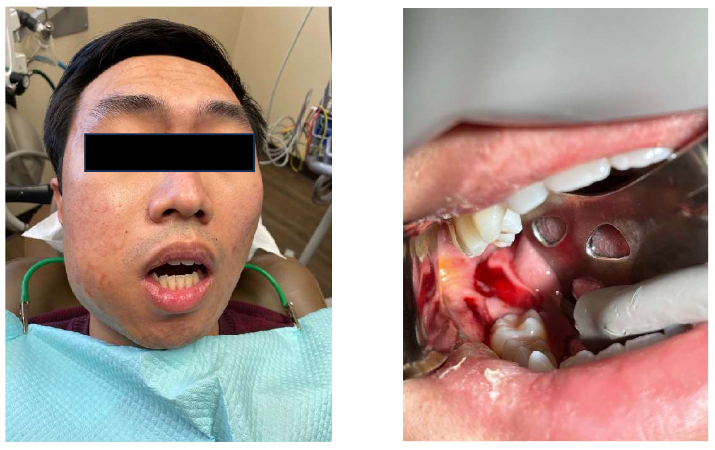
Figure 4.
Impacted #17 with caries, mesial bone loss and distal cupping resorption.
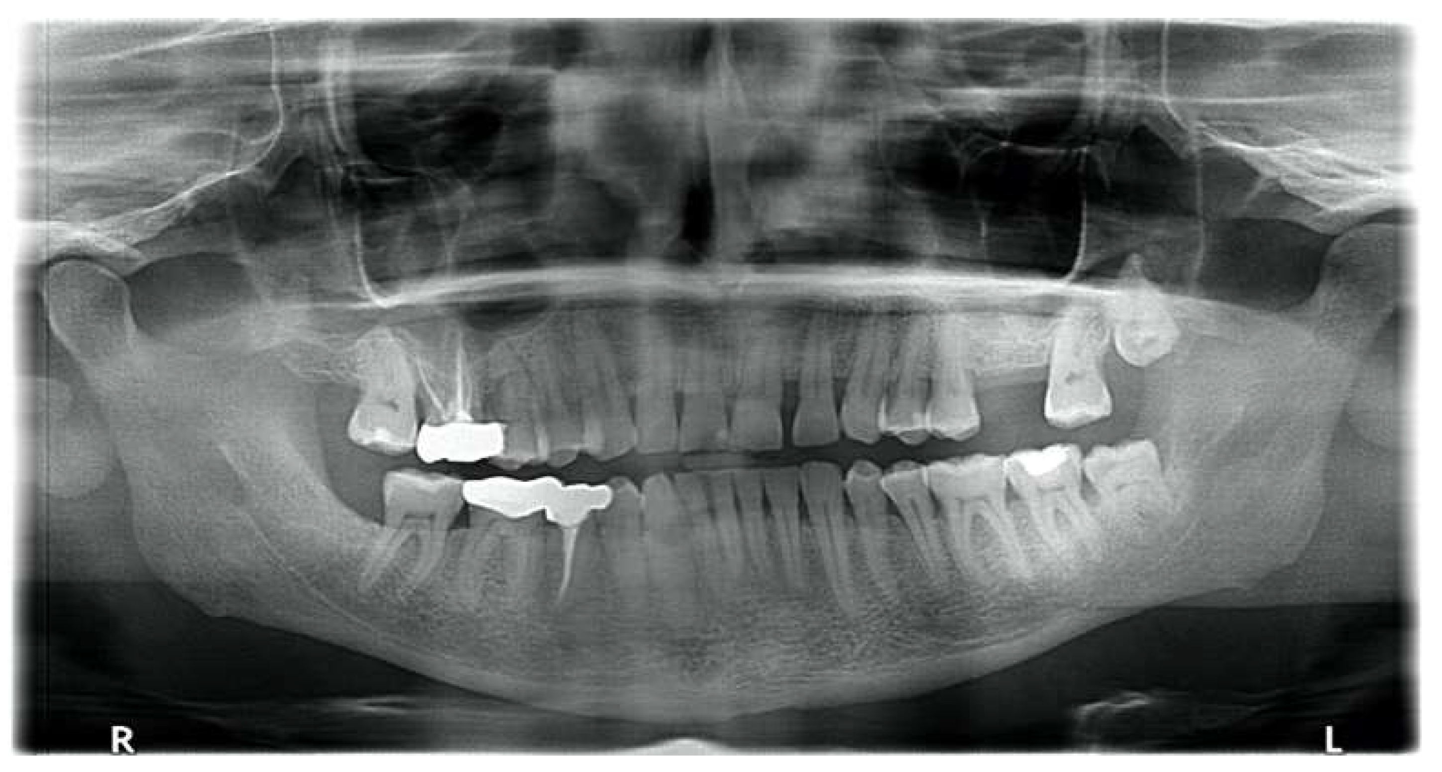
Figure 5.
Anaerobic Organisms: Fusobacterium, Mixed Prevotella, P. micra (75%).
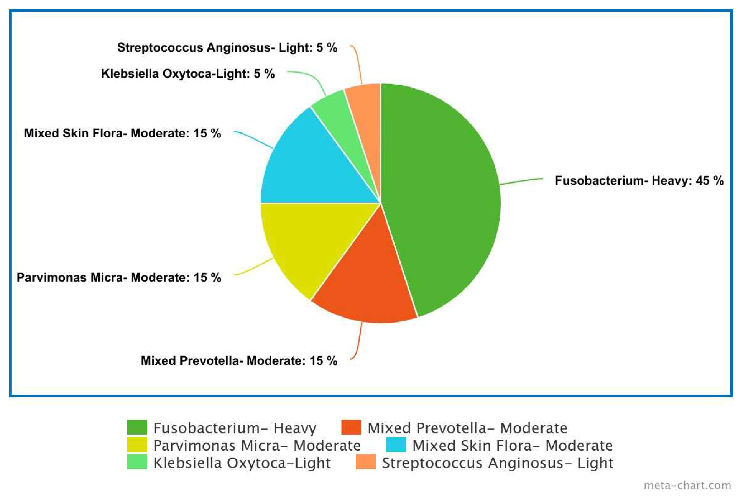

Table 1.
MBC for Prevotella buccae (anaerobic Gram-negative rod).
| Anaerobic Gram-negative rods MBC (μg/mL) |
||
| Ampicillin + Sulbactam | 1 | Susceptible |
| Clindamycin | 256 | Resistant |
| Meropenem | 0.25 | Susceptible |
| Metronidazole | 0.5 | Susceptible |
Table 2.
Susceptibility Testing for Prevotella intermedia and Fusobacterium species.
| Susceptibility | ||
| Prevotella intermedia | ||
| Antibiotic | Interpretation | Value Comment (μg/mL) |
| Ampicillin + Sulbactam | Susceptible | <=0.03 |
| Clindamycin | Susceptible | <=0.03 |
| Meropenem | Susceptible | 0.06 |
| Metronidazole | Susceptible | 0.12 |
| Fusobacterium Species | ||
| Ampicillin + Sulbactam | Susceptible | <=0.03 |
| Clindamycin | Susceptible | 0.06 |
| Meropenem | Susceptible | 0.03 |
| Metronidazole | Susceptible | <=0.03 |
Table 3.
Anaerobic Organisms Other Than Bacteroides spp. and Parabacteroides spp. and their sensitivity to antibiotics. Reprinted with permission from Clinical and Laboratory Standards Institute from: CLSI. Performance Standards for Antimicrobial Susceptibility Testing.30th ed. CLSI supplement M100. Wayne, PA: Clinical and Laboratory Standards Institute; 2020.
| Ampicillin-Sulbactam | Pipercillin-tazobactam | Imipenem | Meropenem | |||||||||
| Anaerobic Organisms | # of strains | %S | %R | # of strains | %S | %R | # of strains | %S | %R | # of strains | %S | %R |
| Prevotella spp. | 29 | 97 | 3 | 63 | 100 | 0 | 29 | 100 | 0 | 92 | 98 | 0 |
| Fusobacterium spp | 20 | 100 | 0 | 55 | 96 | 2 | 75 | 95 | 4 | 20 | 100 | 0 |
| Anaerobic gram positive cocci | - | - | - | 1853 | 99 | 1 | 134 | 99 | 0 | 1647 | 100 | 0 |
| Clostridium perfringens | 15 | 100 | 0 | 410 | 100 | 0 | 23 | 100 | 0 | 417 | 100 | 0 |
| Clostridioides | 76 | 99 | 0 | 542 | 93 | 0 | 480 | 69 | 4 | 609 | 99 | 0 |
| Clostridium spp. | - | 439 | 94 | 1 | 71 | 99 | 0 | 390 | 100 | 0 | ||
| Penicillin | Clindamycin | Moxifloxacin | Metronidazole | |||||||||
| Anaerobic Organisms | # of strains | %S | %R | # of strains | %S | %R | # of strains | %S | %R | # of strains | %S | %R |
| Prevotella spp. | 63 | 100 | 0 | 29 | 69 | 28 | 92 | 66 | 25 | 92 | 99 | 0 |
| Fusobacterium spp | - | - | - | 75 | 77 | 21 | 75 | 68 | 23 | 75 | 95 | 5 |
| Anaerobic gram positive cocci | 1647 | 100 | 0 | 1826 | 97 | 3 | 300 | 72 | 21 | 0 | 100 | 0 |
| Clostridium perfringens | 402 | 90 | 4 | 425 | 83 | 12 | 23 | 83 | 9 | 425 | 100 | 0 |
| Clostridioides | 533 | 6 | 37 | 1013 | 32 | 38 | 480 | 74 | 25 | 1343 | 100 | 0 |
| Clostridium spp. | 390 | 69 | 13 | 461 | 67 | 25 | 71 | 62 | 35 | 461 | 100 | 119 |
Table 4.
The number of resistant isolates and the percentage of resistance against different antibiotics and the prevalence of the corresponding resistance genes in Prevotella isolates. A.C.M. Veloo, W.H. Baas, F.J. Haan, J. Coco, J.W. Rossen. Prevalence of antimicrobial resistance genes in Bacteroides spp. and Prevotella spp. Dutch clinical isolates, Clinical Microbiology and Infection, Volume 25, Issue 9, 2019, Pages 1156.e9–1156.e13, ISSN 1198-743X, https://doi.org/10.1016/j.cmi.2019.02.017. (With permission).
| Resistant strains (n [%]) | ||||
|---|---|---|---|---|
| Amoxicillin | Meropenem | Clindamycin | Metronidazole | |
| Species | ||||
| Breakpoint (mg/L) | R>2 | R>8 | R>4 | R>4 |
| P. baroniae (n=2) | 1 (50.0) | 0 | 0 | 0 |
| P. bergensis (n=3) | 2 (66.7) | 0 | 2 (66.7) | 0 |
| P. bivia (n=17) | 9 (52.9) | 0 | 2 (11.8) | 1 (5.9) |
| P. buccae (n=13) | 5 (38.5) | 0 | 0 | 0 |
| P. buccalis (n=3) | 0 | 0 | 0 | 0 |
| P. copri (n=2) | 1 (50.0) | 0 | 1 (50.0) | 0 |
| P. denticola (n=7) | 4 (57.1) | 0 | 0 | 0 |
| P. disiens (n=4) | 1 (25.0) | 0 | 2 (50.0) | 0 |
| P. histicola (n=2) | 1 (50.0) | 0 | 0 | 0 |
| P. intermedia (n=4) | 1 (25.0) | 0 | 0 | 0 |
| P. jejuni (n=2) | 2 (100) | 0 | 0 | 0 |
| P. melaninogenica (n=21) | 14 (66.7) | 0 | 1 (4.8) | 1 (4.8) |
| P. nigrescens (n=4) | 3 (75.0) | 0 | 1 (25.0) | 0 |
| P. oris (n=2) | 2 (100) | 0 | 0 | 0 |
| P. timonensis (n=6) | 1 (16.7) | 0 | 1 (16.7) | 0 |
| Prevotella spp. (n=7) | 1 (14.3) | 0 | 0 | 0 |
| Total, n (%) | 48 (48.5) | 0 | 10 (10.1) | 2 (2.0) |
Disclaimer/Publisher’s Note: The statements, opinions and data contained in all publications are solely those of the individual author(s) and contributor(s) and not of MDPI and/or the editor(s). MDPI and/or the editor(s) disclaim responsibility for any injury to people or property resulting from any ideas, methods, instructions or products referred to in the content. |
© 2024 by the authors. Licensee MDPI, Basel, Switzerland. This article is an open access article distributed under the terms and conditions of the Creative Commons Attribution (CC BY) license (http://creativecommons.org/licenses/by/4.0/).
Copyright: This open access article is published under a Creative Commons CC BY 4.0 license, which permit the free download, distribution, and reuse, provided that the author and preprint are cited in any reuse.


