Submitted:
27 June 2024
Posted:
28 June 2024
Read the latest preprint version here
Abstract
Leishmaniasis, caused by Leishmania parasites transmitted through sandfly bites, encompasses various clinical forms with distinct immune responses. Mucocutaneous leishmaniasis (ML) exhibits a delicate balance between IFN-γ and IL-10, influencing disease severity. Conversely, cutaneous leishmaniasis (CL) presents varied IFN-γ/IL-10 ratios, affecting prognosis. Leishmania RNA virus 1 (LRV1) and extracellular vesicles modulate ML immunity. In visceral leishmaniasis (VL), severe immune defects necessitate combined host immunity and drug treatment for cure. Cytokines and chemokines serve as diagnostic and prognostic markers in CL. Neutrophils' dual role in leishmaniasis pathogenesis is extensively studied, alongside the NLRP3 inflammasome's regulation of Th1/Th2 responses. Live attenuated LdCen-/- parasites offer promising VL vaccine candidates, with biomarkers like miR-21 aiding efficacy assessment. Neutrophil-DC interactions play pivotal roles in vaccine-induced immunity. Genetically modified live attenuated vaccines and novel formulations elicit robust cellular responses against VL. Understanding cytokine networks is crucial for treatment strategy development. Ongoing research strives to overcome vaccine development challenges and ensure long-term protection against leishmaniasis.
Keywords:
Leishmaniasis
; immune system
; vaccines
; cytokines
; biomarkers
1. Introduction
Leishmaniasis is a parasitic infection that affects approximately 12 million people worldwide, primarily in developing countries[1]. The disease is transmitted by the bite of infected sand flies and can present in three main ways: cutaneous, mucocutaneous, or visceral. “Cutaneous leishmaniasis (CL) is the most common form and causes an open sore at the bite site, which heals in a few months to a year and a half, leaving an unpleasant-looking scar. To date, there are no effective vaccines for leishmaniasis. Many of the treatments available have serious side effects and resistance mechanisms are becoming an increasingly prevalent problem”[1,2,3]. Natural Killer T (NKT) cells are a unique T cell population that recognizes glycolipids. They are a conserved innate-like lymphocyte population with immunomodulating effects in various settings. The role of NKT cells in the resistance or susceptibility towards Leishmania infections is still not well-defined, as there are conflicting data. However, it is proposed the activation of NKT cells for controlling Leishmania infections[1,2,4,5].
The behavior of these cells is influenced by various factors, including the infection site, parasite count, strain virulence, and Leishmania species. NKT cells can be activated through direct or indirect pathways. In the direct pathway, Leishmania glycolipids are presented by CD1d molecules on antigen-presenting cells (such as dendritic cells), leading to cytokine secretion by NKT cells. In the indirect pathway, Leishmania glycolipids stimulate TLR2 in dendritic cells, triggering IL-12 production and subsequent NKT cell activation. The study delves into NKT cell roles in disease development, both in humans and mouse models, and proposes NKT cell activation as a strategy for controlling Leishmania infections[4,5]. Biomarkers and vaccines are two important aspects of leishmaniasis research.
Leishmaniasis, a vector-borne disease caused by the protozoan parasite Leishmania, is endemic in various regions worldwide. Its clinical manifestations range from self-healing skin lesions to severe visceral infections. Recent studies have harnessed large-scale genomic and proteomic analyses to understand infection pathways in both parasites and hosts more effectively than studying individual molecules. Additionally, researchers have identified potential pharmacodynamic biomarkers for different forms of leishmaniasis. Despite these advances, there is currently no approved vaccine for human use against this disease[6,7,8].
2. Immune Response to the Parasite
2.1. Immune Response in Cutaneous Leishmaniasis
The innate immune response serves as our initial defense against pathogens and tissue damage. In cutaneous leishmaniasis (CL), the skin’s innate response involves recognizing pathogenic molecules and activating immune cells like dendritic cells, neutrophils, macrophages, natural killer cells, and the complement system [9] These components play a crucial role in containing the parasite, although Leishmania can manipulate them to its advantage, causing damage in the skin layers[10,11,12].
The innate immune response against Leishmania begins with the recruitment of neutrophils, essential polymorphonuclear cells responsible for phagocytosis and lysis of Leishmania promastigotes. These immune cells are drawn to the infection site by soluble chemotactic factors released by Leishmania and IL-8 in the host. Neutrophils recognize the parasite through innate immune receptors (such as Toll-like receptors TLR2 and TLR4) on their membranes. This recognition is crucial for Leishmania destruction, as it activates antimicrobial substances like reactive oxygen species (ROS), nitric oxide (NO), and neutrophil extracellular traps (NETs). Additionally, neutrophils trigger cytokine release, recruit macrophages, and activate cellular immune responses against the microorganism[10,11].
Beyond neutrophils, other innate cells like inflammatory monocytes, mast cells, and NK cells also influence resistance or susceptibility to Leishmania infection. Recent research highlights how metabolic pathways in innate immune cells activate unique immune signal cascades, alongside cytokine and chemokine signals. However, the intricate interplay between metabolic pathways, epigenetic changes, and immune signaling remains a puzzle in Leishmania pathogenesis. A deeper grasp of early interactions between parasites and innate immune cells is crucial for developing effective and safe vaccines against Leishmaniasis[13].
In this study, researchers explored the impact of an empty bacterial pcDNA3 plasmid on mice infected with Leishmania major. They assessed immune mediators, including IFN-γ, IL-4, IL-10, IgG2a, IgG1, arginase activity, and nitric oxide (NO). The results revealed that pcDNA3 modulated immune responses in favor of host cells, reducing disease severity. Notably, Th2-associated mediators were downregulated, while cellular responses increased, accompanied by elevated NO and IFN-γ levels. The study highlights the potential of pcDNA3 as an immunoenhancing agent for treating zoonotic cutaneous leishmaniasis (ZCL). Further research is needed to understand the underlying mechanisms of pcDNA3’s immunogenic properties against ZCL [14]
In this study, the authors investigated the antileishmanial activity of Urtica dioica extract against zoonotic cutaneous leishmaniasis. They found that Urtica dioica extract can modulate the immune responses in favor of host cells and decrease the disease severity. By quantifying immune mediators such as IFN-γ, IL-4, IL-10, IgG2a, IgG1, arginase activity, and nitric oxide (NO), they observed significant reduction in L. major promastigotes viability. Importantly, the extract showed no toxicity to macrophages but effectively killed L. major amastigotes. These findings highlight the potential of Urtica dioica extract as an immunoenhancing agent for treating zoonotic cutaneous leishmaniasis. Further research is needed to understand the underlying mechanisms of its immunogenic properties.[15].
2.2. Immune Response against Mucocutaneous Leishmaniasis (ML)
This paper investigates the association between the balance of interferon-gamma (IFN-γ) and interleukin-10 (IL-10) and the severity of human Leishmania (Viannia) braziliensis infection. The authors suggest that suitable levels of IFN-γ and IL-10 are essential for the maintenance of protective responses in cutaneous leishmaniasis (CL), while high IFN-γ and low IL-10 production are associated with the severity of mucosal leishmaniasis (ML). The study involved 33 individuals who recovered from L. braziliensis infection, and the cytokines were quantified by enzyme-linked immunosorbent assay (ELISA) in culture supernatants of L. braziliensis-stimulated peripheral blood mononuclear cells (PBMC). The results showed that cured ML cases maintained significantly lower production of IL-10 in comparison to spontaneous healing of CL or asymptomatic individuals. Thus, a high IFN-γ/IL-10 ratio observed in ML can indicate an unfavorable cytokine balance. Conversely, lower IFN-γ/IL-10 balance observed in cured CL, spontaneous healing of CL, and asymptomatic individuals can represent a better-modulated immune response associated with a favorable prognosis. The study is significant because it provides insights into the association between the balance of IFN-γ and IL-10 and the severity of human Leishmania (Viannia) braziliensis infection. The authors suggest that the cytokine balance is essential for the maintenance of protective responses in CL, while high IFN-γ and low IL-10 production are associated with the severity of ML. The study highlights the need for further research in this area to understand the mechanisms underlying the association between the balance of IFN-γ and IL-10 and the severity of human Leishmania (Viannia) braziliensis infection[16].
In this research is discussed the role of Leishmania Viannia guyanensis, LRV1 virus, and extracellular vesicles in the immune response during muco-cutaneous leishmaniasis. The authors argue that the interaction between these three factors can influence the outcome of the immune response to the disease. They suggest that the LRV1 virus can modulate the host’s immune response by inhibiting inflammasome activation and inducing the production of proinflammatory cytokines such as TNFα and IL-12. The authors also highlight the role of extracellular vesicles in the pathogenesis of the disease by promoting the survival of Leishmania parasites and inhibiting the host’s immune response. The article provides a comprehensive review of the current understanding of the role of Leishmania Viannia guyanensis, LRV1 virus, and extracellular vesicles in muco-cutaneous leishmaniasis. The study is significant because it provides insights into the complex interplay between Leishmania Viannia guyanensis, LRV1 virus, and extracellular vesicles in the pathogenesis of muco-cutaneous leishmaniasis. The authors suggest that the interaction between these three factors can influence the outcome of the immune response to the disease. The study highlights the need for further research in this area to understand the mechanisms underlying the interaction between Leishmania Viannia guyanensis, LRV1 virus, and extracellular vesicles in mucocutaneous leishmaniasis. In addition, the cells involved in the pathogenesis of the disease include macrophages, neutrophils and dendritic cells[17].
2.3. Immune Response in Visceral Leishmaniasis (VL)
“The article reviews the factors that drive the recruitment of neutrophils at the site of injection by the sandfly and the molecular mechanisms involved in the uptake of the Leishmania parasite by neutrophils and how the parasite subverts their microbicidal functions. The article also discusses the role of neutrophils in acute and chronic leishmaniasis, with a particular focus on neutrophil-parasite interactions and anti-neutrophil antibody treatments in experimental models of leishmaniasis. The article concludes that neutrophils play a dual protective and permissive role shortly after promastigote infection by reducing incoming parasite burden and subsequently facilitating safe passage of surviving parasites to naive host cells”[18].
In a review the role of neutrophils in acute and chronic leishmaniasis is explored, with a focus on neutrophil-parasite interactions and anti-neutrophil antibody treatments in experimental models. Neutrophils serve a dual role: initially reducing parasite burden and later facilitating safe passage of surviving parasites to naive host cells. Recent findings suggest that neutrophils may also influence chronic parasite persistence. A small clinical trial demonstrated that adding granulocyte–macrophage colony-stimulating factor (GM-CSF) to standard antimonial therapy improved neutropenia in visceral leishmaniasis (VL) patients and reduced secondary infection risk. However, further research is needed to assess the efficacy of anti-neutrophil antibody treatments in humans [12].
VL is a disseminated infection of the lymphoreticular system of the body, which is marked by severe defects in the immune system of the host. The successful cure of VL depends on the immune status of the host in combination with the effects of antileishmanial drugs. The authors of the article discuss the different aspects of adaptive and innate immune responses and explore their role in the protection or pathogenesis of VL. They highlight the importance of cytokines, chemokines, and cellular subsets in the immunology of VL. The authors also discuss the role of regulatory T cells in VL pathogenesis and the potential of immunotherapy targeting IL-10 for the treatment of VL. This article provides a comprehensive review of the involvement and interactions of different immune cells and their cytokines in human visceral leishmaniasis. The article highlights the importance of the immune system in the successful cure of VL and provides insights into the potential of immunotherapy for the treatment of VL[19].
The authors discuss the current understanding of the immune response to VL and the role of cytokines in the protection or pathogenesis of VL. They also highlight the potential of cytokines as diagnostic markers and therapeutic targets for VL. The authors begin by discussing the different aspects of the immune response to VL, including the role of cytokines, chemokines, and cellular subsets. They highlight the importance of cytokines such as IFN-γ, TNF-α, IL-10, and IL-27 in the immunology of VL. The authors also discuss the role of regulatory T cells in VL pathogenesis and the potential of immunotherapy targeting IL-10 for the treatment of VL[20].
It is also discussed the potential of cytokines as diagnostic markers for VL. They highlight the importance of identifying biomarkers that can differentiate between active and latent VL infections. The authors suggest that cytokines such as IFN-γ, TNF-α, and IL-10 could be used as diagnostic markers for VL. Finally, the authors discuss the potential of cytokines as therapeutic targets for VL. They highlight the importance of developing novel immunotherapies that can target cytokines such as IL-10 and IL-27. The authors suggest that immunotherapy targeting IL-10 could be a promising approach for the treatment of VL. This paper provides a comprehensive review of the role of cytokines in the pathogenesis of VL. The authors highlight the importance of cytokines in the immune response to VL and their potential as diagnostic markers and therapeutic targets for VL[20].
2.4. Innate Immune Response
The immune response against Leishmania begins at the site of pathogen entry in the dermis, where promastigotes interact with serum components, activating the complement system via both classical and alternative pathways. Complement opsonization of metacyclic promastigote forms leads to efficient lysis of approximately 90% of inoculated parasites, but Leishmania has developed mechanisms to resist and evade this process [1,21]. Leishmania expresses protein kinases that inhibit complement activation by phosphorylating key components [22,23]
Surface molecules like lipophosphoglycan (LPG) and glycoprotein of 63 kDa (GP63) bind to inactivated C3b (iC3b), preventing complement-mediated lysis and facilitating internalization via complement receptors (CRs) [24] . Sandfly saliva, containing exosomes, gut microbes, and various molecules with immunomodulatory effects, further influences infection outcomes [25,26,27,28]. Resident cells like macrophages, keratinocytes, mast cells, and Langerhans cells express pattern recognition receptors (PRRs), initiating innate and acquired immune response cascades upon encountering Leishmania [29]. Neutrophils [18,30,31], monocytes [32], macrophages [33,34]and dendritic cells (DCs) [35,36,37] are involved in phagocytosing Leishmania parasites. Phagocytosis includes binding to specific receptors, formation of parasitophorous vacuoles, and parasite degradation via reactive oxygen species/reactive nitrogen species (ROS/RNS) [38]. Resident cells like macrophages, keratinocytes, mast cells, and Langerhans cells express pattern recognition receptors (PRRs), initiating innate and acquired immune response cascades upon encountering Leishmania [37,38].
2.5. Inflammasome in Leishmaniasis
Inflammasomes are important components of the innate immune system that help fight bacterial and viral infections. However, their role in regulating adaptive immunity during protozoan parasite infections is not well understood. In this study, researchers found that the NLRP3 inflammasome balances Th1/Th2 responses during leishmaniasis. Mice lacking the inflammasome components NLRP3, ASC, or caspase 1 on a Leishmania-susceptible BALB/c background were resistant to cutaneous L. major infection. The researchers also discovered that production of IL-18 promotes the Th2 cytokine IL-4, which propagates disease in susceptible BALB/c mice. “Neutralization of IL-18 in these animals reduced L. major titers and footpad swelling. The results suggest that activation of the NLRP3 inflammasome is detrimental during leishmaniasis and that IL-18 neutralization has potential as a therapeutic strategy to treat leishmaniasis patients”[39].
It is suggested that the NLRP3 inflammasome plays a dual role in leishmaniasis, both as a protector and a pathogen. The NLRP3 inflammasome is essential for the host’s defense against Leishmania infection by activating the immune system and inducing the production of pro-inflammatory cytokines. The cytokines involved in this process include IL-1β, IL-18, and IL-33. The NLRP3 inflammasome also activates the production of IL-1β, IL-18, and IL-6, which can contribute to the pathogenesis of the disease by inducing excessive inflammation and tissue damage. The cells involved in the dual role of the NLRP3 inflammasome in leishmaniasis include macrophages, neutrophils, and dendritic cells. The balance between the protective and pathogenic roles of the NLRP3 inflammasome is critical for the outcome of the disease, and further research is needed to understand the mechanisms underlying the dual role of the NLRP3 inflammasome in leishmaniasis[40,41,42].
2.6. Leishmania Infection Induces the Production of Reactive Oxygen Species (ROS) and Nitric Oxide (NO) in Macrophages
Leishmania infection in macrophages induces the production of ROS and NO, impacting various signaling pathways including MAPK, NF-κB, and PI3K/Akt. While multiple enzymes contribute to ROS production, NADPH oxidase is crucial in macrophages. Despite ROS production upon infection, Leishmania parasites inhibit ROS generation in phagolysosomes, potentially aiding their survival. However, activated macrophages can still exhibit ROS-dependent killing of Leishmania, with NO playing a role in parasite elimination as well. Studies indicate a dual role for ROS, as they contribute to both host defense and parasite survival, depending on the context of infection and the Leishmania species involved. Furthermore, ROS may influence the establishment of replicative niches within infected macrophages, facilitating long-term parasite survival. Overall, ROS emerge as key players in the intricate interplay between Leishmania parasites and host macrophages, impacting the outcome of infection [38,43,44,45,46,47,48,49,50,51,52,53,54,55,56].
2.7. Summary of the Immune Response to Leishmania spp.
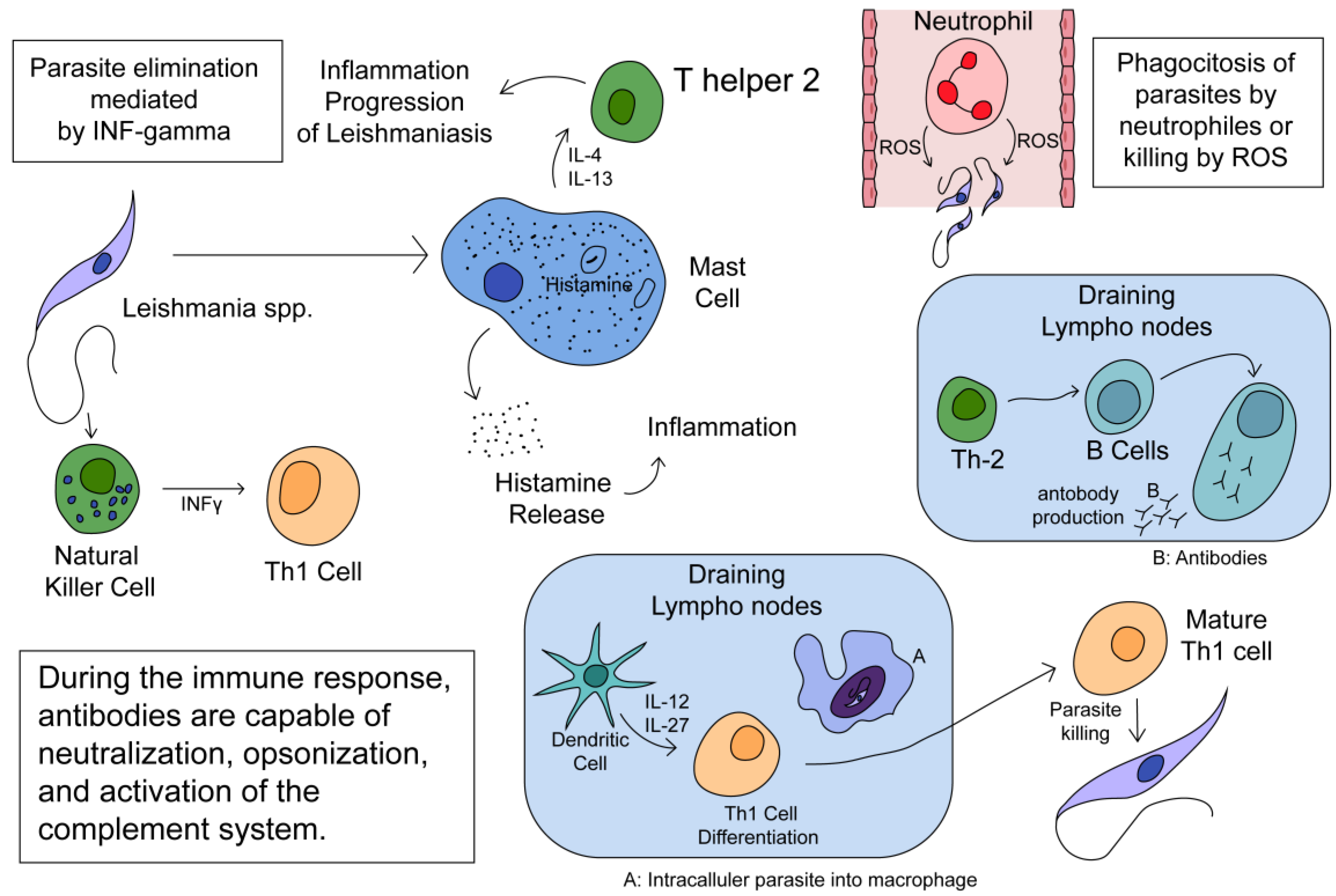
Leishmania infection profoundly affects immune cells, altering their behavior and impacting both innate and adaptive immune responses. Mast cells play a significant role in disease progression by releasing IL-4 and IL-13, promoting Th2 responses and aiding parasite survival. Neutrophils, macrophages, and dendritic cells can either eliminate Leishmania parasites or facilitate their survival within the host. Neutrophils eliminate parasites through phagocytosis, ROS, and NETs release, but Leishmania can evade destruction by inhibiting phagolysosome biogenesis and delaying neutrophil apoptosis. Infected neutrophils also attract more immune cells, supporting parasite survival and pathology. Macrophages exhibit plasticity during leishmaniasis, differentiating into M1 or M2 phenotypes, with M1 macrophages promoting Th1 responses for disease control and M2 macrophages supporting Th2 responses and disease progression. Dendritic cells play a crucial role in regulating immune responses by presenting Leishmania antigens to naïve T cells, influencing their differentiation into either Th1 or Th2 cells. Finally, NK cells contribute to the host defense against Leishmania by secreting IFNγ, which enhances the Th1 response, thus aiding in parasite clearance [29,37,57,58]. Figure 1 shows some aspects of the immune defense in leishmsniasis.
3. Leishmaniasis Biomarkers
A new study aimed to identify the chemokines and cytokines present in the plasma of patients infected with Leishmania guyanensis, the causative agent of cutaneous leishmaniasis. The authors used a multiplex assay to measure the levels of 25 cytokines and chemokines in the plasma of 30 patients with cutaneous leishmaniasis and 30 healthy controls. They found that the levels of several cytokines and chemokines were significantly different between the two groups. For example, the levels of interleukin-1 receptor antagonist (IL-1RA), interleukin-6 (IL-6), and interferon-gamma (IFN-γ) were significantly higher in the plasma of patients with cutaneous leishmaniasis than in healthy controls. Conversely, the levels of interleukin-10 (IL-10) and interleukin-13 (IL-13) were significantly lower in the plasma of patients with cutaneous leishmaniasis than in healthy controls. The authors also performed a principal component analysis (PCA) to identify the cytokines and chemokines that best distinguished between the two groups. They found that the first principal component (PC1) was mainly composed of IL-1RA, IL-6, and IFN-γ, while the second principal component (PC2) was mainly composed of IL-10 and IL-13. The authors suggest that these cytokines and chemokines could be used as biomarkers for the diagnosis and prognosis of cutaneous leishmaniasis. This study provides valuable insights into the cytokine and chemokine profiles of patients with cutaneous leishmaniasis. The authors’ findings suggest that IL-1RA, IL-6, IFN-γ, IL-10, and IL-13 could be useful biomarkers for the diagnosis and prognosis of this disease. Further research is needed to validate these findings and to determine the clinical utility of these biomarkers[59].
The authors discuss the limitations of current vaccines against CVL and the need for new approaches. The main focus of the article is on the potential of biomarkers to improve the efficacy of vaccines against CVL. The authors describe the various types of biomarkers that have been studied, including those related to the immune response, parasite load, and genetic factors. Biomarkers are measurable indicators that can be used to diagnose, predict, or monitor the progression of a disease. In the context of canine visceral leishmaniasis (CVL), researchers have studied various types of biomarkers that could improve the efficacy of vaccines against this disease. One type of biomarker that has been studied is related to the immune response. Researchers have identified several immune-related biomarkers that could be used to predict the efficacy of vaccines against CVL. For example, the levels of certain cytokines, such as IFN-γ and IL-10, have been shown to be associated with protection against CVL. Another type of biomarker that has been studied is related to parasite load. Researchers have investigated the use of parasite-specific biomarkers, such as the K39 antigen, to monitor the progression of CVL and assess the efficacy of vaccines. Finally, genetic factors have also been studied as potential biomarkers for CVL. Researchers have identified several genetic polymorphisms that are associated with susceptibility to CVL, including those related to the MHC class II genes. They call for further research in this area and suggest that a better understanding of the immune response to Leishmania infection will be critical to the development of effective vaccines. In summary, this study provides a comprehensive overview of the current state of research on CVL and the potential of biomarkers to improve the efficacy of vaccines against this disease. Their work highlights the need for continued research in this area and provides a valuable resource for researchers and clinicians working to combat CVL[60].
4. Investigation on the Effects of Treatment of Human Leishmaniasis
In this study, researchers explored the impact of amphotericin B treatment on patients with leishmaniasis, a parasitic disease. By analyzing cytokine production in peripheral blood mononuclear cells (PBMCs), they found that patients with mucocutaneous leishmaniasis (MCL) exhibited higher IFN-γ levels compared to those with cutaneous leishmaniasis (CL). Additionally, MCL patients showed increased IL-12 synthesis. These findings highlight potential avenues for controlling protozoan infections through combined use of immunoenhancing agents and antiprotozoal drugs, emphasizing the need for further research in this area[61].
5. Vaccines against Leishmaniasis
In this research, scientists explored vaccine development against visceral leishmaniasis (VL) and the effectiveness of live attenuated LdCen-/- parasites in animal models. Notably, immunization with LdCen-/- parasites led to robust and lasting protective immunity in pre-clinical animal studies. The study also pinpointed early biomarkers associated with immunogenicity in these parasitic vaccines. Specifically, LdCen-/- infection suppressed microRNA-21 (miR-21) expression in human macrophages, which negatively regulates IL12. This miR-21 could serve as a crucial biomarker for LdCen−/− vaccine efficacy in human clinical trials, facilitating adaptive immunity and Th1 cell development [62].

Table 1.
Some types of vaccines against Leishmaniasis.
| Type of vaccine | Reference |
|---|---|
| Killed parasite antigen | [63] |
| Oral immunization using live Lactococcus lactis co-expressing LACK and IL-12 | [64,65] |
| Live attenuated Centrin gene-deleted Leishmania vaccine | [66] |
| Growth-arrested Leishmania amastigotes used to develop live attenuated vaccines | [67] |
| Live attenuated L. major parasites with p27 gene deletion | [68] |
| Genetically modified live attenuated vaccines for VL | [69] |
| Recombinant protein derived from sandfly saliva against Leishmania infection | [70,71,72,73,74,75,76,77,78,79,80,81,82,83] |
The research revealed that neutrophils play a crucial role in initial host responses to pathogens and can independently activate T cell responses or collaborate indirectly with dendritic cells (DCs). Additionally, direct interactions between neutrophils and T cells were observed following immunization with a live attenuated Leishmania donovani centrin-deleted parasite vaccine (LdCen-/-). However, the specific role of neutrophil-DC interactions in T cell priming during vaccine immunity remains less understood. The study compared LdCen-/- infection with wild-type parasite (LdWT) both in vitro and in vivo, concluding that LdCen-/- parasites induced increased expression of CCL3 in neutrophils, leading to enhanced recruitment of DCs and a robust proinflammatory response, as well as elevated co-stimulatory molecule expression compared to LdWT infection[84].
In a recent immunology study, researchers explored the dynamic interactions between neutrophils and dendritic cells (DCs) during immune responses. The LdCen-/- parasite induced increased CCL3 expression in neutrophils, leading to enhanced DC recruitment and robust proinflammatory responses, ultimately shaping vaccine-induced immunity[85].
This paper discusses the development of biomarkers for genetically modified live attenuated Leishmania vaccines against visceral leishmaniasis. The study highlights the need for clear biomarkers associated with protection and safety for live attenuated parasite vaccines.[86]. The article discusses the development of a vaccine for leishmaniasis. The authors provide a comprehensive overview of the studies conducted by Dr. Nakhasi’s group in developing a vaccine against leishmaniasis based on genetically modified Leishmania lines lacking a centrin-coding gene. They describe the improvements introduced over time and the challenges faced in developing a vaccine for leishmaniasis[66].
This article discusses the development of a tool to generate growth-arrested Leishmania amastigotes for use in developing live attenuated vaccines against visceral leishmaniasis. The authors describe the challenges of developing a vaccine for leishmaniasis and the potential of live attenuated Leishmania parasites as effective vaccine candidates. They also discuss the development of a method to generate growth-arrested Leishmania amastigotes that can be used to develop live attenuated vaccines. The authors describe the advantages of using growth-arrested Leishmania amastigotes over other methods and the potential of this method to develop effective vaccines against visceral leishmaniasis[67]
This study was conducted by Fiuza JA et al. and aimed to evaluate the efficacy of a live attenuated vaccine against L. infantum in dogs. The researchers used a centrin gene-deleted L. donovani parasite as a vaccine candidate and tested it on dogs. The results showed that the vaccine was able to induce a protective immune response against L. infantum in dogs. The study also found that the vaccine was safe and did not cause any adverse effects in the dogs. The study suggests that vaccination using live attenuated L. donovani parasites can provide protection against L. infantum in dogs. However, further research is needed to evaluate the long-term efficacy of the vaccine and its safety in different dog breeds and ages[87].
The study aimed to evaluate the efficacy of a live attenuated vaccine against Leishmania major and Leishmania infantum in BALB/c mice. The researchers used a p27 gene-deleted L. major parasite as a vaccine candidate and tested it on mice. The results showed that the vaccine was able to induce a protective immune response against L. major and L. infantum in mice. The study also found that the vaccine was safe and did not cause any adverse effects in the mice. The study suggests that vaccination using live attenuated L. major parasites with p27 gene deletion can provide protection against L. major and L. infantum in mice. However, further research is needed to evaluate the long-term efficacy of the vaccine and its safety in different animal models and humans[68].
The most critical finding of the research on genetically modified live attenuated vaccines for VL is that they hold great potential for combating the disease. These vaccines are engineered to be less virulent, ensuring safety, while still eliciting a protective immune response. However, achieving this balance requires a thorough understanding of the Leishmania genome and the mechanisms of pathogenesis. Researchers continue to work towards a breakthrough that could significantly impact the fight against VL[88,89]. Genetically modified live attenuated vaccines for VL is that they hold great potential for combating the disease. These vaccines are engineered to be less virulent, ensuring safety, while still eliciting a protective immune response[69]
The main finding of the research on genetically modified live attenuated vaccines for VL is that they generate functional CD4+ skin tissue-resident memory T (TRM) cells, similar to the natural process of leishmanization. These TRM cells play a crucial role in host protection, along with effector T cells, by rapidly responding to infection and producing essential cytokines and chemokines[90,91,92,93,94]. Almeida et al. investigated a novel vaccine formulation for visceral leishmaniasis (VL). They developed a genetically modified live attenuated vaccine using a modified version of the Amastigote 2 antigen and the non-virulent Trypanosoma cruzi CL-14 strain, it elicit a strong cellular immunity response[95].
Th1 concomitant immune response mediated by IFN-γ protects against sand fly delivered Leishmania infection[91]. A novel vaccination approach combining recombinant L. tarentolae (a nonpathogenic lizard protozoan parasite) with a sand fly salivary antigen (PpSP15) shows promise as a protective vaccine against major infection[70].
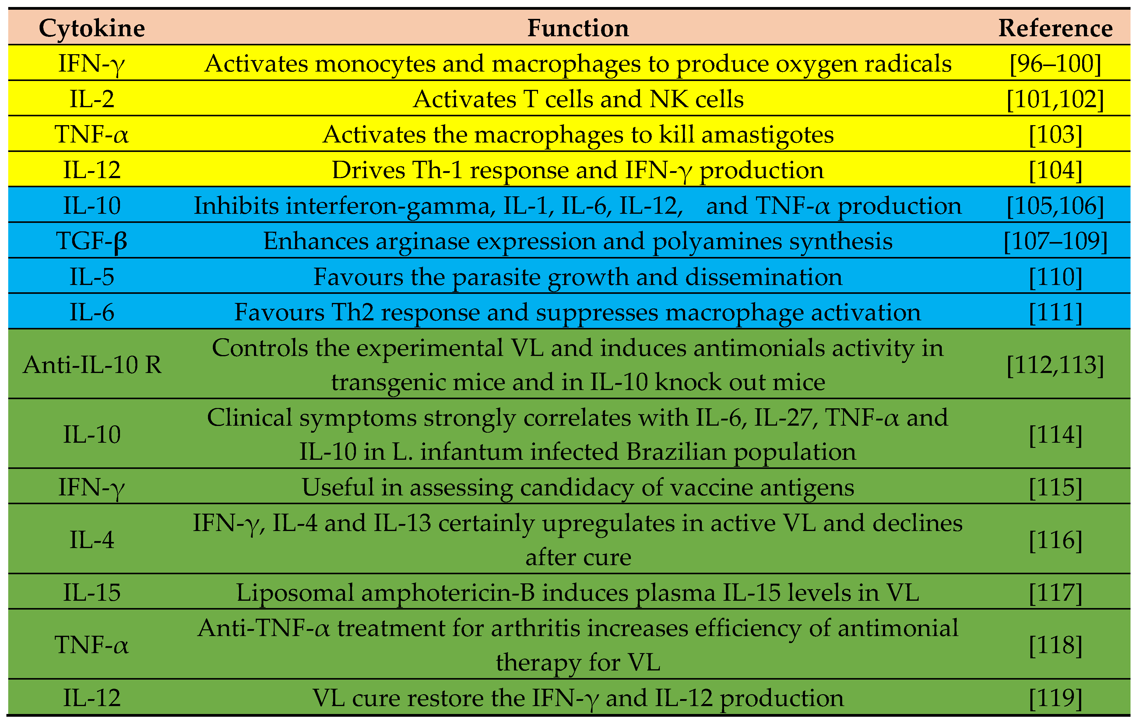
Figure 2.
Cytokines involved in host protection (yellow), disease progression (blue), and used in diagnostic, Chemo/immunotherapy of VL (green). Adapted from[20].
Figure 2.
Cytokines involved in host protection (yellow), disease progression (blue), and used in diagnostic, Chemo/immunotherapy of VL (green). Adapted from[20].
Leishmaniasis, a vector-borne disease prevalent in over 100 countries, lacks an available vaccine for human use. Despite evaluating various candidate antigens, comprehensive biomarkers that serve as reliable indicators of protection or exacerbation remain elusive. While host immune factors associated with disease progression have been identified, no definitive biomarkers have emerged. Researchers propose that combining data from animal models and patients could unveil specific immunity biomarkers, potentially guiding vaccine and drug development against leishmaniasis. Four candidate vaccines have been developed to or near to the clinic[120,121,122]. These are:.
- LEISHF3+ GLA-SE: A recombinant fusion protein delivered with strong Th1-inducing adjuvants.
- LeishDNAvax: A naked multi-epitope DNA vaccine.
- ChAd63-KH: An adenovirus-based vaccine.
- Live genetically attenuated vaccine: A live genetically attenuated vaccine.
These vaccines have been proposed to counter cutaneous and visceral leishmaniasis (CL and VL, respectively). However, the authors of the review article suggest that more research is needed to determine the safety and efficacy of these vaccines. In addition to these candidate vaccines, researchers are also exploring other vaccine strategies such as the use of sand fly saliva proteins as vaccine candidates.
6. Conclusion
The immune response to Leishmania spp. varies depending on the type of infection, with distinct mechanisms identified in mucocutaneous leishmaniasis (ML), visceral leishmaniasis (VL), and cutaneous leishmaniasis (CL). In ML, the balance between interferon-gamma (IFN-γ) and interleukin-10 (IL-10) levels plays a crucial role in disease severity, with high IFN-γ and low IL-10 associated with severe outcomes. Conversely, a lower IFN-γ/IL-10 balance is linked to a more favorable prognosis in CL cases. Furthermore, factors such as the presence of Leishmania RNA virus 1 (LRV1) and extracellular vesicles have been implicated in modulating the host immune response during ML. Understanding these interactions is essential for developing effective interventions against ML. In VL, the immune response is characterized by severe defects, and successful cure relies on both host immune status and antileishmanial drugs. Biomarkers such as cytokines and chemokines have been proposed as diagnostic and prognostic indicators in cutaneous leishmaniasis (CL), aiding in distinguishing active from latent infections and guiding therapeutic strategies. Additionally, the role of neutrophils in both acute and chronic leishmaniasis has been extensively studied, highlighting their dual function in parasite elimination and disease progression. The NLRP3 inflammasome has emerged as a crucial regulator of Th1/Th2 responses during leishmaniasis, with its activation promoting disease propagation in susceptible hosts. However, further research is needed to elucidate the precise mechanisms underlying the role of the inflammasome in leishmaniasis pathogenesis. Overall, a comprehensive understanding of the immune responses to Leishmania infection, including the interplay between cytokines, chemokines, immune cells, and parasitic factors, is vital for the development of effective diagnostic tools, therapeutic interventions, and preventive measures against this complex and debilitating disease.
Research into vaccine development against visceral leishmaniasis (VL) has seen significant progress, particularly with live attenuated LdCen-/- parasites showing robust and lasting protective immunity in animal models. The identification of early biomarkers like miR-21 suppression in human macrophages provides insights into vaccine efficacy in clinical trials. Neutrophils have emerged as key players in host responses to pathogens, with their interactions with dendritic cells (DCs) crucial for T cell priming during vaccine immunity. Live attenuated vaccines, including those lacking the centrin gene or p27 gene deletion, have demonstrated efficacy in inducing protective immune responses against VL in animal models. The development of biomarkers is essential for assessing protection and safety in live attenuated parasite vaccines. Genetically modified live attenuated vaccines for VL show promise in balancing virulence and immunogenicity, with the potential to generate functional CD4+ tissue-resident memory T cells. Novel vaccine formulations, such as those incorporating modified antigens and non-virulent strains, elicit strong cellular immune responses against VL. Cytokines like IFN-γ, IL-12, and TNF-α play critical roles in host protection and disease progression, offering targets for diagnostic and therapeutic interventions. Understanding the intricate cytokine network involved in VL pathogenesis is crucial for developing effective treatment strategies. Overall, ongoing research aims to address challenges in vaccine development, enhance efficacy, and ensure safety against VL, with a focus on harnessing the immune system's potential for long-term protection.
Author Contributions
The manuscript was conceptualized by A.J.-V. and P.E.A. Planning and discussion were conducted by all authors. A.J.-V. wrote the initial draft of the manuscript. A.J.-V. and D.G. reviewed the manuscript. All authors investigated, participated in software, curation, writing or review of the final draft. All authors have read and agreed to the published version of the manuscript.
Funding
This study did not receive any external funding.
Institutional Review Board Statement
Not applicable.
Informed Consent Statement
Not applicable.
Data Availability Statement
The dataset supporting the findings of this study is included within the manuscript and its referenced sources, ensuring comprehensive access to the relevant data for further examination and analysis.
Acknowledgments
The authors would like to sincerely thank West Indian Immunology Society (WIIS) for their assistance.
Conflicts of Interest
The authors declare no conflict of interest.
References
- von Stebut, E. Cutaneous Leishmania Infection: Progress in Pathogenesis Research and Experimental Therapy. Exp. Dermatol. 2007, 16, 340–346. [Google Scholar] [CrossRef] [PubMed]
- Marín, M.; López, M.; Gallego-Yerga, L.; Álvarez, R.; Peláez, R. Experimental Structure Based Drug Design (SBDD) Applications for Anti-Leishmanial Drugs: A Paradigm Shift? Med. Res. Rev. 2023. [Google Scholar] [CrossRef] [PubMed]
- Pace, D. Leishmaniasis. J. Infect. 2014, 69 Suppl 1, S10–S18. [Google Scholar] [CrossRef]
- Bendelac, A.; Savage, P.B.; Teyton, L. The Biology of NKT Cells. Annu. Rev. Immunol. 2007, 25, 297–336. [Google Scholar] [CrossRef] [PubMed]
- Zamora-Chimal, J.; Hernández-Ruiz, J.; Becker, I. NKT Cells in Leishmaniasis. Immunobiology 2017, 222, 641–646. [Google Scholar] [CrossRef]
- Jain, K.; Jain, N.K. Vaccines for Visceral Leishmaniasis: A Review. J. Immunol. Methods 2015, 422, 1–12. [Google Scholar] [CrossRef]
- Veras, P.S.T.; Ramos, P.I.P.; de Menezes, J.P.B. In Search of Biomarkers for Pathogenesis and Control of Leishmaniasis by Global Analyses of Leishmania-Infected Macrophages. Front. Cell. Infect. Microbiol. 2018, 8, 326. [Google Scholar] [CrossRef]
- Seyed, N.; Taheri, T.; Rafati, S.; Taslimi, Y. Identification of Leishmania Vaccine Candidates: A Proteome-Wide Immunoinformatics Approach. Parasitol. Res. 2018. [Google Scholar] [CrossRef]
- Liese, J.; Schleicher, U.; Bogdan, C. The Innate Immune Response against Leishmania Parasites. Immunobiology 2008, 213, 377–387. [Google Scholar] [CrossRef] [PubMed]
- Serrano-Coll, H.; Cardona-Castro, N.; Ramos, A.P.; Llanos-Cuentas, A. Innate Immune Response: Ally or Enemy in Cutaneous Leishmaniasis? Pathog. Dis. 2021, 79. [Google Scholar] [CrossRef]
- Feijó, D.; Tibúrcio, R.; Ampuero, M.; Brodskyn, C.; Tavares, N. Dendritic Cells and Leishmania Infection: Adding Layers of Complexity to a Complex Disease. J Immunol Res 2016, 2016, 3967436. [Google Scholar] [CrossRef]
- Carlsen, E.D.; Liang, Y.; Shelite, T.R.; Walker, D.H.; Melby, P.C.; Soong, L. Permissive and Protective Roles for Neutrophils in Leishmaniasis. Clin. Exp. Immunol. 2015, 182, 109–118. [Google Scholar] [CrossRef] [PubMed]
- Volpedo, G.; Pacheco-Fernandez, T.; Bhattacharya, P.; Oljuskin, T.; Dey, R.; Gannavaram, S.; Satoskar, A.R.; Nakhasi, H.L. Determinants of Innate Immunity in Visceral Leishmaniasis and Their Implication in Vaccine Development. Front. Immunol. 2021, 12, 748325. [Google Scholar] [CrossRef] [PubMed]
- Montakhab-Yeganeh, H.; Shafiei, R.; Najm, M.; Masoori, L.; Aspatwar, A.; Badirzadeh, A. Immunogenic Properties of Empty pcDNA3 Plasmid against Zoonotic Cutaneous Leishmaniasis in Mice. PLoS One 2022, 17, e0263993. [Google Scholar] [CrossRef] [PubMed]
- Badirzadeh, A.; Heidari-Kharaji, M.; Fallah-Omrani, V.; Dabiri, H.; Araghi, A.; Salimi Chirani, A. Antileishmanial Activity of Urtica Dioica Extract against Zoonotic Cutaneous Leishmaniasis. PLoS Negl. Trop. Dis. 2020, 14, e0007843. [Google Scholar] [CrossRef] [PubMed]
- Gomes-Silva, A.; de Cássia Bittar, R.; Dos Santos Nogueira, R.; Amato, V.S.; da Silva Mattos, M.; Oliveira-Neto, M.P.; Coutinho, S.G.; Da-Cruz, A.M. Can Interferon-Gamma and Interleukin-10 Balance Be Associated with Severity of Human Leishmania (Viannia) Braziliensis Infection? Clin. Exp. Immunol. 2007, 149, 440–444. [Google Scholar] [CrossRef]
- Olivier, M.; Zamboni, D.S. Leishmania Viannia Guyanensis, LRV1 Virus and Extracellular Vesicles: A Dangerous Trio Influencing the Faith of Immune Response during Muco-Cutaneous Leishmaniasis. Curr. Opin. Immunol. 2020, 66, 108–113. [Google Scholar] [CrossRef] [PubMed]
- Kupani, M.; Pandey, R.K.; Mehrotra, S. Neutrophils and Visceral Leishmaniasis: Impact on Innate Immune Response and Cross-Talks with Macrophages and Dendritic Cells. J. Cell. Physiol. 2021, 236, 2255–2267. [Google Scholar] [CrossRef] [PubMed]
- Bhattacharya, P.; Ali, N. Involvement and Interactions of Different Immune Cells and Their Cytokines in Human Visceral Leishmaniasis. Rev. Soc. Bras. Med. Trop. 2013, 46, 128–134. [Google Scholar] [CrossRef]
- Dayakar, A.; Chandrasekaran, S.; Kuchipudi, S.V.; Kalangi, S.K. Cytokines: Key Determinants of Resistance or Disease Progression in Visceral Leishmaniasis: Opportunities for Novel Diagnostics and Immunotherapy. Front. Immunol. 2019, 10, 670. [Google Scholar] [CrossRef]
- Regli, I.B.; Passelli, K.; Hurrell, B.P.; Tacchini-Cottier, F. Survival Mechanisms Used by Some Leishmania Species to Escape Neutrophil Killing. Front. Immunol. 2017, 8, 1558. [Google Scholar] [CrossRef]
- Salei, N.; Hellberg, L.; Köhl, J.; Laskay, T. Enhanced Survival of Leishmania Major in Neutrophil Granulocytes in the Presence of Apoptotic Cells. PLoS One 2017, 12, e0171850. [Google Scholar] [CrossRef] [PubMed]
- Verma, S.; Mandal, A.; Ansari, M.Y.; Kumar, A.; Abhishek, K.; Ghosh, A.K.; Kumar, A.; Kumar, V.; Das, S.; Das, P. Leishmania Donovani Inhibitor of Serine Peptidases 2 Mediated Inhibition of Lectin Pathway and Upregulation of C5aR Signaling Promote Parasite Survival inside Host. Front. Immunol. 2018, 9, 63. [Google Scholar] [CrossRef] [PubMed]
- Hermoso, T.; Fishelson, Z.; Becker, S.I.; Hirschberg, K.; Jaffe, C.L. Leishmanial Protein Kinases Phosphorylate Components of the Complement System. EMBO J. 1991, 10, 4061–4067. [Google Scholar] [CrossRef]
- Francesquini, F.C.; Silveira, F.T.; Passero, L.F.D.; Tomokane, T.Y.; Carvalho, A.K.; Corbett, C.E.P.; Laurenti, M.D. Salivary Gland Homogenates from Wild-Caught Sand Flies Lutzomyia Flaviscutellata and Lutzomyia (Psychodopygus) Complexus Showed Inhibitory Effects on Leishmania (Leishmania) Amazonensis and Leishmania (Viannia) Braziliensis Infection in BALB/c Mice. Int. J. Exp. Pathol. 2014, 95, 418–426. [Google Scholar] [CrossRef]
- Lestinova, T.; Vlkova, M.; Votypka, J.; Volf, P.; Rohousova, I. Phlebotomus Papatasi Exposure Cross-Protects Mice against Leishmania Major Co-Inoculated with Phlebotomus Duboscqi Salivary Gland Homogenate. Acta Trop. 2015, 144, 9–18. [Google Scholar] [CrossRef] [PubMed]
- Thiakaki, M.; Rohousova, I.; Volfova, V.; Volf, P.; Chang, K.-P.; Soteriadou, K. Sand Fly Specificity of Saliva-Mediated Protective Immunity in Leishmania Amazonensis-BALB/c Mouse Model. Microbes Infect. 2005, 7, 760–766. [Google Scholar] [CrossRef]
- Abdeladhim, M.; Kamhawi, S.; Valenzuela, J.G. What’s behind a Sand Fly Bite? The Profound Effect of Sand Fly Saliva on Host Hemostasis, Inflammation and Immunity. Infect. Genet. Evol. 2014, 28, 691–703. [Google Scholar] [CrossRef]
- Pacheco-Fernandez, T.; Volpedo, G.; Verma, C.; Satoskar, A.R. Understanding the Immune Responses Involved in Mediating Protection or Immunopathology during Leishmaniasis. Biochem. Soc. Trans. 2021, 49, 297–311. [Google Scholar] [CrossRef]
- Charmoy, M.; Auderset, F.; Allenbach, C.; Tacchini-Cottier, F. The Prominent Role of Neutrophils during the Initial Phase of Infection by Leishmania Parasites. J. Biomed. Biotechnol. 2010, 2010, 719361. [Google Scholar] [CrossRef]
- Passelli, K.; Billion, O.; Tacchini-Cottier, F. The Impact of Neutrophil Recruitment to the Skin on the Pathology Induced by Leishmania Infection. Front. Immunol. 2021, 12, 649348. [Google Scholar] [CrossRef]
- Mohamed, A.M.; Taye, T.; Akuffo, H.O. Mechanisms of Resistance to Leishmania Aethiopica. I. Interferon-Gamma in Combination with a Cytokine (not Tumor Necrosis Factor-Alpha) Is Required, but Cannot Act Alone in the Inhibition of Intracellular Forms of L. Aethiopica in THP1 Cells. Eur. J. Immunol. 1992, 22, 2331–2337. [Google Scholar] [CrossRef] [PubMed]
- Siqueira-Neto, J.L.; Moon, S.; Jang, J.; Yang, G.; Lee, C.; Moon, H.K.; Chatelain, E.; Genovesio, A.; Cechetto, J.; Freitas-Junior, L.H. An Image-Based High-Content Screening Assay for Compounds Targeting Intracellular Leishmania Donovani Amastigotes in Human Macrophages. PLoS Negl. Trop. Dis. 2012, 6, e1671. [Google Scholar] [CrossRef] [PubMed]
- Corradin, S.B.; Buchmüller-Rouiller, Y.; Mauël, J. Phagocytosis Enhances Murine Macrophage Activation by Interferon-Gamma and Tumor Necrosis Factor-Alpha. Eur. J. Immunol. 1991, 21, 2553–2558. [Google Scholar] [CrossRef] [PubMed]
- Argueta-Donohué, J.; Wilkins-Rodríguez, A.A.; Aguirre-García, M.; Gutiérrez-Kobeh, L. Differential Phagocytosis of Leishmania Mexicana Promastigotes and Amastigotes by Monocyte-Derived Dendritic Cells. Microbiol. Immunol. 2016, 60, 369–381. [Google Scholar] [CrossRef] [PubMed]
- Rivera-Fernández, I.; Argueta-Donohué, J.; Wilkins-Rodríguez, A.A.; Gutiérrez-Kobeh, L. Effect of Two Different Isolates of Leishmania Mexicana in the Production of Cytokines and Phagocytosis by Murine Dendritic Cells. J. Parasitol. 2019, 105, 359–370. [Google Scholar] [CrossRef] [PubMed]
- Kautz-Neu, K.; Schwonberg, K.; Fischer, M.R.; Schermann, A.I.; von Stebut, E. Dendritic Cells in Leishmania Major Infections: Mechanisms of Parasite Uptake, Cell Activation and Evidence for Physiological Relevance. Med. Microbiol. Immunol. 2012, 201, 581–592. [Google Scholar] [CrossRef]
- Filardy, A.A.; Costa-da-Silva, A.C.; Koeller, C.M.; Guimarães-Pinto, K.; Ribeiro-Gomes, F.L.; Lopes, M.F.; Heise, N.; Freire-de-Lima, C.G.; Nunes, M.P.; DosReis, G.A. Infection with Leishmania Major Induces a Cellular Stress Response in Macrophages. PLoS One 2014, 9, e85715. [Google Scholar] [CrossRef] [PubMed]
- Gurung, P.; Karki, R.; Vogel, P.; Watanabe, M.; Bix, M.; Lamkanfi, M.; Kanneganti, T.-D. An NLRP3 Inflammasome-Triggered Th2-Biased Adaptive Immune Response Promotes Leishmaniasis. J. Clin. Invest. 2015, 125, 1329–1338. [Google Scholar] [CrossRef]
- Paget, C.; Doz-Deblauwe, E.; Winter, N.; Briard, B. Specific NLRP3 Inflammasome Assembling and Regulation in Neutrophils: Relevance in Inflammatory and Infectious Diseases. Cells 2022, 11. [Google Scholar] [CrossRef]
- Meyers, A.K.; Zhu, X. The NLRP3 Inflammasome: Metabolic Regulation and Contribution to Inflammaging. Cells 2020, 9. [Google Scholar] [CrossRef]
- Raneros, A.B.; Bernet, C.R.; Flórez, A.B.; Suarez-Alvarez, B. An Epigenetic Insight into NLRP3 Inflammasome Activation in Inflammation-Related Processes. Biomedicines 2021, 9. [Google Scholar] [CrossRef] [PubMed]
- Wanasen, N.; Soong, L. L-Arginine Metabolism and Its Impact on Host Immunity against Leishmania Infection. Immunol. Res. 2008, 41, 15–25. [Google Scholar] [CrossRef] [PubMed]
- Diaz-Gandarilla, J.A.; Osorio-Trujillo, C.; Hernández-Ramírez, V.I. Leishmanicidal Activity of NO and H2O2 Alone or in Combination against Leishmania Mexicana. Parasitol. Res. 2013. [Google Scholar]
- Bedard, K.; Krause, K.-H. The NOX Family of ROS-Generating NADPH Oxidases: Physiology and Pathophysiology. Physiol. Rev. 2007, 87, 245–313. [Google Scholar] [CrossRef] [PubMed]
- Nathan, C.; Cunningham-Bussel, A. Beyond Oxidative Stress: An Immunologist’s Guide to Reactive Oxygen Species. Nat. Rev. Immunol. 2013, 13, 349–361. [Google Scholar] [CrossRef] [PubMed]
- Bogdan, C. Nitric Oxide and the Immune Response. Nat. Immunol. 2001, 2, 907–916. [Google Scholar] [CrossRef] [PubMed]
- Antoine, J.C.; Prina, E.; Jouanne, C.; Bongrand, P. Parasitophorous Vacuoles of Leishmania Amazonensis-Infected Macrophages Maintain an Acidic pH. Infect. Immun. 1990, 58, 779–787. [Google Scholar] [CrossRef] [PubMed]
- Nandan, D.; Reiner, N.E. Attenuation of Gamma Interferon-Induced Tyrosine Phosphorylation in Mononuclear Phagocytes Infected with Leishmania Donovani: Selective Inhibition of Signaling through Janus Kinases and Stat1. Infect. Immun. 1995, 63, 4495–4500. [Google Scholar] [CrossRef] [PubMed]
- Green, S.J.; Meltzer, M.S.; Hibbs, J.B., Jr; Nacy, C.A. Activated Macrophages Destroy Intracellular Leishmania Major Amastigotes by an L-Arginine-Dependent Killing Mechanism. J. Immunol. 1990, 144, 278–283. [Google Scholar] [CrossRef]
- Assreuy, J.; Cunha, F.Q.; Epperlein, M.; Noronha-Dutra, A.; O’Donnell, C.A.; Liew, F.Y.; Moncada, S. Production of Nitric Oxide and Superoxide by Activated Macrophages and Killing of Leishmania Major. Eur. J. Immunol. 1994, 24, 672–676. [Google Scholar] [CrossRef] [PubMed]
- Díaz-Gandarilla, J.A.; Osorio-Trujillo, C.; Hernández-Ramírez, V.I.; Salaiza-Suazo, N. Human Monocytes Infected with Leishmania Mexicana Are Primed for IL-12p70 Production. Parasite Immunology 2011, 33, 643–651. [Google Scholar]
- Vieira, L.Q.; Goldschmidt, M.; Nashleanas, M.; Pfeffer, K.; Mak, T. Association of LMP-7 with MHC Class I Molecules Accounts for a Maturation-Dependent Increase in Their Peptide Presentation in Leishmania Amazonensis-Infected Cells. European Journal of Immunology 1996, 26, 1001–1010. [Google Scholar]
- Mohapatra, S. Drug Resistance in Leishmaniasis: Newer Developments. Trop. Parasitol. 2014, 4, 4–9. [Google Scholar] [PubMed]
- Mukbel, R.M.; Patten, C., Jr; Gibson, K.; Ghosh, M.; Petersen, C.; Jones, D.E. Macrophage Killing of Leishmania Amazonensis Amastigotes Requires Both Nitric Oxide and Superoxide. Am. J. Trop. Med. Hyg. 2007, 76, 669–675. [Google Scholar] [CrossRef] [PubMed]
- Farias, L.H.S.; Rodrigues, A.P.D.; Silveira, F.T. New Approaches on Leishmania Therapy and Prevention Based on Understanding of Leishmania Interactions within the Mammalian Host. Veterinary Parasitology 2018, 258, 54–64. [Google Scholar]
- Conde, L.; Maciel, G.; de Assis, G.M.; Freire-de-Lima, L.; Nico, D.; Vale, A.; Freire-de-Lima, C.G.; Morrot, A. Humoral Response in Leishmaniasis. Front. Cell. Infect. Microbiol. 2022, 12, 1063291. [Google Scholar] [CrossRef] [PubMed]
- Volpedo, G.; Pacheco-Fernandez, T.; Holcomb, E.A.; Cipriano, N.; Cox, B.; Satoskar, A.R. Mechanisms of Immunopathogenesis in Cutaneous Leishmaniasis And Post Kala-Azar Dermal Leishmaniasis (PKDL). Front. Cell. Infect. Microbiol. 2021, 11, 685296. [Google Scholar] [CrossRef]
- de Mesquita, T.G.R.; Junior, J.d.E.S.; da Silva, L.D.O.; Silva, G.A.V.; de Araújo, F.J.; Pinheiro, S.K.; Kerr, H.K.A.; da Silva, L.S.; de Souza, L.M.; de Almeida, S.A.; et al. Distinct Plasma Chemokines and Cytokines Signatures in Leishmania Guyanensis-Infected Patients with Cutaneous Leishmaniasis. Front. Immunol. 2022, 13, 974051. [Google Scholar] [CrossRef] [PubMed]
- Giunchetti, R.C.; Silveira, P.; Resende, L.A.; Leite, J.C.; Melo-Júnior, O.A.d.O.; Rodrigues-Alves, M.L.; Costa, L.M.; Lair, D.F.; Chaves, V.R.; Soares, I.D.S.; et al. Canine Visceral Leishmaniasis Biomarkers and Their Employment in Vaccines. Vet. Parasitol. 2019, 271, 87–97. [Google Scholar] [CrossRef] [PubMed]
- Cuna, W.R.; Velasquez, R.; Riva, J.; Guachalla, I.; Rodríguez, C. Enhancement of a TH1 Immune Response in Amphotericin B-Treated Mucocutaneous Leishmaniasis. J. Biomed. Biotechnol. 2007, 2007, 96410. [Google Scholar] [CrossRef]
- Gannavaram, S.; Bhattacharya, P.; Siddiqui, A.; Ismail, N.; Madhavan, S.; Nakhasi, H.L. miR-21 Expression Determines the Early Vaccine Immunity Induced by LdCen-/- Immunization. Front. Immunol. 2019, 10, 2273. [Google Scholar] [CrossRef] [PubMed]
- Mendonça, S.C.F. Differences in Immune Responses against Leishmania Induced by Infection and by Immunization with Killed Parasite Antigen: Implications for Vaccine Discovery. Parasit. Vectors 2016, 9, 492. [Google Scholar] [CrossRef] [PubMed]
- Hugentobler, F.; Di Roberto, R.B.; Gillard, J.; Cousineau, B. Oral Immunization Using Live Lactococcus Lactis Co-Expressing LACK and IL-12 Protects BALB/c Mice against Leishmania Major Infection. Vaccine 2012, 30, 5726–5732. [Google Scholar] [CrossRef] [PubMed]
- Hugentobler, F.; Yam, K.K.; Gillard, J.; Mahbuba, R.; Olivier, M.; Cousineau, B. Immunization against Leishmania Major Infection Using LACK- and IL-12-Expressing Lactococcus Lactis Induces Delay in Footpad Swelling. PLoS One 2012, 7, e30945. [Google Scholar] [CrossRef]
- Volpedo, G.; Bhattacharya, P.; Gannavaram, S.; Pacheco-Fernandez, T.; Oljuskin, T.; Dey, R.; Satoskar, A.R.; Nakhasi, H.L. The History of Live Attenuated Centrin Gene-Deleted Leishmania Vaccine Candidates. Pathogens 2022, 11. [Google Scholar] [CrossRef] [PubMed]
- Selvapandiyan, A.; Dey, R.; Gannavaram, S.; Solanki, S.; Salotra, P.; Nakhasi, H.L. Generation of Growth Arrested Leishmania Amastigotes: A Tool to Develop Live Attenuated Vaccine Candidates against Visceral Leishmaniasis. Vaccine 2014, 32, 3895–3901. [Google Scholar] [CrossRef] [PubMed]
- Elikaee, S.; Mohebali, M.; Rezaei, S.; Eslami, H.; Khamesipour, A.; Keshavarz, H.; Eshraghian, M.R. Leishmania Major p27 Gene Knockout as a Novel Live Attenuated Vaccine Candidate: Protective Immunity and Efficacy Evaluation against Cutaneous and Visceral Leishmaniasis in BALB/c Mice. Vaccine 2019, 37, 3221–3228. [Google Scholar] [CrossRef]
- Moreira, P.O.L.; Nogueira, P.M.; Monte-Neto, R.L. Next-Generation Leishmanization: Revisiting Molecular Targets for Selecting Genetically Engineered Live-Attenuated Leishmania. Microorganisms 2023, 11. [Google Scholar] [CrossRef]
- Zahedifard, F.; Gholami, E.; Taheri, T.; Taslimi, Y.; Doustdari, F.; Seyed, N.; Torkashvand, F.; Meneses, C.; Papadopoulou, B.; Kamhawi, S.; et al. Enhanced Protective Efficacy of Nonpathogenic Recombinant Leishmania Tarentolae Expressing Cysteine Proteinases Combined with a Sand Fly Salivary Antigen. PLoS Negl. Trop. Dis. 2014, 8, e2751. [Google Scholar] [CrossRef]
- Valenzuela, J.G.; Belkaid, Y.; Garfield, M.K.; Mendez, S.; Kamhawi, S.; Rowton, E.D.; Sacks, D.L.; Ribeiro, J.M. Toward a Defined Anti-Leishmania Vaccine Targeting Vector Antigens: Characterization of a Protective Salivary Protein. J. Exp. Med. 2001, 194, 331–342. [Google Scholar] [CrossRef]
- Gomes, R.; Teixeira, C.; Teixeira, M.J.; Oliveira, F.; Menezes, M.J.; Silva, C.; de Oliveira, C.I.; Miranda, J.C.; Elnaiem, D.-E.; Kamhawi, S.; et al. Immunity to a Salivary Protein of a Sand Fly Vector Protects against the Fatal Outcome of Visceral Leishmaniasis in a Hamster Model. Proc. Natl. Acad. Sci. U. S. A. 2008, 105, 7845–7850. [Google Scholar] [CrossRef] [PubMed]
- Collin, N.; Gomes, R.; Teixeira, C.; Cheng, L.; Laughinghouse, A.; Ward, J.M.; Elnaiem, D.-E.; Fischer, L.; Valenzuela, J.G.; Kamhawi, S. Sand Fly Salivary Proteins Induce Strong Cellular Immunity in a Natural Reservoir of Visceral Leishmaniasis with Adverse Consequences for Leishmania. PLoS Pathog. 2009, 5, e1000441. [Google Scholar] [CrossRef] [PubMed]
- Tavares, N.M.; Silva, R.A.; Costa, D.J.; Pitombo, M.A.; Fukutani, K.F.; Miranda, J.C.; Valenzuela, J.G.; Barral, A.; de Oliveira, C.I.; Barral-Netto, M.; et al. Lutzomyia Longipalpis Saliva or Salivary Protein LJM19 Protects against Leishmania Braziliensis and the Saliva of Its Vector, Lutzomyia Intermedia. PLoS Negl. Trop. Dis. 2011, 5, e1169. [Google Scholar] [CrossRef] [PubMed]
- Xu, X.; Oliveira, F.; Chang, B.W.; Collin, N.; Gomes, R.; Teixeira, C.; Reynoso, D.; my Pham, V.; Elnaiem, D.-E.; Kamhawi, S.; et al. Structure and Function of a “Yellow” Protein from Saliva of the Sand Fly Lutzomyia Longipalpis That Confers Protective Immunity against Leishmania Major Infection*. J. Biol. Chem. 2011, 286, 32383–32393. [Google Scholar] [CrossRef]
- Martin-Martin, I.; Chagas, A.C.; Guimaraes-Costa, A.B.; Amo, L.; Oliveira, F.; Moore, I.N.; DeSouza-Vieira, T.S.; Sanchez, E.E.; Suntravat, M.; Valenzuela, J.G.; et al. Immunity to LuloHya and Lundep, the Salivary Spreading Factors from Lutzomyia Longipalpis, Protects against Leishmania Major Infection. PLoS Pathog. 2018, 14, e1007006. [Google Scholar] [CrossRef] [PubMed]
- Lerner, E.A.; Ribeiro, J.M.; Nelson, R.J.; Lerner, M.R. Isolation of Maxadilan, a Potent Vasodilatory Peptide from the Salivary Glands of the Sand Fly Lutzomyia Longipalpis. J. Biol. Chem. 1991, 266, 11234–11236. [Google Scholar] [CrossRef] [PubMed]
- Gomes, R.; Oliveira, F.; Teixeira, C.; Meneses, C.; Gilmore, D.C.; Elnaiem, D.-E.; Kamhawi, S.; Valenzuela, J.G. Immunity to Sand Fly Salivary Protein LJM11 Modulates Host Response to Vector-Transmitted Leishmania Conferring Ulcer-Free Protection. J. Invest. Dermatol. 2012, 132, 2735–2743. [Google Scholar] [CrossRef]
- Oliveira, F.; Rowton, E.; Aslan, H.; Gomes, R.; Castrovinci, P.A.; Alvarenga, P.H.; Abdeladhim, M.; Teixeira, C.; Meneses, C.; Kleeman, L.T.; et al. A Sand Fly Salivary Protein Vaccine Shows Efficacy against Vector-Transmitted Cutaneous Leishmaniasis in Nonhuman Primates. Sci. Transl. Med. 2015, 7, 290ra90. [Google Scholar] [CrossRef]
- Cunha, J.M.; Abbehusen, M.; Suarez, M.; Valenzuela, J.; Teixeira, C.R.; Brodskyn, C.I. Immunization with LJM11 Salivary Protein Protects against Infection with Leishmania Braziliensis in the Presence of Lutzomyia Longipalpis Saliva. Acta Trop. 2018, 177, 164–170. [Google Scholar] [CrossRef]
- Gholami, E.; Oliveira, F.; Taheri, T.; Seyed, N.; Gharibzadeh, S.; Gholami, N.; Mizbani, A.; Zali, F.; Habibzadeh, S.; Bakhadj, D.O.; et al. DNA Plasmid Coding for Phlebotomus Sergenti Salivary Protein PsSP9, a Member of the SP15 Family of Proteins, Protects against Leishmania Tropica. PLoS Negl. Trop. Dis. 2019, 13, e0007067. [Google Scholar] [CrossRef]
- Chagas, A.C.; Oliveira, F.; Debrabant, A.; Valenzuela, J.G.; Ribeiro, J.M.C.; Calvo, E. Lundep, a Sand Fly Salivary Endonuclease Increases Leishmania Parasite Survival in Neutrophils and Inhibits XIIa Contact Activation in Human Plasma. PLoS Pathog. 2014, 10, e1003923. [Google Scholar] [CrossRef] [PubMed]
- Morris, R.V.; Shoemaker, C.B.; David, J.R.; Lanzaro, G.C.; Titus, R.G. Sandfly Maxadilan Exacerbates Infection with Leishmania Major and Vaccinating against It Protects against L. Major Infection. J. Immunol. 2001, 167, 5226–5230. [Google Scholar] [CrossRef] [PubMed]
- Bhattacharya, P.; Dey, R.; Saxena, A.; Karmakar, S.; Ismail, N.; Gannavaram, S.; Dagur, P.K.; Satoskar, M.; Satoskar, S.; De Paoli, S.; et al. Essential Role of Neutrophils in the Protective Immune Response Induced by a Live Attenuated Leishmania Vaccine. J. Immunol. 2020, 205, 3333–3347. [Google Scholar] [CrossRef] [PubMed]
- Bhattacharya, P.; Ismail, N.; Saxena, A.; Gannavaram, S.; Dey, R.; Oljuskin, T.; Akue, A.; Takeda, K.; Yu, J.; Karmakar, S.; et al. Neutrophil-Dendritic Cell Interaction Plays an Important Role in Live Attenuated Leishmania Vaccine Induced Immunity. PLoS Negl. Trop. Dis. 2022, 16, e0010224. [Google Scholar] [CrossRef]
- Gannavaram, S.; Dey, R.; Avishek, K.; Selvapandiyan, A.; Salotra, P.; Nakhasi, H.L. Biomarkers of Safety and Immune Protection for Genetically Modified Live Attenuated Leishmania Vaccines against Visceral Leishmaniasis - Discovery and Implications. Front. Immunol. 2014, 5, 241. [Google Scholar] [CrossRef] [PubMed]
- Fiuza, J.A.; Gannavaram, S.; Santiago, H. da C.; Selvapandiyan, A.; Souza, D.M.; Passos, L.S.A.; de Mendonça, L.Z.; Lemos-Giunchetti, D. da S.; Ricci, N.D.; Bartholomeu, D.C.; et al. Vaccination Using Live Attenuated Leishmania Donovani Centrin Deleted Parasites Induces Protection in Dogs against Leishmania Infantum. Vaccine 2015, 33, 280–288. [Google Scholar] [PubMed]
- Pandey, S.C.; Kumar, A.; Samant, M. Genetically Modified Live Attenuated Vaccine: A Potential Strategy to Combat Visceral Leishmaniasis. Parasite Immunol. 2020, 42, e12732. [Google Scholar] [CrossRef]
- Fernandes, A.P.; Coelho, E.A.F.; Machado-Coelho, G.L.L.; Grimaldi, G., Jr; Gazzinelli, R.T. Making an Anti-Amastigote Vaccine for Visceral Leishmaniasis: Rational, Update and Perspectives. Curr. Opin. Microbiol. 2012, 15, 476–485. [Google Scholar] [CrossRef]
- Ismail, N.; Karmakar, S.; Bhattacharya, P.; Sepahpour, T.; Takeda, K.; Hamano, S.; Matlashewski, G.; Satoskar, A.R.; Gannavaram, S.; Dey, R.; et al. Leishmania Major Centrin Gene-Deleted Parasites Generate Skin Resident Memory T-Cell Immune Response Analogous to Leishmanization. Front. Immunol. 2022, 13, 864031. [Google Scholar] [CrossRef]
- Seyed, N.; Rafati, S. Th1 Concomitant Immune Response Mediated by IFN-γ Protects against Sand Fly Delivered Leishmania Infection: Implications for Vaccine Design. Cytokine 2021, 147, 155247. [Google Scholar] [CrossRef]
- Zhang, W.-W.; Karmakar, S.; Gannavaram, S.; Dey, R.; Lypaczewski, P.; Ismail, N.; Siddiqui, A.; Simonyan, V.; Oliveira, F.; Coutinho-Abreu, I.V.; et al. A Second Generation Leishmanization Vaccine with a Markerless Attenuated Leishmania Major Strain Using CRISPR Gene Editing. Nat. Commun. 2020, 11, 3461. [Google Scholar] [CrossRef]
- Peters, N.C.; Kimblin, N.; Secundino, N.; Kamhawi, S.; Lawyer, P.; Sacks, D.L. Vector Transmission of Leishmania Abrogates Vaccine-Induced Protective Immunity. PLoS Pathog. 2009, 5, e1000484. [Google Scholar] [CrossRef] [PubMed]
- Seyed, N.; Peters, N.C.; Rafati, S. Translating Observations From Leishmanization Into Non-Living Vaccines: The Potential of Dendritic Cell-Based Vaccination Strategies Against Leishmania. Front. Immunol. 2018, 9, 1227. [Google Scholar] [CrossRef] [PubMed]
- Almeida, A.P.M.M.; Machado, L.F.M.; Doro, D.; Nascimento, F.C.; Damasceno, L.; Gazzinelli, R.T.; Fernandes, A.P.; Junqueira, C. New Vaccine Formulations Containing a Modified Version of the Amastigote 2 Antigen and the Non-Virulent Trypanosoma Cruzi CL-14 Strain Are Highly Antigenic and Protective against Leishmania Infantum Challenge. Front. Immunol. 2018, 9, 465. [Google Scholar] [CrossRef] [PubMed]
- Carvalho, E.M.; Bacellar, O.; Brownell, C.; Regis, T.; Coffman, R.L.; Reed, S.G. Restoration of IFN-Gamma Production and Lymphocyte Proliferation in Visceral Leishmaniasis. J. Immunol. 1994, 152, 5949–5956. [Google Scholar] [CrossRef]
- Taylor, A.P.; Murray, H.W. Intracellular Antimicrobial Activity in the Absence of Interferon-Gamma: Effect of Interleukin-12 in Experimental Visceral Leishmaniasis in Interferon-Gamma Gene-Disrupted Mice. J. Exp. Med. 1997, 185, 1231–1239. [Google Scholar] [CrossRef]
- Nathan, C.F.; Hibbs, J.B., Jr. Role of Nitric Oxide Synthesis in Macrophage Antimicrobial Activity. Curr. Opin. Immunol. 1991, 3, 65–70. [Google Scholar] [CrossRef]
- Hart, P.H.; Whitty, G.A.; Piccoli, D.S.; Hamilton, J.A. Control by IFN-Gamma and PGE2 of TNF Alpha and IL-1 Production by Human Monocytes. Immunology 1989, 66, 376–383. [Google Scholar]
- de Waal Malefyt, R.; Haanen, J.; Spits, H.; Roncarolo, M.G.; te Velde, A.; Figdor, C.; Johnson, K.; Kastelein, R.; Yssel, H.; de Vries, J.E. Interleukin 10 (IL-10) and Viral IL-10 Strongly Reduce Antigen-Specific Human T Cell Proliferation by Diminishing the Antigen-Presenting Capacity of Monocytes via Downregulation of Class II Major Histocompatibility Complex Expression. J. Exp. Med. 1991, 174, 915–924. [Google Scholar] [CrossRef]
- Kelly, C.D.; Welte, K.; Murray, H.W. Antigen-Induced Human Interferon-Gamma Production. Differential Dependence on Interleukin 2 and Its Receptor. J. Immunol. 1987, 139, 2325–2328. [Google Scholar] [CrossRef]
- Smith, K.A. Interleukin-2: Inception, Impact, and Implications. Science 1988, 240, 1169–1176. [Google Scholar] [CrossRef] [PubMed]
- Titus, R.G.; Sherry, B.; Cerami, A. Tumor Necrosis Factor Plays a Protective Role in Experimental Murine Cutaneous Leishmaniasis. J. Exp. Med. 1989, 170, 2097–2104. [Google Scholar] [CrossRef] [PubMed]
- Manetti, R.; Parronchi, P.; Giudizi, M.G.; Piccinni, M.P.; Maggi, E.; Trinchieri, G.; Romagnani, S. Natural Killer Cell Stimulatory Factor (interleukin 12 [IL-12]) Induces T Helper Type 1 (Th1)-Specific Immune Responses and Inhibits the Development of IL-4-Producing Th Cells. J. Exp. Med. 1993, 177, 1199–1204. [Google Scholar] [CrossRef] [PubMed]
- Mosmann, T.R. Cytokine Secretion Patterns and Cross-Regulation of T Cell Subsets. Immunol. Res. 1991, 10, 183–188. [Google Scholar] [CrossRef] [PubMed]
- Holaday, B.J.; Pompeu, M.M.; Jeronimo, S.; Texeira, M.J.; Sousa, A. de A.; Vasconcelos, A.W.; Pearson, R.D.; Abrams, J.S.; Locksley, R.M. Potential Role for Interleukin-10 in the Immunosuppression Associated with Kala Azar. J. Clin. Invest. 1993, 92, 2626–2632. [Google Scholar] [CrossRef]
- Corraliza, I.M.; Soler, G.; Eichmann, K.; Modolell, M. Arginase Induction by Suppressors of Nitric Oxide Synthesis (IL-4, IL-10 and PGE2) in Murine Bone-Marrow-Derived Macrophages. Biochem. Biophys. Res. Commun. 1995, 206, 667–673. [Google Scholar] [CrossRef] [PubMed]
- Omer, F.M.; Kurtzhals, J.A.; Riley, E.M. Maintaining the Immunological Balance in Parasitic Infections: A Role for TGF-Beta? Parasitol. Today 2000, 16, 18–23. [Google Scholar] [CrossRef]
- Iniesta, V.; Gómez-Nieto, L.C.; Corraliza, I. The Inhibition of Arginase by N(omega)-Hydroxy-L-Arginine Controls the Growth of Leishmania inside Macrophages. J. Exp. Med. 2001, 193, 777–784. [Google Scholar] [CrossRef] [PubMed]
- Brombacher, F. The Role of Interleukin-13 in Infectious Diseases and Allergy. Bioessays 2000, 22, 646–656. [Google Scholar] [CrossRef]
- Tripathi, P.; Singh, V.; Naik, S. Immune Response to Leishmania: Paradox rather than Paradigm. FEMS Immunol. Med. Microbiol. 2007, 51, 229–242. [Google Scholar] [CrossRef]
- Gomez, M.A.; Contreras, I.; Hallé, M.; Tremblay, M.L.; McMaster, R.W.; Olivier, M. Leishmania GP63 Alters Host Signaling through Cleavage-Activated Protein Tyrosine Phosphatases. Sci. Signal. 2009, 2, ra58. [Google Scholar] [CrossRef] [PubMed]
- Bhattacharyya, S.; Ghosh, S.; Jhonson, P.L.; Bhattacharya, S.K.; Majumdar, S. Immunomodulatory Role of Interleukin-10 in Visceral Leishmaniasis: Defective Activation of Protein Kinase C-Mediated Signal Transduction Events. Infect. Immun. 2001, 69, 1499–1507. [Google Scholar] [CrossRef]
- Dos Santos, P.L.; de Oliveira, F.A.; Santos, M.L.B.; Cunha, L.C.S.; Lino, M.T.B.; de Oliveira, M.F.S.; Bomfim, M.O.M.; Silva, A.M.; de Moura, T.R.; de Jesus, A.R.; et al. The Severity of Visceral Leishmaniasis Correlates with Elevated Levels of Serum IL-6, IL-27 and sCD14. PLoS Negl. Trop. Dis. 2016, 10, e0004375. [Google Scholar] [CrossRef] [PubMed]
- Handman, E. Leishmaniasis: Current Status of Vaccine Development. Clin. Microbiol. Rev. 2001, 14, 229–243. [Google Scholar] [CrossRef]
- Babaloo, Z.; Kaye, P.M.; Eslami, M.B. Interleukin-13 in Iranian Patients with Visceral Leishmaniasis: Relationship to Other Th2 and Th1 Cytokines. Trans. R. Soc. Trop. Med. Hyg. 2001, 95, 85–88. [Google Scholar] [CrossRef] [PubMed]
- d’Ettorre, G.; Ceccarelli, G.; Carnevalini, M.; Forcina, G.; Zaffiri, L.; Massetti, A.P.; Mastroianni, C.M.; Vullo, V. Central Role of Interleukin-15 in Human Immunodeficiency Virus (HIV)-Infected Patients with Visceral Leishmaniasis. Acta Trop. 2006, 99, 83–87. [Google Scholar] [CrossRef]
- Munger, W.; DeJoy, S.Q.; Jeyaseelan, R., Sr; Torley, L.W.; Grabstein, K.H.; Eisenmann, J.; Paxton, R.; Cox, T.; Wick, M.M.; Kerwar, S.S. Studies Evaluating the Antitumor Activity and Toxicity of Interleukin-15, a New T Cell Growth Factor: Comparison with Interleukin-2. Cell. Immunol. 1995, 165, 289–293. [Google Scholar] [CrossRef]
- Bacellar, O.; Brodskyn, C.; Guerreiro, J.; Barral-Netto, M.; Costa, C.H.; Coffman, R.L.; Johnson, W.D.; Carvalho, E.M. Interleukin-12 Restores Interferon-Gamma Production and Cytotoxic Responses in Visceral Leishmaniasis. J. Infect. Dis. 1996, 173, 1515–1518. [Google Scholar] [CrossRef]
- Coutinho De Oliveira, B.; Duthie, M.S.; Alves Pereira, V.R. Vaccines for Leishmaniasis and the Implications of Their Development for American Tegumentary Leishmaniasis. Hum. Vaccin. Immunother. 2020, 16, 919–930. [Google Scholar] [CrossRef]
- Sacks, D.; Kamhawi, S. Molecular Aspects of Parasite-Vector and Vector-Host Interactions in Leishmaniasis. Annu. Rev. Microbiol. 2001, 55, 453–483. [Google Scholar] [CrossRef]
- Melikishvili, M. KEY IMMUNOLOGICAL CONSIDERATIONS FOR LEISHMANIASIS VACCINE DEVELOPMENT. 2023. [Google Scholar]
Disclaimer/Publisher’s Note: The statements, opinions and data contained in all publications are solely those of the individual author(s) and contributor(s) and not of MDPI and/or the editor(s). MDPI and/or the editor(s) disclaim responsibility for any injury to people or property resulting from any ideas, methods, instructions or products referred to in the content. |
© 2024 by the authors. Licensee MDPI, Basel, Switzerland. This article is an open access article distributed under the terms and conditions of the Creative Commons Attribution (CC BY) license (http://creativecommons.org/licenses/by/4.0/).
Copyright: This open access article is published under a Creative Commons CC BY 4.0 license, which permit the free download, distribution, and reuse, provided that the author and preprint are cited in any reuse.


