Preprint
Article

This version is not peer-reviewed.

Robust COVID-19 Vaccine Responses Despite Filarial Co-Infection: Insights from a Lymphatic Filariasis Cohort in Ghana

A peer-reviewed article of this preprint also exists.

Submitted:

02 February 2025

Posted:

03 February 2025

You are already at the latest version

Abstract

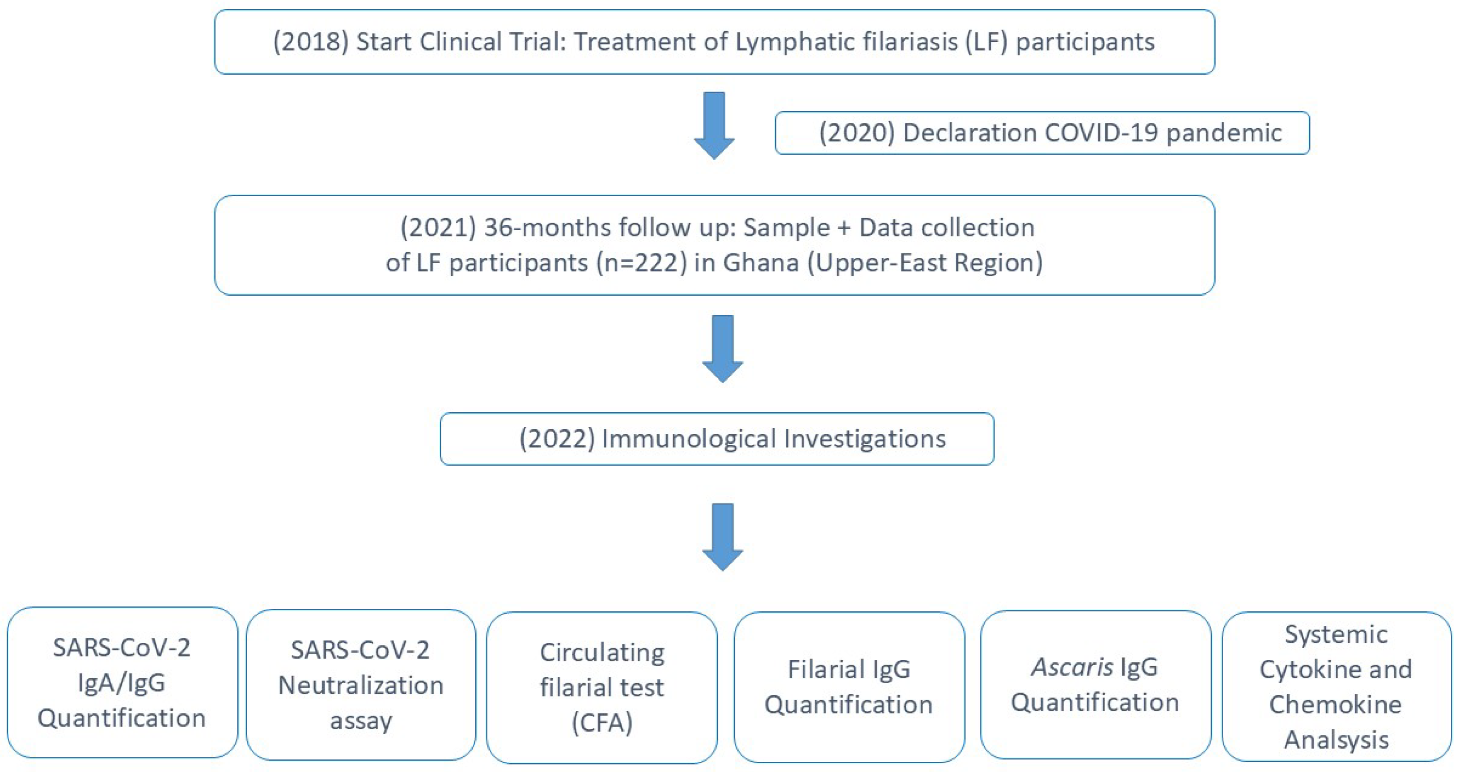
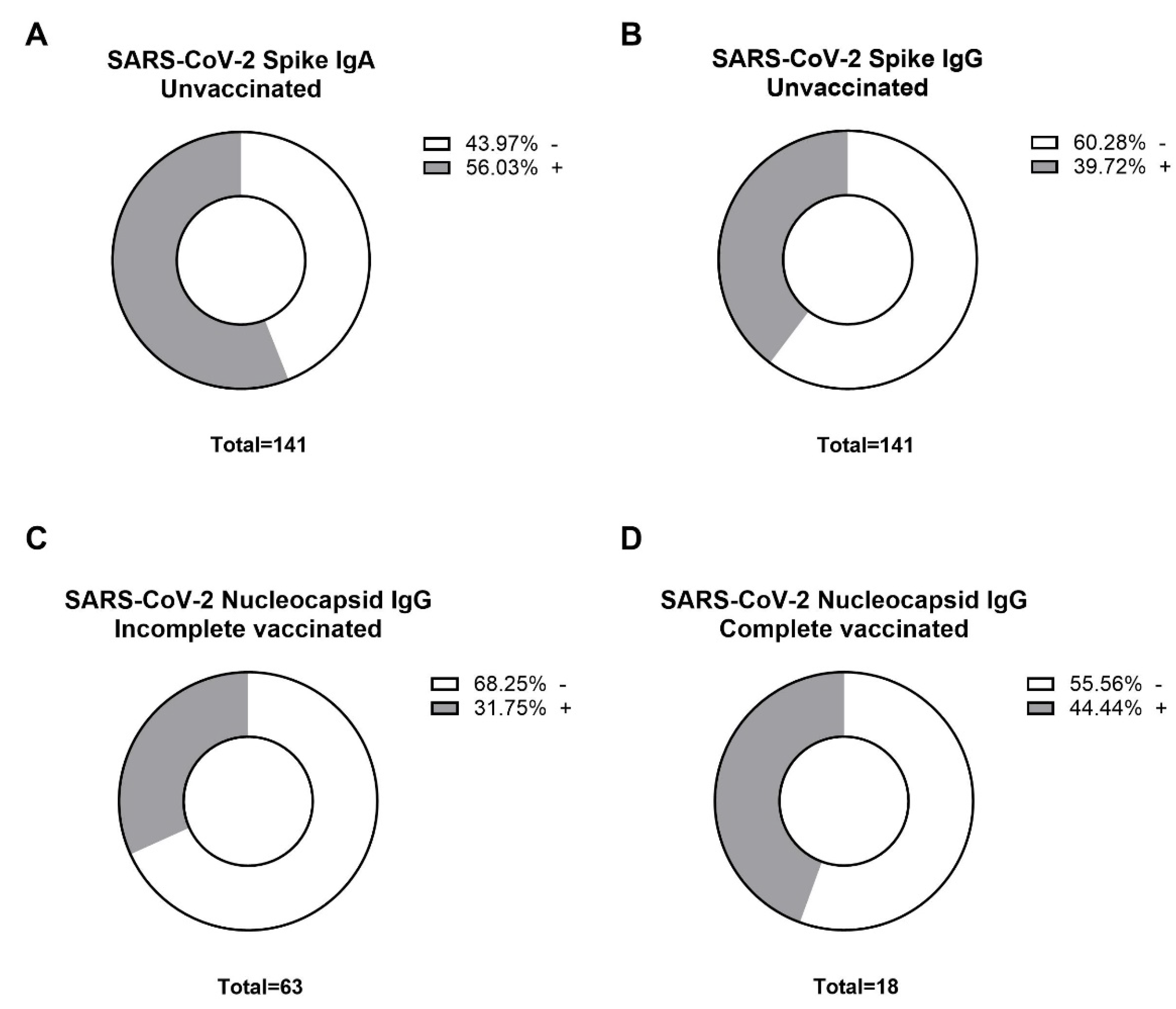
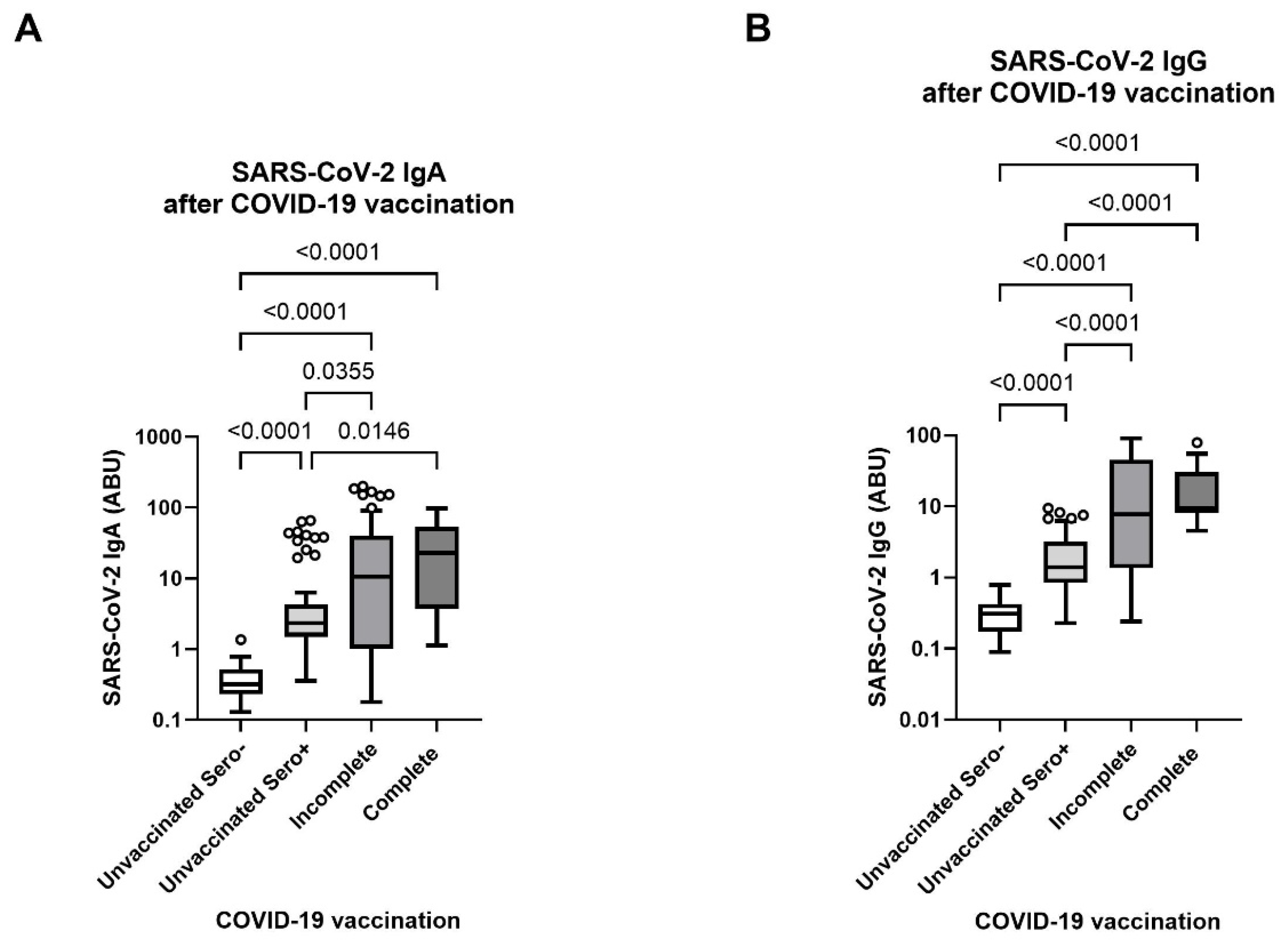
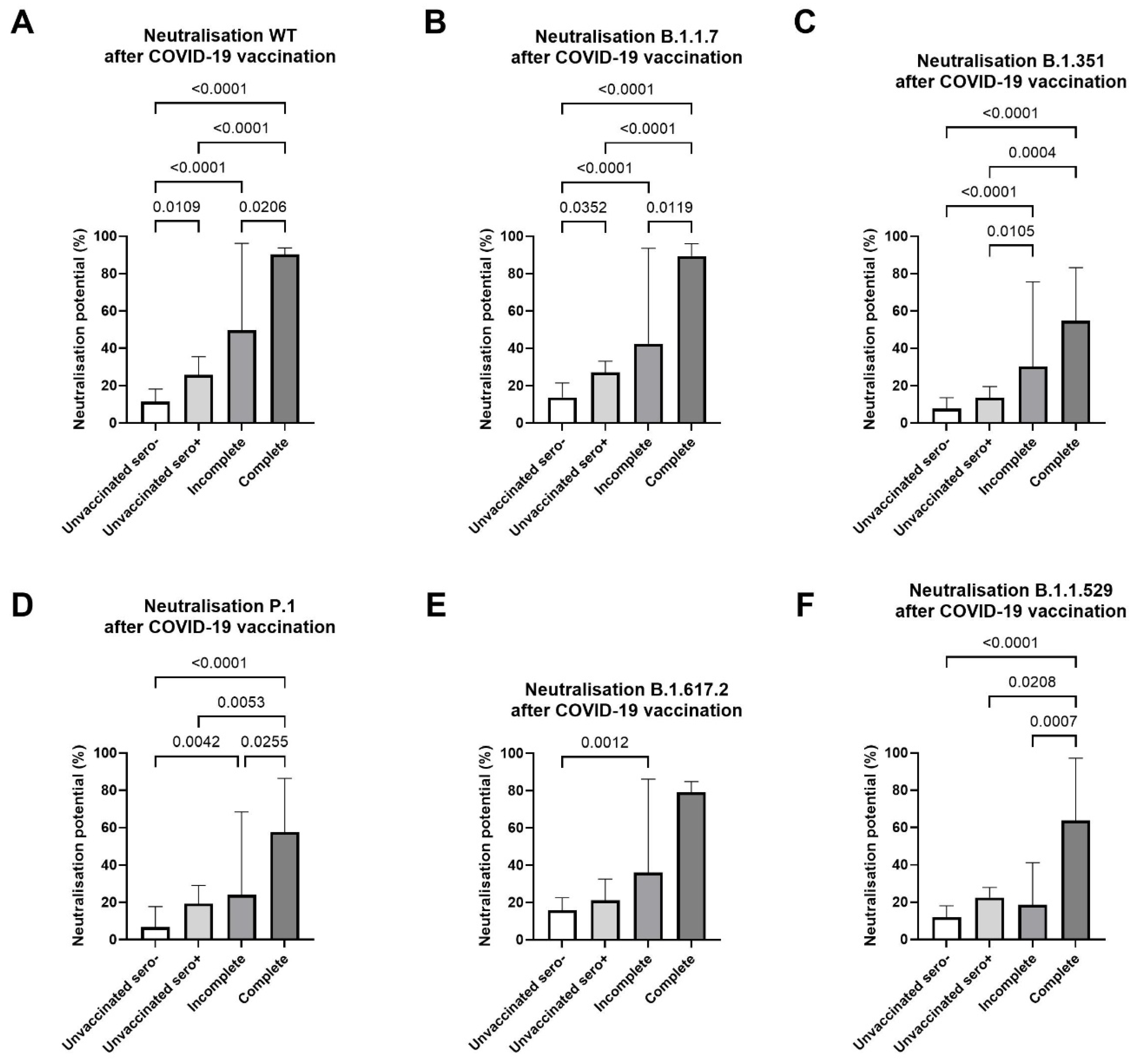
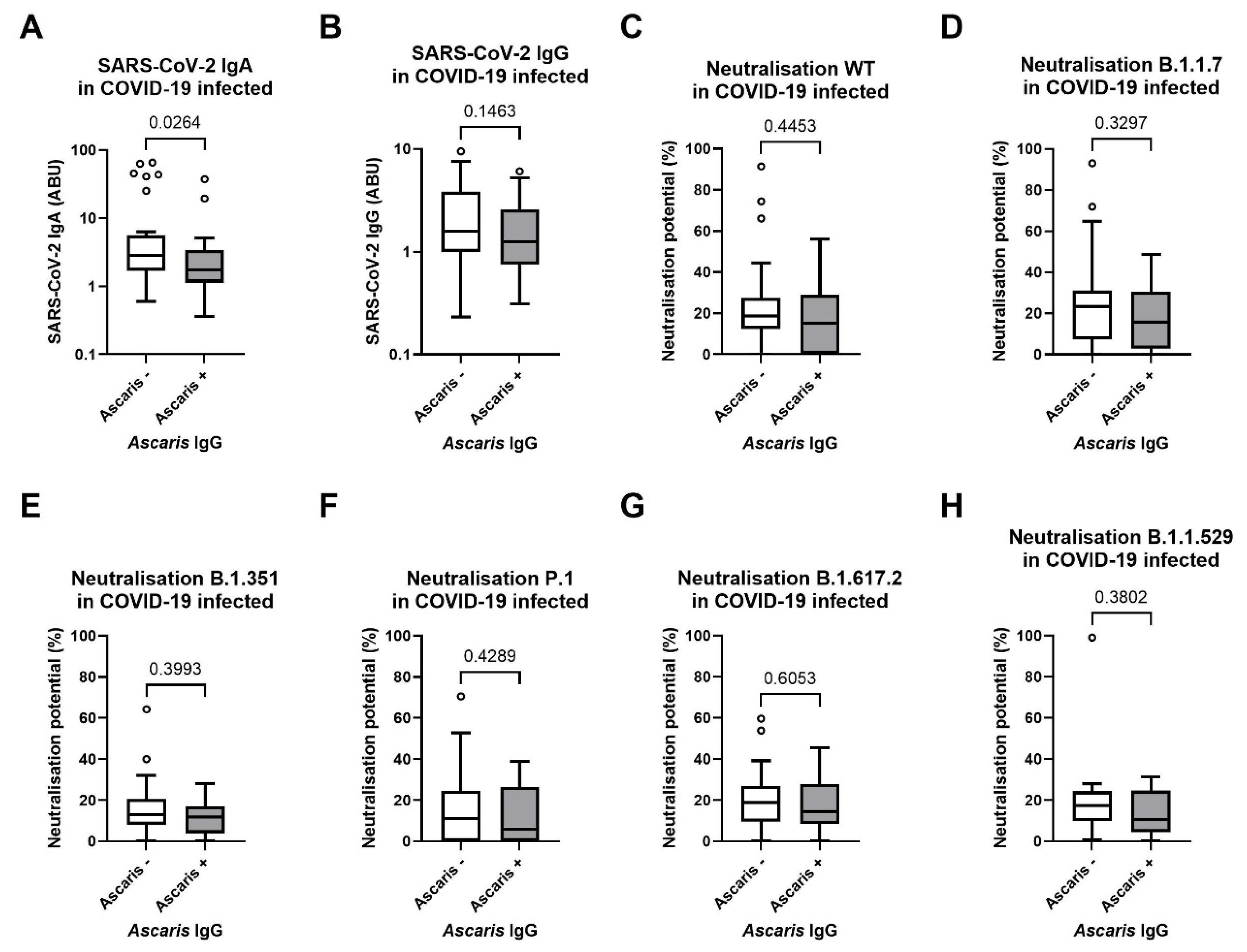
Background/Objectives: Although the COVID-19 pandemic has largely concluded, the varied trajectories it has followed in different regions of the world remain incompletely understood. Intensive research is needed to fully grasp its course and the implications for future global health challenges. Notably, the milder trajectory of the COVID-19 pandemic in Sub-Saharan Africa has defied initial predictions. An emerging body of evidence suggests that, in addition to the continent's younger average age and the lower prevalence of relevant comorbidities, co-infections with helminths may have also impressively shaped the pandemic's milder trajectory in the region. Indeed, helminths are renowned for their ability to modulate human immune responses, which, while potentially beneficial in limiting excessive inflammation, could also diminish vaccine efficacy and impede viral clearance. This study investigated different aspects of the intricate interactions between COVID-19 and Lymphatic Filariasis (LF), a helminth infection caused by parasitic worms such as Wuchereria bancrofti, Brugia malayi, and Brugia timori and endemic to various regions in Sub-Saharan Africa and the tropics. Methods: For this purpose, samples were collected from 222 individuals from endemic areas of Ghana, along with comprehensive clinical and demographic data. The samples include LF patients (n=222) grouped according to their Lymphoedema (LE) stages, as well as COVID-19 vaccinated (n=81) and non-vaccinated individuals (n=141). The expressions of SARS-CoV-2 and filarial-specific antibodies (IgG, IgA) were accessed using ELISA, while Luminex-based immunoassays were employed to measure the expression of SARS-CoV-2 variant-specific neutralizing antibodies. The interplay between vaccine responses, and demographic factors was analyzed using group comparisons with the Kruskal-Wallis or Mann-Whitney U tests. Results: The results indicate that a remarkable portion of unvaccinated individuals (56% IgA seropositive, 39% IgG seropositive) developed antibodies against SARS-CoV-2 despite no confirmed infection. Notably, the study identified a robust antibody response to COVID-19 vaccination, which was independent of the degree of LF pathology or parasitic status. An important observation was the reduced SARS-CoV-2 antibody response in individuals seropositive for Ascaris lumbricoides (p=0.0264), highlighting an interaction between roundworm infection and COVID-19. Conclusion: The study concludes that while COVID-19 vaccination triggers a strong immune response in LF patients, filarial seropositivity might influence the immunogenicity and clinical outcomes of COVID-19, emphasizing the complexity of infectious disease dynamics in co-infected populations.

Keywords: 
;  ;  ;  ;  ;  ;  ;  

1. Introduction

The COVID-19 pandemic, caused by the coronavirus SARS-CoV-2, has been a defining global health crisis of the early 21st century. Its impact has been profound and widespread, affecting every aspect of human life [1,2,3,4,5,6]. As of 11th January 2025, 777,126,421 cases were reported to the WHO, along with 7,079,925 reported COVID-19 deaths globally [7]. Most people infected with the virus display mild to moderate symptoms such as fever, cough, shortness of breath, muscle aches, headache, loss of taste and smell, sore throat, congestion, or runny nose [8]. However, research has indicated that individuals with advanced age, male gender, and comorbidities or certain pre-existing conditions such as cardiovascular disease, diabetes, chronic respiratory disease, or cancer, may have an elevated risk of developing severe responses to COVID-19 due to underlying chronic inflammation or immune system imbalances that might lead to requisition of medical intervention or even death [8,9,10,11]. Approximately 6 to 10% of COVID-19 patients fall into this category and require intensive care, with a significant portion needing mechanical ventilation due to severe respiratory distress [12,13]. The virus enters host cells through the ACE2 receptor, triggering an innate immune response characterized by the production of type I interferons and pro-inflammatory cytokines [14,15]. If the innate response is insufficient, the adaptive immune system is activated, involving T cells and B cells, which produce antibodies to neutralize the virus [16,17,18,19]. In severe cases, a dysregulated immune response can occur, often resulting in a "cytokine storm," characterized by the release of large amounts of pro-inflammatory cytokines and chemokines, such as interleukin-6 (IL-6), tumor necrosis factor-alpha (TNF-α), and interleukin-1beta (IL-1β), which can cause widespread tissue damage leading to acute respiratory distress syndrome (ARDS) and multi-organ failure [14,16]. The cytokine storm is not only a marker of severe disease but also a critical determinant of mortality in COVID-19 patients. In the fight against the pandemic, a critical tool in reducing the severity and mortality has been the development of vaccines. Various platforms have been employed, including mRNA, viral vector-based DNA, and attenuated virus vaccines. Each of these platforms offers unique attributes and immunogenic properties, as well as differing potentials for adverse reactions. mRNA vaccines, such as those developed by Pfizer-BioNTech and Moderna, are notable for their rapid design and production capabilities, which have proven especially useful during the pandemic [20]. Viral vector vaccines, like those from AstraZeneca and Johnson & Johnson, utilize modified viruses to deliver genetic material that induces an immune response [21]. Attenuated virus vaccines use a weakened form of the virus to stimulate immunity without causing disease [21]. These diverse approaches have collectively contributed to the global effort to control COVID-19 through vaccination [21]. Globally, as recorded by Our World Data on January 11th 2025, over 13.72 billion doses of COVID-19 vaccines have been administered, with approximately 1.43 billion doses distributed in the European region and 874 million doses in the African region. Notably, 70.7% of the world's population has received at least one dose of a COVID-19 vaccine. However, this coverage is significantly lower in low-income countries, where only 32.8% of people have received at least one dose [22]. Despite having a low vaccine coverage, African countries have demonstrated a relatively low number of severe cases of COVID-19 [7]. This discrepancy raises important questions about the factors contributing to the lower severity observed in these regions. As the world gradually recovers from the COVID-19 pandemic, the varying impact it has had across different regions underscores our incomplete understanding of its trajectory and broader implications for global health. Despite its devastating global impact [1,2,3,4,5,6], the pandemic's progression in Sub-Saharan Africa has been milder than expected, challenging initial forecasts and emphasizing the need for in-depth investigation into the underlying factors [23]. Emerging hypotheses suggest several potential contributors to this trend, including the continent's younger demographic profile, limited access to diagnostic tests, genetic factors, geographic settings, previous vaccinations, and lower rates of comorbidities [24]. Among these, the hypothesis that co-infections with parasitic diseases including malaria and helminthiasis stand out as particularly compelling [25,26,27]. Particularly, the interactions between helminths and the immune system present paradoxical scenarios during the COVID-19 pandemic. On the one hand, the immunomodulatory effects of these parasites could potentially reduce excessive inflammation, a characteristic of severe COVID-19 cases, thereby lessening symptom severity and lowering mortality rates [26]. This anti-inflammatory impact may have contributed to the observed milder course of COVID-19 in areas with high rates of helminth infections, offering natural resistance against the severe inflammatory reactions leading to severe COVID-19 [28]. On the other hand, the immune modulation induced by helminths might negatively affect vaccine efficacy [29]. This multifaceted scenario highlights the urgency for comprehensive studies that not only unravel the intricate interplay between infectious and parasitic diseases but also inform targeted health interventions and policy-making, crucial for improving resilience against future pandemics. Lymphatic Filariasis (LF), also known as elephantiasis and caused by parasitic worms such as Wuchereria bancrofti, Brugia malayi, and Brugia timori, is a prominent helminth infection affecting humans [30]. It is estimated that at least 36 million people suffer from chronic manifestations of LF, with over 15 million experiencing lymphoedema [31]. LF presents a significant public health challenge in tropical and subtropical areas, including Sub-Saharan Africa, where it causes chronic suffering and disability and imposes a heavy socio-economic burden on communities [31]. Like many helminths, LF parasites exert sophisticated immunoregulatory mechanisms to ensure their survival within the human host [32]. These mechanisms involve a complex interaction between the human immune system and LF parasites, with a notable strategy being the induction of a modified Th2 environment characterized by anti-inflammatory cytokines, such as IL-10 and TGF-β, which help downregulate pro-inflammatory responses and promote parasite tolerance. In helminth endemic regions including Ghana, where LF and COVID-19 coexist, there are unresolved questions about their combined effects on the body's response to the coronavirus, especially in terms of vaccine effectiveness and disease progression. The present study aims to investigate the interaction between COVID-19 and LF within a unique cohort from Ghana, focusing on the response to COVID-19 vaccines and the seroprevalence among individuals with LF. The relationship between viral infections like COVID-19 and chronic parasitic diseases such as LF remains poorly understood, making this research critical for illuminating the complex dynamics involved. The present study investigates various aspects of the complex interactions between Lymphatic Filariasis (LF) and COVID-19, as well as the response to COVID-19 vaccines, leveraging data from a cohort of LF patients in Ghana. This research is particularly significant given the coexistence of these infections in regions where both are endemic. By analyzing how LF may influence the immune response to COVID-19 and the efficacy of the vaccines, the study aims to uncover potential synergies or conflicts that could inform more tailored public health strategies.

2. Materials and Methods

2.1. Study Design and Clinical Characteristics of Participants

During the COVID-19 pandemic, blood samples of Lymphatic filariasis (LF) participants with Lymphoedema (LE) pathology (n=222) were collected in the upper east region of Ghana (Kassena-Nankana District) in December 2021. Participants were enrolled from 12 sub-districts in the study, which was part of a larger and an ongoing clinical trial (“Tackling the Obstacle to fight Filariasis and Podoconiosis” (TAKeOFF) with the ethical approval code CHRPE/AP/525/17 and the controlled trial registration number ISRCTN14042737) [33]. Sample collection was performed in Ghana, and samples were shipped to the Institute of Medical Microbiology, Immunology, and Parasitology (IMMIP) of the University Hospital of Bonn, where the experimental part was performed. A total of 222 participants (34 males and 188 females) were included in the study. Treated lymphatic filariasis participants with lymphoedema pathology in different stages were involved in the study. Participants were divided into two groups according to their lymphoedema (LE) stage: LE Stage 1-3 (Group A) and LE stage 4-6 (Group B). Group A can be subdivided into two treatment arms: 100 mg/day of doxycycline, 200 mg/day of doxycycline, and a placebo group. Group B contained participants who received 200 mg/day of doxycycline and a placebo group. All participants received treatment for 6 weeks. Samples were taken 36 months post-treatment. Each participant gave informed consent for participation. Ethical approval for the study was obtained from the ethical boards of the University Hospital Bonn (Lfd.Nr. 439/20). The lymphoedema staging of the legs was performed at baseline and 36 months follow-up. The overall staging gives the highest achieved stage of a participant. An additional pregnancy test was performed for all female participants; however, all females were negative. The Filariasis Test Strip (FTS) (Alere Scarborough, USA) for detection of circulating filarial antigen (CFA) was carried out 36 months after treatment for all participants. Due to limited COVID-19 test availabilities and capacities, none of the participants (n=222) had ever tested positive for SARS-CoV-2 by PCR or rapid antigen test.
Table 1. Demographic and clinical characteristics of study participants.
Sample Size (n=) 222
Age (Mean ± SD, Min-Max) 46.57 ± 9.21 (17 – 64)
Sex (M / F) 34 (15%) / 188 (85%)
BMI (Min-Max, Mean ± SD) 11.49 – 42.44 (23.32 ± 4.05)
LE Staging (0 / 1 / 2 / 3 / 4 / 5 / 6) 2 / 5 / 130 / 55 / 0 / 1 / 29
0.9% / 2.3% / 58.6% / 24.8% / 0% / 0.5% / 13.1%
Treatment Group (A / B) 198 (89%) / 24 (11%)
COVID-19 Positive test (No / Yes) 222 (100%) / 0
COVID-19 Vaccination (No / 1. Dose / 2. Dose) 141 (63%) / 63 (28%) / 18 (8%)
BCG vaccination (No / Yes / Unknown) 65 (29%) / 139 (63%) / 18 (8%)
Influenza vaccination (No / Yes / Unknown) 65 (29%) / 139 (63%) / 18 (8%)
COVID-19 related symptoms (No / Yes) 186 (84%) / 36 (16%)
SD: Standard Deviation, BMI: Body Mass Index, LE: Lymphoedema, BCG: Bacillus Calmette-Guérin
Figure 1. Flowchart of the study design and timeline for assessing the immunological responses in lymphatic filariasis patients treated with doxycycline. Blood samples were collected in December 2021 during the COVID-19 pandemic from individuals with lymphatic filariasis who were part of a treatment trial (started in 2018) in the Upper-East Region of Ghana. The study included 222 individuals with lymphoedema (LE) pathology. Demographic and clinical information was collected, and immunological investigations were performed, including SARS-CoV-2 IgA/IgG quantification, SARS-CoV-2 neutralization assay, circulating filarial antigen (CFA) test, filarial IgG quantification, Ascaris IgG quantification, and systemic cytokine and chemokine analysis. Sample and data collection occurred in Ghana, while immunological investigations (except for the CFA test) were conducted at the Institute for Medical Microbiology, Immunology, and Parasitology (IMMIP) in Bonn, Germany.
Preprints 148053 g001

2.2. Quantification of SARS-CoV-2-Specific Antibodies

Enzyme-linked immunosorbent assay (ELISA) Kits (Euroimmun, Germany) were used to detect SARS-CoV-2-Spike-specific IgA/IgG antibodies in the plasma of Ghanaian Lymphoedema participants. SARS-CoV-2 Nucleocapsid (NCP)-specific IgG antibody ELISA Kit (Euroimmun, Germany) was used for the detection of NCP-specific antibodies in plasma of COVID-19 vaccinated LE participants, according to the manufacturer’s protocol. Samples were automatically diluted 1:101 in the provided sample buffer and incubated for 60 min at 37°C in a pre-coated 96-well plate. Washing and incubation steps were performed automatically using the predesigned program of Euroimmun’s analyzer I automate. Optical densities (OD) were measured at 450 nm. Ratios <0.8 were considered negative, 0.8-1.1 as equivocal, and >1.1 as positive.

2.3. Detection of Ascaris Lumbricoides Specific IgG

Ascaris lumbricoides IgG ELISA Kit (DRG Instruments GmbH, Marburg, Germany) was used for the detection of specific IgG immunoglobulins in the plasma of LE participants and carried out manually according to the manufacturer’s protocol. Briefly, the samples were diluted (1:101) with the sample dilution buffer. 100 µl of samples/standards and controls were added to each well of the pre-coated 96-well plate and incubated for 1h at 37 °C. The wells were washed 3x with 300 µl washing solution. 100 µl conjugate was added and incubated for 30 min at RT. After another wash step, 100 µl substrate (TMB) was added and incubated for 15 min at RT in the dark. 100 µl of stop solution was added, and optical density (OD) was measured at 450/620nm. The absorbance was converted into units according to the manufacturer’s protocol with the following formula: (Sample absorbance value x 10) / (Cut-off) = DRG Units (DU). Results were interpreted as recommended by the manufacturer: >11 DU as positive, 9-11 DU as equivocal, and <9 DU as negative. The diagnostic specificity is 95 %, and the sensitivity is 100 %. A cross-reaction with antibodies against Toxocara canis, Trichinella, Fasciola, Filaria, and Strongyloides cannot be excluded.

2.4. Detection of Human Filarial-Specific Antibodies

Acanthocheilonema viteae ELISA-Kit (Bordier Affinity Products, Crissier, Switzerland) was used for the quantitative detection of IgG immunoglobulins specific for human filarial infections, including lymphatic filariasis (caused by Wuchereria bancrofti and Brugia malayi), loaosis, onchocerciasis, and mansonelliasis in plasma of Ghanaian LE individuals, according to the manufacturer’s protocol. Briefly, the pre-coated 96-well plate was blocked with 300 µl buffer for 15 min. at RT. The samples were diluted (1:201) with dilution buffer (1X). 100 µl of samples/controls were added to each well and incubated for 30 min at 37 °C. The wells were washed 4x with 300 µl washing solution. 100 µl conjugate was added and incubated for 30 min at 37 °C. After another washing step, 100 µl substrate was added and incubated for 30 min at 37 °C. 100 µl of stop solution was added, and optical density (OD) was measured at 450nm. The absorbance was converted into units according to the manufacturer’s protocol with the following formula: (Sample absorbance value) / (Cut-off absorbance value) = Index. Results were interpreted as recommended by the manufacturer: >1.0 Index as positive and <1.0 as negative. The diagnostic specificity is given at 96 %, and the sensitivity at 95 %. Cross-reactivity occurred with ascariasis, trichinellosis, ancylostomiasis, fasciolosis, and cystic echinococcosis.

2.5. Quantification of SARS-CoV-2 Neutralizing Antibody Levels

Neutralizing SARS-CoV-2 variant-specific antibodies were quantified using the SARS-CoV-2 Variants Neutralizing Antibody 6-plex ProcartaPlex Panel (Thermo Fisher). The assay was performed according to the manufacturer’s instructions and allowed the comparison of neutralizing antibody capacity towards six SARS-CoV-2 variants, including Wildtype (WT), Alpha (B.1.1.7), Beta (B.1.351), Gamma (P.1), Delta (B.1.617.2) and Omicron (B.1.1.529). 50 µl of magnetic capture beads were added to each well of the provided 96-well plate and washed with 150µl 1X washing solution. 25 µl of prediluted samples (1:100) were added, followed by 25 µl assay diluent. Positive and negative control was prepared according to the manufacturer’s recommendation and added to the dedicated wells. Samples were incubated for 2h at RT while shaking (500 rpm/minute). After two washing steps, 25 µl of prediluted 1X detection antibody was added to each well and incubated for 30 min at RT while shaking. After another two washing steps, 50 µl of Streptavidin-PE solution was added to each well and incubated for another 30 min at RT on a plate shaker. After two more washing steps, 120 µl of reading buffer was added to each well, and the plate was incubated for 5 min while shaking before the plate was measured using the MagPix Luminex instrument. The results were analyzed using the following neutralization equation: (1 – (MFI of samples / MFI of negative control)) x 100 = Neutralization (%).

2.6. Quantification of Systemic Cytokine and Chemokine Levels

Cytokine storm 21-plex human ProcartaPlex Panel (Thermo Fisher) was used to quantify systemic cytokines and chemokines in the plasma of Ghanaian Lymphoedema participants. The assay was performed according to the manufacturer’s protocol and involved 21 cytokines and markers related to cytokine release syndrome (CRS), including: G-CSF (CSF-3), GM-CSF, IFN alpha, IFNγ, IL-1 beta, IL-2, IL-4, IL-5, IL-6, IL-8 (CXCL8), IL-10, IL-12p70, IL-13, IL-17A (CTLA-8), IL-18, IP-10 (CXCL10), MCP-1 (CCL2), MIP-1 alpha (CCL3), MIP-1 beta (CCL4), TNF alpha, and TNF beta. 50 µl of magnetic capture beads were added to each well of the provided 96-well plate and washed with 150µl 1X washing solution. 25 µl of undiluted samples were added to the dedicated wells, followed by 25 µl assay diluent. Standard and controls were prepared according to the manufacturer’s recommendation, and samples were incubated for 2h at RT while shaking (500 rpm/minute). After two washing steps, 25 µl of 1X Detection antibody was added to each well and incubated 30 min. at RT while shaking. After another two washing steps, 50 µl of Streptavidin-PE solution was added to each well and incubated for another 30 min. at RT on a plate shaker. After the final two washing steps, 120 µl of reading buffer was added to each well, and the plate was incubated for 5 min. The plate was then measured using the MagPix Luminex instrument, and the results were analyzed using the ProcartaPlex analyst application (Thermo Fisher).

2.7. Statistics

Data were analyzed using GraphPad Prism 10 (La Jolla, CA, USA). We described the characteristics for the study participants using mean values ± (SD), Minimum and Maximum for continuous variables. The values of categorical variables were presented as frequency distribution with percentages. The nonparametric Kruskal-Wallis test, followed by Dunn’s posthoc test, was used for group comparison along with the Mann-Whitney U-test. A p-value <0.05 was considered statistically significant.

3. Results

3.1. Elevated SARS-CoV-2 Seroprevalence Among Unvaccinated Individuals in Ghana

Since the circulation of SARS-CoV-2 in African countries is not well-documented due to the lack of testing capacity and the high prevalence of asymptomatic infections [24], we first evaluated Nucleocapsid (N) and Spike (S) protein seroprevalences to discriminate between vaccine-induced and infection-induced SARS-CoV-2 antibody expressions. The analysis of the unvaccinated cohort (n=141) revealed that a remarkable portion had serological evidence of exposure to SARS-CoV-2: 56% had detectable levels of Spike-specific IgA, and 39% were positive for Spike-specific IgG, as illustrated in Figure 2A and Figure 2B, respectively. These results suggest a considerable degree of previous exposure to the virus among the unvaccinated individuals. Further investigation into the immune responses of vaccinated participants involved a SARS-CoV-2 Nucleocapsid protein-specific (NCP) IgG ELISA. This assay was instrumental in distinguishing between vaccine-induced immunity and immunity developed from natural infection. Our findings showed that among the incompletely vaccinated individuals (n=63), 31.7% tested positive for NCP-specific IgG, indicating a likelihood of previous viral infection (Figure 2C). In contrast, the fully vaccinated group (n=18) demonstrated a higher NCP-seropositivity rate of 44.4% (Figure 2D). These observed rates of seropositivity to the NCP antigen in vaccinated individuals reflect additional viral encounters following vaccination. The disparity in NCP-seropositivity between those with incomplete and complete vaccination courses suggests a complex interplay between vaccination-induced immunity and natural infection.

3.2. Robust SARS-CoV-2-Specific Antibody Response in Lymphatic Filariasis Patients after Incomplete and Complete COVID-19 Vaccination

After confirming a high SARS-CoV-2 seroprevalence in the LF cohort, both among vaccinated and unvaccinated individuals, we first investigated how LF pathology stages influence antibody response to COVID-19 vaccination. The results indicated that Lymphoedema stages of the participants revealed not to influence the SARS-CoV-2 antibody response after vaccination (Figure S1). We then compared the SARS-CoV-2-specific antibody levels in different vaccination and serostatus groups. As shown in Figure 3A,B, the median levels of SARS-CoV-2-specific IgA (Figure 3A) and IgG (Figure 3B) were highest in the completely vaccinated group. The incompletely vaccinated group also showed elevated IgA levels, although there was considerable variability within this group. Both vaccinated groups demonstrated notably higher IgA and IgG levels compared to the unvaccinated seronegative and seropositive groups. These findings highlight that COVID-19 vaccination induces a robust SARS-CoV-2-specific antibody response in LF patients. To further characterize the effect of helminth infections on the COVID-19 vaccination response, we analyzed SARS-CoV-2 IgA and IgG expressions related to their parasitic status. The parasitic status was grouped according to Negative, Single Positive or Double Positive for Circulating filarial antigen (CFA) test, Ascaris lumbricoides IgG and Acanthocheilonema viteae IgG. The data indicated that none of the helminths remarkably affected COVID-19-mediated antibody induction. Among single and double positive participants, elevated antibody responses were observed in the incompletely and completely vaccinated groups (Figure S2). Beside the potential effect of the parasitic status onto the SARS-CoV-2 antibody response, the possible impact onto the systemic cytokine and chemokine profile was further analyzed. However, no effect has been observed and comparable levels of Th1, Th2, Th17 and Treg-related cytokines were detected within plasma of Lymphatic filariasis individuals irrespective of their filarial status (Figure S3).
Figure 2. Seropositivity rates for SARS-CoV-2-specific antibodies between unvaccinated and vaccinated cohorts in Ghana. Donut charts represent the seroprevalence of (A) Spike-specific IgA antibodies in the unvaccinated cohort (n=141), with 56.03% seropositive (grey) and 43.97% seronegative (white); (B) Spike-specific IgG antibodies in the unvaccinated cohort (n=141), with 39.72% seropositive (grey) and 60.28% seronegative (white); (C) Nucleocapsid protein-specific (NCP) IgG antibodies in the partially vaccinated group (n=63), with 31.75% seropositive (grey) and 68.25% seronegative (white); (D) Nucleocapsid protein-specific (NCP) IgG antibodies in the fully vaccinated group (n=18), with 44.44% seropositive (grey) and 55.56% seronegative (white).
Preprints 148053 g002
Figure 3. SARS-CoV-2-specific antibody levels in different vaccination and serostatus groups. Box plots represent Spike specific IgA (A) and IgG (B) antibody expressions in incomplete (n=63) (medium grey) and complete (n=18) COVID-19 vaccinated (dark grey) lymphatic filariasis individuals suffering from lymphoedema pathology compared to COVID-19 unvaccinated seronegative (n=37) (white) and seropositive (n=87) (light grey) participants. Indicated p values were calculated using Kruskal-Wallis’ test followed by Dunn’s comparison post hoc to compare all groups. Bars represent the median ± IQR of antibody binding units (ABU). Significance is accepted if p <0.05.
Preprints 148053 g003

3.3. Elevated Neutralizing Antibody Levels Towards Variants of Concern in COVID-19 Vaccinated Filaria Seropositive Individuals

Since antibody expression may not necessarily translate in neutralizing potential we compared the expression of SARS-CoV-2 neutralizing antibodies in different vaccination and serostatus groups. The neutralizing capacity of SARS-CoV-2 specific antibodies within the incomplete (n=63) and complete (n=18) COVID-19 vaccinated lymphatic filariasis participants was notably elevated compared to the unvaccinated and SARS-CoV-2 seronegative control group (n=37) (Figure 4). By comparing the neutralizing antibody capacity towards six SARS-CoV-2 variants including Wildtype (WT), Alpha (B.1.1.7), Beta (B.1.351), Gamma (P.1), Delta (B.1.617.2) and Omicron (B.1.1.529), higher levels towards the different variants of concern have been observed in the COVID-19 vaccinated groups, except for the Omicron variant where just complete COVID-19 vaccinated individuals higher levels achieved (Figure 4F). However, the COVID-19 unvaccinated but SARS-CoV-2 seropositive group (n=48) showed reduced levels of neutralizing antibodies compared to the vaccinated groups. Interestingly, the SARS-CoV-2 seropositive group had increased neutralizing potential compared to the unvaccinated control group, but only for the Wild type, Alpha, and Gamma variants (Figure 4). In the next step, the influence of an additional COVID-19 infection within the COVID-19 vaccinated groups on the neutralizing capacity of SARS-CoV-2 specific antibodies was analyzed, nevertheless no differences have been observed (Figure S4).
Figure 4. Neutralization potential against different SARS-CoV-2 variants in various vaccination and serostatus groups. Bars represent the expression of SARS-CoV-2 specific neutralizing antibodies in incomplete (n=63) (medium grey) and complete (n=18) COVID-19 vaccinated (dark grey) lymphatic filariasis infected individuals for six major SARS-CoV-2 variants, including Wildtype (WT) (A), Alpha (B.1.1.7) (B), Beta (B.1.351) (C), Gamma (P.1) (D), Delta (B.1.617.2) (E) and Omicron (B.1.1.529) (F) compared to the COVID-19 unvaccinated and SARS-CoV-2 seronegative (n=37) group (white). Indicated p values were calculated using Kruskal-Wallis’ test followed by Dunn’s comparison post hoc to compare all groups. Bars represent the median ± IQR of antibody neutralizing potential (%). Significance is accepted if p <0.05.
Preprints 148053 g004

3.4. Diminished SARS-CoV-2-Specific IgA Response in Ascaris Seropositive Individuals

After observation of a robust antibody response and high neutralizing potential following COVID-19 vaccination, a potential nematode effect on COVID-19 infection-induced responses was evaluated additionally. Therefore, the effect of Ascaris lumbricoides seropositivity on the SARS-CoV-2-Spike-specific IgA/IgG response within COVID-19 infected individuals was analyzed. In regard to IgA expression, our findings indicated a decrease (p=0.0264) in SARS-CoV-2 specific IgA levels among individuals seropositive for Ascaris lumbricoides (Ascaris+) (Figure 5A). The median IgA levels were notably lower (39%) in the Ascaris+ group (n=28) compared to the seronegative (n=35) (Ascaris-) cohort, underscoring the impact of nematode infection on the humoral immune response to SARS-CoV-2. Concerning the SARS-CoV-2 IgG response, no difference was observed in IgG antibody levels between the Ascaris seronegative and positive groups (p=0.1463) (Figure 5B). This suggests that nematode infection status does not markedly influence the IgG-mediated immune response against SARS-CoV-2. Overall, the data suggests that Ascaris lumbricoides infection is associated with a reduced IgA antibody response to SARS-CoV-2, yet it appears to have no crucial effect on the IgG antibody response. Due to the negative effect of filarial seropositivity on the COVID-19 infection-induced SARS-CoV-2 specific IgA antibody response, the potential negative effect on the neutralizing capacity of the SARS-CoV-2 specific antibodies towards six different variants of concern was analyzed. However, comparable neutralization potentials of SARS-CoV-2 antibodies was observed in the Ascaris lumbricoides seropositive group towards all six different VoCs (Figure 5C–H).
Figure 5. Influence of Ascaris lumbricoides IgG seropositivity on COVID-19 infection-induced SARS-CoV-2 Spike-specific antibody response and neutralizing potential. Reduced SARS-CoV-2 IgA levels were seen in the Ascaris positive (n=28) group (grey) compared to the negative (n=35) group (white) (A), as well as comparable SARS-CoV-2 IgG levels (B). Comparable neutralization capacity of SARS-CoV-2 specific antibodies towards six major SARS-CoV-2 variants within Ascaris seropositive COVID-19 infected participants compared to seronegative group (C-H). Indicated p values were calculated using the Mann-Whitney-U test. Bars represent the median ± IQR of antibody binding units (ABU) (A+B) and antibody neutralizing potential (%) (C-H). Significance is accepted if p <0.05.
Preprints 148053 g005

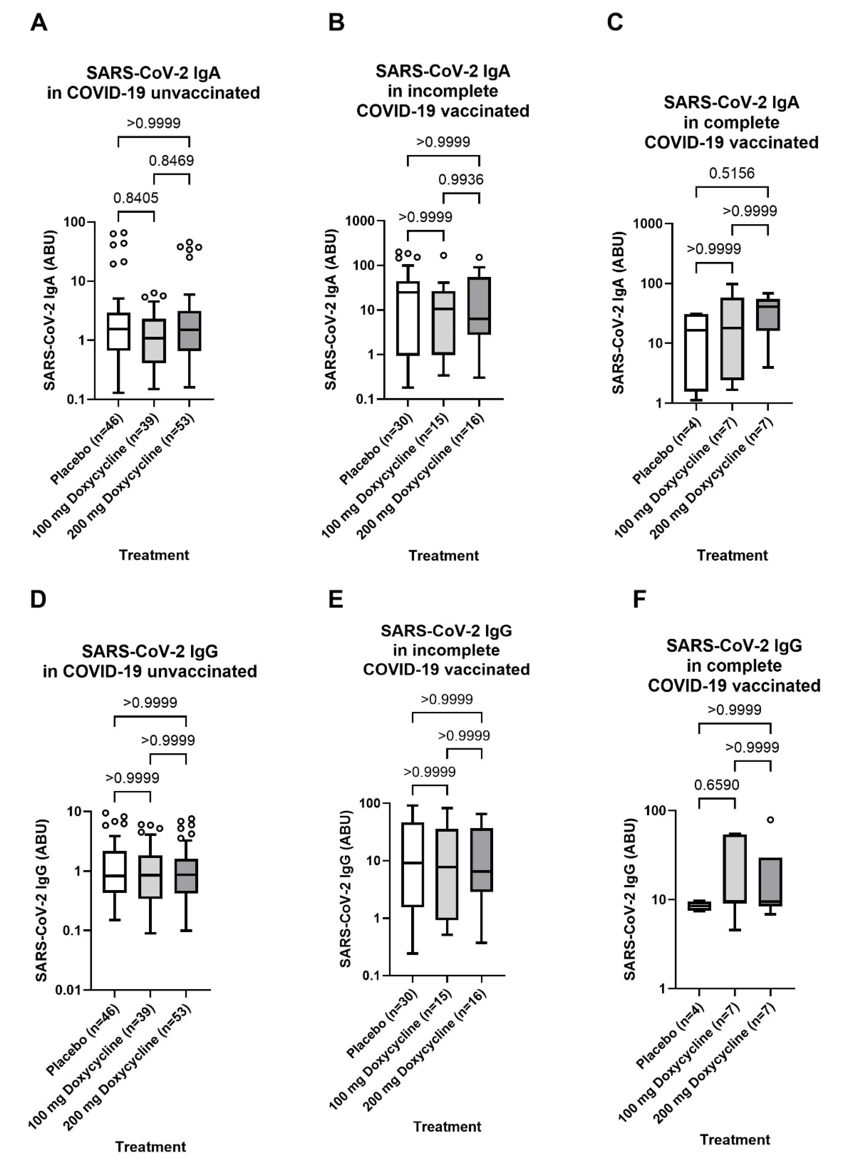
3.5. No Influence of Doxycycline Treatment 36-Months Before Blood Sampling of Lymphatic Filariasis Individuals with Lymphoedema Pathology Compared to the Placebo Control Group

Since all participants were part of a clinical trial and received a six-week doxycycline treatment (100mg/day or 200mg/day) 36-months before blood sampling, a potential effect of treatment on the SARS-CoV-2 specific antibody response post-COVID-19 infection and/or vaccination was analyzed. However, no statistical differences were observed for the SARS-CoV-2 specific IgA and IgG response among the two doxycycline treatment arms (100mg/day and 200mg/day) compared to the placebo control group within unvaccinated, incompletely and completely COVID-19 vaccinated individuals (Figure 6).
Figure 6. No effect of Doxycycline Treatment 36 months before blood sampling on infection- or vaccination-induced SARS-CoV-2 specific antibody response of Lymphatic filariasis participants. Comparable SARS-CoV-2 Spike-specific IgA (A-C) and IgG (D-F) antibody levels among unvaccinated (n=141) (A+D), incomplete (n=63) (B+E) and complete (n=18) (C+F) COVID-19 vaccinated lymphatic filariasis individuals irrespective of a six-week treatment with 100mg/day (light grey) and 200 mg/day (grey) Doxycycline 36-months before blood sampling compared to the Placebo Control group (white). Indicated p values were calculated using Kruskal-Wallis’ test followed by Dunn’s comparison post hoc to compare all groups. Bars represent the median ± IQR of antibody binding units (ABU). Significance is accepted if p < 0.05.
Preprints 148053 g006
Altogether, the present retrospective study provides insight into the ability of filarial seropositivity and/or co-infection to modify COVID-19 vaccine efficacy in humans. It can be stated that an adenovirus-vectored COVID-19 vaccine in lymphatic filariasis individuals induced a strong humoral immune response. This vaccine-induced response was independent of the lymphoedema stage and serological parasitic status. Moreover, notably elevated neutralizing anti-body levels towards six different SARS-CoV-2 variants of concern were observed within the COVID-19 vaccinated groups independent of filarial status.

4. Discussion

Vaccines are one of the most successful medical inventions, crucial for eradicating or controlling common and fatal diseases [34]. For COVID-19, the early rollout of various vaccines significantly contributed to halting the pandemic [35,36,37]. Several studies have shown that helminths can modulate immune responses to vaccines [38,39,40]. Given the prevalence of helminth infections globally, especially in sub-Saharan Africa [41], evaluating COVID-19 vaccine efficacy in helminth-infected populations is essential for optimizing vaccination programs. In this context, the present study explored the humoral immune response following COVID-19 vaccination with AstraZeneca in a lymphatic filariasis (LF) cohort in Ghana. Our initial seroprevalence analyses revealed a remarkable portion of the unvaccinated cohort with serological evidence of exposure to SARS-CoV-2: 56% had detectable levels of Spike-specific IgA, and 39% were positive for Spike-specific IgG. These high seroprevalence rates could be due to asymptomatic infections and limited testing capacity in the region. Our data are comparable with the results of previous prevalence analyses in the Africa [42,43]. Here we uniquely employed Nucleocapsid (N) and Spike (S) protein seroprevalences to differentiate between vaccine-induced and infection-induced antibodies. Our findings imply a considerable degree of previous exposure to the virus among both vaccinated and unvaccinated individuals, which has important implications for public health strategies and our understanding of SARS-CoV-2 spread in similar contexts. Our study found that COVID-19 vaccination induces a robust SARS-CoV-2-specific antibody response in LF patients. The median levels of SARS-CoV-2-specific IgA and IgG were highest in the completely vaccinated group, highlighting the robustness of the antibody response in these individuals. The incompletely vaccinated group also showed elevated IgA levels, although with notable variability. Moreover, our results suggest that LF pathology stages do not influence the SARS-CoV-2 antibody response after vaccination. This finding is important for vaccination strategies in populations with similar health profiles, indicating that COVID-19 vaccines remain effective regardless of LF pathology. We analyzed the impact of circulating filarial antigen (CFA) and filarial serological positivity as well as seropositivity towards other helminth infections (Ascaris lumbricoides) on antibody responses to COVID-19 vaccines and found that these did not considerably affect antibody induction post-COVID-19 vaccination. This suggests that co-infections with these helminths do not pose a barrier to achieve effective immunization against COVID-19. These findings are relevant for understanding the broader implications for co-infected populations and support the continued rollout of COVID-19 vaccines in helminth-endemic regions. A plausible explanation for the lack of crucial impact of helminth infections on the immune response to the AstraZeneca COVID-19 vaccine could be the nature of the vaccine itself. AstraZeneca's vaccine, also known as ChAdOx1 nCoV-19, uses a viral vector based on a weakened version of an adenovirus that causes infections in chimpanzees [44]. This viral vector approach may induce a strong and broad immune response that is less susceptible to modulation by helminth-induced immune changes. Helminths are known to induce a modified type 2 immune response (Th2) characterized by the production of cytokines such as IL-4, IL-5, IL-13 and IL-10, which can potentially modulate vaccine responses [38]. However, the ChAdOx1 nCoV-19 vaccine is designed to stimulate a robust type 1 immune response (Th1) with high levels of IFN-γ and TNF-α, which are crucial for antiviral immunity [45]. The strong Th1 response induced by the vaccine might override the modulatory effects of helminths, resulting in effective immunization. Several potential mechanisms might explain why AstraZeneca's vaccine remains effective in the presence of helminth infections: The use of a viral vector can generate strong immune responses, possibly overcoming any immunomodulatory effects induced by helminths [38,44,45]. The ability of the viral vector to present multiple epitopes could stimulate a broad and diverse immune response, making it more resilient to immune modulation by helminths [46]. Our findings are particularly encouraging for communities in helminth-endemic regions. They suggest that the COVID-19 vaccination programs using AstraZeneca's vaccine can be rolled out without the need for extensive deworming campaigns beforehand. This is crucial for maintaining high vaccination coverage and protecting vulnerable populations from COVID-19, especially in areas where healthcare resources are limited, and helminth infections are prevalent. Another interesting aspect of our data is the robust antibody expression and neutralizing potential against different SARS-CoV-2 variants in completely vaccinated individuals. Among the other groups, only completely vaccinated individuals showed elevated levels of neutralizing antibodies towards the Omicron variant, indicating no circulation or exposure to this variant of concern at the sampling time point. While a relatively elevated neutralizing antibody expression is seen in incompletely vaccinated individuals, a huge variability in antibody expression and neutralizing potential is seen suggesting that some individuals in this group may not be sufficiently protected against severe forms of the infection. This finding aligns with previous literature on the topic [47,48] and confirms the necessity to adopt a full vaccination regime. Our findings also indicated no differences in SARS-CoV-2 antibody responses among participants who received doxycycline treatment 36 months before blood sampling compared to the placebo control group. This suggests that prior doxycycline treatment does not interfere with the immune response to COVID-19 vaccination. Doxycycline in addition to its indirect macrofilaricidal capacity, leading to sterility and death of adult worms [49,50] was also shown to have various immunomodulatory effects through inhibition of metalloproteinases and was suggested as a potential treatment in hospitalized COVID-19 patients [51]. These immunomodulatory effects can potentially affect immune responses to COVID-19 vaccines. To exclude any direct effect of doxycycline on the immune responses to COVID-19 vaccines, we compared SARS-CoV-2 antibody expression in doxycycline treated and placebo patients. Our data indicates that prior doxycycline treatment has no impact on vaccine-induced immunity. This result is relevant for treatment protocols and their potential impact on vaccine-induced immunity, supporting the idea that prior doxycycline treatment can be safely administered without compromising vaccine efficacy. The main limitation of this work is its regional scope in Ghana, which may limit the generalizability of the findings to other regions with different epidemiological profiles. Further studies are needed to confirm these findings in other geographic and demographic contexts. Additionally, despite controlling for many variables, there might still be unaccounted confounders that could have influenced the results. Factors such as variations in individual immune responses, environmental influences, and genetic diversity could potentially impact the observed outcomes. Moreover, the lack of PCR testing for COVID-19 in our cohort could mean that some SARS-CoV-2 exposures were potentially underestimated, as some infected individuals may not express antibodies [52,53]. Moreover, the relative rapid waning of antibody expression after virus encounter may have led to underestimation of asymptomatic individuals [54]. PCR testing was crucial for identifying active infections and understanding the true spread of the virus. In Ghana and other parts of West Africa, limited access to PCR testing has been a significant challenge in accurately tracking COVID-19 cases [55]. In regard to the use of antibody expressions to detect helminth infections, while serological analyses have the potential to detect current and previous helminth infections, which extends the validity and robustness of our conclusions, they may also introduce variability in the detection accuracy and sensitivity. While our study cohort includes only ChAdOx1 nCoV-19 vaccinated individuals, it is important to verify if our findings are confirmed with other vaccines. This was not possible in the present study due to the low COVID-19 vaccine coverage in the region [56], the logistic challenges posed by mRNA-based vaccines in sub-Saharan Africa, but also the high vaccine hesitancy particularly towards mRNA vaccines in the region [56]. In addition, while our study focused on humoral immune responses to COVID-19 vaccines, due to logistical challenges, T cells responses might also play a key role [57]. Despite these limitations intrinsic to the nature of the study, the present study uniquely demonstrates that COVID-19 vaccination with a DNA-vectored vaccine elicits a robust immune response in individuals with LF, independent of their parasitological status.

5. Conclusions

Our study indicates that COVID-19 vaccination elicits a robust immune response in individuals with LF, independent of their helminth infection status. This finding supports the continued rollout of COVID-19 vaccines in helminth-endemic regions, emphasizing the need for comprehensive public health strategies to manage co-infections and improve vaccine coverage. The manuscript provides valuable data and insights into the complex interplay between helminth infections and COVID-19, with important implications for public health policies in endemic regions. However, the identified limitations highlight areas for further research and validation to enhance the applicability of these findings across diverse populations.

Supplementary Materials

The following supporting information can be downloaded at: Preprints.org, Figure S1: Comparable SARS-CoV-2 vaccination-induced IgA/IgG antibody response independent of lymphoedema stage in lymphatic filariasis individuals; Figure S2: No impact of circulating filarial antigen (CFA), Ascaris lumbricoides or Acanthocheilonema viteae serostatus on SARS-CoV-2 specific IgG and IgA expression after COVID-19 vaccination; Figure S3: No effect of the parasitic status on the systemic cytokine and chemokine profile; Figure S4: Comparable neutralizing potential of SARS-CoV-2 antibodies among SARS-CoV-2 Nucleocapsid (NCP) negative/positive incomplete and complete COVID-19 vaccinated lymphatic filariasis individuals; Figure S5: Comparable SARS-CoV-2 neutralizing capacity in incomplete and complete COVID-19 vaccinated lymphatic filariasis individuals towards six variants of concern (VoC) irrespective of parasitic status.

Author Contributions

Conceptualization, T.A., A.H. and J.M.; methodology, J.M., T.A., P.J.K. and J.M.K.; software, J.M. and U.K.S.; validation, J.M. and U.K.S.; formal analysis, T.A, J.N. and J.M.; investigation, T.A. and J.M.; resources, A.Y.D., L.B.D., J.O.M. and D.A.M.; data curation, J.M. and U.K.S.; writing—original draft preparation, J.M. and T.A.; writing—review and editing, J.M., T.A., and A.H.; visualization, J.M. and T.A.; supervision, T.A.; project administration, A.H.; funding acquisition, A.H. and U.K.S. All authors have read and agreed to the published version of the manuscript.

Funding

AH is funded by the Deutsche Forschungsgemeinschaft (DFG, German Research Foundation) under Germany’s Excellence Strategy–EXC2151–390873048”. The TAKeOff project was funded by the German Federal Ministry of Education and Research (BMBF) under the funding code FKZ01KA1611. U.K.S, J.M.K and P.J.K are partially funded by the German Center for Infection Research (TTU 09.703/09.715, TI 07.005, and TTU 03.815). “Alternative treatment strategies using anti-Wolbachial drugs to accelerate elimination of Lymphatic Filariasis and Onchocerciasis” ― funded by the European and Developing Countries Clinical Trial Partnership (EDCTP2) program and supported by the European Union (grant number TMA2018SF-2451-ASTAWOL) to AYD. This publication is supported by the Open Access Publication Fund of the University of Bonn. The funders had no role in study design, data collection, analysis, decision to publish, or manuscript preparation.

Institutional Review Board Statement

The study was conducted in accordance with the Declaration of Helsinki and approved by the Institutional Ethics Committees of the University Hospital Bonn (Lfd.Nr.439/20) and the Committee on Human Research, Publication and Ethics of the Kwame Nkrumah University of Science and Technology (KNUST), Kumasi, with the ethical approval code CHRPE/AP/525/17 and the controlled trial registration number ISRCTN14042737.

Informed Consent Statement

Informed consent was obtained from all subjects involved in the study.

Data Availability Statement

All data generated during this investigation are included in the main text or the supplementary material and datasets are available upon request from the corresponding author.

Acknowledgments

We thank the Ghana-based teams for their support during the recruitment of LF patients.

Conflicts of Interest

The authors declare no conflicts of interest. The funders had no role in the design of the study; in the collection, analyses, or interpretation of data; in the writing of the manuscript; or in the decision to publish the results.

Abbreviations

The following abbreviations are used in this manuscript:
CFA Circulating Filarial Antigen
COVID-19 Coronavirus Disease 2019
LE Lymphoedema
LF Lymphatic Filariasis
NCP Nucleocapsid
SARS-CoV-2 Severe acute respiratory syndrome coronavirus 2
VoC Variant of concern
WT Wildtype

References

  1. Venkatesh, A.; Edirappuli, S. Social distancing in covid-19: What are the mental health implications? Bmj 2020, 369. [Google Scholar] [CrossRef] [PubMed]
  2. Freeman, T.; et al. Illustrating the impact of commercial determinants of health on the global COVID-19 pandemic: Thematic analysis of 16 country case studies. Health Policy 2023, 134, 104860. [Google Scholar] [CrossRef]
  3. Hsieh, E.; et al. Global Perspective on the Impact of the COVID-19 Pandemic on Rheumatology and Health Equity. Arthritis Care Res (Hoboken) 2024, 76, 22–31. [Google Scholar] [CrossRef]
  4. Lau, J.; et al. Understanding the mental health impact of COVID-19 in the elderly general population: A scoping review of global literature from the first year of the pandemic. Psychiatry Res 2023, 329, 115516. [Google Scholar] [CrossRef] [PubMed]
  5. Rahman, M.A.; et al. The WHO has terminated global public health emergency for COVID-19 by the IHR Emergency Committee recommendation: Potential impact analysis. Ann Med Surg (Lond) 2023, 85, 3755–3756. [Google Scholar] [CrossRef] [PubMed]
  6. Shaikh, B.T.; et al. COVID-19 Pandemic and Its Global Impact on the Accessibility and Provision of Maternal and Child Health Care Services. Asia Pac J Public Health 2024, 10105395241250120. [Google Scholar] [CrossRef]
  7. (WHO), W.H.O. COVID-19 Deaths Dashbord. 2024. Available online: https://data.who.int/dashboards/covid19/deaths?n=c.
  8. Li, Q.; et al. Immune response in COVID-19: What is next? Cell Death Differ 2022, 29, 1107–1122. [Google Scholar] [CrossRef]
  9. Noor, F.M.; Islam, M.M. Prevalence and Associated Risk Factors of Mortality Among COVID-19 Patients: A Meta-Analysis. J Community Health 2020, 45, 1270–1282. [Google Scholar] [CrossRef]
  10. Fang, X.; et al. Epidemiological, comorbidity factors with severity and prognosis of COVID-19: A systematic review and meta-analysis. Aging (Albany NY) 2020, 12, 12493–12503. [Google Scholar] [CrossRef]
  11. Peckham, H.; et al. Male sex identified by global COVID-19 meta-analysis as a risk factor for death and ITU admission. Nat Commun 2020, 11, 6317. [Google Scholar] [CrossRef]
  12. Herrera-Esposito, D.; de Los Campos, G. Age-specific rate of severe and critical SARS-CoV-2 infections estimated with multi-country seroprevalence studies. BMC Infect Dis 2022, 22, 311. [Google Scholar] [CrossRef] [PubMed]
  13. Oliveira, E.; et al. ICU outcomes and survival in patients with severe COVID-19 in the largest health care system in central Florida. PLoS ONE 2021, 16, e0249038. [Google Scholar] [CrossRef] [PubMed]
  14. Tay, M.Z.; et al. The trinity of COVID-19: Immunity, inflammation and intervention. Nat Rev Immunol 2020, 20, 363–374. [Google Scholar] [CrossRef]
  15. Blanco-Melo, D.; et al. Imbalanced Host Response to SARS-CoV-2 Drives Development of COVID-19. Cell 2020, 181, 1036–1045.e9. [Google Scholar] [CrossRef] [PubMed]
  16. Vabret, N.; et al. Immunology of COVID-19: Current State of the Science. Immunity 2020, 52, 910–941. [Google Scholar] [CrossRef]
  17. Coveney, C.; et al. Innate immunology in COVID-19-a living review. Part I: Viral entry, sensing and evasion. Oxf Open Immunol 2020, 1, iqaa004. [Google Scholar] [CrossRef]
  18. Li, G.; et al. Coronavirus infections and immune responses. J Med Virol 2020, 92, 424–432. [Google Scholar] [CrossRef]
  19. Vafaeinezhad, A.; Atashzar, M.R.; Baharlou, R. The Immune Responses against Coronavirus Infections: Friend or Foe? Int Arch Allergy Immunol 2021, 182, 863–876. [Google Scholar] [CrossRef]
  20. Okuyama, R. mRNA and Adenoviral Vector Vaccine Platforms Utilized in COVID-19 Vaccines: Technologies, Ecosystem, and Future Directions. Vaccines (Basel) 2023, 11. [Google Scholar] [CrossRef]
  21. Adjobimey, T.; et al. Comparison of IgA, IgG, and Neutralizing Antibody Responses Following Immunization With Moderna, BioNTech, AstraZeneca, Sputnik-V, Johnson and Johnson, and Sinopharm's COVID-19 Vaccines. Front Immunol 2022, 13, 917905. [Google Scholar] [CrossRef]
  22. Edouard Mathieu, H.R.; Rodés-Guirao, L.; Appel, C.; Giattino, C.; Hasell, J.; Macdonald, B.; Dattani, S.; Beltekian, D.; Ortiz-Ospina, E.; Roser, M. Coronavirus Pandemic (COVID-19). 2020. Available online: https://ourworldindata.org/covid-vaccinations.
  23. Oduro-Mensah, D.; et al. Explaining the unexpected COVID-19 trends and potential impact across Africa. F1000Res 2021, 10, 1177. [Google Scholar] [CrossRef] [PubMed]
  24. Bwire, G.; et al. The COVID-19 pandemic in the African continent. BMC Med 2022, 20, 167. [Google Scholar] [CrossRef] [PubMed]
  25. Habibzadeh, F. Malaria and the incidence of COVID-19 in Africa: An ecological study. BMC Infect Dis 2023, 23, 66. [Google Scholar] [CrossRef]
  26. Adjobimey, T.; et al. Negative association between ascaris lumbricoides seropositivity and Covid-19 severity: Insights from a study in Benin. Front Immunol 2023, 14, 1233082. [Google Scholar] [CrossRef]
  27. Adjobimey, T.; et al. Helminth antigens differentially modulate the activation of CD4(+) and CD8(+) T lymphocytes of convalescent COVID-19 patients in vitro. BMC Med 2022, 20, 241. [Google Scholar] [CrossRef]
  28. Tan, L.Y.; Komarasamy, T.V.; Balasubramaniam, V.R. Hyperinflammatory Immune Response and COVID-19: A Double Edged Sword. Front Immunol 2021, 12, 742941. [Google Scholar] [CrossRef]
  29. Natukunda, A.; et al. The effect of helminth infection on vaccine responses in humans and animal models: A systematic review and meta-analysis. Parasite Immunol 2022, 44, e12939. [Google Scholar] [CrossRef] [PubMed]
  30. Pastor, A.F.; et al. Recombinant antigens used as diagnostic tools for lymphatic filariasis. Parasit Vectors 2021, 14, 474. [Google Scholar] [CrossRef]
  31. (WHO), W.H.O. Lymphatic Filariasis. 2024.
  32. Babu, S.; Nutman, T.B. Immunology of lymphatic filariasis. Parasite Immunol 2014, 36, 338–346. [Google Scholar] [CrossRef]
  33. Debrah, L.B.; et al. Adherence to Hygiene Protocols and Doxycycline Therapy in Ameliorating Lymphatic Filariasis Morbidity in an Endemic Area Post-Interruption of Disease Transmission in Ghana. Am J Trop Med Hyg 2024, 111 4_Suppl, 66–82. [Google Scholar] [CrossRef]
  34. Plotkin, S. History of vaccination. Proc Natl Acad Sci U S A 2014, 111, 12283–12287. [Google Scholar] [CrossRef] [PubMed]
  35. Poland, G.A.; Ovsyannikova, I.G.; Kennedy, R.B. SARS-CoV-2 immunity: Review and applications to phase 3 vaccine candidates. Lancet 2020, 396, 1595–1606. [Google Scholar] [CrossRef]
  36. Mohammed, I.; et al. The efficacy and effectiveness of the COVID-19 vaccines in reducing infection, severity, hospitalization, and mortality: A systematic review. Hum Vaccin Immunother 2022, 18, 2027160. [Google Scholar] [CrossRef]
  37. Watson, O.J.; et al. Global impact of the first year of COVID-19 vaccination: A mathematical modelling study. Lancet Infect Dis 2022, 22, 1293–1302. [Google Scholar] [CrossRef] [PubMed]
  38. Maizels, R.M.; McSorley, H.J. Regulation of the host immune system by helminth parasites. J Allergy Clin Immunol 2016, 138, 666–675. [Google Scholar] [CrossRef] [PubMed]
  39. McCarthy, J.S.; Nutman, T.B. Perspective: Prospects for development of vaccines against human helminth infections. J Infect Dis 1996, 174, 1384–1390. [Google Scholar] [CrossRef]
  40. Storey, H.L.; et al. Soil transmitted helminth infections are not associated with compromised antibody responses to previously administered measles and tetanus vaccines among HIV-1 infected, ART naïve Kenyan adults. Parasite Epidemiol Control 2017, 2, 13–20. [Google Scholar] [CrossRef]
  41. (WHO), W.H.O. Soil-transmitted helminth infections. 2023. Available online: https://www.who.int/news-room/fact-sheets/detail/soil-transmitted-helminth-infections.
  42. Adetifa, I.M.O.; et al. Temporal trends of SARS-CoV-2 seroprevalence during the first wave of the COVID-19 epidemic in Kenya. Nat Commun 2021, 12, 3966. [Google Scholar] [CrossRef]
  43. Uyoga, S.; et al. Seroprevalence of anti-SARS-CoV-2 IgG antibodies in Kenyan blood donors. Science 2021, 371, 79–82. [Google Scholar] [CrossRef]
  44. Voysey, M.; et al. Safety and efficacy of the ChAdOx1 nCoV-19 vaccine (AZD1222) against SARS-CoV-2: An interim analysis of four randomised controlled trials in Brazil, South Africa, and the UK. Lancet 2021, 397, 99–111. [Google Scholar] [CrossRef]
  45. Ewer, K.J.; et al. Author Correction: T cell and antibody responses induced by a single dose of ChAdOx1 nCoV-19 (AZD1222) vaccine in a phase 1/2 clinical trial. Nat Med 2021, 27, 1116. [Google Scholar] [CrossRef] [PubMed]
  46. Folegatti, P.M.; et al. Safety and immunogenicity of the ChAdOx1 nCoV-19 vaccine against SARS-CoV-2: A preliminary report of a phase 1/2, single-blind, randomised controlled trial. Lancet 2020, 396, 467–478. [Google Scholar] [CrossRef] [PubMed]
  47. Hvidt, A.K.; et al. Comparison of vaccine-induced antibody neutralization against SARS-CoV-2 variants of concern following primary and booster doses of COVID-19 vaccines. Front Med (Lausanne) 2022, 9, 994160. [Google Scholar] [CrossRef] [PubMed]
  48. Khoury, D.S.; et al. Neutralizing antibody levels are highly predictive of immune protection from symptomatic SARS-CoV-2 infection. Nat Med 2021, 27, 1205–1211. [Google Scholar] [CrossRef]
  49. Aniagyei, W.; et al. Doxycycline Treatment of Mansonella perstans-Infected Individuals Affects Immune Cell Activation and Causes Long-term T-cell Polarization. Clin Infect Dis 2023, 76, e1399–e1407. [Google Scholar] [CrossRef]
  50. Debrah, A.Y.; et al. Doxycycline Leads to Sterility and Enhanced Killing of Female Onchocerca volvulus Worms in an Area With Persistent Microfilaridermia After Repeated Ivermectin Treatment: A Randomized, Placebo-Controlled, Double-Blind Trial. Clin Infect Dis 2015, 61, 517–526. [Google Scholar] [CrossRef]
  51. Dhar, R.; et al. Doxycycline for the prevention of progression of COVID-19 to severe disease requiring intensive care unit (ICU) admission: A randomized, controlled, open-label, parallel group trial (DOXPREVENT.ICU). PLoS ONE 2023, 18, e0280745. [Google Scholar] [CrossRef]
  52. Johannesen, C.K.; et al. Risk Factors for Being Seronegative following SARS-CoV-2 Infection in a Large Cohort of Health Care Workers in Denmark. Microbiol Spectr 2021, 9, e0090421. [Google Scholar] [CrossRef]
  53. Ding, J.; et al. Characteristics and Prognosis of Antibody Non-responders With Coronavirus Disease 2019. Front Med (Lausanne) 2022, 9, 813820. [Google Scholar] [CrossRef]
  54. Hamady, A.; Lee, J.; Loboda, Z.A. Waning antibody responses in COVID-19: What can we learn from the analysis of other coronaviruses? Infection 2022, 50, 11–25. [Google Scholar] [CrossRef]
  55. Adepoju, P. Africa's struggle with inadequate COVID-19 testing. Lancet Microbe 2020, 1, e12. [Google Scholar] [CrossRef] [PubMed]
  56. Moyazzem Hossain, M.; Abdulla, F.; Rahman, A. Challenges and difficulties faced in low- and middle-income countries during COVID-19. Health Policy Open 2022, 3, 100082. [Google Scholar] [CrossRef] [PubMed]
  57. Grifoni, A.; et al. Targets of T Cell Responses to SARS-CoV-2 Coronavirus in Humans with COVID-19 Disease and Unexposed Individuals. Cell 2020, 181, 1489–1501.e15. [Google Scholar] [CrossRef] [PubMed]
Disclaimer/Publisher’s Note: The statements, opinions and data contained in all publications are solely those of the individual author(s) and contributor(s) and not of MDPI and/or the editor(s). MDPI and/or the editor(s) disclaim responsibility for any injury to people or property resulting from any ideas, methods, instructions or products referred to in the content.
Copyright: This open access article is published under a Creative Commons CC BY 4.0 license, which permit the free download, distribution, and reuse, provided that the author and preprint are cited in any reuse.
Prerpints.org logo

Preprints.org is a free preprint server supported by MDPI in Basel, Switzerland.

Subscribe

Disclaimer

Terms of Use

Privacy Policy

Privacy Settings

© 2025 MDPI (Basel, Switzerland) unless otherwise stated