
Submitted:
02 June 2024
Posted:
03 June 2024
You are already at the latest version
Abstract
A surprising insight from the advent of genomic sequencing was that many genes are deeply conserved during evolution. With a particular focus on genes that interact with light in animals, I explore the metaphor of genetic toolkits, which can be operationalized as lists of genes involved in a trait of interest. A fascinating observation is that genes of a toolkit are often used again and again during convergent evolution, sometimes across vast phylogenetic distances. Such a pattern in the evolution of toolkits requires three different stages: origin, maintenance, and redeployment of the genes. The functional origins of toolkit genes might often be rooted in interactions with external environments. The origins of light interacting genes in particular may be tied to ancient responses to photo-oxidative stress, inspiring questions about the extent to which the evolution of other toolkits were also impacted by stress. Maintenance of genetic toolkits over long evolutionary timescales requires gene multifunctionality to prevent gene loss when a trait of interest is absent. Finally, the deployment of toolkit genes in convergently evolved traits like eyes sometimes involves the repeated use of similar, ancient genes but other times involves different genes specific to each convergent origin. How often a particular gene family is used time and again for the same function may depend on how many possible biological solutions are available. When few solutions exist and are maintained, evolution is constrained to use the same genes over and over. However, when many different solutions are possible, the innovative possibilities of evolution are often on display. Therefore, a focus on genetic toolkits highlights the combination of legacy plus innovation that drives the evolution of biological diversity.
Keywords:
convergent evolution
; genetic toolkit
; deep homology
; vision
; eyes
; metaphor
; bioluminescence
1. Introduction
One of the great paradigm shifts induced by the advent of comparative genomics was realizing genomes are not fine-tuned systems optimized for current function, but rather complicated Rube-Goldberg-like devices resulting from the interplay of structure, function, and an uninterruptible history (Jacob 1977; Gould 2002; Delwiche 2004; Rose and Oakley 2007). This transformation in perspective was heralded by new terms like “deep homology” (Shubin et al. 2009) and “genetic toolkit” (Knoll and Carroll 1999; Wilkins 2013). Deep homology refers to the surprising realization that fundamentally similar genetic sequences and developmental pathways are often used to produce similar structures, even in vastly different species. The term helped popularize a growing realization that evolutionary histories of the different levels of organization of convergently evolved traits are often distinct: Even when morphologies are convergent, they may often share homologous elements. Deep homology is closely related to macroevolutionary definitions of “parallel evolution”, whereby convergent traits in distantly related species share a common developmental origin (Gould 2002; Hall 2003). Conceptually related to deep homology is the term “Genetic toolkit”, which was first used to describe genes conserved across vast phylogenetic distances that regulate key developmental processes (Knoll and Carroll 1999; Wilkins 2013). While originally used mainly for conserved genes in animal development, the term “toolkit” has expanded to include sets of genes used commonly in complex biological systems, such as light-interaction and related traits (Speiser, Pankey, et al. 2014; Oakley and Speiser 2015), multicellularity (Rokas 2008), biomineralization (Ramos-Silva et al. 2013; Murdock 2020), behavior (Rittschof and Robinson 2016; Young et al. 2019), secretion (Brückner and Parker 2020) including venom and toxins (Zancolli et al. 2022), bioluminescence (Lau and Oakley 2021; Lau et al. 2024), and other systems that have convergent similarities even in distantly related species. These results from genomics changed our understanding of evolutionary biology, highlighting the role of history, and revealing that the pathways taken by evolution are often guided by a shared genetic heritage, as much as by adaptation and optimization.
As the generality, diversity, and biological reality of deep homologies and genetic toolkits became increasingly apparent, some authors began to list shortcomings of the terms (Scotland 2010; DiFrisco et al. 2023). Scotland (2010) argued that the term deep homology is too imprecise, because it could apply to a wide range of ways that homology varies across levels of organization. Namely, the term could be applied to completely different morphologies like beetle horns and wings, or to analogous but convergent morphologies like vertebrate eyes and insect eyes. In another critique, DiFrisco et al. (2023) argue that the term deep homology lacks a formal integration with developmental mechanisms and does not define what constitutes homology of development, beyond the simple presence of orthologous genes expressed in traits of interest. Instead, they advocate for using experimentally demonstrated Character Identity Mechanisms (ChIMs), rather than just patterns of presence and absence of orthologous genes, and to distinguish true novelties from alternatives such as serial repetition (duplicated or furcated structures that are not individuated, sensu (Serb and Oakley 2005; Wagner 2007; Oakley 2017)). Similarly the term ‘toolkit’ has its own shortcomings, although perhaps not as directly criticized in the literature. First, the expansion of the term from animal development to include many other features and processes like biomineralization and venoms, has changed its scope, perhaps leading to ambiguity of definition. No matter the scope, both deep homology and toolkit terms focus on what is shared, drawing attention away from what evolves and how it evolves, leading Wilkins (2013) to remind researchers interested in genetic toolkits to study not only deeply shared similarities, but also the evolutionary mechanisms that lead to diversity.
Although recognizing shortcomings of the term ‘toolkit’ exist, I will use it here as a metaphor to help understand general patterns of molecular evolution, especially by using a primary example -- the organismal interactions with light and the convergent evolution of eyes in animals. Using the toolkit metaphor follows a long line of evolutionary metaphors like natural selection, phylogenetic trees, The Red Queen Hypothesis, and many more (Olson et al. 2019). As Olson et al (2019) point out, metaphors have the advantage of bringing together scientists with diverse viewpoints. Perhaps a researcher wondering about a venom toolkit will have different perspectives than someone interested in a biomineralization toolkit, but together they could find generalities about how evolution works. At the same time, Olson et al. (2019) list possible drawbacks of metaphors, including the possibility of hindering research by omission. For ‘genetic toolkit’, they note the metaphor -- when applied to animal development -- “hides the fact that most of the genes of the genome are required to produce a new individual, not just a privileged set.” Another disadvantage of metaphors is an expansion of usage, which can lead to less specificity over time, as occurred when the term toolkit expanded from animal development to also include the genetic components for many different types of traits. As a final drawback, metaphors may be reified, treated as though they are real instead of the metaphors they actually are. Despite the drawbacks, Olson et al. (2019) still advocate for the use of metaphor, while keeping in mind the possible omissions and differences of usage. They also urge avoiding arguments over any one true definition, and strongly advocate for operationalizing the metaphor, meaning defining it in practical terms for the specific context at hand.
2. Defining and Operationalizing Genetic Toolkits
Here I will define ‘genetic toolkit’ as a group of genes used as a more-or-less specialized set of functionally related behavioral, physiological, biochemical, and/or developmental processes in a particular taxonomic group. Usually, a genetic toolkit is of interest because convergent evolution led to multiple instances of a trait using a similar toolkit. Many of the toolkit genes will be old and used convergently, a pattern similar to that of deep homology. Yet as I define it here, not all genes of a toolkit need be old, and some genes (usually a functional class like crystallins or luciferases) involved in a particular function may be non-homologous across convergent systems (Piatigorsky 2009; Lau and Oakley 2021). The biomineralization genetic toolkit, which comprises animal genes encoding proteins essential for mineral nucleation, crystal growth, matrix organization, and mineralization, is particularly well studied (Drake et al. 2013; Marcellini et al. 2023; Sleight 2023). By producing enzymes, structural proteins, regulatory factors, and other molecular components, these genes govern the formation and development of biominerals, such as skeletons, which evolved convergently many times (Murdock 2020). Similarly, a light interaction toolkit includes animal genes involved in the biochemistry, physiology, and development of light-interacting structures such as eyes and light-producing photophores, which also evolved convergently many times (v. Salvini-Plawen and Mayr 1977; Picciani et al. 2018; Lau and Oakley 2021; Varney et al. 2024).
In practice, specifically operationalizing metaphorical toolkits could require a huge amount of knowledge, but simply defining the taxonomic, morphological, and functional scope, and a list of genes seems prudent. First, the taxonomic scope of the genetic toolkit is important to define. This matters because an interesting aspect of genetic toolkits is the evolutionary re-use of genes in similar biological features. Usually, if the taxonomic scope becomes great enough, genetic similarities will differ dramatically. For example, although some non-animal eukaryotes have structures that meet a minimum definition of an eye (pigment and photoreceptor sensu Arendt (2003)), the genetic toolkit for making those structures (sometimes called eyespots or ocelloids) is presumably quite different compared to that used in animal eyes (Francis 1967; Larusso et al. 2008; Leander 2008a, 2008b). Second, the morphological and functional (including developmental, behavioral, physiological, and biochemical) scope of the system of interest will also be important: For example, defining a toolkit as “light-interacting genes'' will be more inclusive than “eye genes”. The broader scope would lead to including more genes such as those involved in biological rhythms tuned by light, and possibly genes of bioluminescence, the creation of light. It is obviously not feasible to know all the genes involved in all the light-interacting processes of all the animals. Even within a single model organism, determining which genes are involved in a process or trait is challenging because in addition to clearly specialized genes, many unspecialized genes will be involved. Do we count as part of the light-interaction toolkit the genes involved in the optic lobes of the brain, which process the information from eyes? When can we assume we know the organismal function of a gene in a newly discovered species of interest? Obviously, it is impossible to be comprehensive. Therefore, building and using lists seems a good way to start to operationalize a toolkit, keeping in mind the list will quite probably omit some important genes and may require some difficult decisions about which genes are involved specifically enough in the system of interest. Such lists often take genes involved in functionally specified processes, usually identified from genes of model organisms associated with those processes, and identify similar genes in other species using annotation through similarity or gene phylogenies. Establishing functions mainly in model organisms also introduces biases because gene function in non-model organisms could often be different or difficult to establish.
For studying the evolution of genetic toolkits, light-interaction is particularly compelling because decades of research have established functions of genes involved in structures critical for vision. For example, Nilsson (2013) proposed a series of functional innovations for the evolution of eyes that span from basic light sensing to complex visual processing. Each innovation in Nilsson’s model requires a structural novelty, which can be linked to particular genes. First, photoreceptive cells by themselves enable non-directional light sensing. Next, adding screening pigments enables directional photoreception, sometimes taken as the minimum definition of an eye (Arendt 2003). Further adding lenses or another focusing apparatus enables high-resolution vision. In addition, a visual cycle regenerates visual pigments, perhaps especially important in high-demand visual systems. By relating these functions to specific genes, including transcription factors commonly involved in eye development, Rivera et al (2010) and Speiser et al (2014) built on earlier synthetic work (Buschbeck and Friedrich 2008) to define 109 gene families of a light interaction toolkit in animals. Defining genes of toolkits enables us to explore their evolutionary origins, long-term maintenance, and the frequency, timing, and mechanisms by which these genes collectively evolve new structures and functions.
3. Building Genetic Toolkits
Perhaps the most intriguing aspect of genetic toolkits is that their evolution pre-dates the traits where they are used, leading to questions about how the genes first originated. Genetic toolkits could often emerge through interactions of organisms with their environments. Environmental challenges, ranging from physical stresses to nutrient availability, usually require rapid, flexible, and often complex and integrated genetic responses (West-Eberhard 2003), perhaps contributing to the origins of toolkit genes, even prior to their later deployment in novel traits. These challenges include a broad range of environmental factors and stressors. For example, dramatic environmental shifts during evolutionary history, such as transitions between benthic and pelagic habitats (Lindgren et al. 2012), have frequently served as catalysts for evolutionary novelties. Environmental interactions may affect the evolvability of the genes themselves (Tokuriki and Tawfik 2009) and extend through all levels of biological organization. Therefore, how specific environmental factors such as stressors may contribute to the origin, maintenance, diversification, and deployment of genetic toolkits seems a fruitful direction for future research.
In the case of light-interacting genes, Swafford and Oakley (2019) proposed that a light interaction toolkit arose long before animal eyes as a response to photo-oxidative stressors that are common in most environments. The hypothesis underscores a shift in the paradigm traditionally used to understand the evolution of eyes by indicating that natural selection favoring morphological variations for enhanced light sensing is only part of the story. Instead, Swafford and Oakley (2019) propose that protection against light-induced damage may have been the original function of many genes eventually recruited for vision, which could help explain the persistence of diffuse photosensitivity in animals outside of eyes and the multiple independent origins of eyes from an often shared toolkit. Consistent with the hypothesis Swafford and Oakley (2019) point out that nearly all components of the light-interaction toolkit are currently involved in mitigating harmful effects of light, such as UV damage to DNA, oxidative stress, and aldehyde production. For example, melanin is used as a screening pigment in many eyes and is also crucial for protecting cells from UV damage through complex regulatory mechanisms. Upon UV exposure, cells rapidly darken from melanization, block UV light, and neutralize reactive oxygen species (e.g. Bustamante et al. 1993). Future comparative work could more explicitly test the hypothesis of ancestral roles in protection from light stress. If supported, light stress may have played a crucial role in driving the evolutionary assembly of genes aimed at repairing and preventing light-induced damage, even long before the evolutionary origin of animal eyes. The genes of the toolkit may have originally functioned through regulatory responses to light (e.g. expressing genes in response to light) and/or through constitutive developmental deployment in places normally exposed to light. Either of these may have allowed for expression of multiple genes of ancient organisms in anticipation of or response to light exposure, facilitating evolution into cohesive developmental modules such as the Retinal Determination Network (Kumar 2009). This perspective not only reevaluates the role of stress in the evolution of eyes but also posits photo-oxidative stress itself as a specific driver for the repeated use of toolkit genes in the development of convergently evolved eyes.
Although photo-oxidative stress from external light sources may have contributed to the origin of a light interaction toolkit, and responses to other external stressors may often have been co-opted during the origin of novelties (Wagner et al. 2019; Love and Wagner 2022), how often external stressors have created other genetic toolkits that were later re-deployed for other traits is unclear. Therefore, linking stress and the origins of genetic toolkits could be an interesting focus of future work. Already, we do know of some other connections between stress and toolkits. One toolkit linked to stress is the secretory toolkits of animals. Secretory systems produce an enormous diversity of products, including pheromones, toxins, enzymes, and signaling molecules that play crucial roles in interaction with other organisms and the environment (Brückner and Parker 2020). In particular, high-output secretion processes, especially those used for protein secretion, often use protection mechanisms to guard against protein misfolding and the accumulation of potentially toxic elements in the cell. A particularly important group of genes in this context is the unfolded protein response (UPR), which helps ensure reliable protein folding (Hetz 2012; Hetz et al. 2020), and is often up-regulated in secretory systems with very high demands. A focus on secretory systems further highlights how toolkits will often overlap with other toolkits, a consequence of the interconnectedness of biological systems. Namely, a secretory toolkit could be defined as parts of other toolkits, like biomineralization, bioluminescence, and venom. In the end, more clearly understanding the role of environmental factors – especially stress – in the evolution of many toolkits that often overlap could be an intriguing direction for future research.
4. Maintaining Genetic Toolkits
In order for a genetic toolkit to be used time and again in the origin of convergent traits, many of the genes of the toolkit must be maintained, often for long time periods, even in the absence of the trait itself. One cause of gene maintenance is multi-functionality, wherein genes integral to one trait fulfill crucial roles in other traits (Basu et al. 2008; Copley 2014). Functional versatility then allows genes to be preserved in the absence of a focal trait, even over vast evolutionary spans, facilitating recurrent use of the genes (Marshall et al. 1994).
In the case of the light interaction toolkit, opsins provide a clear example of multifunctionality. In addition to being the photoreceptors of most animal eyes, opsins are deployed in a variety of non-visual contexts across multiple taxa. For instance, in some marine invertebrates, opsins are found as dermal photoreceptors, or as contributors to the synchronization of circadian rhythms to environmental light cycles without eyes (Ramirez et al. 2011; Porter 2016). Additionally, certain species use opsins within their central nervous systems to modulate neuronal activity in response to light (Velarde et al. 2005), perhaps influencing behaviors independent of direct image formation. Opsins also have other functions as thermoreceptors and chemoreceptors (Leung and Montell 2017). These examples underscore versatility of opsins, allowing them to contribute to the organism's fitness through a diversity of functions outside of eyes.
Pigments also serve functions beyond their contribution to the optical properties of eyes. Melanin, for example, is not only used as a screening pigment in a diversity of different eyes (Speiser, DeMartini, et al. 2014), but also protects tissues from ultraviolet (UV) radiation damage. In some arthropods, melanin is crucial for thermoregulation, with darker individuals absorbing more heat in cooler climates (Watt 1968). Melanization is also sometimes involved in immune responses (Ayres and Schneider 2008). Ommochrome, another screening pigment often associated with eye coloration, plays a role in camouflaging and signaling in various arthropods (Insausti and Casas 2008). These pigments' involvement in functions unrelated to vision illustrates a reason for conservation, independent of the eyes themselves.
The concept of gene sharing, as articulated by Piatigorsky (2009), further illustrates the multifunctionality inherent in the genetic toolkit for light interaction. Namely, crystallin is a functional category of genes involved in lens formation and focusing in the eye, most of which serve dual functions as heat shock proteins, detoxification enzymes, and other roles (Tomarev and Piatigorsky 1996; Piatigorsky 2009). While other factors such as pleiotropic constraint could also result in gene maintenance, the preservation of genetic toolkits across vast evolutionary timescales is certainly facilitated by the multifunctional nature of many genes. By serving roles beyond their participation in one trait, genes remain within evolving genomes, available to be co-opted for the development of convergent traits like eyes. This adaptability underscores the dynamic interplay between genetic heritage and evolutionary innovation, where the past roles of genes influence their potential for facilitating future morphological and functional diversity.
5. (Re)-Deploying Genetic Toolkits
Genetic toolkits are of particular interest when employed in the convergent evolution of traits, illustrating how some genes of the toolkit are used time and again in convergent traits; a pattern of deep homology. In contrast, some genes are used rarely in convergent traits, which might be called “pure convergence” or “deep diversity” (Vöcking et al. 2022), because different genes for a particular function in convergent traits may have been co-opted separately. Taken together, this pattern of legacy-plus-innovation leads to complicated evolutionary histories for biological systems. Whether one homologous gene family is used over and over may depend on how many biological solutions exist for the structure and/or function at hand. Deep homology and parallel evolution may be caused by a limited number of genetic solutions, which constrains evolution to repeatedly use similar genes across different lineages to fulfill similar functional roles. A particularly extreme example of strong constraint is luciferin sulfation, a process involved in the metabolism of substrates used in bioluminescent light production. Despite the diversity of bioluminescent organisms, sulfotransferase is the only gene family known to sulfate small molecules, and these genes are used multiple times convergently in the bioluminescence systems of animals as distinct as arthropods and cnidarians, probably to convert easily oxidized substrates to stable storage forms (Lau et al. 2024).
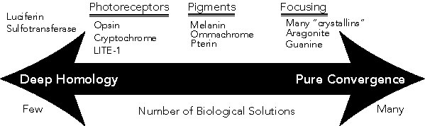
Figure 1.
The continuum from deep homology to pure convergence (or ‘deep diversity’ sensu (Vöcking et al. 2022)) may be influenced by the number of possible biological solutions for a required function. When few solutions exist, evolution is more constrained to using the same genes over and over, as long as those are maintained. At the same time, gene maintenance itself may predispose that gene to being re-used instead of using a new solution.
A similar but less extreme example of constraint is photoreceptor genes, with only a few biological solutions in animals, including opsins, cryptochrome, and LITE-1 (Edwards et al. 2008; Björn 2012). The fairly limited number of animal light receptors suggest that, for certain functions, evolutionary processes gravitate towards a small set of possible solutions, reinforcing the concept of deep homology where functional necessity dictates genetic conservation. At the same time, another consideration is the maintenance of these genes, which could be as strong a determinant of redeployment as the number of functional options. Namely, when a genetic solution for photoreception like opsins is maintained, it is commonly available for co-option, even if many other biological solutions exist. Support for this idea of historical constraint as a driver of deep homology include the fact that non-animals have additional solutions for photoreceptive genes (Björn 2012), indicating a larger suite of biological solutions exists, yet animals usually use opsins as photoreceptors. Furthermore, the photoreceptor LITE-1 exists in C. elegans, a species that lacks opsins. Because LITE-1 is relatively unknown as a photoreceptor elsewhere, perhaps alternatives to opsin are mainly used when opsins are not maintained in a genome.
Conversely, the spectrum also includes pure convergence, where many genetic solutions exist for a single function. This end is exemplified by the diversity of focusing mechanisms in eyes, facilitated by a wide array of genes for crystallins. Crystallin genes are not a single gene family, but rather a functional category, with many different types of genes employed across various species to achieve lens formation. The variety of solutions for focusing structures also includes aragonite lenses (Speiser et al. 2011) and guanine reflectors, illustrating that when multiple viable solutions exist for a function, evolution has used these alternatives, leading to a broad diversity of some genes in convergent evolutionary pathways.
Where a gene or function falls on this continuum may be influenced by how many viable biological solutions exist for a given function. Functions for which there are few effective solutions tend to show deep homology, as evolutionary processes use the same solutions convergently across different lineages. In contrast, when many possible solutions exist, pure convergence becomes more prevalent, with different genetic strategies being adopted to fulfill similar functional roles. This dynamic interplay between deep homology and convergence underscores the complexity of evolutionary biology, illustrating how evolutionary outcomes are shaped by the interaction between genetic heritage, functional requirements, and the stochastic nature of evolutionary processes (Jacob 1977).
6. Summary
Comparative genomics has transformed our understanding of genomes, highlighting complex and often very deep evolutionary histories. To describe the often unexpectedly deep connections between similar features, key terms emerged, such as "deep homology" and "genetic toolkit". Deep homology highlights the shared developmental and genetic bases of similar structures in different species, while the genetic toolkit concept has expanded from focusing solely on animal development to include a variety of biological systems like light interaction, venom, biomineralization, behaviors, and others. These toolkits consist of genes with long conserved functions and genes with new functions, either of which may be used in convergent evolution to evolve similar traits in very different organisms.
The evolutionary significance of genetic toolkits is profound, as they often predate the traits where they are deployed with origins potentially shaped by environmental interactions. The multifunctionality of genes within these toolkits allows for their preservation and reuse in various biological processes. This reuse is evident in the way opsins and pigments serve multiple roles beyond their primary functions, contributing to a wide range of physiological processes. The evolution of these toolkits reflects a dynamic interplay between genetic heritage, environmental pressures, and evolutionary innovation. A limited number of viable genetic solutions can lead to deep homology, while a wider array of solutions fosters pure convergence. This complex landscape showcases how genetic toolkits facilitate both the conservation and diversification of traits across the tree of life, emphasizing the intricate relationship between genetic and developmental pathways and biological functionality.
Acknowledgments
The ideas in this manuscript evolved through many discussions with colleagues, especially labmates. A. Rivera and D. Speiser first helped operationalize a light-interaction toolkit. D. Speiser and A. Swafford and I had many discussions about photo-oxidative stress and vision. E. Lau and I realized the continuum of Figure 1, which I lightheartedly termed “Lau’s Law” in the presentation associated with this manuscript. L. Mesrop taught me about secretory structures and related genetics, and R. Varney about biomineralization. The entire lab and S. McKim provided valuable feedback on an earlier draft. Our lab was supported by NSF DEB-2153773 and IOS-1754770.
References
- Arendt D. 2003. Evolution of eyes and photoreceptor cell types. Int J Dev Biol 47:563–71.
- Ayres JS, Schneider DS. 2008. A signaling protease required for melanization in Drosophila affects resistance and tolerance of infections. PLoS Biol 6:2764–73.
- Basu MK, Carmel L, Rogozin IB, Koonin EV. 2008. Evolution of protein domain promiscuity in eukaryotes. Genome Res 18:449–61.
- Björn LO. 2012. Photobiology: The Science of Light and Life Springer Science & Business Media.
- Brückner A, Parker J. 2020. Molecular evolution of gland cell types and chemical interactions in animals. J Exp Biol 223.
- Buschbeck EK, Friedrich M. 2008. Evolution of Insect Eyes: Tales of Ancient Heritage, Deconstruction, Reconstruction, Remodeling, and Recycling. Evo Edu Outreach 1:448–62.
- Bustamante J, Bredeston L, Malanga G, Mordoh J. 1993. Role of melanin as a scavenger of active oxygen species. Pigment Cell Res 6:348–53.
- Copley SD. 2014. An evolutionary perspective on protein moonlighting. Biochem Soc Trans 42:1684–91.
- Delwiche CF. 2004. The Genomic Palimpsest: Genomics in Evolution and Ecology. Bioscience 54:991–1001.
- DiFrisco J, Wagner GP, Love AC. 2023. Reframing research on evolutionary novelty and co-option: Character identity mechanisms versus deep homology. Semin Cell Dev Biol 145:3–12.
- Drake JL, Mass T, Haramaty L, Zelzion E, Bhattacharya D, Falkowski PG. 2013. Proteomic analysis of skeletal organic matrix from the stony coral Stylophora pistillata. Proc Natl Acad Sci U S A 110:3788–93.
- Edwards SL, Charlie NK, Milfort MC, Brown BS, Gravlin CN, Knecht JE, Miller KG. 2008. A novel molecular solution for ultraviolet light detection in Caenorhabditis elegans. PLoS Biol 6:e198.
- Francis D. 1967. On the eyespot of the dinoflagellate, Nematodinium. J Exp Biol 47:495–501.
- Gould SJ. 2002. The Structure of Evolutionary Theory Harvard University Press.
- Hall BK. 2003. Descent with modification: the unity underlying homology and homoplasy as seen through an analysis of development and evolution. Biol Rev Camb Philos Soc 78:409–33.
- Hetz C. 2012. The unfolded protein response: controlling cell fate decisions under ER stress and beyond. Nat Rev Mol Cell Biol 13:89–102.
- Hetz C, Zhang K, Kaufman RJ. 2020. Mechanisms, regulation and functions of the unfolded protein response. Nat Rev Mol Cell Biol 21:421–38.
- Insausti TC, Casas J. 2008. The functional morphology of color changing in a spider: development of ommochrome pigment granules. J Exp Biol 211:780–89.
- Jacob F. 1977. Evolution and tinkering. Science 196:1161–66.
- Knoll AH, Carroll SB. 1999. Early animal evolution: emerging views from comparative biology and geology. Science 284:2129–37.
- Kumar JP. 2009. The molecular circuitry governing retinal determination. Biochim Biophys Acta 1789:306–14.
- Larusso ND, Ruttenberg BE, Singh AK, Oakley TH. 2008. Type II opsins: evolutionary origin by internal domain duplication? J Mol Evol 66:417–23.
- Lau ES, Goodheart JA, Anderson NT, Liu VL, Mukherjee A, Oakley TH. 2024. Similar enzymatic functions in distinct bioluminescence systems: Evolutionary recruitment of sulfotransferases in ostracod light organs. Biology Letters.
- Lau ES, Oakley TH. 2021. Multi-level convergence of complex traits and the evolution of bioluminescence. Biol Rev Camb Philos Soc 96:673–91.
- Leander BS. 2008a. Different modes of convergent evolution reflect phylogenetic distances: a reply to Arendt and Reznick. Trends Ecol Evol.
- Leander BS. 2008b. A hierarchical view of convergent evolution in microbial eukaryotes. J Eukaryot Microbiol 55:59–68.
- Leung NY, Montell C. 2017. Unconventional Roles of Opsins. Annu Rev Cell Dev Biol 33:241–64.
- Lindgren AR, Pankey MS, Hochberg FG, Oakley TH. 2012. A multi-gene phylogeny of Cephalopoda supports convergent morphological evolution in association with multiple habitat shifts in the marine environment. BMC Evol Biol 12.
- Love AC, Wagner GP. 2022. Co-option of stress mechanisms in the origin of evolutionary novelties. Evolution 76:394–413.
- Marcellini S, Debiais-Thibaud M, Marin F. 2023. The Evolution of Biomineralization in Metazoans Frontiers Media SA.
- Marshall CR, Raff EC, Raff RA. 1994. Dollo’s law and the death and resurrection of genes. Proc Natl Acad Sci U S A 91:12283–87.
- Murdock DJE. 2020. The “biomineralization toolkit” and the origin of animal skeletons. Biol Rev Camb Philos Soc 95:1372–92.
- Nilsson D-E. 2013. Eye evolution and its functional basis. Vis Neurosci 30:5.
- Oakley TH. 2017. Furcation and fusion: The phylogenetics of evolutionary novelty. Dev Biol 431:69–76.
- Oakley TH, Speiser DI. 2015. How Complexity Originates: The Evolution of Animal Eyes. Annu Rev Ecol Evol Syst 46:237–60.
- Olson ME, Arroyo-Santos A, Vergara-Silva F. 2019. A User’s Guide to Metaphors In Ecology and Evolution. Trends Ecol Evol 34:605–15.
- Piatigorsky J. 2009. Gene Sharing and Evolution: The Diversity of Protein Functions Harvard University Press.
- Picciani N, Kerlin JR, Sierra N, Swafford AJM, Ramirez MD, Roberts NG, Cannon JT, Daly M, Oakley TH. 2018. Prolific Origination of Eyes in Cnidaria with Co-option of Non-visual Opsins. Curr Biol 28:2413–19.e4.
- Porter ML. 2016. Beyond the Eye: Molecular Evolution of Extraocular Photoreception. Integr Comp Biol 56:842–52.
- Ramirez MD, Speiser DI, Pankey MS, Oakley TH. 2011. Understanding the dermal light sense in the context of integrative photoreceptor cell biology. Vis Neurosci 28:265–79.
- Ramos-Silva P, Marin F, Kaandorp J, Marie B. 2013. Biomineralization toolkit: the importance of sample cleaning prior to the characterization of biomineral proteomes. Proc Natl Acad Sci U S A.
- Rittschof CC, Robinson GE. 2016. Behavioral Genetic Toolkits: Toward the Evolutionary Origins of Complex Phenotypes. Curr Top Dev Biol 119:157–204.
- Rivera AS, Pankey MS, Plachetzki DC, Villacorta C, Syme AE, Serb JM, Omilian AR, Oakley TH. 2010. Gene duplication and the origins of morphological complexity in pancrustacean eyes, a genomic approach. BMC Evol Biol 10:123.
- Rokas A. 2008. The origins of multicellularity and the early history of the genetic toolkit for animal development. Annu Rev Genet 42:235–51.
- Rose MR, Oakley TH. 2007. The new biology: beyond the Modern Synthesis. Biol Direct 2:30.
- Scotland RW. 2010. Deep homology: a view from systematics. Bioessays 32:438–49.
- Serb JM, Oakley TH. 2005. Hierarchical phylogenetics as a quantitative analytical framework for evolutionary developmental biology. Bioessays 27:1158–66.
- Shubin N, Tabin C, Carroll S. 2009. Deep homology and the origins of evolutionary novelty. Nature 457:818–23.
- Sleight VA. 2023. Cell type and gene regulatory network approaches in the evolution of spiralian biomineralisation. Brief Funct Genomics 22:509–16.
- Speiser DI, DeMartini DG, Oakley TH. 2014. The shell-eyes of the chiton Acanthopleura granulata (Mollusca, Polyplacophora) use pheomelanin as a screening pigment. J Nat Hist 48:2899–2911.
- Speiser DI, Eernisse DJ, Johnsen S. 2011. A Chiton Uses Aragonite Lenses to Form Images. Curr Biol 21:665–70.
- Speiser DI, Pankey MS, Zaharoff AK, Battelle BA, Bracken-Grissom HD, Breinholt JW, Bybee SM, Cronin TW, Garm A, Lindgren AR, Patel NH, Porter ML, Protas ME, Rivera AS, Serb JM, Zigler KS, Crandall KA, Oakley TH. 2014. Using phylogenetically-informed annotation (PIA) to search for light-interacting genes in transcriptomes from non-model organisms. BMC Bioinformatics 15:350.
- Swafford AJM, Oakley TH. 2019. Light-induced stress as a primary evolutionary driver of eye origins. Integr Comp Biol 59:739–50.
- Tokuriki N, Tawfik DS. 2009. Protein dynamism and evolvability. Science 324:203–7.
- Tomarev SI, Piatigorsky J. 1996. Lens Crystallins of Invertebrates. Eur J Biochem 235:449–65.
- Varney RM, Speiser DI, Cannon JT, Aguilar MA, Eernisse DJ, Oakley TH. 2024. A morphological basis for path-dependent evolution of visual systems. Science 383:983–87.
- Velarde RA, Sauer CD, Walden KKO, Fahrbach SE, Robertson HM. 2005. Pteropsin: a vertebrate-like non-visual opsin expressed in the honey bee brain. Insect Biochem Mol Biol 35:1367–77.
- Vöcking O, Macias-Muñoz A, Jaeger SJ, Oakley TH. 2022. Deep Diversity: Extensive Variation in the Components of Complex Visual Systems across Animals. Cells 11.
- v. Salvini-Plawen L, Mayr E. 1977. On the Evolution of Photoreceptors and Eyes. In: Hecht MK, Steere WC, Wallace B, editors. Evolutionary Biology Boston, MA: Springer US. p. 207–63.
- Wagner GP. 2007. The developmental genetics of homology. Nat Rev Genet 8:473–79.
- Wagner GP, Erkenbrack EM, Love AC. 2019. Stress-induced evolutionary innovation: A mechanism for the origin of cell types. Bioessays 41:e1800188.
- Watt WB. 1968. Adaptive significance of pigment polymorphisms in Colias butterflies. I. Variation of melanin pigment in relation to thermoregulation. Evolution 22:437–58.
- West-Eberhard MJ. 2003. Developmental Plasticity and Evolution Oxford University Press.
- Wilkins AS. 2013. “the genetic tool-kit”: The life-history of an important metaphor. In: Advances in Evolutionary Developmental Biology Hoboken, NJ, USA: John Wiley & Sons, Inc. p. 1–14.
- Young RL, Ferkin MH, Ockendon-Powell NF, Orr VN, Phelps SM, Pogány Á, Richards-Zawacki CL, Summers K, Székely T, Trainor BC, Urrutia AO, Zachar G, O’Connell LA, Hofmann HA. 2019. Conserved transcriptomic profiles underpin monogamy across vertebrates. Proc Natl Acad Sci U S A 116:1331–36.
- Zancolli G, Reijnders M, Waterhouse RM, Robinson-Rechavi M. 2022. Convergent evolution of venom gland transcriptomes across Metazoa. Proc Natl Acad Sci U S A 119.
Disclaimer/Publisher’s Note: The statements, opinions and data contained in all publications are solely those of the individual author(s) and contributor(s) and not of MDPI and/or the editor(s). MDPI and/or the editor(s) disclaim responsibility for any injury to people or property resulting from any ideas, methods, instructions or products referred to in the content. |
© 2024 by the authors. Licensee MDPI, Basel, Switzerland. This article is an open access article distributed under the terms and conditions of the Creative Commons Attribution (CC BY) license (http://creativecommons.org/licenses/by/4.0/).
Copyright: This open access article is published under a Creative Commons CC BY 4.0 license, which permit the free download, distribution, and reuse, provided that the author and preprint are cited in any reuse.


