Submitted:
12 December 2023
Posted:
13 December 2023
You are already at the latest version
Abstract
ozone-based chemiluminescence detection (CLD) has been widely applied for determining nitric oxide (NO) and its derived species in many different fields, such as environmental monitoring and biomedical research. In humans CLD was applied to determine exhaled NO and NO metabolites in biological samples. The main advantages of CLD are high sensitivity and selectivity for quantitative analysis in a wide dynamic range. Combining CLD with analytical separation techniques like gas or liquid chromatography allows the analytes to be quantified with less disturbance from matrix components or impurities. Sampling techniques like microdialysis and flow injection analysis may be coupled to CLD with the possibility of real-time monitoring of NO. However, details and precautions in experimental practice need to be addressed and clarified to avoid wrongly estimating. Therefore, using CLD as a detection tool requires a deep understanding of sample preparation procedure and chemical reactions used for liberating NO from its derived species. In this review, we discuss the advantages and pitfalls of CLD for determining NO species, the different applications and combinations with other analytical techniques, and provide general practical notes for sample preparation. These guidelines should help other researchers understand CLD data and select the best procedure for detecting NO species.
Keywords:
Chemiluminescence detection
; NO metabolites
; vascular function
; clinical studies
1. Introduction
Chemiluminescence is defined as the emission of light as result of a chemical reaction. The reactants or intermediates are chemically activated via oxidation into an electronically excited state and alternatively release light by two distinct mechanisms, which are defined as direct and indirect chemiluminescence (Figure 1). In direct chemiluminescence, the chemiluminescent molecules (A) are oxidized to an unstable excited intermediates (C*) that then return to the ground state (C) by releasing energy in form of photons. In indirect chemiluminescence, instead of directly decaying, the excited intermediates (D*) transfer the energy through an optical process to the surrounding fluorophores (E), which become then excited (E*) and release energy by light emission. This phenomenon is called chemiluminescence resonance energy transfer of light.
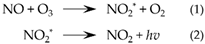
Ozone-based chemiluminescence detection (CLD) for nitric oxide (NO) species is well recognized as a highly sensitive and specific approach to quantify gaseous NO. The detector quantifies the light produced by the reaction of NO with ozone in the gas phase [1]. The reaction produces excited nitrogen dioxide (NO2*) (Reaction 1), which then emits photons when it returns to the ground state (Reaction 2). The emitted light is then amplified by a photomultiplier tube and detected.
Typically, NO is generated in a reaction chamber purged with an inert carrier gas (N2 or Argon), which carries the generated NO along the tubing connecting the chamber to the CLD (which often is referred to as an NO analyzer). The chamber contains a reductive or oxidative solution for acidic or neutral conditions and with/without high temperatures (e.g., 60 degrees). The settings of the reaction chamber depend on the type of analyte and the type of NO derivatives that one may want to quantify. After leaving the reaction chamber, the carrier gas together with NO is then purged into a “NaOH trap”, consisting of a solution of NaOH (1 N), which prevents high-temperature acid vapor to enter and damage the NO analyzer (Figure 2) [2,3].
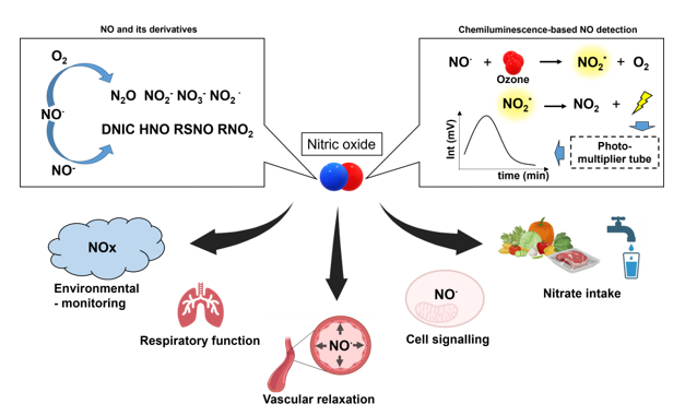
2. NO and its biologically relevant derivatives
•NO, is a moderately-reactive uncharged free radical, which attracted lots of attention in late 1980s for its biological properties. It was found to be the endothelium-derived relaxing factor (EDRF), which lifted the veil of the mystery how the endothelial cells are involved in the vasodilatory effect of smooth muscle cells in the vasculature [3,4,5]. The nature of EDRF as NO was indeed revealed by using CLD [3,6]. The Nobel Prize in Physiology or Medicine for 1998 was awarded jointly to Robert F. Furchgott, Louis J. Ignarro, and Ferid Murad for their discoveries concerning “nitric oxide as a signalling molecule in the cardiovascular system”.
By reacting with other radicals and molecules produced in biological environment, •NO leads to formation of reactive nitrogen species and more stable metabolites like nitrite and nitrate, which are on turn involved in the oxidation or nitrosation of biomolecules and have biological effects on their own [7,8]. These downstream species include dinitrogen trioxide (N2O3), nitrosothiols and nitrosamines (often abbreviated as RSNO and RNNO respectively), intracellular dinitrosyl iron complexes (DNICs), nitroxyl (HNO), •NO2, peroxynitrite (ONOO−), nitrite, and nitrate (Figure 3, adapted from Möller et al.) [9]. Together with reactive oxygen and sulfur species are part of the reactive species interactome [10].
N2O3, is an intermediate in the autoxidation of •NO, and is derived from the reaction of •NO with •NO2 (Reaction 3 and 4). N2O3 can be then hydrolyzed to two molecules of nitrite or rapidly nitrosated thiols and amines leading to RSNO, RNNO and nitrite (Reaction 5 and 6) [8,11,12,13,14] .
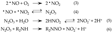
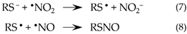
Instead of direct nitrosation of thiols by N2O3, RSNO was also proposed to being formed by reacting RS- with •NO2 to generate RS•, which then reacts with •NO to obtain RSNO (Reaction 7 and 8) [15].
In addition, •NO can target also Fe-Heme and bind to iron from or partially from the chelatable iron pool together with ligands such as glutathione to form DNICs [16].
NO− is formed by one electron reduction of •NO, which is found only as protonated form, HNO [9]. HNO can react with oxygen leading to formation of ONOO−, but it has low relevance under biological conditions due to relatively slow reaction rate (Reaction 9) [17,18].

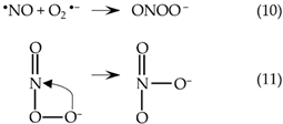
ONOO− is formed also by the reaction between •NO and O2•− (Reaction 10), which can undergo one or two electrons reduction yielding •NO2 and NO2− respectively [19,20]. ONOO− can be also isomerized to NO3− in the presence of metmyoglobin or methemoglobin as catalysts (Reaction 11) [21,22].
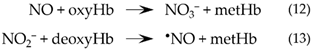
Nitrate can be derived by the reaction between •NO and oxyhemoglobin (Reaction 12) [23]. Under certain conditions such as in the presence of oral bacteria or the enzyme xanthine oxidoreductase, nitrate can be reduced back to nitrite, which is an important source of nitrite for further reduction to •NO in the body [24,25,26,27,28,29].
Nitrite can be reduced to •NO by proteins such as deoxyhemoglobin, deoxymyoglobin and other globins under hypoxic conditions and these mechanisms contribute to many processes such as vasodilation, neurotransmission, immune response and other physiological signallings (Reaction 13) [30,31].
NO was also described to react with deoxyhemoglobin to form nitosylhemoglobin, where NO forms a complex with the iron heme. This was described as a transporter of NO bioactivity and as an intermediate for the formation of s-nitrosohemoglobin as well as an intermediate formed during nitrite reduction into NO [30,32,33,34].
3. Measurement of NO metabolites by chemiluminescence
The NO metabolites need to be converted back to NO in the reaction chamber by reduction (nitrate, nitrite, RSNO, RNNO) or oxidation reactions (NO-Heme or DNIC).
3.1. Reduction of nitrite, RSNO and RNNO by tri-iodide
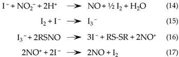
The tri-iodide-based reductive reagent consists of potassium iodide (KI) and iodine (I2) in glacial acetic acid. This mixture is very widely used for reducing nitrite to NO in biological samples (Reaction 14) [2,35]. In addition to nitrite, tri-iodide assay can also be applied for the detection of other biological NO metabolites such as nitrosated (S- or N-nitroso) products (RXNO) (Reaction 15 to 17). Sample preparation for detection of S-nitrosated products (RSNO) requires a pre-treatment of the samples with a stabilization solution containing N-ethylmaleimide (NEM) that prevents artificial S-nitrosation of free thiols. Treatments with NEM have some drawbacks as NEM is often contaminated by nitrite and the contamination may vary from one lot number to another. It is therefore necessary to check contamination before adding NEM to the samples.
3.2. Measurement of nitrate by vanadium chloride
In comparison to nitrite and other species, nitrate is present as much higher concentration in biological specimens and makes the major part of total NO species. To be reduced it requires a stronger reductive reagent as compared to triiodide or an enzymatic reaction catalyzed by a bacterial nitrate reductase. Chemical reduction of nitrate is carried out by vanadium chloride (VCl3) prepared in hydrochloric acid (HCl) in a final concentration of 0.1 mol/L of VCl3 in 2 mol/L HCl (Reaction 18) [36,37]. It is worth mentioning that VCl3/HCl reagent will not just reduce nitrate but also all those metabolites able to be reduced by the tri-iodide method. To obtain the accurate nitrate level, therefore, the results from the tri-iodide assay need to be subtracted from the observed signals in VCl3/HCl method.

Some common reductive and oxidative solutions for the detection of NO metabolites have been listed in Table 1.
4. Multi-level analytical approaches for comprehensive analysis of NO metabolites
4.1. Chemiluminescence coupled with chromatography or mass spectrometry (MS)
CLD, compared to other optical techniques, needs no external light source, has low background signal and, therefore, has a high signal-to-noise ratio and thus is very sensitive [43,44]. For continuously monitoring gaseous pollutants in the air, a chemiluminescence nitrogen detector was developed in 1970 based on the reaction of NO with ozone [1]. Coupling CLD with analytical separative techniques like gas chromatography (GC) or high-performance liquid chromatography (HPLC) allow to combine such a highly sensitive detection technique with powerful separation methods. The selective elution of the chromatography column can separate the target analytes from unwanted interferants or impurities in the sample matrix, which improves the accuracy of CLD and the sensitivity even further [45,46].
4.1.1. Gas chromatography
The chemiluminescence detector for NO species coupled with GC was applied for the detection of atmospheric NO, ammonia, amines, and some nitrogen-containing compounds [1,47,48]. A well-established GC-CLD system requires attention for many critical aspects on both GC and CLD. For GC, the sample injection (split/splitless, which decides a certain ratio of sample entering the GC system), the column (such as type of stationary phase, film thickness, column inner diameter, and length of the column), GC-CLD interface (with a transfer line where analytes travel from GC to the CLD) [48].
Since GC is an analytical technique for the quantitative analysis of volatile compounds, the volatility of the analytes, therefore, is important for their vaporization to enter the gaseous mobile phase and then be transported for further separation on the column. The introduction of the sample in GC might also lead to the failure of the chemiluminescence reaction of NO with ozone. For samples with liquid matrices (like waste water, plasma or biological tissues), the injected samples need to be introduced at a proper flow rate for the combustion at the injection port of GC, which eliminates the risk of water reaching CLD [48]. For higher limits of detection, CLD was also coupled to two-dimensional gas chromatography (GCxGC), with orthogonality for the separation between two applied GC columns, for quantitative analysis of nitrogen-containing compounds in microalgae-based bio-oils, food samples, or urban aerosol samples [49,50,51].
In GC-CLD, GC introduces the chance for further separation on the GC column, however, also brings the requirement for the volatility of the samples. For those complex samples, especially biological tissues or cells, the development of sample preparation method and instrumental parameters as mentioned before for GC-CLD might be time-consuming and costly, which requires more careful consideration and more prudent handling.
4.1.2. Liquid chromatography
Instead of GC, attempts also have been made to couple the CLD for NO species with HPLC, which allows the direct introduction of non-volatile samples in the liquid matrices without vaporization [45,52]. HPLC-CLD was preliminarily applied in the analysis of nitrated polycyclic aromatic hydrocarbons, environmental combustion pollutants, which was reported to encounter the pressure variation issue in the reaction chamber. Incomplete conversion of the mobile phase to the gaseous state before the chemiluminescence reaction leads to reducing the sensitivity, which becomes worse as increasing aqueous content in the mobile phase [53].
To overcome this, a dewatering chamber equipped with drying membrane can be placed between the furnace for the oxidation and the reaction chamber for ozone chemiluminescence, where water is continuously removed from gas streams (including NO) after the oxidation [54]. The modified HPLC-CLD system in the ion chromatography mode was validated for the detection limit of ammonium nitrogen down to 5 ng in wastewater and also the capability of profiling nitrate of 80 ng and nitrite of 160 ng in a nitrite-nitrate mixture [54]. The application of HPLC-CLD for the nitrogen-specific detection of commercially synthesized peptides, including crude peptide mixtures, further demonstrated its superiority over simultaneous ultraviolet detection in peptide profiling [45,55]. On the other hand, HPLC-CLD system was also applied for quantifying the nucleotides and nucleosides standard mixture based on their nitrogen content [52].
An automated system for the analysis of nitrite and nitrate was successfully attempted in 1982, which was then commercialized and developed as ENO-10/ENO-20 and ENO-30 NOx analyzer (Eicom Corporation) [56,57,58]. Such a system was constructed to obtain high sensitivity by coupling HPLC with a diazotization reaction method (Griess reaction). In briefing, nitrite and nitrate are firstly separated on the reverse phase HPLC column, followed by flowing through a copper-loaded reduction column (where the nitrate was reduced to nitrite). Two resolved eluents then react with Griess reagent to form the azo dye compound and detected by spectrophotometer [56,58].
4.1.3. Mass spectrometry
Mass spectrometry is a powerful detection technique based on determination of mass-to-charge ratio (m/z) of target ions and has been applied for the qualitative analysis and quantitation of NO-derived species such as nitrite and nitrate by coupling with chromatography as GC-MS or LC-MS [59,60]. Instead of sole MS detection, the CLD was coupled for on-line quantitative analysis to LC-MS or GC-MS. For the sample class with a known formula, the quantitation with CLD can be calibrated with external standard, which means there is no need for preparing primary standards for the detection of each single analyte (known class but with unknown concentration) then enables high-throughput analysis. Hence, the combination of LC-MS/CLD show the advantage of CLD for quantitation in high-throughput analysis and also brings the power of MS in qualification. An example of this application is the analysis of 24 selected nitrogen-containing compounds [61].
The main pitfall of LC-MS/CLD techniques is the tailing of the peak due to clogging of the splitter and the nebulizer, which can be overcome by the adjustment of flow rate, tubing length, and the replacement by a variable flow splitter [61]. Nevertheless, LC-MS/CLD still provides the chance for a relatively comprehensive view of both quantitative and qualitative analysis. LC-MS/CLD was applied for the identification and quantitation of in vivo metabolites in complex biological matrixes such as bile, urine and plasma [62].
In GC-MS/CLD, a T-splitter was placed after the trapping chamber containing NaOH solution, which splits the NO generated by chemiluminescence reaction into CLD and mass spectrometer [63]. With GC-MS/CLD, total NO can be quantified routinely with the detection of CLD, furthermore, the 14NO and isotope labelled 15NO can be differentiated according to m/z by MS. As an example, an application of this technique is the measurement of nitrite reductase activity in a macrophage cell line lysate. J774.2 macrophage cell lysate was treated with 15NO3−, which was then reduced to 15NO2− by nitrate reductase activity. Both original 14NO2− and produced 15NO2− were reduced with chemiluminescence assay to 14NO and 15NO respectively, followed by MS analysis to distinguish them. In this case, GC-MS/CLD holds the detection limit of NO-related products around 10 nM in a 100-μL sample, which was demonstrated to be beneficial for the study of nitrate reductase activity or related enzymatic pathways [63].
4.2. Coupled with microdialysis
Microdialysis was developed in 1972 based on the concept of collecting samples from small interstitial tissue with a dialysis bag, which then dramatically improved in collecting efficiency by changing the surface area of the dialysis membrane [64]. A special microdialysis probe with three-layer membrane was designed to collect NO in vivo from blood or brain tissues of rats and rabbits, which is then coupled with chemiluminescence-based detection. The integration of such microdialysis sampling system with CLD was proposed for the real-time monitoring of changes in NO concentration in vivo, which provide hints to trace variations in physiological states such as body temperature and evaluate the impact of NO donors on the concentration of NO in blood and tissues [65].
4.3. Coupled with flow injection analysis
Flow injection analysis (FIA) involves the injection of samples into a continuous carrier flow, a liquid stream, which then is mixed with other reagent flows for the reaction before reaching the detector [66]. Aqueous ammonia, nitrite and nitrate were quantified by using a flow injection system equipped with ozone-based NO chemiluminescence detector [67,68]. In this system, an aqueous sample stream was mixed with various reducing reagents (KI for nitrite only and titanium chloride for total nitrate and nitrite) continuously. Moreover, high-temperature combustion was preceded before the generated NO reached the detector to remove aqueous content. The detection limits of 10 nM for nitrite and nitrate were demonstrated by the performance study with standards. Another FIA/chemiluminescence system with and without pre-selective membrane was constructed for the monitoring of peroxynitrite in biological samples based on peroxynitrite-induced luminol chemiluminescence [69]. With this setup, detection limits were 10 and 100 pM for the calibrations with and without membrane respectively, which was proposed to be beneficial for in vivo monitoring of peroxynitrite as combined with microdialysis. A FIA/HPLC system was applied for reproducible determination of plasma nitrite level based on Griess reaction, which enables high-throughput measurements as clinical routine with a detection limit of 10 nM [70]. It helps indicating the correction of plasma nitrite level with endothelial dysfunction and cardiovascular risk factors (such as elevated blood pressure, hypercholesterolemia and etc.).
5. Advantages and drawbacks of chemiluminescence for measuring NO species
5.1. Advantages
Chemiluminescence is currently the most used technique for the quantification of NO [71] because of its high sensitivity, since it allows to detect pM concentration of NO [72], its selectivity, a wide dynamic linear range usually from 0.5 ppb to 5600 ppm NO [71], its ability for real-time monitoring of NO [73], and its commercial availability.
It is important to underline that the ozone-based chemiluminescence can be used not only for the detection of NO, but also for nitrite, nitrate, S-nitrosothiols, nitrosyl-metal complex and N-nitrosamines [2,33,38,74,75,76,77,78].
Moreover, CLD can be easily coupled with other analytical techniques like GC or LC for better accuracy and sensitivity in the quantification of respectively volatile and non-volatile molecules [1,45], or with LC-MS and GC-MS which allow the differentiation and detection of labelled 15NO according to the m/z ratio [63].
5.2. Pitfalls
As mentioned before, the ozone-based chemiluminescence detector can detect pM concentration of nitrite, therefore it is important to avoid contamination coming from the water used for the preparation of reagents, standards, samples and cleaning procedures. For this reason, every step that involves the use of water, must be done with distilled water, filtered by a Milli-Q system which was demonstrated to contain the lowest level of nitrite among the other sources of water [79]. Contaminations is not the only reason why the samples preparation is a critical step. In fact, since NO has a short half-life and nitrite in the blood rapidly reacts with oxyhemoglobin to form nitrate, it is essential that the sample preparation is carried on in conditions that allow to preserve the endogenous NO metabolites. This could require working quickly and many steps during both sample collecting and processing [80]. Regarding the sensitivity of the ozone-based chemiluminescence, it is necessary to mention that different machines have different sensitivities, therefore for comparing data, the measurements need to be done with the same machine [79]. In fact, the reproducibility and sensitivity of the measurement is affected from the ozone gas stream, which is difficult to keep it stable, and from the carrier gas flow rate [81].
Moreover, the chemiluminescence methods required very specific and relatively expensive equipment, time-consuming detection procedure, a frequent equipment maintenance, as well as the knowledge of the chemical reagents used, the reactions that occur during the measurement and the specific operating system for data analysis [71,72]. Concerning these last points, for example, the measurement of S-nitrosothiols, which are released slower than nitrite in tri-iodide, could lead to hard data analysis because of a large width of the peaks [79].
Taking into account the advantages and pitfalls of this method, if the proper precautions are taken to avoid contamination, interference in measurement and errors in data analysis, ozone-based chemiluminescence can be considered one of the best techniques for the determination of NO, mainly due to its high sensitivity, selectivity and possibility of coupling with other analytical techniques.
6. Alternatives for the detection of nitric oxide species
Chemiluminescence detection is one of the most widely used techniques for the determination of NO and NO derivatives, but undoubtedly, many other methods are currently in use. In this section we give a quick overview of the alternatives that can be used, which are also summarized in Table 2.
6.1. Electrochemical sensors
The detection of NO by using electrochemical sensors exploits the phenomena of electrooxidation [82,83] or electroreduction which occur on a metal surface working as an electrode. This method allows a direct NO measurement with high sensitivity and selectivity [82]. One example of electrochemical sensor is the ion-selective electrode (ISE) that can be used specifically for nitrate detection (NO3− - ISE) [84].
6.2. Fluorescence
Fluorescent probes allow an indirect detection of NO via the formation of a fluorescent molecule. The most used fluorescent agents are the 2,3-diaminonaphthalene (DAN) which reacts with nitrite under acidic conditions to produce the fluorescent deprotonated naphthotriazol [85], and the diaminofluoresceins (DAFs) which of NO can be oxidize forming a fluorescent derivative triazolofluorescein [86,87].
6.3. Electron paramagnetic resonance (EPR)
EPR is a specific method to indirectly detect NO formation in cells or tissues using spin traps like Hb, nitronylnitroxides and iron-dithiocarbamate complex [88,89,90]. Moreover, NO-derived produced in biological system like nitrosylhemoglobin (HbNO) and dinitrosyl iron complex can be only defined by EPR spectrum.
6.4. Mass spectrometry
A membrane inlet mass spectrometry (MIMS) was used for measuring NO in aqueous solution (detection limit of 10 nM and linear response to 50 μM). This technique uses a semipermeable membrane that allow to introduce selectively small molecules like NO into the MS [91].
6.5. UV-visible spectrophotometry for NO determination
Spectrometric detection of NO is based on the change of absorption spectrum when oxyhemoglobin is oxidized to methemoglobin in the presence of NO in aqueous solution [92].
6.6. Griess assay
Griess assay is a colorimetric method used for the determination of nitrite. For the nitrate, a reduction from nitrate to nitrite is needed before the assay. This method is based on the formation of a red-violet colored azo compound (λmax ≈ 540 nm) by the reaction of nitrite with the amino group of sulfanilic acid to form a diazonium cation, which then couples to α-naphthylamine [93].

Table 2.
Main techniques applied for the determination of NO and NO derivatives.
| Method | Common applications | Applications | References |
|---|---|---|---|
| Electrochemical sensors | Direct NO detection via electrooxidation or electroreduction; |
Real-time NO quantification in biological system; NO detection in tissues and cells |
[82,83] |
| Fluorescence | Indirect detection of NO via formation of a fluorescent molecule | Detection of NO in cells and tissues | [85,86,87] |
| Electron paramagnetic resonance (EPR) | Indirect detection of NO; Direct detection of HbNO and dinitrosyl iron complex |
NO and HbNO detection in cells and tissues | [88,89,90] |
| Mass spectrometry | Detection of NO via multiple ion detection (MID) | NO detection in aqueous solution | [91] |
|
UV-visible spectrophotometry |
Indirect detection of NO via oxyhemoglobin oxidation | NO formation in cells and tissues | [92,94] |
| Griess assay | Determination of nitrite and nitrate via formation of an azo dye in acidic condition | Determination of nitrite level in biological system | [93,95] |
7. Practical considerations
7.1. General concerns about choosing chemiluminescence as a detection tool for NO species
Many techniques are capable of detecting NO species in the biological samples. However, it is necessary to choose the technique depends on the target. For example, as the main players in the nitrate-nitrite-NO/nitrosothiols biological pathway, nitrite and nitrate can be measured by the Griess assay diazotiation of nitrite (nitrate requires reduction to nitrite first) or by ozone-based chemiluminescence detection of NO [96,97,98,99]. The Griess assay with colorimetric determination of the diazotization product is simple, fast and also affordable (chemicals and UV-vis spectrophotometer are relatively low costs as compared to other instruments). As mentioned before in chapter 5, ozone-based chemiluminescence depends on the availability of the NO analyzer for the direct measurement of gaseous NO or those derived from the reduction of nitrite and nitrate. The detection procedures by using ozone-based chemiluminescence also requires more experience to avoid contamination and for carrying out the reactions, but it is superior for sensitivity.
As reported by many researchers, Griess assay has a limit of detection around 1 μM [99,100,101,102]. In contrast, ozone-based chemiluminescence is able to quantify NO at the range of nM, which is much more sensitive as compared with the Griess assay [103]. Moreover, the direct quantification allows ozone-based chemiluminescence analyzer to measure even exhaled NO [104,105].
The choice of the technique needs to be based on the detection range and the sample type. If the target sample is complex, coupled technique such as HPLC-CLD or GC-CLD can be one of the solutions instead of sole chemiluminescence detection. As mentioned above, such coupling system with chromatography provides additional separation before chemiluminescence detection, which reduces the matrix effect and the chance of under-or over-estimation. Furthermore, MS can bring highly accurate qualitative analysis even possibility of isotopic tracing with 15N-labelled substrate for studies of enzymatic pathway and metabolomics. The GC/MS-CLD approach developed by Cornelius et al. provided the detection limits from 10 to 30 pmol for 14NO2− and 15NO2− as well as the capability of studying enzymatic reduction of nitrate reductase in murine macrophage J774 cell lysates.
7.2. Technical details on the procedure
7.2.1. Carefully choose calibrants and a range of calibration concentrations
As all semiquantitative and quantitative analytical techniques the calibrants should be prepared in a matrix as close as possible as the samples, which means the interference from other compounds in the sample also needs to be included in the calibrants. The classes of target analyte (nitrite, nitrate, etc.) and their estimated level in the samples need to be confirmed to define the calibration range. The detected concentrations of samples need to be located in the linear range of the calibration, otherwise, samples need to be diluted.
7.2.2. Interference from other components in the matrix, non-objective reaction
Chemiluminescence depends on the reaction of NO with ozone. However, some species such as ethylenic hydrocarbons, sulfur compounds, and carbonyls can react with ozone and produce chemiluminescence signals, which may lead to interference for accurate detection [106]. Some substituted ethylenes can react with ozone and produce strong chemiluminescence signals at higher or lower pressure in the analyzer. Sulfur compounds, such as hydrogen sulfide or dimethyl sulfide, can produce intensive signals once they react with ozone. Carbonyls can be introduced from experiment treatment, such as treatment with CO, which interferes with the detection by reacting with ozone. The disturbance of interferants like ethylenic hydrocarbons and sulfur to NO/ozone reaction can be eliminated by increasing detection wavelength to that > 600 nm (440 to 470 nm for ethylenic hydrocarbons, <400 nm for sulfur). However, even some of the interference can be reduced or overcome by methods such as adjusting the wavelength, pressure of the analyzer, or filtering, the artificial contamination needs to be avoided or carefully evaluated before starting.
7.3. Notes for ozone-based chemiluminescence detectiion in biological samples
Some critical points for technical procedures of ozone-based chemiluminescence in biological samples have been summarized in Table 3.
8. Successful application in different fields
The determination of NO and its metabolites has already been used for a long time in different fields. Already fifty years ago CLD was used for identifying and understanding the composition of air to detect the presence of nitrogen species and air pollution [107]. CLD of nitrogen species NOx in the air to the determine air pollution as well as the composition of exhausted air from motors and cars was one of the first applications of CLD of NO species and is still used until today [108,109]. Furthermore, the concentration of NO and NOx is measured in lakes, oceans and rivers as well as drinking water to investigate the pollution of nitrate by increasing use of fertilizer in agriculture [110,111,112].
The analysis of NO species in the gas phase is not only used for environmental monitoring but also used for biomedical and clinical research and diagnosis. In 1991 it was shown that endogenous NO is present in exhaled air of different animal species and humans and can be measured by CLD [113]. Following-up studies could find differences in exhaled NO in asthmatic patients compared to healthy individuals [114]. CLD was also applied to detect NO released by vasoactive drugs in exhaled air in pigs [115]. In this interesting a pioneering study the authors concluded that the measurement of exhaled NO could be a possible indicator of pulmonary endothelial dysfunction or hypertension. Measurement of exhaled NO is still used as a reproducible noninvasive indicator for inflammation of the airways [116,117].
These studies mainly focused on the measurement of NO in exhaled air, but the measurement of NO metabolites in plasma, urine, or tissues is used in various medical fields to investigate the role of endogenous NO production or the effect of intake of nitrate in the body [118,119,120,121]. NO produced in the body is immediately metabolized. Thus, the production of NO by NO synthase (NOS) is measured by NO metabolites [122,123]. Nitrite concentration in plasma was proposed as an indicator of endothelial dysfunction [70,124]. Global eNOS KO mice show decreased circulating nitrite and nitrate metabolites [122,123,125,126]. Recently we found that mice lacking eNOS in the endothelial cells show a decrease in circulating nitrite and nitrate in plasma. However, reactivation of eNOS expression only in endothelial cell in global eNOS KO mice do not recover the nitrite levels in plasma of the mice. This indicate that the levels of nitrite in plasma are not only dependent on eNOS activity in the endothelium. Another finding of this study was that also eNOS expressed in RBCs contribute to the circulating nitrite levels.[97]
In addition, the activity of nNOS and iNOS may also contribute to overall NO metabolites [123]. Recently it has been shown that decreased NO and sulfide availability as detected by CLD is a hallmark of COVID-19 [127].
Due to the variability of circulating NO, nitrate and nitrite levels, these metabolites could not be used as a clinical biomarker until now. However, measuring these metabolites with CLD is still considered as an important approach to understand the NO-pathway in health and disease.
Regarding to nitrate and nitrite levels in the circulation, it is important to note that a main factor of modulating those levels is the intake of nitrate by diet. There are multiple studies using CLD for analyzing nitrate and nitrite levels in food and drinking water [128], which is also important due to the fact that a high intake of nitrate has been considered as harmful and cancerogenic due to the conversion of nitrate to nitrite by bacteria and further metabolism into nitrosamine [129,130]. Furthermore, sodium nitrate and potassium nitrate as well as sodium nitrite and potassium nitrite are used in the food industry especially for preservation of meat as additives [131]. It is, therefore, important to control nitrate and nitrite levels to avoid potential health risks [132]. On the other hand, it has been shown that that the supplementation of nitrate lowers the blood pressure in human and is cardioprotective and shows the importance of the intake of nitrate by dietary [120,121,133].
NO and NO metabolites are also determined in this field using chemiluminescence among other method [134,135]. NO signaling is known to play an important role in plant biology and is involved in multiple processes [136]. Similar to animals and humans, plants can use NADH and nitrite to generate NO but do not express nitric oxide synthase. The measurement of these molecules is not just important for a better insight in the signaling of plants but can also provide information about the accumulation of nitrate in leaves and vegetables of plants due to the use of fertilizer.
To conclude, chemiluminescence detection of nitric oxide is a widely used technique in multiple fields and is continuously improved and adapted to new possible usage.
9. Summary and conclusion
The observation that NO reacts with ozone, leading to the emission of light was first described and used for quantifying NO by amplifying the signal in a photomultiplier. Chemiluminescence-based NO detection allowed researchers to confirm the nature of the EDRF to be NO and afterwards to investigate NO and its derived species in biological samples in a specific, precise and direct way.
The high sensitivity and wide dynamic range make chemiluminescence detection an indispensable tool for quantitative analysis. To increase applicability and specificity of detection for nitrite and nitrate or other metabolites many scientists have combine the advantages of chemiluminescence detection with various analytical separative approaches, such as chromatography, mass spectrometry, microdialysis, and flow injection analysis. These multilevel approaches enable researchers to obtain both quantitative and qualitative profiles including isotope tracing of 14NO and 15NO for enzymatic activity or in vivo monitoring of important NO-derived species such as nitrite and peroxynitrite.
Chemiluminescence-based NO detection has been applied in various fields, including the investigation of environmental pollution related to NOx, biomedical and clinical research on endogenous NO, the study of NO metabolites as indicators of endothelial dysfunction. It is important to note that ozone-based CLD is a highly sensitive method for the detection of NO species, but needs to be operated under controlled conditions. In fact, the system needs to be carefully calibrated, sample preparation and analysis need to be performed taking into consideration sample composition and target analytes, presence of contaminants and molecules that could interfere with the reactions in the chamber or in the detector. Other important factor to consider are sample collection and storage, and technical issues (system leakage, baseline, contamination from air, exhaustion of the ozone in the chamber etc.).
In conclusion, chemiluminescence-based NO determination is a very useful and versatile tool for the quantitative analysis of NO and its metabolites in various samples and can be applied in many fields. The power of chemiluminescence can be further enhanced when coupled with other analytical separative techniques, which deliver a more vivid picture of the status of NO metabolism in various samples. These techniques coupled with cell-specific genetic manipulation of NOS enzyme and/or pharmacological/dietetic intervention will reveal how NO metabolism is regulated in vivo.
Author Contributions
Conceptualization, J.L. and M.C.-K; writing—original draft preparation, J.L.; writing—review and editing, J.L., A.L.B and S.H.; visualization, J.L.; funding acquisition, J.L. and M.C.-K. All authors have read and agreed to the published version of the manuscript.
Funding
This research was funded by research grants from the Research Commission, Medical Faculty of the Heinrich Heine University Duesseldorf (to J.L.), grant number 2022-06; from the German Research Council (DFG) (to M.C.-K), grant number 263779315; from DFG (to M.C.-K), grant number 521638178. .
Acknowledgments
We are indebted with Zhixin Li for help editing the manuscript.
References
- Fontijn, A.; Sabadell, A.J.; Ronco, R.J. Homogeneous chemiluminescent measurement of nitric oxide with ozone. Implications for continuous selective monitoring of gaseous air pollutants. Analytical chemistry 1970, 42, 575–579. [Google Scholar] [CrossRef]
- Feelisch, M.; Rassaf, T.; Mnaimneh, S.; Singh, N.; Bryan, N.S.; Jourd’Heuil, D.; Kelm, M. Concomitant S-, N-, and heme-nitros(yl)ation in biological tissues and fluids: implications for the fate of NO in vivo. FASEB J 2002, 16, 1775–1785. [Google Scholar] [CrossRef] [PubMed]
- Palmer, R.M.; Ferrige, A.G.; Moncada, S. Nitric oxide release accounts for the biological activity of endothelium-derived relaxing factor. Nature 1987, 327, 524–526. [Google Scholar] [CrossRef]
- Ignarro, L.J.; Buga, G.M.; Wood, K.S.; Byrns, R.E.; Chaudhuri, G. Endothelium-derived relaxing factor produced and released from artery and vein is nitric oxide. Proc Natl Acad Sci U S A 1987, 84, 9265–9269. [Google Scholar] [CrossRef]
- Furchgott, R.F.; Cherry, P.D.; Zawadzki, J.V.; Jothianandan, D. Endothelial cells as mediators of vasodilation of arteries. J Cardiovasc Pharmacol 1984, 6 Suppl 2, S336–343. [Google Scholar] [CrossRef]
- Kelm, M.; Feelisch, M.; Spahr, R.; Piper, H.M.; Noack, E.; Schrader, J. Quantitative and kinetic characterization of nitric oxide and EDRF released from cultured endothelial cells. Biochem Biophys Res Commun 1988, 154, 236–244. [Google Scholar] [CrossRef]
- Doyle, M.P.; Hoekstra, J.W. Oxidation of nitrogen oxides by bound dioxygen in hemoproteins. J Inorg Biochem 1981, 14, 351–358. [Google Scholar] [CrossRef] [PubMed]
- Lewis, R.S.; Tannenbaum, S.R.; Deen, W.M. Kinetics of N-Nitrosation in Oxygenated Nitric Oxide Solutions at Physiological pH: Role of Nitrous Anhydride and Effects of Phosphate and Chloride. Journal of the American Chemical Society 1995, 117, 3933–3939. [Google Scholar] [CrossRef]
- Moller, M.N.; Rios, N.; Trujillo, M.; Radi, R.; Denicola, A.; Alvarez, B. Detection and quantification of nitric oxide-derived oxidants in biological systems. J Biol Chem 2019, 294, 14776–14802. [Google Scholar] [CrossRef] [PubMed]
- Cortese-Krott, M.M.; Koning, A.; Kuhnle, G.G.C.; Nagy, P.; Bianco, C.L.; Pasch, A.; Wink, D.A.; Fukuto, J.M.; Jackson, A.A.; van Goor, H.; et al. The Reactive Species Interactome: Evolutionary Emergence, Biological Significance, and Opportunities for Redox Metabolomics and Personalized Medicine. Antioxid Redox Signal 2017, 27, 684–712. [Google Scholar] [CrossRef] [PubMed]
- Goldstein, S.; Czapski, G. Kinetics of Nitric Oxide Autoxidation in Aqueous Solution in the Absence and Presence of Various Reductants. The Nature of the Oxidizing Intermediates. Journal of the American Chemical Society 1995, 117, 12078–12084. [Google Scholar] [CrossRef]
- Goldstein, S.; Czapski, G. Mechanism of the Nitrosation of Thiols and Amines by Oxygenated •NO Solutions: the Nature of the Nitrosating Intermediates. Journal of the American Chemical Society 1996, 118, 3419–3425. [Google Scholar] [CrossRef]
- Bryan, N.S.; Rassaf, T.; Maloney, R.E.; Rodriguez, C.M.; Saijo, F.; Rodriguez, J.R.; Feelisch, M. Cellular targets and mechanisms of nitros(yl)ation: an insight into their nature and kinetics in vivo. Proc Natl Acad Sci U S A 2004, 101, 4308–4313. [Google Scholar] [CrossRef]
- Moller, M.N.; Li, Q.; Vitturi, D.A.; Robinson, J.M.; Lancaster, J.R., Jr.; Denicola, A. Membrane "lens" effect: focusing the formation of reactive nitrogen oxides from the *NO/O2 reaction. Chem Res Toxicol 2007, 20, 709–714. [Google Scholar] [CrossRef]
- Jourd’heuil, D.; Jourd’heuil, F.L.; Feelisch, M. Oxidation and nitrosation of thiols at low micromolar exposure to nitric oxide. Evidence for a free radical mechanism. J Biol Chem 2003, 278, 15720–15726. [Google Scholar] [CrossRef] [PubMed]
- Toledo, J.C., Jr.; Bosworth, C.A.; Hennon, S.W.; Mahtani, H.A.; Bergonia, H.A.; Lancaster, J.R., Jr. Nitric oxide-induced conversion of cellular chelatable iron into macromolecule-bound paramagnetic dinitrosyliron complexes. J Biol Chem 2008, 283, 28926–28933. [Google Scholar] [CrossRef]
- Bartberger, M.D.; Liu, W.; Ford, E.; Miranda, K.M.; Switzer, C.; Fukuto, J.M.; Farmer, P.J.; Wink, D.A.; Houk, K.N. The reduction potential of nitric oxide (NO) and its importance to NO biochemistry. 2002, 99, 10958–10963. [Google Scholar] [CrossRef]
- Smulik, R.; Debski, D.; Zielonka, J.; Michalowski, B.; Adamus, J.; Marcinek, A.; Kalyanaraman, B.; Sikora, A. Nitroxyl (HNO) reacts with molecular oxygen and forms peroxynitrite at physiological pH. Biological Implications. J Biol Chem 2014, 289, 35570–35581. [Google Scholar] [CrossRef]
- Blough, N.V.; Zafiriou, O.C. Reaction of superoxide with nitric oxide to form peroxonitrite in alkaline aqueous solution. Inorganic Chemistry 1985, 24, 3502–3504. [Google Scholar] [CrossRef]
- Ferrer-Sueta, G.; Campolo, N.; Trujillo, M.; Bartesaghi, S.; Carballal, S.; Romero, N.; Alvarez, B.; Radi, R. Biochemistry of Peroxynitrite and Protein Tyrosine Nitration. Chem Rev 2018, 118, 1338–1408. [Google Scholar] [CrossRef]
- Herold, S.; Shivashankar, K. Metmyoglobin and methemoglobin catalyze the isomerization of peroxynitrite to nitrate. Biochemistry 2003, 42, 14036–14046. [Google Scholar] [CrossRef]
- Romero, N.; Radi, R.; Linares, E.; Augusto, O.; Detweiler, C.D.; Mason, R.P.; Denicola, A. Reaction of human hemoglobin with peroxynitrite. Isomerization to nitrate and secondary formation of protein radicals. J Biol Chem 2003, 278, 44049–44057. [Google Scholar] [CrossRef]
- Joshi, M.S.; Ferguson, T.B., Jr.; Han, T.H.; Hyduke, D.R.; Liao, J.C.; Rassaf, T.; Bryan, N.; Feelisch, M.; Lancaster, J.R., Jr. Nitric oxide is consumed, rather than conserved, by reaction with oxyhemoglobin under physiological conditions. Proc Natl Acad Sci U S A 2002, 99, 10341–10346. [Google Scholar] [CrossRef] [PubMed]
- Rosier, B.T.; Buetas, E.; Moya-Gonzalvez, E.M.; Artacho, A.; Mira, A. Nitrate as a potential prebiotic for the oral microbiome. Sci Rep 2020, 10, 12895. [Google Scholar] [CrossRef] [PubMed]
- Millar, T.M.; Stevens, C.R.; Benjamin, N.; Eisenthal, R.; Harrison, R.; Blake, D.R. Xanthine oxidoreductase catalyses the reduction of nitrates and nitrite to nitric oxide under hypoxic conditions. FEBS Lett 1998, 427, 225–228. [Google Scholar] [CrossRef] [PubMed]
- Li, H.; Cui, H.; Liu, X.; Zweier, J.L. Xanthine oxidase catalyzes anaerobic transformation of organic nitrates to nitric oxide and nitrosothiols: characterization of this mechanism and the link between organic nitrate and guanylyl cyclase activation. J Biol Chem 2005, 280, 16594–16600. [Google Scholar] [CrossRef] [PubMed]
- Kapil, V.; Haydar, S.M.; Pearl, V.; Lundberg, J.O.; Weitzberg, E.; Ahluwalia, A. Physiological role for nitrate-reducing oral bacteria in blood pressure control. Free Radic Biol Med 2013, 55, 93–100. [Google Scholar] [CrossRef]
- Lundberg, J.O.; Weitzberg, E. Nitric oxide signaling in health and disease. Cell 2022, 185, 2853–2878. [Google Scholar] [CrossRef]
- Jansson, E.A.; Huang, L.; Malkey, R.; Govoni, M.; Nihlen, C.; Olsson, A.; Stensdotter, M.; Petersson, J.; Holm, L.; Weitzberg, E.; et al. A mammalian functional nitrate reductase that regulates nitrite and nitric oxide homeostasis. Nat Chem Biol 2008, 4, 411–417. [Google Scholar] [CrossRef]
- Keller, T.C.S.t.; Lechauve, C.; Keller, A.S.; Brooks, S.; Weiss, M.J.; Columbus, L.; Ackerman, H.; Cortese-Krott, M.M.; Isakson, B.E. The role of globins in cardiovascular physiology. Physiol Rev 2022, 102, 859–892. [Google Scholar] [CrossRef] [PubMed]
- Cosby, K.; Partovi, K.S.; Crawford, J.H.; Patel, R.P.; Reiter, C.D.; Martyr, S.; Yang, B.K.; Waclawiw, M.A.; Zalos, G.; Xu, X.; et al. Nitrite reduction to nitric oxide by deoxyhemoglobin vasodilates the human circulation. Nat Med 2003, 9, 1498–1505. [Google Scholar] [CrossRef] [PubMed]
- LoBue, A.; Heuser, S.K.; Lindemann, M.; Li, J.; Rahman, M.; Kelm, M.; Stegbauer, J.; Cortese-Krott, M.M. Red blood cell endothelial nitric oxide synthase: A major player in regulating cardiovascular health. Br J Pharmacol 2023. [Google Scholar] [CrossRef]
- Jia, L.; Bonaventura, C.; Bonaventura, J.; Stamler, J.S. S-nitrosohaemoglobin: a dynamic activity of blood involved in vascular control. Nature 1996, 380, 221–226. [Google Scholar] [CrossRef] [PubMed]
- Gladwin, M.T.; Schechter, A.N.; Shelhamer, J.H.; Pannell, L.K.; Conway, D.A.; Hrinczenko, B.W.; Nichols, J.S.; Pease-Fye, M.E.; Noguchi, C.T.; Rodgers, G.P.; et al. Inhaled nitric oxide augments nitric oxide transport on sickle cell hemoglobin without affecting oxygen affinity. J Clin Invest 1999, 104, 937–945. [Google Scholar] [CrossRef]
- Wang, X.; Bryan, N.S.; MacArthur, P.H.; Rodriguez, J.; Gladwin, M.T.; Feelisch, M. Measurement of nitric oxide levels in the red cell: validation of tri-iodide-based chemiluminescence with acid-sulfanilamide pretreatment. J Biol Chem 2006, 281, 26994–27002. [Google Scholar] [CrossRef]
- Smarason, A.K.; Allman, K.G.; Young, D.; Redman, C.W. Elevated levels of serum nitrate, a stable end product of nitric oxide, in women with pre-eclampsia. Br J Obstet Gynaecol 1997, 104, 538–543. [Google Scholar] [CrossRef] [PubMed]
- Abu-Alghayth, M.; Vanhatalo, A.; Wylie, L.J.; McDonagh, S.T.; Thompson, C.; Kadach, S.; Kerr, P.; Smallwood, M.J.; Jones, A.M.; Winyard, P.G. S-nitrosothiols, and other products of nitrate metabolism, are increased in multiple human blood compartments following ingestion of beetroot juice. Redox Biol 2021, 43, 101974. [Google Scholar] [CrossRef] [PubMed]
- Samouilov, A.; Zweier, J.L. Development of chemiluminescence-based methods for specific quantitation of nitrosylated thiols. Anal Biochem 1998, 258, 322–330. [Google Scholar] [CrossRef]
- Marley, R.; Feelisch, M.; Holt, S.; Moore, K. A chemiluminescense-based assay for S-nitrosoalbumin and other plasma S-nitrosothiols. Free Radic Res 2000, 32, 1–9. [Google Scholar] [CrossRef]
- Ewing, J.F.; Janero, D.R. Specific S-nitrosothiol (thionitrite) quantification as solution nitrite after vanadium(III) reduction and ozone-chemiluminescent detection. Free Radic Biol Med 1998, 25, 621–628. [Google Scholar] [CrossRef] [PubMed]
- Fang, K.; Ragsdale, N.V.; Carey, R.M.; MacDonald, T.; Gaston, B. Reductive assays for S-nitrosothiols: implications for measurements in biological systems. Biochem Biophys Res Commun 1998, 252, 535–540. [Google Scholar] [CrossRef] [PubMed]
- Gladwin, M.T.; Wang, X.; Reiter, C.D.; Yang, B.K.; Vivas, E.X.; Bonaventura, C.; Schechter, A.N. S-Nitrosohemoglobin is unstable in the reductive erythrocyte environment and lacks O2/NO-linked allosteric function. J Biol Chem 2002, 277, 27818–27828. [Google Scholar] [CrossRef] [PubMed]
- Yan, Y.; Shi, P.; Song, W.; Bi, S. Chemiluminescence and Bioluminescence Imaging for Biosensing and Therapy: In Vitro and In Vivo Perspectives. Theranostics 2019, 9, 4047–4065. [Google Scholar] [CrossRef]
- Huang, C.; Zhou, W.; Wu, R.; Guan, W.; Ye, N. Recent Advances in Nanomaterial-Based Chemiluminescence Probes for Biosensing and Imaging of Reactive Oxygen Species. Nanomaterials (Basel) 2023, 13. [Google Scholar] [CrossRef]
- Fujinari, E.M.; Manes, J.D. Nitrogen-specific detection of peptides in liquid chromatography with a chemiluminescent nitrogen detector. Journal of Chromatography A 1994, 676, 113–120. [Google Scholar] [CrossRef]
- Brannegan, D.; Ashraf-Khorassani, M.; Taylor, L.T. High-performance liquid chromatography coupled with chemiluminescence nitrogen detection for the study of ethoxyquin antioxidant and related organic bases. J Chromatogr Sci 2001, 39, 217–221. [Google Scholar] [CrossRef] [PubMed]
- Kashihira, N.; Makino, K.; Kirita, K.; Watanabe, Y. Chemiluminescent nitrogen detector-gas chromatography and its application to measurement of atmospheric ammonia and amines. Journal of Chromatography A 1982, 239, 617–624. [Google Scholar] [CrossRef]
- Fujinari, E.M. Gas chromatography-chemiluminescent nitrogen detection: GC-CLND. In Developments in Food Science, Wetzel, D.L.B., Charalambous, G., Eds.; Elsevier: 1998; Volume 39, pp. 385–424.
- Toraman, H.E.; Franz, K.; Ronsse, F.; Van Geem, K.M.; Marin, G.B. Quantitative analysis of nitrogen containing compounds in microalgae based bio-oils using comprehensive two-dimensional gas-chromatography coupled to nitrogen chemiluminescence detector and time of flight mass spectrometer. J Chromatogr A 2016, 1460, 135–146. [Google Scholar] [CrossRef]
- Kocak, D.; Ozel, M.Z.; Gogus, F.; Hamilton, J.F.; Lewis, A.C. Determination of volatile nitrosamines in grilled lamb and vegetables using comprehensive gas chromatography - nitrogen chemiluminescence detection. Food Chem 2012, 135, 2215–2220. [Google Scholar] [CrossRef]
- Ozel, M.Z.; Hamilton, J.F.; Lewis, A.C. New sensitive and quantitative analysis method for organic nitrogen compounds in urban aerosol samples. Environ Sci Technol 2011, 45, 1497–1505. [Google Scholar] [CrossRef]
- Fujinari, E.M.; Damon Manes, J. Nitrogen-specific liquid chromatography detection of nucleotides and nucleosides by HPLC-CLND. In Developments in Food Science, Charalambous, G., Ed.; Elsevier: 1995; Volume 37, pp. 379–396.
- Robbat, A.; Corso, N.P.; Liu, T.Y. Evaluation of a nitrosyl-specific gas-phase chemiluminescence detector with high-performance liquid chromatography. Analytical Chemistry 1988, 60, 173–174. [Google Scholar] [CrossRef]
- Fujinari, E.M.; Courthaudon, L.O. Nitrogen-specific liquid chromatography detector based on chemiluminescence: Application to the analysis of ammonium nitrogen in waste water. Journal of Chromatography A 1992, 592, 209–214. [Google Scholar] [CrossRef]
- Fujinari, E.M.; Manes, J.D.; Bizanek, R. Peptide content determination of crude synthetic peptides by reversed-phase liquid chromatography and nitrogen-specific detection with a chemiluminescent nitrogen detector. Journal of Chromatography A 1996, 743, 85–89. [Google Scholar] [CrossRef]
- Green, L.C.; Wagner, D.A.; Glogowski, J.; Skipper, P.L.; Wishnok, J.S.; Tannenbaum, S.R. Analysis of nitrate, nitrite, and [15N]nitrate in biological fluids. Anal Biochem 1982, 126, 131–138. [Google Scholar] [CrossRef]
- Tanaka, S.; Akaike, T.; Fang, J.; Beppu, T.; Ogawa, M.; Tamura, F.; Miyamoto, Y.; Maeda, H. Antiapoptotic effect of haem oxygenase-1 induced by nitric oxide in experimental solid tumour. Br J Cancer 2003, 88, 902–909. [Google Scholar] [CrossRef]
- Bryan, N.S.; Grisham, M.B. Methods to detect nitric oxide and its metabolites in biological samples. Free Radic Biol Med 2007, 43, 645–657. [Google Scholar] [CrossRef]
- Tsikas, D. Simultaneous derivatization and quantification of the nitric oxide metabolites nitrite and nitrate in biological fluids by gas chromatography/mass spectrometry. Anal Chem 2000, 72, 4064–4072. [Google Scholar] [CrossRef] [PubMed]
- Shin, S.; Fung, H.L. Evaluation of an LC-MS/MS assay for 15N-nitrite for cellular studies of L-arginine action. J Pharm Biomed Anal 2011, 56, 1127–1131. [Google Scholar] [CrossRef]
- Corens, D.; Carpentier, M.; Schroven, M.; Meerpoel, L. Liquid chromatography–mass spectrometry with chemiluminescent nitrogen detection for on-line quantitative analysis of compound collections: advantages and limitations. Journal of Chromatography A 2004, 1056, 67–75. [Google Scholar] [CrossRef] [PubMed]
- Taylor, E.W.; Jia, W.; Bush, M.; Dollinger, G.D. Accelerating the drug optimization process: identification, structure elucidation, and quantification of in vivo metabolites using stable isotopes with LC/MSn and the chemiluminescent nitrogen detector. Anal Chem 2002, 74, 3232–3238. [Google Scholar] [CrossRef] [PubMed]
- Cornelius, J.; Tran, T.; Turner, N.; Piazza, A.; Mills, L.; Slack, R.; Hauser, S.; Alexander, J.S.; Grisham, M.B.; Feelisch, M.; et al. Isotope tracing enhancement of chemiluminescence assays for nitric oxide research. Biol Chem 2009, 390, 181–189. [Google Scholar] [CrossRef] [PubMed]
- Delgado, J.M.; DeFeudis, F.V.; Roth, R.H.; Ryugo, D.K.; Mitruka, B.M. Dialytrode for long term intracerebral perfusion in awake monkeys. Arch Int Pharmacodyn Ther 1972, 198, 9–21. [Google Scholar] [PubMed]
- Yao, D.; Vlessidis, A.G.; Evmiridis, N.P.; Evangelou, A.; Karkabounas, S.; Tsampalas, S. Luminol chemiluminescense reaction: a new method for monitoring nitric oxide in vivo. Analytica Chimica Acta 2002, 458, 281–289. [Google Scholar] [CrossRef]
- Trojanowicz, M.; Pyszynska, M. Flow-Injection Methods in Water Analysis-Recent Developments. Molecules 2022, 27. [Google Scholar] [CrossRef] [PubMed]
- Aoki, T.; Fukuda, S.; Hosoi, Y.; Mukai, H. Rapid flow injection analysis method for successive determination of ammonia, nitrite, and nitrate in water by gas-phase chemiluminescence. Analytica Chimica Acta 1997, 349, 11–16. [Google Scholar] [CrossRef]
- Hendgen-Cotta, U.; Grau, M.; Rassaf, T.; Gharini, P.; Kelm, M.; Kleinbongard, P. Reductive gas-phase chemiluminescence and flow injection analysis for measurement of the nitric oxide pool in biological matrices. Methods Enzymol 2008, 441, 295–315. [Google Scholar] [CrossRef]
- Dai, K.; Vlessidis, A.G.; Evmiridis, N.P. Dialysis membrane sampler for on-line flow injection analysis/chemiluminescence-detection of peroxynitrite in biological samples. Talanta 2003, 59, 55–65. [Google Scholar] [CrossRef] [PubMed]
- Kleinbongard, P.; Dejam, A.; Lauer, T.; Jax, T.; Kerber, S.; Gharini, P.; Balzer, J.; Zotz, R.B.; Scharf, R.E.; Willers, R.; et al. Plasma nitrite concentrations reflect the degree of endothelial dysfunction in humans. Free Radic Biol Med 2006, 40, 295–302. [Google Scholar] [CrossRef] [PubMed]
- Gill, A.; Zajda, J.; Meyerhoff, M.E. Comparison of electrochemical nitric oxide detection methods with chemiluminescence for measuring nitrite concentration in food samples. Anal Chim Acta 2019, 1077, 167–173. [Google Scholar] [CrossRef]
- Potter, M.N.; Green, J.R.; Mutus, B. Fluorescein isothiocyanate, a platform for the selective and sensitive detection of S-Nitrosothiols and hydrogen sulfide. Talanta 2022, 237, 122981. [Google Scholar] [CrossRef] [PubMed]
- Yao, D.; Vlessidis, A.G.; Evmiridis, N.P. Determination of nitric oxide in biological samples. Microchimica Acta 2004, 147, 1–20. [Google Scholar] [CrossRef]
- Garside, C. A chemiluminescent technique for the determination of nanomolar concentrations of nitrate and nitrite in seawater. Marine Chemistry 1982, 11, 159–167. [Google Scholar] [CrossRef]
- Kallakunta, V.M.; Slama-Schwok, A.; Mutus, B. Protein disulfide isomerase may facilitate the efflux of nitrite derived S-nitrosothiols from red blood cells. Redox Biol 2013, 1, 373–380. [Google Scholar] [CrossRef] [PubMed]
- Doctor, A.; Platt, R.; Sheram, M.L.; Eischeid, A.; McMahon, T.; Maxey, T.; Doherty, J.; Axelrod, M.; Kline, J.; Gurka, M.; et al. Hemoglobin conformation couples erythrocyte S-nitrosothiol content to O2 gradients. Proc Natl Acad Sci U S A 2005, 102, 5709–5714. [Google Scholar] [CrossRef] [PubMed]
- Stamler, J.S.; Jaraki, O.; Osborne, J.; Simon, D.I.; Keaney, J.; Vita, J.; Singel, D.; Valeri, C.R.; Loscalzo, J. Nitric oxide circulates in mammalian plasma primarily as an S-nitroso adduct of serum albumin. Proc Natl Acad Sci U S A 1992, 89, 7674–7677. [Google Scholar] [CrossRef]
- Gladwin, M.T.; Shelhamer, J.H.; Schechter, A.N.; Pease-Fye, M.E.; Waclawiw, M.A.; Panza, J.A.; Ognibene, F.P.; Cannon, R.O. , 3rd. Role of circulating nitrite and S-nitrosohemoglobin in the regulation of regional blood flow in humans. Proc Natl Acad Sci U S A 2000, 97, 11482–11487. [Google Scholar] [CrossRef] [PubMed]
- Yang, B.K.; Vivas, E.X.; Reiter, C.D.; Gladwin, M.T. Methodologies for the sensitive and specific measurement of S-nitrosothiols, iron-nitrosyls, and nitrite in biological samples. Free Radic Res 2003, 37, 1–10. [Google Scholar] [CrossRef] [PubMed]
- Piknova, B.; Park, J.W.; Cassel, K.S.; Gilliard, C.N.; Schechter, A.N. Measuring Nitrite and Nitrate, Metabolites in the Nitric Oxide Pathway, in Biological Materials using the Chemiluminescence Method. J Vis Exp 2016. [Google Scholar] [CrossRef] [PubMed]
- Bateman, R.M.; Ellis, C.G.; Freeman, D.J. Optimization of nitric oxide chemiluminescence operating conditions for measurement of plasma nitrite and nitrate. Clin Chem 2002, 48, 570–573. [Google Scholar] [CrossRef]
- Brown, M.D.; Schoenfisch, M.H. Electrochemical Nitric Oxide Sensors: Principles of Design and Characterization. Chem Rev 2019, 119, 11551–11575. [Google Scholar] [CrossRef]
- Shibuki, K. An electrochemical microprobe for detecting nitric oxide release in brain tissue. Neurosci Res 1990, 9, 69–76. [Google Scholar] [CrossRef]
- Wang, H.; Yuan, B.; Yin, T.; Qin, W. Alternative coulometric signal readout based on a solid-contact ion-selective electrode for detection of nitrate. Anal Chim Acta 2020, 1129, 136–142. [Google Scholar] [CrossRef] [PubMed]
- Wiersma, J.H. 2, 3-Diaminonaphthalene as a spectrophotometric and fluorometric reagent for the determination of nitrite ion. Analytical Letters 1970, 3, 123–132. [Google Scholar] [CrossRef]
- Kojima, H.; Sakurai, K.; Kikuchi, K.; Kawahara, S.; Kirino, Y.; Nagoshi, H.; Hirata, Y.; Nagano, T. Development of a fluorescent indicator for nitric oxide based on the fluorescein chromophore. Chem Pharm Bull (Tokyo) 1998, 46, 373–375. [Google Scholar] [CrossRef] [PubMed]
- Li, J.; LoBue, A.; Heuser, S.K.; Leo, F.; Cortese-Krott, M.M. Using diaminofluoresceins (DAFs) in nitric oxide research. Nitric Oxide 2021, 115, 44–54. [Google Scholar] [CrossRef] [PubMed]
- Kleschyov, A.L.; Mollnau, H.; Oelze, M.; Meinertz, T.; Huang, Y.; Harrison, D.G.; Munzel, T. Spin trapping of vascular nitric oxide using colloid Fe(II)-diethyldithiocarbamate. Biochem Biophys Res Commun 2000, 275, 672–677. [Google Scholar] [CrossRef] [PubMed]
- Joseph, J.; Kalyanaraman, B.; Hyde, J.S. Trapping of nitric oxide by nitronyl nitroxides: an electron spin resonance investigation. Biochem Biophys Res Commun 1993, 192, 926–934. [Google Scholar] [CrossRef]
- Piknova, B.; Gladwin, M.T.; Schechter, A.N.; Hogg, N. Electron paramagnetic resonance analysis of nitrosylhemoglobin in humans during NO inhalation. J Biol Chem 2005, 280, 40583–40588. [Google Scholar] [CrossRef] [PubMed]
- Goodwin, J.M.; Chrestensen, C.A.; Moomaw, E.W. Detection of Nitric Oxide by Membrane Inlet Mass Spectrometry. Methods Mol Biol 2018, 1747, 35–47. [Google Scholar] [CrossRef] [PubMed]
- Feelisch, M.; Noack, E.A. Correlation between nitric oxide formation during degradation of organic nitrates and activation of guanylate cyclase. European journal of pharmacology 1987, 139, 19–30. [Google Scholar] [CrossRef]
- Griess, P. Bemerkungen zu der Abhandlung der HH. Weselsky und Benedikt „Ueber einige Azoverbindungen”︁. 1879, 12, 426–428. [Google Scholar] [CrossRef]
- Kelm, M.; Schrader, J. Control of coronary vascular tone by nitric oxide. Circ Res 1990, 66, 1561–1575. [Google Scholar] [CrossRef]
- Guevara, I.; Iwanejko, J.; Dembinska-Kiec, A.; Pankiewicz, J.; Wanat, A.; Anna, P.; Golabek, I.; Bartus, S.; Malczewska-Malec, M.; Szczudlik, A. Determination of nitrite/nitrate in human biological material by the simple Griess reaction. Clin Chim Acta 1998, 274, 177–188. [Google Scholar] [CrossRef] [PubMed]
- Nagababu, E.; Rifkind, J.M. Measurement of plasma nitrite by chemiluminescence. Methods Mol Biol 2010, 610, 41–49. [Google Scholar] [CrossRef]
- Leo, F.; Suvorava, T.; Heuser, S.K.; Li, J.; LoBue, A.; Barbarino, F.; Piragine, E.; Schneckmann, R.; Hutzler, B.; Good, M.E.; et al. Red Blood Cell and Endothelial eNOS Independently Regulate Circulating Nitric Oxide Metabolites and Blood Pressure. Circulation 2021, 144, 870–889. [Google Scholar] [CrossRef] [PubMed]
- Kleinbongard, P.; Rassaf, T.; Dejam, A.; Kerber, S.; Kelm, M. Griess method for nitrite measurement of aqueous and protein-containing samples. Methods Enzymol 2002, 359, 158–168. [Google Scholar] [CrossRef]
- Giustarini, D.; Rossi, R.; Milzani, A.; Dalle-Donne, I. Nitrite and nitrate measurement by Griess reagent in human plasma: evaluation of interferences and standardization. Methods Enzymol 2008, 440, 361–380. [Google Scholar] [CrossRef] [PubMed]
- Tsikas, D. Analysis of nitrite and nitrate in biological fluids by assays based on the Griess reaction: appraisal of the Griess reaction in the L-arginine/nitric oxide area of research. J Chromatogr B Analyt Technol Biomed Life Sci 2007, 851, 51–70. [Google Scholar] [CrossRef]
- Hunter, R.A.; Storm, W.L.; Coneski, P.N.; Schoenfisch, M.H. Inaccuracies of nitric oxide measurement methods in biological media. Anal Chem 2013, 85, 1957–1963. [Google Scholar] [CrossRef] [PubMed]
- Brizzolari, A.; Dei Cas, M.; Cialoni, D.; Marroni, A.; Morano, C.; Samaja, M.; Paroni, R.; Rubino, F.M. High-Throughput Griess Assay of Nitrite and Nitrate in Plasma and Red Blood Cells for Human Physiology Studies under Extreme Conditions. Molecules 2021, 26. [Google Scholar] [CrossRef] [PubMed]
- MacArthur, P.H.; Shiva, S.; Gladwin, M.T. Measurement of circulating nitrite and S-nitrosothiols by reductive chemiluminescence. J Chromatogr B Analyt Technol Biomed Life Sci 2007, 851, 93–105. [Google Scholar] [CrossRef]
- Robbins, R.A.; Floreani, A.A.; Von Essen, S.G.; Sisson, J.H.; Hill, G.E.; Rubinstein, I.; Townley, R.G. Measurement of exhaled nitric oxide by three different techniques. Am J Respir Crit Care Med 1996, 153, 1631–1635. [Google Scholar] [CrossRef] [PubMed]
- Binding, N.; Muller, W.; Czeschinski, P.A.; Witting, U. NO chemiluminescence in exhaled air: interference of compounds from endogenous or exogenous sources. Eur Respir J 2000, 16, 499–503. [Google Scholar] [CrossRef] [PubMed]
- Bates, J.N. Nitric oxide measurement by chemiluminescence detection. Neuroprotocols 1992, 1, 141–149. [Google Scholar] [CrossRef]
- Joseph, D.W.; Spicer, C.W. Chemiluminescence method for atmospheric monitoring of nitric acid and nitrogen oxides. Analytical Chemistry 1978, 50, 1400–1403. [Google Scholar] [CrossRef]
- Kuik, F.; Kerschbaumer, A.; Lauer, A.; Lupascu, A.; von Schneidemesser, E.; Butler, T.M. Top–down quantification of NOx emissions from traffic in an urban area using a high-resolution regional atmospheric chemistry model. Atmos. Chem. Phys. 2018, 18, 8203–8225. [Google Scholar] [CrossRef]
- Suarez-Bertoa, R.; Astorga, C. Impact of cold temperature on Euro 6 passenger car emissions. Environmental Pollution 2018, 234, 318–329. [Google Scholar] [CrossRef]
- Tian, Y.; Wang, K.-K.; Yang, G.-P.; Li, P.-F.; Liu, C.-Y.; Ingeniero, R.C.O.; Bange, H.W. Continuous Chemiluminescence Measurements of Dissolved Nitric Oxide (NO) and Nitrogen Dioxide (NO2) in the Ocean Surface Layer of the East China Sea. Environmental Science & Technology 2021, 55, 3668–3675. [Google Scholar] [CrossRef] [PubMed]
- Kanda, Y.; Taira, M. Flow-Injection Analysis Method for the Determination of Nitrite and Nitrate in Natural Water Samples Using a Chemiluminescence NO<SUB>x</SUB> Monitor. Analytical Sciences 2003, 19, 695–699. [Google Scholar] [CrossRef]
- Bijay, S.; Craswell, E. Fertilizers and nitrate pollution of surface and ground water: an increasingly pervasive global problem. SN Applied Sciences 2021, 3, 518. [Google Scholar] [CrossRef]
- Gustafsson, L.E.; Leone, A.M.; Persson, M.G.; Wiklund, N.P.; Moncada, S. Endogenous nitric oxide is present in the exhaled air of rabbits, guinea pigs and humans. Biochem Biophys Res Commun 1991, 181, 852–857. [Google Scholar] [CrossRef]
- Alving, K.; Weitzberg, E.; Lundberg, J.M. Increased amount of nitric oxide in exhaled air of asthmatics. Eur Respir J 1993, 6, 1368–1370. [Google Scholar] [CrossRef] [PubMed]
- Malmstrom, R.E.; Tornberg, D.C.; Settergren, G.; Liska, J.; Angdin, M.; Lundberg, J.O.; Weitzberg, E. Endogenous nitric oxide release by vasoactive drugs monitored in exhaled air. Am J Respir Crit Care Med 2003, 168, 114–120. [Google Scholar] [CrossRef]
- Berry, M.; Hargadon, B.; Morgan, A.; Shelley, M.; Richter, J.; Shaw, D.; Green, R.H.; Brightling, C.; Wardlaw, A.J.; Pavord, I.D. Alveolar nitric oxide in adults with asthma: evidence of distal lung inflammation in refractory asthma. Eur Respir J 2005, 25, 986–991. [Google Scholar] [CrossRef]
- Sen, P.; Khatri, S.B.; Tejwani, V. Measuring exhaled nitric oxide when diagnosing and managing asthma. Cleve Clin J Med 2023, 90, 363–370. [Google Scholar] [CrossRef] [PubMed]
- Ashworth, A.; Mitchell, K.; Blackwell, J.R.; Vanhatalo, A.; Jones, A.M. High-nitrate vegetable diet increases plasma nitrate and nitrite concentrations and reduces blood pressure in healthy women. Public Health Nutr 2015, 18, 2669–2678. [Google Scholar] [CrossRef]
- Miller, G.D.; Marsh, A.P.; Dove, R.W.; Beavers, D.; Presley, T.; Helms, C.; Bechtold, E.; King, S.B.; Kim-Shapiro, D. Plasma nitrate and nitrite are increased by a high-nitrate supplement but not by high-nitrate foods in older adults. Nutr Res 2012, 32, 160–168. [Google Scholar] [CrossRef]
- Kapil, V.; Milsom, A.B.; Okorie, M.; Maleki-Toyserkani, S.; Akram, F.; Rehman, F.; Arghandawi, S.; Pearl, V.; Benjamin, N.; Loukogeorgakis, S.; et al. Inorganic nitrate supplementation lowers blood pressure in humans: role for nitrite-derived NO. Hypertension 2010, 56, 274–281. [Google Scholar] [CrossRef]
- Larsen, F.J.; Ekblom, B.; Sahlin, K.; Lundberg, J.O.; Weitzberg, E. Effects of dietary nitrate on blood pressure in healthy volunteers. N Engl J Med 2006, 355, 2792–2793. [Google Scholar] [CrossRef] [PubMed]
- Kleinbongard, P.; Dejam, A.; Lauer, T.; Rassaf, T.; Schindler, A.; Picker, O.; Scheeren, T.; Godecke, A.; Schrader, J.; Schulz, R.; et al. Plasma nitrite reflects constitutive nitric oxide synthase activity in mammals. Free Radic Biol Med 2003, 35, 790–796. [Google Scholar] [CrossRef] [PubMed]
- Milsom, A.B.; Fernandez, B.O.; Garcia-Saura, M.F.; Rodriguez, J.; Feelisch, M. Contributions of nitric oxide synthases, dietary nitrite/nitrate, and other sources to the formation of NO signaling products. Antioxid Redox Signal 2012, 17, 422–432. [Google Scholar] [CrossRef]
- Lauer, T.; Preik, M.; Rassaf, T.; Strauer, B.E.; Deussen, A.; Feelisch, M.; Kelm, M. Plasma nitrite rather than nitrate reflects regional endothelial nitric oxide synthase activity but lacks intrinsic vasodilator action. Proc Natl Acad Sci U S A 2001, 98, 12814–12819. [Google Scholar] [CrossRef]
- Wood, K.C.; Cortese-Krott, M.M.; Kovacic, J.C.; Noguchi, A.; Liu, V.B.; Wang, X.; Raghavachari, N.; Boehm, M.; Kato, G.J.; Kelm, M.; et al. Circulating blood endothelial nitric oxide synthase contributes to the regulation of systemic blood pressure and nitrite homeostasis. Arterioscler Thromb Vasc Biol 2013, 33, 1861–1871. [Google Scholar] [CrossRef]
- Park, J.W.; Piknova, B.; Huang, P.L.; Noguchi, C.T.; Schechter, A.N. Effect of blood nitrite and nitrate levels on murine platelet function. PLoS One 2013, 8, e55699. [Google Scholar] [CrossRef] [PubMed]
- Dominic, P.; Ahmad, J.; Bhandari, R.; Pardue, S.; Solorzano, J.; Jaisingh, K.; Watts, M.; Bailey, S.R.; Orr, A.W.; Kevil, C.G.; et al. Decreased availability of nitric oxide and hydrogen sulfide is a hallmark of COVID-19. Redox Biol 2021, 43, 101982. [Google Scholar] [CrossRef] [PubMed]
- Kumari, A.; Bhatoee, M.; Singh, P.; Kaladhar, V.C.; Yadav, N.; Paul, D.; Loake, G.J.; Gupta, K.J. Detection of Nitric Oxide from Chickpea Using DAF Fluorescence and Chemiluminescence Methods. Current Protocols 2022, 2, e420. [Google Scholar] [CrossRef] [PubMed]
- Picetti, R.; Deeney, M.; Pastorino, S.; Miller, M.R.; Shah, A.; Leon, D.A.; Dangour, A.D.; Green, R. Nitrate and nitrite contamination in drinking water and cancer risk: A systematic review with meta-analysis. Environ Res 2022, 210, 112988. [Google Scholar] [CrossRef]
- Tannenbaum, S.R.; Correa, P. Nitrate and gastric cancer risks. Nature 1985, 317, 675–676. [Google Scholar] [CrossRef]
- D’Amore, T.; Di Taranto, A.; Berardi, G.; Vita, V.; Iammarino, M. Nitrate as food additives: Reactivity, occurrence, and regulation. In Nitrate handbook; CRC Press: 2022; pp. 281–300.
- Ziarati, P.; Zahedi, M.; Shirkhan, F.; Mostafidi, M. Potential health risks and concerns of high levels of nitrite and nitrate in food sources. SF Pharma J 2018, 1, 2. [Google Scholar]
- Kapil, V.; Khambata, R.S.; Robertson, A.; Caulfield, M.J.; Ahluwalia, A. Dietary nitrate provides sustained blood pressure lowering in hypertensive patients: a randomized, phase 2, double-blind, placebo-controlled study. Hypertension 2015, 65, 320–327. [Google Scholar] [CrossRef]
- Vishwakarma, A.; Wany, A.; Pandey, S.; Bulle, M.; Kumari, A.; Kishorekumar, R.; Igamberdiev, A.U.; Mur, L.A.J.; Gupta, K.J. Current approaches to measure nitric oxide in plants. Journal of Experimental Botany 2019, 70, 4333–4343. [Google Scholar] [CrossRef]
- Gupta, K.J.; Kaiser, W.M. Production and Scavenging of Nitric Oxide by Barley Root Mitochondria. Plant and Cell Physiology 2010, 51, 576–584. [Google Scholar] [CrossRef] [PubMed]
- Verma, N.; Tiwari, S.; Singh, V.P.; Prasad, S.M. Nitric oxide in plants: an ancient molecule with new tasks. Plant Growth Regulation 2020, 90, 1–13. [Google Scholar] [CrossRef]
Figure 1.
Direct and indirect chemiluminescence. Green: basal state; yellow: excited state. (I) Direct chemiluminescence, A: chemiluminescent molecules, B: oxidant, C*: excited state of intermediate, C: ground state of intermediate; (II) Indirect chemiluminescence, D*: excited state of intermediate, D: ground state of intermediate, E: fluorophore, E*: excited state of fluorophore, E: ground state of fluorophore.
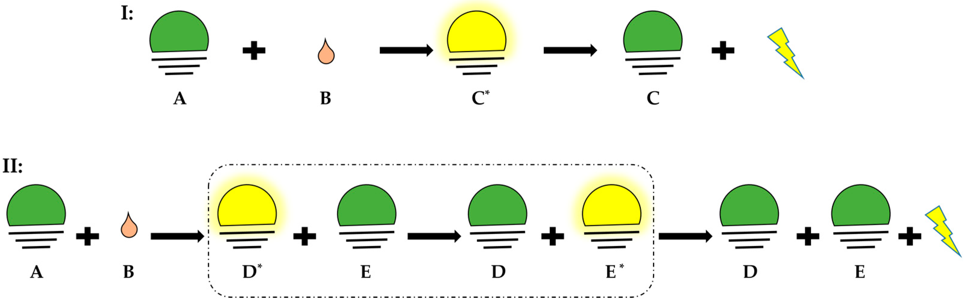
Figure 2.
Apparatus of ozone-based chemiluminescence. A: supply gas, N2, B: supply gas fine control for purging, C: waste, D: injection port of sample, E: heating circulation, F: cooling circulation, G: NaOH trap, H: gas filter, in the NO analyzer: the emission hv is collected by I: optical filter then converted by J: PMT (photomultiplier tube) to amplified signal in mV.
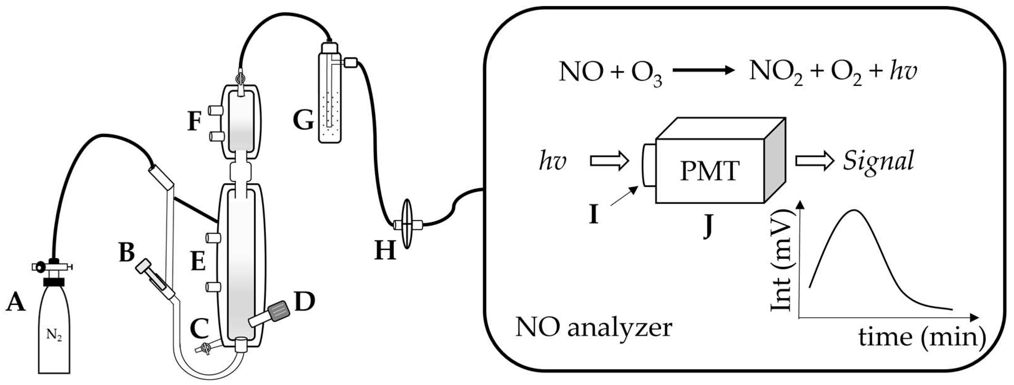
Figure 3.
The reactions of NO• to form its derived species. Nitric oxide (•NO) can react with nitrogen dioxide (•NO2), with N2O3 as intermediate, to generate nitrite (NO2−) and S-nitrosated product (RSNO); •NO can bind to iron (Fe(II)) to form dinitrosyl iron complexes (DNICs); Nitrosyl (HNO) can be formed by reduction of •NO, nitrate (NO3−) can be derived from the reaction between •NO and oxyhemoglobin (HbO2); Peroxynitrite can be obtained by reacting •NO with O2•−, which then can yield NO3−, or be reduced to NO2−, and •NO2 (adapted from Möller et al.)[9].
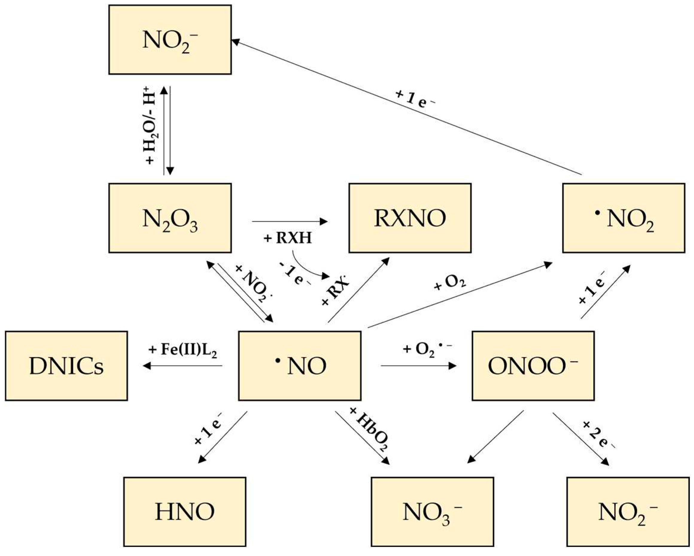
Table 1.
Reductive and oxidative reaction solution for NO metabolites.
| Reaction solution | Conditions | Target NO metabolites |
References |
| Iodine/iodide | 60 mM I−/6 to 20 mM I2/ 1M HCl, RT | NO2−; RNNO, RSNO | [38] |
| 56 mM I−/2 mM I2, 4mM CuCl, CH3COOH, 68°C | [39] | ||
| 45 mM I−/10 mM I2, CH3COOH, 60°C | [2] | ||
| VCl3/H+ | 0.1 M vanadium(III) in 2M HCl | NO3−; NO2−; RNNO; RSNO | [40] |
| Cysteine/CuCl | 1 mM L-cysteine/ 0.1 mM CuCl | RSNO | [41] |
| Hydroquinone/quinone | 0.1 M hydroqui-none/ 0.01 M quinone | RSNO | [38] |
| Ferricyanide |
0.2 M ferricyanide in PBS, pH 7.5 | NO-Heme | [42] |
| 0.05 M ferricyanide in PBS, pH 7.5 | [13] |
Table 3.
Notes for Ozone-based chemiluminescence in biological samples.
| 1. Pretreatment |
Stabilization reagent is prepared for organ tissues and RBCs by adding…
|
Differentiation assistant reagent: measurements in tri-iodide reductive solution in…
|
| 2. Avoiding contamination |
Nitrite contamination is everywhere; therefore, paying attention to…
|
| 3. Minimum time before storage |
The scavenging of the nitrite can be really fast; therefore...
|
| 4. Sample storage |
|
| 5. Measurement procedure |
|
| 6. Technical issues |
|
| 7. pH value affecting the reductive reaction |
|
Disclaimer/Publisher’s Note: The statements, opinions and data contained in all publications are solely those of the individual author(s) and contributor(s) and not of MDPI and/or the editor(s). MDPI and/or the editor(s) disclaim responsibility for any injury to people or property resulting from any ideas, methods, instructions or products referred to in the content. |
© 2023 by the authors. Licensee MDPI, Basel, Switzerland. This article is an open access article distributed under the terms and conditions of the Creative Commons Attribution (CC BY) license (http://creativecommons.org/licenses/by/4.0/).
Copyright: This open access article is published under a Creative Commons CC BY 4.0 license, which permit the free download, distribution, and reuse, provided that the author and preprint are cited in any reuse.


