

Submitted:
20 March 2026
Posted:
23 March 2026
You are already at the latest version
Abstract
The increasing demand for sustainable plant protection products has intensified interest in microbial biocontrol agents (BCAs). The antifungal activity of ten actinobacterial strains of the genus Streptomyces were evaluated against phytopathogenic fungi Botrytis cinerea, Colletotrichum salicis, Fusarium oxysporum and F. graminearum using the dual-culture assay. All tested Streptomyces isolates exhibited antifungal activity, with S. venezuelae MSCL 350 demonstrating the strongest inhibition of fungal growth. The antifungal activity of T. asperellum MSCL 309, Bacillus subtilis MSCL 49, B. subtilis MSCL 1441 and B. stercoris MSCL 897 against twelve Fusarium spp. isolates obtained from oats was evaluated. T. asperellum effectively inhibited F. sporotrichioides, F. oxysporum, F. culmorum and F. poae in dual culture assay while minimal inhibition was observed against the F. graminearum strains used. Soluble metabolites produced by T. asperellum showed strong antifungal activity against Fusarium spp., whereas no significant inhibitory effect of volatile compounds was detected. B. subtilis MSCL 1441 inhibited the growth of all tested Fusarium isolates, while other two Bacillus strains showed no detectable antifungal activity. The results confirm the significant antifungal potential of selected Streptomyces, Trichoderma, and Bacillus strains and support their prospective application as environmentally friendly BCAs against phytopathogenic fungi.
Keywords:
Fusarium
; Streptomyces
; Bacillus
; Trichoderma asperellum
; Colletotrichum salicis
; antifungal
1. Introduction
Phytopathogenic fungi represent one of the major constraints to global crop production, causing significant yield losses and compromising food safety. Among them, the genus Fusarium is particularly important due to its wide distribution in soils and plant residues and its ability to infect a broad range of agricultural crops. Species such as Fusarium oxysporum and F. solani are responsible for vascular wilt, root rot, and seedling diseases in cereals, vegetables, and fruit crops [1,2]. F. graminearum is a causal agent of Fusarium head blight (FHB) of wheat, barley and other small-grain cereals worldwide [3], however other species are also involved. Over the past two decades, the most common species causing FHB in Canada were F. avenaceum, F. equiseti, F. graminearum, F. poae, and F. sporotrichioides [4]. F. avenaceum, F. culmorum, F. graminearum, F. poae and F. sporotrichioides have been identified in Latvian populations of Fusarium species that cause FHB in both spring barley and oat [5]. Fusarium pathogenicity is associated with the secretion of cell wall–degrading enzymes, toxins, and effector molecules that facilitate host penetration, tissue maceration, and systemic colonization [6,7]. In addition to direct plant damage, many Fusarium species produce mycotoxins—including trichothecenes, zearalenone, and fumonisins—that threaten animal and human health [8,9].
F. graminearum and F. oxysporum along with Botrytis cinerea and Colletotrichum spp. are named among the “top 10” based on their scientific/economic importance [10]. WHO also included Fusarium species in the group of high-priority pathogens among fungi that can cause invasive acute and subacute systemic fungal infections in humans, and they may also have inherent resistance to many currently available antifungal agents [11].
B. cinerea, a necrotrophic phytopathogenic fungus, the causal agent of grey mould, has a wide host range, and this fungus causes damage both pre-harvest and during crop storage [12]. Colletotrichum species, hemibiotrophic fungi, cause anthracnose spots and blights in a wide range of woody and herbaceous plants [13]. C. salicis belongs to C. acutatum species complex and is associated with anthracnose of woody hosts [14] and fruit rot in peppers and tomatoes [15].
Growing environmental and regulatory pressure to reduce synthetic fungicide use has intensified interest in biological control strategies. Soil microorganisms with antifungal properties are increasingly considered sustainable alternatives in integrated plant protection systems. Among the most extensively studied and promising biocontrol agents (BCAs) are Trichoderma, Bacillus, and Streptomyces species [16].
Trichoderma species are well-documented antagonists of Fusarium spp. and other soil-borne pathogens. Their biocontrol activity is based on three principal mechanisms: mycoparasitism, antibiosis, and competition for nutrients and ecological niches [17,18]. During mycoparasitism, Trichoderma attaches to pathogen hyphae, forms appressorium-like structures, and secretes hydrolytic enzymes such as chitinases and β-1,3-glucanases, leading to cell wall degradation and growth inhibition [19,20]. In addition, T. asperellum produces secondary metabolites with antifungal activity and can induce systemic resistance in plants, thereby enhancing host tolerance to infection [21]. Commercially available products for agriculture contain various Trichoderma species, including strains of T. asperellum [22].
Members of the genus Bacillus are Gram-positive, endospore-forming bacteria widely distributed in soil environments and known for their stability and adaptability [23]. Their antifungal activity is mainly due to the production of various antifungal volatile and water-soluble compounds, including cyclic lipopeptides such as iturin, surfactin, and fengycin, which disrupt fungal cell membranes and cause leakage of cell contents [24,25]. Additional mechanisms include secretion of lytic enzymes, siderophore-mediated iron competition, and induction of plant defense-related genes [26,27]. Experimental studies have demonstrated significant inhibition of Fusarium growth and sporulation by various Bacillus strains [28]. B. subtilis is a model organism and the most popular workhorse in the biotechnology industry. [29] classified the bioactive metabolites produced by B. subtilis into five classes: non-ribosomal peptides, polyketides, ribosomal peptides, and hybrid and volatile metabolites.
Actinobacteria of the genus Streptomyces are dominant components of soil microbial communities and prolific producers of bioactive secondary metabolites. They account for a substantial proportion of clinically and agriculturally relevant antibiotics [30,31]. In agricultural systems, Streptomyces spp. suppress phytopathogenic fungi through competition, antibiosis, emission of volatile compounds, and secretion of cell wall–degrading enzymes [32,33,34]. The two primary classes of secondary metabolites produced by Streptomyces are non-ribosomal peptides and polyketides [35]. Several isolates have shown strong antagonistic activity against toxigenic Fusarium species and the potential to reduce mycotoxin accumulation [36].
In general, the literature indicates that Trichoderma, Bacillus and Streptomyces species possess complementary antifungal mechanisms, including enzymatic degradation of fungal cell walls, membrane damage and competition for nutrients. These properties underline their potential as key components of sustainable disease management strategies aimed at reducing reliance on chemical fungicides while maintaining crop productivity and health. The aim of this study was to evaluate the antifungal activity of selected Streptomyces, Bacillus, and Trichoderma asperellum strains against phytopathogenic fungi and to assess their potential as BCAs using in vitro assays.
2. Materials and Methods
2.1. Microbial Strains
The antifungal activity of 14 selected microbial strains was evaluated against 16 strains of phytopathogenic fungi (Table 1). All strains were obtained from the Microbial Strain Collection of Latvia (MSCL) and maintained under standardized laboratory conditions.
2.2. Culture Media and Growth Conditions
Fungi were cultured on Malt Extract Agar (MEA, Millipore, India), while bacterial strains were cultured on R2A Agar (Millipore, Germany). B. subtilis and B. stercoris were subcultured 3–4 days prior to testing to ensure active growth. Fungal cultures and Streptomyces were incubated for 7 days at 20–22 °C before antifungal assays.
2.3. Dual Culture Assay
The antifungal activity of antagonist strains was evaluated using a dual culture assay. Agar discs (0.7 cm diameter) from actively growing colonies of phytopathogenic fungi and antagonist strains were placed 4 cm apart on MEA plates. Plates were incubated at 20–22 °C for 7 days. Antifungal activity was quantified by measuring fungal colony diameter and calculating the growth inhibition percentage: Percentage of inhibition = (colony diameter in control—colony diameter in treatment) x 100 / colony diameter in control [37]. Each fungus–antagonist combination was performed in triplicate.
2.4. Agar Well Diffusion Assay
The antifungal activity of Bacillus spp. strains was determined using an agar well diffusion assay. Fusarium suspensions were adjusted to an optical density of 0.16 at 540 nm and uniformly spread on MEA plates. Wells (0.7 cm diameter) were created in the agar and filled with 70 µL of Bacillus suspensions standardized to 0.16 at 540 nm. Plates were incubated at 20–22 °C for 7 days. Antifungal activity was expressed as inhibition zone diameter.
2.5. Assessment of Volatile and Soluble Metabolites of T. asperellum
To determine the contribution of volatile organic compounds (VOCs) produced by T. asperellum, a double-plate assay was performed according to [38]. T. asperellum and the pathogen were inoculated on the surface of agar plates in separate Petri dishes. The lids were removed and the bottom halves of the dishes were placed facing each other so that the inoculated agar surfaces were aligned. The paired dishes were sealed with several layers of Parafilm to prevent the escape of VOCs. Fungal growth inhibition was evaluated after 7 days of incubation at 20–22 °C.
The effect of soluble/diffusible metabolites of T. asperellum was evaluated using a cellophane overlay method [38]. T. asperellum was cultivated on MEA plates covered with sterile cellophane (Bright Ideas Crafts, UK) for 7 days. After removal of the fungal biomass and cellophane, Fusarium discs were transferred onto the conditioned medium. Colony diameter was measured after 48 h to 72 h at 20–22 °C, and growth inhibition percentage was calculated relative to untreated controls.
2.6. Statistical Analysis
All antifungal assays were performed in three independent replicates. Growth inhibition percentage and inhibition zone diameter were calculated as quantitative indicators of antifungal activity. Statistical analyses were conducted using RStudio (version 2024.12.1+563). One-way ANOVA was applied to evaluate the effect of antagonist strain on fungal growth inhibition. In experiments involving multiple phytopathogenic fungi, two-way ANOVA was used to assess the interaction between antagonist strain and fungal species. Duncan’s multiple range test was applied for multiple comparisons. Differences were considered statistically significant at p < 0.05.
3. Results
3.1. Antifungal Activity of Trichoderma asperellum Against Fusarium spp.
3.1.1. Dual Culture Test
The antifungal activity of T. asperellum MSCL 309 against twelve phytopathogenic Fusarium isolates obtained from oats in Latvia was assessed using the dual culture assay. After seven days of incubation, growth inhibition was observed for all tested isolates. The growth inhibition ranged from 44.6% to 78.4% (Table 2, Figure 1), indicating substantial variability in isolate susceptibility. The highest inhibition, 71.0%—78.4%, was observed for both strains of F. sporotrichioides (MSCL 1697 and MSCL 1695), two strains of F. oxysporum (MSCL 1696 and MSCL 1700), F. culmorum MSCL 1693 and F. poae MSCL 1701. Both strains of F. graminearum (MSCL 1691 and MSCL 1694) exhibited significantly (p < 0.05) lower sensitivity. The consistent inhibition across all isolates indicates that T. asperellum possesses broad-spectrum antifungal potential against Fusarium spp., although the degree of inhibition is species and/or isolate-dependent.
3.1.2. Contribution of Volatile Organic Compounds
The role of volatile organic compounds (VOCs) in providing antifungal activity was assessed separately using sealed plate assays. After 72 hours of incubation, no statistically significant differences (p > 0.05) were found between treated and control plates in terms of colony diameter, although slight variations in radial growth were visually observed for some isolates. These findings suggest that VOCs-mediated inhibition was negligible under the experimental conditions used. VOCs were either not synthesized in sufficient concentrations or did not have potent antifungal activity against the tested isolates in this experimental setting. It is also possible that environmental parameters, such as incubation time, headspace volume, or gas exchange conditions, limited VOC accumulation.
3.1.3. Contribution of Soluble Metabolites
In contrast to VOCs, soluble metabolites showed strong antifungal activity. After 48 hours of incubation, colony diameters on control plates ranged from 1.4 to 3.8 cm, while plates pretreated with soluble T. asperellum metabolites showed complete growth inhibition (Figure 2A). After 72 hours, the diameter of the control Fusarium colonies had increased 1.5–2-fold (Figure 2B). However, complete inhibition was still observed for isolates F. graminearum MSCL 1694, both strains of F. sporotrichioides (MSCL 1695 and MSCL 1697), F. oxysporum MSCL 1696, F. tricinctum MSCL 1698 and F. poae MSCL 1701 on pretreated plates. The other isolates also had significantly reduced colony diameters (1.0–1.6 cm), consistent with significant growth inhibition. These results demonstrate that soluble metabolites are the primary contributors to the antifungal activity of T. asperellum at least under in vitro conditions (Table 2, Figure 1).
3.2. Antifungal Activity of Bacillus species Against Fusarium spp.
An experiment conducted using two B. subtilis strains, MSCL 49 and MSCL 1441, and B. stercoris MSCL 897 to evaluate the antifungal activity of bacterial suspensions against twelve Fusarium spp. isolates showed antifungal activity for only one strain, B. subtilis 1441 (Figure 3 and Figure 4). The largest diameter of the zone of inhibition was observed for both F. graminearum strains, MSCL 1694 and MSCL 1691, at 25 mm and 19 mm, respectively. The lowest inhibition was observed for F. culmorum MSCL 1690 (11 mm) and F. sporotrichioides MSCL 1697 (12 mm).
3.3. Antifungal Activity of Streptomyces spp. Against Phytopathogenic Fungi
Ten Streptomyces strains were evaluated against four phytopathogenic fungi using the dual culture assay. Antifungal activity varied markedly depending on both antagonist strain and fungal species (Figure 5). Across all tested Streptomyces strains, S. venezuelae MSCL 350 exhibited the highest antifungal activity against all pathogens, highlighting its superior antagonistic capacity. For F. oxysporum, the maximum inhibition zone diameter was 13.7 mm when paired with S. venezuelae. Other Streptomyces strains showed considerably weaker inhibition, ranging from 0.3 mm to 4.3 mm. F. graminearum demonstrated relatively high resistance, and significant inhibition was observed only in S. venezuelae (18.0 mm), while inhibition was minimal or absent for most other strains.
Botrytis cinerea showed moderate sensitivity. The strongest inhibition was again observed in S. venezuelae (20.7 mm), while the other strains produced inhibition zone diameters ranging from 1.3 mm to 4.3 mm.
The most sensitive species tested was Colletotrichum salicis. The diameter of the inhibition zone for S. venezuelae reached 22.7 mm, and other Streptomyces strains also caused significant inhibition (7.0–9.7 mm). This suggests a greater sensitivity of C. salicis to actinobacterial antagonism compared to F. graminearum and B. cinerea.
Two-way ANOVA analysis confirmed statistically significant differences in fungal sensitivity (p < 0.05), supporting the conclusion that antifungal activity depends not only on antagonist strain but also on intrinsic properties of the phytopathogenic fungus. These findings emphasize the importance of strain selection in BCAs applications and suggest that S. venezuelae MSCL 350 represents a particularly promising candidate for further development.
4. Discussion
The present study demonstrates that antifungal activity is strongly dependent on both the antagonist strain and the phytopathogen species and strain. Among the tested microorganisms, Trichoderma asperellum MSCL 309 and Streptomyces venezuelae MSCL 350 exhibited the highest and most consistent antifungal activity, whereas investigated Bacillus spp. showed comparatively moderate effects.
4.1. Antifungal Mechanisms of Trichoderma asperellum
The dual culture assay revealed broad-spectrum inhibition of all tested Fusarium isolates by T. asperellum, supporting previous reports describing Trichoderma spp. as effective antagonists of soil-borne phytopathogens [17,18]. Experiments show that of the Fusarium species studied, F. graminearum is relatively difficult to control (Table 1). The observed isolate-dependent growth inhibition percentage is consistent with documented variability in susceptibility among Fusarium species and strains [1,2].
The absence of statistically significant inhibition in VOCs assay suggests that VOCs played a limited role under the applied in vitro conditions. Although Trichoderma spp., including T. asperellum, are known to produce VOCs with antifungal properties [39], their efficacy may depend on environmental conditions and metabolite concentration [20]. The present findings suggest that the antagonism mediated by VOCs was negligible compared to other metabolites.
In contrast, soluble metabolites demonstrated strong antifungal activity, including complete inhibition of several isolates. This observation aligns with established mechanisms of Trichoderma antagonism, which involve secretion of hydrolytic enzymes (chitinases, β-1,3-glucanases) and secondary metabolites that degrade fungal cell walls or disrupt membrane integrity [19,20]. Such enzymatic and metabolite-driven inhibition likely represents the primary mechanism of antifungal activity observed in this study.
4.2. Strain-Specific Antifungal Activity of Bacillus spp.
B. subtilis MSCL 1441 showed significant antifungal activity, compared to the B. subtilis MSCL 49 and B. stercoris MSCL 897, confirming the strain-dependent nature of the antagonism. Bacillus spp. are widely recognized for producing cyclic lipopeptides such as iturins, surfactins, and fengycins, which disrupt fungal cell membranes [24,25]. It has been found that both the studied B. subtilis MSCL 1441 and B. stercoris MSCL 897 synthesize surfactins and fengycins, but not iturins [40], however, the antagonistic activity differed significantly, indicating the involvement of other metabolites. B. stercoris belongs to the “subtilis group” and “subtilis subgroup” [41]. It was originally identified as B. subtilis subsp. stercoris, but in 2020, based on genomic analysis, it was elevated to the species level [42].
Additionally, Bacillus spp. may contribute to pathogen inhibition through siderophore-mediated iron competition and induction of plant defense responses [26,27]. However, under the strictly in vitro conditions of this study, the dominant mechanism was likely direct metabolite-mediated inhibition.
B. subtilis MSCL 1441 stood out among all other potential antagonists in that it was able to strongly inhibit the F. graminearum species (Figure 3), which was relatively resistant to the others.
4.3. Superior Antagonistic Potential of Streptomyces venezuelae
The dual culture assay demonstrated that antifungal activity among Streptomyces spp. was species or strain-dependent, with S. venezuelae MSCL 350 producing the largest inhibition zone diameters. This is consistent with the well-documented capacity of Streptomyces spp. to synthesize diverse bioactive secondary metabolites, including antibiotics with antifungal properties [30,31]. The strong inhibition observed in dual culture assay suggests that S. venezuelae MSCL 350 secretes extracellular antifungal metabolites. Similar findings have been reported for other Streptomyces strains capable of suppressing Fusarium spp. and other phytopathogenic fungi via antibiotic production [33,34].
Soil bacterium S. venezuelae is known as an antibiotic (chloramphenicol, jadomycin and pikromycins) producer and differs from many other Streptomyces in its ability to complete its entire life cycle in liquid culture [43,44]. It is possible that this is what allowed this bacterium to display properties of its extracellular metabolites that were not possible for other Streptomyces under liquid cultivation. The expression of secondary metabolite biosynthesis gene clusters in Streptomyces spp. is associated with the complex morphological differentiation of the bacteria during the transition from vegetative to aerial mycelium and subsequent sporulation, therefore these genes are called silent or cryptic, and under laboratory conditions they are usually weakly expressed [45].
Species-dependent sensitivity was evident in this study. Colletotrichum salicis showed the highest sensitivity (Figure 5D), while Fusarium graminearum showed relatively higher resistance (Figure 5B), as in the case of T. asperellum (Table 1). Such differences may be linked to intrinsic pathogenicity mechanisms, cell wall architecture, and adaptive stress responses [6,7].
The consistent activity of S. venezuelae MSCL 350 against multiple phytopathogenic fungi suggests its potential as a broad-spectrum BCA.
5. Conclusions
The present study demonstrates that antifungal activity against phytopathogenic fungi is strongly dependent on both the antagonist strain and the pathogen species. Of the microorganisms tested, T. asperellum MSCL 309 and S. venezuelae MSCL 350 showed the greatest antifungal activity, while B. subtilis MSCL 1441 was the most potent specifically against F. graminearum strains.
T. asperellum MSCL 309 displayed broad-spectrum inhibition against all tested Fusarium isolates. The results indicate that soluble metabolites, rather than VOCs, are the primary contributors to antifungal activity under in vitro conditions.
Within the tested actinobacteria, S. venezuelae MSCL 350 demonstrated superior antagonistic performance. The consistent inhibition observed across multiple phytopathogenic fungi suggests strong secretion of extracellular antifungal metabolites. In contrast, antifungal activity among Bacillus strains was strain-dependent, with only Bacillus subtilis MSCL 1441 showing significant inhibition.
Species and strain-dependent sensitivity was evident among phytopathogenic fungi, highlighting the necessity for targeted biocontrol strategies. The variability in growth inhibition percentage emphasizes the importance of selecting highly effective antagonist strains rather than relying solely on genus-level identification.
From a practical perspective, the results identify T. asperellum MSCL 309, S. venezuelae MSCL 350 and B. subtilis MSCL 1441 as promising candidates for the development of biological control formulations aimed at managing phytopathogenic fungi, particularly Fusarium spp. Future studies should focus on molecular and chemical characterization of antifungal metabolites and evaluation of the performance of the new BCAs under greenhouse and field conditions to assess real-world efficacy and plant growth–promoting potential.
Author Contributions
Conceptualization, M.S. and V.N.; methodology, M.S. and V.N.; software, M.S., V.N. and L.R.; validation, V.N.; formal analysis, M.S. and V.N.; investigation, M.S., L.R., K.J., L.Z. and I.J.; resources, V.N.; data curation, M.S. and V.N.; writing—original draft preparation, M.S. and V.N.; writing—review and editing, M.S. and V.N.; visualization, M.S., V.N., L.R. and K.J.; supervision, V.N. All authors have read and agreed to the published version of the manuscript.
Funding
This research received no external funding.
Institutional Review Board Statement
Not applicable.
Informed Consent Statement
Not applicable.
Data Availability Statement
The original contributions presented in this study are included in the article. Further inquiries can be directed to the corresponding author.
Conflicts of Interest
The authors declare no conflicts of interest.
Abbreviations
The following abbreviations are used in this manuscript:
| BCA | biocontrol agent |
| FHB | Fusarium head blight |
| MSCL | Microbial Strain Collection of Latvia |
| VOCs | volatile organic compounds |
References
- Coleman, J.J. The Fusarium solani Species Complex: Ubiquitous Pathogens of Agricultural Importance. Mol. Plant Pathol. 2016, 17, 146–158. [Google Scholar] [CrossRef] [PubMed]
- Michielse, C.B.; Rep, M. Pathogen Profile Update: Fusarium oxysporum. Mol. Plant Pathol. 2009, 10, 311–324. [Google Scholar] [CrossRef] [PubMed]
- Alisaac, E.; Mahlein, A.-K. Fusarium Head Blight on Wheat: Biology, Modern Detection and Diagnosis and Integrated Disease Management. Toxins 2023, 15, 192. [Google Scholar] [CrossRef]
- Xue, A.G.; Chen, Y.; Seifert, K.; Guo, W.; Blackwell, B.A.; Harris, L.J.; Overy, D.P. Prevalence of Fusarium Species Causing Head Blight of Spring Wheat, Barley and Oat in Ontario during 2001–2017. Can. J. Plant Pathol. 2019, 41, 392–402. [Google Scholar] [CrossRef]
- Treikale, O.; Javoisha, B.; Feodorova-Fedotova, L.; Grantina-Ievina, L.; Volkova, J. Occurrence of Fusarium Species on Small Cereals in Latvia. Communications in Agricultural and Applied Biological Sciences 2015, 80, 551–554. [Google Scholar] [PubMed]
- Doehlemann, G.; Ökmen, B.; Zhu, W.; Sharon, A. Plant Pathogenic Fungi. Microbiol. Spectr. 2017, 5(1), FUNK-0023-2016. [Google Scholar] [CrossRef]
- Han, X.; Kahmann, R. Manipulation of Phytohormone Pathways by Effectors of Filamentous Plant Pathogens. Front. Plant Sci. 2019, 10, 822. [Google Scholar] [CrossRef] [PubMed]
- Ji, F.; He, D.; Olaniran, A.O.; Mokoena, M.P.; Xu, J.; Shi, J. Occurrence, Toxicity, Production and Detection of Fusarium Mycotoxins: A Review. Food Prod. Process. Nutr. 2019, 1, 6. [Google Scholar] [CrossRef]
- Shabeer, S.; Tahira, R.; Jamal, A. Fusarium spp. Mycotoxin Production, Diseases and Their Management: An Overview. J. Agric. Res. 2021, 34, 278–293. [Google Scholar] [CrossRef]
- Dean, R.; van Kan, J.A.L.; Pretorius, Z.A.; Hammond-Kosack, K.E.; Di Pietro, A.; Spanu, P.D.; Rudd, J.J.; Dickman, M.; Kahmann, R.; Ellis, J.; Foster, G.D. The Top 10 Fungal Pathogens in Molecular Plant Pathology. Mol. Plant. Pathol. 2012, 13, 414–430. [Google Scholar] [CrossRef] [PubMed]
- WHO. WHO Fungal Priority Pathogens List to Guide Research, Development and Public Health Action; World Health Organization: Geneva, 2022. [Google Scholar]
- Chen, T.; Zhang, Z.; Chen, Y.; Li, B.; Tian, S. Botrytis cinerea. Curr. Biol. 2023, 33, R460–R462. [Google Scholar] [CrossRef]
- Usman, H.M.; Hussain, M.D.; Karim, M.M.; Nizamani, M.M.; Mubeen, M.; Hussain, S.; Kamran, A.; Wang, Y.; Liu, F.-Q. Colletotrichum: a Versatile Fungal Genus with Diverse Infection Strategies, Host Interactions, and Management Challenges. Phytopathol. Res. 2026, 8, 6. [Google Scholar] [CrossRef]
- Damm, U.; Cannon, P.F.; Woudenberg, J.H.C.; Crous, P.W. The Colletotrichum acutatum Species Complex. Stud. Mycol. 2012, 73, 37–113. [Google Scholar] [CrossRef]
- Manova, V.; Stoyanova, Z.; Rodeva, R.; Boycheva, I.; Korpelainen, H.; Vesterinen, E.; Wirta, H.; Bonchev, G. Morphological, Pathological and Genetic Diversity of the Colletotrichum Species, Pathogenic on Solanaceous Vegetable Crops in Bulgaria. J. Fungi 2022, 8, 1123. [Google Scholar] [CrossRef]
- Nguyen, H.T.; Pham, T.T.; Nguyen, P.T.; Dinh, N.C.G.; Le, M.T.; Nguyen, T.D.; Nguyen, T.T.; Nguyen, V.B. Microbial Biocontrol in Agriculture: from Mechanistic Understanding to Field Application. Discov. Plants 2025, 2, 334. [Google Scholar] [CrossRef]
- Harman, G.E.; Howell, C.R.; Viterbo, A.; Chet, I.; Lorito, M. Trichoderma Species—Opportunistic, Avirulent Plant Symbionts. Nat. Rev. Microbiol. 2004, 2, 43–56. [Google Scholar] [CrossRef]
- Mukhopadhyay, R.; Kumar, D. Trichoderma: A Beneficial Antifungal Agent and Insight into Its Mechanism of Biocontrol Potential. Egypt. J. Biol. Pest Control 2020, 30, 133. [Google Scholar] [CrossRef]
- Brunner, K.; Reithner, B.; Schumacher, R.; Peissl, I.; Seidl, V.; Krska, R.; Zeilinger, S. The G Protein Alpha Subunit Tga1 of Trichoderma atroviride is Involved in Chitinase Formation and Differential Production of Antifungal Metabolites. Fungal Genet. Biol. 2005, 42, 749–760. [Google Scholar]
- Vinale, F.; Sivasithamparam, K.; Ghisalberti, E.L.; Marra, R.; Woo, S.L.; Lorito, M. Trichoderma–Plant–Pathogen Interactions. Soil Biol. Biochem. 2008, 40, 1–10. [Google Scholar] [CrossRef]
- Diaz-Gutierrez, C.; Arroyave, C.; Llugany, M.; Poschenrieder, C.; Martos, S.; Pelaez, C. Trichoderma asperellum as a Preventative and Curative Agent to Control Fusarium Wilt in Stevia rebaudiana. Biol. Control 2021, 155, 104537. [Google Scholar] [CrossRef]
- Arakere, U.C.; Jagannath, S.; Krishnamurthy, S.; Chowdappa, S.; Konappa, N. Chapter 12—Microbial Bio-pesticide as Sustainable Solution for Management of Pests: Achievements and Prospects. Biopesticides;Advances in Bio-inoculant Science 2022, Vol. 2, 183–200. [Google Scholar] [CrossRef]
- Villarreal-Delgado, M.F.; Villa-Rodriguez, E.D.; Cira-Chavez, L.A.; Estrada-Alvarado, M.I.; Parra-Cota, F.I.; Santos-Villalobos, S. The Genus Bacillus as a Biological Control Agent and Its Implications in Agricultural Biosecurity. Mex. J. Phytopathol. 2018, 36, 1. [Google Scholar]
- Falardeau, J.; Wise, C.; Novitsky, L.; Avis, T.J. Ecological and Mechanistic Insights into the Antimicrobial Properties of Bacillus subtilis Lipopeptides on Plant Pathogens. J. Chem. Ecol. 2013, 39, 869–878. [Google Scholar] [CrossRef]
- Gong, M.; Wang, J.D.; Zhang, J.; Yang, H.; Lu, X.F.; Pei, Y.; Cheng, J.Q. Study of the Antifungal Ability of Bacillus subtilis Strain PY-1 and Identification of Its Antifungal Substance (Iturin A). Acta Biochim. Biophys. Sin. 2006, 38, 233–240. [Google Scholar] [CrossRef]
- Fgaier, H.; Eberl, H.J. Antagonistic Control of Microbial Pathogens under Iron Limitation by Siderophore-Producing Bacteria. J. Theor. Biol. 2011, 273, 103–114. [Google Scholar] [CrossRef]
- Kim, J.S.; Lee, J.; Lee, C.H.; Woo, S.Y.; Kang, H.; Seo, S.G.; Kim, S.H. Activation of Pathogenesis-Related Genes by the Rhizobacterium Bacillus sp. JS Inducing Systemic Resistance in Tobacco Plants. Plant Pathol. J. 2015, 31, 195–201. [Google Scholar] [CrossRef]
- Khan, N.; Martinez-Hidalgo, P.; Ice, T.A.; Maymon, M.; Humm, E.A.; Nejat, N.; Sanders, E.R.; Kaplan, D.; Hirsch, A.M. Antifungal Activity of Bacillus Species Against Fusarium and Analysis of Potential Biocontrol Mechanisms. Front. Microbiol. 2018, 9, 2363. [Google Scholar] [CrossRef]
- Iqbal, S.; Begum, F.; Rabaan, A.A.; Aljeldah, M.; Al Shammari, B.R.; Alawfi, A.; Alshengeti, A.; Sulaiman, T.; Khan, A. Classification and Multifaceted Potential of Secondary Metabolites Produced by Bacillus subtilis Group: A Comprehensive Review. Molecules 2023, 28, 927. [Google Scholar] [CrossRef]
- Mahajan, G.B.; Balachandran, L. Antibacterial Agents from Actinomycetes—A Review. Front. Biosci. (Elite Ed.) 2012, 4, 240–253. [Google Scholar] [CrossRef] [PubMed]
- Barka, E.A.; Vatsa, P.; Sanchez, L.; Gaveau-Vaillant, N.; Jacquard, C.; Meier-Kolthoff, J.P.; Klenk, H.P.; Clément, C.; Ouhdouch, Y.; van Wezel, G.P. Taxonomy, physiology, and natural products of Actinobacteria. Microbiol. Mol. Biol. Rev. 2015, 80, 1–43. [Google Scholar] [CrossRef] [PubMed]
- Jeyaram, M.; Jeyaram, Y.; Ayyadurai, T.; Gurusamy, M. Exploration of Streptomycetes as Biocontrol Agents Against Plant Pathogens. In Streptomycetes: Biological Candidates for Sustainable Agriculture; Kaur, T., Kumar, H., Manhas, R.K., Eds.; Springer: Singapore, 2026. [Google Scholar] [CrossRef]
- Colombo, E.M.; Pizzatti, C.; Kunova, A.; Gardana, C.; Saracchi, M.; Cortesi, P.; Pasquali, M. Evaluation of in Vitro Methods to Select Effective Streptomycetes against Toxigenic Fusaria. PeerJ. 2019, 7, e6905. [Google Scholar] [CrossRef] [PubMed]
- Prapagdee, B.; Kuekulvong, C.; Mongkolsuk, S. Antifungal Potential of Extracellular Metabolites Produced by Streptomyces hygroscopicus against Phytopathogenic Fungi. Int. J. Biol. Sci. 2008, 4, 330–337. [Google Scholar] [CrossRef] [PubMed]
- Malfent, F.; Zehl, M.; Kirkegaard, R.H.; Oberhofer, M.; Zotchev, S.B. Genomes and Secondary Metabolomes of Streptomyces spp. Isolated from Leontopodium nivale ssp. alpinum. Front. Microbiol. 2024, 14, 1408479. [Google Scholar] [CrossRef] [PubMed]
- Anh, D.N. Studying on Antifungal Activities of Streptomyces Isolated from Soil and Its Biocontrol Potential Against Fusarium of Chili’s Root Rot Disease. Int. J. Appl. Biol. 2024, 8, 14–28. [Google Scholar] [CrossRef]
- Ommati, F.; Zaker, M. In Vitro and Greenhouse Evaluations of Trichoderma Isolates for Biological Control of Potato Wilt Disease (Fusarium solani). Arch. Phytopathol. Plant Protect. 2012, 45, 1715–1723. [Google Scholar] [CrossRef]
- Sipiczki, M. Identification of Antagonistic Yeasts as Potential Biocontrol Agents: Diverse Criteria and Strategies. Int. J. Food Microbiol. 2023, 406, 110360. [Google Scholar] [CrossRef]
- Chávez-Avilés, M.N.; García-Álvarez, M.; Ávila-Oviedo, J.L.; Hernández-Hernández, I.; Bautista-Ortega, P.I.; Macías-Rodríguez, L.I. Volatile Organic Compounds Produced by Trichoderma asperellum with Antifungal Properties against Colletotrichum acutatum. Microorganisms 2024, 12, 2007. [Google Scholar] [CrossRef]
- Seņkovs, M.; Bērziņa, Z.; Nikolajeva, V.; Nakurte, I. Analysis of Lipopeptides Produced by Bacillus subtilis. Abstract of the 78th Scientific Conference of the University of Latvia. Environmental and Experimental Biology 2020, 18, 53. Available online: https://eeb.lu.lv/EEB/202003/EEB_XVIII_abstracts.pdf.
- Balleux, G.; Höfte, M.; Arguelles-Arias, A.; Deleu, M.; Ongena, M. Bacillus Lipopeptides as Key Players in Rhizosphere Chemical Ecology. Trends Microbiol. 2025, 33, 80–95. [Google Scholar] [CrossRef] [PubMed]
- Dunlap, C.A.; Bowman, M.J.; Zeigler, D.R. Promotion of Bacillus subtilis subsp. inaquosorum, Bacillus subtilis subsp. spizizenii and Bacillus subtilis subsp. stercoris to Species Status. Antonie Van Leeuwenhoek 2020, 113, 1–12. [Google Scholar] [CrossRef]
- Jordan, M.L.; Schlimpert, S. Microbe Profile: Streptomyces venezuelae—a Model Species to Study Morphology and Differentiation in Filamentous Bacteria. Microbiology 2025, 171, 001541. [Google Scholar] [CrossRef] [PubMed]
- Schlimpert, S.; Elliot, M.A. The Best of Both Worlds—Streptomyces coelicolor and Streptomyces venezuelae as Model Species for Studying Antibiotic Production and Bacterial Multicellular Development. J. Bacteriol. 2023, 205, e00153-23. [Google Scholar] [CrossRef] [PubMed]
- Schlimpert, S.; Elliot, M.A. The Best of Both Worlds—Streptomyces coelicolor and Streptomyces venezuelae as Model Species for Studying Antibiotic Production and Bacterial Multicellular Development. J. Bacteriol. 2023, 205, e00153-23. [Google Scholar] [CrossRef] [PubMed]
Figure 1.
Dual culture test of T. asperellum MSCL 309 (on the left side of the Petri dishes) against (A) Fusarium culmorum MSCL 1690, (B) F. graminearum MSCL 1691, (C) F. culmorum MSCL 1692, (D) F. culmorum MSCL 1693, (E) F. graminearum MSCL 1694, (F) F. sporotrichioides MSCL 1695, (G) F. oxysporum MSCL 1696, (H) F. sporotrichioides MSCL 1697, (I) F. tricinctum MSCL 1698, (J) F. oxysporum MSCL 1699, (K) F. oxysporum MSCL 1700 and (L) F. poae MSCL 1701 (on the right side) after seven days of cultivation at 22-24 °C; two repetitions shown.
Figure 1.
Dual culture test of T. asperellum MSCL 309 (on the left side of the Petri dishes) against (A) Fusarium culmorum MSCL 1690, (B) F. graminearum MSCL 1691, (C) F. culmorum MSCL 1692, (D) F. culmorum MSCL 1693, (E) F. graminearum MSCL 1694, (F) F. sporotrichioides MSCL 1695, (G) F. oxysporum MSCL 1696, (H) F. sporotrichioides MSCL 1697, (I) F. tricinctum MSCL 1698, (J) F. oxysporum MSCL 1699, (K) F. oxysporum MSCL 1700 and (L) F. poae MSCL 1701 (on the right side) after seven days of cultivation at 22-24 °C; two repetitions shown.
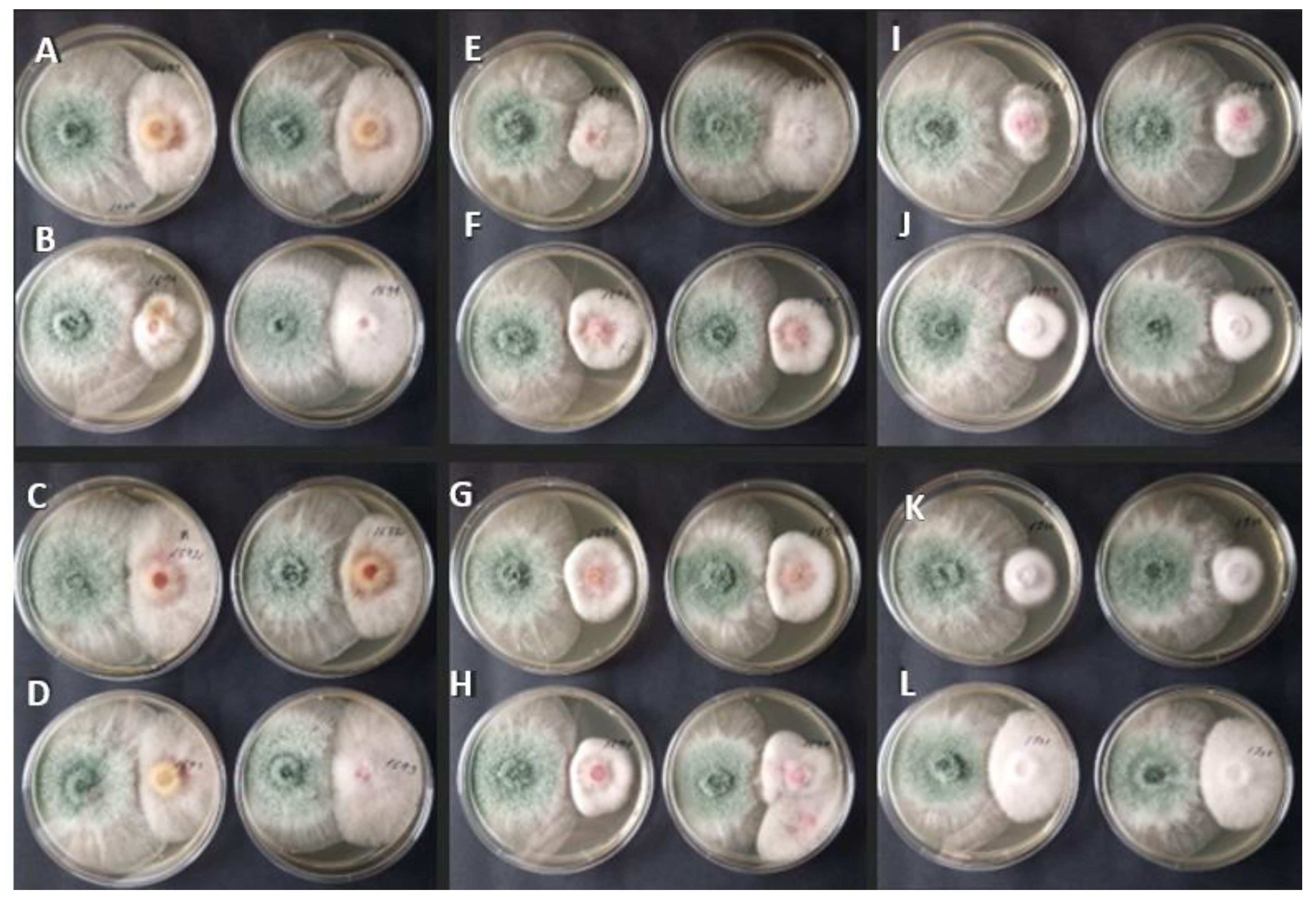
Figure 2.
Fusarium colony diameter (cm ±SD) in plates without (Control) and with (Sample) T. asperellum solutes after 48 (A) and 72 hours (B) of cultivation. Strain transcription: 1. Fusarium culmorum MSCL 1690, 2. F. graminearum MSCL 1691, 3. F. culmorum MSCL 1692, 4. F. culmorum MSCL 1693, 5. F. graminearum MSCL 1694, 6. F. sporotrichioides MSCL 1695, 7. F. oxysporum MSCL 1696, 8. F. sporotrichioides MSCL 1697, 9. F. tricinctum MSCL 1698, 10. F. oxysporum MSCL 1699, 11. F. oxysporum MSCL 1700, and 12. F. poae MSCL 1701. Three replicates.
Figure 2.
Fusarium colony diameter (cm ±SD) in plates without (Control) and with (Sample) T. asperellum solutes after 48 (A) and 72 hours (B) of cultivation. Strain transcription: 1. Fusarium culmorum MSCL 1690, 2. F. graminearum MSCL 1691, 3. F. culmorum MSCL 1692, 4. F. culmorum MSCL 1693, 5. F. graminearum MSCL 1694, 6. F. sporotrichioides MSCL 1695, 7. F. oxysporum MSCL 1696, 8. F. sporotrichioides MSCL 1697, 9. F. tricinctum MSCL 1698, 10. F. oxysporum MSCL 1699, 11. F. oxysporum MSCL 1700, and 12. F. poae MSCL 1701. Three replicates.
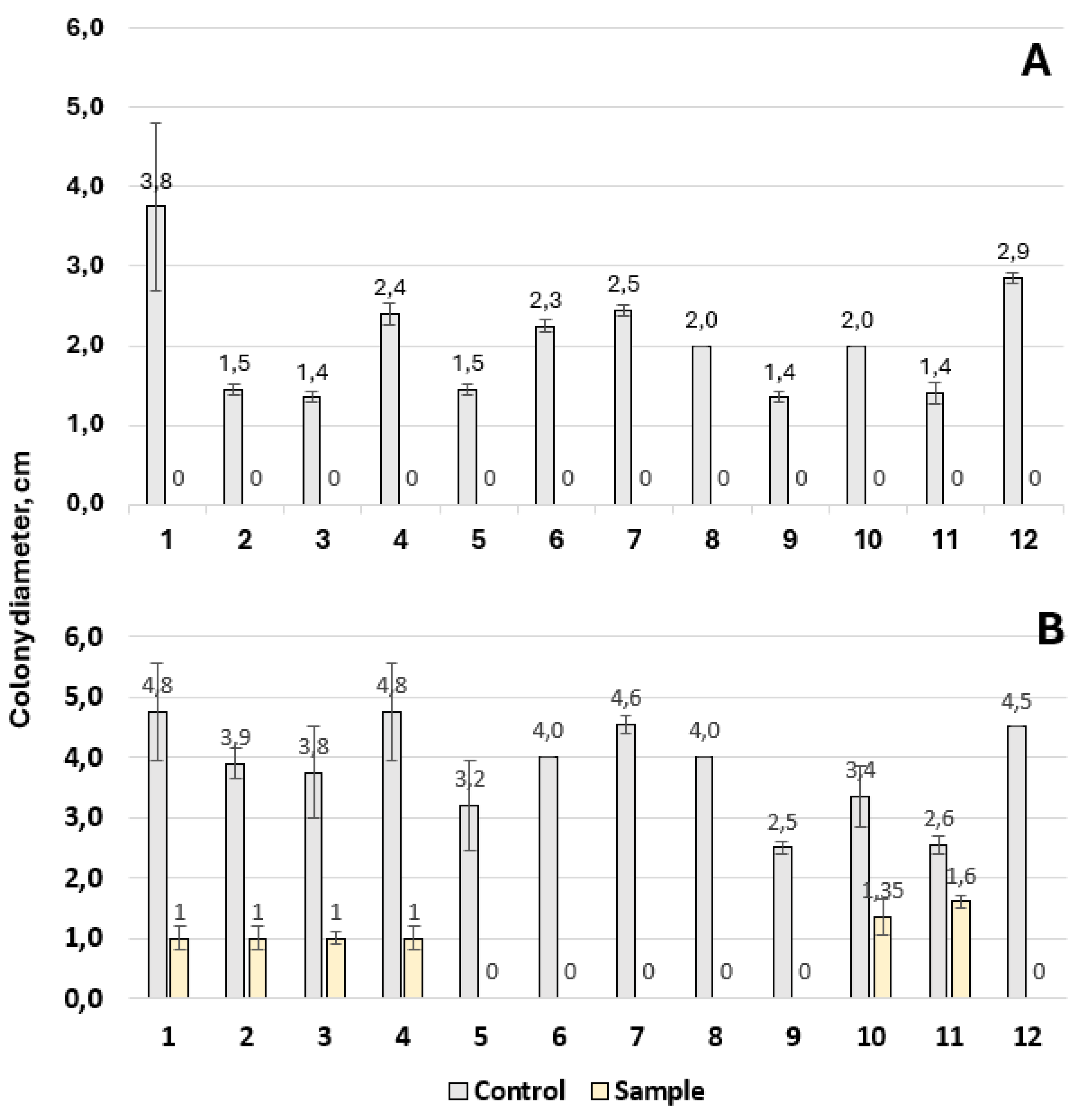
Figure 3.
Diameter of the zone of inhibition of Fusarium (mm ± SD) after seven days of cultivation in agar well assay with B. subtilis MSCL 1441. Strain transcription: 1. Fusarium culmorum MSCL 1690, 2. F. graminearum MSCL 1691, 3. F. culmorum MSCL 1692, 4. F. culmorum MSCL 1693, 5. F. graminearum MSCL 1694, 6. F. sporotrichioides MSCL 1695, 7. F. oxysporum MSCL 1696, 8. F. sporotrichioides MSCL 1697, 9. F. tricinctum MSCL 1698, 10. F. oxysporum MSCL 1699, 11. F. oxysporum MSCL 1700, and 12. F. poae MSCL 1701. Numbers with different letters above the columns are significantly different (p < 0.05).
Figure 3.
Diameter of the zone of inhibition of Fusarium (mm ± SD) after seven days of cultivation in agar well assay with B. subtilis MSCL 1441. Strain transcription: 1. Fusarium culmorum MSCL 1690, 2. F. graminearum MSCL 1691, 3. F. culmorum MSCL 1692, 4. F. culmorum MSCL 1693, 5. F. graminearum MSCL 1694, 6. F. sporotrichioides MSCL 1695, 7. F. oxysporum MSCL 1696, 8. F. sporotrichioides MSCL 1697, 9. F. tricinctum MSCL 1698, 10. F. oxysporum MSCL 1699, 11. F. oxysporum MSCL 1700, and 12. F. poae MSCL 1701. Numbers with different letters above the columns are significantly different (p < 0.05).
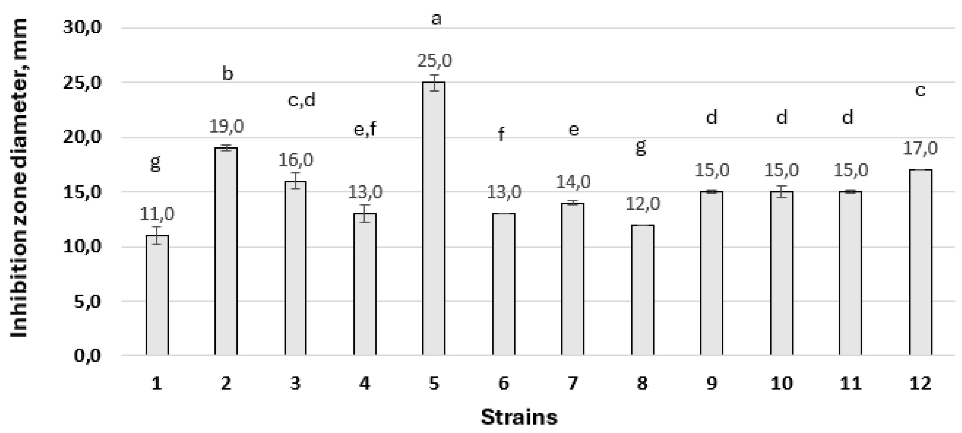
Figure 4.
Effect of three Bacillus strains plated in agar wells on the growth of Fusarium spp. after seven days of cultivation. i—B. stercoris MSCL 897, ii—B. subtilis MSCL 1441, iii—B. subtilis MSCL 49. (A) Fusarium culmorum MSCL 1690, (B) F. graminearum MSCL 1691, (C) F. culmorum MSCL 1692, (D) F. culmorum MSCL 1693, (E) F. graminearum MSCL 1694, (F) F. sporotrichioides MSCL 1695, (G) F. oxysporum MSCL 1696, (H) F. sporotrichioides MSCL 1697, (I) F. tricinctum MSCL 1698, (J) F. oxysporum MSCL 1699, (K) F. oxysporum MSCL 1700, and (L) F. poae MSCL 1701. One of three replicates is shown.
Figure 4.
Effect of three Bacillus strains plated in agar wells on the growth of Fusarium spp. after seven days of cultivation. i—B. stercoris MSCL 897, ii—B. subtilis MSCL 1441, iii—B. subtilis MSCL 49. (A) Fusarium culmorum MSCL 1690, (B) F. graminearum MSCL 1691, (C) F. culmorum MSCL 1692, (D) F. culmorum MSCL 1693, (E) F. graminearum MSCL 1694, (F) F. sporotrichioides MSCL 1695, (G) F. oxysporum MSCL 1696, (H) F. sporotrichioides MSCL 1697, (I) F. tricinctum MSCL 1698, (J) F. oxysporum MSCL 1699, (K) F. oxysporum MSCL 1700, and (L) F. poae MSCL 1701. One of three replicates is shown.
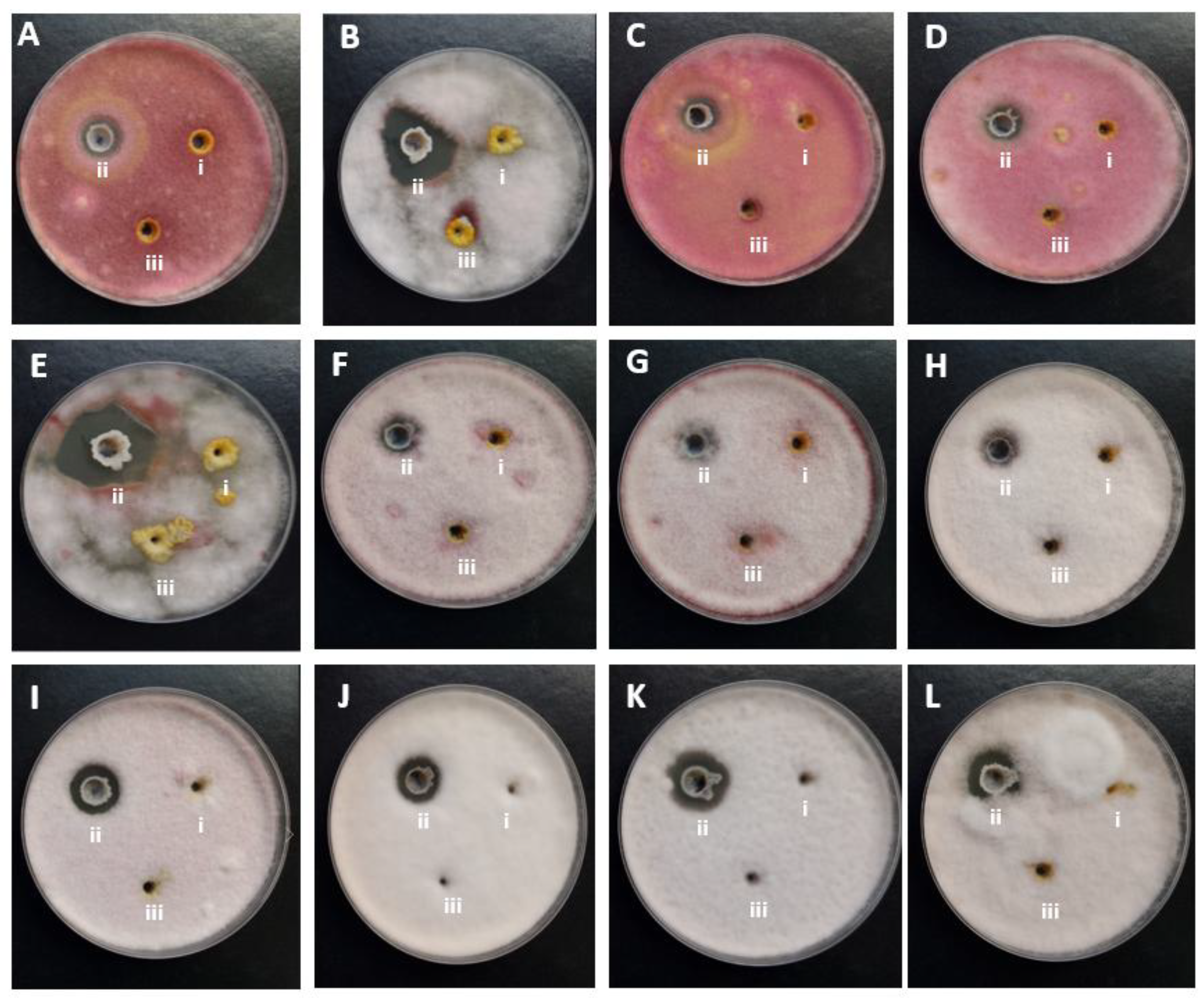
Figure 5.
Inhibition zone size (mm ±SD) of Fusarium oxysporum (A), F. graminearum (B), Botrytis cinerea (C) and Colletotrichum salicis (D) depending on the opposite growing actinobacteria Streptomyces griseus MSCL 346, Streptomyces sp. MSCL 349, S. venezuelae MSCL 350, S. griseus MSCL 351, S. sylvae MSCL 354, Streptomyces sp. MSCL 355, Streptomyces sp. MSCL 415, S. anthocyanicus MSCL 420, S. griseus MSCL 422 and S. griseus MSCL 424, in three replicates after seven days of incubation.
Figure 5.
Inhibition zone size (mm ±SD) of Fusarium oxysporum (A), F. graminearum (B), Botrytis cinerea (C) and Colletotrichum salicis (D) depending on the opposite growing actinobacteria Streptomyces griseus MSCL 346, Streptomyces sp. MSCL 349, S. venezuelae MSCL 350, S. griseus MSCL 351, S. sylvae MSCL 354, Streptomyces sp. MSCL 355, Streptomyces sp. MSCL 415, S. anthocyanicus MSCL 420, S. griseus MSCL 422 and S. griseus MSCL 424, in three replicates after seven days of incubation.
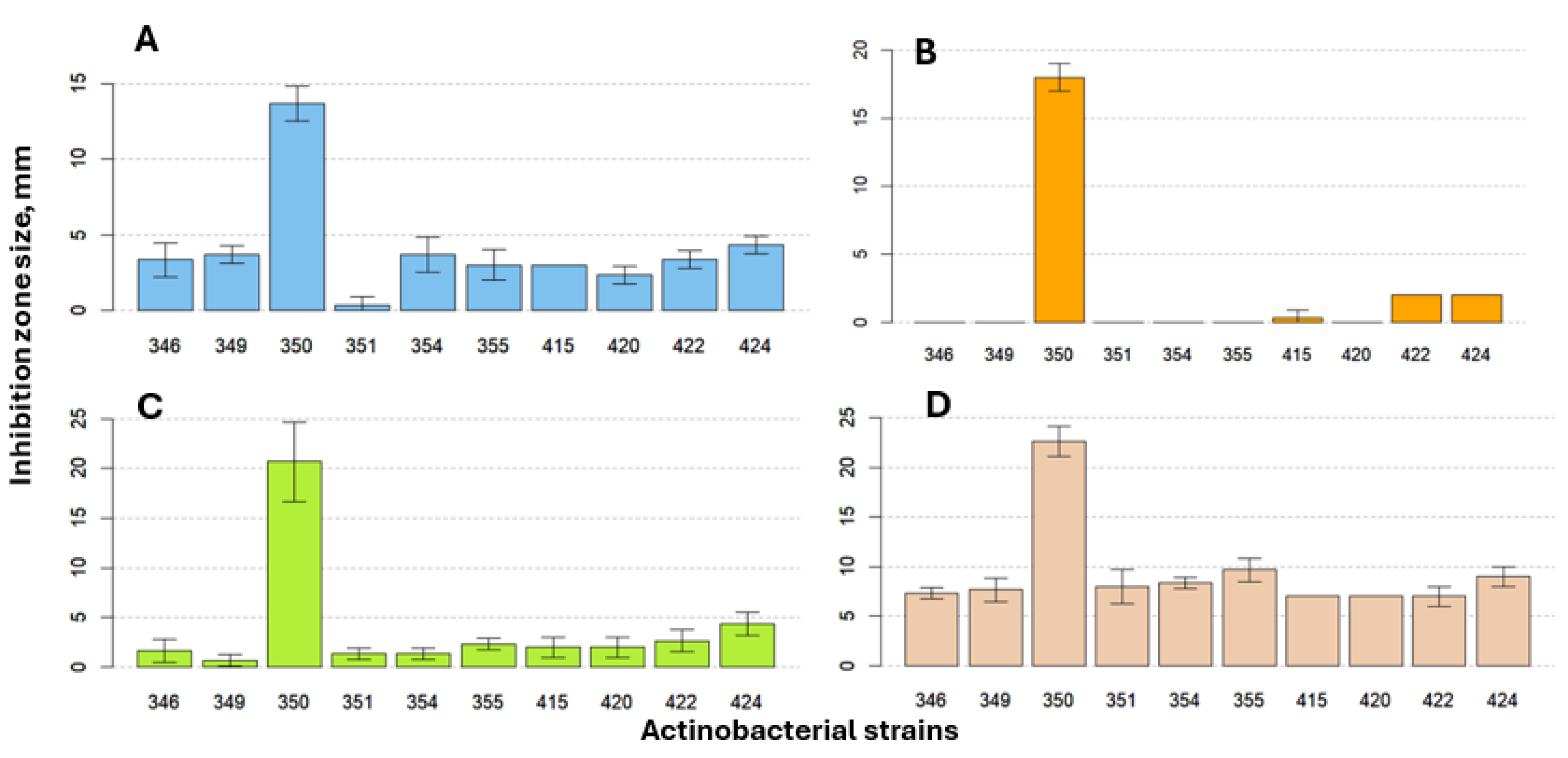

Table 1.
Microorganisms used in the study.
| Species | MSCL number | Origin | Type of identification |
|---|---|---|---|
| Antagonists | |||
| Bacillus subtilis | 1441 | Lupine soil, Latvia | biochemical |
| Bacillus subtilis | 49 | Unknown | biochemical |
| Bacillus stercoris | 897 | Commercial cleaner, Latvia | rRNA gene |
| Streptomyces anthocyanicus | 420 | Garden soil, Latvia | ANI |
| Streptomyces griseus | 346 | Garden soil, Latvia | rRNA gene, ANI |
| Streptomyces griseus | 351 | Garden soil, Latvia | rRNA gene, ANI |
| Streptomyces griseus | 422 | Garden soil, Latvia | ANI |
| Streptomyces griseus | 424 | Garden soil, Latvia | ANI |
| Streptomyces silvae | 354 | Garden soil, Latvia | rRNA gene |
| Streptomyces sp. | 355 | Garden soil, Latvia | rRNA gene |
| Streptomyces sp. | 415 | Garden soil, Latvia | micromorphological |
| Streptomyces sp. | 349 | Garden soil, Latvia | rRNA gene |
| Streptomyces venezuelae | 350 | Garden soil, Latvia | ANI |
| Trichoderma asperellum | 309 | Commercial preparation, Latvia | ITS |
| Pathogens | |||
| Botrytis cinerea | 433 | Liver paste, Sweden | unknown |
| Colletotrichum salicis | 850 | Rhododendron leaves, Riga | ITS, LSU, TUB2, ACT, CHS-1, GAPDH, HIS3 |
| Fusarium oxysporum | 259 | Unknown | unknown |
| Fusarium graminearum | 435 | Oat, Sweden | unknown |
| Fusarium culmorum | 1690 | Avena sativa, Latvia | micromorphological |
| Fusarium culmorum | 1692 | Avena sativa, Latvia | ITS, TEF |
| Fusarium culmorum | 1693 | Avena sativa, Latvia | ITS, TEF |
| Fusarium graminearum | 1691 | Avena sativa, Latvia | ITS, TEF |
| Fusarium graminearum | 1694 | Avena sativa, Latvia | ITS, TEF |
| Fusarium oxysporum | 1696 | Avena sativa, Latvia | micromorphological |
| Fusarium oxysporum | 1699 | Avena sativa, Latvia | ITS, TEF |
| Fusarium oxysporum | 1700 | Avena sativa, Latvia | ITS, TEF |
| Fusarium poae | 1701 | Avena sativa, Latvia | ITS, TEF |
| Fusariumsporotrichioides | 1695 | Avena sativa, Latvia | ITS, TEF |
| Fusariumsporotrichioides | 1697 | Avena sativa, Latvia | ITS, TEF |
| Fusariumtricinctum | 1698 | Avena sativa, Latvia | micromorphological |
Table 2.
Inhibitory effect of T. asperellum against twelve Fusarium strains. Numbers in a column followed by different letters are significantly different (p < 0.05).
Table 2.
Inhibitory effect of T. asperellum against twelve Fusarium strains. Numbers in a column followed by different letters are significantly different (p < 0.05).
| Fusarium strains | Percentage of inhibition under dual culture conditions |
|---|---|
| Fusarium culmorum MSCL 1690 | 49.1 ± 4.4c |
| Fusarium graminearum MSCL 1691 | 44.6 ± 10.5c |
| Fusarium culmorum MSCL 1692 | 46.7 ± 5.8c |
| Fusarium culmorum MSCL 1693 | 71.4 ± 4.5a |
| Fusarium graminearum MSCL 1694 | 48.9 ± 9.4b,c |
| Fusarium sporotrichioides MSCL 1695 | 71.0 ± 3.9a |
| Fusarium oxysporum MSCL 1696 | 74.4 ± 5.6a |
| Fusarium sporotrichioides MSCL 1697 | 78.4 ± 7.0a |
| Fusarium tricinctum MSCL 1698 | 56.7 ± 3.3b |
| Fusarium oxysporum MSCL 1699 | 56.7 ± 6.6b |
| Fusarium oxysporum MSCL 1700 | 74.1 ± 3.5a |
| Fusarium poae MSCL 1701 | 71.0 ± 4.1a |
Disclaimer/Publisher’s Note: The statements, opinions and data contained in all publications are solely those of the individual author(s) and contributor(s) and not of MDPI and/or the editor(s). MDPI and/or the editor(s) disclaim responsibility for any injury to people or property resulting from any ideas, methods, instructions or products referred to in the content. |
© 2026 by the authors. Licensee MDPI, Basel, Switzerland. This article is an open access article distributed under the terms and conditions of the Creative Commons Attribution (CC BY) license (http://creativecommons.org/licenses/by/4.0/).
Copyright: This open access article is published under a Creative Commons CC BY 4.0 license, which permit the free download, distribution, and reuse, provided that the author and preprint are cited in any reuse.