

Submitted:
16 March 2026
Posted:
17 March 2026
You are already at the latest version
Abstract
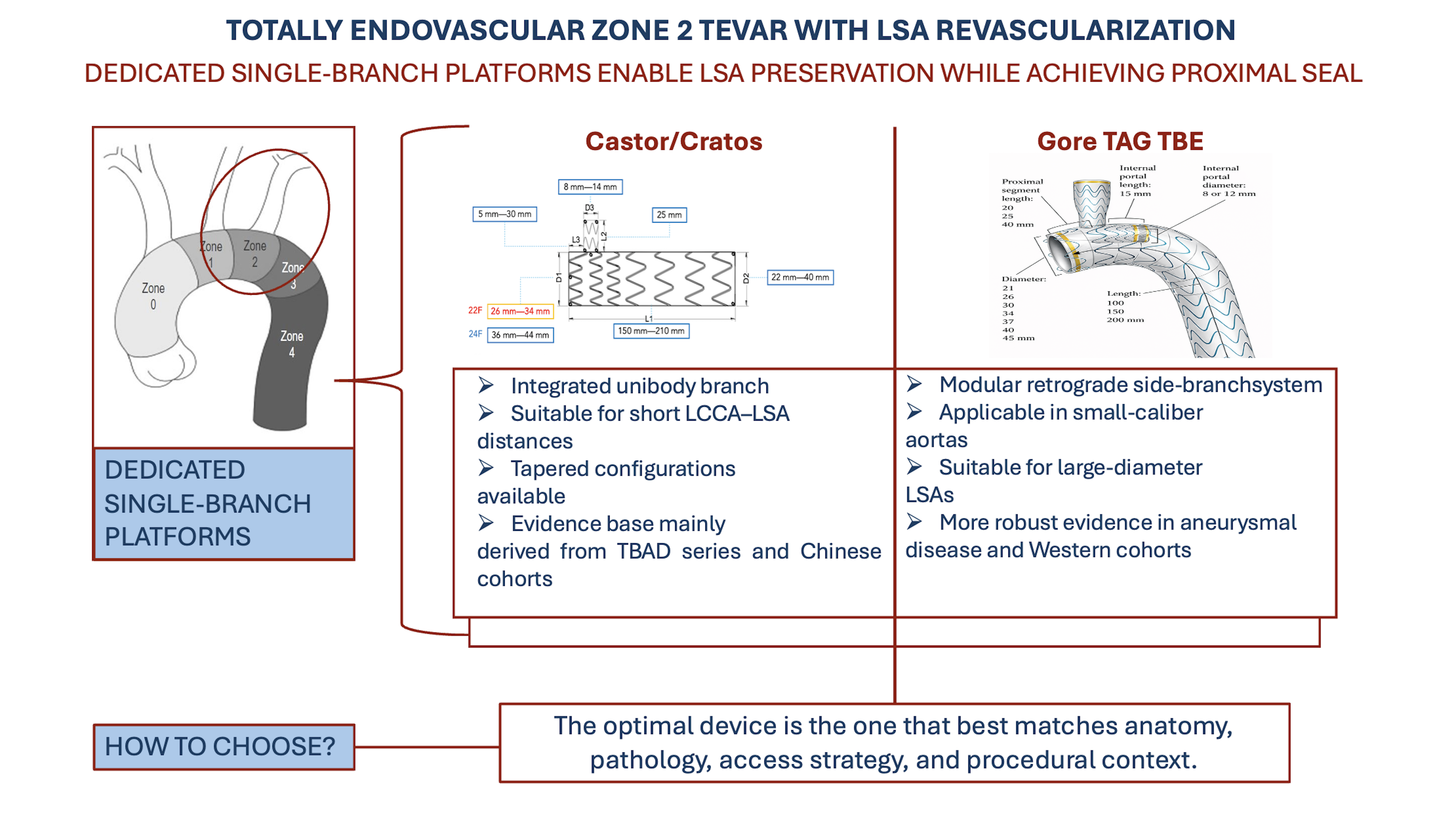
Zone 2 thoracic endovascular aortic repair (TEVAR) frequently requires left subclavian artery (LSA) preservation to maintain vertebrobasilar and upper-extremity perfusion while obtaining a durable proximal seal. Dedicated single-branch endografts were de-veloped to standardize this step and to convert a traditionally hybrid scenario into a reproducible fully endovascular strategy. Two different concepts currently dominate this field: integrated unibody branch platforms, represented by Castor and the sec-ond-generation Cratos, and modular retrograde-branch systems, represented by the Gore TAG Thoracic Branch Endoprosthesis (TBE). The Castor/Cratos evidence base is broader, older, and much more heavily weighted toward type B aortic dissection, including long-term prospective multicenter data and several large real-world cohorts with fa-vorable branch patency and aortic remodeling. By contrast, TBE evidence is expanding rapidly and is supported by prospective midterm data in arch aneurysms as well as by increasingly large post-commercial series and comparative analyses across zones 0–2. Beyond outcomes, the two platforms differ substantially in branch directionality, con-tribution to proximal fixation, modularity, branch diameter range, proximal landing requirements, access profile, and regulatory/off-the-shelf availability, all of which have direct consequences for anatomical suitability in dissection, aneurysm disease, and trauma. This narrative review synthesizes current evidence and proposes an anato-my-first, pathology-aware framework for selecting between Castor/Cratos and TBE in totally endovascular zone 2 TEVAR with LSA revascularization.
Keywords:
TEVAR
; Zone 2
; aortic arch
; left subclavian artery
; Castor
; Cratos
; Gore TBE
; thoracic branch endoprosthesis
; aortic dissection
; endovascular arch repair
1. Introduction
Thoracic endovascular aortic repair (TEVAR) has become the preferred treatment for many lesions of the descending thoracic aorta, including type B thoracic aortic dissection (TBAD), penetrating aortic ulcer (PAU), intramural hematoma (IMH), traumatic aortic injury, and descending thoracic aneurysm [1]. However, the durability of TEVAR remains critically dependent on the quality of the proximal seal [2]. In a substantial proportion of patients, an adequate disease-free proximal landing zone distal to the left subclavian artery (LSA) is not available, and the endograft must therefore be landed in Ishimaru zone 2, inevitably jeopardizing the LSA origin unless dedicated preservation strategies are employed [3,4,5,6,7].
Intentional LSA coverage is not a trivial technical detail. It can alter posterior cerebral circulation, impair collateral pathways to the spinal cord, jeopardize upper-limb perfusion, and create specific risks in patients with prior left internal mammary artery coronary bypass, dominant left vertebral artery, incomplete circle of Willis, or anticipated extensive thoracic coverage [5,6,7,8,9,10]. For that reason, current aortic practice has increasingly moved from routine LSA sacrifice toward selective or systematic preservation, especially in elective cases and in anatomically complex or extensive repairs [7,9,11].
Before the era of dedicated single-branch devices, LSA preservation during zone 2 TEVAR relied on open or hybrid debranching, most commonly carotid–subclavian bypass or transposition, or on purely endovascular workarounds such as chimney/periscope grafting and in situ fenestration. Each of these approaches remains useful, but each carries specific liabilities. Hybrid debranching adds cervical dissection, cranial nerve risk, wound morbidity, and an additional operative step, considerations that should not be underestimated in surgically high-risk or frail patients and that are well recognized more broadly in the literature on complex thoracoabdominal aortic repair [12,13]; chimney techniques introduce the risk of gutters and type Ia endoleaks; fenestration may compromise graft integrity and can be technically demanding, especially in hostile arch anatomy [9,14,15].
Dedicated single-branch platforms were designed to address these shortcomings by incorporating LSA preservation into the primary repair construct itself. Conceptually, however, not all branched devices solve the problem in the same way. One philosophy is represented by Castor (MicroPort Medical, Shanghai, China) and later Cratos (MicroPort Medical, Shanghai, China), which use a unibody aortic graft with an integrated antegrade side branch. The other is represented by the Gore TAG Thoracic Branch Endoprosthesis (TBE) (W.L. Gore and Associates, Flagstaff, AZ, USA), which uses a modular aortic component with an internal portal and a separate retrograde side-branch component [5,6,7,16,17,18].
This distinction is not merely descriptive. It affects proximal sealing mechanics, branch fixation, freedom from migration, minimum required proximal landing length, branch sizing flexibility, access strategy, and the number of components needed to complete the repair. It also partly explains the different evidence ecosystems in which the devices evolved. Castor was introduced earlier and therefore accumulated a much larger and longer clinical literature, but the overwhelming majority of that literature concerns TBAD and originates from China [5,16,19,20,21,22,23]. By contrast, TBE entered a Western environment dominated by distal arch aneurysm, mixed arch pathology, and regulatory pathways that emphasized prospective feasibility and post-commercial real-world registries [6,7,17,24,25].
Accordingly, the central problem is not simply whether one device is “better” than the other. Rather, the question is which device is more appropriate for which anatomy, pathology, and procedural scenario. This review therefore focuses on totally endovascular zone 2 TEVAR with LSA preservation and compares Castor/Cratos and TBE through four complementary lenses: anatomical suitability, device design, procedural strategy, and clinical evidence.
2. Anatomical Considerations
2.1. Zone 2 Sealing: More than a Nominal Landing Length
The success of zone 2 TEVAR depends not only on how many millimeters of “healthy” aorta are available between the Left Common Carotid Artery (LCCA) and the LSA, but also on how that segment behaves in three-dimensional space. The proximal landing segment is short, highly curved, and subject to significant inner–outer curvature mismatch. Arch angulation, calcification, mural thrombus, and the relative apposition of the graft along the outer curvature all influence the likelihood of bird-beak, type Ia endoleak, migration, and adverse aortic remodeling [2,4,9,26,27,28].
These issues become even more pronounced in TBAD. In acute dissection, one is not treating a static aneurysmal lumen but a dynamic geometry with true-lumen compression, tapered distal diameters, branch-vessel relationships driven by the flap, and a proximal landing segment that may be short, fragile, or immediately adjacent to the primary entry tear. This explains why some platforms that perform very well in distal arch aneurysm do not necessarily translate seamlessly into acute dissection, and vice versa [3,4,6,17,26].
This distinction is reflected in the literature. Castor series repeatedly focus on the interplay between proximal seal, remodeling, migration, and branch patency in TBAD [3,4,5,16,19,20,21,22,23,27,29], whereas TBE studies more often evaluate mixed or aneurysmal arch cohorts in which the central question is procedural safety, branch durability, and the capacity to replace hybrid bypass strategies [6,7,17,25].
2.2. LCCA–LSA Distance: A Major Discriminator Between Platforms
Perhaps the most important anatomical discriminator between the platforms is the available distance between the distal LCCA and the LSA. For TBE, this distance is critical because the modular portal configuration requires a longer segment of proximal zone 2 to accommodate the portal and covered seal without jeopardizing the LCCA. In the Greek single-center TBE experience, the authors explicitly state that a minimum of 2 cm between the end of the LCCA and the end of the LSA is required for TBE use [30]. Feasibility studies and Instruction For Use (IFU)-based sizing analyses similarly use thresholds around 15–20 mm, reflecting the need for a meaningful proximal covered segment before the branch portal [28,31].
By contrast, Castor was designed specifically to function in shorter zone 2 anatomies. Its integrated branch can be positioned very proximally relative to the proximal edge of the aortic graft. The offset from the proximal end of the main body to the branch is available from 5 mm up to 30 mm. This short-offset architecture is highly relevant in the subset of patients in whom the disease reaches immediately beneath the LSA or in whom the LCCA–LSA gap is too short for a modular portal-based solution. Clinical data support this anatomical intuition. Chen et al. specifically treated TBAD with an “insufficient anchoring region” using Castor, demonstrating that the device can be used successfully when the available proximal landing segment is very limited [3]. In the comparative anatomical feasibility analysis of acute TBAD by Lang et al., Castor suitability reached 82%, whereas TBE off-the-shelf suitability was only 22%, largely because the longer seal requirements and non-tapered modular configuration excluded many dissection anatomies [26].
2.3. LSA Diameter and Branch Sizing: Why Western Aneurysm Anatomy Matters
The second key anatomical discriminator is the diameter of the target vessel. Castor and Cratos offer branch diameters generally ranging from 6 to 14 mm. This range is entirely appropriate for most TBAD patients and for many Asian series, and it is mirrored by the branch-diameter ranges reported in several clinical studies [16,19,33]. However, it can become limiting in larger, more degenerative proximal subclavian anatomies, particularly in Western aneurysm populations. TBE has a clear advantage in this domain. TBE provides branch options up to 20 mm, with treated-vessel diameter ranges extending up to 18 mm depending on the portal configuration. This broader branch range is repeatedly emphasized in TBE feasibility studies and technical reviews [28,30,31,34]. As a result, TBE can accommodate larger LSAs without forcing undersizing or reliance on adjunctive relining. This is particularly relevant in degenerative arch aneurysm, in elderly patients, and more broadly in Western populations, where larger supra-aortic vessel diameters are not uncommon.
The practical consequence is that TBE may remain fully endovascular and fully device-based in anatomies in which Castor/Cratos would require an additional covered stent in the side branch to bridge a diameter mismatch. Indeed, in the European Castor feasibility study by Leone et al., large LSA diameter was the single most important cause of non-feasibility, accounting for more than half of all exclusions [32].
2.4. Distal Tapering in Acute TBAD: A Core Advantage of Integrated Tapered Platforms
Acute TBAD often produces a marked proximal-to-distal mismatch between the aortic diameter in zone 2 and the distal true lumen. This is one of the main reasons why devices designed around more uniform aneurysmal anatomy may become difficult to size. Lang et al. showed that for TBE in acute TBAD, the principal reason for non-feasibility was distal diameter mismatch, usually because the distal aorta was too small relative to the available TBE configurations [26]. This finding is highly relevant because it mechanistically explains why TBE acute TBAD series frequently require additional distal components and why the platform’s pure off-the-shelf suitability in dissection is relatively low [17,24,35,36,37].
Castor, on the other hand, is available with proximal diameters of 26–44 mm and distal diameters of 20–44 mm, thereby allowing tapered main-body configurations with up to 8 mm of tapering, which may be particularly advantageous in acute TBAD, where pronounced proximal-to-distal diameter mismatch is frequently encountered. This seems minor on paper, but in acute TBAD it is very important. A tapered main body is intrinsically more compatible with proximal seal in zone 2 and distal treatment in a smaller, compressed true lumen. This is one of the reasons Castor has accumulated such a dissection-heavy literature and why its anatomical suitability in acute TBAD is markedly higher than that of TBE in comparative modeling [5,19,20,21,22,23,26,29].
2.5. Iliac Access Feasibility and the Concept of “True Feasibility”
Another relevant anatomical issue is whether the patient can physically accommodate the device from an access perspective. This is especially important because thoracic branched devices are bulkier than standard TEVAR systems and because the older, more aneurysmal patients in whom TBE is often used frequently have diseased iliofemoral axes.
Vacirca et al. helpfully separated Anatomical Feasibility (AF) from Iliac Feasibility (IF) and reported a True Feasibility (TF) rate of 85%, down from 92% when access constraints were added. Importantly, access feasibility was significantly lower in women, highlighting that large-profile devices are not evaluated properly if one stops at arch anatomy alone [28].
Most Castor feasibility studies do not formally provide this arch-plus-access analysis. Leone et al. examined morphology in detail but did not define a separate true-feasibility category [32]. Lang et al. focused on arch suitability rather than access suitability [26]. This methodological asymmetry matters, because it can make Castor suitability appear more favorable on paper when access has not been assessed with the same rigor. At the same time, the profile of the Castor family is often advantageous: the original system uses a 24F sheath, whereas Cratos can drop to 22F in smaller and intermediate sizes [16]. The larger TBE configurations, by contrast, can require up to 26F introducers.
3. Evolution of LSA Preservation in Zone 2 TEVAR
The shift from open or hybrid debranching to fully endovascular LSA preservation has been driven by two parallel needs: reducing the invasiveness of the procedure and standardizing the technical result. Hybrid carotid–subclavian bypass remains durable, but it introduces an extra incision, potential nerve injury, wound complications, and, importantly, a second operative step that partly offsets the minimally invasive advantage of TEVAR. Chimney grafting avoids neck surgery, but gutter-related endoleak remains a structural concern. In situ fenestration is attractive and versatile, but it is operator-dependent, less standardized, and potentially problematic in heavily calcified or highly angulated arches [7,9,14,15].
Dedicated single-branch endografts therefore represent the next logical step in the evolution of zone 2 TEVAR. They embed the branch solution within the primary aortic repair and aim to reduce variability. Yet, even within this category, there are two very different philosophies. Castor was the first widely used dedicated single-branch zone 2 thoracic device and was initially developed to preserve the LSA in TBAD. Its strong association with dissection is therefore historical as well as anatomical [5,19,20,23]. TBE emerged later in a Western regulatory and commercial context, with a broader focus on arch pathology and with immediate relevance not only to zone 2 but also to zones 0 and 1 through a modular branch concept and proximal extension options [6,7,17,34].
This divergence in origins matters because it has shaped the evidence base. The Castor literature is larger but less geographically diverse; the TBE literature is newer but more Western and more heterogeneous in pathology. Any meaningful comparison between them must therefore distinguish between device performance and the clinical ecosystem in which the device was adopted [5,6,7,17,25].
4. Device Design Comparison
4.1. Unibody Branch Versus Modular Retrograde Branch
The most fundamental distinction is architectural. Castor and Cratos are unibody devices in which the aortic component and the side branch are part of a single integrated structure. TBE is a modular system consisting of an aortic component with an internal portal and a separate side-branch component [6,34]. This design distinction influences almost every downstream aspect of the procedure.
In Castor/Cratos, the side branch is deployed together with the main graft, and the branch is oriented in an antegrade, more physiologic direction. In TBE, the branch is inserted retrogradely through a portal and therefore functions as an additional modular component [6,17,25,34]. This is not simply a matter of elegance. Castor/Cratos behaves as a single integrated construct, whereas TBE inherently introduces at least one component junction even before any distal aortic extensions are added. In addition, it requires deployment of a separate bridging branch into the LSA, which increases procedural complexity and may expose the repair to component-related failure modes such as malalignment, junctional endoleak, or loss of apposition. Similar concerns are well known from other multicomponent endovascular systems [38], including fenestrated and branched endovascular aneurysm repair [39,40,41,42].
4.2. Active Fixation and Sealing Contribution of the Integrated Branch
One of the strongest conceptual arguments in favor of Castor/Cratos is that the integrated side branch contributes not only to LSA perfusion but also to proximal construct stabilization. Because the side branch is part of the main graft, it acts as an anchoring vector into the target vessel. The branch is not merely connected to the system; it is the system. This may be particularly relevant in dissection or aneurysm, where the proximal seal is short, the aortic wall is vulnerable, and migration or displacement may develop even when the immediate angiographic result looks acceptable. This hypothesis is supported indirectly by clinical evidence. In the acute TBAD comparison by Liu et al., Castor had 0% stent displacement versus 20% with standard TEVAR and markedly lower reintervention rates at 2 years [27]. Although this was not a direct Castor-versus-TBE study, it supports the idea that an integrated branch can stabilize the proximal zone 2 repair. Likewise, Chen et al. reported favorable early results in very short anchoring zones, again supporting the notion that Castor’s side branch is functionally relevant to proximal fixation [3].
TBE, by contrast, does not derive proximal sealing strength from the side branch in the same way. The branch is retrograde and modular, and the proximal seal is determined by the covered aortic component and its relation to the native arch. The side branch preserves flow, but it does not meaningfully reduce the proximal landing requirement, which remains a core anatomical constraint in the TBE system [28,31].
4.3. Modularity: Versatility on One Side, More Overlap Zones on the Other
The main advantage of TBE modularity is versatility. The system can be configured across zones 0, 1, and 2, and the same fundamental aortic component/side-branch logic can be used in different arch scenarios [6,7,17,25,34]. This is a major practical advantage and one reason TBE has become so attractive in Western practice, particularly when distal arch aneurysm, prior ascending replacement, or more proximal arch involvement must be addressed without open sternotomy.
The downside is equally obvious: modularity means more components, more interfaces, and often more adjunctive extensions. The real-world TBE literature repeatedly shows this. Distal extensions were common in the Dutch single-center European experience [36], in the large Cleveland Clinic post-commercial series [17], in Pang’s zone 0–2 cohort [25], and in the urgent Padua case series, in which all five patients required distal extension [37]. These are not trivial technical details. Every additional overlap zone is another place where apposition, kinking, separation, or device-related failure can occur, and this logic is well known from other branched aortic platforms [38,39].
The same principle applies to the bridging side-branch component. No contemporary series suggests that TBE branch durability is poor; on the contrary, branch patency is excellent in most reports [6,7,25]. However, the fact remains that TBE introduces an additional modular connection and often additional distal thoracic components, whereas Castor/Cratos tries to minimize the number of moving parts at the level of the arch itself [6,7,17,25,34].
4.4. Proximal Offset and Short-Neck Advantage
Another decisive design feature is the proximal branch offset. In the Castor/Cratos system, the distance from the proximal edge of the main body to the branch can range from 5 mm to 30 mm. This allows very short proximal configurations and makes the device usable in anatomies with minimal space between the LCCA and the LSA.
TBE does not have this degree of short-offset flexibility. Its modular portal design requires a more generous proximal seal and has translated in practice into threshold distances of roughly 15–20 mm in feasibility work, or around 2 cm in single-center clinical interpretation [28,30,31]. This difference is one of the clearest practical advantages of Castor/Cratos and is a major reason why it performs better in dissection-centered anatomical feasibility analyses [26].
4.5. Branch Diameter Range: A Real Advantage for TBE in Larger LSAs
If Castor/Cratos has the advantage in short-neck anatomy, TBE has the clearer advantage in large LSA anatomy. Branch diameters up to 20 mm allow TBE to accommodate larger target vessels that would exceed the intended Castor side-branch range [28,30,31]. In practical terms, this is often more relevant in degenerative aneurysm disease than in acute TBAD. Many dissection patients have target-vessel diameters comfortably within the Castor range, which partly explains why Castor performs so well in that pathology. However, once LSA diameter exceeds roughly 13–14 mm, the integrated-branch platform may require adjunctive relining or may become infeasible outright [32].
4.6. Delivery Profile and Access
The delivery profile comparison also deserves a nuanced reading. The classic Castor system uses 24F across its range, whereas Cratos reduces this to 22F up to 34 mm in the multicenter second-generation study [16]. TBE, depending on size, may require 20F to 26F introducers. This means that smaller TBE configurations can be comparable or even advantageous, but larger TBE configurations become less favorable from an iliac-access standpoint, especially in aneurysm patients and women [28].
Upper-extremity access goes in the opposite direction. Castor/Cratos generally requires 7–8F branch access [5,33]. TBE commonly uses smaller brachial or axillary access, typically around 4-5F in practice, which can reduce the need for open exposure and facilitate percutaneous workflows [30,37]. This distinction is not negligible, because larger upper-extremity sheaths may more frequently necessitate surgical exposure to mitigate access-related complications [43]. Therefore upper-extremity access strategy should be considered an integral part of procedural planning rather than a secondary technical detail. Thus, TBE may be easier from the arm and harder from the groin. Castor/Cratos may be easier from the groin, particularly in the Cratos generation, but more demanding from the supra-aortic access point.
4.7. Maneuverability and the Cratos Response to First-Generation Limitations
One legitimate criticism of first-generation integrated devices is maneuverability. Castor requires careful rotational alignment during advancement, and the learning curve can be non-trivial. The Scandinavian multicenter study is particularly informative here: the authors explicitly describe wire wrapping as a potential issue during advancement, emphasize the need for familiarity with the system, and report one technical failure caused by displacement during off-IFU zone 1 plus laser-fenestration manipulation. They also document one episode of premature release of the branch cap, which was corrected by removing and remounting the device before successful implantation [11]. These details matter because they move the discussion beyond abstract design concepts and confirm that first-generation Castor is technically elegant but operator-sensitive.
Cratos appears to address many of these limitations. Li et al. reported that the second-generation system incorporates design refinements aimed at improving orientation control and proximal conformability (the delivery system features an additional dedicated trigger that allows control of proximal apposition and helps mitigate bird-beak formation), with encouraging early safety and 12-month branch patency [16]. The first Western Cratos case, performed for late TEVAR migration, also highlighted improved rotational control and a more favorable delivery profile, supporting the idea that Cratos is not just a rebranding but a response to true technical limitations of first-generation Castor [18].
5. Procedural Strategies
5.1. Preoperative Planning and Device Selection
For both device families, preoperative Computed Tomography Angiography (CTA) remains the essential planning modality. At a minimum, the following variables should be carefully assessed in order to guide optimal device selection for Zone 2 implantation [26,28,31,32]:
1. proximal landing-zone diameter and length on the outer curvature, inner curvature, and centerline;
2. LCCA–LSA distance, specifically the distance from the distal edge of the LCCA to the proximal edge of the LSA (critical for Castor/Cratos), the distance from the distal edge of the LCCA to the distal edge of the LSA, and the distance from the center of the LCCA ostium to the distal edge of the LSA (both key measurements for TBE); in all cases, these measurements should be obtained along the outer curvature.
3. LSA diameter and the length of the prevertebral segment.
4. distal thoracic diameter;
5. iliac-femoral access diameters, calcification, and tortuosity.
The different device configurations are illustrated in Figure 1 for Castor and Cratos, and in Figure 2 for TBE.
The diameters and lengths covered by the available configurations of the different devices are summarized in Table 1.
5.2. Device Implantation Procedures
Based on the largest available clinical series [6,16,18,21,33,34], the most practical procedural setup is generally, under general anesthesia, a three-access strategy, consisting of two femoral accesses (one main working access and one ancillary access) and one left upper-extremity access. For Castor/Cratos, because the upper-extremity branch access typically ranges around 7–8F, several authors have favored a small surgical brachial cutdown in order to minimize access-related complications. By contrast, Gore TBE usually requires a smaller upper-extremity working access, commonly around 4–5F in routine practice, and can therefore often be managed through simple percutaneous radial or brachial access. As for ballooning, proximal molding of the main graft is generally left to the operator’s discretion, as is distal ballooning, unless the distal sealing zone lies within another implanted device or across a graft overlap. With regard to the subclavian branch, routine ballooning is not mandatory for Castor/Cratos, although it is advisable in the presence of branch kinking or suboptimal expansion. By contrast, for TBE, the Instructions for Use recommend systematic ballooning of the side-branch component at three levels: at the overlap zone within the retrograde internal portal, along the mid-portion of the branch component, and distally within the LSA. Finally, deployment of the main graft is strongly recommended under controlled hypotension and bradycardia.
5.2.1. Castor Implantation Technique
A through-and-through wire is usually established between the left upper-extremity access, most commonly through a 7F brachial sheath,and one femoral access, typically the right, using a snaring maneuver. After the guidewire has been externalized, the branch catheter is advanced from the upper-extremity access and connected to the pre-flushed side branch of the device. The main graft, mounted on a 24F delivery system, is then introduced from the femoral access and advanced into the arch under fluoroscopic guidance. Particular attention is paid to maintaining coaxial alignment and to preventing wire twisting during advancement. The radiopaque markers are used to confirm correct rotational orientation, with the branch directed cranially toward the LSA origin. If malrotation or wire entrapment is recognized, the device can be withdrawn into the sheath and carefully reoriented before re-advancement. Final deployment is usually performed under controlled pharmacological hypotension and bradycardia in order to improve positional accuracy. Release of the branch into the LSA is then completed under tension on the upper-extremity wire, allowing precise engagement and stabilization of the branch component. The overall procedural workflow is summarized in Video S1.
5.2.2. Cratos Implantation Technique
A through-and-through brachiofemoral wire was established using standard snaring techniques. After placement of a stiff guidewire into the ascending aorta, the delivery catheter introduced from the brachial access was retrieved through the femoral sheath and docked into the pre-flushed Cratos branch. The main graft, pre-mounted on a 24F or, in selected configurations, a 22F delivery system, was then advanced from the femoral access. Radiopaque markers were aligned with the LSA origin under fluoroscopic guidance. The improved rotational control of the Cratos introducer, positioned in the proximal descending thoracic aorta, allowed fine orientation adjustments even in the presence of marked tortuosity. In cases of malrotation, the device could be partially resheathed within the introducer and safely repositioned before redeployment. After release of the main graft through the dedicated deployment knob, the delivery system features an additional dedicated trigger that allows control of proximal apposition and helps mitigate bird-beak formation. Controlled tension on the brachial wire then enabled accurate release of the branch into the LSA. The overall procedural workflow is summarized in Video S2.
5.2.3. TBE Implantation Technique
From the left brachial access, a guidewire is advanced and then retrieved from the right femoral access, thereby creating a left brachiofemoral through-and-through wire. Over a stiff aortic guidewire and with the brachiofemoral rail in place, the main device is advanced from the primary femoral access (typically 20–26 Fr, depending on the selected configuration) into the intended position and deployed after angiographic confirmation. Release is achieved using the dedicated deployment handle in a single-step maneuver. During this phase, maximal control of the delivery system is essential to prevent inadvertent forward or backward movement of the graft. The dedicated branch component for the left subclavian artery is subsequently advanced over the brachiofemoral through-and-through wire, deployed, and balloon-molded. The overall procedural workflow is summarized in Video S3.
5.3. Deployment Pitfalls
For Castor/Cratos, rotational alignment and avoidance of wire wrapping are central.
For TBE, the main procedural issue is not so much rotation of a unibody graft but the orchestration of a modular system: proper portal alignment, safe branch advancement, sufficient overlap, and careful planning of any distal extensions. This complexity is manageable, but it means that “totally endovascular” does not necessarily mean “single-device” or “simple.”
5.4. Completion Imaging and Surveillance
At completion angiography, the operator should document:
• absence of type Ia/Ib/Ic/III endoleak;
• branch patency;
• branch geometry and absence of kinking;
• absence of displacement or malapposition;
Follow-up CTA is particularly important in dissection because the immediate goal is not merely technical success but durable remodeling. Several Castor studies provide serial remodeling data, including false-lumen thrombosis and true-lumen expansion [5,19,20,22,29,33]. TBE follow-up is more often reported in terms of branch durability, survival, and reintervention, especially in aneurysm cohorts [6,7,17,25]. Figure 3, Figure 4 and Figure 5 show postoperative 3D CT reconstructions of a Castor, a Cratos, and a TBE, respectively.
6. Clinical Evidence
6.1. Literature Identification Strategy
This review was conducted as a structured narrative review rather than a formal PRISMA systematic review. Searches were performed in PubMed and Scopus, up to 25th February 2026, using combinations of the following keywords: “thoracic branch endoprosthesis”, “Gore TBE”, “single-branched TEVAR”, “Castor stent graft”, “Cratos stent graft”, “zone 2 TEVAR”, “left subclavian artery preservation”, “LSA revascularization”, “aortic dissection involving the left subclavian artery”, “distal arch aneurysm”, and “blunt thoracic aortic injury”. Reference lists of key feasibility studies and major clinical cohorts were screened manually to identify additional relevant publications.
Eligible articles included English-language clinical series, comparative cohorts, anatomical feasibility studies, meta-analyses, and high-relevance case reports focused specifically on dedicated single-branch platforms for totally endovascular arch repair involving zone 2 with LSA preservation. The final dataset included 32 studies [3,4,5,6,7,9,11,16,17,18,19,20,21,22,23,24,25,26,27,28,29,30,31,32,33,34,35,36,37,44,45,46]. The implantation-outcome studies are summarized in Table 2. Feasibility studies were analyzed separately from implantation-outcome studies to avoid conflating anatomical suitability with procedural or clinical durability [26,28,31,32] and are summarized in Table 3.
6.2. Two Evidence Ecosystems Rather than One Head-to-Head Literature
The most important interpretive point is that Castor/Cratos and TBE do not yet share a balanced, directly comparable evidence environment.
The Castor/Cratos literature is clearly broader and older. It includes a prospective multicenter trial with long-term follow-up [5], multiple large retrospective or real-world multicenter series [16,19,20,21], midterm and long-term remodeling data [22,29], and a systematic review/meta-analysis focused on TBAD [23]. However, this evidence is overwhelmingly dissection-driven and largely Chinese. Western experience exists, but it remains far more limited and includes mainly small elective cohorts, case reports, and a few multicenter European studies with relatively short follow-up [11,18,45,46].
The TBE literature is different. It is younger but more geographically Western. It is anchored by the prospective feasibility trial in aortic arch aneurysm [6] and has rapidly expanded through post-commercial single-center, multicenter, and comparative real-world studies [7,17,24,35,37,44]. It also includes a substantial amount of acute-pathology and trauma evidence [24,31,35,37,44]. In other words, TBE was not first adopted primarily in dissection but rather in a broader arch-repair landscape.
This matters because it means that many apparent differences between Castor/Cratos and TBE may actually reflect differences in pathology, patient selection, and regional practice rather than intrinsic device superiority. A platform that looks excellent in acute TBAD may not have been exposed equally to large aneurysmal arches; a platform with elegant aneurysm data may not yet have been stress-tested across the full acute-dissection anatomical spectrum [5,6,7,23,26].
6.3. Castor/Cratos: Strengths and Limits of the Current Evidence
The strongest component of the Castor literature is unquestionably TBAD. The multicenter prospective trial by Jing et al. remains a landmark study, with 73 patients, high technical success, no perioperative stroke, excellent 6-year survival, and durable branch patency [5]. This is complemented by large retrospective series showing technical success above 95%, low neurological event rates, and meaningful midterm follow-up [19,20,21]. The meta-analysis by Yao et al. further consolidates this picture, reporting pooled technical success of 97.5%, perioperative stroke of 0%, and 30-day mortality below 1%, although all pooled evidence remained observational and of low to very low certainty [23].
Castor has also shown consistent remodeling effects. Tian et al. reported long-term patency and favorable false-lumen thrombosis out to nearly six years [22]. Yuan et al. showed clear true-lumen expansion and false-lumen reduction, while also drawing attention to the fact that distal abdominal remodeling may be less predictable than thoracic remodeling [29]. These studies collectively reinforce the idea that Castor is not merely a branch-patency device but a dissection-specific remodeling platform [5,19,20,22,23,29].
However, the limitations are equally important. Most data come from China, where practice patterns, patient anatomy, and regulatory access are not identical to Western settings [5,16,19,20,21,23]. Western Castor evidence remains fragmentary. The Italian single-center experience by Rizza et al. included only ten patients and was elective and custom-made [46]. The Polish multicenter study by Żołnierczuk et al. showed encouraging feasibility but also a 4.8% Retrograde Type A Dissection (RTAD) rate and type I endoleak/bird-beak rates of nearly 10% [45]. The Scandinavian multicenter study fills an important gap by providing the largest contemporary European Castor series: 23 patients, 96% technical success, 95% side-branch patency, no 30-day major adverse events, but also 23% bird-beak and two reinterventions for distal stent graft–induced new entry tears, underlining that Western experience is improving but still short-term and not free of device- or anatomy-related failure modes [11].
6.4. TBE: Strengths and Limits of the Current Evidence
TBE’s most solid foundation remains the prospective multicenter feasibility trial in distal arch aneurysm, which demonstrated 100% technical success, zero 30-day mortality, 93% branch patency at 3 years, and excellent freedom from rupture, migration, and reintervention [6]. This is a high-quality benchmark study and remains one of the most important arguments in favor of TBE in aneurysmal distal arch disease.
Post-commercial experience has broadened the picture. Pang et al. described early outcomes across zones 0–2, with 100% technical success, acceptable 30-day mortality, and preserved branch patency, but also frequent need for distal extensions and proximal cuffs [25]. Lou et al. extended this by showing that post-commercial TBE can be applied broadly in real-world patients, including mixed aneurysm and dissection anatomy, albeit with a notable 16.4% endoleak rate and frequent adjunctive procedures [17]. Most importantly from a comparative-practice standpoint, Satam et al. demonstrated in a zone 2 comparison that TBE was associated with lower Myocardial Infarction (MI) and Acute Kidney Injury (AKI), shorter procedures and hospitalization, greater sac regression, and better freedom from reintervention than TEVAR combined with carotid–subclavian bypass [7].
TBE’s acute-pathology evidence is also growing. DiLosa and colleagues reported multicenter and single-center acute-pathology experiences [24,35], while Spertino et al. demonstrated feasibility in urgent acute aortic syndromes, albeit with universal use of distal extensions [37]. Trauma-specific evidence is another TBE strength: the device has both comparative clinical data against standard TEVAR with LSA coverage and dedicated anatomical feasibility modeling in Blunt Thoracic Aortic Injury (BTAI) [31,44].
The limitations of TBE are different from those of Castor. The device is not hampered by a lack of Western data, but much of its real-world literature still has relatively short follow-up [17,24,25,35,36,37]. Its data are also more heterogeneous in pathology and zone, making it harder to isolate “pure zone 2” performance from broader arch-repair experience [7,17,25]. Finally, the acute TBAD literature remains less mature than the Castor literature, and anatomical feasibility in dissection is lower when strict off-the-shelf criteria are applied [26].
The main advantages and limitations of the different devices are summarized in Table 4.
7. Patient Selection
7.1. When Castor/Cratos Is Generally Favored
First, Castor/Cratos is favored in acute or subacute TBAD with inadequate proximal landing or minimal LCCA–LSA distance. This is the setting in which its evidence is strongest and its design is most logically aligned with the anatomy [3,4,5,16,19,20,21,22,26,27,29].
Second, Castor/Cratos is attractive when the operator wants a more integrated construct with fewer modular arch junctions. The lower displacement and reintervention rates seen versus standard TEVAR in acute TBAD support this preference [27].
7.2. When TBE Is Generally Favored
First, TBE is favored in distal arch aneurysm and in mixed elective arch pathology where larger branch diameters and a formally broader arch role are important [6,7,17,25].
Second, TBE is preferred when the target vessel is large, particularly when the LSA diameter approaches or exceeds the comfortable range of Castor [28,30,31,32].
Third, TBE is highly attractive when the goal is to avoid carotid–subclavian bypass in a patient who is anatomically suitable for a fully endovascular zone 2 solution, because the strongest comparative real-world data against hybrid bypass currently support TBE [7].
7.3. Practical Nuance: Regulatory Off-the-Shelf Status Versus Real-World Logistics
TBE is unequivocally an off-the-shelf modular platform, with formal approval from the Food and Drug Administration (FDA) and European Conformity (CE) marking under the Medical Device Regulation (MDR), and its use in the contemporary arch literature already spans zones 0, 1, and 2 [6,7,17,34].
Castor/Cratos occupies a more nuanced position. In daily practice, particularly in markets with high experience and established distributor logistics, operators may experience the platform as functionally close to off-the-shelf because many configurations exist and can be sourced relatively quickly. However, the currently available European Castor documentation still defines the device as custom-made and available on a named-patient basis only, whereas in China—where it was approved in 2017 by the China Food and Drug Administration (CFDA), now the National Medical Products Administration (NMPA)—it has effectively functioned as an off-the-shelf solution in routine practice. The Scandinavian multicenter experience likewise described Castor as a custom-made device, with delivery times generally around 2 weeks and longer for less common configurations. This regulatory and logistical difference is not trivial, as it may restrict acute applicability outside highly specialized centers and remains a relevant practical distinction from TBE [11].
8. Limitations of the Evidence
Several limitations must be acknowledged.
First, the literature remains strongly pathology-imbalanced. Castor/Cratos is primarily a TBAD literature; TBE is primarily a Western mixed-arch and aneurysm literature [5,6,7,17,23,25].
Second, the geographic imbalance is substantial. Most mature Castor data come from China, whereas most TBE data come from North America and Europe . This may influence apparent feasibility and outcome patterns because vessel diameters, treatment algorithms, and referral pathways are not identical.
Third, follow-up remains asymmetric. Castor has long-term dissection follow-up [5,22], whereas many TBE studies remain shorter-term outside the prospective aneurysm trial [7,17,24,35,36,37].
9. Future Perspectives
The next phase of this field should move in four directions.
- The first is standardized anatomical reporting. Every study on branched zone 2 TEVAR should report, at minimum, LCCA–LSA distance, LSA diameter, LSA–vertebral artery distance, proximal and distal aortic diameters, tapering, access-vessel diameters, and whether true feasibility includes iliac assessment [26,28,31,32].
- The second is pathology-stratified comparison. Castor/Cratos and TBE should not be compared globally but within clearly separated groups: acute TBAD, chronic dissecting aneurysm, distal arch aneurysm, PAU/IMH, and trauma.
- The third is longer-term Western data, especially for Castor/Cratos and especially for Cratos. The Scandinavian, Polish, and Italian experiences are encouraging but still too short and too small to balance the Western TBE literature convincingly [11,45,46]. Conversely, TBE needs more mature acute-dissection-specific data before it can challenge Castor’s dominance in that pathology.
- Finally, future refinements of TBE platform may also involve improved delivery-system control. In particular, a potential strategy to reduce forward or backward device jumping during deployment and to improve control of proximal bird-beak formation could be the incorporation of features conceptually similar to those of the Gore TAG Conformable Thoracic Stent Graft (W.L. Gore and Associates, Flagstaff, AZ, USA), which was developed to enhance deployment precision and proximal conformability.
10. Conclusions
Dedicated single-branch endografts have transformed zone 2 TEVAR from a frequently hybrid procedure into a genuinely totally endovascular strategy in appropriately selected patients. However, Castor/Cratos and TBE are not interchangeable solutions to the same problem.
Castor/Cratos currently has the larger and more mature clinical literature, with the clearest strength in type B aortic dissection, where short proximal landing zones, distal tapering, and the need for durable remodeling make the integrated branch concept especially attractive. Yet this literature is predominantly Chinese, and Western data remain limited to small elective or early multicenter experiences.
TBE, in contrast, is a modular, truly off-the-shelf Western platform with formal multi-zone arch applicability and excellent branch-diameter flexibility. Its strongest data come from distal arch aneurysm and mixed real-world arch pathology, where it has shown durable midterm branch performance and, in zone 2, a clear advantage over hybrid carotid–subclavian bypass in contemporary comparative practice.
Thus, the most rational contemporary approach is anatomy-first and pathology-aware. When the proximal zone 2 segment is very short, when dissection tapering dominates the anatomy, and when an integrated branch may add proximal stability, Castor/Cratos is often the more attractive platform. When the LSA is large, the pathology is aneurysmal, or a formal off-the-shelf arch solution with broader zone applicability is required, TBE may be the better choice. The real question is therefore not whether Castor/Cratos or TBE is universally superior, but which one better matches the patient in front of the operator.
Supplementary Materials
The following supporting information can be downloaded at: https://www.mdpi.com/article/doi/s1,; Video S1: Castor Deployment video; Video S2: Cratos Deployment video; Video S3: TBE Deployment video.
Author Contributions
Conceptualization, A.M.,L.D.M. and W.M.; methodology, A.M. ; software, A.M. and G.G.C.; validation, A.M., W.M., L.D.M. and A.D.G.; formal analysis, A.M. and W.M.; investigation, C.B., A.G., M.A., M.P. and F.M.; resources,R.C.; data curation, A.M.; writing—original draft preparation, A.M.; writing—review and editing, A.M., L.D.M. and W.M.; visualization, W.M.; supervision, L.D.M.; project administration, A.M. and W.M.; funding acquisition, L.D.M. All authors have read and agreed to the published version of the manuscript.”.
Funding
This research received no external funding.
Institutional Review Board Statement
Not applicable.
Informed Consent Statement
Not applicable.
Data Availability Statement
No new datasets were generated or analyzed in this study. All data discussed in this review are available in the cited published articles.
Acknowledgments
No additional individuals contributed to this work, and no external editorial assistance was used.
Conflicts of Interest
The authors declare no conflicts of interest.
Abbreviations
The following abbreviations are used in this manuscript:
| 3D | Three-dimensional |
| AF | Anatomical Feasibility |
| AKI | Acute Kidney Injury |
| BEVAR | Branched Endovascular Aortic Repair |
| BTAI | Blunt Thoracic Aortic Injury |
| CE | European Conformity |
| CFDA | China Food and Drug Administration |
| CSB | Carotid-Subclavian Bypass |
| CT | Computed Tomography |
| CTA | Computed Tomography Angiography |
| FDA | Food and Drug Administration |
| IF | Iliac Feasibility |
| IFU | Instruction For Use |
| IMH | Intramural Hematoma |
| LCCA | Left Common Carotid Artery |
| LSA | Left Subclavian Artery |
| LVA | Left Vertebral Artery |
| MAE | Major Adverse Event |
| MDR | Medical Device Regulation |
| MI | Myocardial Infarction |
| NMPA | National Medical Products Administration |
| PAU | Penetrating Aortic Ulcer |
| PRISMA | Preferred Reporting Items for Systematic Reviews and Meta-Analyses |
| RTAD | Retrograde Type A Dissection |
| SCI | Spinal Cord Ischemia |
| SINE | Stent Graft-Induced New Entry |
| STABILISE | Stent-Assisted Balloon-Induced Intimal Disruption and Relamination in Aortic Dissection Repair |
| TBE | Thoracic Branch Endoprosthesis |
| TBAD | Type B Aortic dissection |
| TEVAR | Thoracic Endovascular Aortic Repair |
| TF | True Feasibility |
References
- Wanhainen A, Gombert A, Antoniou GA, Fidalgo Domingos LA, Gouveia e Melo R, Grabenwöger M, et al. European Society for Vascular Surgery (ESVS) 2026 Clinical Practice Guidelines on the Management of Descending Thoracic and Thoraco-Abdominal Aortic Diseases. European Journal of Vascular and Endovascular Surgery 2025. [CrossRef]
- Skrypnik D, Kalmykov E, Bischoff MS, Meisenbacher K, Klotz R, Hagedorn M, et al. Late Endograft Migration After Thoracic Endovascular Aortic Repair: A Systematic Review and Meta-analysis. Journal of Endovascular Therapy 2024;31:7–18. [CrossRef]
- Chen W, Liu D, Chen T, Liu J, Guo Y, Ye B. Treatment for Stanford type B aortic dissection with insufficient anchoring region using castor integrated branched aortic stent graft. Front Cardiovasc Med 2024;11. [CrossRef]
- Cheng Z, Zhang H, Pu J, Schoenhagen P, Zhao L, Qiao H, et al. Impact on early outcome after endovascular repair of type B dissection without proximal landing zone using Castor single-branched stent graft—a retrospective cohort study. Cardiovasc Diagn Ther 2024;14:18–28. [CrossRef]
- Jing Z, Lu Q, Feng J, Zhou J, Feng R, Zhao Z, et al. Endovascular Repair of Aortic Dissection Involving the Left Subclavian Artery by Castor Stent Graft: A Multicentre Prospective Trial. European Journal of Vascular and Endovascular Surgery 2020;60:854–61. [CrossRef]
- Liang NL, Dake MD, Fischbein MP, Bavaria JE, Desai ND, Oderich GS, et al. Midterm Outcomes of Endovascular Repair of Aortic Arch Aneurysms with the Gore Thoracic Branch Endoprosthesis. European Journal of Vascular and Endovascular Surgery 2022;64:639–45. [CrossRef]
- Satam K, Fereydooni A, Liu BC, Eshraghian E, Cabot J, Yang L, et al. Real-World Outcomes of the Gore Thoracic Branch Endoprosthesis in Aortic Arch Zones 0–2, with a Zone 2 Comparison to TEVAR With Carotid–Subclavian Bypass. J Vasc Surg 2026. [CrossRef]
- Miceli F, Ascione M, Cangiano R, Marzano A, Di Girolamo A, Gagliardo G, et al. Neurological Complications After Thoracic Endovascular Repair (TEVAR): A Narrative Review of the Incidence, Mechanisms and Strategies for Prevention and Management. J Pers Med 2026;16:77. [CrossRef]
- Zhang Z, Sun G, Ye J, Liu B, Wang Y, Li Y, et al. Comparing endovascular techniques for left subclavian artery revascularization during zone 2 thoracic endovascular aortic repair for type B acute aortic syndromes: a retrospective cohort study. Front Cardiovasc Med 2025;12. [CrossRef]
- Bradshaw RJ, Ahanchi SS, Powell O, Larion S, Brandt C, Soult MC, et al. Left subclavian artery revascularization in zone 2 thoracic endovascular aortic repair is associated with lower stroke risk across all aortic diseases. J Vasc Surg 2017;65:1270–9. [CrossRef]
- Saers S, Srinanthalogen R, Ohrlander T, Strömberg S, Resch TA, Budtz-Lilly J, et al. Initial experience and results of a single-branched TEVAR system in Scandinavia. J Cardiovasc Surg (Torino) 2025;66:369–77. [CrossRef]
- Saucy F, Probst H, Hungerbühler J, Maufroy C, Ricco J-B. Impact of Frailty and Sarcopenia on Thirty-Day and Long-Term Mortality in Patients Undergoing Elective Endovascular Aortic Aneurysm Repair: A Systematic Review and Meta-Analysis. J Clin Med 2024;13:1935. [CrossRef]
- Cuozzo S, Sbarigia E, Jabbüür J, Marzanü A, D’Amico C, Brizzi V, et al. Impact of frailty on outcomes of patients undergoing elective endovascular thoraco-abdominal aortic aneurysm repair. Journal of Cardiovascular Surgery 2024;65:515–22. [CrossRef]
- Shi R, Wooster M. Hybrid and Endovascular Management of Aortic Arch Pathology. J Clin Med 2024;13:6248. [CrossRef]
- Ozcinar E, Dikmen N, Baran C, Buyukcakir O, Kandemir M, Yazicioglu L. Comparative Retrospective Cohort Study of Carotid-Subclavian Bypass versus In Situ Fenestration for Left Subclavian Artery Revascularization during Zone 2 Thoracic Endovascular Aortic Repair: A Single-Center Experience. J Clin Med 2024;13:5043. [CrossRef]
- Li X, Liu X, Wang D, Wei M, Zhong Z, Liu J, et al. Second-generation unibody single-branched stent-graft for thoracic endovascular aortic repair of type B aortic dissection. Eur Radiol 2025. [CrossRef]
- Lou X, Vargo PR, Caputo F, Lyden S, Kramer B, Koprivanac M, et al. Initial Post-Commercialization Experience Using a Thoracic Branch Endoprosthesis: Broad Application to Real-World Patients. European Journal of Cardio-Thoracic Surgery 2026;68. [CrossRef]
- Marzano A, Gagliardo di Carpinello G, Palombo F, Ascione M, Di Girolamo A, Miceli F, et al. Management of Late TEVAR Migration Using a Novel Branched Endograft: First Western Experience. JACC Case Rep 2026. [CrossRef]
- Li M, Yuan L, Wang D, Wang Y, Piao H, Liu K. Midterm Outcomes of Castor Stent Graft in Treatment of Type B Aortic Dissection Involving Left Subclavian Artery: A Retrospective Single-Center Study. Catheterization and Cardiovascular Interventions 2025;106:1233–43. [CrossRef]
- Li X, Zhou Q, Li C, Wan Z, Zhang H, Cai N, et al. Thoracic endovascular aortic repair with unibody single-branched stent-graft for type B aortic dissection: a real-world multicenter study. Int J Surg 2025;111:941–9. [CrossRef]
- Ren J, Chen Y, Erdemutu E, Ma M, Liu Z, Zhu J, et al. Midterm Outcomes of Multicenter Castor Single-Branch Stent Graft Use in the Treatment of Thoracic Aortic Diseases. Journal of Endovascular Therapy 2025;32:2105–14. [CrossRef]
- Tian Y, Wang C, Xie P. Mid-term outcomes of left subclavian artery revascularization with Castor stent graft in treatment of type B aortic dissection in left subclavian artery. Journal of Interventional Medicine 2023;6:74–80. [CrossRef]
- Yao S, Chen X, Liao Y, Ding G, Li D, Qin G, et al. Systematic review and meta-analysis of type B aortic dissection involving the left subclavian artery with a Castor stent graft. Front Cardiovasc Med 2022;9. [CrossRef]
- DiLosa KL, Manesh M, Kanamori LR, Chan M, Magee GA, Fleischman F, et al. Multi-center experience with an off-the-shelf single retrograde thoracic branch endoprosthesis for acute aortic pathology. J. Vasc. Surg., vol. 81, Elsevier Inc.; 2025, p. 839–46. [CrossRef]
- Pang HJ, Warren AS, Dansey KD, Burke C, DeRoo S, Sweet MP, et al. Early outcomes of endovascular repairs of the aortic arch using thoracic branch endoprosthesis. J. Vasc. Surg., vol. 80, Elsevier Inc.; 2024, p. 22–31. [CrossRef]
- Lang J, Meuli L, Dueppers P, Zimmerman A, Reutersberg B. Suitability of Single-Branched Thoracic Endografts for the Treatment of Acute Type B Aortic Dissection—An Anatomical Feasibility and Comparative Study. J Clin Med 2026;15:558. [CrossRef]
- Liu S, Sun Z, Qin J, Dong Y, Luo B. The efficacy and safety of castor single-branch stent graft implantation in the treatment of type B acute aortic dissection: a retrospective analysis. J Thorac Dis 2025;17:3762–71. [CrossRef]
- Vacirca A, Faggioli G, Caputo S, Di Leo A, Gallitto E, Gargiulo M. Anatomical Feasibility of a Thoracic Branched Endograft for Aortic Pathology Requiring Proximal Sealing in Zone 2. European Journal of Vascular and Endovascular Surgery 2025;70:448–55. [CrossRef]
- Yuan Z, Zhang L, Cai F, Wang J. Clinical outcomes and aortic remodeling after Castor single-branched stent-graft implantation for type B aortic dissections involving left subclavian artery. Front Cardiovasc Med 2024;11. [CrossRef]
- Kratimenos T, Tachmetzidi Papoutsi D, Petaloudis P, Ntinou N, Papadopoulou M, Panou V, et al. Single center experience with a novel single-branched thoracic stent graft. CVIR Endovasc 2025;8. [CrossRef]
- Rogalska A, Flinn-Patterson A, Navarro M, Combs S, Hart T, Causey M. The anatomic feasibility of thoracic branched endoprosthesis in the treatment of blunt thoracic aortic injury. Front Surg 2026;12. [CrossRef]
- Leone N, Andreoli F, Bartolotti LAM, Migliari M, Baresi GF, Saitta G, et al. Anatomical feasibility of a ‘semi-custom’ unibody single-branch endograft in previous zone 2 thoracic endovascular aortic repair. European Journal of Cardio-Thoracic Surgery 2023;64. [CrossRef]
- Marzano A, Martinelli O, Miceli F, Ascione M, di Marzo L, Mansour W. Endovascular repair of subacute complicated type B aortic dissections using the Castor branched endograft and the STABILISE technique. Journal of Vascular Surgery Cases, Innovations and Techniques 2025;11. [CrossRef]
- SCHMID BP, KANAMORI LR, ODEN-BRUNSON H V., BABOCS D, HUANG Y, MIRANDA J, et al. Technical considerations and contemporary literature review of the Gore Thoracic Branch Endoprosthesis®. J Cardiovasc Surg (Torino) 2026. [CrossRef]
- DiLosa K, Pozolo C, Heafner T, Humphries M, Kwong M, Maximus S. Early experience with the Gore TAG thoracic branch endoprosthesis for treatment of acute aortic pathology. Journal of Vascular Surgery Cases, Innovations and Techniques, vol. 10, Society for Vascular Surgery; 2024. [CrossRef]
- Dirven M, Geuzebroek GSC, Nauta FJH, Van Der Vijver RJ, Knaapen L, Geeraedts TEA, et al. An Initial Single-Center European Experience with the Gore Thoracic Branch Endoprosthesis. Interdisciplinary Cardiovascular and Thoracic Surgery 2026;41. [CrossRef]
- Spertino A, Marrocco S, Zavatta M, Squizzato F, Piazza M, Antonello M. Gore Tag Thoracic Branch Endoprosthesis in Acute Aortic Syndromes: A Case Series. Journal of Endovascular Therapy 2025. [CrossRef]
- Cuozzo S, Miceli F, Marzano A, Martinelli O, Gattuso R, Sbarigia E. Surgery for late type Ia/IIIb endoleak from a fabric tear and stent fracture of AFX2 stent graft. Journal of Vascular Surgery Cases, Innovations and Techniques 2022;8:458–61. [CrossRef]
- Becker D, Sikman L, Ali A, Mosbahi S, F. Prendes C, Stana J, et al. Analysis of Target Vessel Instability in Fenestrated Endovascular Repair (f-EVAR) in Thoraco-Abdominal Aortic Pathologies. J Clin Med 2024;13:2898. [CrossRef]
- Cuozzo S, Marzano A, Martinelli O, Jabbour J, Molinari A, Brizzi V, et al. Early Experience with Inner Branch Stent–Graft System for Endovascular Repair of Thoraco-Abdominal and Pararenal Abdominal Aortic Aneurysm. Diagnostics 2024;14:2612. [CrossRef]
- Squizzato F, Piazza M, Isernia G, Pratesi G, Gatta E, Ferri M, et al. Use of an Off the Shelf Inner Branch Thoraco-abdominal Endograft for the Treatment of Juxtarenal and Pararenal Aortic Aneurysms. European Journal of Vascular and Endovascular Surgery 2025;69:837–45. [CrossRef]
- Piazza M, Squizzato F, Ferri M, Pratesi G, Gatta E, Orrico M, et al. Outcomes of off-the-shelf preloaded inner branch device for urgent endovascular thoraco-abdominal aortic repair in the ItaliaN Branched Registry of E-nside EnDograft. J Vasc Surg 2024;80:1350-1360.e4. [CrossRef]
- Bertoglio L, Oderich G, Melloni A, Gargiulo M, Kölbel T, Adam DJ, et al. Multicentre International Registry of Open Surgical Versus Percutaneous Upper Extremity Access During Endovascular Aortic Procedures. European Journal of Vascular and Endovascular Surgery 2023;65:729–37. [CrossRef]
- DiLosa K, Anaya D, Weiss N, Callcut R, Mell MW, Maximus S. Single retrograde thoracic branch endoprosthesis versus standard endovascular repair with subclavian coverage for treatment of blunt thoracic aortic injuries. J. Vasc. Surg., vol. 82, Elsevier Inc.; 2025, p. 1209–14. [CrossRef]
- Żołnierczuk M, Rynio P, Rybicka A, Głowiński J, Milnerowicz A, Pormanczuk K, et al. Initial Multicenter Experience With the New Castor Arch Branched Device in Europe: A Middle-term Results Study. Journal of Endovascular Therapy 2024. [CrossRef]
- Rizza A, Trimarchi G, Di Sibio S, Bastiani L, Murzi M, Palmieri C, et al. Preliminary Outcomes of Zone 2 Thoracic Endovascular Aortic Repair Using Castor Single-Branched Stent Grafts: A Single-Center Experience. J Clin Med 2023;12. [CrossRef]
Figure 1.
Available Castor/Cratos configurations.

Figure 2.
Available Gore TBE configurations.

Figure 3.
Postoperative 3D CTA reconstruction following Castor implantation combined with the STABILISE technique.
Figure 3.
Postoperative 3D CTA reconstruction following Castor implantation combined with the STABILISE technique.

Figure 4.
Postoperative 3D CT reconstruction following Cratos implantation combined with branched endovascular aortic repair (BEVAR).
Figure 4.
Postoperative 3D CT reconstruction following Cratos implantation combined with branched endovascular aortic repair (BEVAR).
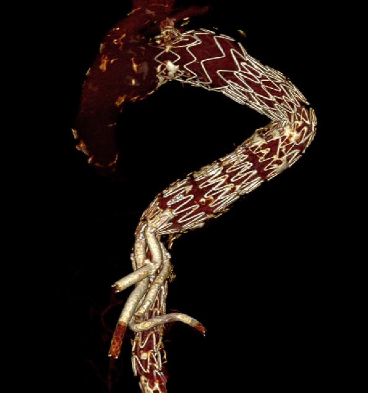
Figure 5.
Postoperative 3D CT reconstruction after TBE implantation combined with a distal straight thoracic stent-graft module.
Figure 5.
Postoperative 3D CT reconstruction after TBE implantation combined with a distal straight thoracic stent-graft module.
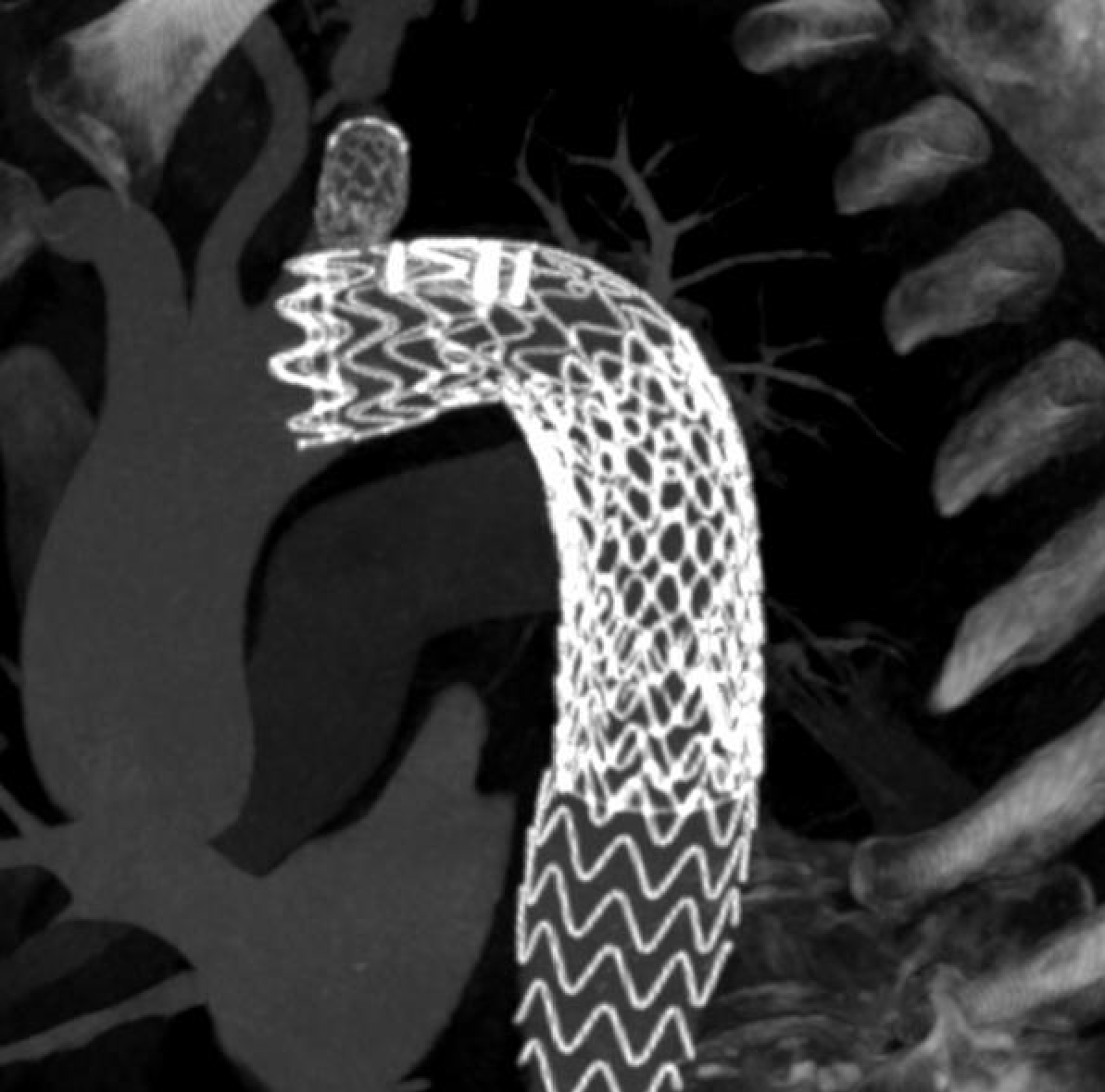

Table 1.
The table summarizes the diameters and lengths covered by the different device configurations, together with the delivery-system profiles required for implantation.
Table 1.
The table summarizes the diameters and lengths covered by the different device configurations, together with the delivery-system profiles required for implantation.
| Parameter | Castor / Cratos | TBE |
| Intended proximal aortic treatment range | 22–41mm | 16–42 mm |
| Intended distal aortic treatment range | 16–41mm | 16–42 mm |
| Tapering within branched component | Up to 8 mm | Not available |
| Branched component length | 100–210 mm | 100/150/200 mm |
| Proximal branch offset / short-zone capability | 5–30 mm | Longer portal-based geometry required |
| LSA diameter covered | 5–13 mm (branch 6-14 mm) | 5–18 mm (side branch 8-20 mm) |
| Prevertebral / branch landing length | 25–30 mm (European Market) | 25–30 mm |
| Internal portal options | - | 8 or 12 mm (14F) |
| LCCA–LSA spacing required | ≥5 mm | ≥15 mm short distance ≥20 mm long distance |
| LSA–LVA distance required | >25 mm | >25 mm |
| Aortic delivery profile | 24F Castor; 22F Cratos up to 34 mm | 20–26F |
| Upper-extremity access | 7-8F | 4-5F |
Table 2.
Implantation-outcome studies included in the review and their key findings.
| Ref | Device | Study type | N | Main pathology | Main message |
| [3] | Castor | Single-center retrospective |
26 | TBAD with insufficient anchoring region |
100% technical success; favorable early safety in very short proximal landing anatomy |
| [4] | Castor | Single-center retrospective |
41 | TBAD without proximal landing zone | Short proximal landing predicts bird-beak and early adverse events |
| [44] | TBE vs standard TEVAR |
Single-center retrospective |
65 | BTAI1 | TBE reduced early complications versus standard TEVAR with subclavian coverage |
| [24] | TBE | Multicenter retrospective |
107 | Acute aortic pathology |
Broad acute applicability; acceptable 30-day outcomes, but RTAD2 observed in acute dissection subset |
| [35] | TBE | Single-center retrospective |
20 | Acute aortic pathology |
100% technical success; no 30-day aortic mortality |
| [36] | TBE | Single-center retrospective |
20 | Mixed | 100% technical success; frequent distal extension; no 30-day mortality/stroke/ SCI3 |
| [5] | Castor | Multicenter prospective |
73 | TBAD | Landmark long-term prospective study; durable 6-year survival and branch patency |
| [30] | TBE | Single-center retrospective |
12 | Mixed | Reiterates IFU-driven anatomy; early clinical outcomes acceptable |
| [19] | Castor | Single-center retrospective |
150 | TBAD | Large midterm dissection cohort; strong patency and acceptable RTAD2/stroke rates |
| [16] | Cratos | Multicenter prospective |
89 | TBAD | Second-generation platform improves profile and maneuverability; promising 12-month results |
| [20] | Castor | Multicenter retrospective |
180 | TBAD | Large contemporary dissection cohort with excellent technical success |
| [6] | TBE | Multicenter prospective |
40 | Aortic arch aneurysm |
Strongest TBE durability study; 3-year branch patency 93% |
| [27] | Castor vs standard TEVAR |
Single-center retrospective |
73 | TBAD | Lower complications, reintervention, and displacement with Castor |
| [17] | TBE | Single-center retrospective |
55 | Mixed | Broad real-world use; frequent adjuncts and non-negligible endoleak rate |
| [18] | Cratos | Case report | 1 | Late TEVAR migration/ aneurysm |
First Western Cratos use; highlights maneuverability and profile advantages |
| [33] | Castor + STABILISE4 | Case series | 3 | TBAD | Effective integration with STABILISE |
| [25] | TBE | Single-center retrospective |
40 | Mixed | Strong branch patency but frequent distal extensions and cuffs |
| [21] | Castor | Multicenter retrospective |
106 | Mixed | Good 2-year survival and branch patency; some zone 1 extensions |
| [46] | Castor | Single-center retrospective |
10 | Mixed | First Italian experience; excellent early results but highly selected, custom-made cohort |
| [11] | Castor | Multicenter retrospective |
23 | Mixed | Largest contemporary European series; 96% technical success, 95% branch patency, no 30-day MAE5, but 23% bird-beak and two distal SINE6-related reinterventions |
| [7] | TBE vs TEVAR+ CSB7 |
Single-center retrospective |
125 | Mixed | Strong real-world evidence favoring TBE over hybrid bypass in zone 2 |
| [37] | TBE | Single-center retrospective |
5 | Acute aortic pathology |
Feasible in urgent cases, though all needed distal extension |
| [22] | Castor | Multicenter retrospective |
32 | TBAD | Long follow-up with strong branch patency and remodeling |
| [23] | Castor | Meta-analysis | 415 | TBAD | Best pooled Castor dataset, but low certainty evidence |
| [29] | Castor | Single-center retrospective |
29 | TBAD | Good early safety and remodeling analysis |
| [9] | Castor vs chimney vs fenestration | Single-center retrospective |
133 | Acute aortic pathology |
Similar broad outcomes; technique-specific complication profiles |
| [45] | Castor | Multicenter retrospective |
21 | Mixed | Encouraging but includes RTAD and bird-beak; reinforces need for careful Western appraisal |
1 Blunt thoracic aortic injury; 2 Retrograde type A dissection; 3 Spinal cord ischemia; 4 Stent-Assisted Balloon-Induced Intimal Disruption and Relamination of Aortic Dissection;5 Major adverse event; 6 Stent graft–induced new entry; 7 Carotid-Subclavian bypass.
Table 3.
Feasibility studies included in the review and their key findings.
| Ref. | Device(s) | Population | N | Main feasibility finding |
| [26] | Castor vs TBE | Acute TBAD | 100 | Castor suitability 82% vs TBE 22% off-the-shelf; tapering is central |
| [32] | Castor | Prior zone 2 TEVAR cohort | 72 | Feasibility 68.1%; large LSA diameter major cause of exclusion |
| [31] | TBE | BTAI1 | 66 | Only ~56% met IFU criteria in trauma |
| [28] | TBE | Mixed pathology needing zone 2 seal | 93 | AF 92%, true feasibility 85%; iliac access and sex differences important |
1 Blunt thoracic aortic injury.
Table 4.
Main advantages and limitations of the different devices.
| Domain | Castor / Cratos: main advantages | Castor / Cratos: main limitations | Gore TAG TBE: main advantages | Gore TAG TBE: main limitations |
| Evidence base | Broader and older overall literature, with the most mature dedicated evidence in TBAD, including prospective multicenter and long-term data. | Most evidence is dissection-centered and predominantly Chinese; Western and European series remain smaller, more heterogeneous, and generally shorter in follow-up. | Strong Western evidence base, including prospective midterm data in aortic arch aneurysms and growing real-world multicenter experience across zones 0–2. | The literature is overall younger and less mature in acute TBAD than the Castor literature; many post-commercial reports still have limited follow-up. |
| Pathology fit | Particularly well suited to TBAD, especially in short proximal landing zones and in anatomies with marked proximal-to-distal diameter mismatch. | Less extensively studied in aneurysm-predominant Western arch anatomy; branch diameter ceiling may reduce applicability in large-caliber LSAs. | Particularly attractive in distal arch aneurysm and mixed arch pathology, with strong branch durability and broad clinical applicability in Western practice. In addition, its applicability in aortic diameters as small as 16 mm may represent a specific advantage in younger trauma patients with small native thoracic aortas. | Lower pure off-the-shelf suitability in acute TBAD when strict anatomical criteria are applied, particularly because of tapering-related distal mismatch. |
| Aortic diameter treatment envelope | Proximal diameters 26–44 mm and distal diameters 20–44 mm, allowing tapered main-body configurations with up to 8 mm of tapering. | Does not extend to very small proximal aortic diameters, which may limit applicability in selected young trauma patients with small native aortas. | IFU-based aortic sizing starts from 16 mm and extends to 42 mm intended aortic diameter, which is advantageous in small, non-dilated thoracic aortas. | No true tapered branched main body; relevant proximal-to-distal mismatch often requires adjunctive distal extensions, particularly in acute dissection. |
| Short zone 2 anatomy / proximal landing requirement |
Major advantage in very short LCCA–LSA anatomies because the branch can arise very close to the proximal edge of the graft; particularly valuable in dissection. | Requires meticulous planning and precise rotational alignment to fully exploit this short-offset advantage. | Performs well in appropriately sized, more regular zone 2 anatomies and is supported by formal IFU-driven planning criteria. | Requires a longer proximal zone 2 segment than Castor/Cratos; short LCCA–LSA distance is a frequent cause of non-feasibility. |
| LSA branch diameter range |
Adequate for most dissection anatomies and standard LSA calibers . | Branch diameter is generally limited to 14 mm; larger LSAs may require adjunctive covered stenting or may be anatomically unsuitable. | Clear advantage in large LSAs, with branch options up to 20 mm and intended treated-vessel ranges up to 18 mm in the IFU. | Greater branch flexibility is obtained through a modular side-branch system rather than an integrated construct. |
| Core architecture |
Integrated unibody design with antegrade branch; conceptually behaves as a single construct and avoids an arch-level modular junction. | Castor may be technically more demanding to orient and deploy in tortuous arches. | Modular design provides versatility and broad applicability across the contemporary arch spectrum. | Inherently introduces at least one component junction even before distal extensions are added and requires implantation of a separate bridging side-branch component into the LSA. |
| Sealing and fixation concept |
The integrated antegrade branch may contribute to proximal construct stabilization and may reduce dependence on a long proximal neck | Despite this conceptual advantage, proximal seal quality still depends on anatomy and deployment accuracy; bird-beak and type Ia endoleak remain possible in hostile arches. | Excellent branch patency and reproducibility in aneurysm and mixed arch cohorts. | The retrograde branch does not contribute to proximal sealing in the same way as an integrated antegrade branch; proximal sealing requirements therefore remain more demanding. |
| Modularity / number of components | Single integrated construct at the arch level, without the need for a separate branch bridging component. | Less versatile than a modular arch platform when more proximal arch extension is required. | Major advantage in versatility: can be used across zones 0–2 and combined with adjunctive components when needed. | Modularity increases procedural steps and introduces component-related failure modes, including malalignment, junction-related endoleak, and overlap-dependent durability concerns; distal extensions are frequently required in real-world practice. |
| Maneuverability / bird-beak control | Cratos specifically improves maneuverability and delivery-system control compared with Castor and was developed to improve proximal conformability and reduce bird-beak formation. | Castor may be less forgiving during rotational alignment and arch navigation, especially in tortuous anatomy. | Familiar modular Gore platform for many arch operators and reproducible in experienced centers. | The published TBE literature has not described a dedicated bird-beak-control feature analogous to that proposed for second-generation Cratos. |
| Deployment control | Integrated design may reduce arch-level component interaction during release. Cratos further improves deployment control compared with Castor. | Castor still requires careful rotational orientation and precise branch alignment. | Standardized modular deployment strategy with well-described procedural workflow. | Accurate release requires meticulous stabilization; the IFU warns that forward or backward movement during deployment may result in inaccurate positioning, effectively creating a tendency to “jump” if wire tension, arch curvature, and delivery-system control are not optimized. |
| Femoral access profile | Castor has a stable 24F profile, and Cratos reduces this to 22F up to 34 mm, which may be advantageous in borderline iliofemoral anatomy. | Large-profile femoral access is still required, especially for classic Castor, and may remain limiting in small or diseased iliac axes. | Smaller TBE configurations may have favorable access profiles. | Large-diameter TBE configurations may require up to 26F introducers, reducing true feasibility in women and in hostile iliac anatomy. |
| Upper-extremity access | Familiar and effective branch access strategy, widely used in published Castor series. | Upper-extremity access is typically larger (around 7–8F), which may more often prompt limited surgical brachial exposure to reduce access-related complications. | Usually requires smaller upper-extremity (4-5F) working access in routine practice, facilitating percutaneous radial or brachial strategies. | - |
| Regulatory / logistic profile |
In China, Castor has effectively functioned as an off-the-shelf platform since approval by CFDA; (NMPA). In selected European markets, some common configurations may be available with relatively short lead times. | Despite this practical availability in selected settings, Castor is not approved as an off-the-shelf platform in Europe. Current European documentation still describes it as a custom-made device supplied on a named-patient basis only Cratos has so far been reported clinically under a custom-made framework rather than a standard off-the-shelf CE pathway. Not Available for sale in the United States. |
TBE is a true off-the-shelf modular platform, supported by FDA approval and CE marking. | - |
Disclaimer/Publisher’s Note: The statements, opinions and data contained in all publications are solely those of the individual author(s) and contributor(s) and not of MDPI and/or the editor(s). MDPI and/or the editor(s) disclaim responsibility for any injury to people or property resulting from any ideas, methods, instructions or products referred to in the content. |
© 2026 by the authors. Licensee MDPI, Basel, Switzerland. This article is an open access article distributed under the terms and conditions of the Creative Commons Attribution (CC BY) license (http://creativecommons.org/licenses/by/4.0/).
Copyright: This open access article is published under a Creative Commons CC BY 4.0 license, which permit the free download, distribution, and reuse, provided that the author and preprint are cited in any reuse.