

Submitted:
04 March 2026
Posted:
05 March 2026
You are already at the latest version
Abstract
Objective: The study investigates the effects of modeling liquids (MLs) on the staining of composite resins, with a focus on unichromatic resins. Materials and methods: The research was carried out by subjecting samples of monochromatic resin (2mm height x 6mm internal diameter) to the immersion protocol in coffee solution (Nescafé Tradição Forte), with color evaluation after 21 days. Results: Statistics showed that the adhesive group presented greater color change when compared to the modeling group (p = .000). There was no statistically significant difference between water and experimental staining (= 0.104). Among the staining group factors, there was a difference for ∆E in the interactions mC-mE (p = 0.004), mC-aC (p < 0.001), mC-aE (p < 0.001), cE-aC (p = 0.015), cE-aE (p = 0.007), cC-aC (p = 0.033) and cC-aE (p = 0.017). Conclusion: These results indicate the need for further clinical studies on the applicability of modeling liquids to support decision-making in clinical practice.
Keywords:
composite resin
; modeling liquids
; staining
; color
; surface properties
1. Introduction
The continuous advancement of materials and techniques in restorative dentistry aims to enhance clinical outcomes and streamline daily procedures [1]. Among these innovations, the development of composite resins and their application techniques has significantly improved the optical and mechanical properties of restorations, enabling them to better mimic the characteristics of natural teeth [2].
Despite these advancements, composite resins retain a viscous consistency that poses challenges in proper insertion, anatomical contouring, and adaptation to cavity walls. To facilitate handling, clinicians often use lubricants such as alcohol, adhesives, or modeling resins [3]. Adhesive systems, particularly non-simplified versions that lack solvents, can serve as modeling liquids by utilizing only their final hydrophobic resin layer [4,5].
Modeling liquids (MLs) have emerged as specialized materials designed to enhance the sculpting and finishing of direct restorations while reducing surface tension. Applied in small quantities to dental instruments, MLs do not significantly alter the viscosity of composite resins but improve their adaptability and reduce the incorporation of air bubbles [6,7]. These materials, composed primarily of methacrylates such as urethane dimethacrylate (UDMA), bisphenol A-glycidyl methacrylate (Bis-GMA), or triethylene glycol dimethacrylate (TEGDMA), can be applied between increments or in the final layer of restorative material to create a smoother surface and decrease working time [8,9,10,11]. This approach, commonly referred to as Direct Dental Modeling (DDM), has gained recognition for its efficiency in restorative procedures [9].
In parallel with improvements in handling techniques, monochromatic composite resins—often referred to as single-shade or “chameleon” composites—have been introduced to simplify shade selection. These materials are formulated to adapt optically to the surrounding dental tissues by modifying their apparent color based on environmental lighting and observation angle, reducing the effects of metamerism and minimizing the need for multiple shades [12,13]. However, the esthetic performance of these materials is highly dependent on surface integrity and long-term optical stability.
Color stability is a critical factor for the longevity and esthetic success of composite restorations, particularly when exposed to chromogenic challenges such as dietary beverages [14,15]. Previous studies evaluating the influence of modeling liquids and adhesives on composite resins have reported conflicting results. While some investigations suggest that modeling liquids do not significantly affect color stability [6,7], others indicate increased discoloration depending on the chemical composition of the lubricant used [14].
Although several studies have evaluated the influence of modeling liquids on the color stability of conventional multi-shade composite resins, evidence regarding their interaction with monochromatic composite resins remains scarce. Given that the optical performance of single-shade composites relies on their chameleon effect and surface integrity, surface modifications induced by modeling approaches may disproportionately affect their long-term color stability. To date, no study has specifically investigated the impact of adhesive systems and dedicated modeling resins on the color stability of unichromatic composites under staining conditions. Therefore, the present study aimed to evaluate the stability of a monochromatic composite resin finished with different modeling approaches after immersion in staining solutions over a 21-day period, providing evidence to support clinical decision-making.
2. Materials and Methods
2.1. Study Design and Sample Calculation
This study was designed as an in vitro, exploratory, controlled experimental investigation aiming to evaluate the influence of different modeling approaches on the color stability of a unichromatic composite resin. The experimental protocol was structured to ensure standardization, reproducibility, and minimization of confounding variables, following previously established methodologies for color stability assessment of resin-based composites.
The sample size (n = 6 per group) was based considering the controlled in vitro design and the magnitude of ΔE differences reported in previous studies [8]. This sample size was deemed sufficient to detect relevant differences in color variation. Independent variables were the modeling approach (no modeling liquid, modeling resin, or adhesive system used as modeling liquid) and the immersion medium (distilled water or coffee solution). The dependent variable was color change (ΔE) measured after an accelerated aging protocol.
2.2. Sample Preparation
Thirty-six disc-shaped specimens were fabricated using a unichromatic composite resin (Vittra APS Unique, FGM Dental Group, Joinville, SC, Brazil). A standardized 3D-printed mold was used to obtain specimens measuring 2 mm in thickness and 6 mm in diameter, dimensions selected to ensure uniform light penetration during polymerization and compatibility with spectrophotometric analysis.
Specimens were randomly allocated into three experimental groups according to the modeling approach employed during fabrication:
- I)
- Control group (c): composite resin inserted without the use of any modeling liquid;
- II)
- Modeling resin group (m): composite resin finished using a dedicated modeling resin (Assist Modeling APS, FGM Dental Group, Brazil);
- III)
- Adhesive group (a): composite resin finished using an adhesive system (Optibond FL, Kerr Corporation, USA) as a modeling liquid.
For all groups, the composite resin was inserted into the mold in a single increment. When applicable, the modeling liquid was applied using a fine disposable brush, and excess material was carefully removed using sterile gauze before contact with the composite resin. The modeling liquid was then used exclusively during sculpting of the final surface, simulating routine clinical practice.
A polyester strip and a glass slide were positioned over the composite surface prior to polymerization to standardize specimen thickness and surface flatness.
Polymerization was performed using a LED curing unit (Quazar, FGM Dental Group, Brazil) with an irradiance of 1000 mW/cm², verified prior to specimen fabrication. Each specimen was light-cured for 20 seconds, following the manufacturer’s recommendations.
After removal of the polyester strip and glass slide, an additional light-curing cycle was performed to ensure complete polymerization of the superficial layer. This step was applied uniformly to all specimens, regardless of the experimental group.
No finishing or polishing procedures were performed after polymerization. This decision was intentional and aimed at preserving the superficial resin-rich layer formed during specimen fabrication, which is clinically relevant when modeling liquids are used during the sculpting of composite restorations.
After fabrication, specimens were stored in distilled water at 37°C for 48 hours to allow post-polymerization reactions and stress relaxation prior to baseline color measurements.
2.3. Baseline Color Analysis
Baseline color measurements were obtained using a spectrophotometer (Vita Easyshade V, Vita Zahnfabrik, Bad Säckingen, Germany), operating according to the CIE L*a*b* color space.
Before measurement, specimens were gently rinsed with distilled water and air-dried. Each specimen was positioned consistently against a neutral background, and multiple readings were obtained to minimize measurement variability. The mean values of L*, a*, and b* coordinates were recorded for subsequent analysis.
2.4. Immersion and Storage Protocol
After baseline measurements, specimens were immersed individually in 6 mL of either distilled water or coffee solution for 21 days at 37°C, simulating accelerated aging and prolonged exposure to commonly consumed staining beverages.
The coffee solution was prepared by dissolving 15 g of coffee powder (Nescafé Tradição Forte, Nestlé, Brazil) in 500 mL of distilled water, following a standardized protocol. The solution was renewed daily to maintain staining potential throughout the experimental period.
2.5. Final Color Analysis
After completion of the immersion period, specimens were removed from the solutions, rinsed with distilled water, gently dried, and subjected to final color measurements using the same spectrophotometric protocol employed at baseline.
Color change (ΔE) was calculated using the formula: ΔE = [(ΔL)² + (Δa)² + (Δb)²]¹ᐟ².
2.6. Statistical Analysis
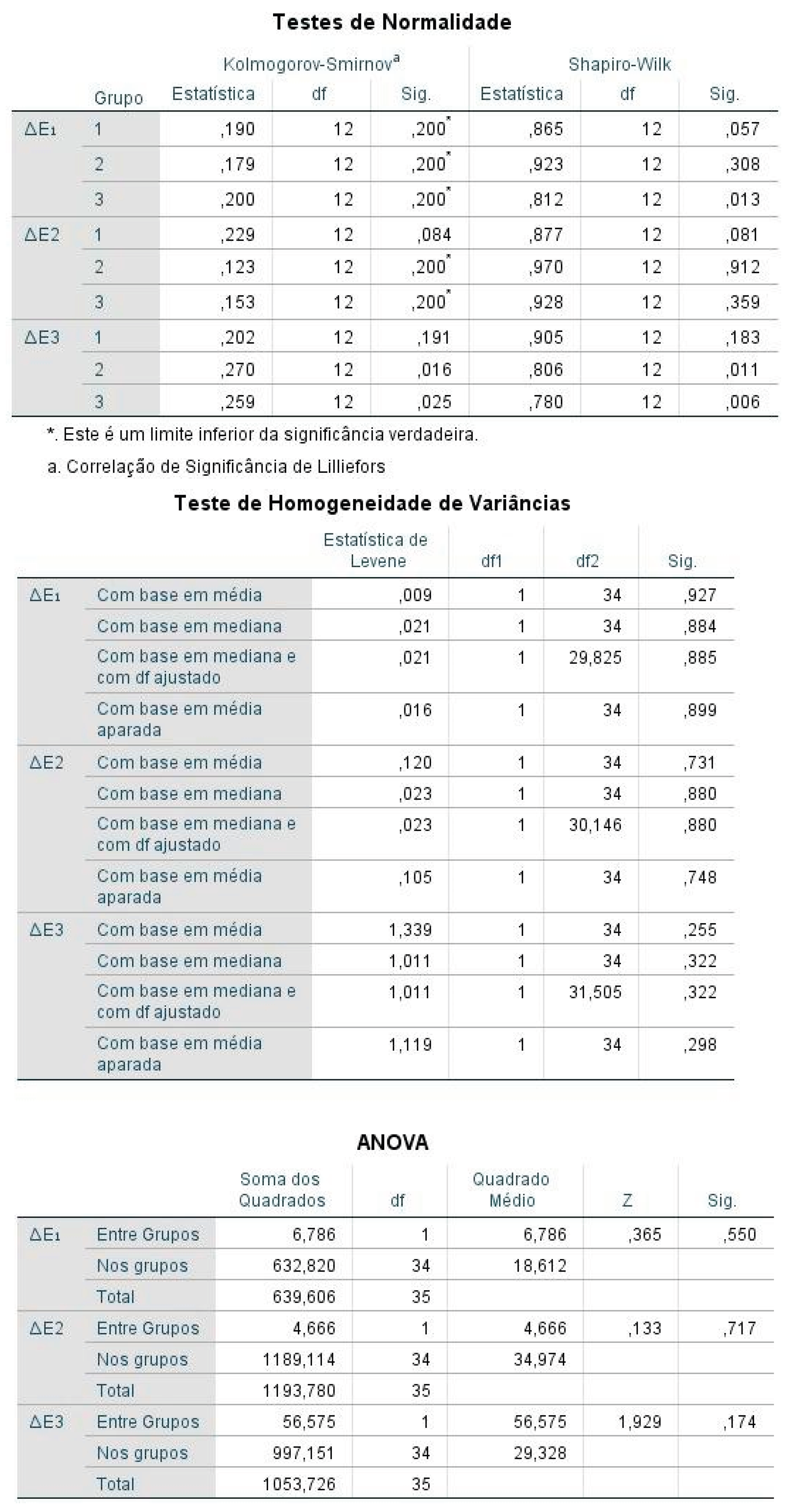
Data distribution was assessed using the Shapiro–Wilk test. As the data did not meet normality assumptions, non-parametric statistical tests were applied.
Comparisons among independent groups were performed using the Kruskal–Wallis test, while pairwise comparisons and analysis of immersion media were conducted using the Mann–Whitney U test. Statistical analyses were performed using SPSS software (version 25.5), with the significance level set at α = 0.05.
3. Results
The Kruskal–Wallis test revealed statistically significant differences in ΔE values among the experimental groups (p < 0.001). Specimens finished with adhesive used as a modeling liquid exhibited the highest color change, with mean ΔE values substantially higher than those observed in both the control group and the modeling resin group. No statistically significant difference was detected between the control group and the modeling resin group, indicating comparable color stability behavior (Table 1).
Regarding immersion media, the Mann–Whitney U test demonstrated (Table 2) no statistically significant difference between specimens stored in distilled water and those immersed in coffee solution (p = 0.104). This pattern was consistent across all experimental groups, indicating that the modeling approach exerted a greater influence on color change than the immersion medium.
Interaction analysis between modeling approach and immersion medium showed (Table 3) that specimens finished with adhesive presented higher ΔE values regardless of the storage solution. Conversely, specimens finished with modeling resin or without modeling liquid demonstrated more homogeneous ΔE distributions between distilled water and coffee immersion. Importantly, all experimental groups exhibited ΔE values exceeding the clinically acceptable threshold of 3.3.
4. Discussion
Use of modeling liquids (MLs) has become increasingly common in restorative dentistry due to their ability to improve handling, reduce stickiness, and facilitate anatomical sculpting of composite resins [20,21]. However, concerns remain regarding their potential impact on the optical stability of restorations, particularly when applied to contemporary unichromatic composite resins whose esthetic performance relies heavily on surface integrity and optical interaction with surrounding tooth structures [12,13].
The main finding of the present study was that the use of an adhesive system as a modeling liquid resulted in significantly greater color change (ΔE) compared with both the control group and the dedicated modeling resin group, regardless of the immersion medium. This result indicates that the modeling approach exerts a stronger influence on color stability than the staining solution itself. In contrast, specimens finished with a dedicated modeling resin exhibited color stability comparable to that of specimens restored without any modeling liquid, suggesting that not all lubricating agents produce detrimental optical effects [6,7].
Discoloration observed in the adhesive group may be explained by the chemical composition and hydrophilicity of adhesive systems. Adhesives commonly contain monomers such as Bis-GMA, HEMA, and TEGDMA, which are known to increase water sorption and facilitate hydrolytic degradation of the polymer network [22,23]. Bis-GMA, in particular, exhibits higher susceptibility to water uptake due to its hydroxyl groups, promoting matrix swelling and microstructural changes over time [27]. Additionally, the presence of HEMA increases the hydrophilic character of the adhesive layer, further enhancing water diffusion and pigment penetration [24]. These phenomena likely contributed to the increased ΔE values observed in specimens finished with adhesive used as a modeling liquid.
From a material science perspective, the interaction between modeling agents and the superficial resin matrix plays a critical role in determining long-term optical behavior. Previous studies have demonstrated that the superficial resin-rich layer formed during composite placement is more susceptible to water sorption and solubility, especially when enriched with low-molecular-weight or hydrophilic monomers [11,16]. The presence of this layer may accelerate hydrolytic degradation, leading to changes in refractive index and increased light scattering at the resin–filler interface, which ultimately manifests as clinically perceptible color alteration [28,29]. These mechanisms may be particularly relevant in unichromatic composite resins, where optical blending is highly dependent on surface continuity and translucency rather than intrinsic pigmentation [12,31].
Another factor that may contribute to the increased discoloration observed when adhesive systems are used as modeling liquids is the potential elution of unreacted monomers from the superficial layer. Studies evaluating the chemical composition and elution behavior of dental adhesives have shown that residual monomers such as Bis-GMA, TEGDMA, and UDMA may be released over time, especially under aqueous storage conditions [26,27]. This elution process not only weakens the polymer network but may also increase surface porosity, facilitating pigment absorption and intrinsic staining [22,23]. In contrast, materials formulated specifically as modeling resins tend to present a more homogeneous polymer structure, which may explain their comparatively improved color stability observed in the present study [6,9].
From a clinical standpoint, the findings of this investigation highlight the importance of selecting modeling strategies that balance handling convenience with long-term esthetic performance. Although adhesive systems may offer short-term benefits during sculpting procedures, their chemical composition and hydrophilic nature may compromise the optical stability of restorations over time [7,21]. This consideration is especially critical when working with single-shade composite resins, whose success relies on sustained optical integration with surrounding tooth structure [12,13]. Therefore, clinicians should be aware that material choices made during the modeling phase may have a direct impact on restoration longevity and esthetic predictability, reinforcing the need for evidence-based selection of modeling agents in daily practice [6,10].
Conversely, the dedicated modeling resin evaluated in this study is primarily composed of UDMA-based monomers, which have been associated with lower water sorption and improved color stability when compared with Bis-GMA–rich formulations [27]. The absence of highly hydrophilic monomers may explain the more favorable optical behavior observed in this group. These findings corroborate previous reports indicating that the chemical nature of the lubricant plays a decisive role in determining the long-term esthetic performance of composite resins [6,9,10].
Interestingly, no statistically significant difference was observed between distilled water and coffee immersion for any of the experimental groups. This finding contrasts with several studies reporting pronounced staining effects associated with coffee exposure [26,27]. However, the absence of polishing in the present study may explain this result. The superficial resin-rich layer formed during specimen fabrication was intentionally preserved to evaluate the direct influence of modeling liquids on the restoration surface [11]. This unpolished surface is more susceptible to water sorption and intrinsic color changes, which may have masked differences between staining solutions. Clinically, this approach is relevant, as modeling liquids are often applied during sculpting of the final increment, and the superficial layer may remain partially unmodified until finishing procedures are performed.
Despite differences among modeling approaches, all experimental groups exhibited ΔE values exceeding the commonly accepted clinical acceptability threshold of 3.3 [25]. This result highlights the vulnerability of unpolished composite surfaces to optical degradation, even in the absence of aggressive staining agents. Light scattering at the resin–filler interface, matrix degradation, and changes in refractive index resulting from water uptake may collectively contribute to the observed discoloration [28,29]. These mechanisms are particularly critical in unichromatic composite resins, whose chameleon effect depends on precise optical continuity between the restoration surface and surrounding dental tissues [12,13].
The findings of this study are especially relevant in the context of monochromatic composite resins. Unlike conventional multi-shade composites, single-shade materials rely on their ability to adapt optically to adjacent tooth structures, making them more sensitive to surface alterations [12,31]. Any modification of the superficial resin layer—whether chemical or topographical—may compromise this adaptive behavior and negatively affect esthetic outcomes over time. While previous investigations have predominantly focused on conventional composites [6,7,14], the present study addresses an important gap by evaluating the interaction between modeling approaches and unichromatic composites.
Differences between the present findings and previous studies reporting minimal color changes associated with adhesive-based modeling may be attributed to methodological variations, including polishing protocols, immersion periods, and adhesive composition [6,7,31]. Studies incorporating surface polishing may have removed the resin-rich superficial layer, thereby reducing the impact of hydrophilic monomers on color stability [11]. Additionally, variations in adhesive formulations—particularly regarding solvent content and monomer composition—may account for discrepancies in reported outcomes [24,30].
Within the limitations of this in vitro investigation, the results suggest that the indiscriminate use of adhesive systems as modeling liquids should be approached with caution, particularly when working with unichromatic composite resins. Dedicated modeling resins appear to offer a more stable alternative, preserving optical properties while maintaining the handling benefits sought during restorative procedures.
5. Conclusion
Within the limitations of this in vitro study, it was concluded that adhesive systems used as modeling liquids resulted in greater color change of unichromatic composite resin, whereas the dedicated modeling resin demonstrated better color stability. These findings highlight the importance of appropriate selection of modeling approaches to ensure long-term esthetic success of direct composite restorations.
References
- Borgia, E; Baron, R; Borgia, JL. Quality and Survival of Direct Light-Activated Composite Resin Restorations in Posterior Teeth: A 5- to 20-Year Retrospective Longitudinal Study. J Prosthodont. 2019, 28, e195–203. [Google Scholar] [CrossRef] [PubMed]
- Pfeifer, CS. Polymer-Based Direct Filling Materials. Dent Clin North Am. 2017, 61, 733–50. [Google Scholar] [CrossRef] [PubMed]
- Kosewski, J; Kosewski, P; Mielczarek, A. Influence of Instrument Lubrication on Properties of Dental Composites. Eur J Dent. 2022, 16, 719–28. [Google Scholar] [CrossRef] [PubMed]
- Mendes, JL; Paiva, PR da S; Vasconcelos, RG; Vasconcelos, MG. Modeladores de Resina Composta: Uso e Repercussões Clínicas. Arch Heal Investig. 2021, 11, 506–12. Available online: https://www.archhealthinvestigation.com.br/ArcHI/article/view/5563.
- Mendes de Araujo Aquino, MP; De Castro, CA; Pereira do Carmo, GP; Lima, LF; Ribeiro Amaral, ML; Martins Mendes, GA. Avaliação Da Resistência Mecânica De Resina Composta Incorporada Com Líquidos Modeladores. Sci Investig Dent. 2021, 26, 76–84. [Google Scholar] [CrossRef]
- Sedrez-Porto, JA; Münchow, EA; Brondani, LP; Cenci, MS; Pereira-Cenci, T. Effects of modeling liquid/resin and polishing on the color change of resin composite. Braz Oral Res. 2016, 30, 1–9. [Google Scholar] [CrossRef]
- Araujo, FS; Barros, MCR; Santana, MLC; de Jesus Oliveira, LS; Silva, PFD; Lima, G da S; et al. Effects of adhesive used as modeling liquid on the stability of the color and opacity of composites. J Esthet Restor Dent. 2018, 30, 427–33. [Google Scholar] [CrossRef]
- Krajangta, N; Ninbanjong, S; Khosook, S; Chaitontuak, K; Klaisiri, A. Effects of Immediate Coating on Unset Composite with Different Bonding Agents to Surface Hardness. Eur J Dent. 2022, 16, 828–32. [Google Scholar]
- Münchow, EA; Sedrez-Porto, JA; Piva, E; Pereira-Cenci, T; Cenci, MS. Use of dental adhesives as modeler liquid of resin composites. Dent Mater. 2016, 32, 570–7. [Google Scholar] [CrossRef]
- de Paula, FC; de S, Valentin R; Borges, BCD; Medeiros, MC dos S; de Oliveira, RF; da Silva, AO. Effect of Instrument Lubricants on the Surface Degree of Conversion and Crosslinking Density of Nanocomposites. J Esthet Restor Dent. 2016, 28, 85–91. [Google Scholar] [CrossRef]
- Sedrez-Porto, JA; Münchow, EA; Valente, LL; Cenci, MS; Pereira-Cenci, T. New material perspective for endocrown restorations: Effects on mechanical performance and fracture behavior. Braz Oral Res. 2019, 33. [Google Scholar] [CrossRef] [PubMed]
- Pimenta, C; Guimarães, A; Melquiades Barbosa De Oliveira, E; Da, A; Torres, S; Pereira Isolan, C; et al. Resinas compostas monocromáticas e seu efeito camaleão: uma revisão sistemática Monochrome composite resins and their chameleon effect: a systematic review. Periódico não identificado.
- Anwar, RS; Hussein, YF; Riad, M. Optical behavior and marginal discoloration of a single shade resin composite with a chameleon effect: a randomized controlled clinical trial. BDJ Open, 2024; 10. [Google Scholar]
- Aydın, N; Karaoğlanoğlu, S; Ersöz, B. Effect of Modeling Liquid Use on Color and Whiteness Index Change of Composite Resins. Cumhur Dent J. 2022, 25, 119–23. [Google Scholar] [CrossRef]
- Melo, AM dos S; dos Santos, TJS; Tertulino, MD; Medeiros, MC dos S; da Silva, AO; Borges, BCD. Degree of conversion, translucency and intrinsic color stability of composites during surface modeling with lubricants. Braz J Oral Sci. 2018, 17. [Google Scholar] [CrossRef]
- Schmitt, VL; Naufel, FS; Pardo, F; Nahsan, S; Baseggio, W; Camilotti, V; et al. Avaliação da sorção e solubilidade de uma resina composta em diferentes meios líquidos. Rev Odontol UNESP 2011, 10, 265–9. [Google Scholar]
- Baldissera, RA. Programa de Pós-Graduação em Odontologia; Tese, 2012; pp. 1–83. [Google Scholar]
- de Almeida, L; Santin, DC; Maran, BM; Naufel, FS; Schmitt, VL. Avaliação do manchamento e da rugosidade superficial de materiais restauradores diretos após diferentes sistemas de polimento: estudo in vitro. Rev Odontol UNESP 2019, 48. [Google Scholar] [CrossRef]
- Ozera, EH; Pascon, FM; Correr, AB; Puppin-Rontani, RM; de Castilho, AR; Correr-Sobrinho, L; et al. Color stability and gloss of esthetic restorative materials after chemical challenges. Braz Dent J. 2019, 30, 52–7. [Google Scholar] [CrossRef]
- Santos, ER; dos; Möller, L. Influência do líquido modelador na resistência à compressão de duas resinas compostas: estudo in vitro. Rev Odontol Bras Cent. 2023, 32, 200–12. [Google Scholar] [CrossRef]
- Maalekipour, M; Safari, M; Barekatain, M; Fathi, A. Effect of Adhesive Resin as a Modeling Liquid on Elution of Resin Composite Restorations. In Int J Dent; 2021. [Google Scholar]
- Paolone, G; Mazzitelli, C; Zechini, G; Scolavino, S; Goracci, C; Scotti, N; et al. Influence of Modeling Liquids and Universal Adhesives Used as Lubricants on Color Stability and Translucency of Resin-Based Composites. Coatings 2023, 13. [Google Scholar] [CrossRef]
- Silva, EDEM. Evelyn De Moura Silva. Documento não periódico 2023. [Google Scholar]
- Paravina, R.D.; Perez, M.M.; Ghinea, R. Acceptability and perceptibility thresholds in dentistry: A comprehensive review of clinical and research applications. J. Esthet. Restor. Dent. 2015, 27, S1–S9. [Google Scholar] [CrossRef] [PubMed]
- Van Landuyt, KL; Snauwaert, J; De Munck, J; Peumans, M; Yoshida, Y; Poitevin, A; et al. Systematic review of the chemical composition of contemporary dental adhesives. Biomaterials 2007, 28, 3757–85. Available online: https://linkinghub.elsevier.com/retrieve/pii/S0142961207003596. [CrossRef]
- Sideridou, I.; Achilias, D.S. Elution study of unreacted Bis-GMA, TEGDMA, UDMA, and Bis-EMA from dental resins and resin composites. J. Biomed. Mater. Res. B Appl. Biomater. 2005, 74, 617–626. [Google Scholar] [CrossRef]
- Kerpel, F; Bamberg, AC; Nicoloso, GF; Miotti, LL; Dalmolin, A; Swarowsky, LA; et al. Effect of different placement techniques on color stability and surface roughness of resin composites. Rev Odontol UNESP 2021, 50, 1–10. [Google Scholar] [CrossRef]
- Lopes E dos, S; Linhares, TS; Garone-Netto, N; Lago, ADN. Avaliação do efeito de bebidas quanto ao manchamento de resinas compostas. Rev Pesq Saúde 2016, 17, 147–50. [Google Scholar]
- Ribeiro, J; Jorge, P. Em função de composição e sistema de fotoativação – avaliação através de [informação incompleta]. 2006. [Google Scholar]
- Esvercutti, RV; Evaristo Pantolfi, AC; Felizardo, KR. Resinas Unicromáticas - Uma Alternativa Para a Estratificação De Cor Em Restaurações De Resina Composta - Relato De Caso Clínico. Braz J Implantol Heal Sci. 2023, 5, 2058–82. [Google Scholar] [CrossRef]
- Cubukcu, I; Gundogdu, I; Gul, P. Color match analysis of single-shade and multi-shade composite resins using spectrophotometric and visual methods after bleaching. Dent Mater J. 2023, 42, 2023–115. Available online: https://www.jstage.jst.go.jp/article/dmj/42/6/42_2023-115/_article.

Table 1.
Color evaluation (∆E) in relation to groups.
| Groups | Average | Median | DP | AI | p | |
|---|---|---|---|---|---|---|
| c | 10,25 a | 9,67 | 2,66 | 3,74 | ||
| ∆E | m | 10,10 a | 7,49 | 5,20 | 10,70 | ,000 |
| a | 18,34 b | 17,37 | 3,62 | 3,01 | ||
*Different lowercase letters indicate statistically different results.
Table 2.
Color evaluation (∆E) in relation to staining.
| Staining | Average | Median | DP | AI | p |
|---|---|---|---|---|---|
| C | 11,64 | 9,88 | 6,09 | 10,54 | |
| ∆E | ,104 | ||||
| E | 14,15 | 15,53 | 4,63 | 7,30 |
Table 3.
Color evaluation (∆E) in relation to group X staining.
| GxM | Average | Median | DP | AI | p | |
|---|---|---|---|---|---|---|
| cC | 10,68 a | 9,88 | 9,88 | 4,71 | ||
| cE | 7,46 a | 9,67 | 2,24 | 4,41 | ||
| ∆E | mC | 5,90 a | 5,75 | ,91 | 1,12 | |
| mE | 14,31 b | 16,03 | 4,03 | 6,84 | ,000 | |
| aC | 18,35 b | 17,23 | 4,53 | 4,73 | ||
| aE | 18,33 b | 17,76 | 2,88 | 4,21 |
*Different lowercase letters indicate statistically different results.
Disclaimer/Publisher’s Note: The statements, opinions and data contained in all publications are solely those of the individual author(s) and contributor(s) and not of MDPI and/or the editor(s). MDPI and/or the editor(s) disclaim responsibility for any injury to people or property resulting from any ideas, methods, instructions or products referred to in the content. |
© 2026 by the authors. Licensee MDPI, Basel, Switzerland. This article is an open access article distributed under the terms and conditions of the Creative Commons Attribution (CC BY) license (http://creativecommons.org/licenses/by/4.0/).
Copyright: This open access article is published under a Creative Commons CC BY 4.0 license, which permit the free download, distribution, and reuse, provided that the author and preprint are cited in any reuse.