
Submitted:
21 January 2026
Posted:
22 January 2026
You are already at the latest version
Abstract
The objective of this study was to synthesize silver nanoparticles (AgNPs) utilizing an eco-friendly approach with Ocimum lamiifolium leaf extract as a biological reducing agent. The research focused on investigating how various experimental conditions influenced the stability and particle size of the AgNPs. The characterization of the synthesized nanoparticles involved multiple techniques, including X-ray diffraction (XRD), scanning electron microscopy (SEM), Fourier-transform infrared spectroscopy (FTIR), UV-visible spectroscopy, particle size analysis, Polydispersity Index (PDI), and zeta potential measurements. During the reduction process, a noticeable color change from colorless to grey indicated successful conversion of Ag+ to Ag°. The UV-vis spectra revealed a maximum absorption at 467 nm, confirming nanoparticle formation. The average particle size was found to be 65.37 nm with a PDI of 0.241, suggesting a relatively uniform size distribution. The zeta potential was measured at -29.85 mV, indicating good colloidal stability over time. FTIR analysis identified various functional groups associated with phytochemicals, supporting the role of plant compounds in reduction and stabilization. XRD patterns confirmed the face-centered cubic (FCC) crystalline structure of the AgNPs. Furthermore, antibacterial testing showed increased inhibitory zones with higher AgNP concentrations, with the minimum inhibitory zone of 4 mm and maximum of 15.45 mm against E. coli. The green synthesis mechanism involved reduction, stabilization, membrane disruption, reactive oxygen species (ROS) production, and bacterial cell death. Overall, AgNPs produced via Ocimum lamiifolium extract demonstrated enhanced stability and effective antibacterial activity, highlighting the potential of plant-based green synthesis methods for biomedical applications.
Keywords:
antibacterial activities
; particle size
; reducing agents
; silver nanoparticles
; Ocimum lamiifolium leaves extract
1. Introduction
The production of nanoparticles (NPs) employing safe, dependable, non-toxic, and environmentally sustainable processing methods has been the focus of substantial research and development efforts in recent years [1]. These advancements aim to enhance the utilization of nanoparticles and address pressing needs, particularly in the biomedical sector [2]. It has been proven that appropriate NPs can be utilized for various chemical and biological applications due to their unique physicochemical characteristics [3]. Green synthesis methods, which offer practical alternatives to conventional chemical processes, include microbial, enzymatic, and plant-based techniques [4,5,6]. The integration of NPs with enhanced safety profiles and little environmental effect into pharmaceutical and therapeutic applications is made possible by these activities.
Biosensors, water treatment, biotechnology, biomolecules, electronics, medication delivery, catalysis, textiles, and bioengineering are just a few of the numerous applications of NPs [7]. NPs are classified into various categories, including organic, biological, hybrid, and inorganic materials [8,9,10,11,12]. Among these, inorganic metallic nanoparticles (MNPs) such as those of silver, gold, nickel, platinum, zinc, and iron, as well as their oxide forms, have garnered considerable attention [13]. Particularly, since their sizes, forms, and compositions are closely related to their physical, chemical, and optical properties, nanoscale materials with a size range of 1 to 100 nm have garnered substantial attention across various sectors, from industrial to therapeutic applications [14]. Due to their superiority in nanomedicine applications, particularly cancer diagnostics and therapy, silver nanoparticles (AgNPs), one of the most important and exciting nanomaterials among the extensively researched NPs, have recently drawn a lot of attention [15,16]. AgNPs play a significant role in various applications beyond medicine.
AgNPs have been thoroughly investigated by numerous researchers in recent years for the production of antimicrobial textiles, nano-silver-containing polymers, freezers, dishwashing machines, plastic films, and the removal of dyes from wastewater. However, regulating the size and distribution of the NPs during synthesis is an important step that can be accomplished by selecting an acceptable synthesis process, efficient reducing agents, and suitable stabilizers [17,18]. AgNPs have been observed in various shapes, including spheres, disks, rods, and prisms, with different sizes.
AgNPs can be generally produced using three distinct methods: chemical, biological, and physical methods [19,20,21]. However, certain disadvantages of physical approaches have been noted, including the need for costly equipment, the need to sustain high temperatures, the need for a large amount of space, the lengthy process, and the wide range of sizes and forms of the resultant particles [19]. Chemical procedures, another main strategy, are less expensive, but they still require high working temperatures and utilize hazardous and toxic substances that are severely harmful to the environment [22]. Therefore, it is not difficult to select production conditions that result in nanoparticles of the required size. For the reasons outlined above, physical and chemical processes are considered irrelevant in terms of their environmentally beneficial qualities [10]. The biological technique is receiving a lot of interest from the research community as they hunt for ways to synthesize AgNPs that are non-hazardous, non-toxic, rapid, economical, require less temperature, cost-effective, biocompatible, ecologically friendly, and sustainable. As a result, the biological process, which has been investigated and documented by multiple researchers for the synthesis of AgNPs, can be regarded as a green approach [23,24]. Plant-based extracts have been employed as reducing agents to prepare AgNPs for biosynthesis.
Numerous plant extracts have been investigated thus far as biological reducing agents for the preparation of AgNPs. Cinnamomum camphora, banana peel, Caccinia macranthera, Rosmarinus officinalis leaf extract, Artemisia kopetdaghensis, Chenopodium murale, Gmelina arborea, and Capsicum annuum L. were found to be noteworthy for acting as potential reducing agents to produce NPs, according to other studies. Although extracts from Aloe vera, Salvia spinosa, aqueous saffron, Cynodon dactylon leaves, Streptomyces sp., Camellia sinensis, Vernonia amygdalina, Azadirachta indica aqueous leaf extract, and Acalypha indica leaf were used to make AgNPs [25,26]. Flavonoids, polyphenols, proteins, alkaloids, phenols, amino acids, saponins, steroids, triterpenoids, glycosides, and tannins are among the metabolites found in extracts from various parts of these plants that are primarily responsible for the reduction of metallic ions into MNPs. In the case of AgNPs, a redox reaction has occurred, reducing Ag+ to Ag0. In addition, the plant extract serves as a capping agent for the biosynthesized nanoparticles [27,28]. AgNPs become more stable as a result of these phytochemicals.
Ocimum lamiifolium is an erect, hairy, perennial, vigorous branching shrub or herb that can reach a height of 3 m. It is a member of the genus Ocimum within the family Lamiaceae. It thrives in grasslands between 1,200 and 2,900 m, near roads and streams, in wilderness areas, and on the edge of forests. Ocimum lamiifolium has long been utilized in traditional medicine across the globe. It is used to treat conditions like dropsy, asthma, gastric ulcers, hepatitis, and gonorrhea. The plant is commonly known as "dama-kassie" in Ethiopia. Crushed Ocimum lamiifolium leaves are used to stop nose bleeding, while fresh leaves are pressed and inhaled to cure colds and coughs and as an eye rinse for eye infections. Additionally, it is used to treat infections and inflammatory diseases as well as illnesses like pyrexia, eye disease, cough, cold, cutaneous leishmaniasis, headache, and herpes (kusil). Traditional methods of treating Mich include squeezing and sniffing fresh Ocimum lamiifolium leaves [29]. The Ethiopian Ocimum lamiifolium crude leaf extract was shown to be rich in many phytochemicals, including alkaloids, tannins, saponins, flavonoids, terpenoids, cardiac glycosides, anthraquinones, and phenols, according to the GC-MS data for the antibacterial impact, based on the research have done by Sahalie, Abrha [30]. In light of this, no research was done to examine the synthesis of AgNPs utilizing extract from a variety of Ethiopian Ocimum lamiifolium leaves for the use of antibacterial activities. Furthermore, XRD, FTIR, dynamic light scattering (DLS), Zeta potential, ultraviolet-visible (UV-Vis) spectroscopy, and antibacterial activity were used to characterize the produced AgNPs.
2. Materials and Methods
2.1. Materials and Chemicals
In this experiment, distilled water and silver nitrate (AgNO3, 98.5%) were purchased from UDYOG (India), and Whatman filter paper (No.1, 90 mm, UK). Bio and Emerging Technology Institute (BETin) provided barium chloride and Mueller-Hinton agar. The Ethiopian Public Health Institute provided the gram-negative E. coli (ATCC 25972) bacteria. The Akaki, Ethiopia, provided the fresh leaves of the Ocimum lamiifolium plant. Ocimum lamiifolium leaves were cleaned with distilled water and shade-dried for a week in order to prepare the extract.
2.2. Synthesis of AgNPs Using Ocimum lamiifolium Extract
AgNPs were produced using a biological reduction process using a modified version of the Asif, Yasmin [31] and Tessema, Gonfa [25]. Ocimum lamiifolium leaf extract was used to make the AgNPs. The leaves were first cleaned with distilled water and allowed to dry in the shade for a week. A coffee grinder was then used to grind the dried leaves. Next, 20 g of the leaf powder and 200 mL of distilled water were added to a 250 mL conical flask, which was then heated for 20 minutes at 90 ± 5 °C. The solution was then vacuum-filtered through Whatman filter paper (No. 1, 90 mm, UK) and kept in a refrigerator at 4 °C until the AgNPs were synthesized. ` Ocimum lamiifolium leaf extract (10 mL) was added to four flasks containing 100 mL of distilled water and varying concentrations of AgNO3 (1.5, 3, 6, and 9 mM) in order to synthesize AgNPs. The flasks were set on a hot plate, and the water in the vessel was continuously stirred magnetically for 15 minutes to maintain a temperature of 65 ± 5 °C. The color of the solution changed from colorless to light gray, indicating the production of AgNPs. The flasks were taken off the heating plates after the color shift was seen. The flasks were kept under a magnetic stirrer while the solutions cooled to room temperature in order to prevent AgNPs from clumping together. The samples were then subjected to ultrasonic waves (Sonorex Digiplus DLUltrasonic Baths, Bandelin, Germany) for 15 minutes after being chilled at 4 °C for 24 h. The AgNPs reduction process was successful when the solutions were cooled and agitated until they turned into a light grey colloidal solution [32]. AgNPs synthesis using plant leaf extract is a more cost-effective, dependable, and ecologically friendly method. The detailed procedure for making leaf extract, which is used to create silver nanoparticles, is shown in Figure 1. As shown in panel (a), the process begins with gathering fresh Ocimum lamiifolium leaves. After a thorough washing to remove any dirt or contaminants, the leaves are dried and then ground into a fine powder, as shown in panel (b). As shown in panel (c), this leaf powder is further processed by grinding to expand the surface area, enabling effective extraction of bioactive chemicals. Utilizing the phytochemicals found in Ocimum lamiifolium leaves, this extract acts as a natural reducing and stabilizing agent in the synthesis of silver nanoparticles.
2.3. Characterization of Synthesized AgNPs
2.3.1. Droplet Size and Distribution
The size distributions of AgNPs were determined using dynamic light scattering (DLS ZEN3600, United Kingdom). After centrifuging the mixture for 15 minutes at 10,000 rpm, the diameters of the colloidal AgNPs in the suspension were measured.
2.3.2. UV-Vis Spectroscopic Analysis
UV-Vis spectroscopy (Jasco V-770, UV-Vis-JASCO Co., Tokyo, Japan) was used to measure the absorption spectra of colloidal AgNPs in the 200–800 nm range with a resolution of 1 nm. Using the technique described by Dehnavi, Raisi [33] and Tessema, Gonfa [25], UV-Vis spectroscopy was used to track the reduction of the silver ions (from Ag+ to Ag°). AgNPs colloidal spectrum analysis was conducted using distilled water as a control.
2.3.3. Zeta Potential
Through emulsion characterization utilizing zeta potential analysis, the electrostatic stability of the generated emulsions containing AgNPs based on Ocimum lamiifolium leaf extract and various emulsifiers was evaluated. Zeta potential (ZEN3600, United Kingdom) measurements using electrophoretic light scattering were used to determine the surface charge of the emulsion droplets; larger absolute values indicate stronger electrostatic repulsion and, consequently, greater stability against coalescence and phase separation. This measure is crucial for assessing how effectively combinations of emulsifiers and natural extracts maintain emulsion integrity over time [34]. In order to determine the ideal formation of AgNPs that result in maximum stability.
2.3.4. Fourier Transform Infra-Red Spectroscopy Analysis
FTIR spectra frequently offer details about the colloidal AgNPs that were synthesized. The data was recorded in the wavenumber range of 4000–400 cm-1 using an FTIR spectroscopy device (Thermo Scientific FTIR, IS50). The AgNPs-containing solutions were dried at 90 ± 5 °C for 12 h. Additionally, the dried powders were subjected to FTIR spectroscopy analysis using the attenuated total reflection technique. In this investigation, a known amount of dried AgNPs was placed on a copper grid, and the FTIR peaks were used to analyze the functional groups.
2.3.5. X-Ray Diffraction Studies
A CuK emission of acceleration voltage at 40 kV and current at 44 mA was examined using XRD analytical equipment (Model-Ultima-IV) from a scan range of 3 to 80° of 2θ at a speed of 10.0 degree/min. The colloidal AgNPs were dried out for 12 h at 90 ± 5 °C before XRD scanning was performed. One gram of dry powder sample was mashed in a mortar and pestle to create a fine, homogenous mixture. The powders of AgNPs made using both techniques of tri-sodium citrate and Ocimum lamiifolium leaf extract as a reducing agent were then scanned by XRD spectrometry.
2.4. Antibacterial Performance Evaluation of AgNPs Mixed with the Leaf Extract
The process involved preparing bacterial suspensions and culture medium for testing. 38 grams of brain-heart infusion broth were autoclaved after being dissolved in 1,000 mL of sterile distilled water. To reach the exponential phase, E. coli cultures were cultivated in this medium for 24 h at 37 °C. After that, the cells were collected, centrifuged for 5 minutes at 10,000 rpm, rinsed with sterile water, and then centrifuged one more time. They were subsequently resuspended in sterile water. A solution of 1% H2SO4 and 1% barium chloride dihydrate was used to create a 0.5 McFarland standard to standardize the bacterial concentration, which was calculated to be roughly 1.5 × 10⁸ CFU/mL. In addition, 38 g of Mueller-Hinton agar was combined with 1,000 mL of sterilized distilled water to create agar plates, which were then incubated in a biosafety cabinet. After 24 h, 100 µL of E. coli was extracted from the growth plates for testing. To create the test solutions, the bacterial culture was resuspended in sterile distilled water, sonicated, and autoclaved after different amounts (0.5, 1, 2, 4, and 8 mL) of crude. Similar studies were conducted by Magnani [35], indicating that this research builds upon or relates to prior work in the field.
Zone of Inhibition Test for E. coli
The experiment was conducted in accordance with the Clinical and Laboratory Standards Institute's (CLSI M100) guidelines, with minor adjustments made to accommodate the specific experimental conditions [36]. For 24 h, 100 µL of E. coli was cultured in the medium. Next, the synthesis of AgNPs with the range of (0.5, 1, 2, 4, and 8 mL) was done. The method described somewhere was used to sonicate and autoclave the solution [37]. The samples were placed in an autoclave, where the medium underwent autoclave sterilization for 15 min at 121 °C, followed by a shaking incubator at 150 rpm at 37 °C. Then, the sterilized medium (20 mL) was aseptically poured onto sterile, 90 mm-diameter petri dishes, cooled to a molten state, and then allowed to cool to room temperature in the sterile setting in the biological safety cabinet (BIOBASE, Chia). The bacterial strain was sub-cultured in brain-heart infusion broth for 24 h at 37 °C, and then suspended in sterile nutritional broth with the 0.5 MacFarland standard adjusted.
Sterile cotton swabs were used to swab the designated test microorganisms (100 µL each) into sterile plates containing Mueller-Hinton agar. The Mueller-Hinton agar was swabbed and then dried. Blue tips were used to punch 8-mm-diameter wells into the inoculated agar medium. Add 100 µL of each sample (0.5, 1, 2, 4, and 8 mL in solution) to the wells within 15 minutes. Similarly, the same amount of gentamycin and distilled water were pipetted for the positive and negative controls, respectively. Before being incubated at 37 °C for 24 hours for both bacterial strains, all dishes were pre-incubated at room temperature for 2 h to enable uniform diffusion of the sample solution into the agar medium. The diameter of the bacterial growth inhibition zone was measured in millimeters in order to assess the antibacterial activity [38]. Each experiment was carried out three times.
3. Results and Discussion
3.1. Particle Size Distribution Result Analysis Using DLS
The colloidal silver nanoparticles (AgNPs) produced with Ocimum lamiifolium leaf extract as a natural reducing agent are shown in Fig. 1f. During the synthesis procedure, 10 mL of the leaf extract was added to a 100 mL aqueous solution of silver nitrate (AgNO₃) at a concentration of 3 mM. Initially, this mixture was stored at room temperature. It was then heated on a hot plate while being continuously stirred by a magnetic stirrer. For around fifteen minutes, the solution was heated in a water bath kept at 65 ± 5 °C. AgNPs were formed at this time, as evidenced by a noticeable color shift from colorless to light gray. To ensure homogeneous particle production and stabilization, the mixture was then allowed to cool gradually to room temperature while still being agitated. The average size of the produced AgNPs was about 65.37 nm, according to Dynamic Light Scattering (DLS) analysis presented in Figure 2a. The size distribution showed a very narrow distribution centered on a modest particle size, ranging from as little as 1 nm to 99.2 nm. These findings imply that the phytochemicals in the leaf extract of Ocimum lamiifolium efficiently promoted the reduction of silver ions and regulated the growth of nanoparticles, resulting in the creation of AgNPs with appropriate size properties. Furthermore, Table 1 provides a comparative overview of the AgNPs obtained in this study with those reported in other research works, which employed both chemical and biological synthesis methods. This comparison highlights the influence of different synthesis strategies on nanoparticle size, distribution, and overall characteristics, emphasizing the potential of plant-based green synthesis for producing stable and moderately sized AgNPs. Similar studies were conducted by Gildas, Parfait [39]. Several natural sources, including fructose, Caccinia macranthera seed extract, Ocimum lamiifolium, Vernonia amygdalina leaf extract, and Cynodon dactylon leaves, were used to reduce AgNPs. Several methods were used to characterize the produced nanoparticles, and the results were in good agreement with those found in the literature. This study employed natural sources as efficient and sustainable reducing agents for the manufacture of silver nanoparticles [25,40]. The phytochemicals in the plant extract have an impact on nucleation and growth processes during synthesis, as evidenced by the average particle size and distribution pattern seen in this instance. Overall, these findings show how the manufacturing method used affects the size and distribution of nanoparticles, which may have an impact on their stability and biological activity.
For the synthesis of AgNPs employing reducing agents extracted from Ocimum lamiifolium leaves, the observed PDI (Polydispersity Index) of 0.241 shows a comparatively narrow size distribution, indicating a homogenous population of nanoparticles. For stable emulsions, a PdI of less than 0.3 is often preferable; nevertheless, for many applications, a value of 0.289 is an acceptable homogeneity. The findings were consistent with other studies, such as Khiavi, Heshmati [35], who investigated the use of Rosa canina and Heracleum persicum extracts in the production of mayonnaise enriched with encapsulated extracts and Tween 80 as a surfactant. The results demonstrated that the PDI of 0.258 [41], and Jain and Pandey [42] produced from nano emulsion gel containing Prosopis cineraria, Aerva javanica, and Fagonia indica extracts with a PDI of 0.259. This low PDI value indicates that the synthesis procedure was carefully managed, producing nanoparticles with uniform sizes—a critical component for guaranteeing dependable antibacterial action. In addition to providing a sustainable and environmentally friendly method, using Ocimum lamiifolium leaves as a natural reducing agent enhances the biocompatibility and efficacy of AgNPs.
3.2. UV-Vis Spectroscopy Analysis
Previous studies have demonstrated that silver ions can be reduced into AgNPs when subjected to reducing agents such chemicals or plant extracts, and a color shift can show this [43]. Figure 3 displays the prepared AgNPs' energy gap and UV-vis absorption spectra. The UV-vis absorption spectra of the colloidal AgNPs solution made for this investigation are shown in Figure 3a. The greatest absorption peak of the generated AgNPs was observed at 452 nm. These peaks show the creation of variously shaped and sized AgNPs. Table 1 displays the average particle size, the spectrum at which maximal absorption was detected, the energy gap (eV) of the current AgNPs, and the values reported for earlier studies. This outcome was in line with research on other reducing agents, including fructose, Cynodon dactylon leaves, Vernonia amygdalina leaf extract, and Caccinia macranthera seed extract. AgNPs with comparable absorption peaks were also generated by these agents [25,44]. After two months, there was no discernible change in the prepared AgNPs' stability. The outcome demonstrates that the stability of the current AgNPs is superior to that of the chemically generated AgNPs [16]. Figure 2(b) illustrates the large energy difference in the biosynthetic processes used to create NPs, as revealed by the findings. The changes in AgNPs' crystal structure brought on by plant extract may be the source of the bandgap value shift [45]. Equation 1 was used to compute the band gap energy.
where a represents the E absorbance coefficient and hv (eV) is the gap band energy. As seen in Figure 3, the bandgap energy (Eg) of AgNPs was estimated by projecting the linear portion of the plot of (h)2 versus h (eV) (a and b) to the x-axis. A is an integer constant, and n is the bandgap energy, which varies depending on whether the transition is direct (n = 2) or indirect (n = 0.5).
The average particle diameters, wavelengths at which maximum absorption was detected, and energy gaps of the produced silver nanoparticles (AgNPs) utilizing different green reducing agents across several experiments are compared in Table 1. AgNPs with an average size of 65.37 nm, maximal absorbance at 467 nm, and an energy gap of 5.344 eV were produced in the current study using Ocimum lamiifolium leaf extract. Previous works reported smaller or comparable particle sizes, such as Vernonia amygdalina leaves extract producing particles around 63.08 and 70-90 nm with a maximum absorption at 452 and 480 nm and a slightly higher energy gap of 5.34 eV [25,46]. A wider range of particle sizes have been shown in other studies using green synthesis techniques, ranging from 8–10 nm and an absorbance of 451 nm with Cynodon dactylon leaves [47] and a wider particle size range 37 nm to larger sizes up to 89 nm with Caccinia macrantherase seed extract, which showed a clear maximum absorption at 663 nm and an energy gap of 3.1 eV [45]. Overall, these results demonstrate the variation in nanoparticle properties depending on the plant extract utilized, and the present study provides important information on the synthesis of AgNPs with particular optical and electrical characteristics.
Table 1 illustrates how the effective synthesis of AgNPs is influenced by the concentration of the precursor solution (silver nitrate), the type of reducing agent utilized, and the quantity of that agent. Furthermore, a biological approach using Ocimum lamiifolium leaf extract proved to be reliable, economical, and environmentally beneficial in comparison to the chemical process.
3.3. Zeta Potential
Figure 2b illustrates the dispersion of zeta potential values in an AgNPs synthesis employing leaf extract from Ocimum lamiifolium. The zeta potential, which was measured up to 60 days after storage, showed that the particles in the colloid had a significantly negative surface charge. In this study, the final zeta potential result was -29.85. The strong electrostatic repulsion between the particles, which prevents dispersed droplets from aggregating and coalescing and increases the stability of the colloid, is indicated by these high negative zeta potential values. The limited range of zeta potential values, which are focused around a peak near -30 mV and represent a homogeneous charge distribution, further supports a stable colloid system. Similar studies conducted by Khiavi, Heshmati [41] investigated the use of Rosa canina and Heracleum persicum extracts in the production of mayonnaise enriched with encapsulated extracts and Tween 20 as a surfactant. The results demonstrated that the zeta potential of these emulsions ranged from approximately -33.2 mV to -29.2 mV. These values indicate a strong negative surface charge on the emulsion droplets, which is essential for maintaining stability by providing electrostatic repulsion that prevents droplet aggregation [41]. Because well-dispersed nanoparticles with the right surface charge may effectively cling to and penetrate bacterial cells, disrupting their functions and resulting in microbial death, this stability is essential for maximizing the antibacterial activity of the AgNPs. Overall, zeta potential study correlates with the potential for increased antibacterial activity and validates the successful manufacture of stable AgNPs utilizing leaf extracts from Ocimum lamiifolium.
3.4. Fourier Transform Infra-Red Spectroscopy
A popular technique for determining a material's functional groups is FTIR spectroscopy. In this work, the AgNPs produced by Ocimum lamiifolium leaf extract were examined using FTIR. The AgNPs samples' FTIR spectra revealed many peaks between 3224 and 430 cm-1, suggesting the presence of active functional groups. Figure 4 shows the FTIR spectrum of AgNPs made with Ocimum lamiifolium leaf extract as a reducing agent. The hydrogen-nitrogen [53,54] Alcohol and phenol groups' stretching vibration, which produces absorption bands at 3224 cm-1 [55]. Nevertheless, hydroxyl groups, alkanes, and aldehydes hydrocarbon groups were found in biologically produced AgNPs. The peaks formed at 2887-2924 cm-1 because these groups were linked to C-H symmetrical stretching [56]. The functional groups of esters, methyl-symmetrical C-H bending, and carbohydrates are represented by the peaks at 1255, 1334, and 1361 cm-1, respectively. These groups show how the plant extract stabilizes and caps the AgNPs. There was also another peak at 1573–1599 cm-1, which might be caused by the stretching vibration of C=N bonds in amides or C=C bonds in aromatic rings [57]. The band at 1361 cm-1 revealed the nitro compound's N=O symmetry stretching feature, whereas the band at 1042 cm-1 was ascribed to the proteins' C-N of amines stretch vibration [58]. The C-O stretching vibrations of the phenolic and alcoholic functional groups are represented by the two significant peaks in the spectra that are located between 821 and 828 cm-1. These peaks indicate that the sample contains hydroxyl groups bonded to aliphatic or aromatic carbon atoms [56]. When the alkynes had a range of 630–770 cm-1, the acetylenic C–H bend displayed comparable characteristics to the other connections. This suggested a potential relationship between the bond strength of the C–H bend and the alkyne frequency [59]. Amines, which include alkaloids, phenols, flavonoids, amino acids, glycosides, anthraquinones, saponins, and tannins, are among the common phytochemicals found in the production of AgNPs from plant extracts. These substances can affect the size and form of AgNPs by binding to their surface and donating electrons.
3.5. X-Ray Diffraction Analysis
Figure 5a displays the XRD patterns of the dried AgNPs made using Ocimum lamiifolium leaf extract as reducing agents. With distinct peaks, the XRD examination verified that the AgNPs were crystalline. The peaks line up with the silver face-centered cubic structure's (111), (200), (220), and (311) planes [54]. At 2 theta values of 29.56, 32.34, 33.94, 38.22, 41.32, 44.34, 46.32, 54.92, and 77.46°, the AgNPs showed a diffraction peak that corresponded to the (104), (006), (110), (111), (202), (200), (018), (211), (220), and (311) planes of the AgNPs FCC structure. With minor differences in the lattice parameters, the diffraction angles for the present AgNPs samples showed a comparable crystalline structure [60]. For synthesized AgNPs, the prominent diffraction peaks are shown at 2° of 38.22°. The face-centered cubic lattice plane (111) of pure silver AgNPs is represented by these peaks. The (111) facets and planes are preferentially aligned parallel to the substrate's surface [61]. The crystalline size of AgNPs was determined using the Scherrer equation in Equation 2, and it was 29.18 nm for greenly produced NPs.
where, FWHM (Full width at half-maximum), k = 0.90, λ is 0.154 nm, and angle.
3.6. Scanning Electron Microscopy
A high-magnification scanning electron microscopy (SEM) micrograph illustrating the morphology of biosynthesized silver nanoparticles (AgNPs) produced by Ocimum lamiifolium leaf extract-mediated synthesis is presented Fig. 5b. Because the natural capping agents from the plant extract stabilize the particles and prevent excessive growth or agglomeration, the nanoparticles appear as roughly spherical or near-spherical, rounded and uniform in form, irregular, granular, and aggregated clusters, which is typical of biologically generated metallic nanoparticles. Individual nanoparticles most likely measure in the nanometer scale, which is normally between 20 and 70 nm. The scale bar shows that the particles are in the size range of a few micrometers. These outcomes closely match with the work of Khan, Shaheen [62] and Tailor, Yadav [63]. From this work have been confirmed the larger silver particle size may be due to the aggregation of the smaller size [62,63]. Similarly reported by Yerragopu, Hiregoudar [64] due to the rough surface area of AgNPs a greater roughness value was obtained. This is because of AgNPs tend to cluster together when they are deposited on surfaces [64]. The prepared AgNPs made using a green approach where the nanoparticles appear to be produced fairly uniformly and to have an almost spherical morphology, despite the fact that their dimensions are so small. The high porosity of our product was further demonstrated by the absence of agglomerated nanocomposites in the SEM pictures [45,58,65,66,67]. AgNPs prepared using green method compared with a reducing agent of trisodium citrate. From these results shows AgNPs prepared using green method was free in aggregation, spherical shape and looking porous particle AgNPs prepared using the chemical method.
3.7. Zone of Inhibition Results
Synthesizing AgNPs then tested their antibacterial effects on E. coli at different concentrations (0.5, 1, 2, 4, and 8 mL). Then, using the zone of inhibition method, the bactericidal activity was evaluated against bacterial cultures with a density of 1.5ⅹ108 CFU/mL. Figure 6 show the average size of the inhibitory zone (in mm) around the various pellets for each bacterial strain. From the experiments, we observed that all AgNPs concentrations inhibited of E. coli effectively. In this experiment, we used gentamycin as a positive control and sterilized distilled water as a negative control.
The diameter of the inhibitory zone against E. coli was measured in order to assess the antibacterial activity of AgNPs. The results showed that the inhibitory zone increased with the increase in AgNPs concentration; for the same bacterial concentration, the minimum inhibitory zone diameter was 4 mm, and the maximum inhibitory zone diameter was 15.45 for E. coli. The results of this study demonstrated that the AgNPs had good antibacterial activity against E. coli, which may be related to the bacteria's outer membrane that acts as a barrier to AgNPs [68]. Therefore, the AgNPs could be considered a promising material for antibacterial applications against E. coli. A green reducing agent and a straightforward microwave irradiation procedure were used by Dahlous, Abd-Elkader [69] to create AgNPs immobilized on silica particles in an environmentally acceptable manner. They assess the AgNPs' initial antimicrobial efficacy against the prevalent pathogenic bacteria, E. coli. According to the study's findings, AgNPs has outstanding antibacterial efficacy, preventing bacterial growth by over 90%. The AgNPs had greater antibacterial activity against E. coli (15.45 mm), which is consistent with this investigation. AgNPs immobilized on silica can be prepared using their straightforward, economical, and eco-friendly technique for a variety of uses in nanomedicine, catalysis, and sensing [69]. The antibacterial activity of Ocimum lamiifolium leaf extract based synthesis of AgNPs was compared with earlier described AgNPs synthesized using Azadirachta indica fruit pulp extract [52], Cynodon dactylon grass leaf extract [49], Vernonia amygdalina leaves extract [46], and Leaf extract of Ficus hispida Linn.f [70], which presented zone of inhibition test of 15-16, 12, 8.46-9.85, 12-14 mm, respectively. This study's findings align with previous research using different plant extracts for AgNP synthesis (Table 2), suggesting consistent antibacterial activity across different synthesis methods.
3.8. Proposed Mechanisms of AgNPs
The Figure 7 has presented how Ocimum lamiifolium leaf extract are used as environmentally friendly reducing and stabilizing agents in the green synthesis of silver nanoparticles (AgNPs) for antibacterial activity. Flavonoids, alkaloids, tannins, proteins, and sugars are examples of phytochemicals found in plant extracts that convert silver ions (Ag⁺) from silver nitrate (AgNO₃) into elemental silver (Ago0) nanoparticles while also stabilizing them to avoid aggregation. By adhering to the cell membrane, enhancing membrane permeability, and entering the cell, the generated AgNPs interact with bacterial cells [63], although the precise mechanism underlying this activity is still unknown. Once internalized, AgNPs damage proteins and DNA, interfere with respiratory enzymes, produce reactive oxygen species (ROS), and block vital metabolic pathways. The efficacy of green-synthesized AgNPs as non-toxic, economical, and ecologically friendly antibacterial agents is demonstrated by their combined actions, which cause membrane rupture, oxidative stress, and eventually bacterial cell death. According to a study by Ali, Karam [71], reactive oxygen species produced by plant-based silver nanoparticles interfere with bacterial physiology. These ROS cause oxidative damage and hinder vital biological processes by attacking proteins, lipids, and DNA. By successfully weakening antibiotic-resistant microorganisms, this technique improves antibiotic efficacy against multidrug-resistant infections and offers environmentally friendly treatment possibilities.
4. Conclusions
The study aimed to investigate the potential of biological synthesis of AgNPs using the reducing agents of Ocimum lamiifolium leaf extract and silver nitrate. These biologically prepared AgNPs were found to be more stable, with a homogenous nature and require less temperature. The prepared AgNPs using the green method exhibited maximum UV-Vis absorption at a wavelength of 467. The average diameter of the resulting AgNPs was determined to be 65.37 nm for the biologically produced AgNPs. The largest crystal peak in this case corresponded to (111), which has been validated by the XRD crystallinity of the product. FTIR analysis of the dried powdered AgNPs samples revealed several functional groups and phytochemical characteristic agents. Additionally, the antibacterial activities of AgNPs result in an increased inhibitory zone with increasing AgNP concentration. For the same bacterial concentration, the minimum inhibitory zone diameter was 4 mm, and the maximum inhibitory zone diameter was 15.45 mm for E. coli. In a nutshell, the biological properties of Ocimum lamiifolium leaf extract were found to be well supported for stability throughout storage, offering a straightforward procedure, a cost-effective, quick green chemistry technique, as well as attaining the excellent property of AgNPs.
Data Availability
Data will be made available on request.
Acknowledgments
Acknowledgment of financial support is a crucial aspect of academic writing, reflecting the collaborative nature of research. The University of Kwazulu Natal in South Africa has demonstrated their commitment to advancing knowledge by providing financial assistance for scholarly work. This support not only facilitates the research process but also strengthens the academic relationships between institutions and researchers.
Declaration of Competing Interest
The authors declare that they have no known competing financial interests or personal relationships that could have appeared to influence the work reported in this paper.
References
- Olaniyan, O.F.; et al. Advances in Green Synthesis and Application of Nanoparticles from Crop Residues: A Comprehensive Review. Scientific African 2025, e02654. [Google Scholar] [CrossRef]
- Rokunuzzaman, M.K. The Nanotech Revolution: Advancements in Materials and Medical Science. Journal of Advancements in Material Engineering 2024, 9(2), 1–10. [Google Scholar]
- Yusuf, A.; et al. Nanoparticles as drug delivery systems: a review of the implication of nanoparticles’ physicochemical properties on responses in biological systems. Polymers 2023, 15(7), 1596. [Google Scholar] [CrossRef]
- Seth, R.; Meena, A.; et al. Enzymes-based nanomaterial synthesis: an eco-friendly and green synthesis approach. Clean Technologies and Environmental Policy 2024, 1–24. [Google Scholar] [CrossRef]
- Kaliraja, T.; et al. Eco-friendly synthesis of silver nanoparticles from ligustrum ovalifolium flower and their catalytic applications. Nanomaterials 2025, 15(14), 1087. [Google Scholar] [CrossRef] [PubMed]
- Chang, X.; et al. Zingiber officinale polysaccharide silver nanoparticles: a study of its synthesis, structure elucidation, antibacterial and immunomodulatory Activities. Nanomaterials 2025, 15(14), 1064. [Google Scholar] [CrossRef] [PubMed]
- Malode, S.J.; et al. Bio-based nanomaterials for smart and sustainable nano-biosensing and therapeutics. Industrial Crops and Products 2025, 234, 121603. [Google Scholar] [CrossRef]
- Song, J.; et al. Hierarchy of hybrid materials. Part-II: The place of organics-on-inorganics in it, their composition and applications. Frontiers in Chemistry 2023, 11, 1078840. [Google Scholar] [CrossRef] [PubMed]
- Tian, F.; et al. Organismal Function Enhancement through Biomaterial Intervention. Nanomaterials 2024, 14(4), 377. [Google Scholar] [CrossRef]
- Harish, V.; et al. Nanoparticle and nanostructure synthesis and controlled growth methods. Nanomaterials 2022, 12(18), 3226. [Google Scholar] [CrossRef]
- Dadashi, J.; et al. Lead (II)-Azido metal–organic coordination polymers: synthesis, structure and application in PbO nanomaterials preparation. Nanomaterials 2022, 12(13), 2257. [Google Scholar] [CrossRef] [PubMed]
- Barros, M.; et al. Chromogenic chemodosimeter based on capped silica particles to detect spermine and spermidine. Nanomaterials 2021, 11(3), 818. [Google Scholar] [CrossRef] [PubMed]
- Nagime, P.V.; et al. Metallic nanostructures: an updated review on synthesis, stability, safety, and applications with tremendous multifunctional opportunities. Pharmaceutical Nanotechnology 2025. [Google Scholar] [CrossRef]
- Eker, F.; et al. A comprehensive review of nanoparticles: from classification to application and toxicity. Molecules 2024, 29(15), 3482. [Google Scholar] [CrossRef] [PubMed]
- Jangid, H.; et al. Advancing biomedical applications: an in-depth analysis of silver nanoparticles in antimicrobial, anticancer, and wound healing roles. Frontiers in pharmacology 2024, 15, 1438227. [Google Scholar] [CrossRef]
- Sati, A.; et al. Silver nanoparticles (AgNPs): comprehensive insights into bio/synthesis, key influencing factors, multifaceted applications, and toxicity─ a 2024 update. ACS omega 2025, 10(8), 7549–7582. [Google Scholar] [CrossRef]
- Wibawa, P.J.; et al. Green synthesized silver nanoparticles immobilized on activated carbon nanoparticles: antibacterial activity enhancement study and its application on textiles fabrics. Molecules 2021, 26(13), 3790. [Google Scholar] [CrossRef]
- Budama, L.; et al. A new strategy for producing antibacterial textile surfaces using silver nanoparticles. Chemical engineering journal 2013, 228, 489–495. [Google Scholar] [CrossRef]
- Dhaka, A.; et al. A review on biological synthesis of silver nanoparticles and their potential applications. Results in Chemistry 2023, 6, 101108. [Google Scholar] [CrossRef]
- Lasmi, F.; et al. Silver Nanoparticles (AgNPs), Methods of Synthesis, Characterization, and Their Application: A Review. Plasmonics 2025, 1–34. [Google Scholar] [CrossRef]
- Tessema, B.; Gonfa, G.; Hailegiorgis, S.M. Synthesis of modified silica gel supported silver nanoparticles for the application of drinking water disinfection: a review. Results in Engineering 2024, 22, 102261. [Google Scholar] [CrossRef]
- Anik, A.H.; Toha, M.; Tareq, S.M. Occupational chemical safety and management: A case study to identify best practices for sustainable advancement of Bangladesh. Hygiene and Environmental Health Advances 2024, 12, 100110. [Google Scholar] [CrossRef]
- Zahoor, M.; et al. A review on silver nanoparticles: Classification, various methods of synthesis, and their potential roles in biomedical applications and water treatment. Water 2021, 13(16), 2216. [Google Scholar] [CrossRef]
- Noga, M.; et al. Toxicological aspects, safety assessment, and green toxicology of silver nanoparticles (AgNPs)—critical review: state of the art. International Journal of Molecular Sciences 2023, 24(6), 5133. [Google Scholar] [CrossRef]
- Tessema, B.; et al. Synthesis and characterization of silver nanoparticles using reducing agents of bitter leaf (Vernonia amygdalina) extract and tri-sodium citrate. Nano-Structures & Nano-Objects 2023, 35, 100983. [Google Scholar] [CrossRef]
- Elmitwalli, O.S.M.M.S.; et al. Green synthesis of metal nanoparticles using cinnamomum-based extracts and their applications. Nanotechnology, Science and Applications 2025, 93–114. [Google Scholar] [CrossRef]
- Bocso, N.-S.; Butnariu, M. The biological role of primary and secondary plants metabolites. Journal of Nutrition and Food Processing 2022, 5(3), 1–7. [Google Scholar]
- Bharadwaj, K.K.; et al. Green synthesis of gold nanoparticles using plant extracts as beneficial prospect for cancer theranostics. Molecules 2021, 26(21), 6389. [Google Scholar] [CrossRef]
- Birhanu, G.; et al. Isolation of ursolic acid from the leaves of Ocimum lamiifolium collected from Addis Ababa Area, Ethiopia. Afr. J. Biotechnol 2020, 19, 65–70. [Google Scholar]
- Sahalie, N.A.; Abrha, L.H.; Tolesa, L.D. Chemical composition and antimicrobial activity of leave extract of Ocimum lamiifolium (Damakese) as a treatment for urinary tract infection. Cogent Chemistry 2018, 4(1), 1440894. [Google Scholar] [CrossRef]
- Asif, M.; et al. Green synthesis of silver nanoparticles (AgNPs), structural characterization, and their antibacterial potential. Dose-response 2022, 20(2), 15593258221088709. [Google Scholar] [CrossRef]
- Khojasteh-Taheri, R.; et al. Green synthesis of silver nanoparticles using Salvadora persica and Caccinia macranthera extracts: cytotoxicity analysis and antimicrobial activity against antibiotic-resistant bacteria. Applied Biochemistry and Biotechnology 2023, 195(8), 5120–5135. [Google Scholar] [CrossRef]
- Dehnavi, A.S.; Raisi, A.; Aroujalian, A.; et al. Control size and stability of colloidal silver nanoparticles with antibacterial activity prepared by a green synthesis method. Synthesis and reactivity in inorganic, Metal-organic, and Nano-metal chemistry 2013, 43(5), 543–551. [Google Scholar] [CrossRef]
- Kibici, D.; Kahveci, D. Effect of emulsifier type, maltodextrin, and β-cyclodextrin on physical and oxidative stability of oil-in-water emulsions. Journal of Food Science 2019, 84(6), 1273–1280. [Google Scholar] [CrossRef]
- Magnani, M. Detection and enumeration of Bacteria, yeast, viruses, and protozoan in foods and freshwater; Springer Nature, 2021. [Google Scholar]
- Humphries, R.; et al. Overview of changes to the clinical and laboratory standards institute performance standards for antimicrobial susceptibility testing, M100. Journal of clinical microbiology 2021, 59(12). [Google Scholar] [CrossRef] [PubMed]
- Sinvula, S. Green synthesis of silver nanoparticles using Moringa oleifera extracts and their evaluation against antibiotic resistant strains of escherichia coli and staphylococcus aureus; University of Namibia, 2022. [Google Scholar]
- Nikam, S.P.; et al. Antibiotic eluting poly (ester urea) films for control of a model cardiac implantable electronic device infection. Acta Biomaterialia 2020, 111, 65–79. [Google Scholar] [CrossRef]
- Gildas, F.N.; et al. Phytoassisted synthesis of biogenic silver nanoparticles using Vernonia amygdalina leaf extract: characterization, antibacterial, anti-inflammatory, and acute toxicity profile. bioRxiv 2024, 2024.10. 31.621348. [Google Scholar]
- Krkobabić, A.; et al. Plant-assisted synthesis of Ag-based nanoparticles on cotton: Antimicrobial and cytotoxicity studies. Molecules 2024, 29(7), 1447. [Google Scholar] [CrossRef]
- Khiavi, H.D.; et al. The application of nanostructured lipid carrier for encapsulation of Rosa canina and Heracleum persicum extracts and production of mayonnaise enriched with encapsulated extracts. Journal of Agriculture and Food Research 2025, 19, 101742. [Google Scholar] [CrossRef]
- Jain, S.; Pandey, A. Formulation development and optimization of nanoemulsion gel containing Prosopis cineraria, Aerva javanica, and Fagonia indica extracts for treatment of arthritis. Journal of Applied Pharmaceutical Science 2025, 16(1), 218–231. [Google Scholar] [CrossRef]
- Ghoshal, G.; Singh, M.; et al. Characterization of silver nano-particles synthesized using fenugreek leave extract and its antibacterial activity. Materials Science for Energy Technologies 2022, 5, 22–29. [Google Scholar] [CrossRef]
- Arroyo, G.; et al. Synthesis and characterization of silver nanoparticles prepared with carrasquilla fruit extract (Berberis hallii) and evaluation of its photocatalytic activity. Catalysts 2021, 11(10), 1195. [Google Scholar] [CrossRef]
- Sabouri, Z.; et al. Plant-based synthesis of Ag-doped ZnO/MgO nanocomposites using Caccinia macranthera extract and evaluation of their photocatalytic activity, cytotoxicity, and potential application as a novel sensor for detection of Pb2+ ions. Biomass Conversion and Biorefinery 2022, 1–13. [Google Scholar] [CrossRef]
- Airemwen, C.; Obarisiagbon, A. Formulation of silver nanoparticles from the leaves extract of Vernonia amygdalina. The Nig J. Pharm 2023, 57(1), 459–466. [Google Scholar]
- Sahu, N.; et al. Synthesis and characterization of silver nanoparticles using Cynodon dactylon leaves and assessment of their antibacterial activity. Bioprocess and biosystems engineering 2013, 36, 999–1004. [Google Scholar] [CrossRef] [PubMed]
- Abdel-Aziz, M.S.; et al. Antioxidant and antibacterial activity of silver nanoparticles biosynthesized using Chenopodium murale leaf extract. Journal of Saudi Chemical Society 2014, 18(4), 356–363. [Google Scholar] [CrossRef]
- Sharma, R.; et al. Cynodon dactylon leaf extract assisted green synthesis of silver nanoparticles and their anti-microbial activity. Advanced Science, Engineering and Medicine 2013, 5(8), 858–863. [Google Scholar] [CrossRef]
- Ajayi, A.; et al. Biogenic Synthesis of Silver Nanoparticles with Bitter Leaf (Vernonia amygdalina) Aqueous Extract and Its Effects on Testosterone-Induced Benign Prostatic Hyperplasia (BPH) in Wistar Rat. Chemistry Africa 2021, 4(4), 791–807. [Google Scholar] [CrossRef]
- Nzekekwu, A.; Abosede, O. Green synthesis and characterization of silver nanoparticles using leaves extracts of neem (Azadirachta indica) and bitter leaf (Vernonia amygdalina). Journal of Applied Sciences and Environmental Management 2019, 23(4), 695–699. [Google Scholar] [CrossRef]
- ALAMPALLY, B.B.; et al. Green Synthesis of Silver Nanoparticles Using Azadirachta Indica (Neem) Fruit Pulp Extract and Their Antioxidant, Antibacterial, and Anticancer Activity. [CrossRef]
- Desalegn, T.; Ravikumar, C.; Murthy, H.A. Eco-friendly synthesis of silver nanostructures using medicinal plant Vernonia amygdalina Del. leaf extract for multifunctional applications. Applied Nanoscience 2021, 11(2), 535–551. [Google Scholar] [CrossRef]
- Sivalingam, A.M.; Pandian, A.; Rengarajan, S. Green synthesis of silver nanoparticles using Vernonia amygdalina leaf extract: characterization, antioxidant, and antibacterial properties. Journal of Inorganic and Organometallic Polymers and Materials 2024, 34(7), 3212–3228. [Google Scholar] [CrossRef]
- Abbas, O.; et al. Phenolic compound explorer: A mid-infrared spectroscopy database. Vibrational Spectroscopy 2017, 92, 111–118. [Google Scholar] [CrossRef]
- Pasieczna-Patkowska, S.; Cichy, M.; Flieger, J. Application of Fourier transform infrared (FTIR) spectroscopy in characterization of green synthesized nanoparticles. Molecules 2025, 30(3), 684. [Google Scholar] [CrossRef]
- Arokiyaraj, S.; et al. Green synthesis of silver nanoparticles using Rheum palmatum root extract and their antibacterial activity against Staphylococcus aureus and Pseudomonas aeruginosa. Artificial cells, nanomedicine, and biotechnology 2017, 45(2), 372–379. [Google Scholar] [CrossRef]
- Jyoti, K.; Baunthiyal, M.; Singh, A. Characterization of silver nanoparticles synthesized using Urtica dioica Linn. leaves and their synergistic effects with antibiotics. Journal of Radiation Research and Applied Sciences 2016, 9(3), 217–227. [Google Scholar] [CrossRef]
- Romei, M.G.; et al. Frequency changes in terminal alkynes provide strong, sensitive, and solvatochromic Raman probes of biochemical environments. The Journal of Physical Chemistry B 2022, 127(1), 85–94. [Google Scholar] [CrossRef]
- Nagaonkar, D.; Rai, M.; et al. Sequentially reduced biogenic silver-gold nanoparticles with enhanced antimicrobial potential over silver and gold monometallic nanoparticles. Advanced Materials Letters 2015, 6(4), 334–341. [Google Scholar] [CrossRef]
- Samanta, S.; et al. Synthesis of silver nanostructures of varying morphologies through seed mediated growth approach. Journal of Molecular Liquids 2010, 153(2-3), 170–173. [Google Scholar] [CrossRef]
- Khan, A.; et al. Novel synthesis and characterization of silver nanoparticles from leaf aqueous extract of Aloe vera and their anti-microbial activity. J Nanosci Nano-Technol 2017, 1, 103. [Google Scholar]
- Tailor, G.; et al. Green synthesis of silver nanoparticles using Ocimum canum and their anti-bacterial activity. Biochemistry and Biophysics Reports 2020, 24, 100848. [Google Scholar] [CrossRef]
- Yerragopu, P.S.; et al. Chemical synthesis of silver nanoparticles using tri-sodium citrate, stability study and their characterization. International Research Journal of Pure and Applied Chemistry 2020, 21(3), 37–50. [Google Scholar] [CrossRef]
- Taghavizadeh Yazdi, M.E.; et al. Anticancer, antimicrobial, and dye degradation activity of biosynthesised silver nanoparticle using Artemisia kopetdaghensis. Micro & Nano Letters 2020, 15(14), 1046–1050. [Google Scholar] [CrossRef]
- Sabouri, Z.; et al. Facile green synthesis of Ag-doped ZnO/CaO nanocomposites with Caccinia macranthera seed extract and assessment of their cytotoxicity, antibacterial, and photocatalytic activity. Bioprocess and Biosystems Engineering 2022, 45(11), 1799–1809. [Google Scholar] [CrossRef]
- Nasab, N.K.; et al. Green-based synthesis of mixed-phase silver nanoparticles as an effective photocatalyst and investigation of their antibacterial properties. Journal of Molecular Structure 2020, 1203, 127411. [Google Scholar] [CrossRef]
- Tahir, I.; et al. Antimicrobial coating of biologically synthesized silver nanoparticles on surgical fabric and surgical blade to prevent nosocomial infections. Heliyon 2024, 10(17). [Google Scholar] [CrossRef] [PubMed]
- Dahlous, K.A.; et al. Eco-friendly method for silver nanoparticles immobilized decorated silica: synthesis & characterization and preliminary antibacterial activity. Journal of the Taiwan Institute of Chemical Engineers 2019, 95, 324–331. [Google Scholar] [CrossRef]
- Ramesh, A.; et al. A Facile plant mediated synthesis of silver nanoparticles using an aqueous leaf extract of Ficus hispida Linn. f. for catalytic, antioxidant and antibacterial applications. South African journal of chemical engineering 2018, 26, 25–34. [Google Scholar]
- Ali, H.M.; et al. Reactive oxygen species induced oxidative damage to DNA, lipids, and proteins of antibiotic-resistant bacteria by plant-based silver nanoparticles. 3 Biotech 2023, 13, 414. [Google Scholar] [CrossRef]
Figure 1.
Preparation of leaf extract for the synthesis of silver nanoparticles. (a) Leaf of Ocimum lamiifolium. (b) Leaf powder. (c) Grounded leaf. (d) Packed in PE bag (e) Leaf Extract, and (f) Synthesis of AgNPs, respectively.
Figure 1.
Preparation of leaf extract for the synthesis of silver nanoparticles. (a) Leaf of Ocimum lamiifolium. (b) Leaf powder. (c) Grounded leaf. (d) Packed in PE bag (e) Leaf Extract, and (f) Synthesis of AgNPs, respectively.
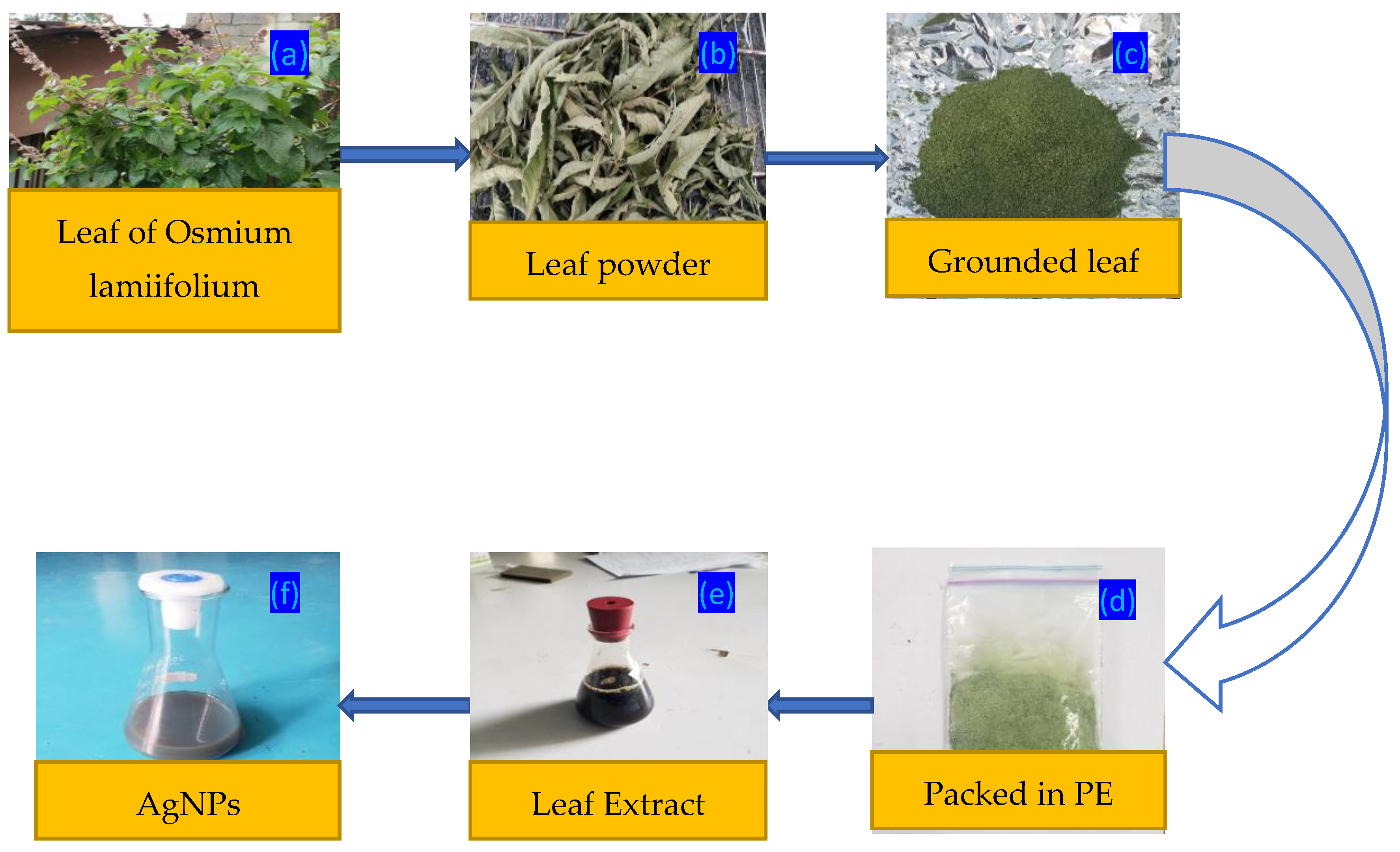
Figure 2.
(A) Particle size and (B) zeta potential of Ocimum lamiifolium leaf extract based AgNPs synthesis.
Figure 2.
(A) Particle size and (B) zeta potential of Ocimum lamiifolium leaf extract based AgNPs synthesis.
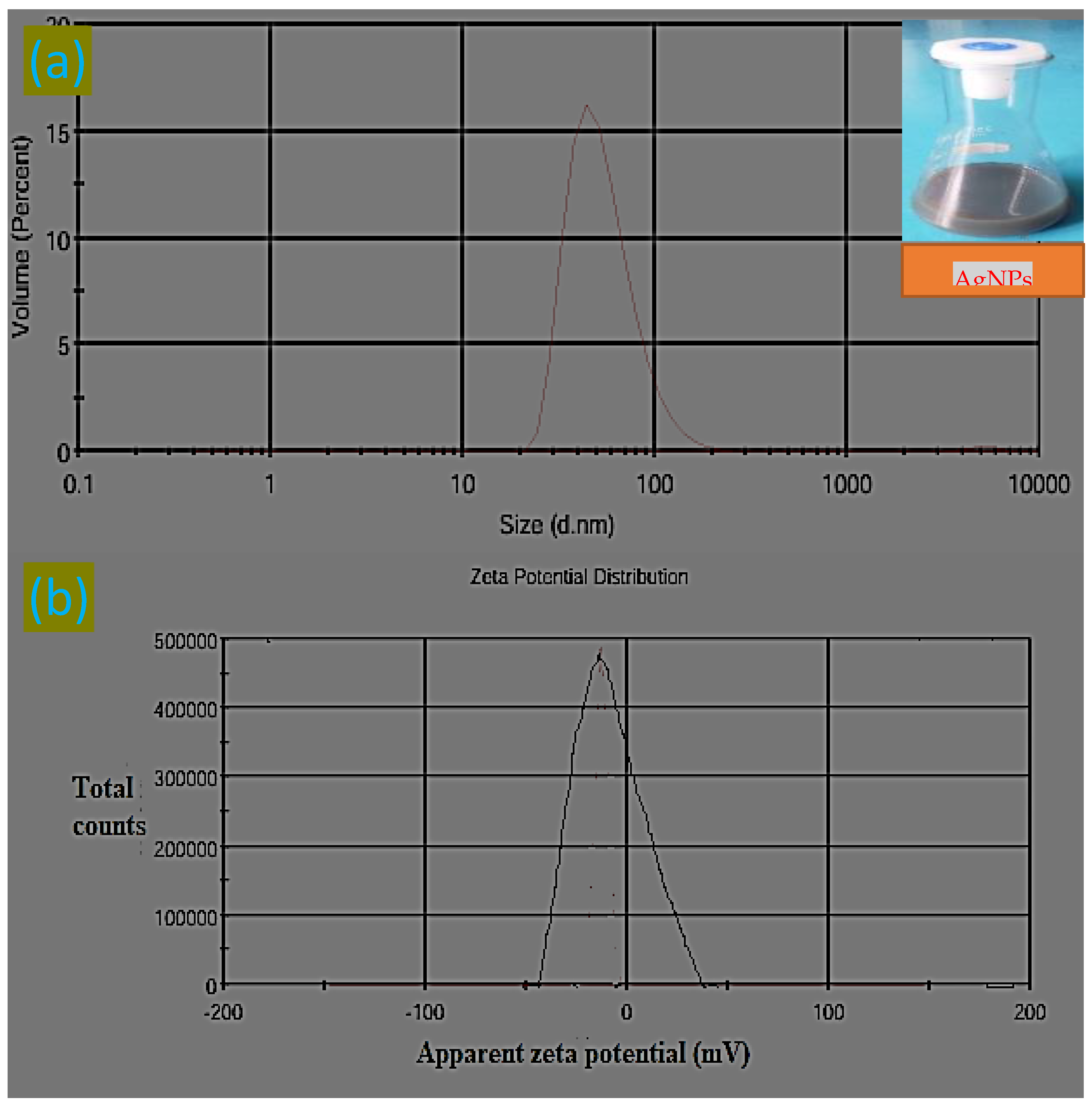
Figure 3.
AgNPs made sustainably are shown in (a) in the UV-vis absorption spectrum and (b) in the energy bandgap.
Figure 3.
AgNPs made sustainably are shown in (a) in the UV-vis absorption spectrum and (b) in the energy bandgap.
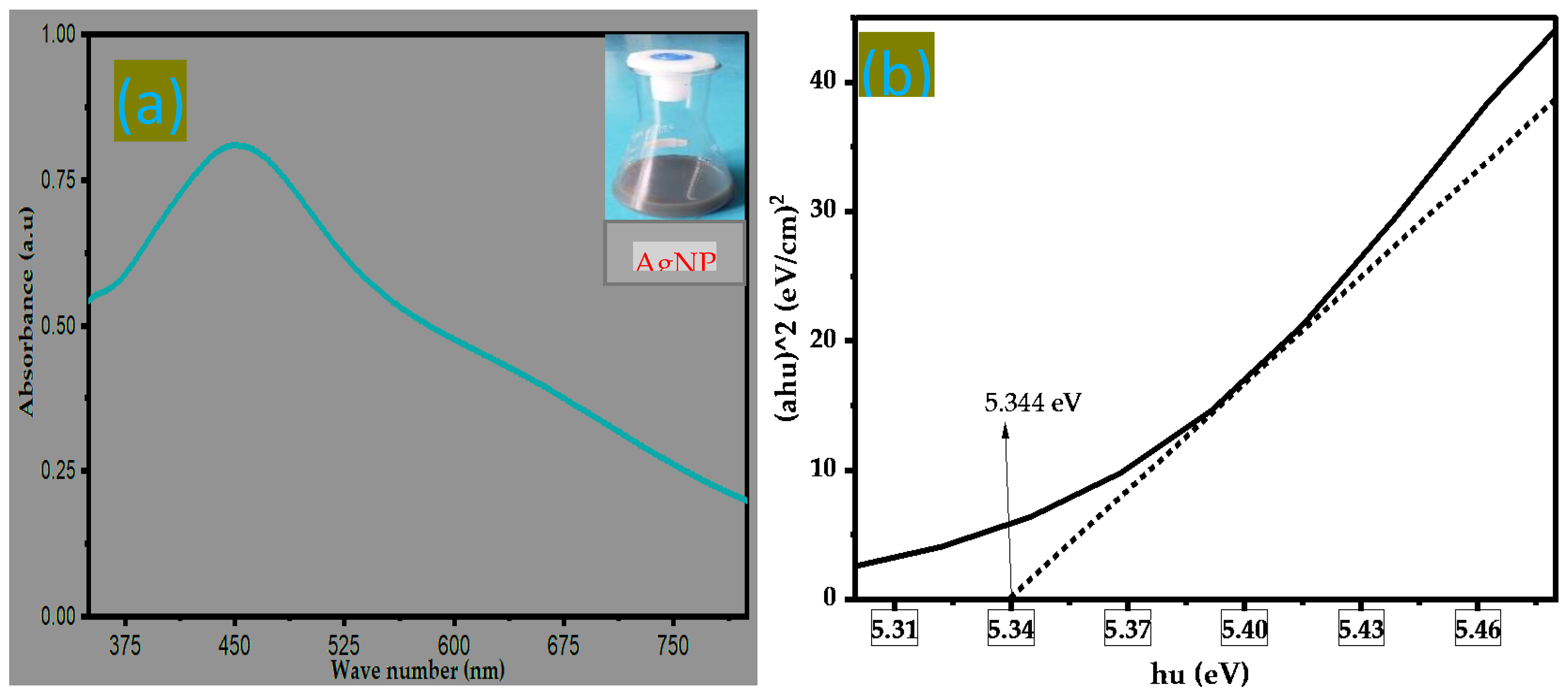
Figure 4.
FTIR spectra of AgNPs produced using Ocimum lamiifolium leaf extract.
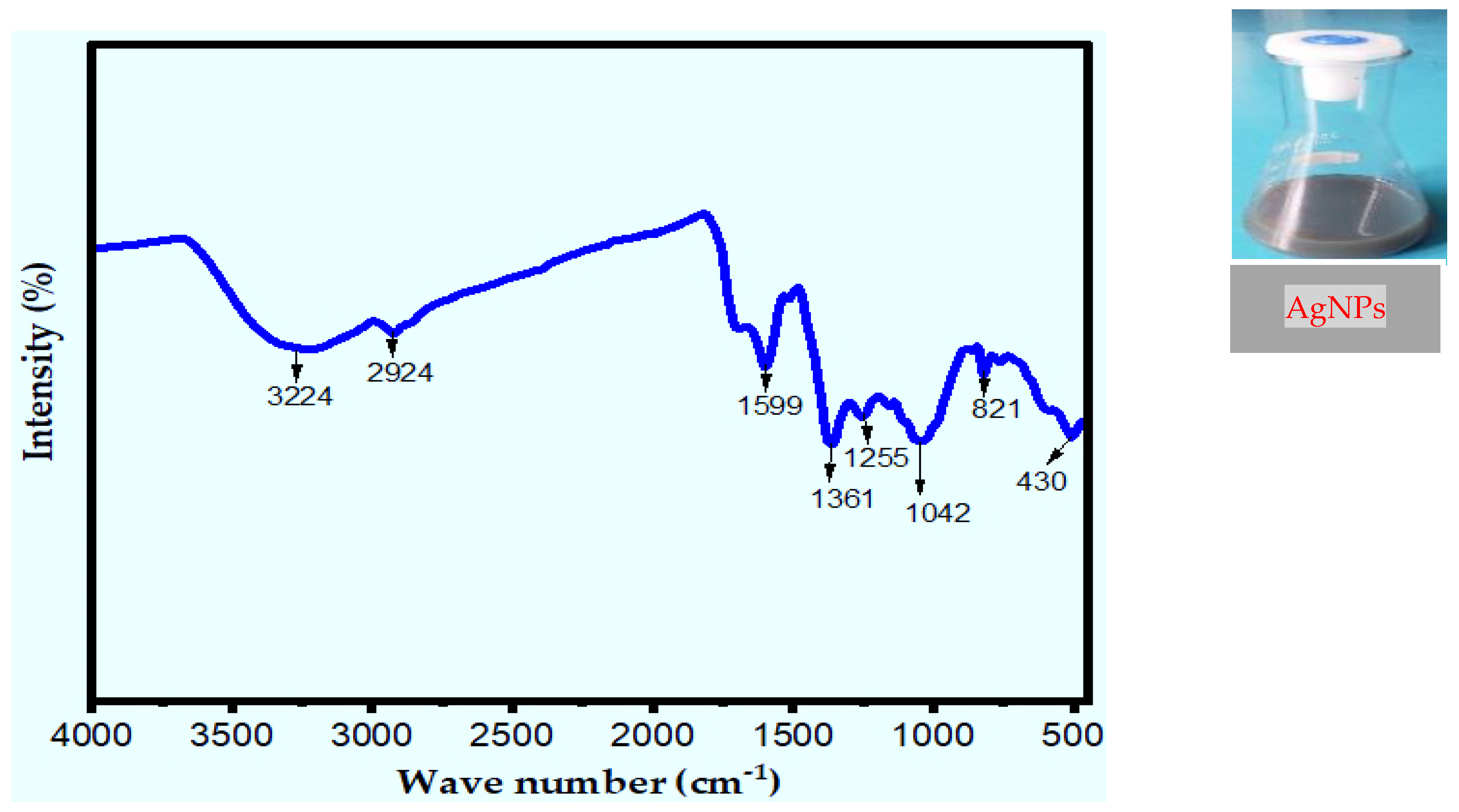
Figure 5.
XRD pattern (a) and SEM (b) of synthesize AgNPs using silver nitrate solution and Ocimum lamiifolium leaf extract.
Figure 5.
XRD pattern (a) and SEM (b) of synthesize AgNPs using silver nitrate solution and Ocimum lamiifolium leaf extract.
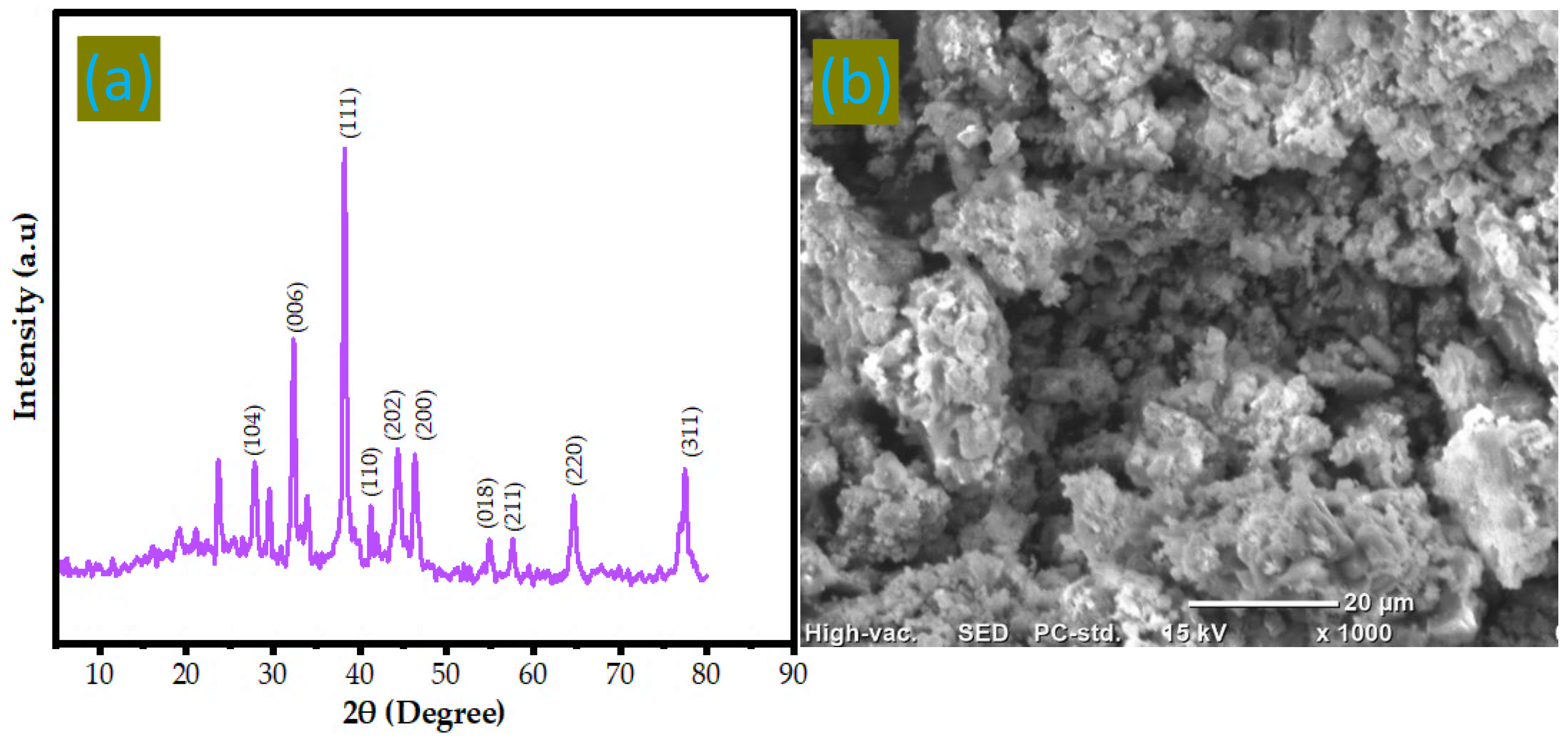
Figure 6.
Zone of inhibition for the synthesis of AgNPs, a (4 mL), and b (8 mL) of E. coli.
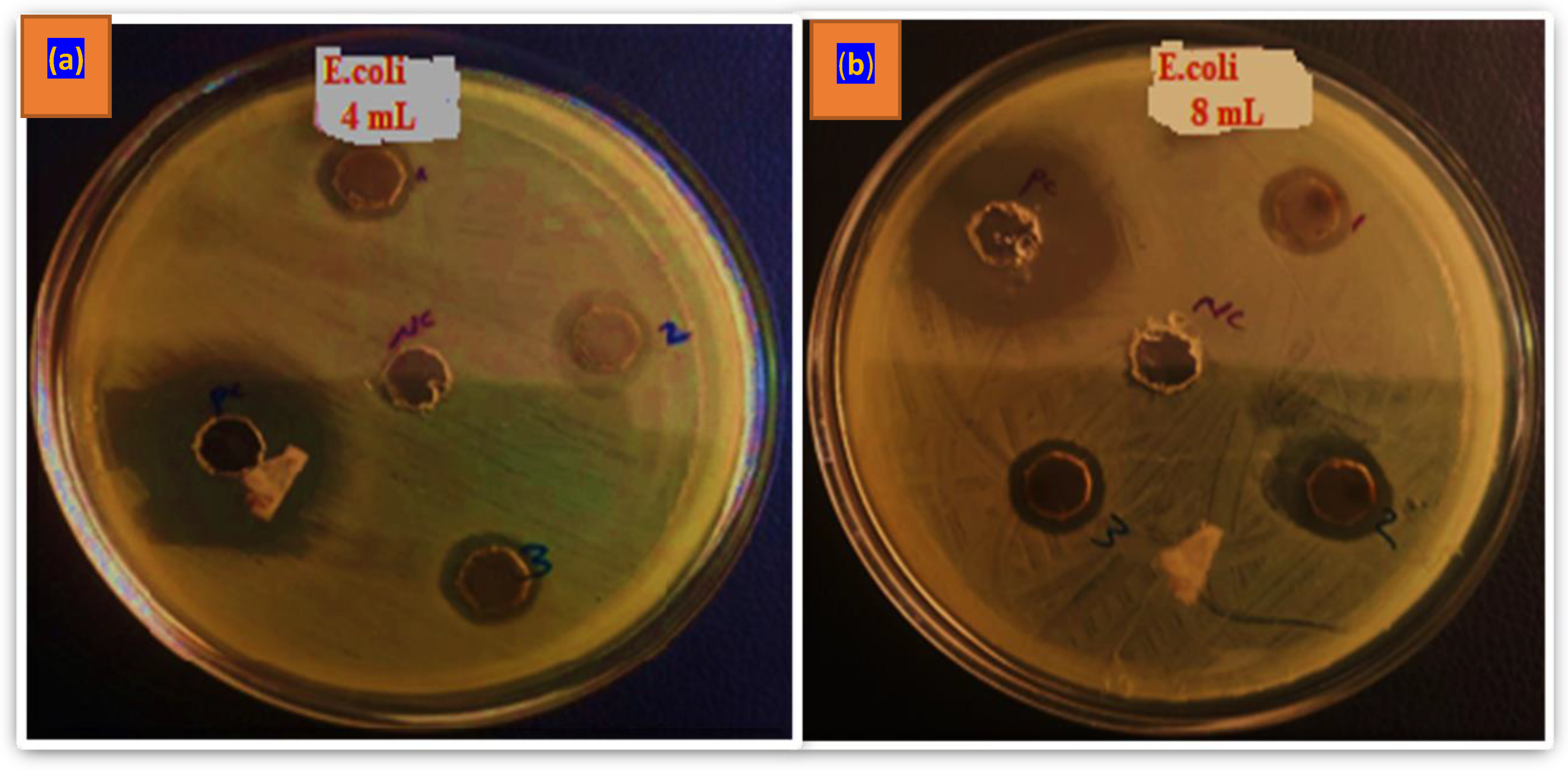
Figure 7.
The green synthesis of AgNPs using Ocimum lamiifolium leaf extract is depicted in the mechanism, emphasizing the processes of reduction, stabilization, membrane rupture, ROS production, and bacterial cell death.
Figure 7.
The green synthesis of AgNPs using Ocimum lamiifolium leaf extract is depicted in the mechanism, emphasizing the processes of reduction, stabilization, membrane rupture, ROS production, and bacterial cell death.
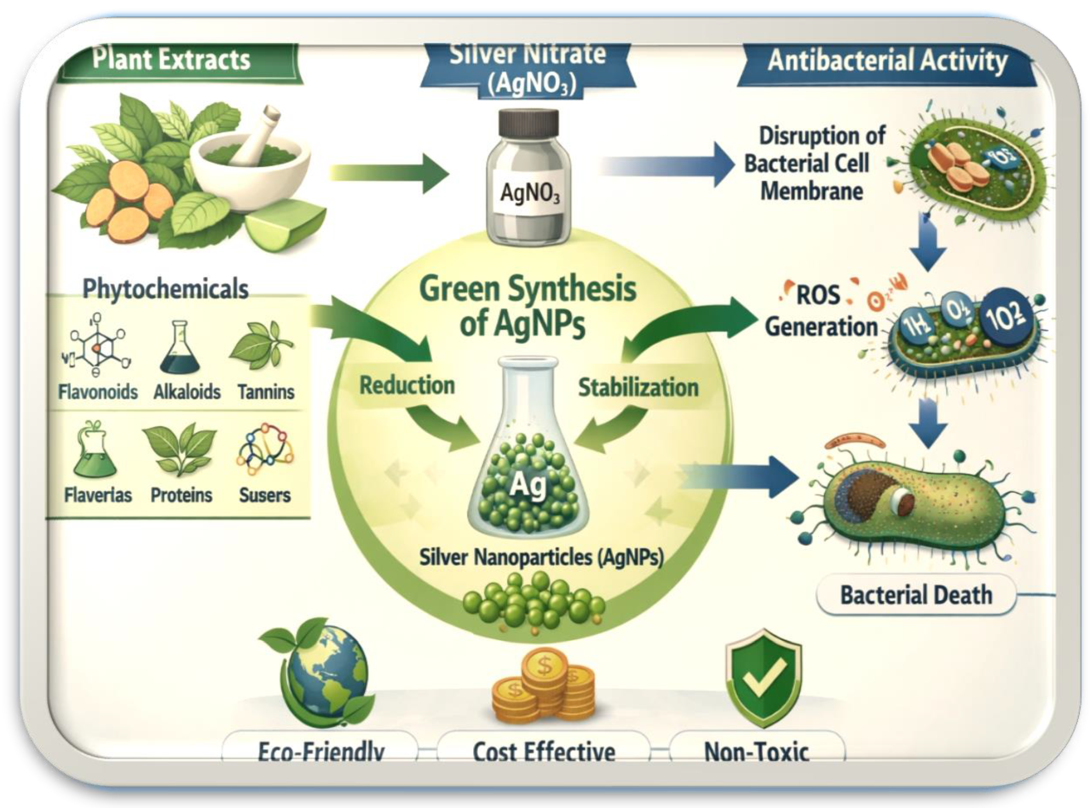

Table 1.
Average particle size, wavelength at which maximum absorption was observed and energy gap the present AgNPs and values reported for previous works.
Table 1.
Average particle size, wavelength at which maximum absorption was observed and energy gap the present AgNPs and values reported for previous works.
| Reducing agents | Methods | Average particle size (nm) | UV-vis analysis | Energy gap (eV) | References |
| Ocimum lamiifolium leaves extract | Green |
65.37 | 467 |
5.344 | Current research work |
| Vernonia amygdalina leaves extract | Green | 63.08 | 452 | 5.234 | [25] |
| Vernonia amygdalina leaves extract | Green | 70-90 | 480 | - | [46] |
| Chenopodium murale leaf | Green | 30-50 | 440 | - | [48] |
| Caccinia macranthera seed extract | Green | 37-89 | 663 | 3.1 | [45] |
| Cynodon dactylon grass leaf extract | Green | 78.66 | 417 | [49] | |
| Vernonia amygdalina leaves extract | Green | 34 | 450 | - | [50] |
| Cynodon dactylon leaves | Green | 8-10 | 451 | - | [47] |
| Azadirachta indica leaves extract | Green | - | 455 | [51] | |
| Azadirachta indica fruit pulp extract | Green | 36.8 | 400–475 | - | [52] |
Table 2.
AgNPs made using several documented techniques exhibit antibacterial action.
| Plant source | Tested pathogen | Zone of inhibition (mm) | References |
| Ocimum lamiifolium leaves extract | E. coli | 15.45 | Current research work |
| Azadirachta indica fruit pulp extract | E. coli | 15 | [52] |
| S. aureus | 16 | ||
| Streptococcus | 16 | ||
| Cynodon dactylon grass leaf extrac | E. coli | 12 | [49] |
| Vernonia amygdalina leaves extract | S. aureus | 8.46-9.85 | [46] |
| Leaf extract of Ficus hispida Linn.f | E. coli | 14 | [70] |
| Bacillus subtilis | 12 |
Disclaimer/Publisher’s Note: The statements, opinions and data contained in all publications are solely those of the individual author(s) and contributor(s) and not of MDPI and/or the editor(s). MDPI and/or the editor(s) disclaim responsibility for any injury to people or property resulting from any ideas, methods, instructions or products referred to in the content. |
© 2026 by the authors. Licensee MDPI, Basel, Switzerland. This article is an open access article distributed under the terms and conditions of the Creative Commons Attribution (CC BY) license (http://creativecommons.org/licenses/by/4.0/).
Copyright: This open access article is published under a Creative Commons CC BY 4.0 license, which permit the free download, distribution, and reuse, provided that the author and preprint are cited in any reuse.


