Preprint
Article

This version is not peer-reviewed.

High Occurrence of Diverse ESBL-, AmpC- and Carbapenemase-Producing Escherichia coli and Klebsiella pneumoniae in Surface Waters, Southern Italy, 2023-2024

Submitted:

13 January 2026

Posted:

14 January 2026

You are already at the latest version

Abstract

Antimicrobial resistance (AMR) is recognised as a major global public health threat, with the environment increasingly acknowledged as a key reservoir and dissemination pathway for resistant bacteria and resistance genes. In this study, 148 surface water samples were collected between 2023 and 2024 from six rivers and three canals discharging wastewater into two lake waters in southern Italy to assess the occurrence and genomic features of extended-spectrum β-lactamase (ESBL)-, AmpC- and carbapenemase-producing Escherichia coli and Klebsiella pneumoniae. Relevant isolates were obtained using selective culturing, and tested for antimicrobial susceptibility by broth microdilution. Major β-lactam resistance genes were detected by Real-Time PCR. Whole-genome sequencing (WGS) was performed on presumptive carbapenemase-producing isolates. ESBL- and/or carbapenemase-producing Enterobacterales were detected in 67.6% of samples, yielding 176 non-duplicate isolates. The most prevalent gene was blaCTX-M, detected in 79.3% of positive isolates (96/121), while carbapenemase genes were detected in 20.6% (25/121) of isolates, mainly blaOXA-48 and blaVIM. WGS analysis revealed occurrence of clinically relevant high-risk clones, such as K. pneumoniae ST512/ST307 carrying blaKPC-3 and E. coli ST10 harboring blaOXA-244. These findings demonstrate widespread contamination of surface waters with clinically relevant resistant Enterobacterales and highlight the importance of integrating environmental compartments into One Health AMR surveillance frameworks.

Keywords: 
;  ;  ;  ;  ;  ;  

1. Introduction

Antimicrobial resistance (AMR) has been recognised by the World Health Organization (WHO) as one of the major global health threats, driven by the rise of multidrug-resistant (MDR) bacteria and the progressive reduction of effective therapeutic options. Projections estimate that AMR could cause up to 10 million deaths annually by 2050 [1]. Among Enterobacterales, the predominant resistance mechanism to β-lactams is the production of enzymes capable of hydrolysing and inactivating such antimicrobials. More than 2,000 naturally occurring β-lactamase variants have been described to date, including clinically relevant families such as class A penicillinases, extended-Spectrum β-Lactamases (ESBLs; e.g., TEM, SHV, VEB, CTX-M), AmpC cephalosporinases (e.g., CMY, FOX, DHA, ACT, MOX), and carbapenemases belonging either to serine-β-lactamases (e.g., KPC, IMI, SME, GES) or metallo-β-lactamases (e.g., NDM, VIM, IMP) [2]. Environmental detection of antimicrobial-resistant bacteria (ARBs) has recently received increasing interest due to the potential health risk posed by bacteria associated with humans, livestock or wildlife at the interface with anthropogenic environments[3]. Historically, research has focused primarily on clinical, veterinary and food-producing animal settings. However, in the last decade, the environment has been increasingly recognised as playing a significant role in the development and dissemination of AMR [4]. The environment contributes to AMR through two major processes [4]. First, it serves as a vehicle for the dissemination of already resistant bacteria between humans, or between animals and humans. ARBs may be released into the environment through municipal wastewater [5], irrigation with reclaimed water [6], agricultural practices [7], and the application of treated sewage sludge as biosolids [8]. There is evidence that resistant bacteria can subsequently re-enter the human microbiome via environmental exposure [9], for example through ingestion of water contaminated by sewage during recreational activities [10], consumption of fresh produce irrigated with surface water [11,12], or more generally in settings with inadequate sanitation. Second, the environment acts as a reservoir and facilitator for the evolution of AMR. Surface waters can constitute resistance hotspots where antibiotic resistance genes (ARGs) disseminate—favoured by bacteriophages, integrons and other mobile genetic elements—and new resistant strains can be generated through horizontal gene transfer [13]. Although the presence of β-lactamase–encoding genes in bacteria isolated from surface waters and wastewater treatment plants has been reported in several geographical settings [14,15], robust and harmonised environmental surveillance data remain limited. In particular, no region-specific data are currently available for Apulia, a large region in Southern Italy characterised by high population mobility and seasonal tourist influx. This study aims to generate baseline evidence on the occurrence and distribution of ESBL-, AmpC- and carbapenemase-producing Escherichia coli and Klebsiella pneumoniae in surface waters of Apulia, thereby supporting environmental antimicrobial resistance surveillance, improving the identification of potential environmental exposure pathways, and contributing to public health risk assessment within a One Health framework.

2. Materials and Methods

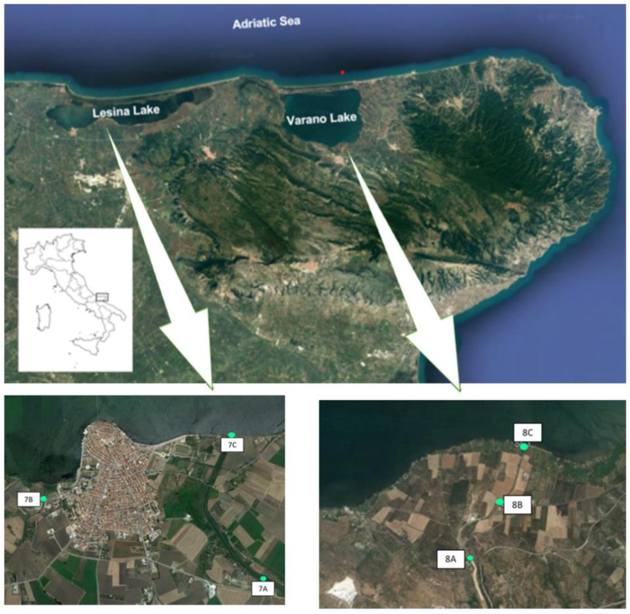
2.1. Sampling Points

Water samples were collected from six rivers and three canals conveying water into two lakes of the Apulia region between 2023 and 2024. The rivers included Candelaro (4 sampling points; 1A, 1B, 1C, 1D), Fortore (1 sampling point; 2A), Cervaro (3 sampling points; 3A, 3B, 3C), Carapelle (2 sampling points; 4A, 4B), Triolo (1 sampling point; 5A) and Ofanto (1 sampling point; 6A) rivers (Figure 1 and Table 1). Rivers were selected for sampling based on the annual mean E. coli concentrations recorded in 2021 at each monitoring site, as reported by ARPA Puglia (https://www.arpa.puglia.it/pagina2975_ii-ciclo-sessennale-2016-2021.html). Specifically, sampling sites with the highest average E. coli values were prioritised, given the role of E. coli as an indicator of faecal contamination in surface waters. In addition, sampling points were also chosen on the basis of their proximity to agricultural land observed using satellite imagery (Google, 2023). Sampling stations within the canals were strategically located along channels receiving wastewater discharges from adjacent settlements into the lake systems. Overall, a total of six sampling points was set for this study, three for Lake Lesina (7A, 7B and 7C) and three for Lake Varano (8A, 8B and 8C) (Figure 2 and Table 1). For Lesina lake, it was chosen to sample the waters of the Cammarata canal within which wastewater from the settlements of Lesina and Poggio Imperiale are conveyed. Of the three sampling points, two (7A, 7C) were placed along the Cammarata canal, 7A at the outlet of the wastewater treatment plant and 7C at the incile of the Cammarata canal into the lagoon, respectively. A third sampling point (7B) was fixed along the La Fara canal, which collects wastewater from an area close to the town of Lesina affected by various agricultural and livestock activities and discharges it into the lagoon (Figure 2). For Varano lake, it was chosen to sample the waters of the San Francesco canal within which wastewater from the town of Cagnano Varano is conveyed. The sampling point 8A is located at the outlet of the wastewater treatment plant, 8B in an area of the canal near which there is a livestock farm, and a third sampling point (8C) at the incile of the San Francesco canal in the lagoon (Figure 2). The analyzed canals constitute critical control points for the environmental quality of the lakes, as they play a pivotal role in maintaining the ecological equilibrium of the lagoon system and in safeguarding the safety and long-term sustainability of associated economic activities.

2.2. Sampling Procedure

From May 2023 to December 2024, a total of 148 surface water samples were collected from surface water bodies in the Apulia region, including six rivers and three canals conveying water into two lakes (Figure 1, Figure 2, Table 1). For each surface water body, a minimum of ten samples were collected and analysed (Table 1). On each sampling occasion, a volume of 500 mL was collected at each site directly by filling a sterile glass container with surface water. The samples were transferred under refrigerated conditions to the laboratories of the Istituto Zooprofilattico Sperimentale della Puglia e della Basilicata (IZS PB, Foggia, Italy). The samples were stored at 4 °C and analysed within 24 h.

2.3. Microbiological Screening, Isolation and Identification of Target Bacteria

The isolation procedure aimed to selectively detect resistant bacteria from the examined samples. For the isolation of target microorganisms, 100 mL of each water sample was filtered (0.45 μm; Sartorius Stedim Biotech GmbH, Goettingen, Germany). To select for ESBL-producing Enterobacterales, the filter was placed in 100 mL of Buffer Peptone Water (BPW) (Thermo Fisher Scientific, Rhone, MI) and incubated at 42 °C for 18-24 hours. Subsequently, 100 μL of each dilution (up to 10-3) was plated on CHROMagar ESBL plates (CHROMagar, Paris, France) and incubated at 42 °C for 18-24 hours. For the isolation of carbapenemase-producing Enterobacterales (CPE), the filter was placed in 100 mL of Mossel broth (EE Broth) (Scharlab S.L., Sentmenat, Spain) and the enrichment incubated at 42 °C for 18-24 hours. Subsequently, 100 μL of each dilution (up to 10 -2) was plated on CHROMagar mSuperCARBA plates (CHROMagar, Paris, France) and incubated at 42°C for 18-24 hours (Savin et al., 2020). After incubation, up to three presumptive colonies of E. coli and Klebsiella spp. per sample were subcultured onto Blood Agar plates (Liofilchem, Roseto degli Abruzzi, Italy), incubated at 37 °C for 24 hours and subjected to identification by MALDI-TOF mass spectrometry (Bruker Daltonics, Milan, IT). The isolates were cryopreserved in Microbank (PRO-LAB DIAGNOSTICS, Richmond Hill, ON, Canada) and stored at –80°C.

2.4. Phenotypic Detection of Antimicrobial Resistance

Phenotypic antimicrobial susceptibility was assessed using the broth microdilution method, following EUCAST guidelines (version 15.0; www.eucast.org) [16]. GN4F and EUVSEC2 panels (Thermo Fisher Scientific Diagnostics, Netherlands) were used. Plates were incubated at 37 °C for 24 h, and MICs were read using the Thermo Scientific™ Sensititre™ Vizion™ system. E. coli ATCC 25922 and K. pneumoniae ATCC 700603 served as quality control strains. Results were interpreted according to epidemiological cut-off values (ECOFFs) provided by the European Committee on Antimicrobial Susceptibility Testing (EUCAST) (www.eucast.org) [16]. Multidrug resistance (MDR) was defined as non-susceptibility to at least one agent in three or more antimicrobial classes, according to Magiorakos et al. [17].

2.5. Genotypic Detection of ESBL- and Carbapenemase-Encoding Genes and Characterization by Whole-Genome Sequencing

Automated bacterial DNA extraction was performed on the Maxwell RSC Extraction System (Promega, USA) using the Maxwell RSC PureFood Pathogen kit (Promega, USA). The quantity (ng/μL) and purities (260/280 and 260/230 ratios) of the extracted DNA were measured spectrophotometrically using the IMPLEN NanoPhotometer N60 (Implen GmbH, München, Germany). DNA from all isolates was used for real-Time PCR using the Allplex EnteroDR Assay kit for the detection of blaNDM, blaKPC, blaOXA-48, blaVIM, blaIMP, blaCTX-M genes (Seegene Inc, Seoul, Republic of Korea). DNA from isolates that tested positive in PCR for carbapenemase genes was also characterised by whole-genome sequencing (WGS). Genomic DNA was sequenced using Illumina short-read technology on a NextSeq platform with 2 × 151 bp reads. Short-read assembly was performed with SPAdes v3.15.2. The quality of raw sequencing and assembly data was assessed using the analytical tool BIFROST (https://github.com/ssi-dk/bifrost/). For K. pneumoniae strains, species identification, multilocus sequence typing (MLST), resistance gene detection and plasmid replicon typing were performed using the Pathogenwatch platform (https://pathogen.watch). For E. coli, all bioinformatics analyses were performed on the public ARIES Galaxy server [18].

2.6. Data Availability and Sequence Deposition

Whole-genome sequencing data generated in this study have been deposited in the NCBI Sequence Read Archive (SRA) under BioProject accession number PRJNA1358062.

3. Results

3.1. Selective Isolation of Presumptive ESBL- and Carbapenemase-Producing Enterobacterales from Surface Water

A total of 148 water samples were cultured to isolate ESBL- and/or carbapenemase-producing Enterobacterales. All surface water sites tested positive, on at least one sampling occasion, for presumptive ESBL- and/or carbapenemase-producing Escherichia coli and Klebsiella pneumoniae, as indicated by growth on selective media. Isolates were recovered on CHROMagar ESBL and/or mSuperCARBA selective media from 67.6% (100/148) of the samples, yielding 176 strains, one strain per sample: 57.4% (101/176) E. coli and 42.6% (75/176) K. pneumoniae.

3.2. Detection of ESBL- and Carbapenemase-Encoding Genes by Multiplex Real-Time PCR

A total of 176 isolates (75 K. pneumoniae and 101 E. coli) were analysed by PCR, revealing that 31.2% (55/176) of isolates were negative for all tested genes (Supplementary data) and 68.8% (121/176) of isolates carried at least one of the investigated resistance genes.
The most prevalent resistance gene was blaCTX-M, detected in 79.3% (96/121) of alone or in combination with carbapenemase-encoding genes. Carbapenemase genes were detected in 20.6% (25/121) of isolates, including blaOXA-48 (3.3%, 4/121), blaKPC (1.7%, 2/121), blaVIM (9.1%, 11/121), or in combination with other genes (blaCTX-M + blaKPC, 2.5%, 3/121; blaCTX-M + blaVIM, 2.5%, 3/121; blaCTX-M + blaOXA-48 1.7%, 2/121) (Table 1). Among E. coli, 30.7% (31/101) were negative for all antibiotic resistance genes tested by PCR (11 isolated from CHROMagar mSuperCARBA, 20 from CHROMAgar ESBL) (Supplementary data). Moreover, 80% (56/70) of E. coli isolates positive for resistance genes were recovered from CHROMagar ESBL, whereas the remaining 20% (14/70) were obtained from CHROMagar mSuperCARBA.
blaCTX-M was the most prevalent gene (84.3%, 59/70), mainly blaCTX-M alone (80%, 56/70), followed by combinations with blaVIM (1.4%, 1/70) and blaOXA-48 (2.9%, 2/70). Carbapenemase genes were identified in 20% (14/70), including blaOXA-48 (4.3%, 3/70), blaVIM (8.6%, 6/70), and combined patterns mentioned above. Among K. pneumoniae, 32% (24/75) were PCR negative (11 isolated from CHROMagar mSuperCARBA, 13 from CHROMAgar ESBL) (Supplementary data). blaCTX-M was again the most common determinant (88.2%, 45/51), either alone (78.4%, 40/51) or in combination with blaKPC (5.8%, 3/51) or or blaVIM (5.8%, 3/51). Carbapenemases were detected in 21.6% (11/51), including blaOXA-48 (2%, 1/51), blaVIM (9.8%, 5/51), and combined profiles (Table 1).

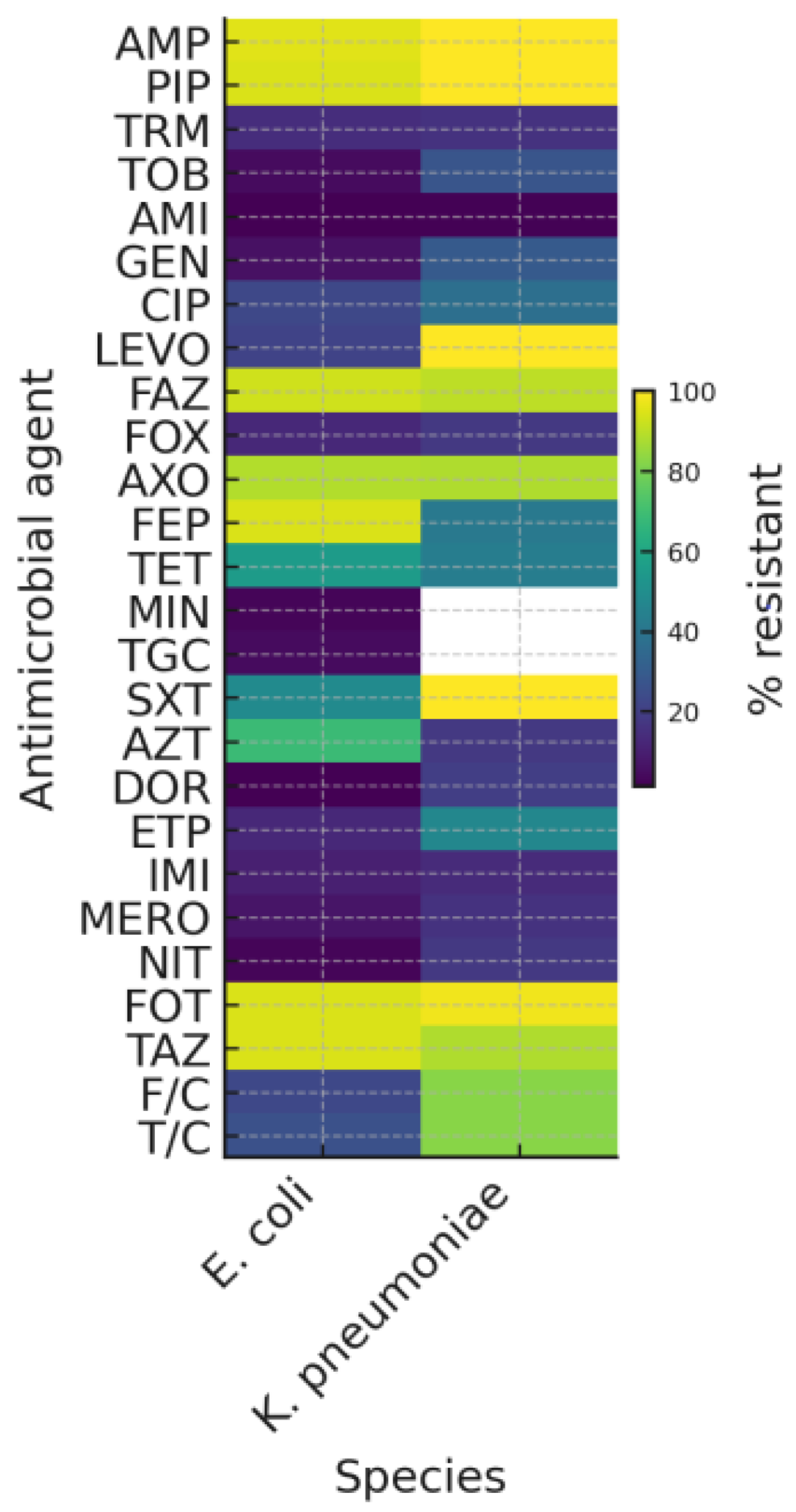
3.3. Phenotypic and Genotypic Detection of Antimicrobial Resistance

Antimicrobial susceptibility testing was performed on all 176 isolates (Table 2, Figure 3). Among the 31 E. coli strains that tested negative for all resistance genes by PCR, 19 isolates were susceptible to all antibiotics tested, 11 showed resistance to up to three antibiotic classes, and only one isolate was multidrug-resistant (Supplementary Data). For K. pneumoniae, 24 isolates tested negative in the PCR analysis, and their antimicrobial profiles are reported in the Supplementary Data. Overall, resistance patterns varied between E. coli and K. pneumoniae, with multidrug resistance observed in both species (Table 3 and Figure 3).

3.4. Whole Genome Sequencing (WGS) of Isolates Positive for Carbapenemase-Encoding Genes by PCR

All (25/121) of Enterobacterales PCR-positive for carbapenemase-encoding were subjected to WGS. Table 4 summarizes genomic data for the 25 isolates (56% (14/25) E. coli and 44% (11/25) K. pneumoniae), including detected β-lactamase genes (carbapenemases, ESBL/AmpC), additional antimicrobial resistance (AMR) determinants, multilocus sequence types (MLST), and plasmid replicon types. WGS confirmed the presence of carbapenemase-encoding genes in 68% (17/25) of isolates, of which 58.8% (10/17) were E. coli and 41.2% (7/17) K. pneumoniae. Among E. coli, carbapenemase genes were blaOXA-244 (4/10, 40%), followed by blaVIM-4 (3/10, 30%), blaOXA-181 (1/10, 10%) and blaKPC-3 (2/10, 20%). In K. pneumoniae, carbapenemase genes included blaKPC-3 (3/7, 42.85%) and blaVIM-1 (4/7, 57.14%). ESBL/AmpC genes were detected in 36.0% (9/25) of isolates, including five E. coli and four K. pneumoniae. The most frequently detected ESBL/AmpC genes were blaCTX-M-15, blaSHV-12, blaSHV-28, blaTEM-52B, and blaDHA-1. Multilocus sequence typing (MLST) revealed considerable diversity. High-risk clones such as K. pneumoniae ST512 and ST307 carrying blaKPC-3 and E. coli ST10 carrying blaOXA-244 were identified (Table 4). In addition to β-lactam resistance determinants, multiple acquired resistance genes were detected: 72% of isolates carried aminoglycoside-modifying enzymes (aac, aph), 64% harboured sulfonamide resistance genes (sul1 or sul2), 60% carried dfrA variants (trimethoprim resistance), 44% had macrolide resistance genes (mph(A)), and 40% possessed tetracycline resistance genes (tet(A/B/D)). The isolates showed marked genomic diversity, encompassing 12 distinct MLST types (e.g. ST10, ST43, ST307, ST512, ST540, ST746, ST1721), and multiple plasmid incompatibility groups, most frequently IncFIB (56%), IncFIA (48%), IncX1 (28%), IncR (20%), and Col-type plasmids (32%).

4. Discussion

Antimicrobial resistance (AMR) represents a major global public health threat, and the World Health Organisation (WHO) classifies ESBL- and carbapenemase-producing Enterobacterales among the highest priority pathogens [19]. The environment is increasingly recognised as a key compartment in the transmission and persistence of AMR [20], with anthropogenic activities considered major drivers of carbapenem resistance dissemination [21,22]. This study provides evidence of widespread contamination of surface waters in the Apulia Region (southern Italy) with ESBL- and carbapenemase-producing E. coli and K. pneumoniae. Using selective culture methods, 176 non-duplicate isolates were recovered from 148 samples collected across eight surface water bodies, with ESBL- or carbapenemase-producing E. coli or K. pneumoniae detected in 68.8% of the analysed samples. These findings confirm that surface waters can function as important reservoirs and dissemination pathways for AMR, particularly in areas influenced by wastewater discharge and agricultural activities [23,24] and support the integration of environmental surveillance into One Health AMR monitoring frameworks. Marked spatial heterogeneity in E. coli concentrations was observed across surface waters, indicating variable faecal contamination from anthropogenic sources, including untreated or inadequately treated wastewater, agricultural runoff and livestock effluents. According to previous reports [25,26], E. coli was the most frequently isolated species (57.4%), in line with its recognised ubiquity and persistence in aquatic environments [27], where it can act as a reservoir and vector for horizontally transferable resistance genes [28]. The co-occurrence of K. pneumoniae (42.6%) further suggests faecal contamination and potential contributions from both human and animal sources [10]. Notably, all sampling sites yielded presumptive ESBL- and carbapenemase-producing strains on at least one sampling occasion, suggesting widespread environmental dissemination of resistant clones. Phenotypic antimicrobial susceptibility revealed high occurrence of resistance to multiple antibiotic classes. The heatmap highlights clear species-specific differences in the distribution of phenotypes across antimicrobial classes. The high proportions of resistance observed in both E. coli and K. pneumoniae to aminopenicillins and extended-spectrum cephalosporins should be interpreted in light of the study design, as isolates were obtained using selective culture media. This targeted isolation approach was intentionally applied to enhance the recovery of resistant Enterobacterales and, by definition, enriches for resistance to the corresponding antimicrobial classes. Therefore, the observed resistance profiles do not reflect the background prevalence of antimicrobial resistance in the sampled environments but rather the phenotypic characteristics of the selected isolate collection. Nevertheless, the identified resistance patterns are in line with findings from EU-level surveillance reports documenting the widespread occurrence of ESBL-producing Enterobacterales in environmental and food-production settings [16]. The detection of isolates growing on selective chromogenic media but testing negative for the ESBL- and carbapenemase-encoding genes targeted by PCR can be explained by the intrinsic characteristics of the screening approach. Chromogenic media such as CHROMagar ESBL and CHROMagar mSuperCARBA are designed to maximise sensitivity and may therefore allow the growth of Enterobacterales with reduced β-lactam susceptibility due to intrinsic or low-level resistance mechanisms, even in the absence of classical ESBL or carbapenemase genes. Similar limitations, including reduced specificity and occasional growth of PCR-negative isolates, have been reported previously [29,30]. In addition, phenotypic resistance patterns not explained by the PCR targets may reflect the presence of alternative β-lactam resistance mechanisms not covered by the multiplex assay, such as non-CTX-M ESBLs, chromosomal AmpC overexpression, porin alterations or efflux mechanisms [30]. These findings highlight the complementary value and limitations of selective culture and targeted molecular assays in environmental AMR surveillance and support the use of broader genomic approaches when detailed characterisation of resistance determinants is required [31]. Comparison between phenotypic antimicrobial resistance and genomic sequencing-based predictions showed a high degree of concordance. Among ESBL genes, blaCTX-M was the most frequently detected (79.3%), in agreement with its global predominance in both clinical and environmental settings [32,33]. Its similar prevalence in E. coli and K. pneumoniae suggests sustained selective pressure in the environment, potentially driven by antimicrobial residues and/or untreated wastewater inputs [34]. Carbapenemase genes were detected in 20.6% (25/121) of isolates carrying at least one of the investigated resistance genes, mainly blaVIM, blaKPC and blaOXA-48-like variants, which are widely disseminated in Europe [35,36,37]. The observed co-occurrence of multiple resistance determinants, including combinations of ESBL- and carbapenemase-encoding genes, indicates the presence of complex resistance profiles within the analysed isolates. While such genetic constellations are commonly associated with mobile genetic elements and horizontal gene transfer in aquatic environments [38], the present findings should be interpreted cautiously, as no specific analyses of plasmids or other mobile genetic elements were performed, and no conclusions can therefore be drawn regarding the underlying mechanisms of gene dissemination. WGS of 25 presumptive carbapenemase-producing isolates confirmed carbapenemase genes in 68%, supporting the reliability of PCR screening but also highlighting occasional discrepancies between phenotypic and genotypic methods [31]. Detected carbapenemase variants included blaKPC-3, blaVIM-1, blaVIM-4, blaOXA-244 and blaOXA-181, confirming that surface waters can act as convergence points for multiple resistance genes [39]. WGS also revealed substantial clonal diversity. The detection of high-risk clinical lineages such as K. pneumoniae ST512 and ST307 carrying blaKPC-3, and E. coli ST10 carrying blaOXA-244, is particularly noteworthy, as these lineages are commonly associated with healthcare-associated outbreaks in Italy and Europe [40,41,42,43]. Their presence in rivers and canals suggests possible bidirectional exchange between clinical environments and natural ecosystems [42,44]. In addition, several sequence types (STs) typically associated with environmental or zoonotic reservoirs were identified, including E. coli ST602, ST746 and ST1721 and K. pneumoniae ST34, ST45 and ST1537, which have been increasingly reported in surface waters, wastewater effluents and animal-associated settings [4,45,46,47,48,49]. Freshwater ecosystems receiving inputs from wastewater treatment plants, agricultural effluents and stormwater runoff are recognised as hotspots of microbial evolution, where multiple selective pressures can promote the emergence and maintenance of resistant strains [13,34]. Plasmid replicon typing showed a predominance of IncF, IncX and IncN plasmids, which are key vectors for ESBL and carbapenemase dissemination [50,51]. However, due to availability of short-read data only, we could not associate ESBL and carbapenemase genes to plasmid replicons. Taken together, these findings indicate that surface waters in the Apulia Region act both as reservoirs of clinically relevant high-risk clones and as dynamic environments supporting the persistence and evolution of environmentally adapted Enterobacterales. This highlights the permeability of ecological boundaries between human, animal and environmental compartments and reinforces the importance of integrating environmental surveillance into One Health AMR monitoring systems. Targeted interventions addressing wastewater treatment, agricultural runoff and environmental contamination are urgently needed to mitigate the environmental spread of carbapenemase-producing Enterobacterales in Italy.

Supplementary Materials

The following supporting information can be downloaded at the website of this paper posted on Preprints.org. Table S1: Antibiotic resistance profiles of E. coli isolates negative for all genes tested by PCR.; Table S2: Antibiotic susceptibility testing of Klebsiella pneumoniae isolates negative for antimicrobial resistance genes tested by PCR.

Author Contributions

Conceptualization, G.N., A.C.C., M.G.B, G.L.S. and V.B.; methodology, G.N., A.C.C. and M. G. B.; software, G.N. and V.B.; investigation, G.N., A.C.C., M. G. B, A.D., R.C., M.G.C., I.L., A.S., M.N., T. S.; resources, G.L.S.; data curation, G.N., A.C.C., G.L.S. and V.B.; writing—review and editing, G.N., A.C.C., M.G.B., G.L.S. and V.B.; supervision, G.N., M.G.B, V.B. and G.L.S. All authors have read and agreed to the published version of the manuscript. All authors have read and agreed to the published version of the manuscript.

Funding

This Project was funded by Italian Ministry of Health (IZSPB 05/22 RC).

Institutional Review Board Statement

Not applicable.

Informed Consent Statement

Not applicable.

Data Availability Statement

Whole-genome sequencing data generated in this study have been deposited in the NCBI Sequence Read Archive (SRA) under BioProject accession number PRJNA1358062. All other data supporting the findings of this study are available from the corresponding authors upon reasonable request.

Acknowledgments

None declared.

Conflicts of Interest

The authors declare no conflicts of interest.

References

  1. Clifford, K.; Desai, D.; Prazeres Da Costa, C.; Meyer, H.; Klohe, K.; Winkler, A.; Rahman, T.; Islam, T.; Zaman, M.H. Antimicrobial Resistance in Livestock and Poor Quality Veterinary Medicines. Bull. World Health Organ. 2018, 96, 662–664. [CrossRef]
  2. Bonomo, R.A. β-Lactamases: A Focus on Current Challenges. Cold Spring Harb Perspect Med 2017, 7, a025239. [CrossRef]
  3. Xu, X.; Zhou, W.; Xie, C.; Zhu, Y.; Tang, W.; Zhou, X.; Xiao, H. Airborne Bacterial Communities in the Poultry Farm and Their Relevance with Environmental Factors and Antibiotic Resistance Genes. Science of The Total Environment 2022, 846, 157420. [CrossRef]
  4. Bengtsson-Palme, J.; Kristiansson, E.; Larsson, D.G.J. Environmental Factors Influencing the Development and Spread of Antibiotic Resistance. FEMS Microbiology Reviews 2018, 42. [CrossRef]
  5. Marathe, N.P.; Pal, C.; Gaikwad, S.S.; Jonsson, V.; Kristiansson, E.; Larsson, D.G.J. Untreated Urban Waste Contaminates Indian River Sediments with Resistance Genes to Last Resort Antibiotics. Water Research 2017, 124, 388–397. [CrossRef]
  6. Christou, A.; Agüera, A.; Bayona, J.M.; Cytryn, E.; Fotopoulos, V.; Lambropoulou, D.; Manaia, C.M.; Michael, C.; Revitt, M.; Schröder, P.; et al. The Potential Implications of Reclaimed Wastewater Reuse for Irrigation on the Agricultural Environment: The Knowns and Unknowns of the Fate of Antibiotics and Antibiotic Resistant Bacteria and Resistance Genes – A Review. Water Research 2017, 123, 448–467. [CrossRef]
  7. Jechalke, S.; Broszat, M.; Lang, F.; Siebe, C.; Smalla, K.; Grohmann, E. Effects of 100 Years Wastewater Irrigation on Resistance Genes, Class 1 Integrons and IncP-1 Plasmids in Mexican Soil. Front. Microbiol. 2015, 6. [CrossRef]
  8. Gigliucci, F.; Barbieri, G.; Veyrunes, M.; Chiani, P.; Marra, M.; Carollo, M.; Knijn, A.; Brambilla, G.; Morabito, S. Characterization of the Resistome and Antibiotic-Resistant Bacteria in Top Soil Improvers and Irrigation Waters Devoted to Food Production: A Case Study from Italy. Environ Sci Pollut Res 2025, 32, 14691–14705. [CrossRef]
  9. Stanton, I.C.; Bethel, A.; Leonard, A.F.C.; Gaze, W.H.; Garside, R. Existing Evidence on Antibiotic Resistance Exposure and Transmission to Humans from the Environment: A Systematic Map. Environ Evid 2022, 11, 8. [CrossRef]
  10. Leonard, A.F.C.; Zhang, L.; Balfour, A.J.; Garside, R.; Hawkey, P.M.; Murray, A.K.; Ukoumunne, O.C.; Gaze, W.H. Exposure to and Colonisation by Antibiotic-Resistant E. Coli in UK Coastal Water Users: Environmental Surveillance, Exposure Assessment, and Epidemiological Study (Beach Bum Survey). Environment International 2018, 114, 326–333. [CrossRef]
  11. O’Flaherty, E.; Solimini, A.G.; Pantanella, F.; De Giusti, M.; Cummins, E. Human Exposure to Antibiotic Resistant-Escherichia Coli through Irrigated Lettuce. Environment International 2019, 122, 270–280. [CrossRef]
  12. Rahman, M.; Alam, M.-U.; Luies, S.K.; Kamal, A.; Ferdous, S.; Lin, A.; Sharior, F.; Khan, R.; Rahman, Z.; Parvez, S.M.; et al. Contamination of Fresh Produce with Antibiotic-Resistant Bacteria and Associated Risks to Human Health: A Scoping Review. IJERPH 2021, 19, 360. [CrossRef]
  13. Berglund, B. Environmental Dissemination of Antibiotic Resistance Genes and Correlation to Anthropogenic Contamination with Antibiotics. Infection Ecology & Epidemiology 2015, 5, 28564. [CrossRef]
  14. Schwartz, T.; Kohnen, W.; Jansen, B.; Obst, U. Detection of Antibiotic-Resistant Bacteria and Their Resistance Genes in Wastewater, Surface Water, and Drinking Water Biofilms. FEMS Microbiology Ecology 2003, 43, 325–335. [CrossRef]
  15. Bouki, C.; Venieri, D.; Diamadopoulos, E. Detection and Fate of Antibiotic Resistant Bacteria in Wastewater Treatment Plants: A Review. Ecotoxicology and Environmental Safety 2013, 91, 1–9. [CrossRef]
  16. European Food Safety Authority (EFSA); European Centre for Disease Prevention and Control (ECDC) The European Union Summary Report on Antimicrobial Resistance in Zoonotic and Indicator Bacteria from Humans, Animals and Food in 2021–2022. EFS2 2024, 22. [CrossRef]
  17. Magiorakos, A.-P.; Srinivasan, A.; Carey, R.B.; Carmeli, Y.; Falagas, M.E.; Giske, C.G.; Harbarth, S.; Hindler, J.F.; Kahlmeter, G.; Olsson-Liljequist, B.; et al. Multidrug-Resistant, Extensively Drug-Resistant and Pandrug-Resistant Bacteria: An International Expert Proposal for Interim Standard Definitions for Acquired Resistance. Clinical Microbiology and Infection 2012, 18, 268–281. [CrossRef]
  18. Knijn, A.; Michelacci, V.; Orsini, M.; Morabito, S. Advanced Research Infrastructure for Experimentation in genomicS (ARIES): A Lustrum of Galaxy Experience 2020.
  19. Sati, H.; Carrara, E.; Savoldi, A.; Hansen, P.; Garlasco, J.; Campagnaro, E.; Boccia, S.; Castillo-Polo, J.A.; Magrini, E.; Garcia-Vello, P.; et al. The WHO Bacterial Priority Pathogens List 2024: A Prioritisation Study to Guide Research, Development, and Public Health Strategies against Antimicrobial Resistance. The Lancet Infectious Diseases 2025, 25, 1033–1043. [CrossRef]
  20. Larsson, D.G.J.; Flach, C.-F. Antibiotic Resistance in the Environment. Nat Rev Microbiol 2022, 20, 257–269. [CrossRef]
  21. Mills, M.C.; Lee, J. The Threat of Carbapenem-Resistant Bacteria in the Environment: Evidence of Widespread Contamination of Reservoirs at a Global Scale. Environmental Pollution 2019, 255, 113143. [CrossRef]
  22. Schwermer, C.U.; Krzeminski, P.; Anglès d’Auriac, M.; Gjeitnes, M.; Moe, J.; Bellanger, X.; Bürgmann, H.; Carapeto García, R.; Crespo Iniesta, P.; Dagot, C.; et al. Pilot Study on Antimicrobial Resistance Monitoring in European Surface Waters - Final Report of the Eionet Working Group; Zenodo, 2025;
  23. Baquero, F.; Martínez, J.-L.; Cantón, R. Antibiotics and Antibiotic Resistance in Water Environments. Current Opinion in Biotechnology 2008, 19, 260–265. [CrossRef]
  24. Rizzo, L.; Manaia, C.; Merlin, C.; Schwartz, T.; Dagot, C.; Ploy, M.C.; Michael, I.; Fatta-Kassinos, D. Urban Wastewater Treatment Plants as Hotspots for Antibiotic Resistant Bacteria and Genes Spread into the Environment: A Review. Science of The Total Environment 2013, 447, 345–360. [CrossRef]
  25. Bong, C.W.; Low, K.Y.; Chai, L.C.; Lee, C.W. Prevalence and Diversity of Antibiotic Resistant Escherichia Coli From Anthropogenic-Impacted Larut River. Front. Public Health 2022, 10, 794513. [CrossRef]
  26. Crettels, L.; Champon, L.; Burlion, N.; Delrée, E.; Saegerman, C.; Thiry, D. Antimicrobial Resistant Escherichia Coli Prevalence in Freshwaters in Belgium and Human Exposure Risk Assessment. Heliyon 2023, 9, e16538. [CrossRef]
  27. García-Aljaro, C.; Ballesté, E.; Muniesa, M.; Jofre, J. Determination of crAssphage in Water Samples and Applicability for Tracking Human Faecal Pollution. Microbial Biotechnology 2017, 10, 1775–1780. [CrossRef]
  28. Zhang, S.; Abbas, M.; Rehman, M.U.; Huang, Y.; Zhou, R.; Gong, S.; Yang, H.; Chen, S.; Wang, M.; Cheng, A. Dissemination of Antibiotic Resistance Genes (ARGs) via Integrons in Escherichia Coli: A Risk to Human Health. Environmental Pollution 2020, 266, 115260. [CrossRef]
  29. Wilkinson, K.M.; Winstanley, T.G.; Lanyon, C.; Cummings, S.P.; Raza, M.W.; Perry, J.D. Comparison of Four Chromogenic Culture Media for Carbapenemase-Producing Enterobacteriaceae. J Clin Microbiol 2012, 50, 3102–3104. [CrossRef]
  30. Noster, J.; Thelen, P.; Hamprecht, A. Detection of Multidrug-Resistant Enterobacterales—From ESBLs to Carbapenemases. Antibiotics 2021, 10, 1140. [CrossRef]
  31. Bortolaia, V.; Kaas, R.S.; Ruppe, E.; Roberts, M.C.; Schwarz, S.; Cattoir, V.; Philippon, A.; Allesoe, R.L.; Rebelo, A.R.; Florensa, A.F.; et al. ResFinder 4.0 for Predictions of Phenotypes from Genotypes. Journal of Antimicrobial Chemotherapy 2020, 75, 3491–3500. [CrossRef]
  32. Cantón, R.; Coque, T.M. The CTX-M β-Lactamase Pandemic. Current Opinion in Microbiology 2006, 9, 466–475. [CrossRef]
  33. Pehrsson, E.C.; Tsukayama, P.; Patel, S.; Mejía-Bautista, M.; Sosa-Soto, G.; Navarrete, K.M.; Calderon, M.; Cabrera, L.; Hoyos-Arango, W.; Bertoli, M.T.; et al. Interconnected Microbiomes and Resistomes in Low-Income Human Habitats. Nature 2016, 533, 212–216. [CrossRef]
  34. Karkman, A.; Do, T.T.; Walsh, F.; Virta, M.P.J. Antibiotic-Resistance Genes in Waste Water. Trends in Microbiology 2018, 26, 220–228. [CrossRef]
  35. Giani, T.; Pini, B.; Arena, F.; Conte, V.; Bracco, S.; Migliavacca, R.; AMCLI-CRE Survey Participants; Pantosti, A.; Pagani, L.; Luzzaro, F.; et al. Epidemic Diffusion of KPC Carbapenemase-Producing Klebsiella Pneumoniae in Italy: Results of the First Countrywide Survey, 15 May to 30 June 2011. Euro Surveill 2013, 18, 20489.
  36. Potron, A.; Poirel, L.; Nordmann, P. Emerging Broad-Spectrum Resistance in Pseudomonas Aeruginosa and Acinetobacter Baumannii : Mechanisms and Epidemiology. International Journal of Antimicrobial Agents 2015, 45, 568–585. [CrossRef]
  37. Dantas Palmeira, J.; Do Arte, I.; Ragab Mersal, M.M.; Carneiro Da Mota, C.; Ferreira, H.M.N. KPC-Producing Enterobacterales from Douro River, Portugal—Persistent Environmental Contamination by Putative Healthcare Settings. Antibiotics 2022, 12, 62. [CrossRef]
  38. Partridge, S.R.; Kwong, S.M.; Firth, N.; Jensen, S.O. Mobile Genetic Elements Associated with Antimicrobial Resistance. Clin Microbiol Rev 2018, 31, e00088-17. [CrossRef]
  39. Lupo, A.; Coyne, S.; Berendonk, T.U. Origin and Evolution of Antibiotic Resistance: The Common Mechanisms of Emergence and Spread in Water Bodies. Front. Microbio. 2012, 3. [CrossRef]
  40. Van Beek, J.; Räisänen, K.; Broas, M.; Kauranen, J.; Kähkölä, A.; Laine, J.; Mustonen, E.; Nurkkala, T.; Puhto, T.; Sinkkonen, J.; et al. Tracing Local and Regional Clusters of Carbapenemase-Producing Klebsiella Pneumoniae ST512 with Whole Genome Sequencing, Finland, 2013 to 2018. Eurosurveillance 2019, 24. [CrossRef]
  41. Grundmann, H.; Glasner, C.; Albiger, B.; Aanensen, D.M.; Tomlinson, C.T.; Andrasević, A.T.; Cantón, R.; Carmeli, Y.; Friedrich, A.W.; Giske, C.G.; et al. Occurrence of Carbapenemase-Producing Klebsiella Pneumoniae and Escherichia Coli in the European Survey of Carbapenemase-Producing Enterobacteriaceae (EuSCAPE): A Prospective, Multinational Study. The Lancet Infectious Diseases 2017, 17, 153–163. [CrossRef]
  42. Wyres, K.L.; Holt, K.E. Klebsiella Pneumoniae as a Key Trafficker of Drug Resistance Genes from Environmental to Clinically Important Bacteria. Current Opinion in Microbiology 2018, 45, 131–139. [CrossRef]
  43. Monaco, M.; Giani, T.; Raffone, M.; Arena, F.; Garcia-Fernandez, A.; Pollini, S.; Network EuSCAPE-Italy, C.; Grundmann, H.; Pantosti, A.; Rossolini, G.M. Colistin Resistance Superimposed to Endemic Carbapenem-Resistant Klebsiella Pneumoniae: A Rapidly Evolving Problem in Italy, November 2013 to April 2014. Eurosurveillance 2014, 19. [CrossRef]
  44. Ludden, C.; Coll, F.; Gouliouris, T.; Restif, O.; Blane, B.; Blackwell, G.A.; Kumar, N.; Naydenova, P.; Crawley, C.; Brown, N.M.; et al. Defining Nosocomial Transmission of Escherichia Coli and Antimicrobial Resistance Genes: A Genomic Surveillance Study. The Lancet Microbe 2021, 2, e472–e480. [CrossRef]
  45. Ben Said, L.; Jouini, A.; Klibi, N.; Dziri, R.; Alonso, C.A.; Boudabous, A.; Ben Slama, K.; Torres, C. Detection of Extended-Spectrum Beta-Lactamase (ESBL)-Producing Enterobacteriaceae in Vegetables, Soil and Water of the Farm Environment in Tunisia. International Journal of Food Microbiology 2015, 203, 86–92. [CrossRef]
  46. Di Pilato, V.; Pollini, S.; Miriagou, V.; Rossolini, G.M.; D’Andrea, M.M. Carbapenem-Resistant Klebsiella Pneumoniae : The Role of Plasmids in Emergence, Dissemination, and Evolution of a Major Clinical Challenge. Expert Review of Anti-infective Therapy 2024, 22, 25–43. [CrossRef]
  47. Manges, A.R.; Geum, H.M.; Guo, A.; Edens, T.J.; Fibke, C.D.; Pitout, J.D.D. Global Extraintestinal Pathogenic Escherichia Coli (ExPEC) Lineages. Clin Microbiol Rev 2019, 32, e00135-18. [CrossRef]
  48. Totsika, M.; Beatson, S.A.; Sarkar, S.; Phan, M.-D.; Petty, N.K.; Bachmann, N.; Szubert, M.; Sidjabat, H.E.; Paterson, D.L.; Upton, M.; et al. Insights into a Multidrug Resistant Escherichia Coli Pathogen of the Globally Disseminated ST131 Lineage: Genome Analysis and Virulence Mechanisms. PLoS ONE 2011, 6, e26578. [CrossRef]
  49. Hendriksen, R.S.; Bortolaia, V.; Tate, H.; Tyson, G.H.; Aarestrup, F.M.; McDermott, P.F. Using Genomics to Track Global Antimicrobial Resistance. Front. Public Health 2019, 7, 242. [CrossRef]
  50. Villa, L.; García-Fernández, A.; Fortini, D.; Carattoli, A. Replicon Sequence Typing of IncF Plasmids Carrying Virulence and Resistance Determinants. Journal of Antimicrobial Chemotherapy 2010, 65, 2518–2529. [CrossRef]
  51. Rozwandowicz, M.; Brouwer, M.S.M.; Fischer, J.; Wagenaar, J.A.; Gonzalez-Zorn, B.; Guerra, B.; Mevius, D.J.; Hordijk, J. Plasmids Carrying Antimicrobial Resistance Genes in Enterobacteriaceae. Journal of Antimicrobial Chemotherapy 2018, 73, 1121–1137. [CrossRef]
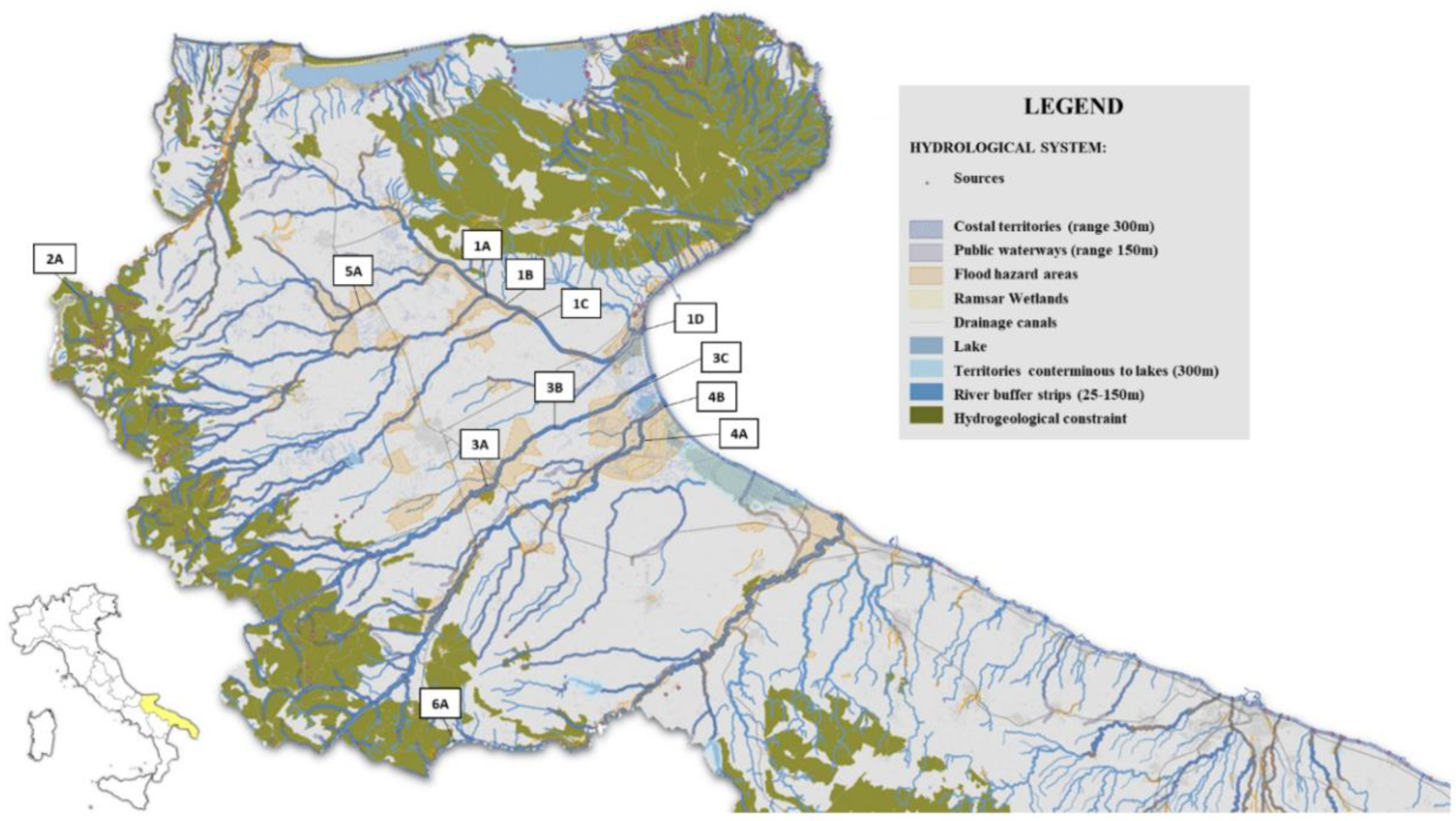
Figure 1. Map of rivers sampling points.
Preprints 194132 g001
Figure 2. Map of lakes sampling points.
Preprints 194132 g002
Figure 3. Heatmap of AMR profiles of Escherichia coli and K. pneumoniae isolated.
Preprints 194132 g003
Table 1. Sampling points and geographical coordinates.
N. Sampling points Surface water bodies of
Apulia Region
LAT (degrees, minutes, seconds-milliseconds) LONG (degrees, minutes, seconds-milliseconds) Annual average E. coli count
(CFU/100 mL)1
Municipality
1 1A Candelaro River 41°37' 34" N 15°38' 7" E 4715,8 San Marco in Lamis
2 1B Candelaro River 41°36' 36" N 15°40' 4" E 12374,8 San Marco in Lamis
3 1C Candelaro River 41°35' 58" N 15°42' 18" E 2240,0 San Marco in Lamis
4 1D Candelaro stream 41°34' 25" N 15°53' 6" E 1795,8 Manfredonia
5 2A Fortore River 41°38' 50" N 15°2' 40" E 505,6 Casalnuovo Monterotaro
6 3A Cervaro River 41°24' 4" N 15°39' 8" E 679,0 Foggia
7 3B Cervaro River 41°25' 37" N 15°40' 4" E 6228,8 Foggia
8 3C Cervaro River- 41°31' 17" N 15°53' 55" E 516,7 Manfredonia
9 4A Carapelle Torrent 41°23' 51" N 15°48' 51" E 245,1 Cerignola
10 4B Carapelle Torrent 41° 29' 26" N 15°55' 14" E 2420,8 Zapponeta
11 5A Triolo Torrent 41° 38' 51" N 15°32' 44" E 7749,2 Rignano Garganico
12 6A Ofanto River 41° 05' 29" N 15° 34' 20" E 1434,3 Rocchetta S.Antonio
13 7A Cammarata canal (Lesina lake) 41° 51' 34" N 15°21' 27" E data not available Lesina
14 7B La Fara canal (Lesina lake) 41° 51' 48" N 15° 21' 24" E data not available Lesina
15 7C Cammarata canal (Lesina lake) 41° 51' 54" N 15°21'19" E data not available Lesina
16 8A San Francesco canal (Varano lake) 41° 50 '17" N 15°46' 32" E data not available Varano
17 8B San Francesco canal (Varano lake) 41°50'34"N 15°45'07" E data not available Varano
18 8C San Francesco canal (Varano lake) 41°50'37" N 15°46'25"E data not available Varano
1.A limit of < 5,000 CFU/100 mL is recommended.
Table 2. Occurrence of ESBL- and carbapenemase-encoding genes in E. coli and K. pneumoniae isolated on selective agar plates.
Organism No. of PCR-positive isolates Resistance genes in Enterobacterales isolates
blaCTX-M CRE* (25/121, 20.6%)
blaOXA-48 blaKPC blaVIM blaCTXM+ blaKPC blaCTX-M+ blaVIM blaCTX-M-+blaOXA-48
K. pneumoniae 51/75 (68%) 40/51 (78.4%) 1/51
(2%)
- 5/51 (9.8%) 3/51
(5.9%)
2/51
(3.9%)
-
E. coli 70/101 (69.3%) 56/70 (80%) 3/70 (4.3%) 2/70 (2.9%) 6/70 (8.6%) - 1/70
(1.4%)
2/70
(2.9%)
Total No. 121/176 (68.8%) 96/121 (79.3%) 4/121
(3.3%)
2/121 (1.7%) 11/121
(9.1%)
3/121
(2.5%)
3/121
(2.5%)
2/121 (1.7%)
* CRE, carbapenem-resistant Enterobacterales.
Table 3. Antimicrobial susceptibility profiles of presumptive ESBL- and carbapenemase-producing Enterobacterales isolates.
Moleculesa No. of E. coli categorized as resistant (%) No. of K. pneumoniae categorized as resistant (%) MIC*
(mg/L)
E. coli
MIC*
(mg/L)
K. pneumoniae
Penicillins AMP 67/70 (95.71) - >8 **
PIP 67/70 (94.28) - >8 **
Penicillin derivatives*** TRM 10/70 (14.28) 8/51 (15.68) >16 >8
Aminoglycosides TOB 3/70 (4.28) 14/51 (27.45) >4 >2
AMI 1/70 (1.42) 1/51 (1.96) >8 >8
GEN 4/70 (5.71) 15/51 (29.41) >2 >2
Fluoroquinolones CIP 16/70 (22.85) 19/51 (37.25) >0.06 >0.125
LEVO 16/70 (21.42) 11/51 (21.56) >0.125 >0.25
First generation cephalosporins FAZ 65/70 (92.85) 46/51 (90.19) >4 >4
Second-generation cephalosporins FOX 9/70 (12.85) 9/51 (17.64) >1 >8
Third- and fourth-generation AXO 62/70 (88.57) 45/51 (88.23) >2 >2
FEP 66/70 (94.28) 21/51 (41.17) >0.25 >0.125
Tetracyclines TET 39/70 (55.71) 22/51 (43.13) >8 >8
MIN 2/70 (2.85) 8/51 (15.68) >4 >8
TGC 3/70 (4.28) 2/51 (3.92) >0.5 >2
Sulfonamides SXT 34/70 (48.57) 36/51 (70.58) >4 >4
Monobactam AZT 48/70 (68.57) 9/51 (17.64) >0.25 >4
Carbapenems DOR 1/70 (1.42) 10/51 (19.60) >0.06 >0.125
ETP 9/70 (12.85) 24/51 (47.05) >0.06 >0.03
IMI 7/70 (10.00) 7/51 (13.72) >0.5 >1
MERO 7/70 (7.14) 8/51 (15.68) >0.06 >0.125
Nitrofurans NIT 2/70 (2.85) - >64 **
ESBL confirmatory tests FOT 67/70 (94.28) 50/51 (98.03) >0.25 >0.25
TAZ 67/70 (94.28) 45/51 (88.23) >1 >1
F/C 16/70 (22.85) 42/51 (82.35) >0.25 >0.25
T/C 18/70 (25.71) 42/51 (82.35) >1 >0.5
a Abbreviations: AMP, ampicillin; PIP, piperacillin; TRM, temocillin; TOB, tobramycin; AMI, amikacin; GEN, gentamicin; CIP, ciprofloxacin; LEVO, levofloxacin; FAZ, cefazolin; FOX, cefoxitin; AXO, ceftriaxone; FEP, cefepime; TET, tetracycline; MIN, minocycline; TGC, tigecycline; SXT, trimethoprim-sulfamethoxazole; AZT, aztreonam; DOR, doripenem; ETP, ertapenem; IMI, imipenem; MERO, meropenem; NIT, nitrofurantoin. ESBL confirmatory tests: FOT, cefotaxime; TAZ, ceftazidime; F/C, cefotaxime-clavulanic acid; T/C, ceftazidime-clavulanic acid. * The MIC values reported in the table represent the epidemiological cut-off values (ECOFFs) used for categorisation, except for ESBL confirmatory tests, which were interpreted according to EUCAST phenotypic criteria. **For K. pneumoniae, no ECOFFs were applied for ampicillin, piperacillin and nitrofurantoin, as this species is considered intrinsically resistant or not a target organism according to EUCAST. *** Temocillin susceptibility was interpreted as an ESBL screening marker according to EUCAST recommendations.
Table 4. Whole-genome sequencing results of 25 presumptive carbapenemase-producing Enterobacterales.
N. Sampling points Date ID Species Multiplex PCR results Carbapenemase ESBL_AmpC Other AMR genes MLST Plasmid type
1 1B 11/23 IZSPB_EC01 E. coli blaCTX-M
blaOXA-48
blaOXA-244 blaCTX-M-15 blaTEM-1B, qnrS1, aph(6)-Id, aph(3'')-Ib, sul2, dfrA14, tet(A) ST10 IncFIB(K), IncFIB(AP0019)
2 1B 06/24 IZSPB_KP01 K. pneumoniae blaCTX-M blaOXA-48 blaKPC-3 - blaTEM-1A, blaOXA-9, aadA3, aac(3)-IIa, aadA2, aph(3')-Ia, aac(6')-Ib, mph(A), sul1, dfrA12, catA1 ST512 IncFIB(K), ColRNAI, IncQ1, IncFIB(pQil)
3 1B 07/24 IZSPB_EC02 E. coli blaVIM - blaTEM-52B ant(3'')-Ia, lnu(F) ST602 IncX1, IncI1-I(Alpha)
4 1D 02/24 IZSPB_EC03 E. coli blaOXA-48 blaOXA-244 - - ST43 IncFIB(AP0019)
5 1D 05/24 IZSPB_EC04 E. coli blaKPC blaKPC-3 - - ST1139 IncI(Gamma), IncFIB(pQil)
6 1D 05/24 IZSPB_KP02 K. pneumoniae blaCTX-MblaKPC blaKPC-3 blaSHV-28, blaCTX-M-15 blaTEM-1A, blaOXA-1, blaOXA-9, qnrB1, aac(3)-IIa, aph(6)-Id, aph(3'')-Ib, sul2, dfrA14, aac(6')-Ib-cr ST307 IncFIB(K), IncN4, IncFIB(pQil)
7 2A 02/24 IZSPB_KP03 K. pneumoniae blaOXA-48 - - - ST3442 IncFIB(K)(pCA), RepB
8 3B 02/24 IZSPB_KP04 K. pneumoniae blaVIM blaVIM-1 blaSHV-12 qnrS1, aph(3')-XV, mph(A), sul1, dfrA14, catB2 ST1537 IncA
9 3B 07/24 IZSPB_KP05 K. pneumoniae blaCTX-MblaKPC blaKPC-3 blaTEM-1A, blaOXA-9, tet(D) ST45 IncFIB(K), Col440II, ColRNAI, IncFIB(pQil)
10 3B 08/24 IZSPB_KP06 K. pneumoniae blaVIM - blaDHA-1 blaOKP-B-2, qnrB4, sul1, dfrA1 ST2059 IncFIB(K), IncR, Col440I
11 3B 09/24 IZSPB_KP07 K. pneumoniae blaVIM - - - ST983 negative
12 3B 09/24 IZSPB_EC05 E. coli blaVIM - - blaTEM-1B, aph(3'')-Ib, tet(B) ST Novel* IncFIB (AP0019)
13 4A 03/24 IZSPB_EC06 E. coli blaOXA-48 blaOXA-181 - blaTEM-1B, aac(3)-IId, aph(6)-Id, aph(3')-Ia, aph(3'')-Ib, aadA5, mph(A), erm(B), sul1, sul2, dfrA17, floR, catA1 ST542 IncFII, IncX1, IncX1, Col (BS512), Col156, IncQ1
14 5A 02/24 IZSPB_EC07 E. coli blaOXA-48 blaOXA-244 - - ST746 negative
15 5A 02/24 IZSPB_EC08 E. coli blaVIM blaVIM-4 - - ST Novel* IncFIB(K), IncFIA(HI1)
16 5A 02/24 IZSPB_KP08 K. pneumoniae blaVIM blaVIM-1 - aph(3')-XV, mph(A), sul1, catB2 ST Novel* repB(R1701), IncFIB(K) (pCA), IncFIB(pKPHS1, Col440I
17 5A 03/24 IZSPB_KP09 K. pneumoniae blaVIM blaVIM-1 - qnrS1, aph(3')-XV, mph(A), sul1, dfrA14, catB2 ST34 IncFIB(K), IncN, IncR
18 5A 03/24 IZSPB_EC09 E. coli blaOXA-48 blaOXA-244 blaCTX-M-15 blaTEM-1B, qnrS1, aph(6)-Id, aph(3'')-Ib, sul2, dfrA14, tet(A) ST540 IncFIB(K), IncFIB(AP0019)
19 5A 03/24 IZSPB_KP10 K. pneumoniae blaCTX-MblaVIM - blaCTX-M-15 qnrS1, aph(6)-Id, aadA2, aph(3')-Ia, aph(3'')-Ib, mph(A), sul1, sul2, dfrA12, catA2 ST469 IncFIB(K)(pCA), IncFIB(pKPHS1, RepB
20 5A 04/24 IZSPB_EC10 E. coli blaCTX-MblaVIM blaVIM-4 - - ST1721 negative
21 5A 04/24 IZSPB_KP11 K. pneumoniae blaVIM blaVIM-1 - aph(3')-XV, sul1, catB2 ST Novel* IncFIB(K), IncFIA(HI1), IncX1, IncX1, IncR, Col(pHAD28), Col440I, Col(pHAD28)
22 5A 06/24 IZSPB_EC11 E. coli blaVIM blaVIM-4 - - ST1721 IncFIB(pHCM2)
23 8A 08/24 IZSPB_EC12 E. coli blaVIM - blaSHV-12 qnrS1, aph(3'')-Ib, tet(A), floR ST2144 IncFIA, IncFIA, IncX1, IncX1, ColpVC, IncFIB(AP0019)
24 8B 02/24 IZSPB_EC13 E. coli blaKPC blaKPC-3 - - ST141 IncN
25 8B 08/24 IZSPB_EC14 E. coli blaVIM - blaSHV-12 qnrS1, aph(3'')-Ib, tet(A), floR ST2144 IncFII(29), IncFIA, IncFIA, IncX1, IncX1, ColpVC, IncFIB(AP0019)
Disclaimer/Publisher’s Note: The statements, opinions and data contained in all publications are solely those of the individual author(s) and contributor(s) and not of MDPI and/or the editor(s). MDPI and/or the editor(s) disclaim responsibility for any injury to people or property resulting from any ideas, methods, instructions or products referred to in the content.
Copyright: This open access article is published under a Creative Commons CC BY 4.0 license, which permit the free download, distribution, and reuse, provided that the author and preprint are cited in any reuse.
Prerpints.org logo

Preprints.org is a free preprint server supported by MDPI in Basel, Switzerland.

Subscribe

Disclaimer

Terms of Use

Privacy Policy

Privacy Settings

© 2026 MDPI (Basel, Switzerland) unless otherwise stated