Preprint
Article

This version is not peer-reviewed.

Design of DEGDA-Crosslinked Semi-IPN Dextran/Inulin Hydrogels for Antitumor Drug Delivery

A peer-reviewed article of this preprint also exists.

Submitted:

23 December 2025

Posted:

23 December 2025

You are already at the latest version

Abstract

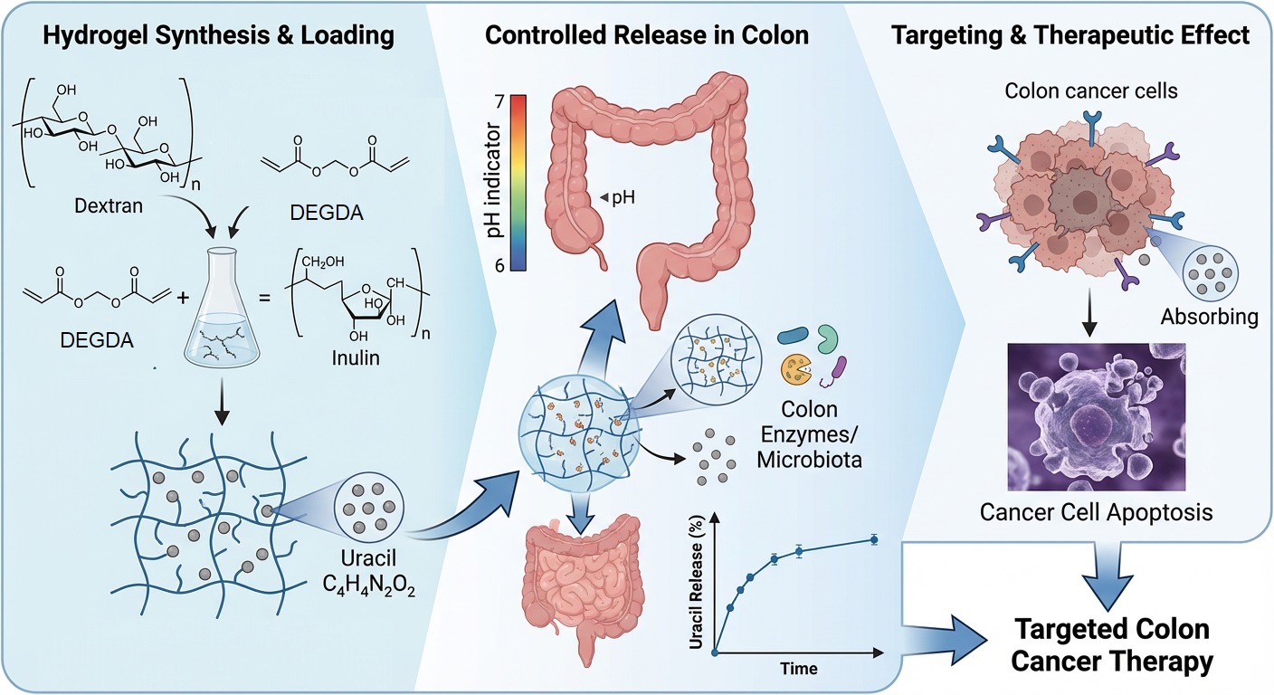
Colon-targeted drug delivery systems are of considerable interest for improving the therapeutic efficacy of anticancer agents while minimizing systemic side effects. In this study, semi-interpenetrating polymer network (semi-IPN) hydrogels based on methacrylated dextran and native inulin were designed as biodegradable carriers for the colon-specific delivery of uracil as a model antitumor compound. The hydrogels were synthesized via free-radical polymerization, using diethylene glycol diacrylate (DEGDA) as a crosslinking agent at varying concentrations (5, 7.5, and 10 wt%), and their structural, thermal, and biological properties were systematically evaluated. Fourier transform infrared spectroscopy (FTIR) confirmed successful crosslinking and physical incorporation of uracil through hydrogen bonding. At the same time, differential scanning calorimetry (DSC) revealed an increase in glass transition temperature (Tg) with increasing crosslinking density (149, 153, and 156 °C, respectively). Swelling studies demonstrated relaxation-controlled, first-order swelling kinetics under physiological conditions (pH 7.4, 37 °C), and high gel fraction values (84.75, 91.34, 94.90%, respectively) indicated stable network formation. All formulations exhibited high encapsulation efficiencies (>86%), which increased with increasing crosslinker content, consistent with the observed gel fraction values. Simulated in vitro gastrointestinal digestion showed negligible drug release under gastric conditions and controlled release in the intestinal phase, primarily governed by crosslinking density. Antimicrobial assessment against Escherichia coli and Staphylococcus epidermidis, used as an initial or indirect indicator of cytotoxic potential, revealed no inhibitory activity, suggesting low biological reactivity at the screening level. Overall, the results indicate that DEGDA-crosslinked dextran/inulin semi-interpenetrating polymer network (semi-IPN) hydrogels represent promising carriers for colon-targeted antitumor drug delivery.

Keywords: 
;  ;  ;  

1. Introduction

According to GLOBOCAN 2022 data, colorectal cancer ranks third in incidence worldwide, following breast and lung cancer, and second in terms of cancer-related mortality. Chemotherapy remains a cornerstone of colorectal cancer treatment. However, due to the non-selective nature of most anticancer drugs, which exhibit comparable affinity toward both healthy and malignant cells, severe systemic side effects and limited patient tolerance are frequently observed. This limitation is particularly critical in patients over the age of 55, who represent the predominant demographic affected by this disease [1]. To minimize adverse effects and maximize the therapeutic concentration of the drug at the tumor site, the development of suitable drug carriers plays a crucial role. These systems are primarily designed to encapsulate the active compound and enable controlled and site-specific release through different mechanisms, largely dictated by the structural and physicochemical design of the carrier. Nanoparticle-based systems can facilitate cellular internalization, thereby increasing drug accumulation within tumor cells. In addition, certain carriers can be engineered to preferentially accumulate in cancer tissue through passive or active targeting mechanisms [2]. Several research efforts have focused on designing micelles, coating systems, conjugates, complexes, microparticles, and nanoparticles for drug encapsulation and controlled delivery. However, such approaches are often complex, time-consuming, and require advanced expertise and substantial investment [3]. Also, some of them are limited to one group of drugs. To overcome these limitations, simply synthesized hydrogels capable of entrapping high amounts of drug have emerged as promising materials for controlled drug delivery. Depending on their composition, particularly the presence of ionizable functional groups, hydrogels can release the active compound in response to specific pH values characteristic of tumor tissue. Nevertheless, pH-triggered release alone is not sufficiently reliable, given the high variability in pH across different segments of the digestive tract and the inability to achieve a quantitatively controlled release at the target site. For this reason, hydrogel systems designed to degrade specifically in the colorectal region offer a more efficient strategy for targeted drug delivery. For effective colon-targeted drug delivery, active compounds must reach the colorectal region intact [4,5,6]. This can be achieved by formulating hydrogels from biopolymers that remain resistant to degradation in the upper gastrointestinal tract but are selectively degraded in the intestinal phase by enzymes present in the colonic microflora. Dextran and inulin, both generally recognized as safe (GRAS), are excellent candidates for developing colon-targeted delivery systems. They can form matrices that resist enzymatic degradation under gastric conditions, enabling drug release in the intestinal environment through biodegradation mediated by colorectal microbiota enzymes. The 1,6-α-D-glucosidic linkages in dextran are susceptible to hydrolysis by the action of dextranase enzymes [7]. In the human colon, these enzymes are predominantly produced by anaerobic gram-negative bacteria of the genus Bacteroides [8], which represent the dominant members of the anaerobic intestinal microbiota, making up approximately 30% of the total cultured microflora [9]. At physiological pH (7.0–7.5), the main effect of the enzyme activity is reflected in the rapid release of D-glucose, where the degradation is mainly limited to the outer parts of the molecule. The specific activity of dextranase has been determined in the mucous membrane of the human intestine: in the small intestine it is related to intracellular enzymes of mammalian cells, while in the large intestine the dominant activity is related to the surface of the cell membrane of bacteria of the genus Bacteroides or to the enzymes that these bacteria secrete into the external environment [10]. Among polysaccharides degradable by intestinal microflora, inulin has recently attracted considerable attention due to its potential in targeted drug delivery to the large intestine and numerous beneficial physiological effects [11]. Inulin is a natural fructan, a complex biopolymer composed of β-(2→1)-linked fructose units, and ranks among the most abundant carbohydrates in nature after starch. This biopolymer is a dietary prebiotic fiber and the most abundant polysaccharides that occur in nature after starch [12]. As a dietary prebiotic fiber, inulin is resistant to hydrolysis by gastric and small-intestinal enzymes, thus reaching the colon intact, where it is fermented by inulinases and inulin-lyases produced by Bifidobacteria—gram-positive bacteria that are natural inhabitants of the large intestine [13]. Since 1995, numerous studies have demonstrated the potential of dextran- and inulin-based hydrogels for the colon-targeted delivery of active compounds. Hovgaard and Brøndsted synthesized carriers based on aliphatic diisocyanate-crosslinked dextran, which showed biodegradability in a human colonic fermentation model, confirming the potential of dextran derivatives for targeted drug delivery in the distal parts of the gastrointestinal tract [14]. In a related approach, Kim et al. reported the development of hydrogels composed of glycidyl methacrylate–modified dextran and poly(acrylic acid), synthesized via UV-initiated polymerization for colon-specific drug delivery applications [15]. Numerous subsequent studies highlighted inulin hydrogels as effective carriers for the controlled and targeted release of antitumor drugs and bioactive substances in the colon. In order to achieve an inulin hydrogel with low swelling capacity and high stability in acidic environments, Van den Mooter et al. modified inulin with methacrylic anhydride, after which they derivatized it with succinic acid and cross-linked it with UV radiation [16]. In another study, hydrogels were obtained by esterification of inulin with pyromellitic dianhydride at room temperature in dimethylformamide (DMF) [17]. In the study by Maris et al., hydrogels were synthesized by copolymerizing methacrylated inulin (IN-MA) with bis(methacryloylamino)azobenzene (BMAAB), an aromatic azo agent, and with 2-hydroxyethyl methacrylate (HEMA) or methacrylic acid (MA), thus obtaining pH- and enzyme-sensitive systems suitable for targeted drug delivery to the colon [18].
In our previous study [19], we investigated the influence of inulin incorporation into dextran-based hydrogel formulations on their digestion behavior, concluding that the presence of inulin inhibited the degradation of hydrogels synthesized in aqueous media, applying an energy-efficient method for synthesizing methacrylated dextran/methacrylated inulin-based hydrogels at room temperature. This study demonstrated that inulin addition provided a protective effect against hydrogel degradation under simulated gastric conditions. In our next study [20], various crosslinking agents were evaluated for the preparation of dextran-based hydrogels in dimethyl sulfoxide, revealing that diethylene glycol diacrylate (DEGDA) produced hydrogels with the most favorable morphological characteristics. Therefore, in the present work, we employed a design strategy based on a semi-interpenetrating biopolymer hydrogel composed of methacrylated dextran and native inulin, using DEGDA as a crosslinking agent, to encapsulate uracil as a model for an antitumor drug, representing a logical continuation and integration of our previously reported results.

2. Results and Discussion

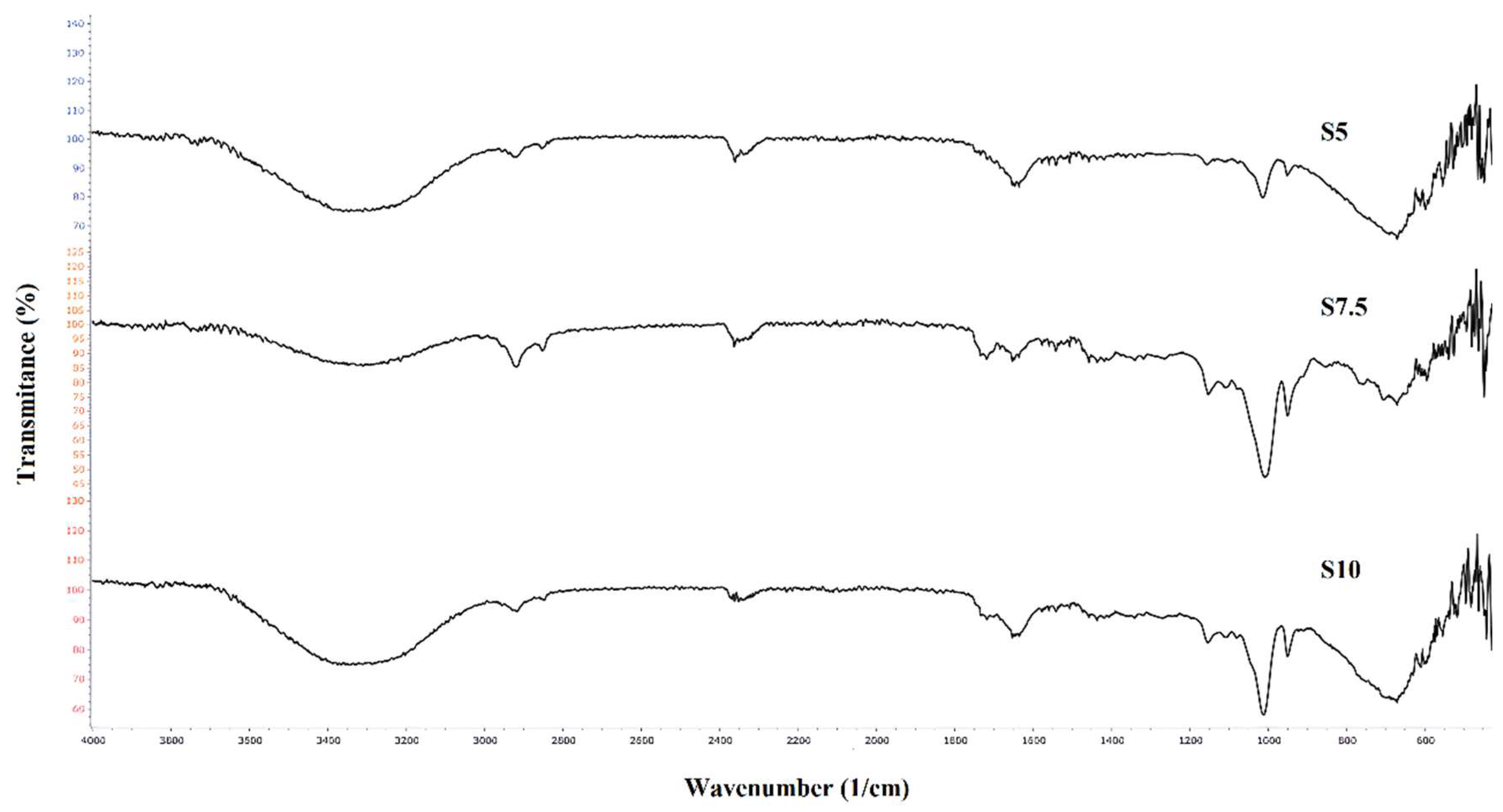
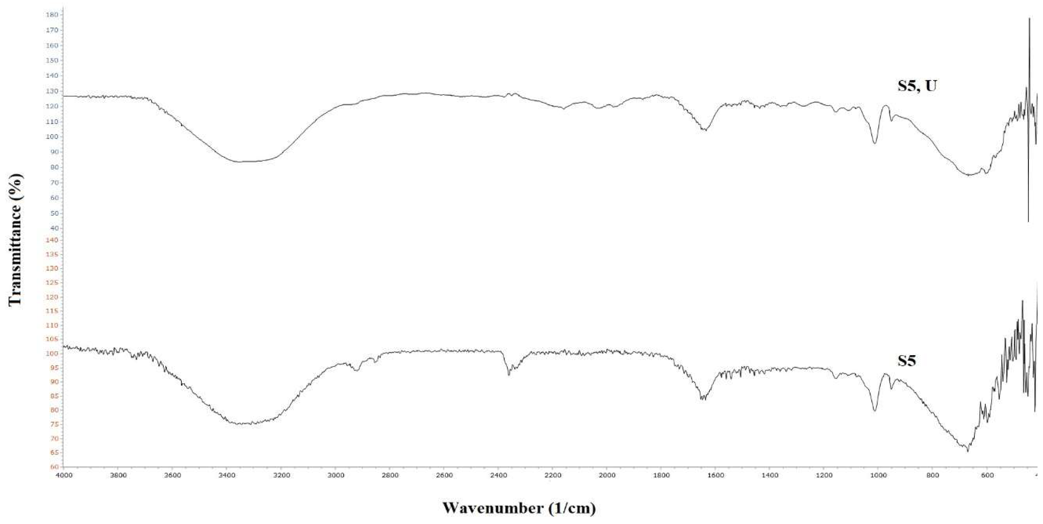
2.1. Results of Infrared spectroscopy with Fourier transformation (FTIR) analysis

The FTIR spectra of hydrogels Dex-DEGDA 5 (S5), Dex-DEGDA 7.5 (S7.5), and Dex-DEGDA 10 (S10) are presented in Figure 1 and show no significant differences among samples of identical composition containing different amounts of crosslinking agent. A broad band centered at 3310–3312 cm⁻¹ corresponds to overlapping O–H stretching vibrations originating from dextran, inulin, and residual hydroxyl groups within the network. Two peaks in the 2900–2850 cm⁻¹ region are attributed to C–H stretching of CH₂ groups originating from methacrylated dextran, inulin (Figure S1, Supporting Information), and DEGDA (Figure S2, Supporting Information). The weak bands observed at 2158 and 2027 cm⁻¹ in the FTIR spectrum of neat DEGDA—assigned to overtone/combination bands associated with the acrylate C=C system—are not present in the hydrogel spectra, confirming the consumption of vinyl groups during crosslinking. The band at 1719 cm⁻¹ corresponds to the ester carbonyl stretching vibration of methacrylated dextran and DEGDA. It becomes more pronounced in hydrogels with higher DEGDA content, consistent with its characteristic presence in the DEGDA spectrum (Figure S2, Supporting Information). The peak at 1652–1650 cm⁻¹ is attributed to the bending vibration of absorbed/bound water. Peaks in the 1435–1335 cm⁻¹ range correspond to CH₂ and CH₃ bending modes. The band at 1250–1150 cm⁻¹ originates from asymmetric C–O–C stretching vibrations of ester and ether groups, while the peak at 1155–1151 cm⁻¹ corresponds specifically to C–O–C asymmetric stretching. The band at 1113–1107 cm⁻¹ is assigned to C–C–O stretching coupled with O–H bending vibrations. A peak at 1012 cm-1 corresponds to asymmetric C–O–C stretching of glycosidic linkages in dextran and inulin coupled with C–O stretching of secondary OH group. The peak at 951 cm⁻¹, visible in all spectra, corresponds to vibrations of the pyranose ring and C–O–C (glycosidic) stretching
Figure 2 compares the FTIR spectra of the neat S5 hydrogel and uracil–loaded S5 hydrogel. Upon uracil loading, the band corresponding to the O–H stretching vibrations originating from dextran, inulin, and residual hydroxyl groups becomes slightly broader and shifted toward lower wavenumbers, suggesting the formation of additional hydrogen bonds between the hydroxyl groups of the biopolymers and the carbonyl and amide functionalities of uracil. In the FTIR spectrum of uracil-loaded hydrogel, two weak peaks appear at 2158 and 2027 cm-1, arising probably from the combination of C=O stretching, and N–H in-plane bending (scissoring). Overall, the observed band shifts and intensity variations confirm successful uracil encapsulation, governed by hydrogen bonding. This mode of incorporation is consistent with the high encapsulation efficiency and suggests that uracil is physically entrapped within the hydrogel matrix without disrupting the crosslinked network architecture.

2.2. Results of DSC analysis

The glass transition temperature (Tg) increases with increasing content of the crosslinking agent (Figure 3), reflecting a direct correlation between the amount of DEGDA in the hydrogel formulation and the resulting crosslinking density [21]. Accordingly, the hydrogel containing the lowest DEGDA content (S5) exhibits the lowest Tg value (149 °C). A progressive increase in crosslinker concentration, and thus crosslinking density, results in higher Tg values, reaching 153 °C and 156 °C for hydrogels S7.5 and S10, respectively. Thermal degradation of the hydrogels occurs in the temperature range of 190–202 °C. Given that the Tg values of all hydrogels are substantially higher than the temperature of the swelling medium, polymer chain mobility is highly restricted under swelling conditions. As a result, polymer chain relaxation proceeds more slowly than solvent diffusion within the hydrogel network, indicating that the swelling behavior is relaxation-controlled and follows a non-Fickian mechanism [22].

2.3. Results of swelling properties analysis

The swelling ratio versus time is presented in Figure 4. Equilibrium swelling ratio (ESR) values increase with decreasing crosslinking agent content, reflecting the reduced network density and greater chain mobility at lower DEGDA concentrations [21]. In comparison with the swelling behaviour of the dextran-based hydrogel prepared with 10 wt% DEGDA reported in our previous study [20], the semi-interpenetrating hydrogel of comparable composition (S10) exhibits a similar swelling profile, indicating that the introduction of the secondary polymer does not significantly disturb the overall network integrity at this crosslinking level. Furthermore, the incorporation of inulin increases its hydrophilicity and the presence of multiple hydroxyl groups capable of forming hydrogen bonds with water molecules. However, at pH 7.4, this hydrogel exhibits a slightly lower ESR value than at pH 6, due to the differences in biopolymer–biopolymer and biopolymer–buffer interactions under these conditions. At pH 6, enhanced hydrogen bonding between hydrophilic functional groups and water molecules promotes higher water uptake and network expansion. In contrast, at pH 7.4, the swelling is reduced, which may be related to a partial screening of hydrophilic groups and a more compact network structure, leading to decreased osmotic driving force for water diffusion into the hydrogel [23]. Swelling measurements were done in triplicate, and the results of the statistical analysis were given in the Supporting Information (Table S1).
To evaluate whether the swelling behavior of the prepared hydrogels follows first-order kinetics, the values of ln [Se/(Se-S)] calculated according to Equation 4 were plotted as a function of time (Figure S3, Supporting Information). The first-order swelling rate constants (Table 1) were determined as the absolute values of the slopes of the corresponding linear regressions [22]. For all investigated samples, a linear relationship was obtained at pH 7.4 and a temperature of 37 °C, indicating good agreement with the first-order kinetic model and confirming that the swelling process of the hydrogels can be adequately described by first-order kinetics under the applied conditions. The linearity of the plots was further confirmed by high coefficients of determination (R2 = 0.97-0.99, Table 1), indicating an excellent agreement between the experimental swelling data and the first-order kinetic model for all hydrogel samples at pH 7.4 and 37 °C. The obtained values demonstrate a strong correlation between the experimental results and the applied kinetic model, thereby confirming the reliability and applicability of the first-order approach for describing the swelling behavior of the prepared hydrogels.

2.4. Results of gel fraction (GF) determination

The gel fraction (GF) increased with increasing content of the crosslinking agent, reflecting the corresponding increase in crosslinking density within the hydrogel network (Table 2). For all investigated samples, the GF values exceeded 84%, indicating a high degree of network formation. Notably, hydrogel S10, which contained the highest amount of crosslinking agent, exhibited the highest GF value, confirming the formation of a densely crosslinked and stable hydrogel structure. Measurements were done in triplicate, and the statistical analysis was provided in the Supporting Information (Table S2).

2.5. Results of encapsulation efficiency (EE) determination

Encapsulation efficiency (EE) values increase with increasing crosslinking agent content (Table 3). Accordingly, the hydrogel synthesized with 10 wt% DEGDA (S10) exhibits the highest EE, which can be attributed to its highest crosslinking density. Nevertheless, all samples show relatively high EE values (>86%), indicating efficient encapsulation across the entire series and confirming that the applied synthesis and loading procedure is well-suited for the incorporation of the active compound, even at lower degrees of crosslinking. Measurements were done in triplicate, and the statistical analysis was provided in the Supporting Information (Table S3).

2.6. Results of simulated in vitro gastrointestinal digestion (GID)

The results of the in vitro digestion process, expressed as the amount of released uracil, are presented in Table 4. The lowest uracil release was observed for sample S10, which can be attributed to its highest crosslinking density. In comparison with our previous study [20], this value is very close to that obtained for a dextran-based hydrogel synthesized with the same DEGDA content but without the addition of inulin. This finding indicates that the incorporation of inulin into the semi-interpenetrating hydrogel network does not significantly influence uracil release under identical conditions. In contrast, hydrogels synthesized with lower DEGDA contents exhibited uracil release already during the gastric phase, which is most likely a consequence of reduced entrapping efficiency associated with a lower crosslinking density. Furthermore, the release behavior in the intestinal phase was inversely related to the crosslinking density, with higher DEGDA contents leading to a decreased uracil release.

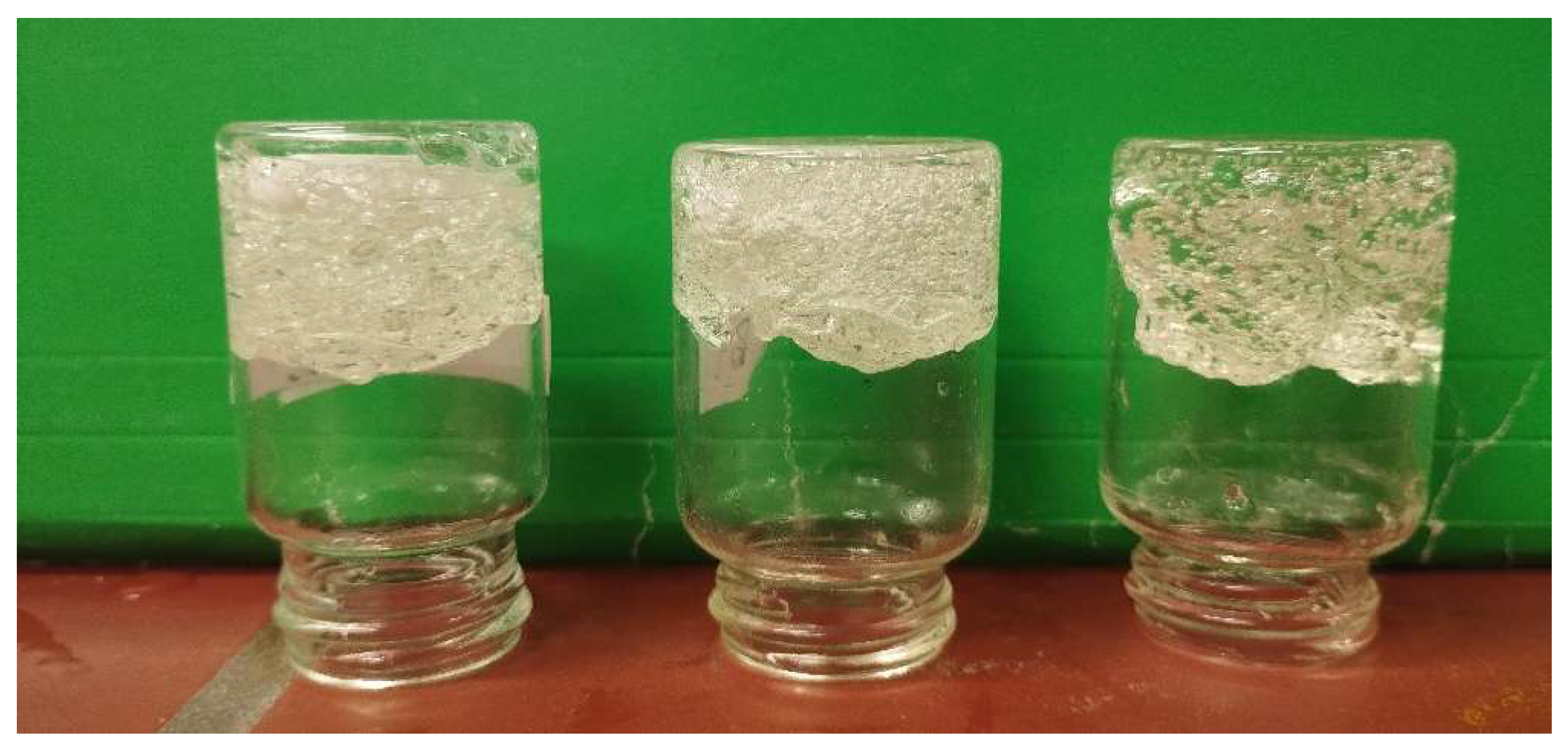
2.7. Results of antimicrobial assessment

In accordance with the principles of ISO 10993 [24] for the biological evaluation of materials, antimicrobial activity testing may be applied as an initial screening tool to assess the potential biological reactivity of newly developed hydrogels. Considering these aspects, Escherichia coli ATCC 8739 (Gram-negative) and Staphylococcus epidermidis ATCC 12228 (Gram-positive) generally exhibit higher sensitivity to external stressors than mammalian cells; therefore, the antibacterial activity of prepared hydrogels was examined using these model microorganisms. The observed differences in sensitivity originate from inherent biological and structural distinctions between prokaryotic and eukaryotic cells. Bacterial cells are characterized by a rigid cell wall containing peptidoglycan and, in the case of Gram-negative bacteria, an additional outer membrane, rendering them more susceptible to damage induced by reactive species, osmotic imbalance, or membrane-disrupting agents. In contrast, mammalian cells possess more robust and tightly controlled plasma membranes, along with complex intracellular regulatory and repair systems. Moreover, bacteria have a limited ability to tolerate environmental stress and therefore experience a rapid loss of viability under adverse physicochemical conditions. Mammalian cells, however, are capable of activating adaptive survival mechanisms, including enhanced oxidative stress resistance, regulated apoptotic pathways, and efficient repair of cellular damage. As a result, a wide range of materials—such as polymers, crosslinkers, and nanoparticles—can exert pronounced antibacterial effects at concentrations that remain non-toxic to mammalian cells, with microbial inhibition occurring at substantially lower exposure levels [25].
The disc diffusion assay provides information on the release of low-molecular-weight or diffusible components from the hydrogel matrix that are capable of interfering with cellular integrity or metabolic activity. Since such effects may arise from non-selective mechanisms that could also affect mammalian cells, pronounced antibacterial activity may indicate a potential risk of cytotoxicity. Antimicrobial activity was assessed by measuring the diameter of the inhibition zone, and the results are summarized in Table 5. The absence of antimicrobial activity in the hydrogel samples strongly indicates that they are unlikely to exhibit cytotoxic effects toward mammalian cells [20]. Therefore, antimicrobial measurements can be considered a complementary and preliminary approach that may partially substitute cytotoxicity testing at the early stage of material development, particularly for comparative screening of formulations. This strategy allows prioritization of samples for further biological evaluation and reduces unnecessary use of mammalian cell-based assays.

3. Conclusions

Semi-interpenetrating biodegradable polymer network (semi-IPN) hydrogels based on methacrylated dextran and native inulin were successfully synthesized using three different amounts of diethylene glycol diacrylate (DEGDA) as a crosslinking agent. Prepared hydrogels were systematically evaluated as potential carriers for colon-targeted drug delivery. Structural and thermal analyses confirmed efficient crosslinking and the formation of stable hydrogel networks, with increasing crosslinking density resulting in higher glass transition temperatures, gel fraction values, and encapsulation efficiencies. The hydrogels exhibited controlled, relaxation-driven swelling behavior under physiological conditions (pH 7.4, 37 °C), and demonstrated high stability during simulated gastric digestion. Uracil release during in vitro gastrointestinal digestion was strongly governed by crosslinking density, with more densely crosslinked systems effectively preventing premature drug release and enabling controlled delivery in the intestinal phase. The absence of antimicrobial activity against both Gram-positive and Gram-negative bacteria suggests minimal release of biologically aggressive species, supporting the suitability of these materials for further biological evaluation.
From a future perspective, these results provide a solid foundation for advancing dextran/inulin semi-IPN hydrogels toward more comprehensive biological validation. Future studies will focus on advancing the in vitro gastrointestinal model by incorporating inulinase, as well as on performing standardized in vitro cytotoxicity testing in accordance with ISO 10993 guidelines. Further investigations will include evaluation of antitumor efficacy using relevant colorectal cancer cell lines and a detailed assessment of degradation behavior in the presence of colonic enzymes and microbiota. In addition, extending this hydrogel platform to encapsulate clinically relevant chemotherapeutic agents and evaluating its in vivo performance will be essential steps toward potential translational and clinical applications.

4. Materials and Methods

4.1. Materials

Dextran from Leuconostoc (Mw ≈ 40,000 g/mol, CAS 9004-54-0), inulin from chicory (Mw ≈ 500–3600 g/mol, CAS 9005-80-5; free glucose < 0.05% and free fructose < 0.05% determined by enzymatic assay; fructose-to-glucose ratio ≥ 20:1), glycidyl methacrylate, diethylene glycol diacrylate (DEGDA), initiator azobisobutyronitrile (AIBN), NaCl, NaHCO₃, KCl, KH₂PO₄, MgCl₂·6H₂O, (NH₄)₂CO₃, CaCl₂·2H₂O, Pefabloc®, dextranase, pepsin, bile salts, pancreatin, and uracil (analytical grade) were all obtained from Sigma-Aldrich (St. Louis, MO, USA). Dimethyl sulfoxide (DMSO) was obtained from Merck KGaA (Darmstadt, Germany). 4-Dimethylaminopyridine (4-DMAP) was purchased from Thermo Fisher Scientific Inc. (Waltham, MA, USA), whereas acetone and 96% ethanol were supplied by Zorka (Šabac, Serbia).

4.2. Preparation of hydrogels

Hydrogels were synthesized through a two-step procedure. In the first step, dextran was modified following the method described in our previous studies [19,20] to obtain dextran–methacrylate (Dex–MA). In the second step, Dex–MA and inulin were dissolved in DMSO at 50 °C in a relative mass ratio of 90:10, with a total polymer concentration of 20 wt%. After complete dissolution, di(ethylene glycol) diacrylate (DEGDA) was added as a crosslinker in varying amounts (5, 7.5, and 10 wt% relative to the total Dex-MA mass). The temperature was then increased to 80 °C, and 0.09 g of AIBN, dissolved in 1 mL of DMSO, was introduced as a free-radical initiator. The polymerization reaction proceeded for 20 minutes, after which the system reached the gel point, yielding the crosslinked hydrogel network (Figure 5). The degree of dextran methacrylation was reported in our previous study as approximately 1.2 hydroxyl groups per repeating unit modified with glycidyl methacrylate [19].
Drug-loaded hydrogels were prepared by in situ incorporation of uracil through the addition of 0.02 g of uracil to a DMSO solution containing methacrylated dextran and inulin, before the addition of AIBN. Uracil was chosen as a model compound due to its structural similarity to the anticancer drug 5-fluorouracil and its well-documented safety. This approach enables meaningful assessment of the drug delivery system under conditions that realistically reflect the behavior of 5-fluorouracil, while eliminating the risks and regulatory constraints associated with handling cytotoxic chemotherapeutic agents, as described in our previous studies [19,20].

4.3. Infrared spectroscopy with Fourier transformation (FTIR) analysis

FTIR analysis of the samples was conducted using a Shimadzu IRTracer-100 Fourier Transform Infrared Spectrometer (Kyoto, Japan) equipped with MIRacle 10 ATR-FTIR with ZnSe crystal. The analysis was performed over a wavelength range of 4000 to 400 cm⁻¹, averaging 40 scans at a spectral resolution of 4 cm⁻¹. Data were processed with LabSolutions IR software.

4.4. DSC analysis

To investigate the influence of the crosslinking agent content on the hydrogel glass transition temperature (Tg), differential scanning calorimetry (DSC) measurements were carried out using a TA Instruments Q20 equipment (New Castle, DE, USA). Samples were heated from 25 to 250 °C at a heating rate of 10 °C/min under a nitrogen atmosphere. The Tg values were determined from the midpoint of the heat-capacity step (∆Cp), with a standard uncertainty of u(T) = 0.5 °C.

4.5. Analysis of swelling properties

The swelling properties of colon-targeted hydrogel carriers synthesized using dextran, inulin, and DEGDA were reported in our previous study. In this study, we wanted to investigate the swelling capacity of prepared hydrogels at physiological pH 7.4, at 37 °C, in order to simulate intestinal conditions, under which enzymatic activity is most pronounced. At this pH range, enzymes involved in polysaccharide degradation exhibit high activity, which leads to the cleavage of glycosidic bonds and the rapid release of D-glucose. Monitoring the swelling behavior under these conditions, therefore, provides relevant insight into the structural stability and water uptake of the hydrogel network in an environment that closely mimics in vivo intestinal conditions. Equilibrium swelling ratio (ESR) was determined using Equation 1:
E S R   % = W t W i W i   · 100 %
Where mt is the weight of the gel after swelling for a given time (1, 2, 3, 4, or 5 hours), and mi initial weight of the dry gel (xerogel).
The swelling kinetics were evaluated under the assumption that the swelling process follows first-order kinetics, as described by Equation 2 [22]:
d S d t =   K 1 · ( S e S )
In this equation, S represents the swelling ratio at time t, S e denotes the equilibrium swelling ratio, and K 1   ( h 1 ) is the first-order kinetic constant. Integration of Equation (3) using the initial conditions t = 0 to t, and S = 0 to S yields Equation 3:
l n S e S S e =   K 1 · t

4.6. Gel fraction determination

Gel fraction, used as an indicator of the crosslinking degree achieved with different amounts of crosslinking agent, was determined according to Equation 4:
G f   =   W E S R W i · 100 %
Where WERS denotes the mass of the gel after reaching equilibrium swelling and subsequent drying to constant weight, while Wi represents the initial mass of the xerogel before immersion in the buffer.

4.7. Determination of encapsulation efficiency

For the determination of encapsulation efficiency, the method described by Erceg et al [19] was applied. Encapsulation efficiency (EE) of hydrogels has been determined using Equation 5:
E E   % = w t w i · 100 %
where wt represents the total amount of uracil incorporated into the xerogel, and wi represents the initial amount of uracil incorporated during hydrogel synthesis (0.2 g per gram of dry xerogel). The total uracil content was evaluated using a two-step extraction and quantification procedure. First, xerogel samples were submerged in DMSO for 12 h to enable complete uracil release, after which the suspensions were centrifuged and filtered to eliminate insoluble material. In the following step, uracil concentrations in the collected supernatants were measured by UV–Vis spectrophotometry Agilent BioTek EPOCH 2 (Santa Clara, California, USA). A calibration curve prepared from uracil standards dissolved in DMSO was used to ensure accurate determination. All analyses were conducted in triplicate, and the obtained data are presented in Table S3.

4.9. Simulated in vitro gastrointestinal digestion (GID)

A standardized static in vitro digestion model was applied following the protocol reported by Kostić et al. (2021) [26], with minor modifications introduced in the intestinal digestion stage. Because the hydrogel formulations are intended to be administered as hard gelatin capsules filled with xerogel granules, the oral phase of digestion was excluded from the experimental design. In this dosage form, the material is swallowed directly and does not undergo retention, disintegration, or interaction with saliva in the oral cavity before gastric release; therefore, inclusion of the oral phase was considered irrelevant. The procedure was described in our previous papers [19,20]. Three experimental groups were prepared in triplicate. Digestion was initiated with the gastric phase, with 0.25 g of xerogel (corresponding to 2.5 g of the rehydrated material) encapsulated in gelatin capsules (26.1 mm). The compositions of SGF and SIF were prepared according to the INFOGEST protocol [27] described in our previous studies [19,20].
Uracil quantification by UV–Vis spectrophotometry was carried out following a method modified from Khajehsharifi and Soleimanzadegan (2013) [28], in combination with procedures established in our earlier studies [19,20]. Spectral measurements were performed using an Agilent BioTek EPOCH 2 photodiode array UV–Vis spectrophotometer controlled by GEN5 software and fitted with a quartz cuvette of 1 cm optical path length. Sample preparation involved dispersing approximately 25 mg of material in 25 mL of distilled water, followed by sonication for 5 min to achieve complete dispersion. The resulting suspension was subsequently diluted to obtain a stock solution with a concentration of 100 mg/mL.

4.8. Antimicrobial assessment

As in our previous study [20], the antibacterial properties of the neat hydrogels were evaluated against Escherichia coli ATCC 8739 (Gram-negative) and Staphylococcus epidermidis ATCC 12228 (Gram-positive), which are frequently associated with bacterial infections. Lyophilized reference cultures were obtained from the American Type Culture Collection (ATCC, Manassas, USA) and maintained in the culture collection of the Institute of Food Technology, University of Novi Sad. Stock cultures were preserved at −80 °C in Trypto-Casein Soy Broth (TSB; Biokar BK046 HA, Beauvais, France) supplemented with 15% (v/v) glycerol. Before analysis, bacterial strains were revived by streaking onto tryptic soy agar (TSA; Oxoid CM0131, Hampshire, UK) and incubated for 24 h at 37 °C. For antimicrobial testing, freshly grown colonies were transferred into phosphate-buffered saline (PBS; Oxoid, Hampshire, UK; pH 7.3), and the cell density was adjusted to correspond to a 0.5 McFarland standard. The suspensions were subsequently diluted in TSB to obtain a final concentration of 1 × 10⁶ CFU/mL. Antibacterial activity of three dextran-based samples was determined using the disc diffusion assay, following the protocol described by Šuput et al. (2024) [30] with minor modifications. Ampicillin (Bioanalyse, Ankara, Turkey) was included as a positive control to verify bacterial susceptibility. Briefly, 100 µL of the standardized bacterial suspension (1 × 10⁶ CFU/mL) was uniformly spread onto TSA plates. Sterile paper discs (approximately 6 mm in diameter) were then placed on the agar surface and loaded with 10 µL of dextran-based sample stock solutions (256,000 µg/mL). The plates were incubated at 37 °C for 24 h, after which antibacterial efficacy was evaluated by measuring the diameter of the inhibition zones surrounding each disc. Results were expressed as mean inhibition zone diameters (mm), and all experiments were performed in triplicate for each bacterial strain.
Antimicrobial activity measurements can serve as an initial or indirect indicator of cytotoxic potential, particularly in the early-stage biological evaluation of hydrogel materials. The disc diffusion assay reflects the ability of compounds released from the hydrogel matrix to diffuse into the surrounding medium and disrupt cellular integrity. Since both bacterial and mammalian cells rely on membrane integrity and metabolic function for survival, pronounced antibacterial effects—especially against Gram-positive and Gram-negative strains may suggest non-selective cytotoxic mechanisms, such as membrane disruption or oxidative stress induction.

4.9. Statistical analysis

All measurements were performed in triplicate and are reported as mean values ± standard deviation. Statistical analysis of the experimental data was conducted using Microsoft Excel 2010. Mean values, standard deviations, and ranges for the swelling ratio, gel fraction, color parameters, and plateau elastic modulus were calculated in Excel, while the standard errors of the intercept and slope of the linearly fitted curves were also determined using Microsoft Excel 2010.

Author Contributions

Conceptualization, T.E., methodology, T.E.; M.R., R.T. software, T.E., M.R., A.T., validation, T.E. formal analysis, T.E., M.R.; investigation, T.E., M.R., M.M., A.C.K., A.T.; resources, T.E., M.R., M.M., A.C.K., A.T. data curation, T.E., M.R., A.C.K., A.T. writing—original draft preparation, T.E., M.R., writing—review and editing, T.E., M.R., R.T., A.C.K., A.T..; visualization, T.E..; supervision, T.E. project administration, A.T.; funding acquisition, A.T. All authors have read and agreed to the published version of the manuscript.

Funding

This work was supported by the project Interreg, co-founded by the European Union, „ Targeted delivery of antitumor drugs employing biodegradable carriers ABIDERS HR-RS00148“.

Conflicts of Interest

The authors declare no conflicts of interest.

Abbreviations

The following abbreviations are used in this manuscript:
DMF Dimethylformamide
BMAAM Bis(methacryloylamino)azobenzene
HEMA 2-hydroxyethyl methacrylate
MA Methacrylic acid
DMSO Dimethyl sulfoxide
4-DMAP 4-Dimethylaminopyridine
AIBN Azobisobutyronitrile
DEGDA Diethylene glycol diacrylate
Dex–MA Dextran-methacrylate

References

  1. Bray, F.; Laversanne, M.; Sung, H.; Ferlay, J.; Siegel, R.L.; Soerjomataram, I.; Jemal, A. Global Cancer Statistics 2022: GLOBOCAN Estimates of Incidence and Mortality Worldwide for 36 Cancers in 185 Countries. CA Cancer J. Clin. 2024, 74, 229–263. [Google Scholar] [CrossRef]
  2. Li, J.; Wang, Q.; Xia, G.; Adilijiang, N.; Li, Y.; Hou, Z.; Fan, Z.; Li, J. Recent Advances in Targeted Drug Delivery Strategy for Enhancing Oncotherapy. Pharmaceutics 2023, 15, 2233. [Google Scholar] [CrossRef]
  3. Kumar, S.; Tummala, H. Development of Soluble Inulin Microparticles as a Potent and Safe Vaccine Adjuvant and Delivery System. Mol. Pharm. 2013, 10, 1845–1853. [Google Scholar] [CrossRef]
  4. Getachew, M.; Atnaf, A.; Kiflu, M.; 1, A.B.; et al. Inulin-based colon targeted drug delivery systems: advancing site-specific therapeutics. Title of Thesis. Level of Thesis;Discov Mater., Degree-Granting University, Location of University, Date of Completion, 2025; p. 114. [Google Scholar] [CrossRef]
  5. Ayala-Fuentes, J.C.; Gallegos-Granados, M.Z.; Villarreal-Gómez, L.J.; Antunes-Ricardo, M.; Grande, D.; Chavez-Santoscoy, R.A. Optimization of the Synthesis of Natural Polymeric Nanoparticles of Inulin Loaded with Quercetin: Characterization and Cytotoxicity Effect. Pharmaceutics 2022, 14, 1013. [Google Scholar] [CrossRef] [PubMed]
  6. Shao, H.; Liu, M.; Jiang, H.; Zhang, Y. Polysaccharide-based Drug Delivery Targeted Approach for Colon Cancer Treatment: A Comprehensive Review. Int. J. Biol. Macromol. 2025, 302, 139177. [Google Scholar] [CrossRef]
  7. Hultin, E.; Nordström, L. Investigations on Dextranase. I. On the Occurrence and the Assay of Dextranase. Acta Chem. Scand. 1949, 3, 1405–1417. [Google Scholar] [CrossRef]
  8. Hehre, E.J.; Sery, T.W. Dextran-Splitting Anaerobic Bacteria from the Human Intestine. J. Bacteriol. 1952, 63, 424–426. [Google Scholar] [CrossRef] [PubMed]
  9. Drasar, B.S.; Hill, M.J. Human Intestinal Flora; Academic Press: New York, NY, USA, 1974; pp. 15–43. [Google Scholar]
  10. Sery, T.W.; Hehre, E.J. Degradation of Dextrans by Enzymes of Intestinal Bacteria. J. Bacteriol. 1956, 71, 373–380. [Google Scholar] [CrossRef]
  11. Mensink, M.A.; Frijlink, H.W.; van der Voort Maarschalk, K.; Hinrichs, W.L.J. Inulin, a Flexible Oligosaccharide. I: Review of Its Physicochemical Characteristics. Carbohydr. Polym. 2015, 130, 405–419. [Google Scholar] [CrossRef] [PubMed]
  12. Giri, S.; Dutta, P.; Giri, T.K. Inulin-Based Carriers for Colon Drug Targeting. J. Drug Deliv. Sci. Technol. 2021, 64, 102595. [Google Scholar] [CrossRef]
  13. Flint, H.J.; Scott, K.P.; Duncan, S.H.; Louis, P.; Forano, E. Microbial Degradation of Complex Carbohydrates in the Gut. Gut Microbes 2012, 3, 289–306. [Google Scholar] [CrossRef] [PubMed]
  14. Hovgaard, L.; Brøndsted, H. Dextran Hydrogels for Colon-Specific Drug Delivery. J. Control. Release 1995, 36, 159–166. [Google Scholar] [CrossRef]
  15. Kim, I.S.; Oh, I.J. Drug Release from the Enzyme-Degradable and pH-Sensitive Hydrogel Composed of Glycidyl Methacrylate Dextran and Poly(acrylic Acid). Arch. Pharm. Res. 2005, 28, 983–987. [Google Scholar] [CrossRef]
  16. Van den Mooter, G.; Vervoort, L.; Kinget, R. Characterization of Methacrylated Inulin Hydrogels Designed for Colon Targeting: In Vitro Release of BSA. Pharm. Res. 2003, 20, 303–307. [Google Scholar] [CrossRef]
  17. Afinjuomo, F.; Barclay, T.G.; Song, Y.; Parikh, A.; Petrovsky, N.; Garg, S. Synthesis and Characterization of a Novel Inulin Hydrogel Crosslinked with Pyromellitic Dianhydride. React. Funct. Polym. 2019, 134, 104–111. [Google Scholar] [CrossRef]
  18. Maris, B.; Verheyden, L.; Van Reeth, K.; Samyn, C.; Augustijns, P.; Kinget, R.; Van den Mooter, G. Synthesis and Characterisation of Inulin-Azo Hydrogels Designed for Colon Targeting. Int. J. Pharm. 2001, 213, 143–152. [Google Scholar] [CrossRef]
  19. Erceg, T.; Radosavljević, M.; Miljić, M.; Cvetanović Kljakić, A.; Baloš, S.; Špoljarić, K.M.; Ćorić, I.; Glavaš-Obrovac, L.; Torbica, A. The Influence of Synthesis Parameters on the Properties of Dextran-Based Hydrogels for Colon-Targeted Antitumor Drug Delivery Part I: Room Temperature Synthesis of Dextran/Inulin Hydrogels for Colon-Targeted Antitumor Drug Delivery. Gels 2025, 11, 1011. [Google Scholar] [CrossRef]
  20. Erceg, T.; Radosavljević, M.; Miljić, M.; Cvetanović-Kljakić, A.; Baloš, S.; Mišković Špoljarić, K.; Ćorić, I.; Glavaš-Obrovac, L.; Torbica, A. Part II: The Influence of Crosslinking Agents on the Properties of Dextran-Based Hydrogels for Targeted Antitumor Drug Delivery to the Colon. Gels 2025. [Google Scholar] [CrossRef]
  21. Erceg, T.; Vukić, N. Architecture of Hydrogels. In Fundamentals to Advanced Energy Applications; Kumar, A., Gupta, R., Eds.; CRC Press: Boca Raton, FL, USA, 2023. [Google Scholar] [CrossRef]
  22. Erceg, T.; Brakus, G.; Stupar, A.; et al. Synthesis and Characterization of Chitosan–Acrylic Acid-Based Hydrogels and Investigation of the Properties of a Bilayered Design with Incorporated Alginate Beads. J. Polym. Environ. 2022, 30, 3737–3760. [Google Scholar] [CrossRef]
  23. Ali, W.; Gebert, B.; Altinpinar, S.; Mayer-Gall, T.; Ulbricht, M.; Gutmann, J.S.; Graf, K. On the Potential of Using Dual-Function Hydrogels for Brackish Water Desalination. Polymers 2018, 10, 567. [Google Scholar] [CrossRef] [PubMed]
  24. ISO 10993-5:2009; Biological Evaluation of Medical Devices—Part 5: Tests for In Vitro Cytotoxicity. International Organization for Standardization: Geneva, Switzerland, 2009.
  25. Ghegoiu, L.; Predoi, D.; Iconaru, S.L.; Ciobanu, S.C.; Trusca, R.; Motelica-Heino, M.; Badea, M.L.; Stefanescu, T.F. Development of dextran-coated zinc oxide nanoparticles with antimicrobial properties. J. Compos. Biodegrad. Polym. 2024, 12, 1–6. [Google Scholar] [CrossRef]
  26. Kostić, A.Ž.; Milinčić, D.D.; Stanisavljević, N.S.; Gašić, U.M.; Lević, S.; Kojić, M.O.; Tešić, Ž.Lj.; Nedović, V.; Barać, M.B.; Pešić, M.B. Polyphenol Bioaccessibility and Antioxidant Properties of In Vitro Digested Spray-Dried Thermally-Treated Skimmed Goat Milk Enriched with Pollen. Food Chem. 2021, 351, 129310. [Google Scholar] [CrossRef] [PubMed]
  27. Brodkorb, A.; Egger, L.; Alminger, M.; Alvito, P.; Assunção, R.; Ballance, S.; Bohn, T.; Bourlieu-Lacanal, C.; Boutrou, R.; Carrière, F.; et al. INFOGEST Static In Vitro Simulation of Gastrointestinal Food Digestion. Nat. Protoc. 2019, 14, 991–1014. [Google Scholar] [CrossRef] [PubMed]
  28. Khajehsharifi, H.; Soleimanzadegan, S. Partial Least Squares Method for Simultaneous Spectrophotometric Determination of Uracil and 5-Fluorouracil in Spiked Biological Samples. Appl. Chem. Today 2013, 7, 31–38. [Google Scholar] [CrossRef]
  29. Šuput, D.; Pezo, L.; Rakita, S.; Spasevski, N.; Tomičić, R.; Hormiš, N.; Popović, S. Camelina sativa Oilseed Cake as a Potential Source of Biopolymer Films: A Chemometric Approach to Synthesis, Characterization, and Optimization. Coatings 2024, 14, 95. [Google Scholar] [CrossRef]
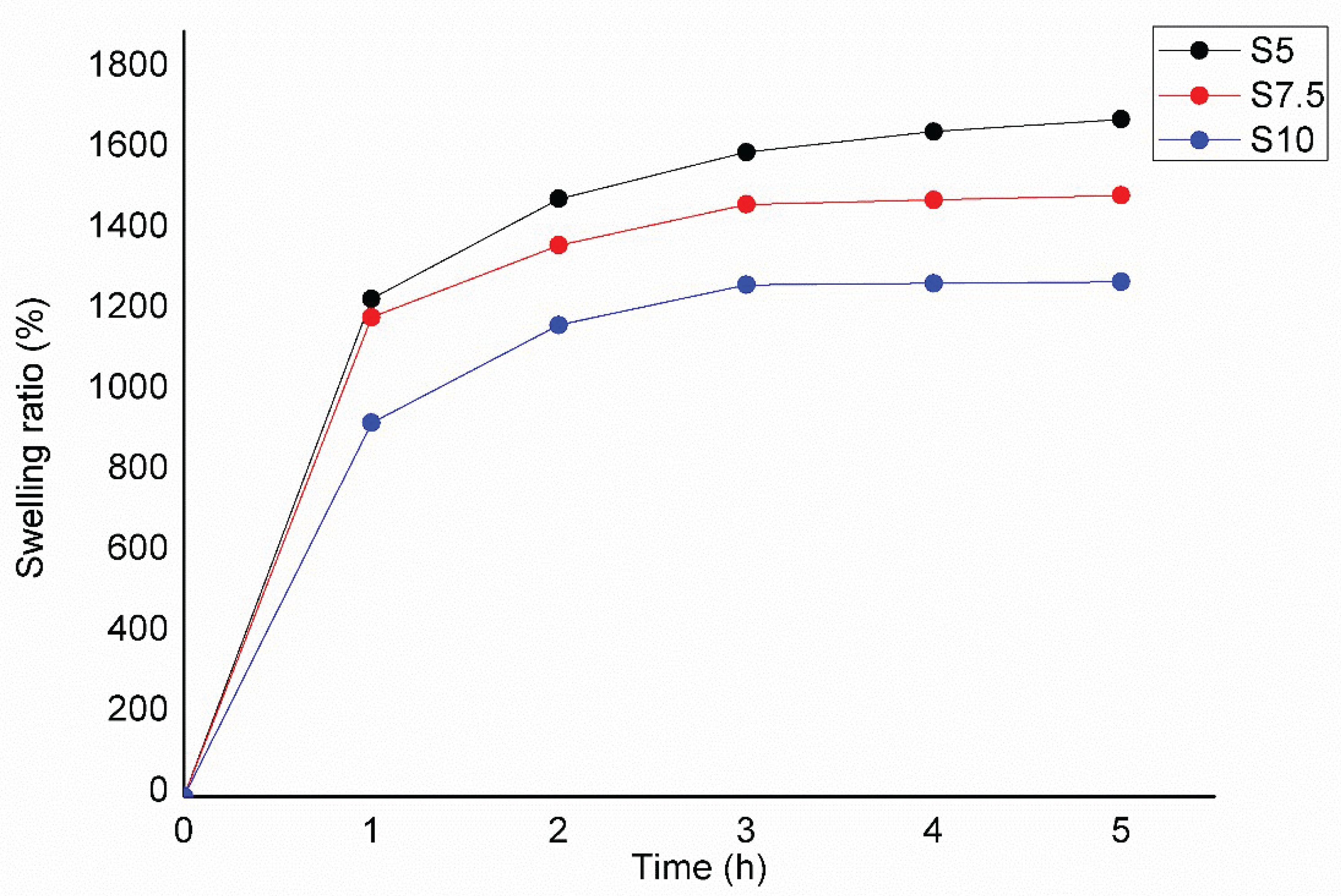
Figure 1. FTIR spectra of semi-IPN dextran/inulin hydrogels.
Preprints 191076 g001
Figure 2. FTIR spectra of neat semi-INP dextran/inulin hydrogel (S5), and uracil-loaded semi-INP dextran/inulin hydrogel (S5, U).
Preprints 191076 g002
Figure 3. DSC thermograms for neat semi-INP dextran/inulin hydrogel.
Preprints 191076 g003
Figure 4. Swelling ratio versus time for the prepared semi-IPN hydrogels.
Preprints 191076 g004
Figure 5. Hydrogels with a rising amount of crosslinking agent DEGDA from left to right (T1, T2, T3).
Preprints 191076 g005
Table 1. Estimated K1 values for the first—order kinetics (Equation 4).
Hydrogels K1, pH 7.4, 37 °C R2
S5 0.97 0.99
S7.5 1.24 0.98
S10 1.55 0.97
Table 2. Results of gel fraction (GF) analysis.
Hydrogels GF (%)
S5 84.75a ± 1.12
S7.5 91.34b ± 1.42
S10 94.90c ± 0.60
Table 3. Encapsulation efficiency of prepared hydrogels.
Uracil–loaded hydrogels Encapsulation efficiency (%)
S5 86.13 ± 0.88a
S7.5 89.67 ± 1.15b
S10 92.45 ± 1.12c
Table 4. Amounts of uracil released from the prepared hydrogels as a result of enzymatic biodegradation during the gastric and intestinal phases.
Uracil–loaded hydrogels Gastric phase release, µg/ml Intestinal phase release, µg/ml Gastric phase release, % Intestinal phase release, % Total released amount of drug, %
S5, U 6.17 ± 0.22a 91.82 ± 3.78a 4.93 ± 0.21a 78.79 ± 2.97a 83.73a ± 3.02a
S7.5, U 4.50 ± 0.19b 65.91 ± 2.63b 3.60 ± 0.15b 52.73 ± 2.11b 56.33 ± 2.62b
S10, U 0.00c 57.24c ± 2.86c 0.00c 45.79 ± 2.29c 45.79 ± 2.29c
Values are expressed as mean ± standard deviation. Different superscript letters within the same column denote statistically significant differences (p < 0.05). The total releasable amount was 125 μg/mL, calculated based on a loading of 20 mg per g of dry carrier and 0.25 g of dry material.
Table 5. Inhibitory activity of dextran-based formulations against Escherichia coli ATCC 8739 and Staphylococcus epidermidis ATCC 12228.
Inhibition zone (mm)
Sample number: Escherichia coli ATCC 8739 Staphylococcus epidermidis ATCC 12228
Ampicilin 10 mcg 10 15
S5 - -
S7.5 - -
S10 - -
Disclaimer/Publisher’s Note: The statements, opinions and data contained in all publications are solely those of the individual author(s) and contributor(s) and not of MDPI and/or the editor(s). MDPI and/or the editor(s) disclaim responsibility for any injury to people or property resulting from any ideas, methods, instructions or products referred to in the content.
Copyright: This open access article is published under a Creative Commons CC BY 4.0 license, which permit the free download, distribution, and reuse, provided that the author and preprint are cited in any reuse.
Prerpints.org logo

Preprints.org is a free preprint server supported by MDPI in Basel, Switzerland.

Subscribe

Disclaimer

Terms of Use

Privacy Policy

Privacy Settings

© 2026 MDPI (Basel, Switzerland) unless otherwise stated