Preprint
Review

This version is not peer-reviewed.

Ion Channels as Targets of the Vitamin D Receptor: A Long Journey with a Promising Future

Submitted:

22 December 2025

Posted:

24 December 2025

You are already at the latest version

Abstract

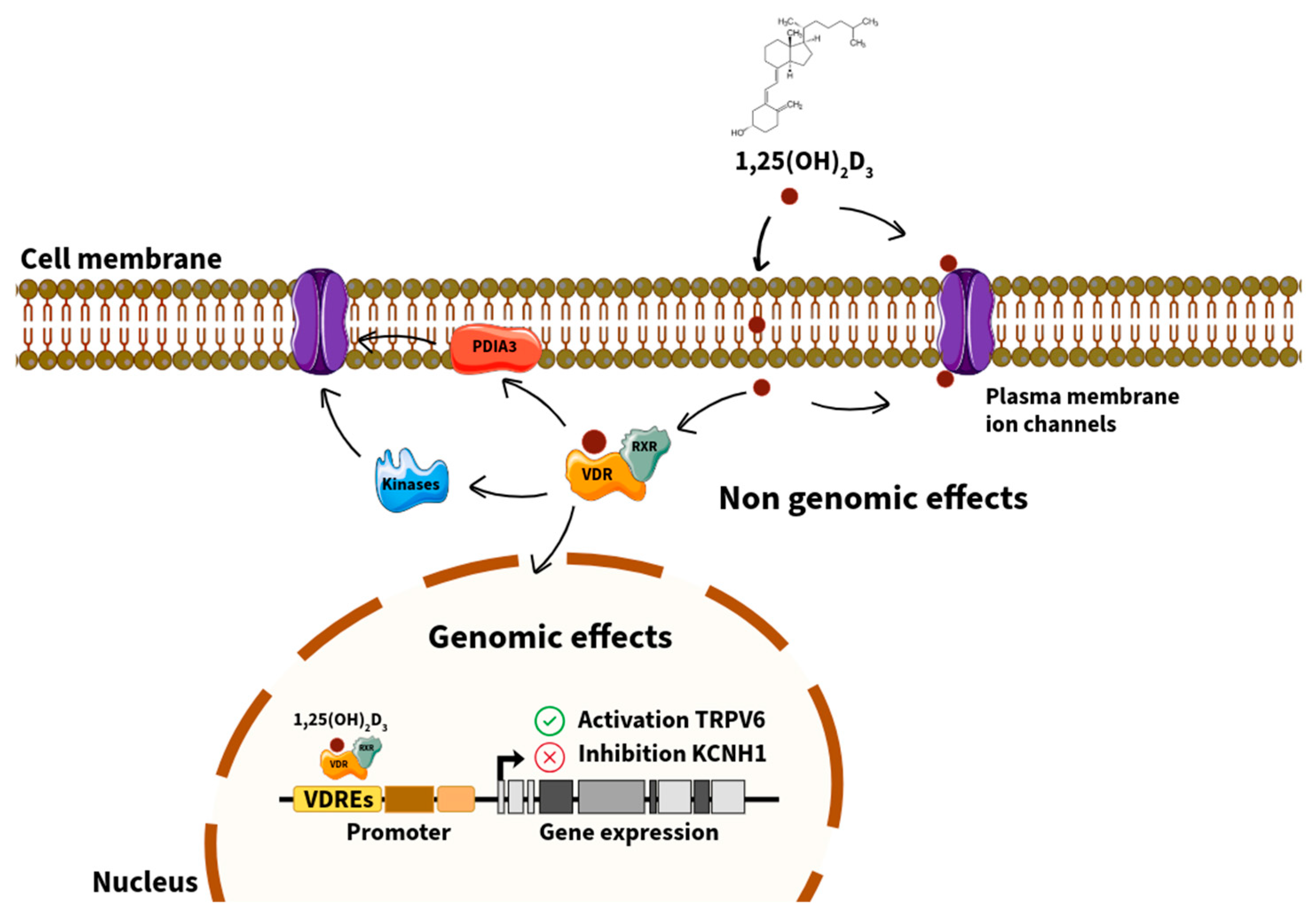
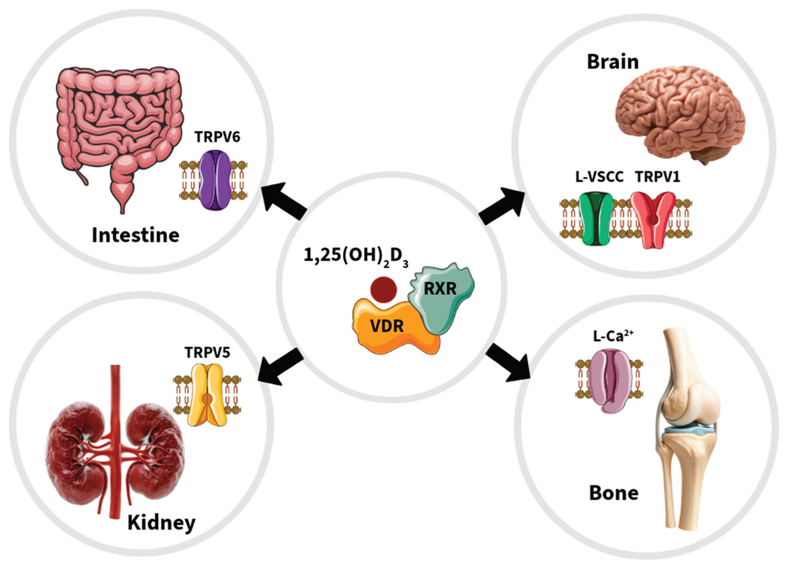
The vitamin D receptor (VDR) acts as both a nuclear transcription factor and a non-genomic mediator that regulates a broad spectrum of physiological processes beyond calcium and phosphate homeostasis. VDR plays an important role in the modulation of ion channels across multiple tissues, including osteoblasts, renal and intestinal epithelial cells, neurons, and vascular smooth muscle. These regulatory mechanisms encompass genomic actions through vitamin D response elements in target genes—such as TRPV5, TRPV6, KCNK3, and KCNH1—as well as rapid, non-genomic actions at the plasma membrane involving protein disulfide isomerase A3 and associated signaling cascades. VDR-mediated transcriptional control of calcium, potassium, and chloride channels contributes to the fine-tuning of cellular excitability, calcium transport, and mitochondrial function. Evidence also implicates VDR–ion channel crosstalk in various pathological contexts, including renal cell carcinoma, breast and cervical cancers, pulmonary arterial hypertension, and osteoporosis. Understanding the molecular interplay between VDR and ion channels provides new perspectives on the pleiotropic effects of vitamin D and offers promising therapeutic opportunities in oncology, cardiovascular disease, and skeletal disorders. This review synthesizes previous and current evidence on the genomic and non-genomic mechanisms underlying VDR–ion channel regulation and highlights novel frontiers in vitamin D signaling relevant to human health and disease.

Keywords: 
;  ;  

1. Introduction

In 1928, the German chemist Adolf Windaus successfully isolated two forms of vitamin D: one derived from plants, designated vitamin D2 (also called calciferol), and the other derived from animal skin, identified as vitamin D3 (also called cholecalciferol). Eight years after this discovery, he elucidated the chemical structure of vitamin D3.
Vitamin D3, is synthesized in the skin upon exposure to sunlight, and vitamin D2 is derived from dietary sources. Vitamin D₃ is converted to 25-hydroxyvitamin D3 (25(OH)D3; also called 25-hydroxycholecalciferol) in the liver, which then circulates in the bloodstream and undergoes a second hydroxylation at the 1α-position in the kidney, producing the biologically active form that regulates calcium and phosphate homeostasis: 1α,25-dihydroxyvitamin D3 (1,25(OH)2D3; also called 1α,25-dihydroxycholecalceferol and calcitriol).
Dietary vitamin D2 and vitamin D3 are metabolized through the same activation process, involving 25-hydroxylation in the liver and subsequent 1α-hydroxylation in the kidney.
Vitamin D is crucial for bone health, as it contributes to the maintenance of strong bones and teeth. Additionally, it plays a role in immune function and is vital for normal growth and development [1,2].
Vitamin D, while primarily recognized for its critical role in calcium metabolism and bone mineralization, is also implicated in a variety of physiological and pathological processes, including cancer, immune modulation, cardiovascular diseases, and metabolic syndrome. Most vitamin D’s effects are mediated through the vitamin D receptor (VDR), which regulates a substantial number of target genes and consequently influences numerous cellular pathways. Notably, VDR is expressed in nearly all human cell types and have been shown to modulate the transcription of approximately 3% of human genes [3].
Research has demonstrated that steroid hormones exert rapid modulatory effects on ion channel activity across various cellular systems through multiple molecular mechanisms. For instance, the rapid actions of 1,25(OH)2D3 on Cl- and Ca2+ currents are linked to secretory activities in osteoblasts and bone mass formation [4]. However, the genomic effects of 1,25(OH)2D3 on ion channels remain insufficiently explored. A comprehensive analysis of transcriptomic data from genes encoding ion channel proteins, which exhibit differential expression under 1,25(OH)2D3 treatment, revealed that the majority of genes affected by 1,25(OH)2D3 belong to the potassium channel family, including voltage-activated potassium channels, calcium-activated potassium channels, and two-pore domain potassium channels [5]. Given the significance of 1,25(OH)2D3 and its receptor as biological regulators and their association with ion channels in the body, we deemed it pertinent to review the existing literature on the functional link between them, the associated molecular mechanisms, and their relationship with certain diseases.

2. VDR: Genomic and Non-Genomic Mechanisms

2.1. The VDR Functions as Both a Nuclear Transcription Factor and a Non-Genomic Mediator

VDR is a ligand-dependent transcription factor, with 1,25(OH)2D3 and lithocholic acid (LCA) serving as endogenous ligands [6,7]. VDR is a member of the nuclear receptor superfamily [8], which encompasses receptors for glucocorticoids, mineralocorticoids, sex hormones, thyroid hormones, and vitamin A metabolites. The primary target tissues of VDR include the bone, kidney, and intestine, where it plays a crucial role in maintaining calcium homeostasis (calcemic effects). However, VDR is also expressed in various other tissues, contributing to non-calcemic effects, such as immune response, innate immunity, and the maintenance of barrier function in the skin and intestinal tract [9]. Upon ligand binding, VDR is activated and forms a heterodimer with the retinoid X receptor (RXR) [10], which subsequently binds to a gene region known as the vitamin D response element (VDRE) to regulate the transcription of target genes. While the principal function of VDR is transcription-mediated genomic action, it has been reported to partially participate in the acute phase calcium influx mechanism as part of the non-genomic actions of 1,25(OH)2D3 [11] (Figure 1).

2.2. Structural Domains and Isoforms of VDR

VDR is a protein with a molecular weight of 50-60 kDa, which was cloned and sequenced in 1987 [6]. X-ray crystallographic analysis elucidated its binding model with 1,25(OH)2D3 in 2000 [12], and with LCA in 2013 [13]. The nuclear receptor superfamily is characterized by an N-terminal domain containing a DNA-binding domain and a C-terminal domain containing a ligand-binding site. Compared to other nuclear receptors, VDR possesses a shorter N-terminal region located at the terminus of the DNA-binding domain. The DNA-binding domain comprises two zinc fingers that form tetrahedral structures with cysteine and zinc, adopting a loop or finger-like configuration. The zinc finger at the N-terminal side determines the specificity of the VDRE sequence. The C-terminal region encompasses a ligand-binding domain essential for heterodimerization with the RXR. Additionally, the activation function 2 domain at the C-terminal end is crucial for interactions with transcription cofactors, such as steroid receptor coactivator (SRC) and vitamin D receptor interacting protein (DRIP) [14]. VDR exists in several isoforms. Human VDRB1 is a transcript variant in which the N-terminal 50 amino acids of VDR are extended, exhibiting cell- and ligand-selective transcriptional activities distinct from those of the wild-type (VDRA) [15,16]. VDR1, in which intron 8 is transcribed following exon 8 of rat VDR, is expressed at low levels in the kidney and intestine and exhibits a dominant negative function compared to the wild-type (VDR0) [17]. The VDR gene contains multiple single nucleotide polymorphisms, with the representative BsmI single nucleotide polymorphism located in intron 8, which has been implicated in bone metabolism and muscle function [18,19]. In contrast, teleost fish, which underwent whole-genome duplication during evolution, possess VDRα and VDRβ. In medaka fish, VDRβ exhibits ligand responsiveness similar to that of terrestrial organisms, whereas the response of VDRα is notably weak [20].

2.3. Regulation by 1,25(OH)2D3 and Its Synthetic Analogs

VDREs present in VDR target genes are characterized by a direct repeat of two hexanucleotide half-elements with a three-nucleotide spacer (called direct repeat 3) or an everted repeat of two half-elements with a six-nucleotide spacer (called everted repeat 6). The VDR-RXR heterodimer on the VDRE then forms a transcription complex through interactions with cofactors, thereby facilitating transcription [21,22]. Synthetic analogs that bind to VDR are categorized into secosteroids and non-secosteroids. Alfacalcidol (1α-hydroxyvitamin D3), a synthetic secosteroidal analog, is clinically used to treat osteoporosis. It is a prodrug that is converted to 1,25(OH)2D3 by 25-hydroxylase in the body [23]. Maxacalcitol is used to treat psoriasis and secondary hyperparathyroidism. Compared to 1,25(OH)2D3, maxacalcitol exhibits weaker VDR activation and binding affinity, as well as a shorter half-life, resulting in reduced hypercalcemia as a side effect [24]. While 1,25(OH)2D3 and its prodrug alfacalcidol promote bone resorption as a side effect, eldecalcitol, introduced in 2011 in Japan, demonstrates an anti-osteoporotic effect with a milder bone resorption effect than alfacalcidol [25]. Although eldecalcitol is a weak VDR agonist, it is more stable and resistant to hydroxylation by the metabolic enzyme cytochrome P450 (CYP) 24A1 than 1,25(OH)2D3, allowing it to exert a sustained effect in the body [26]. Paricalcitol, a 19-nor vitamin D derivative with the 19th exomethylene ring removed, has been used to treat secondary hyperparathyroidism. MART-10, another 19-nor vitamin D derivative, exhibits antitumor effects and resistance to degradation by metabolic enzymes [27,28]. Further development of these compounds is anticipated in the future. Various secosteroidal vitamin D derivatives have been synthesized by modifying the side chains; however, the complete separation of calcemic effects from other effects remains a challenge. LY2108491, a non-secosteroidal compound, improves psoriasis in models without elevating blood calcium levels [29]. Conversely, LCA acetate and LCA propionate, derivatives based on the LCA structure, activate VDR in the intestine and kidney without increasing blood calcium levels [30,31]. Dcha-20 is an LCA-type non-secosteroid compound that significantly enhances the VDR-binding affinity of LCA. The development of LCA-type VDR ligands is expected to yield selective VDR action and differences in stability and absorption efficiency in vivo, compared to secosteroid-type compounds [32].

2.4. Interactions with Co-Regulators and Membrane-Associated Signaling Proteins

The VDR transcriptional complex comprises coactivators and corepressors that modulate the transcriptional activity of VDR. These cofactor proteins possess LxxLL motifs, where ‘L’ denotes leucine and ‘x’ represents any amino acid, facilitating their binding to VDR. Upon ligand binding, the VDR recruits RXR and coactivators to form a transcription complex. Among these, SRC1, SRC2, and SRC3 exhibit histone acetyltransferase (HAT) activity [33], recruiting additional coactivators such as CBP/p300 and p/CAF, which also possess HAT activity [34]. HAT activity promotes transcription by loosening the chromatin structure. DRIP, another coactivator with LxxLL motifs, specifically DRIP205, lacks HAT activity [9], spans the region from the VDRE to the transcription start site, and directly associates with RNA polymerase II [35]. Furthermore, proteins such as PGC-1α [36], NCoA62 [37], Smad3 [38], and Ets-1 [39] have been identified to interact with VDR. In the absence of a ligand or upon antagonist binding, the VDR interacts with corepressors. The nuclear receptor corepressor (called NCoR) [40] and silencing mediator for retinoid or thyroid hormone receptors (called SMRT) [41], which function as VDR corepressors, recruit histone deacetylase 3 and methyltransferases to condense the chromatin structure [42]. Additionally, corepressors such as Hairless [43] and Alien [44] have been implicated in the suppression of VDR transcription.
The role of VDR on the cell membrane is limited, and the protein disulfide isomerase A3 (PDIA3, also called ERp60, ERp57, Grp58, or 1,25-MARRS) is implicated in the activity of vitamin D on the cell membrane [45]. Active vitamin D prompts an intracellular calcium influx within 1–10 min in ROS17/2.8 and chicken-derived muscle cells [46]. PDIA3 is involved in this acute calcium influx induced by active vitamin D [47], and the absence of PDIA3 negates vitamin D-mediated responses [48,49]. Additionally, vitamin D induces protein kinase C (PKC) activation in VDR-deficient ROS24/1 cells [50]. Both VDR and PDIA3 are localized on the cell membrane and interact with Caveolin-1 to engage in intracellular signaling. Consequently, an indirect interaction between VDR and PDIA3 cannot be excluded [51].

3. VDR Signaling and Ion Channels: Mechanistic Insights

VDR is integral to the modulation of ion channel activity across various cellular tissues. Investigating VDR as a regulator of gene expression and ion channel activity is essential for enhancing our understanding of the physiological roles of vitamin D in the human body. The regulation of ion channels by 1,25(OH)2D3 is mediated through at least four mechanisms: a) the classical, long-term action of the ligated VDR within the nucleus; b) the rapid non-genomic action of 1,25(OH)2D3 or ligated VDR at the plasma membrane and within the cytoplasm; c) alterations in the properties of the lipid bilayer housing the channels, which affect conductivity and the likelihood of channel openings; and d) direct binding to the channel protein, independent of the VDR [5]. Genes commonly regulated by 1,25(OH)2D3 that encode channel proteins include transient receptor potential vanilloid (TRPV), two-pore-domain potassium channel (KCN) (KCN subfamily K; KCNK), calcium-regulated potassium (KCNN and KCNMA), and chloride intracellular channels [5,52].

3.1. Transcriptional Regulation of Ion Channel Genes via VDREs

VDR facilitates the transcription of calcium channels, TRPV5 and TRPV6, through the action of 1,25(OH)2D3 [53]. The TRPV6 gene comprises five VDREs [54], while the TRPV5 gene contains four putative VDREs [55]. Notably, duodenal Trpv6 mRNA expression is reduced by 90% in VDR knockout (KO) mice, indicating that VDR is the primary regulator of this gene [56]. Additionally, the transcriptional induction of Trpv6 by 1,25(OH)2D3 is ten-fold in the duodenum and four-fold in the kidney, suggesting the presence of tissue-specific induction mechanisms [57]. The mechanism of TRPV6 expression has been thoroughly examined among VDR-regulated ion channels and transporters. A detailed analysis using chromatin immunoprecipitation by Meyer et al. demonstrated that VDR and RXR form heterodimers on the VDRE of the human TRPV6 promoter in a 1,25(OH)2D3-dependent manner, resulting in the recruitment of SRC1 and RNA polymerase II in intestine-derived cells [54]. Consequently, these genes are direct targets of the VDR. The renal sodium-dependent phosphate transporter gene (called NaPi-3; gene symbol: SLC23A1) is also a VDR target and contains a VDRE in its promoter [58]. Comprehensive RNA-sequencing analysis revealed that ZnT10 (gene symbol: SLC30A10), a zinc-manganese transporter, is induced by 1,25(OH)2D3 in Caco-2 cells, with the VDRE region on the ZnT10 gene confirmed [59]. Another endogenous VDR ligand, LCA, induces the expression of the intestinal manganese transporter Slc30a10 through VDR-dependent regulation [60]. Thus, VDR not only regulates calcium transport, its primary physiological function, but also modulates the expression of phosphorus and other ion transporters.

3.2. Indirect Modulation Through Second Messengers and Kinase Cascades

The regulation of ion channel expression, particularly that of TRPV6, is influenced by various factors in both VDR-dependent and independent manners. Prolactin enhances local 1,25(OH)2D3 levels by upregulating CYP27B1 expression, thereby increasing TRPV6 mRNA expression [61]. VDR-mediated induction of prolactin expression may contribute to the amplification of TRPV6 induction [62]. The involvement of p38 and the p38-interacting molecule GADD45 in 1,25(OH)2D3-induced TRPV6 expression has been noted, although it remains to be determined whether the p38 signaling cascade directly targets TRPV6 mRNA or other post-transcriptional modification factors, such as miRNA [63]. Additionally, p38 and c-Jun N-terminal kinase enhance VDR activity in conjunction with 1,25(OH)2D3 in the breast cancer cell line MCF7 [64]. VDR activity is partially regulated by phosphorylation, with casein kinase II positively influencing transcription by phosphorylating serine 208 of VDR in a 1,25(OH)2D3-dependent manner [65], which enhances the interaction between VDR and coactivator DRIP205 [66]. ATM, a DNA damage response molecule, targets serine residues 208 and 222 of VDR, positively regulating VDR activity [67]. Conversely, PKCβ [68,69] and protein kinase A target serine 51 and serine 182 of VDR, respectively, negatively regulating its activity. protein kinase A also inhibits the heterodimerization of VDR and RXR [70,71]. Phosphorylation of VDR may play a role in regulating ion channel expression in the intestine and kidney.
The regulation of TRPV6 expression independent of VDR may indirectly influence VDR-dependent TRPV6 expression. The induction of TRPV6 mRNA in response to low calcium levels, independent of VDR, represents the most appropriate control mechanism [72]. Additionally, estradiol induces TRPV6 expression in an estrogen receptor (ER) α-dependent manner, without involving the VDR [73]. Notably, ERα deficiency results in a 50% reduction in TRPV6 expression, whereas ERβ does not contribute to this process [73]. Furthermore, TRPV6 expression is induced by glucocorticoids [74], the plant compound quercetin [75], short-chain fatty acids, oligosaccharides [76], and the serum/glucocorticoid regulated kinases 1 and 3, and protein kinase B/Akt pathways [77]. The vitamin D-VDR pathway activates Akt signaling in VDR target tissues [78], suggesting potential intracellular crosstalk between these pathways.

3.3. Epigenetic and Post-Transcriptional Regulation

In addition to phosphorylation, VDR and its target ion channels are regulated by post-translational modifications, such as ubiquitination, SUMOylation, and miRNA interactions. The transcription cofactor SUG1 [79] and cell cycle-related CDK11p58 promote VDR ubiquitination [80]. Meanwhile, TRPV6 is one of the targets of the ubiquitin E3 ligase Neddd45-2 [81]. There are no reports on miRNAs that directly target TRPV6, but they may exert effects via miR-27b [82] and miR-125b [83], which target VDR. The localization of these miRNA expressions in VDR target tissues may be involved in the regulation of ion channels. SUMO2 is modified by PIDIA4 to suppress VDR activity and is deSUMOylated in a ligand-dependent manner [84]. In addition, SUMO specific peptidases 1 and 2 selectively act on intestinal and kidney cell lines to enhance VDR transcriptional activity [85].

4. Regulation of Ion Channel Function by VDR in Different Cellular Contexts

VDR is integral to the regulation of ion channel function in various cells and tissues (Figure 2). This regulation is mediated through both genomic and non-genomic mechanisms, which affect the expression and activity of ion channels. The specific outcomes of VDR-mediated ion channel regulation are contingent on the cellular context, underscoring the complexity and versatility of this regulatory mechanism. Among the initial studies demonstrating the regulatory influence of vitamin D on ion channel expression are those concerning the epithelial calcium channel TRPV6. Herein, we present scientific evidence detailing some of the most extensively studied examples of VDR’s influence of VDR on the expression and activity of ion channels.

4.1. Osteoblast Cells

Osteoblasts have been extensively investigated to elucidate the mechanisms of action associated with vitamin D and VDR. Given that osteoblasts contain various ion channels within their plasma membrane [86,87], they have been deemed appropriate for initial studies on the effects of VDR on ion channel function [4]. For instance, the hormone 1,25(OH)2D3 facilitates the activation of voltage-dependent L-type calcium channels in ROS 17/2.8 osteosarcoma cells [88]. Furthermore, 1,25(OH)2D3 augments chloride currents through mechanosensitive anion channels under conditions of significant membrane depolarization [89]. These rapid actions of 1,25(OH)2D3, which modulate the voltage sensitivity of Ca²⁺ channels and enhance the amplitude of Cl⁻ currents, suggest that vitamin D may engage distifnct signaling mechanisms at the plasma membrane in osteoblasts. In another study, the modulation of ion channel activity by 1,25(OH)2D3 was examined in calvarial osteoblasts isolated from VDR-KO and wild-type mice. The authors concluded that 1,25(OH)2D3 modulates ion channel activities exclusively in calvarial osteoblasts with functional VDR, and this effect is linked to exocytosis [90].

4.2. Intestinal and Kidney Cells

The action of 1,25(OH)2D3 to sustain calcium and phosphorus homeostasis through the facilitation of intestinal calcium absorption and renal calcium reabsorption was linked in early research to its ability to induce the expression of calbindin D9K and/or D28K, as well as plasma membrane calcium ATPase 1b [91,92]. TRPV6 is the principal ion channel in the intestinal epithelial cell membranes that is responsible for calcium entry [54]. TRPV5 represents a class of highly selective calcium channel proteins predominantly located in the kidney, associated with calcium transport, and plays a crucial role in maintaining intracellular calcium concentration stability [93]. The TRPV5/V6 channels in kidney and intestinal cells are among the primary targets for the regulation of calcium homeostasis by hormones [94].
1,25(OH)2D3 has been shown to induce the expression of TRPV6 and calbindin D9K in the intestine and TRPV5, calbindin D9K, and D28K in the kidneys of wild-type mice in a time- and dose-dependent manner [57]. The capacity of 1,25(OH)2D3 to induce TRPV6 gene expression and facilitate calcium transport has also been demonstrated in vitro using human colon-derived Caco-2 cells [95]. These studies suggest that 1,25(OH)2D3 regulates the expression of TRPV genes, which are essential for the intestinal absorption and renal reabsorption of calcium. Subsequent research identified VDREs in the upstream region of the TRPV6 gene, which mediate the cis-actions of 1,25(OH)2D3 to modulate intestinal TRPV6 expression [54]. Additional reports indicate that 1,25(OH)2D3 induces TRPV6 mRNA expression at lower concentrations than those required for the induction of CYP24A1, a VDR target gene involved in vitamin D inactivation, in human intestinal SW480 cells. This suggests an alternative mechanism for vitamin D signaling in TRPV6 induction. 1,25(OH)2D3 treatment induced the expression of GADD45A, which encodes the growth arrest and DNA damage inducible α (GADD45α) MAPK kinase activator, prior to TRPV6 expression, and GADD45A knockdown reduced TRPV6 induction by 1,25(OH)2D3l. These findings suggest that GADD45α plays a role in enhancing vitamin D signaling in TRPV6 expression [63].
Research conducted by Wood et al. (2001) demonstrated that 1,25(OH)2D3 induces a tenfold increase in TRPV6 mRNA expression in the Caco-2 human intestinal cell line. Furthermore, the study observed a significant elevation in TRPV6 mRNA levels, exceeding a five-fold increase, as early as four hours after 1,25(OH)2D3 treatment. This finding suggests a pivotal role for this calcium transporter in vitamin D-dependent calcium absorption [96]. In a separate investigation involving VDR-KO mice, researchers assessed the expression of TRPV calcium channels implicated in transcellular calcium transport. The calcium transport protein TRPV6 was found to be abundantly expressed at the mRNA level in the duodenum; however, its expression was markedly reduced by more than 90% in VDR-KO mice maintained on a normal-calcium diet. This observation indicates that the expression of the duodenal epithelial calcium channel TRPV6 is dependent on vitamin D [97].

4.3. Neuron Cells

In addition to its classical roles in regulating the absorption of calcium and phosphorus in the intestines, bones, and kidneys, as well as bone mineralization, vitamin D3 has been shown to possess neuroprotective functions. These functions are mediated through neuronal calcium regulation, antioxidative pathways, immunomodulation, and detoxification [98]. Direct neuroprotective effects of 1,25(OH)2D3 have been observed at low concentrations, including the selective downregulation of L-type voltage-sensitive Ca2+ channel expression in brain neurons [99]. These findings suggest that the Ca2+-regulatory functions of 1,25(OH)2D3 extend to neurons and may contribute to neuronal survival. TRPV1, a calcium-selective channel, is activated by heat, low pH, and various endogenous and exogenous agonists [100]. This channel is expressed in various tissues throughout the human body, including neurons, immune T-cells, nociceptive C fibers that innervate the airways, and airway epithelial cells [101,102].
25(OH)D3 acts as a partial agonist of the TRPV1 channel. This vitamin D metabolite directly binds to TRPV1 within the same vanilloid binding pocket as capsaicin, enabling it to weakly activate the channel while concurrently antagonizing the effects of full agonists such as capsaicin and oleoyl dopamine. In addition to direct activation, 25(OH)D3 inhibits PKC-mediated potentiation of TRPV1 activity, thereby demonstrating multiple regulatory pathways. This discovery identifies TRPV1 as a novel receptor for the biological actions of vitamin D, thereby expanding our understanding of the traditional nuclear vitamin D receptor [103]. This finding provides evidence supporting the concept of a novel vitamin D/TRPV1 axis, which may elucidate some of the beneficial effects of vitamin D in disease states in which TRPV1 expression and vitamin D deficiency are known to coincide [104].

4.4. Ion Channels from the Plasma and Mitochondrial Membranes

Numerous studies have established a significant association between 1,25(OH)2D3 and mitochondrial function in various cell types. Various ion channels have been identified in the mitochondrial membranes, with the majority classified as potassium channels. To date, eleven distinct potassium channels have been identified in the inner mitochondrial membrane: (1) small-conductance calcium-regulated potassium channel (mitoSKCa; encoded by KCNN1-3); (2) intermediate-conductance calcium-regulated potassium channel (mitoIKCa; encoded by KCNN4); (3) large-conductance calcium-regulated potassium channel (mitoBKCa; encoded by KCNMA1); (4) large-conductance potassium channel DEC isoform (mitoBKCa-DEC; encoded by KCNMA1); and (5) two-pore domain potassium channel [5]. A study has demonstrated for the first time that 1,25(OH)2D3 modulates mitochondrial potassium channels, directly influencing the activity of the mitochondrial large-conductance Ca²⁺-regulated potassium channel (mitoBKCa) in human astrocytoma cells, while not affecting the mitochondrial calcium-independent two-pore domain potassium channel (mitoTASK-3) in human keratinocytes [105].

5. Pathophysiological Relevance of VDR–Ion Channel Interactions

5.1. Cancer

Epidemiological research suggests that vitamin D insufficiency may play an etiological role in the development of various cancers. Over the decades, 1,25(OH)2D3 and vitamin D analogs have been extensively investigated in preclinical studies because of their potential as anticancer agents. These compounds exhibit antiproliferative effects, activate apoptotic pathways, inhibit angiogenesis, and may exert effects through immune mechanisms [106,107]. In addition to its other functions, 1,25(OH)2D3 regulates microRNAs, which are crucial in cancer biology, by modulating their expression profiles and influencing certain components of the miRNA processing machinery [108,109]. VDR is implicated in the regulation of cancer cell proliferation through complex mechanisms involving ion channels and metabolic regulation (Table 1).

5.2. Renal Cell Carcinoma (RCC)

1,25(OH)2D3 and VDR have been implicated in reducing the risk of renal cell carcinoma (RCC) [110,111]. The expression of VDR in normal kidney tissue diminishes during malignant transformation to RCC and is correlated with RCC prognosis [112,113]. These observations suggest that VDR may play a critical role in RCC development and progression. VDR and 1,25(OH)2D3 have been reported to regulate the activity of TRPV5. In vitamin D-deficient or VDR-KO mice, the mRNA and protein expression levels of TRPV5 were reduced, whereas 1,25(OH)2D3 administration significantly increased TRPV5 mRNA expression in the kidneys. Consequently, TRPV5 expression is highly dependent on vitamin D intake. Furthermore, the human TRPV5 promoter contains several consensus vitamin D-responsive elements [56,114]. Functional experiments documented in a published study demonstrated that VDR overexpression decreased cell proliferation, migration, and invasion, while promoting apoptosis in RCC cells. Conversely, VDR knockdown had the opposite effect. TRPV5 expression levels are inversely correlated with VDR, with VDR overexpression downregulating TRPV5 expression and knockdown upregulating it in RCC cells [115]. These findings indicate that VDR acts as a tumor suppressor in RCC cells by inhibiting proliferation, migration, and invasion through the regulation of TRPV5 expression.

5.3. Breast and Cervical Cancer

Numerous studies have demonstrated an association between low VDR levels and aggressive breast cancer. Furthermore, a reduction in VDR is linked to neoplastic transformation, which diminishes cancer cell sensitivity to the antiproliferative effects of 1,25(OH)2D3 [116]. This underscores the significance of VDR activity in regulating the gene interaction network. The large-conductance Ca2+-activated K+ channel, KCa1.1, is associated with high-grade and poorly differentiated tumors in breast cancer [117]. A study has elucidated the effects of VDR agonists on the expression and activity of KCa1.1 in human breast cancer cells. Treatment with VDR agonists resulted in decreased KCa1.1 expression at both the transcript and protein levels in MDA-MB-453 cells, leading to the inhibition of depolarization responses induced by paxilline, a specific KCa1.1 blocker. This provides mechanistic insights, indicating that the Ca2+-activated K+ channel KCa1.1 is a downstream target of VDR signaling [118].
Interactions between VDR and ion channels play crucial roles in cellular proliferation. Specifically, 1,25(OH)2D3 suppresses the expression of the Kv10.1 potassium channel (also called EAG1; encoded by the gene KCNH1), a member of the ether-à-go-go (EAG) family of voltage-gated potassium channels, through VDR-dependent mechanisms, resulting in decreased proliferation of breast cancer cells [119]. Furthermore, 1,25(OH)2D3 downregulates Kv10.1 channels in cancer cells derived from the breast and cervix, leading to a reduction in Kv10.1 protein levels at various incubation periods [120]. Kv10.1 is considered clinically significant because of its transformative activity and ectopic expression in clinical tumors [121]. Ectopic expression has been observed in various tumor cell lines and is associated with cell proliferation [122,123]. Additional studies have demonstrated the overexpression of Kv10.1 in tumors from clinical samples, suggesting its potential as a promising target for cancer therapy [124]. Research on cervical cancer cells has indicated that the effect of 1,25(OH)2D3 on KCNH1 expression is VDR-dependent. It has been proposed that this mRNA repression by 1,25(OH)2D3 involves an E-box type VDRE, which functions as a negative regulatory element in the KCNH1 promoter [125].

5.4. Pulmonary Arterial Hypertension

Pulmonary arterial hypertension (PAH) is a complex chronic condition characterized by pulmonary vascular remodeling and vasoconstriction of the pulmonary artery (PA). This vascular remodeling results from the excessive and uncontrolled proliferation and resistance to apoptosis of PA smooth muscle cells (PASMCs) and endothelial cells, leading to intimal thickening and PA obliteration. Vitamin D deficiency is significantly more prevalent in patients with PAH than in the general population or even in patients with other cardiovascular diseases. In both PAH patients and animal models, lower vitamin D levels are associated with a worse functional class [126,127]. A recent report indicated that VDR is expressed in several cell types within the lung, including PASMCs, and is downregulated in the lungs and PASMCs of PAH patients. Treatment with 1,25(OH)2D3 can restore VDR expression, upregulate KCNK3 and bone morphogenetic protein 4, and downregulate survivin gene expression in PASMCs, exerting an antiproliferative effect by modulating the survivin and bone morphogenetic protein signaling pathways [128]. Notably, low levels of lung VDR are associated with several pulmonary diseases, including chronic obstructive pulmonary disease and idiopathic pulmonary fibrosis, and correlate with a poor prognosis [129,130].
Several studies have indicated that KCNK3 may be a target of VDR [131,132,133], and a VDRE has been identified in the promoter region of the KCNK3 gene [134]. 1,25(OH)2D3 has been shown to significantly upregulate KCNK3 mRNA expression in cultured PASMCs from both control subjects and patients with PAH (26). However, the antiproliferative effects of 1,25(OH)2D3 appear unaffected by KCNK3 inhibition, suggesting that the upregulation of KCNK3 does not contribute to the antiproliferative effects induced by 1,25(OH)2D3 [128]. Voltage-dependent (Kv) or K2P potassium channels are crucial in pulmonary circulation as they regulate the membrane potential, arterial tone, cell proliferation, and survival of PASMCs. Ionic channel remodeling, primarily characterized by dysfunctional Kv1.5 and TASK-1 channels, is a common feature of PAH in both animal models and humans [135,136,137,138].
Kv7 channels are critical regulators of vascular tone in both healthy and diseased states. Activation of Kv7 channels induces an outward efflux of K+, leading to hyperpolarization and relaxation, as well as exerting antiproliferative effects on vascular smooth muscle cells [139,140,141]. Previous studies have identified potential VDREs in the promoters of genes encoding Kv7.1, Kv7.3, and KCNE4 channels [142]. Vitamin D deficiency is highly prevalent among patients with PAH, affecting 95% of this population [126,127]. Low levels of vitamin D are associated with poor prognosis, and both VDR mRNA and protein are downregulated in the lungs of patients with PAH.
A recent investigation examined the function of VDR within the pulmonary vasculature of mice, specifically assessing whether the ablation of Vdr in mice leads to pulmonary arterial dysfunction, with a focus on the regulation of Kv7 channels. The study identified upregulation of the ancillary subunit KCNE4 in the lungs of VDR-KO mice, which aligns with the discovery of a consensus VDRE in both human and mouse KCNE4 genes. Notably, PASMCs from VDR-KO mice exhibited enhanced Kv7 currents and an increased relaxant response to the Kv7 channel activator retigabine. Mice deficient in VDR did not exhibit an evident PAH phenotype [143]. Collectively, these findings indicate a negative regulatory role of VDR on KCNE4 within the pulmonary vasculature, adversely affecting Kv7 channel activity [143] (Table 2).

5.5. Osteoporosis

Osteoporosis is a systemic skeletal disorder characterized by reduced bone mass and deterioration of bone microarchitecture, leading to increased bone fragility and a heightened risk of fractures [144]. A prevalence report that included 86 studies from five continents indicated that the global prevalence rate of osteoporosis is 18.3%, with only 31-36% of individuals over the age of 70 years maintaining normal bone health [145]. The etiology of osteoporosis may be idiopathic or secondary to factors such as hypogonadism, vitamin D deficiency, insufficient calcium intake, and prolonged excessive alcohol consumption, ultimately resulting in an osteopenic skeleton and an increased risk of developing osteoporosis [146].
Bone mineral density (BMD) is commonly employed as an indicator of bone strength, and the diagnosis of osteoporosis is based on its assessment. BMD is affected by both genetic and environmental factors, including physical activity, smoking, and dietary habits [147]. Bone remodeling is a continuous process throughout life that encompasses osteoclast-mediated bone resorption and osteoblast-driven bone formation [148]. Furthermore, osteocytes, which are embedded within the bone matrix, contribute to this remodeling process and are currently regarded as the primary source of molecules that regulate osteoclast and osteoblast activities. The formation and differentiation of osteoclasts are regulated by complex interactions between cytokines, hormones, immune factors, gut microbiota, and cellular aging. Among the various cytokines and hormones that influence osteoclast formation and function, macrophage colony-stimulating factor (M-CSF) and receptor activator of nuclear factor-kB ligand (RANKL) are critical [149].
Genes postulated to contribute to osteoporosis include those implicated in bone formation and remodeling (e.g., LRP5), those involved in hormone signaling pathways (e.g., VDR and ESR1), and those encoding bone structural proteins (e.g., COL1A1) [150].
Dysregulation of the Vitamin D/VDR system has been associated with various bone pathologies, including osteoporosis. Recent advancements have elucidated the role of the VD/VDR system in modulating the activity of bone cells, such as osteoblasts and osteoclasts, and have highlighted how VDR polymorphisms may influence bone mineral BMD and fracture risk [151].
Activation of the VDR modulates the equilibrium between osteoblast-mediated bone formation and osteoclast-mediated bone resorption by regulating the expression of receptor activator of nuclear factor κΒ ligand (RANKL) and osteoprotegerin (OPG) [152,153]. RANKL serves as a critical osteoclastogenic factor, whereas OPG functions as a decoy receptor, inhibiting RANKL and thereby preventing osteoclast activation [152,153]. VDR activation in osteoblasts reduces RANKL expression and increases OPG expression, thereby suppressing osteoclastogenesis and maintaining bone homeostasis. Extensive research has been conducted globally on the association between VDR gene polymorphisms, particularly ApaI, TaqI, and BsmI, and osteoporosis [154,155].
Bone metabolism in osteoporosis is intricately associated with the expression of Ca2+ channels in osteocytes, rendering their regulation essential for effective disease management. Calcium channels play a pivotal role in skeletal homeostasis by facilitating processes that govern extracellular and intracellular Ca2+ equilibrium. The differentiation and apoptosis of osteoclasts are extensively regulated by Ca2+ channels located in the cell membrane [149].
Transient receptor potential (TRP) channels, recognized for their high calcium permeability, have several members that are significantly associated with osteoporosis. These channels, comprising six subfamilies—TRPC, TRPV, TRPP, TRPM, TRPA, and TRPML—are integral to the regulation of Ca2+ balance during bone homeostasis [156]. Specifically, TRPV1 facilitates osteoclast differentiation, as evidenced by TRPV1-deficient mice, which exhibit a reduced number of osteoclasts [157]. Furthermore, TRPV5 and TRPV6 are involved in the regulation of osteoclast size and number, with TRPV6 acting as a negative regulator [158,159,160]. TRPV6 knockdown is associated with a significant increase in bone resorption [161] (Table 3).
Although the precise mechanisms through which VDR-mediated transcription facilitates osteogenesis and osteoblastic bone formation while concurrently inhibiting osteocyte senescence and osteoclastic bone resorption remain to be elucidated, the critical role of the VDR in bone formation and its contribution to bone-associated pathologies continues to represent an important area of investigation [162]. Given the critical involvement of the VDR pathway in osteoporosis pathogenesis, targeting this pathway presents substantial therapeutic potential.

6. Conclusions and Perspectives

The intricate relationship between the VDR and ion channels represents an essential and evolving area of research in molecular and cellular physiology. Over the past few decades, substantial progress has been made in elucidating how vitamin D and its active metabolites influence ion fluxes through both genomic and non-genomic mechanisms. Genomic actions, primarily mediated by ligand-activated VDR binding to VDREs, regulate the transcription of key channel genes, such as TRPV5, TRPV6, KCNK3, and KCNH1. These interactions control calcium and potassium channel expression, contributing to the maintenance of calcium-phosphate balance, membrane potential, and cellular excitability. In parallel, non-genomic actions occurring within seconds to minutes modulate channel activity through membrane-associated VDR, alternative binding pockets, and signaling intermediates, including protein kinase C and cyclic AMP. Together, these dual mechanisms enable the VDR to coordinate both the expression and function of ion channels, ensuring a finely tuned homeostatic network across tissues.
Experimental studies in osteoblasts and renal and intestinal epithelial cells have revealed that 1,25-dihydroxyvitamin D₃ rapidly modulates voltage-gated calcium and chloride currents, underscoring the receptor’s involvement in exocytosis, secretion, and bone formation. Simultaneously, the transcriptional control of TRPV channels and calbindins underlies vitamin D–dependent calcium absorption and reabsorption. Furthermore, mitochondrial ion channels have recently emerged as novel VDR targets, highlighting the potential link between vitamin D signaling and bioenergetic regulation. Growing evidence suggests that VDR–ion channel interactions extend beyond calcium homeostasis to include broader physiological functions, from cell proliferation and differentiation to neuronal excitability and cardiovascular control.
Clinically, disturbances in VDR signaling and ion channel expression are associated with multiple diseases, including osteoporosis, chronic kidney disease, pulmonary hypertension, and various forms of cancer. In this context, dysregulated calcium and potassium fluxes may contribute to aberrant cell proliferation, resistance to apoptosis, and altered metabolic profiles. Thus, understanding the molecular interplay between VDR and ion channels not only enhances our knowledge of basic physiology but also provides new therapeutic opportunities.
Traditional Chinese medicine (TCM) and bioactive compounds derived from natural sources have attracted increasing interest from scholars. Notably, TCM formulations and TCM-derived bioactive compounds have demonstrated the potential to modulate VDR activity [162]. Numerous medicinal herbs used in TCM, commonly referred to as kidney tonics, have been evaluated for their effects on bone metabolism in the treatment of osteoporosis in both laboratory settings and clinical trials [163]. Bu-Shen-Jian-Pi-Yi-Qi therapy, a traditional Chinese medicine, was assessed for its potential therapeutic efficacy in an alcohol-induced osteoporosis rat model, and its underlying mechanism was investigated. Bu-Shen-Jian-Pi-Yi-Qi therapy inhibited bone loss and promoted bone formation in an alcoholic osteoporosis model, as evidenced by increased BMD and elevated VDR expression at the mRNA level [164], suggesting a relationship between VDR expression and bone metabolism in alcoholic osteoporosis.
Further investigation is required to substantiate the biological relationship between VDR function and osteoporosis. Considering the physiological linkage between VDR and various ion channels in bone metabolism, elucidating the cell signaling networks and biological mechanisms connecting these elements within the context of osteoporosis is anticipated to facilitate the development of novel therapeutic strategies.
Several questions remain unanswered. The precise molecular identity of membrane-associated VDR complexes, structural basis for VDR–channel protein interactions and downstream signaling cascades require further elucidation using high-resolution biochemical and biophysical methods. Moreover, integrating transcriptomic and electrophysiological approaches is crucial for capturing the dynamic nature of VDR-mediated ion channel regulation. Future studies should also explore the tissue-specific variability of these mechanisms and their relevance in pathological states and aging.
In conclusion, the VDR–ion channel axis constitutes a multifaceted signaling paradigm that bridges genomic and rapid extranuclear pathways. Continued exploration of this field promises not only to expand our comprehension of vitamin D biology but also to open promising perspectives for precision medicine, where the modulation of receptor–channel interactions could become a key strategy for restoring ionic and metabolic homeostasis in human health and disease.

Author Contributions

Conceptualization, V.C.-O., R.J.G.-D., M.I, M.M. and L.A.P.; figures and tables, V.C.-O., R.J.G.-D.; writing—original draft preparation, V.C.-O., R.J.G.-D., and M.I.; writing—review and editing, M.M., and L.A.P. All authors have read and agreed to the published version of the manuscript.

Funding

This research received no external funding.

Institutional Review Board Statement

Not applicable.

Informed Consent Statement

Not applicable.

Data Availability Statement

No new data were created or analyzed in this study. Data sharing is not applicable to this article.

Conflicts of Interest

The authors declare no conflicts of interest.

Abbreviations

The following abbreviations are used in this manuscript:
1,25(OH)2D3 1,25-dihydroxyvitamin D3
25(OH)D3 25-hydroxyvitamin D3
BMD Bone mineral density
CYP Cytochrome P450
DRIP Vitamin D receptor interacting protein
EAG Ether-à-go-go
ER Estrogen receptor
HAT Histone acetyltransferase
KCN Potassium channel
KO knockout
LCA Lithocholic acid
OPG osteoprotegerin
PDIA3 Protein disulfide isomerase A3
PA Pulmonary artery
PAH Pulmonary artery hypertension
PASMC Pulmonary artery smooth muscle cell
PKC Protein kinase C
RANKL Receptor activator of nuclear factor kB ligand
RCC Renal cell carcinoma
RXR Retinoic X receptor
SRC Steroid receptor coactivator
TCM Traditional Chinese medicine
TRP Transient receptor potential
TRPV Transient receptor potential vanilloid
VDR Vitamin D receptor
VDRE Vitamin D response element

References

  1. Wolf, G. The Discovery of Vitamin D: The Contribution of Adolf Windaus. The Journal of Nutrition 2004, 134(6), 1299–1302. [Google Scholar] [CrossRef]
  2. González-Duarte, RJ. Una vitamina que no es vitamina. Revista ¿Cómo ves? 2013, 174(15), 30–33. [Google Scholar]
  3. Voltan, G; Ceccato, F; Cannito, M; Camozzi, V; Ferrarese, M. Vitamin D: An Overview of Gene Regulation, Ranging from Metabolism to Genomic Effects. Genes 2023, 14(9), 1691. [Google Scholar] [CrossRef] [PubMed]
  4. Zanello, LP; Norman, A. 1α,25(OH) 2 Vitamin D 3 actions on ion channels in osteoblasts. Steroids 2006, 71(4), 291–297. [Google Scholar] [CrossRef]
  5. Olszewska, AM; Zmijewski, MA. Genomic and non-genomic action of vitamin D on ion channels – Targeting mitochondria. Mitochondrion 2024, 77, 101891. [Google Scholar] [CrossRef]
  6. Mcdonnell, DP; Mangelsdorf, DJ; Haussler, MR; O’Malley, BW; Pike, JW. Molecular cloning of complementary DNA encoding the avian receptor for vitamin D. Science 1987, 235(4793), 1214–1217. [Google Scholar] [CrossRef]
  7. Makishima, M; Whitfield, GK; Evans, RM; Xie, W; Haussler, MR; Domoto, H; et al. Vitamin D receptor as an intestinal bile acid sensor. Science 2002, 296(5571), 1313–1316. [Google Scholar] [CrossRef] [PubMed]
  8. Rosen, CJ; Adams, JS; Bikle, DD; Black, DM; Demay, MB; Manson, JE; et al. The nonskeletal effects of vitamin D: an Endocrine Society scientific statement. Endocrine reviews 2012, 33(3), 456–492. [Google Scholar] [CrossRef] [PubMed]
  9. Walters, MR. Newly identified actions of the vitamin D endocrine system. Endocrine Reviews 1992, 13(4), 719–764. [Google Scholar]
  10. Kurokawa, R; Glass, CK; Han, Z; Yu, VC; Naar, A; Silverman, S; et al. Differential orientations of the DNA-binding domain and carboxy-terminal dimerization interface regulate binding site selection by nuclear receptor heterodimers. Genes & Development 1993, 7(7b), 1423–1435. [Google Scholar]
  11. Donati, S; Falsetti, I; Brandi, ML; Miglietta, F; Iantomasi, T; Palmini, G; et al. Rapid Nontranscriptional Effects of Calcifediol and Calcitriol. Nutrients 2022, 14(6), 1291. [Google Scholar] [CrossRef] [PubMed]
  12. Rochel, N; Wurtz, JM; Mitschler, A; Klaholz, B; Moras, D. The Crystal Structure of the Nuclear Receptor for Vitamin D Bound to Its Natural Ligand. Molecular Cell. 2000, 5(1), 173–179. [Google Scholar] [CrossRef]
  13. Masuno, H; Ikura, T; Morizono, D; Orita, I; Yamada, S; Shimizu, M; et al. Crystal structures of complexes of vitamin D receptor ligand-binding domain with lithocholic acid derivatives. Journal of Lipid Research 2013, 54(8), 2206–2213. [Google Scholar] [CrossRef] [PubMed]
  14. Rachez, C; Chang, CPB; Atkins, GB; Lazar, MA; Gamble, M; Freedman, LP. The DRIP complex and SRC-1/p160 coactivators share similar nuclear receptor binding determinants but constitute functionally distinct complexes. Molecular and Cellular Biology 2000, 20(8), 2718–2726. [Google Scholar] [CrossRef]
  15. Gardiner, EM; Esteban, LM; Fong, C; Allison, SJ; Flanagan, JL; Kouzmenko, AP; et al. Vitamin D receptor B1 and exon 1d: functional and evolutionary analysis. The Journal of Steroid Biochemistry and Molecular Biology 2004, 89(1–5), 233–238. [Google Scholar] [CrossRef]
  16. Esteban, LM; Fong, C; Amr, D; Cock, TA; Allison, SJ; Flanagan, JL; et al. Promoter-, cell-, and ligand-specific transactivation responses of the VDRB1 isoform. Biochemical and Biophysical Research Communications 2005, 334(1), 9–15. [Google Scholar] [CrossRef]
  17. Ebihara, K; Hasegawa, T; Masuhiro, Y; Kitamoto, T; Uematsu, Y; Ono, T; et al. Intron retention generates a novel isoform of the murine vitamin D receptor that acts in a dominant negative way on the vitamin D signaling pathway. Molecular and Cellular Biology 1996, 16(7), 3393–3400. [Google Scholar] [CrossRef]
  18. Gennari, L; Becherini, L; Falchetti, A; Masi, L; Massart, F; Brandi, ML. Genetics of osteoporosis: role of steroid hormone receptor gene polymorphisms. The Journal of Steroid Biochemistry and Molecular Biology 2002, 81(1), 1–24. [Google Scholar] [CrossRef] [PubMed]
  19. Grundberg, E; Brandstrom, H; Ljunggren, O; Ribom, E; Kindmark, A; Mallmin, H. Genetic variation in the human vitamin D receptor is associated with muscle strength, fat mass and body weight in Swedish women. European Journal of Endocrinology 2004, 150(3), 323–328. [Google Scholar] [CrossRef]
  20. Howarth, DL; Law, SHW; Barnes, B; Hall, JM; Hinton, DE; Moore, L; et al. Paralogous Vitamin D Receptors in Teleosts: Transition of Nuclear Receptor Function. Endocrinology 2008, 149(5), 2411–2422. [Google Scholar] [CrossRef]
  21. Umesono, K; Murakami, KK; Thompson, CC; Evans, RM. Direct repeats as selective response elements for the thyroid hormone, retinoic acid, and vitamin D 3 receptors. Cell 1991, 65(7), 1255–1266. [Google Scholar] [CrossRef] [PubMed]
  22. Thummel, KE; Brimer, C; Yasuda, K; Thottassery, J; Senn, T; Lin, Y; et al. Transcriptional control of intestinal cytochrome P-4503A by 1alpha,25-dihydroxy vitamin D3. Molecular Pharmacology 2001, 60(6), 1399–1406. [Google Scholar] [CrossRef]
  23. Kubodera, N. A New Look at the Most Successful Prodrugs for Active Vitamin D (D Hormone): Alfacalcidol and Doxercalciferol. Molecules 2009, 14(10), 3869–3880. [Google Scholar] [CrossRef]
  24. Kobayashi, T; Okano, T; Takeuchi, A; Nishii, Y; Kubodera, N; Tsugawa, N; et al. The binding properties, with blood proteins, and tissue distribution of 22-oxa-1 alpha,25-dihydroxyvitamin D3, a noncalcemic analogue of 1 alpha, 25-dihydroxyvitamin D3, in rats. Journal of biochemistry 1994, 115(3), 373–380. [Google Scholar] [CrossRef] [PubMed]
  25. Uchiyama, Y; Higuchi, Y; Takeda, S; Masaki, T; Shira-Ishi, A; Sato, K; et al. ED-71, a vitamin D analog, is a more potent inhibitor of bone resorption than alfacalcidol in an estrogen-deficient rat model of osteoporosis. Bone 2002, 30(4), 582–588. [Google Scholar] [CrossRef]
  26. Ono, Y. Multifunctional and potent roles of the 3-hydroxypropoxy group provide eldecalcitol’s benefit in osteoporosis treatment. The Journal of Steroid Biochemistry and Molecular Biology 2013, 139, 88–97. [Google Scholar] [CrossRef]
  27. Ono, K; Takayama, H; Waku, K; Kittaka, A; Saito, N; Kishimoto, S; et al. Efficient synthesis of 2-modified 1alpha,25-dihydroxy-19-norvitamin D3 with Julia olefination: high potency in induction of differentiation on HL-60 cells. J Org Chem. 2003, 68(19), 7407–7415. [Google Scholar] [CrossRef]
  28. Yasuda, K; Ikushiro, S; Kamakura, M; Takano, M; Saito, N; Kittaka, A; et al. Human cytochrome P450-dependent differential metabolism among three 2α-substituted-1α,25-dihydroxyvitamin D3 analogs. The Journal of Steroid Biochemistry and Molecular Biology 2012, 133, 84–92. [Google Scholar] [CrossRef]
  29. Ma, Y. Identification and characterization of noncalcemic, tissue-selective, nonsecosteroidal vitamin D receptor modulators. Journal of Clinical Investigation 2006, 116(4), 892–904. [Google Scholar] [CrossRef]
  30. Adachi, R; Honma, Y; Masuno, H; Kawana, K; Shimomura, I; Yamada, S; et al. Selective activation of vitamin D receptor by lithocholic acid acetate, a bile acid derivative. Journal of Lipid Research 2005, 46(1), 46–57. [Google Scholar] [CrossRef] [PubMed]
  31. Ishizawa, M; Matsunawa, M; Adachi, R; Uno, S; Ikeda, K; Masuno, H; et al. Lithocholic acid derivatives act as selective vitamin D receptor modulators without inducing hypercalcemia. Journal of Lipid Research 2008, 49(4), 763–772. [Google Scholar] [CrossRef]
  32. Sasaki, H; Yoshihara, A; Kagechika, H; Hirata, N; Masuno, H; Kanda, Y; et al. Lithocholic Acid Derivatives as Potent Vitamin D Receptor Agonists. J Med Chem. 2020, 64(1), 516–526. [Google Scholar] [CrossRef]
  33. Spencer, TE; Zhou, J; Jenster, G; Allis, CD; O’Malley, BW; Mckenna, NJ; et al. Steroid receptor coactivator-1 is a histone acetyltransferase. Nature 1997, 389(6647), 194–198. [Google Scholar] [CrossRef]
  34. Ogryzko, VV; Schiltz, RL; Russanova, V; Howard, BH; Nakatani, Y. The Transcriptional Coactivators p300 and CBP Are Histone Acetyltransferases. Cell. 1996, 87(5), 953–959. [Google Scholar] [CrossRef]
  35. Chiba, N; Suldan, Z; Freedman, LP; Parvin, JD. Binding of Liganded Vitamin D Receptor to the Vitamin D Receptor Interacting Protein Coactivator Complex Induces Interaction with RNA Polymerase II Holoenzyme. Journal of Biological Chemistry 2000, 275(15), 10719–10722. [Google Scholar] [CrossRef] [PubMed]
  36. Savkur, RS; Bramlett, KS; Stayrook, KR; Nagpal, S; Burris, TP. Coactivation of the human vitamin D receptor by the peroxisome proliferator-activated receptor gamma coactivator-1 alpha. Molecular Pharmacology 2005, 68(2), 511–517. [Google Scholar] [CrossRef] [PubMed]
  37. Baudino, TA; Kraichely, DM; Jefcoat, SC; Winchester, SK; Partridge, NC; Macdonald, PN. Isolation and Characterization of a Novel Coactivator Protein, NCoA-62, Involved in Vitamin D-mediated Transcription. Journal of Biological Chemistry 1998, 273(26), 16434–16441. [Google Scholar] [CrossRef]
  38. Yanagisawa, J; Miyazono, K; Masuhiro, Y; Toriyabe, T; Kato, S; Yanagi, Y; et al. Convergence of transforming growth factor-beta and vitamin D signaling pathways on SMAD transcriptional coactivators. Science 1999, 283(5406), 1317–1321. [Google Scholar] [CrossRef]
  39. Tolón, RM; Castillo, AI; Jiménez-Lara, AM; Aranda, A. Association with Ets-1 causes ligand- and AF2-independent activation of nuclear receptors. Molecular and Cellular Biology 2000, 20(23), 8793–8802. [Google Scholar] [CrossRef]
  40. Hörlein, AJ; Heinzel, T; Glass, CK; Näär, AM; Kamei, Y; Söderström, M; et al. Ligand-independent repression by the thyroid hormone receptor mediated by a nuclear receptor co-repressor. Nature 1995, 377(6548), 397–404. [Google Scholar] [CrossRef] [PubMed]
  41. Chen, JD; Evans, RM. A transcriptional co-repressor that interacts with nuclear hormone receptors. Nature 1995, 377(6548), 454–457. [Google Scholar] [CrossRef]
  42. Perissi, V; Rosenfeld, MG; Jepsen, K; Glass, CK. Deconstructing repression: evolving models of co-repressor action. Nat Rev Genet. 2010, 11(2), 109–123. [Google Scholar] [CrossRef]
  43. Xie, Z; Oda, Y; Chang, S; Bikle, DD. Hairless Suppresses Vitamin D Receptor Transactivation in Human Keratinocytes. Endocrinology 2005, 147(1), 314–323. [Google Scholar] [CrossRef]
  44. Polly, P; Carlberg, C; Baniahmad, A; Moehren, U; Heinzel, T; Herdick, M. VDR-Alien: a novel, DNA-selective vitamin D3receptor-corepressor partnership. FASEB j 2000, 14(10), 1455–1463. [Google Scholar]
  45. Nemere, I.; Safford, S. E.; Rohe, B.; DeSouza, M. M.; Farach-Carson, M. C. Identification and characterization of 1,25D3-membrane-associated rapid response, steroid (1,25D3-MARRS) binding protein. J Steroid Biochem Mol Biol 2004, 89-90(1-5), 281–5. [Google Scholar] [CrossRef]
  46. Civitelli, R; Avioli, LV; Kim, YS; Hruska, KA; Gunsten, SL; Fujimori, A; et al. Nongenomic Activation of the Calcium Message System by Vitamin D Metabolites in Osteoblast-like Cells. Endocrinology 1990, 127(5), 2253–2262. [Google Scholar] [CrossRef]
  47. Nemere, I; Boyan, BD; Farach-Carson, MC; Safford, SE; Rohe, B; Norman, AW; et al. Ribozyme knockdown functionally links a 1,25(OH)2D3 membrane binding protein (1,25D3-MARRS) and phosphate uptake in intestinal cells. Proc Natl Acad Sci USA 2004, 101(19), 7392–7397. [Google Scholar] [CrossRef]
  48. Nemere, I; Garbi, N; Hammerling, G. Intestinal Cell Calcium Uptake and the Targeted Knockout Of the 1,25D3-MARRS Receptor/PDIA3/Erp57. The FASEB Journal 2010, 24(S1). [Google Scholar] [CrossRef]
  49. Wang, Y; Chen, J; Lee, CSD; Nizkorodov, A; Riemenschneider, K; Martin, D; et al. Disruption of Pdia3 gene results in bone abnormality and affects 1α,25-dihydroxy-vitamin D 3-induced rapid activation of PKC. The Journal of Steroid Biochemistry and Molecular Biology 2010, 121(1–2), 257–260. [Google Scholar] [CrossRef] [PubMed]
  50. Boyan, BD; Mckinney, N; Schwartz, Z; Sylvia, VL. Membrane actions of vitamin D metabolites 1alpha,25(OH)2D3 and 24R,25(OH)2D3 are retained in growth plate cartilage cells from vitamin D receptor knockout mice. J of Cellular Biochemistry 2003, 90(6), 1207–1223. [Google Scholar] [CrossRef] [PubMed]
  51. Chen, J; Doroudi, M; Cheung, J; Grozier, AL; Schwartz, Z; Boyan, BD. Plasma membrane Pdia3 and VDR interact to elicit rapid responses to 1α,25(OH)2D3. Cellular Signalling 2013, 25(12), 2362–2373. [Google Scholar] [CrossRef] [PubMed]
  52. Li, J; Witonsky, D; Sprague, E; Alleyne, D; Bielski, MC; Lawrence, KM; et al. Genomic and epigenomic active vitamin D responses in human colonic organoids. Physiological Genomics 2021, 53(6), 235–248. [Google Scholar] [CrossRef] [PubMed]
  53. Okano, T; Tsugawa, N; Morishita, A; Kato, S. Regulation of gene expression of epithelial calcium channels in intestine and kidney of mice by 1 α,25-dihydroxyvitamin D 3. The Journal of Steroid Biochemistry and Molecular Biology 2004, 89(1–5), 335–338. [Google Scholar] [CrossRef]
  54. Meyer, MB; Watanuki, M; Pike, JW; Shevde, NK; Kim, S. The Human Transient Receptor Potential Vanilloid Type 6 Distal Promoter Contains Multiple Vitamin D Receptor Binding Sites that Mediate Activation by 1,25-Dihydroxyvitamin D3 in Intestinal Cells. Molecular Endocrinology 2006, 20(6), 1447–1461. [Google Scholar] [CrossRef]
  55. Weber, K; Erben, RG; Rump, A; Adamski, J. Gene Structure and Regulation of the Murine Epithelial Calcium Channels ECaC1 and 2. Biochemical and Biophysical Research Communications 2001, 289(5), 1287–1294. [Google Scholar] [CrossRef]
  56. Van Cromphaut, SJ; Van Herck, E; Collen, D; Dewerchin, M; Bouillon, R; Stockmans, I; et al. Duodenal calcium absorption in vitamin D receptor-knockout mice: functional and molecular aspects. Proc Natl Acad Sci USA 2001, 98(23), 13324–13329. [Google Scholar] [CrossRef] [PubMed]
  57. Song, Y; Peng, X; Porta, A; Takanaga, H; Peng, JB; Hediger, MA; et al. Calcium transporter 1 and epithelial calcium channel messenger ribonucleic acid are differentially regulated by 1,25 dihydroxyvitamin D3 in the intestine and kidney of mice. Endocrinology 2003, 144(9), 3885–3894. [Google Scholar] [CrossRef]
  58. Taketani, Y; Segawa, H; Chikamori, M; Morita, K; Tanaka, K; Kido, S; et al. Regulation of Type II Renal Na+-dependent Inorganic Phosphate Transporters by 1,25-Dihydroxyvitamin D3: IDENTIFICATION OF A VITAMIN D-RESPONSIVE ELEMENT IN THE HUMAN NAPI-3 GENE. Journal of Biological Chemistry 1998, 273(23), 14575–14581. [Google Scholar] [CrossRef]
  59. Claro Da Silva, T; Hiller, C; Gai, Z; Kullak-Ublick, GA. Vitamin D3 transactivates the zinc and manganese transporter SLC30A10 via the Vitamin D receptor. The Journal of Steroid Biochemistry and Molecular Biology 2016, 163, 77–87. [Google Scholar] [CrossRef]
  60. Ahmad, TR; Higuchi, S; Bertaggia, E; Hung, A; Shanmugarajah, N; Guilz, NC; et al. Bile acid composition regulates the manganese transporter Slc30a10 in intestine. Journal of Biological Chemistry 2020, 295(35), 12545–12558. [Google Scholar] [CrossRef]
  61. Ajibade, DV; Dhawan, P; Fechner, AJ; Meyer, MB; Pike, JW; Christakos, S. Evidence for a Role of Prolactin in Calcium Homeostasis: Regulation of Intestinal Transient Receptor Potential Vanilloid Type 6, Intestinal Calcium Absorption, and the 25-Hydroxyvitamin D3 1α Hydroxylase Gene by Prolactin. Endocrinology 2010, 151(7), 2974–2984. [Google Scholar] [CrossRef]
  62. Castillo, AI; Jimenez-Lara, AM; Tolon, RM; Aranda, A. Synergistic activation of the prolactin promoter by vitamin D receptor and GHF-1: role of the coactivators, CREB-binding protein and steroid hormone receptor coactivator-1 (SRC-1). Molecular Endocrinology 1999, 13(7), 1141–1154. [Google Scholar] [CrossRef] [PubMed]
  63. Ishizawa, M; Akagi, D; Yamamoto, J; Makishima, M. 1α,25-Dihydroxyvitamin D3 enhances TRPV6 transcription through p38 MAPK activation and GADD45 expression. The Journal of Steroid Biochemistry and Molecular Biology 2017, 172, 55–61. [Google Scholar] [CrossRef]
  64. Qi, X; Pramanik, R; Wang, J; Schultz, RM; Maitra, RK; Han, J; et al. The p38 and JNK Pathways Cooperate to trans-Activate Vitamin D Receptor via c-Jun/AP-1 and Sensitize Human Breast Cancer Cells to Vitamin D3-induced Growth Inhibition. Journal of Biological Chemistry 2002, 277(29), 25884–25892. [Google Scholar] [CrossRef]
  65. Jurutka, PW; Hsieh, JC; Macdonald, PN; Terpening, CM; Haussler, CA; Haussler, MR; et al. Phosphorylation of serine 208 in the human vitamin D receptor. The predominant amino acid phosphorylated by casein kinase II, in vitro, and identification as a significant phosphorylation site in intact cells. Journal of Biological Chemistry 1993, 268(9), 6791–6799. [Google Scholar] [CrossRef]
  66. Arriagada, G; Paredes, R; Olate, J; Van Wijnen, A; Lian, JB; Stein, GS; et al. Phosphorylation at serine 208 of the 1α,25-dihydroxy Vitamin D3 receptor modulates the interaction with transcriptional coactivators. The Journal of Steroid Biochemistry and Molecular Biology 2007, 103(3–5), 425–429. [Google Scholar] [CrossRef]
  67. Ting, HJ; Messing, E; Sheu, TJ; Yasmin-Karim, S; Hsu, JW; Bao, BY; et al. A Positive Feedback Signaling Loop between ATM and the Vitamin D Receptor Is Critical for Cancer Chemoprevention by Vitamin D. Cancer Research 2012, 72(4), 958–968. [Google Scholar] [CrossRef] [PubMed]
  68. Hsieh, JC; Samuels, DS; Haussler, CA; Terpening, CM; Haussler, MR; Jurutka, PW; et al. Human vitamin D receptor is selectively phosphorylated by protein kinase C on serine 51, a residue crucial to its trans-activation function. Proc Natl Acad Sci USA 1991, 88(20), 9315–9319. [Google Scholar] [CrossRef] [PubMed]
  69. Hsieh, JC; Jurutka, PW; Nakajima, S; Galligan, MA; Haussler, CA; Shimizu, Y; et al. Phosphorylation of the human vitamin D receptor by protein kinase C. Biochemical and functional evaluation of the serine 51 recognition site. Journal of Biological Chemistry 1993, 268(20), 15118–15126. [Google Scholar] [CrossRef]
  70. Jurutka, PW; Hsieh, JC; Haussler, MR. Phosphorylation of the Human 1,25-Dihydroxyvitamin D3 Receptor by cAMP-Dependent Protein-Kinase, In Vitro, and in Transfected COS-7 Cells. Biochemical and Biophysical Research Communications 1993, 191(3), 1089–1096. [Google Scholar] [CrossRef]
  71. Hsieh, JC; Dang, HTL; Galligan, MA; Whitfield, GK; Haussler, CA; Jurutka, PW; et al. Phosphorylation of human vitamin D receptor serine-182 by PKA suppresses 1,25(OH)2D3-dependent transactivation. Biochemical and Biophysical Research Communications 2004, 324(2), 801–809. [Google Scholar] [CrossRef] [PubMed]
  72. Khattar, V; Wang, L; Peng, JB. Calcium selective channel TRPV6: Structure, function, and implications in health and disease. Gene 2022, 817, 146192. [Google Scholar] [CrossRef] [PubMed]
  73. Van Cromphaut, S; Dijcks, F; Ederveen, A; Carmeliet, G; Stockmans, I; Rummens, K; et al. Intestinal calcium transporter genes are upregulated by estrogens and the reproductive cycle through vitamin D receptor-independent mechanisms. Journal of Bone and Mineral Research 2003, 18(10), 1725–1736. [Google Scholar] [CrossRef]
  74. Kim, MH; Lee, GS; Jung, EM; Choi, KC; Jeung, EB. The negative effect of dexamethasone on calcium-processing gene expressions is associated with a glucocorticoid-induced calcium-absorbing disorder. Life Sciences 2009, 85(3–4), 146–152. [Google Scholar] [CrossRef]
  75. Inoue, J; Choi, JM; Yoshidomi, T; Sato, R; Yashiro, T. Quercetin Enhances VDR Activity, Leading to Stimulation of Its Target Gene Expression in Caco-2 Cells. JNSV 2010, 56(5), 326–330. [Google Scholar] [CrossRef]
  76. Fukushima, A; Aizaki, Y; Sakuma, K. Short-Chain Fatty Acids Induce Intestinal Transient Receptor Potential Vanilloid Type 6 Expression in Rats and Caco-2 Cells. The Journal of Nutrition 2009, 139(1), 20–25. [Google Scholar] [CrossRef]
  77. Sopjani, M; Kunert, A; Klaus, F; Föller, M; Lang, F; Laufer, J; et al. Regulation of the Ca2+ Channel TRPV6 by the Kinases SGK1, PKB/Akt, and PIKfyve. J Membrane Biol. 2009, 233(1–3), 35–41. [Google Scholar] [CrossRef]
  78. Zhu, X; Wu, S; Guo, H. Active Vitamin D and Vitamin D Receptor Help Prevent High Glucose Induced Oxidative Stress of Renal Tubular Cells via AKT/UCP2 Signaling Pathway. BioMed Research International 2019, 2019(9), 1–7. [Google Scholar] [CrossRef]
  79. Masuyama, H; Macdonald, PN. Proteasome-mediated degradation of the vitamin D receptor (VDR) and a putative role for SUG1 interaction with the AF-2 domain of VDR. J Cell Biochem. 1998, 71(3), 429–440. [Google Scholar] [CrossRef]
  80. Chi, Y; Hong, Y; Zong, H; Wang, Y; Zou, W; Yang, J; et al. CDK11 p58 represses vitamin D receptor-mediated transcriptional activation through promoting its ubiquitin–proteasome degradation. Biochemical and Biophysical Research Communications 2009, 386(3), 493–498. [Google Scholar] [CrossRef]
  81. Zhang, W; Na, T; Wu, G; Jing, H; Peng, JB. Down-regulation of Intestinal Apical Calcium Entry Channel TRPV6 by Ubiquitin E3 Ligase Nedd4-2. Journal of Biological Chemistry 2010, 285(47), 36586–36596. [Google Scholar] [CrossRef]
  82. Mohri, T; Takagi, S; Yokoi, T; Nakajima, M; Komagata, S. MicroRNA regulates human vitamin D receptor. Intl Journal of Cancer 2009, 125(6), 1328–1333. [Google Scholar] [CrossRef]
  83. Li, F; Du, Y; Shi, Y; Ma, Y; Zhang, A. 1α,25-DihydroxyvitaminD3 prevents the differentiation of human lung fibroblasts via microRNA-27b targeting the vitaminD receptor. International Journal of Molecular Medicine 2015, 36(4), 967–974. [Google Scholar] [CrossRef] [PubMed]
  84. Jena, S; Lee, WP; Doherty, D; Thompson, PD. PIAS4 represses vitamin D receptor-mediated signaling and acts as an E3-SUMO ligase towards vitamin D receptor. The Journal of Steroid Biochemistry and Molecular Biology 2012, 132(1–2), 24–31. [Google Scholar] [CrossRef]
  85. Lee, WP; Thompson, PD; Lemau, J; Jena, S; Furmick, J; Schimdt, J; et al. Sentrin/SUMO Specific Proteases as Novel Tissue-Selective Modulators of Vitamin D Receptor-Mediated Signaling. PLoS ONE 2014, 9(2), e89506. [Google Scholar] [CrossRef] [PubMed]
  86. Chesnoy-Marchais, D; Fritsch, J. Voltage-gated sodium and calcium currents in rat osteoblasts. The Journal of Physiology 1988, 398(1), 291–311. [Google Scholar] [CrossRef]
  87. Gosling, M; Poyner, DR; Smith, JW. Characterization of a volume-sensitive chloride current in rat osteoblast-like (ROS 17/2.8) cells. The Journal of Physiology 1995, 485 Pt 3)(3, 671–682. [Google Scholar] [CrossRef]
  88. Caffrey, JM; Farach-Carson, MC. Vitamin D3 Metabolites Modulate Dihydropyridine-sensitive Calcium Currents in Clonal Rat Osteosarcoma Cells. Journal of Biological Chemistry 1989, 264(34), 20265–20274. [Google Scholar] [CrossRef]
  89. Zanello, LP; Norman, AW. Stimulation by 1α,25(OH)2-Vitamin D3 of Whole Cell Chloride Currents in Osteoblastic ROS 17/2.8 Cells: A STRUCTURE-FUNCTION STUDY. Journal of Biological Chemistry 1997, 272(36), 22617–22622. [Google Scholar] [CrossRef]
  90. Zanello, LP; Norman, AW. Rapid modulation of osteoblast ion channel responses by 1alpha,25(OH)2-vitamin D3 requires the presence of a functional vitamin D nuclear receptor. Proc Natl Acad Sci USA 2004, 101(6), 1589–1594. [Google Scholar] [CrossRef] [PubMed]
  91. Feher, JJ. Facilitated calcium diffusion by intestinal calcium-binding protein. American Journal of Physiology-Cell Physiology 1983, 244(3), C303–C307. [Google Scholar] [CrossRef]
  92. Wasserman, RH. Intestinal absorption of calcium and phosphorus. Federation proceedings 1981, 40(1), 68–72. [Google Scholar]
  93. Den Dekker, E; Hoenderop, JGJ; Nilius, B; Bindels, RJM. The epithelial calcium channels, TRPV5 & TRPV6: from identification towards regulation. Cell Calcium 2003, 33(5–6), 497–507. [Google Scholar] [CrossRef] [PubMed]
  94. Tomilin, VN; Cherezova, AL; Negulyaev, YA; Semenova, SB. TRPV5/V6 Channels Mediate Ca(2+) Influx in Jurkat T Cells Under the Control of Extracellular pH. J of Cellular Biochemistry 2015, 117(1), 197–206. [Google Scholar] [CrossRef]
  95. Fleet, JC; Eksir, F; Hance, KW; Wood, RJ. Vitamin D-inducible calcium transport and gene expression in three Caco-2 cell lines. American Journal of Physiology-Gastrointestinal and Liver Physiology 2002, 283(3), G618–G625. [Google Scholar] [CrossRef]
  96. Wood, RJ; Taparia, S; Tchack, L. 1,25-Dihydroxyvitamin D3 increases the expression of the CaT1 epithelial calcium channel in the Caco-2 human intestinal cell line. BMC Physiol. 2001, 1(1), 11. [Google Scholar] [CrossRef]
  97. Bouillon, R; Carmeliet, G; Van Cromphaut, S. Intestinal calcium absorption: Molecular vitamin D mediated mechanisms. J of Cellular Biochemistry 2003, 88(2), 332–339. [Google Scholar] [CrossRef] [PubMed]
  98. Kubis, AM; Piwowar, A. The new insight on the regulatory role of the vitamin D3 in metabolic pathways characteristic for cancerogenesis and neurodegenerative diseases. Ageing Research Reviews 2015, 24 Pt B, 126–137. [Google Scholar] [CrossRef]
  99. Brewer, LD; Porter, NM; Landfield, PW; Chen, KC; Thibault, V; Langub, MC. Vitamin D Hormone Confers Neuroprotection in Parallel with Downregulation of L-Type Calcium Channel Expression in Hippocampal Neurons. J Neurosci 2001, 21(1), 98–108. [Google Scholar] [CrossRef]
  100. Caterina, MJ; Tominaga, M; Julius, D; Rosen, TA; Schumacher, MA; Levine, JD. The capsaicin receptor: a heat-activated ion channel in the pain pathway. Nature 1997, 389(6653), 816–824. [Google Scholar] [CrossRef] [PubMed]
  101. Lee, LY; Gu, Q. Role of TRPV1 in inflammation-induced airway hypersensitivity. Current Opinion in Pharmacology 2009, 9(3), 243–249. [Google Scholar] [CrossRef]
  102. Bertin, S; To, K; Gwack, Y; Stanwood, SR; Dong, H; Touma, R; et al. The ion channel TRPV1 regulates the activation and proinflammatory properties of CD4+ T cells. Nat Immunol 2014, 15(11), 1055–1063. [Google Scholar] [CrossRef]
  103. Long, W; Philippaert, K; Soni, S; Panigrahi, R; Kelly, R; Light, PE; et al. Vitamin D is an endogenous partial agonist of the transient receptor potential vanilloid 1 channel. The Journal of Physiology 2020, 598(19), 4321–4338. [Google Scholar] [CrossRef] [PubMed]
  104. Long, W; Johnson, J; Kalyaanamoorthy, S; Light, P. TRPV1 channels as a newly identified target for vitamin D. Channels 2021, 15(1), 360–374. [Google Scholar] [CrossRef]
  105. Olszewska, AM; Sieradzan, AK; Bednarczyk, P; Szewczyk, A; Żmijewski, MA. Mitochondrial potassium channels: A novel calcitriol target. Cell Mol Biol Lett 2022, 27(1). [Google Scholar] [CrossRef]
  106. Deeb, KK; Johnson, CS; Trump, DL. Vitamin D signalling pathways in cancer: potential for anticancer therapeutics. Nat Rev Cancer 2007, 7(9), 684–700. [Google Scholar] [CrossRef]
  107. Strzykalska-Augustyniak, A; Psurski, M; Zachary, H; Filip-Psurska, B; Kłopotowska, D; Milczarek, M; et al. Calcitriol and Tacalcitol Modulate Th17 Differentiation Through Osteopontin Receptors: Age-Dependent Insights from a Mouse Breast Cancer Model. ImmunoTargets and Therapy 2025, 14(7). [Google Scholar] [CrossRef]
  108. González-Duarte, RJ; Díaz, L; Hidalgo-Miranda, A; Avila, E; Romero-Córdoba, S; Cázares-Ordoñez, V; et al. Calcitriol increases Dicer expression and modifies the microRNAs signature in SiHa cervical cancer cells. Biochem Cell Biol. 2015, 93(4), 376–384. [Google Scholar] [CrossRef]
  109. González-Duarte, RJ; Larrea, F; Avila, E; Cázares-Ordoñez, V; Díaz, L; Ortíz, V. The expression of RNA helicase DDX5 is transcriptionally upregulated by calcitriol through a vitamin D response element in the proximal promoter in SiHa cervical cells. Mol Cell Biochem. 2015, 410(1–2), 65–73. [Google Scholar] [CrossRef] [PubMed]
  110. Muller, D; Peeters, P; Olsen, A; Katzke, V; Tumino, R; Palli, D; et al. Circulating 25-hydroxyvitamin D3 in relation to renal cell carcinoma incidence and survival in the EPIC cohort. American Journal of Epidemiology 2014, 180(8), 810–820. [Google Scholar] [CrossRef] [PubMed]
  111. Muller, DC; Janout, V; Midttun, Ø; Zaridze, D; Brennan, P; Scelo, G; et al. Circulating 25-hydroxyvitamin D3 and survival after diagnosis with kidney cancer. Cancer Epidemiology, Biomarkers & Prevention 2015, 24(8), 1277–1281. [Google Scholar] [CrossRef]
  112. Blomberg Jensen, M; Andersen, CB; Nielsen, JE; Bagi, P; Jørgensen, A; Juul, A; et al. Expression of the vitamin D receptor, 25-hydroxylases, 1α-hydroxylase and 24-hydroxylase in the human kidney and renal clear cell cancer. The Journal of Steroid Biochemistry and Molecular Biology 2010, 121(1–2), 376–382. [Google Scholar] [CrossRef]
  113. Obara, W; Konda, R; Akasaka, S; Nakamura, S; Sugawara, A; Fujioka, T. Prognostic Significance of Vitamin D Receptor and Retinoid X Receptor Expression in Renal Cell Carcinoma. Journal of Urology 2007, 178(4), 1497–1503. [Google Scholar] [CrossRef]
  114. Hoenderop, JGJ; Mu[Combining Diaeresis]Ller, D; Willems, PHGM; Hartog, A; Van Der Kemp, AWCM; Sweep, F; et al. Calcitriol controls the epithelial calcium channel in kidney. Journal of the American Society of Nephrology: JASN 2001, 12(7), 1342–1349. [Google Scholar] [CrossRef] [PubMed]
  115. Chen, Y; Huang, H; He, X; Wu, Y; Liu, X; Zhang, F; et al. Vitamin D receptor suppresses proliferation and metastasis in renal cell carcinoma cell lines via regulating the expression of the epithelial Ca2+ channel TRPV5. PLoS ONE 2018, 13(4), e0195844. [Google Scholar] [CrossRef]
  116. Stachowicz-Suhs, M; Łabędź, N; Milczarek, M; Kłopotowska, D; Filip-Psurska, B; Maciejczyk, A; et al. Vitamin D3 reduces the expression of M1 and M2 macrophage markers in breast cancer patients. Sci Rep. 2024, 14(1). [Google Scholar] [CrossRef]
  117. Oeggerli, M; Güth, U; Kunzelmann, K; Sauter, G; Zlobec, I; Tian, Y; et al. Role of KCNMA1 in Breast Cancer. PLoS ONE 2012, 7(8), e41664. [Google Scholar] [CrossRef]
  118. Khatun, A; Kito, H; Fujimoto, M; Niwa, S; Ohya, S; Suzuki, T. Down-Regulation of Ca2+-Activated K+ Channel KCa1.1 in Human Breast Cancer MDA-MB-453 Cells Treated with Vitamin D Receptor Agonists. IJMS 2016, 17(12), 2083. [Google Scholar] [CrossRef]
  119. García-Becerra, R; Díaz, L; Camacho, J; Barrera, D; Ordaz-Rosado, D; Morales, A; et al. Calcitriol inhibits Ether-à go-go potassium channel expression and cell proliferation in human breast cancer cells. Experimental Cell Research 2009, 316(3), 433–442. [Google Scholar] [CrossRef] [PubMed]
  120. Avila, E; García-Becerra, R; Rodríguez-Rasgado, JA; et al. Calcitriol down-regulates human ether a go-go 1 potassium channel expression in cervical cancer cells. Anticancer Research 2010, 30(7), 2667–2672. [Google Scholar] [PubMed]
  121. Cázares-Ordoñez, V; Pardo, LA. Kv10.1 potassium channel: from the brain to the tumors. Biochem Cell Biol. 2017, 95(5), 531–536. [Google Scholar] [CrossRef]
  122. Pardo, LA; Stühmer, W; Camacho, J; Brüggemann, A. Cell Cycle–related Changes in the Conducting Properties of r-eag K+ Channels. The Journal of Cell Biology 1998, 143(3), 767–775. [Google Scholar] [CrossRef] [PubMed]
  123. Pardo, LA. Oncogenic potential of EAG K(+) channels. The EMBO Journal 1999, 18(20), 5540–5547. [Google Scholar] [CrossRef]
  124. Toplak, Ž; Hendrickx, LA; Abdelaziz, R; Shi, X; Peigneur, S; Tomašič, T; Tytgat, J; Peterlin-Mašič, L; Pardo, LA. Overcoming challenges of HERG potassium channel liability through rational design: Eag1 inhibitors for cancer treatment. Med Res Rev. 2022, 42(1), 183–226. [Google Scholar] [CrossRef]
  125. Cázares-Ordoñez, V; Avila, E; Larrea, F; Ishizawa, M; Ortíz, V; Díaz, L; et al. A cis-acting element in the promoter of human ether à go-go 1 potassium channel gene mediates repression by calcitriol in human cervical cancer cells. Biochem Cell Biol. 2014, 93(1), 94–101. [Google Scholar] [CrossRef]
  126. Callejo, M; Mondejar-Parreño, G; Esquivel-Ruiz, S; Olivencia, MA; Moreno, L; Blanco, I; et al. Total, Bioavailable, and Free Vitamin D Levels and Their Prognostic Value in Pulmonary Arterial Hypertension. JCM 2020, 9(2), 448. [Google Scholar] [CrossRef] [PubMed]
  127. Tanaka, H; Kataoka, M; Isobe, S; Yamamoto, T; Shirakawa, K; Endo, J; et al. Therapeutic impact of dietary vitamin D supplementation for preventing right ventricular remodeling and improving survival in pulmonary hypertension. PLoS ONE 2017, 12(7), e0180615. [Google Scholar] [CrossRef] [PubMed]
  128. Callejo, M; Serrano-Navarro, A; Perez-Vizcaino, F; Cogolludo, A; Olivencia, MA; Morrell, N; et al. Vitamin D receptor and its antiproliferative effect in human pulmonary arterial hypertension. Sci Rep. 2024, 14(1). [Google Scholar] [CrossRef]
  129. Fei, J; Zhao, H; Li, JB; Cao, W; Hu, B; Fu, L. Low Vitamin D Status Is Associated with Epithelial-Mesenchymal Transition in Patients with Chronic Obstructive Pulmonary Disease. The Journal of Immunology 2019, 203(6), 1428–1435. [Google Scholar] [CrossRef]
  130. Tzilas, V; Bouros, E; Barbayianni, I; Karampitsakos, T; Kourtidou, S; Ntassiou, M; et al. Vitamin D prevents experimental lung fibrosis and predicts survival in patients with idiopathic pulmonary fibrosis. Pulmonary Pharmacology & Therapeutics 2019, 55, 17–24. [Google Scholar] [CrossRef]
  131. Milani, C; Katayama, MLH; De Lyra, EC; Welsh, J; Campos, LT; Brentani, MM; et al. Transcriptional effects of 1,25 dihydroxyvitamin D3 physiological and supra-physiological concentrations in breast cancer organotypic culture. BMC Cancer 2013, 13(8). [Google Scholar] [CrossRef] [PubMed]
  132. Shalhoub, V; Shatzen, EM; Haas, K; Pan, Z; Young, J; Martin, D; et al. Chondro/osteoblastic and cardiovascular gene modulation in human artery smooth muscle cells that calcify in the presence of phosphate and calcitriol or paricalcitol. J of Cellular Biochemistry 2010, 111(4), 911–921. [Google Scholar] [CrossRef]
  133. Campos, LT; Brentani, H; Roela, RA; Katayama, MLH; Lima, L; Rolim, CF; et al. Differences in transcriptional effects of 1α,25 dihydroxyvitamin D3 on fibroblasts associated to breast carcinomas and from paired normal breast tissues. The Journal of Steroid Biochemistry and Molecular Biology 2012, 133(1), 12–24. [Google Scholar] [CrossRef]
  134. Callejo, M; Moreno, L; Barreira, B; Olivencia, MA; Morales-Cano, D; Mondejar-Parreño, G; et al. Vitamin D deficiency downregulates TASK-1 channels and induces pulmonary vascular dysfunction. American Journal of Physiology-Lung Cellular and Molecular Physiology 2020, 319(4), L627–L640. [Google Scholar] [CrossRef]
  135. Antigny, F; Humbert, M; Ruffenach, G; Tremblay, E; Hautefort, A; Fadel, E; et al. Potassium Channel Subfamily K Member 3 (KCNK3) Contributes to the Development of Pulmonary Arterial Hypertension. Circulation 2016, 133(14), 1371–1385. [Google Scholar] [CrossRef] [PubMed]
  136. Gurney, AM; Kempsill, FEJ; Tate, RJ; Osipenko, ON; Macmillan, D; Mcfarlane, KM. Two-pore domain K channel, TASK-1, in pulmonary artery smooth muscle cells. Circulation Research 2003, 93(10), 957–964. [Google Scholar] [CrossRef]
  137. Lambert, M; Humbert, M; Michel, JB; Hautefort, A; Mendes-Ferreira, P; Kotsimbos, T; et al. Loss of KCNK3 is a hallmark of RV hypertrophy/dysfunction associated with pulmonary hypertension. Cardiovascular Research 2018, 114(6), 880–893. [Google Scholar] [CrossRef]
  138. Yuan, XJ; Wang, J; Juhaszova, M; Gaine, SP; Rubin, LJ. Attenuated K + channel gene transcription in primary pulmonary hypertension. The Lancet 1998, 351(9104), 726–727. [Google Scholar] [CrossRef]
  139. Mondejar-Parreño, G; Perez-Vizcaino, F; Cogolludo, A. Kv7 Channels in Lung Diseases. Front Physiol 2020, 11. [Google Scholar] [CrossRef] [PubMed]
  140. Barrese, V; Stott, JB; Greenwood, IA. KCNQ-Encoded Potassium Channels as Therapeutic Targets. Annu Rev Pharmacol Toxicol 2017, 58(1), 625–648. [Google Scholar] [CrossRef]
  141. Mackie, AR; Byron, KL. Cardiovascular KCNQ (Kv7) potassium channels: physiological regulators and new targets for therapeutic intervention. Molecular Pharmacology 2008, 74(5), 1171–1179. [Google Scholar] [CrossRef]
  142. Wang, TT; Lallemant, B; Nagai, Y; Konstorum, A; Tavera-Mendoza, LE; Libby, E; et al. Large-Scale in Silico and Microarray-Based Identification of Direct 1,25-Dihydroxyvitamin D3 Target Genes. Molecular Endocrinology 2005, 19(11), 2685–2695. [Google Scholar] [CrossRef]
  143. Olivencia, MA; Paternoster, E; Villegas-Esguevillas, M; Sancho, M; Adão, R; Larriba, MJ; et al. Vitamin D Receptor Deficiency Upregulates Pulmonary Artery Kv7 Channel Activity. IJMS 2023, 24(15), 12350. [Google Scholar] [CrossRef]
  144. Chandra, A; Rajawat, J. Skeletal Aging and Osteoporosis: Mechanisms and Therapeutics. IJMS 2021, 22(7), 3553. [Google Scholar] [CrossRef]
  145. Salari, N; Darvishi, N; Bartina, Y; Larti, M; Kiaei, A; Hemmati, M; et al. Global prevalence of osteoporosis among the world older adults: a comprehensive systematic review and meta-analysis. J Orthop Surg Res 2021, 16(1). [Google Scholar] [CrossRef]
  146. Chakkalakal, DA. Alcohol-Induced Bone Loss and Deficient Bone Repair; Clinical &amp: Alcoholism; Experimental Research, 1 Dec 2005; Volume 29, 12, pp. 2077–2090. [Google Scholar]
  147. Pedrera-Canal, M; Moran, JM; Vera, V; Roncero-Martin, R; Lavado-Garcia, JM; Aliaga, I; et al. Lack of Influence of Vitamin D Receptor BsmI (rs1544410) Polymorphism on the Rate of Bone Loss in a Cohort of Postmenopausal Spanish Women Affected by Osteoporosis and Followed for Five Years. PLoS ONE 2015, 10(9), e0138606. [Google Scholar] [CrossRef] [PubMed]
  148. Tang, CH. Osteoporosis: From Molecular Mechanisms to Therapies. IJMS 2020, 21(3), 714. [Google Scholar] [CrossRef] [PubMed]
  149. Hao, Y; Yang, S; Chen, X; Sun, M; Yang, N. The role of calcium channels in osteoporosis and their therapeutic potential. Front Endocrinol 2024, 15. [Google Scholar] [CrossRef] [PubMed]
  150. Xu, XH; Yang, TL; Papasian, CJ; Deng, HW; Dong, SS; Guo, Y; et al. Molecular genetic studies of gene identification for osteoporosis: the 2009 update. Endocrine Reviews 2010, 31(4), 447–505. [Google Scholar] [CrossRef]
  151. Li, Y; Zhao, P; Jiang, B; Liu, K; Zhang, L; Wang, H; et al. Modulation of the vitamin D/vitamin D receptor system in osteoporosis pathogenesis: insights and therapeutic approaches. J Orthop Surg Res. 2023, 18(1). [Google Scholar] [CrossRef]
  152. Nakamichi, Y; Kato, S; Udagawa, N; Yamamoto, Y; Takahashi, N; Mizoguchi, T; et al. VDR in Osteoblast-Lineage Cells Primarily Mediates Vitamin D Treatment-Induced Increase in Bone Mass by Suppressing Bone Resorption. J of Bone & Mineral Res. 2017, 32(6), 1297–1308. [Google Scholar]
  153. Yamamoto, Y; Yoshizawa, T; Fukuda, T; et al. Vitamin D receptor in osteoblasts is a negative regulator of bone mass control. Endocrinology 2013, 154(3), 1008–1020. [Google Scholar] [CrossRef]
  154. Ansari, MGA; Wani, KA; Abdi, S; Alnaami, AM; Hussain, SD; Mohammed, AK; et al. Vitamin D Receptor Gene Variants Susceptible to Osteoporosis in Arab Post-Menopausal Women. CIMB 2021, 43(3), 1325–1334. [Google Scholar] [CrossRef]
  155. Jiang, LL; Zhang, C; Zhang, Y; Ma, F; Guan, Y. Associations between polymorphisms in VDR gene and the risk of osteoporosis: a meta-analysis. Archives of Physiology and Biochemistry ahead-of-print(ahead-of-print). 2020, 1637–1644. [Google Scholar] [CrossRef]
  156. Li, H. TRP Channel Classification. Advances in experimental medicine and biology 2017, 976, 1–8. [Google Scholar]
  157. He, LH; Yang, HQ; Liu, M; Zhao, L; Xiao, E; Zhang, T; et al. TRPV1 deletion impaired fracture healing and inhibited osteoclast and osteoblast differentiation. Sci Rep. 2017, 7(1). [Google Scholar] [CrossRef]
  158. Chen, F; Yang, YO; Chen, A; Ye, T; Ni, B. Knockout of TRPV6 Causes Osteopenia in Mice by Increasing Osteoclastic Differentiation and Activity. Cell Physiol Biochem. 2014, 33(3), 796–809. [Google Scholar] [CrossRef] [PubMed]
  159. Chamoux, E; Bisson, M; Payet, MD; Roux, S. TRPV-5 Mediates a Receptor Activator of NF-κB (RANK) Ligand-induced Increase in Cytosolic Ca2+ in Human Osteoclasts and Down-regulates Bone Resorption. Journal of Biological Chemistry 2010, 285(33), 25354–25362. [Google Scholar] [CrossRef] [PubMed]
  160. Van Der Eerden, BCJ; Van Der Eerden, BCJ; Van Der Eerden, BCJ; Uitterlinden, AG; Hoenderop, JGJ; Schoenmaker, T; et al. The epithelial Ca2+ channel TRPV5 is essential for proper osteoclastic bone resorption. Proc Natl Acad Sci USA 2005, 102(48), 17507–17512. [Google Scholar] [CrossRef] [PubMed]
  161. Ma, J; Zhu, L; Zhou, Z; Song, T; Yang, L; Yan, X; et al. The calcium channel TRPV6 is a novel regulator of RANKL-induced osteoclastic differentiation and bone absorption activity through the IGF–PI3K–AKT pathway. Cell Proliferation 2020, 54(1). [Google Scholar] [CrossRef]
  162. Yao, M; Akintibu, AM; Yan, H; Oduro, PK. Modulation of the vitamin D receptor by traditional Chinese medicines and bioactive compounds: potential therapeutic applications in VDR-dependent diseases. Front Pharmacol. 2024, 15(9). [Google Scholar] [CrossRef] [PubMed]
  163. Leung, PC; Siu, WS. Herbal Treatment for Osteoporosis: A Current Review. Journal of Traditional and Complementary Medicine 2013, 3(2), 82–87. [Google Scholar] [CrossRef] [PubMed]
  164. Ren, SJ; Wang, YQ; Jiang, YC; Hu, NW; Dong, QP; Xu, WM; et al. Bu-Shen-Jian-Pi-Yi-Qi Therapy Prevents Alcohol-Induced Osteoporosis in Rats. American journal of therapeutics 2016, 23(5), e1135–e1142. [Google Scholar] [CrossRef] [PubMed]
Figure 1. The dual role of the vitamin D receptor (VDR) as a genomic and non-genomic mediator. The active form of vitamin D, 1,25-dihydroxyvitamin D₃ (1,25(OH)2D3), binds to cytosolic or membrane-associated VDR to initiate rapid, non-genomic responses involving protein disulfide isomerase A3 (PDIA3), kinases, and plasma membrane ion channels. These interactions trigger secondary messenger cascades that modulate ion fluxes and cellular signaling within seconds to minutes. In parallel, nuclear VDR acts as a ligand-dependent transcription factor by binding to vitamin D response elements (VDREs) in the promoter regions of target genes, leading to transcriptional activation of transient receptor vanilloid 6 (TRPV6) and repression of the potassium channel KCNH1. Together, these mechanisms illustrate the integrated nature of VDR signaling, linking rapid extranuclear responses to long-term genomic regulation.
Preprints 190849 g001
Figure 2. Regulation of ion channel function by the vitamin D receptor (VDR) in different tissues. The active form of vitamin D, 1,25-dihydroxyvitamin D₃ [1,25(OH)₂D₃], binds to VDR to regulate the expression and/or activity of multiple ion channels across diverse cellular contexts. In the intestine, VDR controls the transcription of the epithelial calcium channel transient receptor potential vanilloid 6 (TRPV6); in the kidney, it regulates TRPV5, which mediates calcium reabsorption. In bone, 1,25(OH)₂D₃–VDR signaling modulates L-type voltage-sensitive calcium channels (L-VSCC) in osteoblasts, while in the brain, VDR has been associated with the regulation of TRPV1 and neuronal calcium homeostasis. Together, these interactions highlight the broad influence of vitamin D signaling on ion transport and calcium balance in mammalian physiology.
Preprints 190849 g002
Table 1. Ion channels regulated by vitamin D and vitamin D receptor (VDR) in cancer.
Ion Channel/
Protein
Type of Regulation (Genomic/Non-genomic) Functional/Physiological Effect References
TRPV5 Genomic. Vitamin D response elements (VDREs) present in TRPV5 promoter region; transcriptional regulation by VDR. Maintains calcium transport in renal cells; VDR acts as a tumor suppressor in renal cell carcinoma by modulating TRPV5 to inhibit proliferation, migration, and invasion. Van Cromphaut et al., 2001; Hoenderop et al., 2001; Chen et al., 2018.
KCa1.1 (BKCa, KCNMA1) Genomic. Transcriptional repression mediated by VDR activation. Decreases depolarization responses and inhibits cell proliferation in breast cancer cells. Khatun et al., 2016; Oeggerli et al., 2012.
Kv10.1 Genomic. Negative VDRE (E-box) identified in KCNH1 promoter. Reduces potassium channel expression, leading to lower proliferation and oncogenic potential in breast and cervical cancer cells. Avila et al., 2010; García-Becerra et al., 2010; Cázares-Ordoñez et al., 2015; Cázares-Ordoñez & Pardo, 2017.
Summary of ion channels modulated by 1,25-dihydroxybitamin D3/VDR, their regulatory mechanism, and functional consequences in tumor biology (renal cell carcinoma, breast, and cervical cancer).
Table 2. Ion channels regulated by vitamin D and vitamin D receptor (VDR) in pulmonary arterial hypertension.
Ion Channel/
Protein
Type of Regulation (Genomic/Non-genomic) Functional/Physiological Effect References
TASK-1 Genomic. VDRE identified in promoter. Improves repolarization; partial antiproliferative effect. However, KCNK3 inhibition does not block 1,25(OH)2D3-induced antiproliferation. Callejo et al., 2020; Milani et al., 2013; Shalhoub et al., 2010; Campos et al., 2013; Callejo et al., 2024.
Kv7 regulatory subunit 4 Genomic. VDRE present in KCNE4 promoter Overexpression enhances Kv7 activity and K⁺ currents, increasing PASMC relaxation. Olivencia et al., 2023; Wang et al., 2005.
Kv7.x (KCNQ1, KCNQ3, Kv7.1–Kv7.5) Genomic. Predicted VDREs Activation causes K⁺ efflux, hyperpolarization, and relaxation with antiproliferative effects in PASMCs. Wang et al., 2005; Barrese et al., 2018; Mondejar-Parreño et al., 2020; Mackie & Byron, 2008.
Kv1.5 Non-genomic/indirect (no VDRE reported) Loss of Kv1.5 currents favors depolarization and PASMC proliferation. Antigny et al., 2016; Gurney et al., 2003; Lambert et al., 2018; Yuan et al., 1997.
TASK-1/K2P (two-pore domain K⁺ channels) Genomic. VDRE present in KCNK3 promoter Regulates pulmonary arterial tone and PASMC proliferation. Callejo et al., 2020; Tanaka et al., 2017.
Summary of ion channels modulated by 1,25-dixydroxyvitamin D3 (1,25(OH)2D3)/VDR, their regulatory mechanism, and functional consequences in pulmonary artery hypertension. VDRE, vitamin D response element; KCN, potassium channel; PASMC, pulmonary artery smooth muscle cell.
Table 3. Ion channels regulated by vitamin D and vitamin D receptor (VDR) in bone physiology and their potential role in osteoporosis.
Ion Channel/
Protein
Type of Regulation (Genomic/Non-genomic) Functional/Physiological Effect References
TRPV1 Non-genomic/indirect. Ca²⁺-mediated signaling. Increases osteoclastogenesis and bone resorption via Ca²⁺ influx. He et al., 2017.
TRPV5 Genomic. VDRE present in promoter; transcriptional activation by VDR. Controls osteoclast size and Ca²⁺ transport; deficiency reduces calcium reabsorption and bone mineralization. Chen et al., 2014; van der Eerden et al., 2005.
TRPV6 Genomic. VDRE-dependent regulation. Reduces osteoclast activity; TRPV6 knockdown increases bone resorption. Chamoux et al., 2010; Ma et al., 2021; Chen et al., 2014.
TRP Channel Family (TRPC, TRPV, TRPM, TRPA, TRPML) Mixed genomic and non-genomic. Regulate Ca²⁺ influx, osteoclast differentiation, and osteocyte signaling for bone remodeling. Li, 2017; Hao et al., 2024.
Summary of ion channels modulated by 1,25(OH)2D3/VDR, their regulatory mechanism, and functional consequences in osteoporosis. TRP, transient receptor potential; TRPV, transient receptor vanilloid; VDRE, vitamin D response element.
Disclaimer/Publisher’s Note: The statements, opinions and data contained in all publications are solely those of the individual author(s) and contributor(s) and not of MDPI and/or the editor(s). MDPI and/or the editor(s) disclaim responsibility for any injury to people or property resulting from any ideas, methods, instructions or products referred to in the content.
Copyright: This open access article is published under a Creative Commons CC BY 4.0 license, which permit the free download, distribution, and reuse, provided that the author and preprint are cited in any reuse.
Prerpints.org logo

Preprints.org is a free preprint server supported by MDPI in Basel, Switzerland.

Subscribe

Disclaimer

Terms of Use

Privacy Policy

Privacy Settings

© 2025 MDPI (Basel, Switzerland) unless otherwise stated