Preprint
Hypothesis

This version is not peer-reviewed.

Rebuilding Spinal Circuit Computation Through a Patient-Specific Interneuron Precision Model

Submitted:

10 December 2025

Posted:

15 December 2025

You are already at the latest version

Abstract

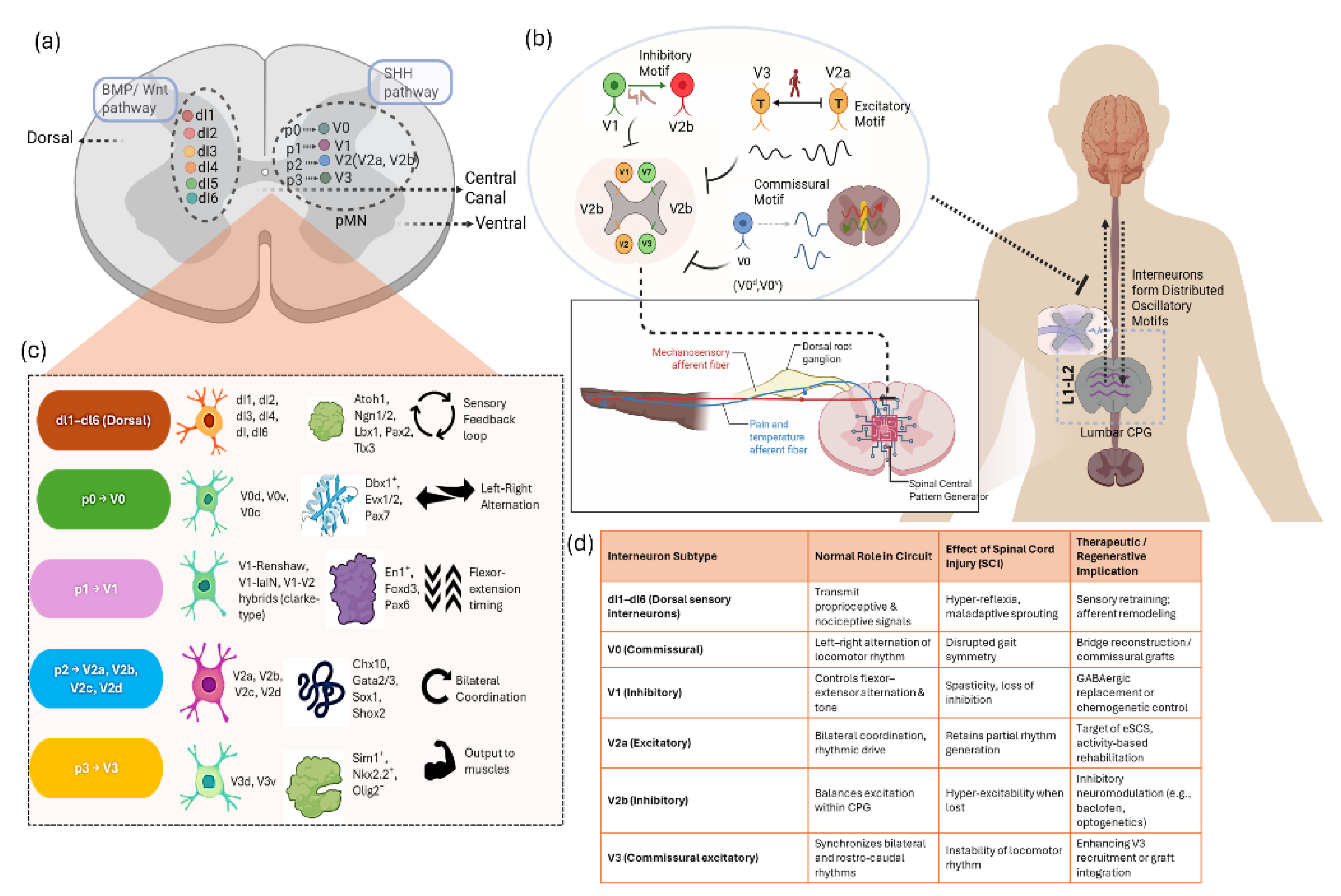
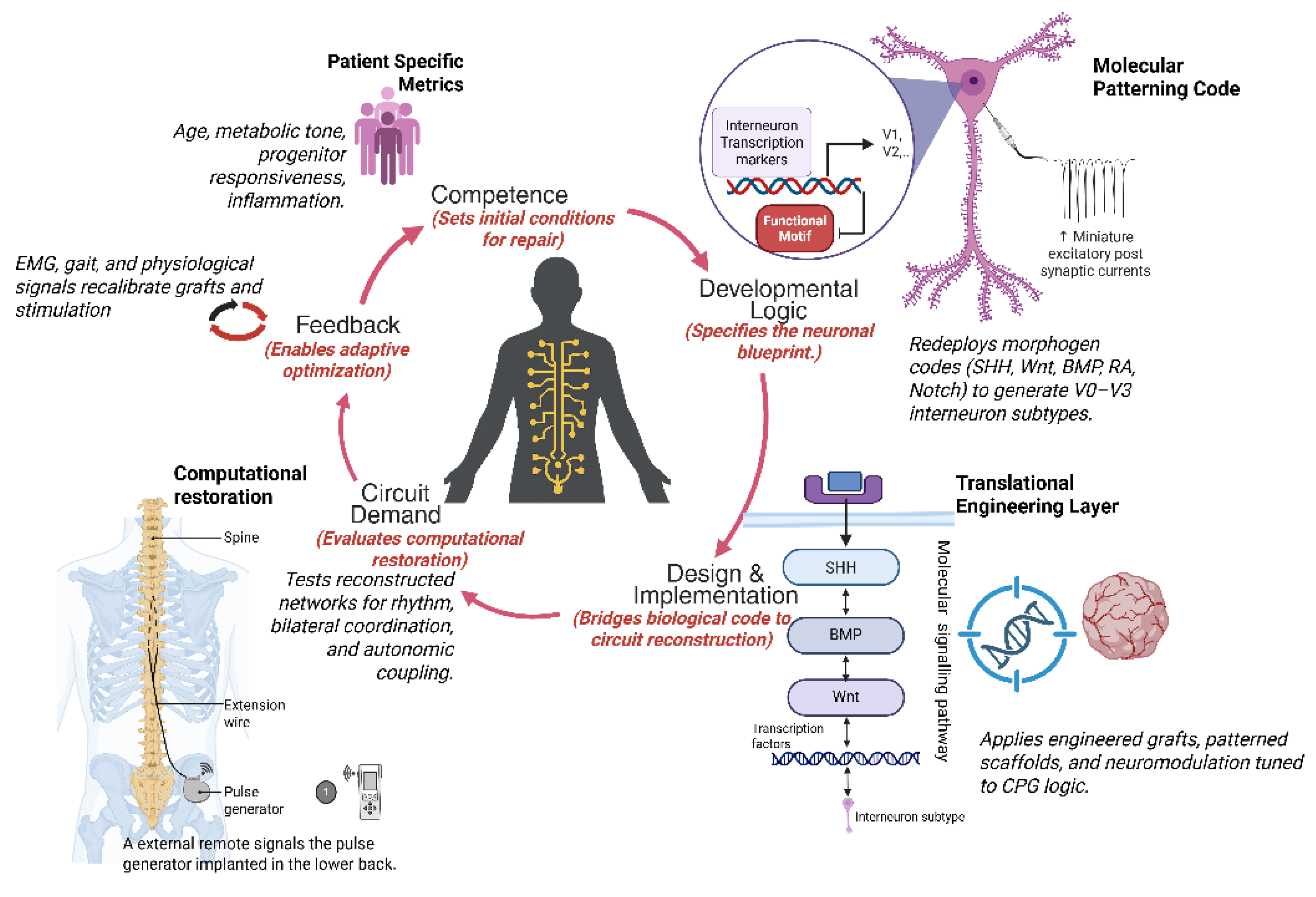
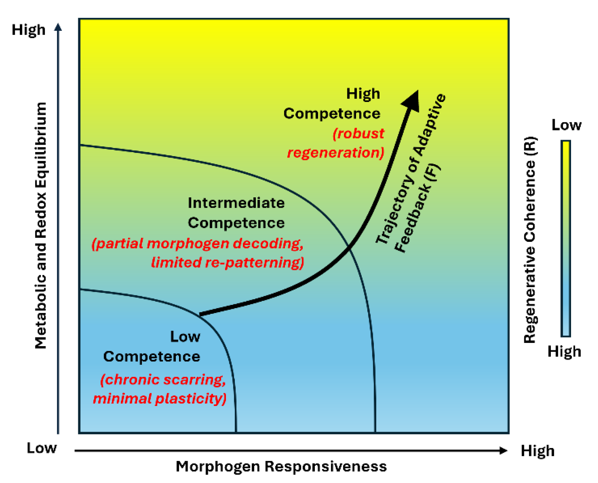
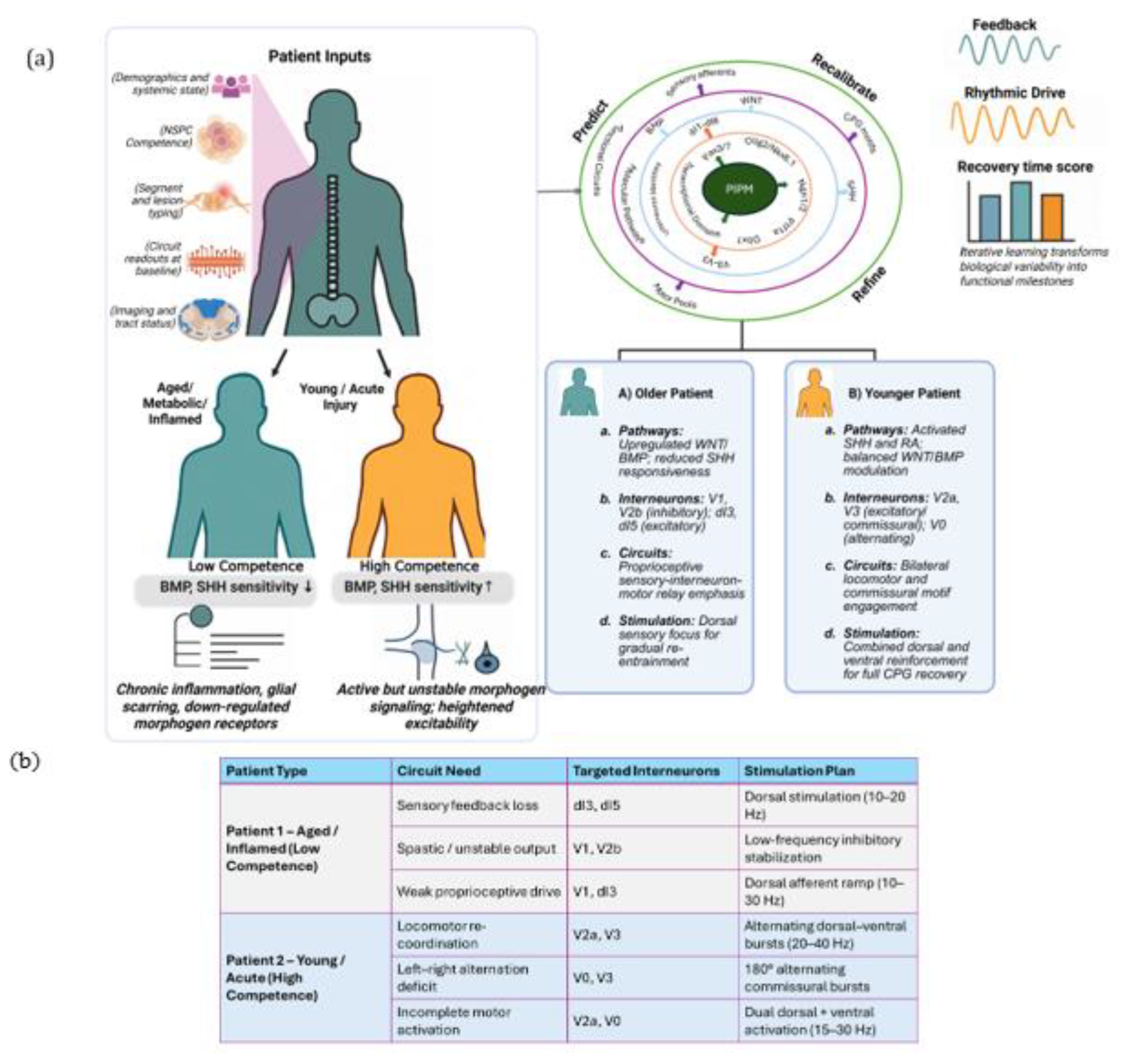
Spinal interneurons constitute the computational core of spinal circuitry, integrating excitatory and inhibitory inputs to generate the rhythmic patterns that drive locomotor, postural, and autonomic control. Their developmental logic, molecular diversity, and adaptive plasticity make them central determinants of functional recovery after spinal cord injury. Yet most regenerative strategies continue to emphasize cellular replacement rather than the restoration of the computational integrity of spinal networks. In this review, we reframe spinal repair as the reconstitution of circuit computation. We synthesize current insights into how embryonic patterning programs defined by SHH, Wnt, and BMP gradients, refined by Notch and retinoic acid signaling, and consolidated by axon guidance cues, establish interneuron diversity, connectivity, and network symmetry that together encode the logic of motor coordination. Spinal cord injury disrupts this developmental logic, fragmenting excitatory and inhibitory balance and desynchronizing rhythmic modules, while residual circuits retain latent capacity for resynchronization through plasticity and neuromodulation. Building upon this developmental and computational continuum, we propose the Patient Specific Interneuron Precision Model (PIPM), a closed loop framework that links patient specific biological states including progenitor competence, morphogen sensitivity, and metabolic tone to circuit level computation and recovery potential. By bridging molecular, physiological, and clinical insights, the PIPM establishes a systems logic that unifies biological competence with circuit recovery, positioning interneuron computation as the conceptual foundation for precision spinal cord regeneration.

Keywords: 
;  ;  ;  ;  ;  ;  ;  ;  ;  

1. Overview

The spinal cord functions as an autonomous computational network capable of generating patterned motor and autonomic output independent of supraspinal input [1,2]. This intrinsic capability arises from the organization of spinal circuits, within which interneurons, comprising diverse excitatory and inhibitory populations, play a central computational role as components of the central pattern generator (CPG) [3,4]. Through their structured synaptic connectivity, these interneurons encode the rhythm, directionality, and coordination of muscle activation across motor pools, sustaining locomotion, posture, and visceral regulation [5,6]. Rather than serving as passive relay elements, interneurons act as the intrinsic logic units of spinal computation [7,8]. Their diversity, spanning multiple progenitor domains and neurotransmitter identities, confers modularity and adaptability, and distinct subtypes mediate left-right alternation, flexor-extensor transitions, and sensory gain control [4,8,9]. Interneurons also establish distributed circuits that dynamically adjust to biomechanical and sensory inputs, facilitating partial functional recovery after injury [3,10,11]. Despite this central role, most regenerative interventions remain agnostic to interneuron subtype identity or circuit-level organization. As a result, structural replacement without restoring the diversity and connectivity of interneurons has yielded inconsistent or incomplete recovery outcomes [12,13,14].
Conventional transplantation of neural stem or progenitor cells frequently results in incomplete lineage specification and fails to restore the precise excitatory-inhibitory balance required for rhythmic coordination, leading to structural repair that yields limited or incoherent function [12,14,15]. These limitations reveal a fundamental conceptual gap, suggesting that replacing neurons does not equate to restoring computation of spinal circuits [16,17,18]. Adding further complexity, the biological competence of human neural stem/progenitor cells (NSPCs) is profoundly heterogeneous [19,20]. This variability directly influences the ability of transplanted or endogenous progenitors to generate appropriate interneuron subtypes and integrate into existing spinal circuits [21,22]. Age, systemic metabolism, chronic inflammation, and prior neurological injury reshape progenitor transcriptional programs and morphogen responsiveness, constraining interneuron differentiation and circuit integration [23,24,25,26]. Aging induces progenitor quiescence and narrows the spectrum of interneuron lineages produced, while astrocytic and immune reactivity sculpt permissive or restrictive microenvironments that govern interneuron survival and connectivity [22,24,27]. These patient-specific variables likely underlie the inconsistent interneuron patterning and circuit outcomes observed across ostensibly similar regenerative interventions. At the systems level, several computational and biological models have been proposed to guide spinal repair, ranging from morphogen-based developmental maps to network simulations of CPG. However, most remain generalized and do not account for patient-specific variability in interneuron competence, subtype identity, or network integration [28,29,30]. Consequently, regenerative strategies derived from such population-level frameworks often fail to predict or reproduce patient-specific outcomes, highlighting a major conceptual gap between cellular potential and the restoration of functional interneuron circuitry [23,30]. To address this gap, we introduce the Patient-specific Interneuron Precision Model (PIPM), a conceptual framework that reframes spinal regeneration as the restoration of circuit computation rather than structural continuity. The model positions interneurons as the core computational units of spinal circuitry and integrates developmental patterning logic, patient-specific biological competence, and circuit-level feedback to iteratively reconstruct rhythmic and coordinated function. Through the integration of cellular state variables and emergent network dynamics, the PIPM establishes a mechanistic foundation for predicting regenerative trajectories and refining interventions across defined interneuron subtypes and circuit motifs.

2. Developmental & Circuit Logic

Spinal interneurons arise from a spatially ordered developmental program that translates dorsoventral patterning cues into the structural and computational architecture of the mature spinal cord [31,32]. Gradients of Sonic Hedgehog (ventral floor plate) and BMP/Wnt (dorsal roof plate) establish progenitor domains (p0-p3 and dI1-dI6) through graded Gli signaling and cross-repressive transcriptional networks involving Dbx1, En1, Chx10, Lhx3, and Sim1 (Figure 1a). Temporal regulation by Notch and retinoic acid signaling further refines lineage competence, while axon guidance systems including Netrins, Slits, Ephrins, and Semaphorins direct projection trajectories that consolidate spatial identity into circuit organization [32,33]. The coordinated action of these molecular programs produces a reproducible map of interneuron subtypes whose intrinsic electrophysiological and synaptic properties reflect their developmental origin (Figure 1b,c). Each class contributes a discrete computational role within the central pattern generator (CPG) network. V0 neurons derived from Dbx1 progenitors project commissurally to coordinate left-right alternation, whereas V1 interneurons defined by En1 expression regulate flexor-extensor transitions and temporal precision [33,34]. V2 interneurons diverge into excitatory V2a (Chx10⁺) and inhibitory V2b (Gata2/3⁺) subtypes that balance excitation and inhibition within motor pools, while V3 neurons marked by Sim1 expression maintain bilateral rhythm and robustness [35,36]. Dorsal interneurons (dI1-dI6) integrate proprioceptive and cutaneous inputs to couple sensory feedback to motor output [31]. Collectively, these excitatory, inhibitory, and commissural motifs embody the fundamental logic of spinal computation, in which developmental identity dictates intrinsic excitability and connectivity to generate patterned locomotor and autonomic output [30]. This developmental framework also delineates the boundaries of regeneration after injury, linking each subtype’s embryonic specification to its selective vulnerability and therapeutic potential (Figure 1d). Excitotoxic and inflammatory cascades preferentially eliminate inhibitory and commissural interneurons, while chronic gliosis and extracellular matrix remodeling degrade residual connectivity and conduction fidelity [37,38]. The resulting imbalance in inhibitory and excitatory drive manifests as spasticity, hyperreflexia, and neuropathic pain, while loss of commissural coordination abolishes rhythmic stepping [38,39]. Such deficits reflect a breakdown of interneuron computation rather than mere neuronal loss.

3. Injury-Induced Computation Loss

Following spinal cord injury (SCI), the coordinated computation of spinal networks collapses as rhythmogenic modules lose excitatory–inhibitory balance and cross-segmental synchrony [30,37,40]. Disconnection of descending input destabilizes interneuron ensembles, producing aberrant firing, spastic reflex loops, and fragmented locomotor output30,41. Within the lesion and spared tissue, surviving interneurons attempt limited reorganization through collateral sprouting, dendritic remodeling, and partial re-engagement of local feedback circuits [39,42]. Electrophysiological mapping shows that ventral interneurons can reroute sensory input and transiently reconstitute rhythmic activity [43,44,45], but these emergent patterns remain unstable without supraspinal coordination [46,47]. Rehabilitative and sensory-evoked activity transiently restore excitability within dormant central-pattern-generator (CPG) nodes, yet the recovered dynamics lack phase precision and fade without continued stimulation48.Neuromodulatory drive through epidural or transcutaneous stimulation can re-entrain surviving circuits by reinstating baseline membrane excitability [49,50], and targeted activation of specific interneuron subclasses (V0, V1, V2) or transplantation of stem-cell-derived V2a/V1 populations can partially rebalance excitation and inhibition [51,52]. Collectively, these observations reveal that recovery depends less on neuronal replacement than on reinstating coherent interneuron computation, highlighting that the core deficit of SCI is computational, not structural.
Beneath this circuit-level collapse lies a progressive molecular exhaustion that erodes the transcriptional programs sustaining interneuron identity and adaptive capacity [30,53,54]. Progenitor competence declines as aging, metabolic tone, and inflammation suppress Notch–EGF, SHH, and Wnt signaling, reducing stemness factors such as SOX2, NES, and MKI67 [22,55]. Attenuated SHH/Wnt activity and loss of BMP antagonism diminish GLI1–β-catenin signaling and downstream factors (Chx10, Evx1, Lhx3), shifting progenitors toward dorsalized or gliogenic fates and destabilizing CPG architecture [31,54,56]. These molecular deficits compress the regenerative bandwidth of the spinal cord by coupling transcriptional fatigue with reduced morphogen sensitivity [23,57]. Consequently, regenerative outcomes vary widely across individuals, driven by differences in progenitor competence, molecular accessibility, and circuit reintegration capacity [23,57]. This heterogeneity highlights that spinal repair follows no uniform biological rule but instead arises from patient-specific configurations of cellular, molecular, and computational parameters [22,58,59]. It is therefore important to understand how these multilevel variables interact to shape regenerative computation.

4. Patient-Specific Interneuron Precision Model (PIPM) Linking Molecular State to Circuit Recovery

The Patient-Specific Interneuron Precision Model (PIPM) provides a unified translational framework built on a closed-loop computational architecture that integrates individual-specific molecular competence, developmental encoding, circuit computation, and adaptive feedback (Figure 2). Within this system, regeneration operates as an iterative inference process that continuously predicts, tests, and corrects deviations between biological state and network output until coherent spinal dynamics are re-established. The framework positions spinal repair as a control architecture in which biological parameters define the initial state, developmental logic translates them into structural configurations, circuit computation evaluates functional recovery, and adaptive feedback progressively recalibrates both biological and engineering layers. In this framework, regeneration begins with the establishment of patient-specific competence, representing the intrinsic capacity of neural stem and progenitor cells to decode morphogenetic signals and generate functional interneuron subtypes. This competence is not a fixed trait, but a multidimensional state defined by morphogen sensitivity, redox equilibrium, metabolic tone, and inflammatory load. Together, these variables set the initial conditions for repair and determine the range of accessible regenerative trajectories. Among these states, high competence reflects efficient morphogen decoding, balanced oxidative metabolism, and accessible chromatin architecture, all of which sustain transcriptional programs required for neuronal diversification and integration [22,24]. Conversely, diminished competence reflects energy deficit, oxidative stress, and pro-inflammatory load, producing restrictive differentiation and network fragmentation [22,25]. Patient-specific metrics such as age, systemic metabolism, and immune activity shift this balance and thereby reshape the regenerative potential of the spinal cord [23,57]. Once competence is defined, the system redeploys the developmental logic that originally constructed the spinal cord. Morphogen gradients of Sonic Hedgehog, BMP, Wnt, Notch, and retinoic acid are decoded through transcriptional networks to generate canonical interneuron subtypes-V0 to V3 and dI1 to dI6 that serve as the computational primitives of spinal circuitry. These subtypes encode distinct rhythmic and regulatory functions. V0 commissural neurons coordinate left-right alternation, V1 interneurons control flexor-extensor phasing, V2a and V2b units stabilise excitatory-inhibitory balance, and V3 neurons sustain bilateral rhythmicity [3,4,41]. The success of regeneration therefore lies not in cellular yield but in the accurate reinstatement of these functional motifs that restore feedback gain and temporal coherence within locomotor and autonomic circuits.
At the systems level, the spinal cord operates as a network of interconnected oscillatory circuits that generate stable rhythmic patterns under healthy conditions [2,60]. Injury displaces these trajectories into desynchronised regions of state space, producing fragmented computation and unstable motor patterns [37]. Regeneration succeeds when network dynamics return to stable physiological states, marked by the re-emergence of coordinated rhythm, phase stability, and synchrony across motor pools characteristic of intact circuitry [30,61]. Functional recovery can thus be described in terms of computational fidelity rather than structural completeness. Observable metrics such as electromyographic coherence, reflex modulation, gait periodicity, and autonomic variability serve as system observers that continuously assess performance and provide error signals to drive recalibration [62,63]. These error signals propagate backward through the PIPM architecture, adjusting morphogen exposure, graft pre-patterning, interneuron composition, and stimulation parameters to correct deviations from the desired computational trajectory. Engineered grafts, patterned scaffolds, and closed-loop neuromodulatory interfaces act as actuators that implement these corrections [64,65,66]. Their parameters are dynamically tuned through physiological feedback, progressively aligning biological growth with functional demand. Each cycle of feedback updates the system’s internal state, reducing phase error, enhancing intersegmental coherence, and moving the network toward a regime of stable rhythmic output. Through these iterative adaptations, the spinal cord transitions from disordered, low-competence configurations to synchronised, high-coherence states within the competence-morphogen manifold.
The interaction between competence and feedback defines the regenerative landscape of the PIPM (Figure 3). In this framework, morphogen responsiveness is plotted against metabolic-redox equilibrium, with regenerative coherence expressed as a gradient from low to high. Low-competence zones are dominated by chronic scarring and minimal plasticity, intermediate states exhibit partial morphogen decoding and limited re-patterning, and high-competence regions support robust regeneration driven by efficient signal decoding and metabolic stability. The trajectory of adaptive feedback (F) traces the system’s ascent through this landscape, representing the progressive refinement of molecular and circuit parameters under continuous control. Over successive cycles of metabolic adjustment and inflammatory modulation, the system’s competence landscape remodels itself, widening the regenerative manifold and fostering the gradual re-establishment of coherent circuit architecture. Operationally, the PIPM generates a patient-specific matrix linking competence class, target interneuron subtype, and neuromodulatory strategy. Quantitative thresholds derived from system observers define progression and rollback boundaries, ensuring adaptive precision across biological and technological domains. Through this recursive integration of measurement and correction, spinal regeneration becomes a self-learning control process in which developmental patterning, physiological feedback, and computational restoration converge. The model thus reframes regeneration as the recovery of spinal computation rather than the replacement of lost cells or a tissue, a process in which rhythm, symmetry, and control emerge from the continuous coupling of molecular state, network topology, and adaptive feedback.

5. Clinical Translation

For clinical translation, we operationalize the PIPM using patient relevant biological and electrophysiological inputs to quantify how competence directs interneuron targeting, stimulation design, and recovery trajectories. Competence values derived from systemic, molecular, and circuit readouts set interneuron priorities and stimulation parameters, producing individualized trajectories toward coherent locomotor output (Figure 4a,b). Within a low-competence regime, typical of aging, metabolic burden, or persistent inflammation, impaired responsiveness to morphogens coincides with scarring and redox imbalance [22,67]. Under these conditions, lineage output shifts toward V1 and V2b inhibitory phenotypes and dorsal sensory classes dI3 to dI5 [3,41]. These populations stabilize segmental relay activity yet provide limited rhythmic adaptability, and the resulting network state expresses weak phase coordination and reduced bilateral symmetry. When metabolic tone and redox control are restored, the effective morphogen window widens and the probability space for Chx10+ V2a and Evx1+ V0 lineages increases, which supports the re-emergence of locomotor symmetry and more reliable rhythmic drive [4,7]. In a high-competence regime, more common in younger or acutely injured contexts, morphogen decoding remains intact and progenitor excitability is higher, which expands the accessible regenerative space [42,59]. Here, balanced engagement of V2a and V3 excitatory interneurons with V0 commissural modules re-establishes alternation across sides and strengthens central pattern generator dynamics [42]. Recovery follows from restoring the proportion and timing of inhibitory and excitatory activity rather than from amplifying either alone [33,35]. Iterative adjustment of morphogen exposure, graft composition, and stimulation frequency improves entrainment and phase stability, with modeled coherence values approaching those observed in empirical electrophysiology [68].
The intervention matrix in Figure 4b summarizes how competence-defined biology can be translated into therapeutic design. Each competence profile aligns with a distinct circuit demand, interneuron target, and stimulation plan, defining a reproducible framework for precision-guided spinal repair. Profiles with low competence favor from dorsal sensory-biased inputs in the 10-30 Hz range combined with strategies that stabilize inhibition, which fortify relay integrity while competence is being conditioned upward [69]. Whereas, high competence conditions benefit from alternating dorsal and ventral activation in the 20-40 Hz range together with deliberate recruitment of commissural pathways to consolidate bilateral coordination [70]. These relationships delineate a mechanistic framework where molecular competence directs circuit-level strategy and stimulation design, providing a streamlined pathway from developmental biology to translational repair.

6. Conclusions

The convergence of developmental biology, patient-specific modeling, and circuit neuroscience is gradually redefining spinal cord injury treatment from generalized repair to precision-guided regeneration [23,71,72]. The Patient-Specific Interneuron Precision Model (PIPM) embodies this transition by viewing spinal repair as the restoration of computation rather than structure. Through the integration of molecular competence, developmental logic, and circuit feedback, the PIPM provides a systems framework in which regenerative outcomes can be iteratively refined by biological input. This perspective positions recovery as an adaptive learning process within the injured spinal network, guided by measurable parameters of progenitor responsiveness, morphogen sensitivity, and circuit coherence. Existing developmental and computational models have advanced our understanding of spinal circuitry, yet most remain generalized and do not incorporate patient-specific biological variation [10,68,73]. The PIPM extends these frameworks by linking molecular state to circuit-level performance and embedding individual variability directly into regenerative logic. Rather than replacing prior models, it complements them by offering a mechanism to integrate patient data, developmental mechanisms, and physiological feedback into a coherent design framework. Recent advances now make it feasible to evaluate these predictions in controlled human-derived system [65,74,75]. Human spinal organoids, assembloids, and microphysiological platforms reproduce segmental identity and rhythmic network behavior, enabling quantitative readouts of progenitor competence and responsiveness [65,74,76]. When combined with multi-omics profiling, electrophysiological mapping, and computational inference, they can help predict recovery trajectories and inform graft patterning or stimulation design [77,78]. Integration with bioelectronic interfaces and wearable sensors further supports adaptive, feedback-driven modulation of therapy in vivo [79,80]. Key challenges remain, including variability among induced pluripotent stem cell lines, incomplete vascular and biomechanical realism in vitro, and the need for rigorous translational and regulatory frameworks for closed-loop neuromodulation [61,81,82]. Even so, patient-specific computational models such as the PIPM offer a pragmatic step toward individualized regenerative strategies that align therapeutic design with measurable biological competence. Here, patient variability becomes a parameter to harness rather than a limitation to overcome, guiding the development of more coherent and reproducible recovery outcomes.

Author Contributions

Sasi Kumar Jagadeesan (SKJ) and Eve Chung Tsai (ET) conceptualized the review and developed the Patient-Specific Interneuron Precision Model (PIPM) framework. SKJ performed the literature synthesis, figure design, and drafting of the original manuscript. Ryan Vimukthie Sandarage (RVS) contributed to visualization, and critical editing. ET provided conceptual refinement, and funding acquisition. All authors discussed the content, revised the text, and approved the final version of the manuscript.

Funding

This work was supported by the Josette Robertson and Joan Johnston Family Foundation Fund (Grant No. JRJJ-2024-001), the Michael T. Richard Clinical Research Fellowship in Neurosurgery at the University of Ottawa Brain and Mind Research Institute (Grant No. MTRF-23-567), the Department of Surgery at the University of Ottawa (Grant No. DOS-2024-045), and the Society of Neurological Surgeons RUNN Course Resident Award (Grant No. RUNN-2024-678).

Competing Interests

The authors declare that they have no competing interests.

Acknowledgments

The authors express their gratitude to the families of organ donors who consented to spinal cord tissue donation for research purposes. We thank the Trillium Gift of Life Network and the organ recovery teams at The Ottawa Hospital for their coordination and support. Technical assistance from the StemCore Facility and the Imaging Core at the Ottawa Hospital Research Institute is gratefully acknowledged.

Ethics Statement

This work does not involve human participants, human biological materials, identifiable personal data, or animal experimentation. As no experimental procedures were performed, ethical approval was not required in accordance with institutional and international guidelines.

Informed Consent

No human subjects or identifiable personal data were included in this study; therefore, informed consent was not applicable.

References

  1. Sengupta, M. & Bagnall, M. W. Spinal Interneurons: Diversity and Connectivity in Motor Control. Annu Rev Neurosci 46, 79–99 (2023). [CrossRef]
  2. Kiehn, O. Decoding the organization of spinal circuits that control locomotion. Nat Rev Neurosci 17, 224–238 (2016).
  3. Zholudeva, L. V. et al. Spinal Interneurons as Gatekeepers to Neuroplasticity after Injury or Disease. The Journal of Neuroscience 41, 845–854 (2021). [CrossRef]
  4. Gosgnach, S. Spinal inhibitory interneurons: regulators of coordination during locomotor activity. Front Neural Circuits 17, (2023).
  5. Grillner, S. & El Manira, A. Current Principles of Motor Control, with Special Reference to Vertebrate Locomotion. Physiol Rev 100, 271–320 (2020). [CrossRef]
  6. Wilson, A. C. & Sweeney, L. B. Spinal cords: Symphonies of interneurons across species. Front Neural Circuits 17, 1146449 (2023).
  7. Ampatzis, K., Song, J., Ausborn, J. & El Manira, A. Separate Microcircuit Modules of Distinct V2a Interneurons and Motoneurons Control the Speed of Locomotion. Neuron 83, 934–943 (2014).
  8. Ziskind-Conhaim, L. & Hochman, S. Diversity of molecularly defined spinal interneurons engaged in mammalian locomotor pattern generation. J Neurophysiol 118, 2956–2974 (2017). [CrossRef]
  9. Talpalar, A. E. et al. Dual-mode operation of neuronal networks involved in left–right alternation. Nature 500, 85–88 (2013).
  10. Asboth, L. et al. Cortico–reticulo–spinal circuit reorganization enables functional recovery after severe spinal cord contusion. Nat Neurosci 21, 576–588 (2018). [CrossRef]
  11. Wagner, F. B. et al. Targeted neurotechnology restores walking in humans with spinal cord injury. Nature 563, 65–71 (2018). [CrossRef]
  12. Kvistad, C. E., Kråkenes, T., Gavasso, S. & Bø, L. Neural regeneration in the human central nervous system—from understanding the underlying mechanisms to developing treatments. Where do we stand today? Front Neurol 15, (2024).
  13. Fischer, G. et al. Advancements in neuroregenerative and neuroprotective therapies for traumatic spinal cord injury. Front Neurosci 18, (2024). [CrossRef]
  14. Lu, P. et al. Long-Distance Growth and Connectivity of Neural Stem Cells after Severe Spinal Cord Injury. Cell 150, 1264–1273 (2012).
  15. Ni, W. et al. Immunomodulatory and Anti-inflammatory effect of Neural Stem/Progenitor Cells in the Central Nervous System. Stem Cell Rev Rep 19, 866–885 (2023). [CrossRef]
  16. Tetzlaff, W. et al. A Systematic Review of Cellular Transplantation Therapies for Spinal Cord Injury. J Neurotrauma 28, 1611–1682 (2011).
  17. YAMASHITA, T. & ABE, K. Recent Progress in Cell Reprogramming Technology for Cell Transplantation Therapy. Neurol Med Chir (Tokyo) 56, 97–101 (2016). [CrossRef]
  18. Dulin, J. N. & Lu, P. Bridging the injured spinal cord with neural stem cells. Neural Regen Res 9, (2014). [CrossRef]
  19. Ottoboni, L., von Wunster, B. & Martino, G. Therapeutic Plasticity of Neural Stem Cells. Front Neurol 11, (2020).
  20. Yang, Y. et al. Epigenetic regulation and factors that influence the effect of iPSCs-derived neural stem/progenitor cells (NS/PCs) in the treatment of spinal cord injury. Clin Epigenetics 16, 30 (2024). [CrossRef]
  21. Elhabbari, K., Sireci, S., Rothermel, M. & Brunert, D. Olfactory deficits in aging and Alzheimer’s—spotlight on inhibitory interneurons. Front Neurosci 18, (2024).
  22. Szegedi, V. et al. Aging-associated weakening of the action potential in fast-spiking interneurons in the human neocortex. J Biotechnol 389, 1–12 (2024).
  23. Jagadeesan, S. K., Sandarage, R. V., Mathiyalagan, S. & Tsai, E. C. Personalized Stem Cell-Based Regeneration in Spinal Cord Injury Care. Int J Mol Sci 26, 3874 (2025). [CrossRef]
  24. Smith, L. K., White, C. W. & Villeda, S. A. The systemic environment: at the interface of aging and adult neurogenesis. Cell Tissue Res 371, 105–113 (2018).
  25. Lecanu, L., Jay Waldron & Althea McCourty. Aging differentially affects male and female neural stem cell neurogenic properties. Stem Cells Cloning 119 (2010) doi:10.2147/SCCAA.S13035.
  26. Lengefeld, J. et al. Cell size is a determinant of stem cell potential during aging. Sci Adv 7, (2021). [CrossRef]
  27. Li, M., Guo, H., Carey, M. & Huang, C. Transcriptional and epigenetic dysregulation impairs generation of proliferative neural stem and progenitor cells during brain aging. Nat Aging 4, 62–79 (2024).
  28. Lu, Q., Wang, X. & Tian, J. A new biological central pattern generator model and its relationship with the motor units. Cogn Neurodyn 16, 135–147 (2022).
  29. Rybak, I. A., Shevtsova, N. A., Lafreniere-Roula, M. & McCrea, D. A. Modelling spinal circuitry involved in locomotor pattern generation: insights from deletions during fictive locomotion. J Physiol 577, 617–639 (2006). [CrossRef]
  30. Rybak, I. A., Dougherty, K. J. & Shevtsova, N. A. Organization of the Mammalian Locomotor CPG: Review of Computational Model and Circuit Architectures Based on Genetically Identified Spinal Interneurons(1,2,3). eNeuro 2, (2015). [CrossRef]
  31. Vallstedt, A. & Kullander, K. Dorsally derived spinal interneurons in locomotor circuits. Ann N Y Acad Sci 1279, 32–42 (2013).
  32. Goyal, R., Spencer, K. A. & Borodinsky, L. N. From Neural Tube Formation Through the Differentiation of Spinal Cord Neurons: Ion Channels in Action During Neural Development. Front Mol Neurosci 13, (2020). [CrossRef]
  33. Francius, C. et al. Identification of Multiple Subsets of Ventral Interneurons and Differential Distribution along the Rostrocaudal Axis of the Developing Spinal Cord. PLoS One 8, e70325 (2013).
  34. Chapman, P. D. et al. A brain-wide map of descending inputs onto spinal V1 interneurons. Neuron 113, 524-538.e6 (2025). [CrossRef]
  35. Chopek, J. W., Nascimento, F., Beato, M., Brownstone, R. M. & Zhang, Y. Sub-populations of Spinal V3 Interneurons Form Focal Modules of Layered Pre-motor Microcircuits. Cell Rep 25, 146-156.e3 (2018).
  36. Lundfald, L. et al. Phenotype of V2-derived interneurons and their relationship to the axon guidance molecule EphA4 in the developing mouse spinal cord. Eur J Neurosci 26, 2989–3002 (2007). [CrossRef]
  37. Kumru, H., Vidal, J., Kofler, M., Portell, E. & Valls-Solé, J. Alterations in Excitatory and Inhibitory Brainstem Interneuronal Circuits after Severe Spinal Cord Injury. J Neurotrauma 27, 721–728 (2010).
  38. Hughes, D. I. & Todd, A. J. Central Nervous System Targets: Inhibitory Interneurons in the Spinal Cord. Neurotherapeutics 17, 874–885 (2020). [CrossRef]
  39. Flynn, J. R., Graham, B. A., Galea, M. P. & Callister, R. J. The role of propriospinal interneurons in recovery from spinal cord injury. Neuropharmacology 60, 809–822 (2011).
  40. Courtine, G. et al. Transformation of nonfunctional spinal circuits into functional states after the loss of brain input. Nat Neurosci 12, 1333–1342 (2009). [CrossRef]
  41. Deska-Gauthier, D. & Zhang, Y. The functional diversity of spinal interneurons and locomotor control. Curr Opin Physiol 8, 99–108 (2019).
  42. Zavvarian, M.-M., Hong, J. & Fehlings, M. G. The Functional Role of Spinal Interneurons Following Traumatic Spinal Cord Injury. Front Cell Neurosci 14, (2020). [CrossRef]
  43. Harkema, S. et al. Effect of epidural stimulation of the lumbosacral spinal cord on voluntary movement, standing, and assisted stepping after motor complete paraplegia: a case study. Lancet 377, 1938–47 (2011).
  44. Park, J., Kim, Y. G., Kim, T. & Baek, M. Electrical Stimulation of the M1 Activates Somatostatin Interneurons in the S1: Potential Mechanisms Underlying Pain Suppression. eNeuro 12, ENEURO.0541-24.2025 (2025).
  45. Taccola, G. et al. Dynamic electrical stimulation enhances the recruitment of spinal interneurons by corticospinal input. Exp Neurol 371, 114589 (2024). [CrossRef]
  46. Balbinot, G. et al. Functional electrical stimulation therapy for upper extremity rehabilitation following spinal cord injury: a pilot study. Spinal Cord Ser Cases 9, 11 (2023).
  47. Singh, R. E. et al. Epidural stimulation restores muscle synergies by modulating neural drives in participants with sensorimotor complete spinal cord injuries. J Neuroeng Rehabil 20, 59 (2023). [CrossRef]
  48. Duysens, J. & Forner-Cordero, A. A controller perspective on biological gait control: Reflexes and central pattern generators. Annu Rev Control 48, 392–400 (2019).
  49. Golowasch, J. Neuromodulation of central pattern generators and its role in the functional recovery of central pattern generator activity. J Neurophysiol 122, 300–315 (2019).
  50. Fu, X. et al. Gq neuromodulation of BLA parvalbumin interneurons induces burst firing and mediates fear-associated network and behavioral state transition in mice. Nat Commun 13, 1290 (2022). [CrossRef]
  51. Ljunggren, E. E., Haupt, S., Ausborn, J., Ampatzis, K. & El Manira, A. Optogenetic Activation of Excitatory Premotor Interneurons Is Sufficient to Generate Coordinated Locomotor Activity in Larval Zebrafish. The Journal of Neuroscience 34, 134–139 (2014).
  52. Falgairolle, M. & O’Donovan, M. J. Optogenetic Activation of V1 Interneurons Reveals the Multimodality of Spinal Locomotor Networks in the Neonatal Mouse. The Journal of Neuroscience 41, 8545–8561 (2021).
  53. Jessell, T. M. Neuronal specification in the spinal cord: inductive signals and transcriptional codes. Nat Rev Genet 1, 20–29 (2000). [CrossRef]
  54. Briscoe, J. & Ericson, J. The specification of neuronal identity by graded sonic hedgehog signalling. Semin Cell Dev Biol 10, 353–362 (1999).
  55. Navarro Negredo, P., Yeo, R. W. & Brunet, A. Aging and Rejuvenation of Neural Stem Cells and Their Niches. Cell Stem Cell 27, 202–223 (2020).
  56. Ribes, V. & Briscoe, J. Establishing and Interpreting Graded Sonic Hedgehog Signaling during Vertebrate Neural Tube Patterning: The Role of Negative Feedback. Cold Spring Harb Perspect Biol 1, a002014–a002014 (2009). [CrossRef]
  57. Mayhew, J. A. et al. Age-related gene expression changes in lumbar spinal cord: Implications for neuropathic pain. Mol Pain 16, 1744806920971914 (2020). [CrossRef]
  58. Vanacore, G., Christensen, J. B. & Bayin, N. S. Age-dependent regenerative mechanisms in the brain. Biochem Soc Trans 52, 2243–2252 (2024).
  59. Eavri, R. et al. Interneuron Simplification and Loss of Structural Plasticity As Markers of Aging-Related Functional Decline. The Journal of Neuroscience 38, 8421–8432 (2018).
  60. Kiehn, O. Development and functional organization of spinal locomotor circuits. Curr Opin Neurobiol 21, 100–109 (2011). [CrossRef]
  61. Wenger, N. et al. Spatiotemporal neuromodulation therapies engaging muscle synergies improve motor control after spinal cord injury. Nat Med 22, 138–145 (2016).
  62. Håkansson, S. et al. Data-driven prediction of spinal cord injury recovery: An exploration of current status and future perspectives. Exp Neurol 380, 114913 (2024).
  63. Guaraldi, P. et al. Effects of Spinal Cord Injury Site on Cardiac Autonomic Regulation: Insight from Analysis of Cardiovascular Beat by Beat Variability during Sleep and Orthostatic Challenge. J Funct Morphol Kinesiol 7, 112 (2022).
  64. Kadoya, K. et al. Spinal cord reconstitution with homologous neural grafts enables robust corticospinal regeneration. Nat Med 22, 479–487 (2016).
  65. Xue, W. et al. Generation of dorsoventral human spinal cord organoids via functionalizing composite scaffold for drug testing. iScience 26, 105898 (2023). [CrossRef]
  66. Lorach, H. et al. Walking naturally after spinal cord injury using a brain–spine interface. Nature 618, 126–133 (2023).
  67. Abbadie, C., Trafton, J., Liu, H., Mantyh, P. W. & Basbaum, A. I. Inflammation Increases the Distribution of Dorsal Horn Neurons That Internalize the Neurokinin-1 Receptor in Response to Noxious and Non-Noxious Stimulation. The Journal of Neuroscience 17, 8049–8060 (1997). [CrossRef]
  68. Griffin, J. M. & Bradke, F. Therapeutic repair for spinal cord injury: combinatory approaches to address a multifaceted problem. EMBO Mol Med 12, (2020).
  69. Reali, C. & Russo, R. E. Neuronal intrinsic properties shape naturally evoked sensory inputs in the dorsal horn of the spinal cord. Front Cell Neurosci 7, (2013). [CrossRef]
  70. Jervis Rademeyer, H., Gauthier, C., Masani, K., Pakosh, M. & Musselman, K. E. The effects of epidural stimulation on individuals living with spinal cord injury or disease: a scoping review. Physical Therapy Reviews 26, 344–369 (2021).
  71. Ausborn, J., Shevtsova, N. A. & Danner, S. M. Computational Modeling of Spinal Locomotor Circuitry in the Age of Molecular Genetics. Int J Mol Sci 22, 6835 (2021). [CrossRef]
  72. Bareyre, F. M. et al. The injured spinal cord spontaneously forms a new intraspinal circuit in adult rats. Nat Neurosci 7, 269–277 (2004).
  73. Shevtsova, N. A. et al. Organization of left-right coordination of neuronal activity in the mammalian spinal cord: Insights from computational modelling. J Physiol 593, 2403–26 (2015).
  74. Buchner, F., Dokuzluoglu, Z., Grass, T. & Rodriguez-Muela, N. Spinal Cord Organoids to Study Motor Neuron Development and Disease. Life 13, 1254 (2023). [CrossRef]
  75. Zhou, G., Pang, S., Li, Y. & Gao, J. Progress in the generation of spinal cord organoids over the past decade and future perspectives. Neural Regen Res 19, 1013–1019 (2024).
  76. Ajongbolo, A. O. & Langhans, S. A. Brain Organoids and Assembloids—From Disease Modeling to Drug Discovery. Cells 14, 842 (2025).
  77. Peng, R. et al. Spatial multi-omics analysis of the microenvironment in traumatic spinal cord injury: a narrative review. Front Immunol 15, (2024). [CrossRef]
  78. Rasoolinejad, M. et al. Machine learning predicts improvement of functional outcomes in spinal cord injury patients after inpatient rehabilitation. Frontiers in Rehabilitation Sciences 6, (2025).
  79. Mariello, M. Reliability and stability of Bioelectronic Medicine: a critical and pedagogical perspective. Bioelectron Med 11, 16 (2025).
  80. Giagiozis, M. et al. Feasibility and Sensitivity of Wearable Sensors for Daily Activity Monitoring in Spinal Cord Injury Trials. Neurorehabil Neural Repair 39, 814–825 (2025). [CrossRef]
  81. Volpato, V. & Webber, C. Addressing variability in iPSC-derived models of human disease: guidelines to promote reproducibility. Dis Model Mech 13, (2020).
  82. Fan, X. et al. Strategies to overcome the limitations of current organoid technology - engineered organoids. J Tissue Eng 16, (2025). [CrossRef]
Figure 1. Developmental and functional logic of spinal interneuron diversity and its translational relevance. (a) Dorsoventral morphogen gradients (BMP/Wnt dorsally and SHH ventrally) organize the embryonic spinal cord into discrete progenitor domains (p0-p3, dI1-dI6) that encode spatial and molecular identity through transcriptional cross repression.(b) These progenitors generate ventral interneuron subtypes (V0-V3) and dorsal sensory classes (dI1-dI6) that assemble into inhibitory, excitatory, and commissural motifs forming the core of the CPG. V0 commissural neurons (Dbx1⁺) coordinate left and right alternation, V1 inhibitory neurons (En1⁺) regulate flexor and extensor timing, V2a (Chx10⁺) and V2b (Gata2/3⁺) interneurons balance excitation and inhibition, and V3 excitatory commissural neurons (Sim1⁺) stabilize bilateral rhythm. Dorsal interneurons integrate proprioceptive and nociceptive input, linking sensory feedback to locomotor and autonomic control. (c) Domain specific transcription factor combinations delineate lineage boundaries and encode circuit function, while temporal morphogen exposure (SHH, Wnt, BMP, Notch, RA) defines dorsal and ventral bias and developmental competence. (d) Functional mapping of interneuron subtypes illustrates how developmental specification predicts circuit vulnerability and therapeutic potential after spinal cord injury (SCI). Selective loss of V0 neurons disrupts commissural symmetry, imbalance in V1 and V2b networks induces spasticity, and diminished V2a or V3 activity weakens rhythmic drive. Subtype directed grafting, neuromodulation, and molecular reprogramming offer strategies to restore specific computational modules within spinal networks.
Preprints 189176 g001
Figure 2. The Patient-specific Interneuron Precision Model (PIPM): an adaptive framework linking biological inputs, developmental logic, engineering design, and circuit feedback for spinal cord regeneration. The PIPM illustrates the logical flow of spinal circuit reconstruction as a closed loop control process that restores computation rather than simply replacing cells. The model progresses through four interconnected domains: Competence, Developmental Logic, Design and Implementation, and Circuit Demand with Feedback, each representing a stage of biological computation. Competence defines the regenerative starting conditions, integrating patient-specific variables such as progenitor responsiveness, metabolic tone, and inflammatory state. Developmental Logic redeploys morphogen pathways (SHH, Wnt, BMP, RA, Notch) to generate V0-V3 interneuron subtypes and establish the neuronal blueprint. Design and Implementation bridges molecular code to circuit reconstruction through engineered grafts, patterned scaffolds, and neuromodulation tuned to CPG logic. Circuit Demand and Feedback evaluate rhythmic, bilateral, and autonomic coherence, using EMG, gait, and physiological outputs as feedback signals that recalibrate graft patterning and stimulation parameters. Patient-specific biological inputs are processed through this recursive framework, allowing developmental logic to be computationally re-engaged for adaptive and precision-guided spinal regeneration.
Preprints 189176 g002
Figure 3. Regenerative Competence Landscape in the PIPM Framework. A state-space representation of regenerative potential as a function of morphogen decoding and metabolic-redox equilibrium. Patient-specific conditions define a position within this landscape, where regenerative coherence (R) increases along an adaptive trajectory (F). Competence zones reflect the capacity of progenitor systems to respond to guidance cues, with high-competence regions enabling robust circuit reconstruction and low-competence zones associated with scarring and impaired plasticity.
Preprints 189176 g003
Figure 4. Clinical translation of the Patient-Specific Interneuron Precision Model (PIPM). The PIPM integrates patient-derived biological inputs to simulate regenerative trajectories across competence states. (a) Low-competence conditions, common in older, metabolically impaired, or chronically inflamed patients, are characterized by attenuated SHH/BMP signaling, glial scarring, and reduced morphogen responsiveness, biasing differentiation toward inhibitory (V1, V2b) and proprioceptive (dI3–dI5) interneurons that stabilize relay circuits but limit rhythmic flexibility.In high-competence conditions, preserved morphogen sensitivity and progenitor excitability expand the regenerative space toward excitatory (V2a, V3) and commissural (V0) populations that restore bilateral rhythm and phase symmetry. (b) The translation matrix links each competence profile to targeted interneuron recruitment and stimulation parameters, positioning progenitor competence as a functional biomarker of recovery velocity, circuit coherence, and responsiveness to neuromodulatory intervention.
Preprints 189176 g004
Disclaimer/Publisher’s Note: The statements, opinions and data contained in all publications are solely those of the individual author(s) and contributor(s) and not of MDPI and/or the editor(s). MDPI and/or the editor(s) disclaim responsibility for any injury to people or property resulting from any ideas, methods, instructions or products referred to in the content.
Copyright: This open access article is published under a Creative Commons CC BY 4.0 license, which permit the free download, distribution, and reuse, provided that the author and preprint are cited in any reuse.
Prerpints.org logo

Preprints.org is a free preprint server supported by MDPI in Basel, Switzerland.

Subscribe

Disclaimer

Terms of Use

Privacy Policy

Privacy Settings

© 2025 MDPI (Basel, Switzerland) unless otherwise stated