Preprint
Article

This version is not peer-reviewed.

Genetic Identification of Wood-Destroying Fungi in Weakened Scots Pine (Pinus sylvestris L.) Forests Using DNA Barcoding

Submitted:

05 December 2025

Posted:

08 December 2025

You are already at the latest version

Abstract

Weakened forest stands provide a favorable environment for the development of saprotrophic and necrotrophic fungi. Traditional identification methods for these ecological groups of fungi are not always effective. However, modern DNA barcoding methods based on sequencing DNA regions of the studied fungal specimens and comparing them 31 with the nucleotide sequences of previously classified organisms often allow for precise species identification. Thus, to identify fungi using barcoding in weakened Scots pine (Pinus sylvestris L.) stands in the Central Forest-Steppe of European Russia (Voronezh, Voronezh Region), 55 mycelium and fruiting body samples were collected from conifer litter and Scots pine damaged butt part of the trunk and roots. After morphological and ecological description and systematization of the collection, DNA extraction was performed, the ITS1 and ITS2 regions were sequenced in 21 samples, and 11 species of xylotrophic fungi were identified, of which three species — Hirschioporus fuscoviolaceus (Ehrenb.) Donk, Gymnopilus penetrans (Fr.) Murrill, and Ganoderma applanatum (Pers.) Pat. — infect living wood. The remaining eight representatives of the mycobiota were saprotrophic fungi involved in the mineralization of organic residues. Two phyla, Ascomycota and Basidiomycota, were identified, with the most samples belonging to Basidiomycota and the class Agaricomycetes with seven orders. The predominance of Basidiomycota species indicates stages III-IV mycogenic xylolysis of the weakened stands analyzed. The DNA barcoding method complements the results of morphological and ecological analysis and provides a more complete mycological picture of wood-destroying fungi in weakened pine forests that are difficult to identify.

Keywords: 
;  ;  ;  ;  ;  

1. Introduction

Modern molecular genetic approaches to the identification of organisms, such as DNA barcoding, make it possible to differentiate morphologically difficult to distinguish species. These organisms include fungi that cause wood rot. DNA barcoding is currently widely used for this purpose [1,2,3]. This method has several advantages: identification from a 54 single sample, regardless of morphological features or life cycle stage, and identification accuracy. 55 DNA barcoding makes it possible to determine the species composition of environmental samples [4], to treat fungal diseases at early stages 56 of their development, and to monitor the spatial and temporal patterns of fungal distributions and migrations. DNA barcoding is a potent approach for rapid identification of fungal specimens, generating novel species hypotheses, and guiding biodiversity and ecological studies [1]. The most commonly used nucleotide sequence for fungal systematics and taxonomy is the internal transcribed spacer (ITS) sequence of nuclear DNA [2], which enables species identification. The method is based on comparing the analyzed unknown DNA sequence with homologous sequences in a database. Basic Local Alignment Search Tool (BLAST [5]) comparison and search algorithms are used for this purpose. Currently, this method is widely used in both agriculture and forestry, enabling a more accurate and complete description of fungal communities, including the identification of pathogens and their antagonists.
Disadvantages of barcoding include the presence of pseudogenes in the genome, which can contribute to misidentification [6]. Due to insufficient intra- and interspecific variability, species identification can also be difficult for some taxa [7]. Furthermore, the universality of primers may be limited, and they may not amplify target genes in some species. Results are verified by comparison with previously deposited sequences from publicly available databases, which are constantly being updated but possibly do not include all species [8].
Pine forests are widespread worldwide, with Scots pine (Pinus sylvestris L.) being the most widespread and important species of the genus Pinus in Eurasia and one of the most ecologically and economically important tree species in Europe [9]. According to a recent press release of the Federal Forestry Agency (Rosleshoz) of the Ministry of Natural Resources and Environment of the Russian Federation, the area covered by forest vegetation in the Russian Federation was approximately 766 million hectares, of which pine trees accounted for about 15.5%, which is equivalent to ~118.7 million hectares [10]. Scots pine accounts for 99% of the conifer forests of the Central Forest-Steppe of European Russia [11].
Adverse weather conditions are the primary cause of forest degradation 81 and decline. A range of weather and climate events and their consequences impact forest ecosystems, leading to their transformation. The combination of rising surface temperatures and reduced precipitation or precipitation patterns in some regions of the country (for example, in the south of European Russia) leads to an increase in the frequency and number of droughts, as well as an increase in the incidence of forest fires and the area burned. Furthermore, the expansion of pest and disease habitats is a significant factor in forest vulnerability to climate change [12]. According to the Federal State Statistics Service of the Ministry of Economic Development of the Russian Federation (Rosstat) data summarized by Zamolodchikov et al. [13]; Section 2.2.1 “The main natural forest disturbances and climate inter-linkages”, page 21], damage to Russian forests in 2014–2017 was predominantly caused by fires (63%), insects (15%), weather conditions (11%), diseases (10%), and other factors such as industrial pollution (~1%).
The development of epiphytoties of fungal diseases is facilitated by a combination of factors acting simultaneously: presence of a source or focus of infection and disease vectors, weather, climate, and edaphic conditions favorable for the pathogen and unfavorable for the host plant, the absence of active competitors or antagonists, and anthropogenic impacts leading to the weakening of trees [14]. The region’s geographic location determines the fragmentation of forest stands, which is characteristic of the forest-steppe zone. At the southern border of forest distribution, trees experience a combination of negative factors, the most severe of which are droughts [15]. Lack of moisture not only negatively affects the activity of physiological processes in trees but also disrupts interactions with mycorrhizal fungi, while pathogen activity increases [16,17]. One of the most dangerous pathogens for conifer stands, including Scots pine stands, is the root fungus Heterobasidion annosum (Fr.) Bref. [18,19], and Cenangium ferruginosum Fr., which cause severe tree mortality [17,20]. These pathogens cause significant damage to conifer forests worldwide.
The species composition of wood-destroying biotrophic fungi that cause rot damage (wood decay) to pine remains unchanged along the entire longitudinal gradient of pine distribution within the Russian Plain. Only the occurrence of individual wood-destroying fungi species changes significantly. The main species of wood-destroying biotrophic fungi include Climacocystis borealis (Fr.) Kotl. & Pouzar, Heterobasidion annosum (Fr.) Bref., Phaeolus schweinitzii (Fr.) Pat., Porodaedalea chrysoloma (Fr.) Fiasson & Niemelä, and Porodaedalea pini (Brot.) Murrill] [=Phellinus pini (Brot.) A. Ames] [21]. Currently, there are a number of scientific publications on the identification of fungal species in Scots pine stands in the Central Forest-Steppe of European Russia [e.g., 21], but DNA barcoding was not used in these or similar studies yet. Therefore, the main aim of this study was the molecular genetic identification of fungi causing wood rot in Scots pine stands in weakened stands using DNA barcoding.

2. Materials and Methods
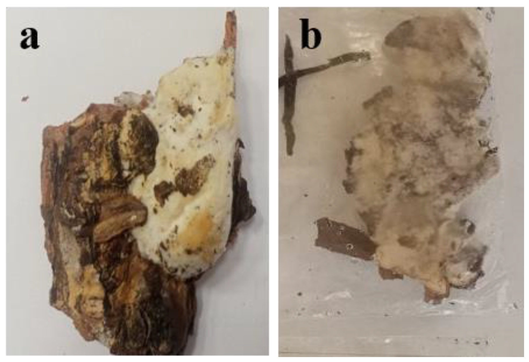
Fungi Sampling

All studied Scots pine stands were planted in suburban and urban forests; their ages ranged from 40 to 80 years (Table 1). In total, 28 samples were collected from suburban forests and 27 from the urban “Northern Forest” (NF, Voronezh City) plantation. 
The study used the mycelium of sapro- and necrotrophic fungi collected from forest conifer litter (litter), roots and butt parts of Scots pine in foci of root fungus infection in weakened pine stands of the Central forest-steppe of the European part of Russia in the Voronezh region. In total, over 60 Voronezh region sample plots of Scots pine stands were analyzed by forest mensuration descriptions; 43 of them (125.1 hectares) were weakened stands, displaying a combination of root fungal infestations. Five permanent sample plots with weakened Scots pine stands (No. 77, 79, 80, 83, and NF) were selected to collect mycelium from fungi that cause wood rot (Table 1). Weakened stands included stands with the following characteristics: 1) the presence of pathological mortality, namely dried-out and wind-fallen trees with a diameter close to or above the average, with a predominance of weakened trees with a sparse crown, reduced growth, but not more than half, and individual dried branches, 2) severely weakened with a very sparse crown, light green, gray, matte or two-thirds eaten needles, weak growth (less than half of the usual), the presence of drying or dried branches, possible significant mechanical damage to the trunk, dry tops, signs of damage by diseases and pests of the trunk, root paws, branches, and needles, 3) drying with a very sparse crown, crumbling needles, gray, yellowish or yellow-green color, absence of needle development on the shoot, very weak or completely absent growth, 4) drying out of more than two-thirds of the branches, dry branches of more than 50%, damage to the trunk and root paws by more than two-thirds, and signs of infestation by stem pests). The “Northern Forest” (NF) permanent sample plot is located in Voronezh City. The other four permanent sample plots are located in the Voronezh suburban area: No. 77, 79, 80, and 83 compartments of the Prigorodnoye forestry, the Left Bank district forestry of the Voronezh region, the educational and experimental forestry of the G.F. Morozov Voronezh State Forestry University, as well as the Northern Forest Nature Park, Voronezh (Figure 1).
Fungi mycelium was collected in the spring and summer. During collection, the collection location and substrate - conifer litter, tree, or a tree part -- were recorded. If it was collected from a tree, then, its condition and the exact location of the mycelium (trunk or root) were recorded. The individual tree specific parts, such as the root of a fallen tree, a stump from a logging operation, etc., were also recoderded, as well as the morphological and ecological characteristics of the samples. In total, 55 samples were collected from all study plots. All collected samples were assigned unique ID names. For example, sample name “79/3 root” has the following meaning: “79” is the compartment number, “3” is the number of the sample collected in that compartment, and “root” is the specific location on the tree from which the sample was taken (root in this case). Samples that were preliminarily assigned to Heterobasidion annosum based on morphological and ecological description (for example, samples 79/3 root and 79/7 root collected in compartment 79 from the roots of a fresh windfall, and excavated tree roots, respectively, were sown on a nutrient medium to obtain pure cultures (Figure 2) and were then identified as Trametes hirsute based on DNA barcoding. Malt-peptone agar was used as a growth medium, with the following composition: malt extract – 30 g, peptone – 1 g, agar – 20 g, distilled water – 1 L. Mycelial fragments from the samples were placed in the center of the growth medium.
The grown samples were then transferred to a new, clean growth medium. The process of multiple transfers was repeated until a pure culture was obtained. The experiment was repeated 10 times. After obtaining pure cultures of the fungal samples, they were identified. Pure fungal cultures were stored in test tubes on malt-peptone agar in the refrigerator at 4 °C. The cultures were maintained by periodic subcultures, every six months. After labeling and systematization of the samples, a process of their morphological and ecological description was carried out, a general table of samples was compiled, DNA was isolated and used for barcoding.

DNA Isolation and PCR

DNA was isolated from fungi cultures using the PROBA-GS reagent kit (DNA-Technology, Moscow, Russia). DNA concentration was determined using an F-7000 spectrophotometer (Hitachi High-Technologies Corporation, Tokyo, Japan) at a wavelength of 260 nm. The purity of the resulting preparations was assessed by the A260/A280 ratio.
DNA regions containing ITS1 и ITS2 were amplified using PCR with universal forward ITS1 (TCCGTAGGTGAACCTGCGG) and reversal ITS4 (TCCTCCGCTTATTGATATGC) primers in the following mixture: deionized water – 16 µL, DNA – 2 µL, ITS1 – 1 µL (10 µM), ITS4 – 1 µL (10 µM), 5x ScreenMix-HS buffer (Eurogen, Russia) – 5 µL.
The PCR was performed using a MasterCycler Personal Thermocycler (Eppendorf, Hamburg, Germany) and the following program: initial heating at 95 °C for 5 min, 37 cycles of denaturation at 95 °C for 30 sec, primer annealing at 54 °C for 30 sec, and chain elongation at 72 °C for 45 sec. The PCR products were checked using 2% agarose gel electrophoresis and visualized using a QuantumM-312B transilluminator (Helikon, Moscow, Russia) at 312 nm. A DNA ladder 100 bp+ (Eurogen, Moscow, Russia) was used to determine the product length. The gel fragment containing the desired DNA fragment was cut out from the agarose gel and purified using the Cleanup Standard kit (Eurogen, Moscow, Russia).

DNA Sequencing and Barcoding

Sequencing of purified PCR products was performed on a NANOFOR 05 genetic analyzer (Synthol, Moscow, Russia) using the BigDye Terminator v3.1 Cycle Sequencing Kit (Thermo Fisher Scientific, Waltham, MA, USA). Taxonomic identification of nucleotide reads was performed in the NCBI GenBank database (https://www.ncbi.nlm.nih.gov) using BLAST [5] and the BOLD System [22].
Phytopathogen taxa names are provided according to the MycoBank on-line database [23].

3. Results

In total, 55 samples were described and systematized. Fungal identification was based on a comprehensive approach that included morphological diagnostics of the study objects, a study of the characteristic symptoms of fungal disease development in forest stands, and DNA barcoding. Of the 55 samples studied, we obtained barcodes for only 15, representing 11 species. Their morphological and ecological properties are described in Table 2.
The taxonomic position of the identified wood-decay fungi species is presented in Table 3.
Bioinformatic analysis of the obtained barcodes allowed us to identify 15 samples with a high degree of accuracy, representing representatives of 11 different taxa. The nucleotide sequences of the identified fungi were deposited in the NCBI GenBank, and their sequence accession numbers are presented in Table 1, along with a brief description of the morphological and ecological properties of the identified wood-decay fungi.
Photos of mycelium and fruiting bodies with established taxonomic affiliation are presented in Supplementary Figure S1.
Based on the results of identification, morphological and ecological description and taxonomy, a database “Collection of fruiting bodies and mycelium of saprotrophic and necrotrophic fungi in pine plantations of Voronezh and the Voronezh region” was compiled (Supplementary Table S1).
DNA barcoding has made it possible to systematize the following representatives of pathogenic fungi.
The species Dichomitus squalens (P. Karst.) D.A. Reid was identified in three samples (Table 2). Based on morphological and ecological description, its fruiting body is white with a yellowish tint, leathery, of indefinite shape, has a mushroom odor, hymenophore is tubular. The fungus is a xylosaprotroph. Usually found on conifer wood, but can also grow and destroy deciduous trees. It grows after fires on dead conifer wood – pine (Pinus), fir (Abies), juniper (Juniperus), larch (Larix), spruce (Picea), hemlock (Tsuga), pseudotsuga (Pseudotsuga), and arborvitaes (Thuja). In Western Siberia, it has been recorded on Scots pine (Pinus sylvestris) and Siberian stone pine (Pinus sibirica Du Tour). It is capable of effectively decomposing cellulose and lignin. It causes a rapidly developing, white, pitted rot; in the final stage, the wood splits into fibers. This species is rare and is listed in the Red Data Book of the Republic of Karelia [24,25]. The ITS sequence matched the Dichomitus squalens sequence in the NCBI GenBank (accession number ON088332.1) with 100% identity and 99.64% coverage. This pathogen was detected in three samples tested.
The Genus Sistotrema sp. was found in two samples (Table 2). It represents saprotrophic organisms with the ability to decompose wood; this process is called white rot. According to the morphological and ecological description, it is white in color, with a yellowish or whitish-yellow tint. It has no distinct shape, can cover small areas of wood, and forms fruiting bodies in the form of a flat cap, attached laterally to the substrate. The cap is white with brown speckles on top. A black stripe runs along the edge of the cap. The hymenophore is tubular. They live on dead wood and plant detritus in the upper layers of the forest litter. They form small fruiting bodies on decaying wood and quite often on the dead fruiting bodies of other wood fungi. They cause a visually white rot; ground-dwelling species are capable of forming ectomycorrhizas. Most species of the genus Sistotrema live on dead wood and appear to be saprotrophic, although whether they are capable of degrading wood remains an open question [26,27]. ITS nucleotide sequences identified Sistotrema sp. in the two studied samples with 98.28% and 93.73% coverage, respectively, and 99% identity to the nucleotide sequence of Sistotrema sp. in the NCBI GenBank (accession number ON561591.1).
A fungus of the Genus Athelia Pers. was identified in the sample 83/3.2 root. According to the morphological and ecological description, its fruiting body has no distinct shape, appearing as a dry, dense, white coating; the hymenophore is not expressed. Fungi of this genus are widespread in the Northern Hemisphere; their high species diversity is observed in dark conifer taiga forests. Most representatives of the genus develop on dead wood and small woody plant debris, causing inactive white rot. Some species parasitize algae, mosses, and attack lichen thalli. For a number of representatives, Rhizoctonia-like imperfect stages were noted [28]. The nucleotide sequence of the ITS was 96.99% identical with 92% coverage to the sequence of Athelia sp. in the NCBI GenBank (accession number KP814461.1).
The species Xenopolyscytalum pinea Crous was identified in the sample NF 4. According to the morphological and ecological description, it has no distinct shape, appearing as a white coating, sometimes forming sparse white compactions; the hymenophore is not pronounced. It inhabits conifer forest litter [29]. The ITS region sequenced in DNA isolated from pine needle litter was 99.60% identical with 95% coverage to the nucleotide sequence of Xenopolyscytalum pinea in the NCBI GenBank (accession number MH864121.1).
The species Xylodon flaviporus (Berk. & M.A. Curtis ex Cooke) Riebesehl & E. Langer was identified in the sample 13. According to the morphological and ecological description, its fruiting body is leathery, thin, without a clear shape, light yellow in color, whitish in places, and has a tubular hymenophore. Fungi of the genus Xylodon are found in a wide variety of locations worldwide in temperate, tropical, and subtropical regions of the Americas, Asia, and Europe. They grow on the wood of both conifers and deciduous trees, as well as on ferns such as Cyathea. They play an important role in the ecosystem as white rot fungi, degrading some of the most resistant macromolecules using extracellular ligninolytic enzymes used in biotechnology [30]. The ITS sequence of DNA isolated from the fruiting body was 92.08% identical with 98% coverage to the X. flaviporus sequence in the NCBI GenBank (accession number PQ498256.1).
The species Sertulicium vernale by Spirin & Volobuev was identified in the sample NF 21. According to the morphological and ecological description, the fungus has no distinct shape, appears as a dense, moist layer in cracks and cavities of wood, is yellow in color, and has a tubular hymenophore. The fungus is widespread, growing on dead wood of conifers and deciduous trees [31]. The sequenced ITS region was 94.99% identical with 93% coverage to the S. vernale nucleotide sequence in the NCBI GenBank (accession number NR_173909.1). In addition, the sequenced ITS in one of the studied samples matched one of the S. vernale sequence in the NCBI GenBank (accession number MT002311.1), but it was only 83.78% identical with 100% coverage.
The species Gymnopilus penetrans (Fr.) Murrill was identified in the sample NF 22. According to the morphological and ecological description, the mycelium has no distinct shape, appearing as a dense, web-like white coating; the hymenophore is not pronounced. The fungus is widespread, growing on dead wood: conifer and deciduous stumps, logs, chips, fallen trees, and sawdust; it sometimes parasitizes living trees. It has a fruiting body in the form of a cap and a stem. The cap reaches 3 to 8 cm in diameter and has a highly variable shape: from rounded in young specimens to convex and even prostrate in more mature specimens. The cap is brown with a rufous tint, darker in the center. The surface is dry and smooth to the touch, becoming oily after exposure to moisture. The gills are narrow but closely spaced, gently descending along the stipe. In young fruiting bodies, they are yellow, but as the fungus grows, they change color to rusty-brown. The spore powder, which is secreted in copious quantities by Gymnopylus penetrans, is also of the same color [32,33]. Pathogen identification was based on the morphological characteristics of the fruiting body. The sequenced ITS region of DNA isolated from the fruiting body was 99.69% identical with 99% coverage to the G. penetrans sequence in GenBank (accession number AY281002.1).
The species Ganoderma applanatum (Pers.) Pat. was identified in the sample 23. According to the morphological and ecological description, the fruiting body was leathery, cap-shaped, attached laterally to the substrate, gray-brown in color, white in places with a dark yellow bloom, and had a tubular hymenophore (partly labyrinthine). Fruiting bodies are perennial and can persist for several years, increasing in size and forming new pore layers as they grow. G. applanatum is one of the most widespread polypores in the world. As a wood-destroying fungus, it causes heartwood rot in various trees. It can also grow as a pathogen on living sapwood, especially on older trees that are sufficiently moist. It can be a common cause of decay and mortality in beech and poplar, and, less commonly, in other tree genera, including alder, apple, elm, horse chestnut, maple, pedunculate oak, holm oak, walnut, willow, hemlock, Douglas-fir, and spruce. G. applanatum is more common on dead trees than on living ones [34,35]. The sequenced DNA region, including ITS1 and ITS2, was 98.57% identical with 99% coverage to the G. applanatum sequence in the NCBI GenBank (accession number MK351671.1).
The species Trametes hirsute was identified in two samples, №3 and №7r. It is a common species of Basidomycete fungi growing on tree bark, also called white rot fungus [36]. According to the morphological and ecological description, it has fruiting bodies in the form of lateral, thin caps, gray on the upper side, with stiff pubescence [37]. They were initially attributed to Heterobasidion annosum (Fr.) Bref. based on a complex of morphological characters, but the sequenced sequence of ITS1 and ITS2 in DNA isolated from the bark of the infected branch was 98.02% identical with 98% coverage to the T. hirsuta sequence in the NCBI GenBank (accession number PV108805.1).
The species Trichaptum fuscoviolaceus (Hirschioporus fuscoviolaceus) was identified in the sample NF 3. According to the morphological description, its fruiting body is a cap-shaped one, attached laterally to the substrate. The upper surface of the fruiting body is brown, and the hymenophore is greenish. The hymenophore is tubular-labyrinth-shaped. It is found on living trees and dead wood of conifers throughout the year, causing white sapwood rot with a cellular structure; decay is quite active [38]. The sequenced ITS region in DNA isolated directly from the fungal mycelium was 98.99% identical with 97% coverage to the Trichaptum fuscoviolaceum sequence in the NCBI GenBank (accession number MF319120.1).
Two samples (NF 18, 20) were identified as Trechispora sp. According to the morphological description, it has no distinct shape, appears as a dense white coating, and has a tubular hymenophore. It participates in the decomposition of dead wood and other plant materials. It is found in forested areas worldwide and is a saprotroph [39]. The sequenced ITS region of DNA isolated from the two dead wood samples showed 98.66% and 95.28% identity, respectively, with 96% coverage of the Trechispora sp. sequence in the NCBI GenBank (accession number JF519107.1).
The sequenced ITS in DNA from sample 13 was also only 80.75% identical with 83% overlap to the nucleotide sequence of the mold Mucor racemosus in the NCBI GenBank (accession number OP709983.1). According to the morphological description, the colonies had a color from dark gray to light gray [40,41]. A characteristic feature of the M. racemosus species is dimorphism, that is, the ability to appear as both filamentous and yeast-like forms. M. racemosus can be found on leaves, rotten wood, and roots [41].
The Genus Pluteus sp. was identified in the sample 14 according to the morphological description. The gray stem and cap, the dark brown gill margins, and the rotten wood as the growth substrate might testify that this sample represents the Pluteus similis species (Justo et al. 2011, 2014). However, the sequenced ITS sequence was only 86.30% identical with partial 11% coverage to the P. similis sequence in the NCBI GenBank (accession number MN738672.1), and this sample needs additional genetic verification.
The sample 18 from permanent sample plot 80 was identified as Hymenoscyphus sp., represented by Ascomycetous fungi [43]. It has a white or white-yellow color. Some species of Hymenoscyphus sp. form a dark gray and brown coating on leaves [44] and are causative agents of various diseases of tree stands. For example, H. fraxineus is a pathogen of ash (Fraxinus excelsior L.). Ascospores of the fungus germinate on the surface of tree leaves, infect leaf tissue, lead to bark rot and death of young shoots [45]. The sequenced ITS region was 90.14% identical with 99% coverage to the nucleotide sequence of Hymenoscyphus sp. in the NCBI GenBank (accession number MK907738.1).

4. Discussion

A comparison of our results with studies of the xylotrophic fungal complex in Southern Urals [46,47], Northern Poland [48], and SE Norway [49] shows that the species composition may be significantly higher than in our study. For instance, in contrast to the 11 taxa we identified, long-term studies have recorded 138 species in a 87-year-old Scots pine forest stand in the Torzym forest in Northern Poland [48] and 75 species in the steppe zone of the Southern Urals in Orenburg oblast [46], 39 of which were collected from the Scots pine trees [47]. However, among 39 species only three species (Fomitopsis pinicola, Heterobasidion annosum (Fr.) Bref., and Porodaedalea pini (Brot.) Murrill) were collected from the weakened pine trees [47]. Research by Kwaśna et al. [48] showed that dead pine wood is colonized by representatives of three classes, including Zygomycota (19 species), Ascomycota (90 species), and Basidiomycota (29 species). Stokland and Larsson [49] found even greater diversity in SE Norway, up to 290 species on spruce and pine logs. These differences may be primarily due to the specifics of mycelial sampling: in our study, samples were collected from the forest litter and the surface of the trunk butt and roots. Importantly, the samples were primarily collected from living, weakened trees. The stand’s condition, composition, and age are factors influencing the species diversity of xylotrophic fungi. It is believed that the greatest species diversity of xylotrophic fungi is achieved as mycogenic xylolysis progresses at stages III-IV, with the majority of fungi belonging to aphyllophoric fungi. Stages III-IV are characterized by the dominance of the cellulose-degrading fungus Neoantrodia serialis, as well as the lignin-degrading fungi Stereum sanguinolentum and Trichaptum fuscoviolaceum [50]. Among the fungi identified on pine wood, we found Trichaptum fuscoviolaceus.
The species composition of wood-destroying fungi of the biotrophic complex that cause pine rot remains unchanged across the entire longitudinal gradient of pine distribution within the Russian Plain [21]. The main species of wood-destroying biotrophic fungi include Climacocystis borealis (Fr.) Kotl. et Pouzar, Heterobasidion annosum (Fr.) Bref., Phaeolus schweinitzii (Fr.) Pat., Porodaedalea chrysoloma (Fr.) Fiasson et Niemela], Phellinus pini (Thore: Fr.) A. Ames [= Porodaedalea pini (Brot.: Fr.) Murrill]. The listed species were not detected in our study; two samples collected from roots and initially attributed to Heterobasidion annosum (Fr.) Bref. based on a complex of morphological characters, were then identified as Trametes hirsute based on DNA barcoding.
Only three species identified in the study can infect living wood: Trichaptum fuscoviolaceus, Gymnopilus penetrans, and Ganoderma applanatum. Thus, the study provided important novel data from the use of DNA barcoding to identify wood-destroying fungi in weakened pine forests. The observed specificity, as well as the low number of identified species, indicate the need to continue the study, including stands in both good and bad sanitary condition and more analysis of stumps and fallen wood. This will provide a more complete picture of the dependence of biotrophic and saprotrophic fungi on the forest condition.

5. Conclusions

The study identified 11 xylotrophic fungal species, three of which attack living wood: Trichaptum fuscoviolaceus, Gymnopilus penetrans, and Ganoderma applanatum. The remaining 8 are saprotrophic fungi involved in the mineralization of organic matter. A detailed study of the morphological and ecological characteristics of wood-destroying fungi in weakened pine forests using DNA barcoding provides a more complete mycological picture of the study area and will allow us to propose the most effective treatment and preventative measures to reduce epiphytotic diseases of pine dendroflora. This study should be continued with an examination of pine forests in different conditions.

Supplementary Materials

The following supporting information can be downloaded at the website of this paper posted on Preprints.org. Supplementary materials are presented in Table S1 and Figure S1. The following supporting information can be downloaded at the website of this paper posted on Preprints.org.

Author Contributions

For research articles with several authors, a short paragraph specifying their individual contributions must be provided. The following statements should be used “Conceptualization, A.A. and K.V.; methodology, M.Y.; software, E.Y., I.G.; validation, K.V., M.Y. and I.V.; formal analysis, I.V.; investigation, I.V., E.Y.; resources, A.A.; data curation, M.Y., O.A.; writing—original draft preparation, A.A., I.V., M.Y.; writing—review and editing, K.V.; visualization, I.V.; supervision, K.V.; project administration, A.A.; funding acquisition, A.A. All authors have read and agreed to the published version of the manuscript.”.

Funding

This study was funded by the Russian Science Foundation grant № 24-26-20120.

Institutional Review Board Statement

Not applicable .

Informed Consent Statement

Not applicable.

Acknowledgments

The authors express their sincere gratitude to G. M. Melkumov (Voronezh State University) for consultations during the article preparation.

Conflicts of Interest

The authors declare no conflict of interest.

References

  1. Xu, J. Fungal DNA barcoding. Genome 2016, 59, 913–932. [Google Scholar] [CrossRef]
  2. Ćelepirović, N.; Agbaba, S.N.; Vlahović, M.K. DNA barcoding of fungi in the forest ecosystem of the Psunj and Papuk Mountains in Croatia. South-East European forestry 2020, 11, 145–152. [Google Scholar] [CrossRef]
  3. Guo, M.; Yuan, C.; Tao, L.; Cai, Y.; Zhang, W. Life barcoded by DNA barcodes. Conservation Genetics Resources 2022, 14, 351–365. [Google Scholar] [CrossRef] [PubMed]
  4. Valentini, A.; Pompanon, F.; Taberlet, P. DNA barcoding for ecologists. Trends in Ecology and Evolution 2009, 24, 110–7. [Google Scholar] [CrossRef]
  5. BLAST: Basic Local Alignment Search Tool. Available online: https://blast.ncbi.nlm.nih.gov/Blast.cgi (accessed on 9 June 2025).
  6. Gong, S.; Ding, Y.; Wang, Y.; Jiang, G.; Zhu, C. Advances in DNA barcoding of toxic marine organisms. International Journal of Molecular Sciences 2018, 19, 2931. [Google Scholar] [CrossRef] [PubMed]
  7. Purty, R.S.; Chatterjee, S. DNA barcoding: an effective technique in molecular taxonomy. Austin Journal of Biotechnology & Bioengineering 2016, 3, 1059. Available online: https://austinpublishinggroup.com/biotechnology-bioengineering/fulltext/ajbtbe-v3-id1059.php (accessed on 25 October 2025).
  8. Ji, Y.; Huotari, T.; Roslin, T.; Schmidt, N.S.; Wang, J.; Yu, D.W.W.; Ovaskainen, O. Spikepipe: a metagenomic pipeline for the accurate quantification of eukaryotic species occurrences and intraspecific abundance change using DNA barcodes or mitogenomes. Molecular Ecology Resources 2020, 20, 256–267. [Google Scholar] [CrossRef]
  9. Brichta, J.; Vacek, S.; Vacek, Z.; Cukor, J.; Mikeska, M.; Bílek, L.; Šimůnek, V.; Gallo, J.; Brabec, P. Importance and potential of Scots pine (Pinus sylvestris L.) in 21st century. Central European Forestry Journal 2023, 69, 3–20. [Google Scholar] [CrossRef]
  10. Rosleskhoz Rosleskhoz: ploshchadʹ zemelʹ lesnogo fonda za 2023 god vyrosla na 107 tys. ga [Press release]. Federal Forestry Agency of Russia. 21 May 2024. Available online: https://rosleshoz.gov.ru/news/federal/rosleskhoz-ploshchad-zemel-lesnogo-fonda-za-2023-god-vyrosla-na-107-tysyach-gektarov-n10998 (accessed on 26 October 2025). (In Russian).
  11. Musievsky, A.L. Trends in the dynamics of the forest fund of the Central forest-steppe of the European part of Russia for the period 1966-2017. Proceedings of the Saint Petersburg Forestry Research Institute 2022, 2, 58–69, (In Russian with English Abstract). [Google Scholar] [CrossRef]
  12. Lipka, O.N.; Korzukhin, M.D.; Zamolodchikov, D.G.; Dobrolyubov, N.Yu.; Krylenko, S.V.; Bogdanovich, A.Yu.; Semenov, S.M. A Role of Forests in Natural Systems Adaptation to Climate Change. Russian Journal of Forest Science (Lesovedenie) 2021, 5, 531–546, (In Russian with English abstract). [Google Scholar] [CrossRef]
  13. Zamolodchikov, D.; Shvidenko, A.; Bartalev, S.; Kulikova, E.; Held, A.; Valentini, R.; Lindner, M. State of Russian forests and forestry. Chapter 2. In Russian forests and climate change. What Science Can Tell Us 11; Leskinen, P, Lindner, M, Verkerk, PJ, Nabuurs, GJ, Van Brusselen, J, Kulikova, E, Hassegawa, M, Lerink, B, Eds.; European Forest Institute, 2020; p. 21. [Google Scholar] [CrossRef]
  14. Varentsova, E.Y.; Mamaev, N.A.; Martirova, M.B. Scots pine and Norway spruce phytopathological condition in forest stands of the Leningrad region. Izvestia Sankt-Peterburgskoj Lesotexnicheskoj Akademii 2023, 244, 131–149, (In Russian with English Abstract). [Google Scholar] [CrossRef]
  15. Camarero, J.; Gazol, A.; Sangüesa-Barreda, G.; Vergarechea, M.; Sánchez, R.A.; Cattaneo, N.; Vicente-Serrano, S.M. Tree growth is more limited by drought in rear-edge forests most of the times. Forest Ecosystems 2021, 8, 25. [Google Scholar] [CrossRef]
  16. Boczoń, A.; Hilszczańska, D.; Wrzosek, M.; Szczepkowski, A.; Sierota, Z. Drought in the forest breaks plant–fungi interactions. European Journal of Forest Research 2021, 140, 1301–1321. [Google Scholar] [CrossRef]
  17. Ryu, M.; Mishra, R.C.; Jeon, J.; Lee, S.K.; Bae, H. Drought-induced susceptibility for Cenangium ferruginosum leads to progression of Cenangium-dieback disease in Pinus koraiensis. Scientific Reports 2018, 8, 16368. [Google Scholar] [CrossRef]
  18. Garbelotto, M.; Gonthier, P. Biology, epidemiology, and control of Heterobasidion species worldwide. Annual Review of Phytopathology 2013, 51, 39–59. [Google Scholar] [CrossRef]
  19. Shen, S.; Zhang, X.; Jian, S. The distributional range changes of European Heterobasidion under future climate change. Forests 2024, 15, 1863. [Google Scholar] [CrossRef]
  20. Jurc, D.; Jurc, M.; Sieber, T.N.; Bojovic, S. Endophytic Cenangium ferruginosum (Ascomycota) as a reservoir for an epidemic of Cenangium dieback in Austrian pine. Phyton – Annales Rei Botanicae (Horn, Austria). 2000, 40, pp. 103–108. Available online: https://www.researchgate.net/profile/Thomas-Sieber-2/publication/279662682_Endophytic_Cenangium_ferruginosum_Ascomycota_as_a_reservoir_for_an_epidemic_of_Cenangium_dieback_in_Austrian_pine/links/560d5abf08aeed9d13751f5a/Endophytic-Cenangium-ferruginosum-Ascomycota-as-a-reservoir-for-an-epidemic-of-Cenangium-dieback-in-Austrian-pine.pdf (accessed on 17 September 2025).
  21. Storozhenko, V.G. The structure of tree stands and wood-destroying fungi of native pine biogeocoenoses of the Russian plain. Siberian Forest Journal 2015, 4, 30–39, (In Russian with English Abstract). [Google Scholar] [CrossRef]
  22. BOLD – The Barcode of Life Data Systems. Available online: https://boldsystems.org (accessed on 9 June 2025).
  23. Mycobank - Fungal Databases, Nomenclature & Species Banks [online]. 6 August 2025. Available online: http://www.mycobank.org.
  24. López, S.; Theelen, B.; Manserra, S.; Issak, T.Y.; Rytioja, J.; Mäkelä, M.R.; de Vries, R.P. Functional diversity in Dichomitus squalens monokaryons. IMA Fungus 2017, 8, 17–25. [Google Scholar] [CrossRef]
  25. Ageev, D.V.; Bulkonova, T.M. Dichomitus squalens - Mushrooms of Siberia. 2025. Available online: https://mycology.su/dichomitus-squalens.html (accessed on 9 June 2025).
  26. Hallenberg, N. A taxonomic analysis of the Sistotrema brinkmannii complex (Corticiaceae, Basidiomycetes). Mycotaxon 1984, 21, 389–411. [Google Scholar] [CrossRef]
  27. Nagy, L.G.; Riley, R.; Tritt, A.; Adam, C.; Daum, C.; Floudas, D.; Sun, H.; Yadav, J.S.; Pangilinan, J.; Larsson, K.-H.; Matsuura, K.; Barry, K.; Labutti, K.; Kuo, R.; Ohm, R.A.; Bhattacharya, S.S.; Shirouzu, T.; Yoshinaga, Y.; Martin, F.M.; Grigoriev, I.V.; Hibbett, D.S. Comparative genomics of early-diverging mushroom-forming fungi provides insights into the origins of lignocellulose decay capabilities. Molecular Biology and Evolution 2016, 33, 959–970. [Google Scholar] [CrossRef] [PubMed]
  28. Zmitrovich, I. Genus Athelia Pers. in Russia. Turczaninowia 2004, 7(4), 22–46. (In Russian) [Google Scholar]
  29. Koukol, O. A new species of Infundichalara from pine litter. Mycotaxon 2012, 120, 343–352. [Google Scholar] [CrossRef]
  30. Cho, Y.; Kim, J.S.; Dai, Y.; Gafforov, Y.; Lim, Y.W. Taxonomic evaluation of Xylodon (Hymenochaetales, Basidiomycota) in Korea and sequence verification of the corresponding species in GenBank. PeerJ 2021, 9, e12625. [Google Scholar] [CrossRef]
  31. Spirin, V.; Volobuev, S.; Viner, I.; Miettinen, O. On Sistotremastrum and similar-looking taxa (Trechisporales, Basidiomycota). Mycological Progress 2021, 20, 453–476. [Google Scholar] [CrossRef]
  32. Lincoff, G.; Mitchel, D. Toxic and hallucinogenic mushroom poisoning; Van Nostrand Reinhold Publishing: New York, 1977; p. 267 p. ISBN -10 0442245807. [Google Scholar]
  33. Kirk, P.M.; Paul, F.; Cannon, D.; Minter, W.; Stalpers, J.A. Dictionary of the Fungi. In CAB International Publishing; 2008; ISBN -10 0851998267. [Google Scholar]
  34. Biek, D. The mushrooms of northern California; Spore Prints: Redding, CA, 1984; ISBN 0-9612020-0-9. ISSN OCLC 10870632. [Google Scholar]
  35. Zmitrovich, I.; Volobuev, S.; Dudka, V.; Zhukova, E.; Sidelnikova, M.; Bondartseva, M. Ganoderma applanatum (Polyporales, Basidiomycota) at the Saint Petersburg area. Mycology and Phytopathology (Mikologiya i Fitopatologiya) 2019, 53, 354–362. [Google Scholar] [CrossRef]
  36. Begum, H.A.; Ahmad, W.; Rafiq, N.; Ali, H.; Hussain, S.; Ali, B.; Ullah, I.; Ahmed, I.; Khan, B.; Khan, A. Exploring the pharmacological potential of Trametes hirsuta (White Rot Fungi): Analgesic, anti-Inflammatory, antispasmodic and antimicrobial activities. Pure and Applied Biology 2023, 12, 1183–1193. [Google Scholar] [CrossRef]
  37. Trudell, S.; Ammirati, J. Mushrooms of the Pacific Northwest. In Timber Press Field Guides; Timber Press: Portland, OR, 2009; p. 264. [Google Scholar] [CrossRef]
  38. GBIF Secretariat. GBIF Backbone Taxonomy. 2023. [Google Scholar] [CrossRef]
  39. Liu, S.L.; He, S.H.; Wang, X.W.; May, T.W.; He, G.; Chen, S.L.; Zhou, L.W. Trechisporales emended with a segregation of Sistotremastrales ord. nov. (Basidiomycota). Mycosphere 2022, 13, 862–954. [Google Scholar] [CrossRef]
  40. El-Morsy, E.M.; Nour El-Dein, M.M.; El-Didamoney, S.M.M. Mucor racemosus as a biosorbent of metal ions from polluted water in Northern Delta of Egypt. Mycosphere 2013, 4, 1118–1131. [Google Scholar] [CrossRef]
  41. Lübbehüsen, T.; Nielsen, J.; McIntyre, M. Morphology and physiology of the dimorphic fungus Mucor circinelloides (syn. M. racemosus) during anaerobic growth. Mycological Research 2003, 107, 223–230. [Google Scholar] [CrossRef] [PubMed]
  42. Bogar, B.; Szakacs, G.; Pandey, A.; Abdulhameed, S.; Linden, J.; Tengerdy, R. Production of phytase by Mucor racemosus in solid-state fermentation. Biotechnology Progress 2003, 19, 312–319. [Google Scholar] [CrossRef]
  43. Caldcluvia paniculosa (2022) CABI Compendium; CABI International Publishing, 17 09 2025. [CrossRef]
  44. Hietala, A.; Agan, A.; Nagy, N.E.; Børja, I.; Timmermann, V.; Drenkhan, R.; Solheim, H. The native Hymenoscyphus albidus and the invasive Hymenoscyphus fraxineus are similar in their necrotrophic growth phase in Ash leaves. Frontiers in Microbiology 2022, 13, 892051. [Google Scholar] [CrossRef]
  45. Elfstrand, M.; Chen, J.; Cleary, M.; Halecker, S.; Ihrmark, K.; Karlsson, M.; Davydenko, K.; Stenlid, J.; Stadler, M.; Durling, M.B. Comparative analyses of the Hymenoscyphus fraxineus and Hymenoscyphus albidus genomes reveals potentially adaptive differences in secondary metabolite and transposable element repertoires. BMC Genomics 2021, 22, 503. [Google Scholar] [CrossRef] [PubMed]
  46. Safonov, M.A.; Safonova, T.I. Wood-destroying fungi observed on tree and shrub introduced species in the steppe zone of the Southern Urals (Orenburg oblast). Journal of Agriculture and Environment 2023, 11, 1–5. [Google Scholar] [CrossRef]
  47. Safonov, M.A.; Safonova, T.I. Xylotrophic fungi on major forest-forming species in Orenburg oblast. International Research Journal 2023b, 4, 1–6, (In Russian with English Abstract). [Google Scholar] [CrossRef]
  48. Kwaśna, H.; Mazur, A.; Łabędzki, A.; Kuźmiński, R.; Łakomy, P. Communities of fungi in decomposed wood of oak and pine. Forest Research Papers 2016, 77, 261–275. [Google Scholar] [CrossRef]
  49. Stokland, J.; Larsson, K-H. Legacies from natural forest dynamics: different effects of forest management on wood-inhabiting fungi in pine and spruce forests. Forest Ecology and Management 2011, 261, 707–1721. [Google Scholar] [CrossRef]
  50. Neklyaev, S.E.; Larina, G.E.; Seraya, L.G. Features of active mycogenic xylolysis on Scots pine in the zone of coniferous-deciduous forests. Izvestiya Sankt-Peterburgskoj Lesotexnicheskoj Akademii 2023, 244, 164–183, (in Russian with English Abstract). [Google Scholar] [CrossRef]
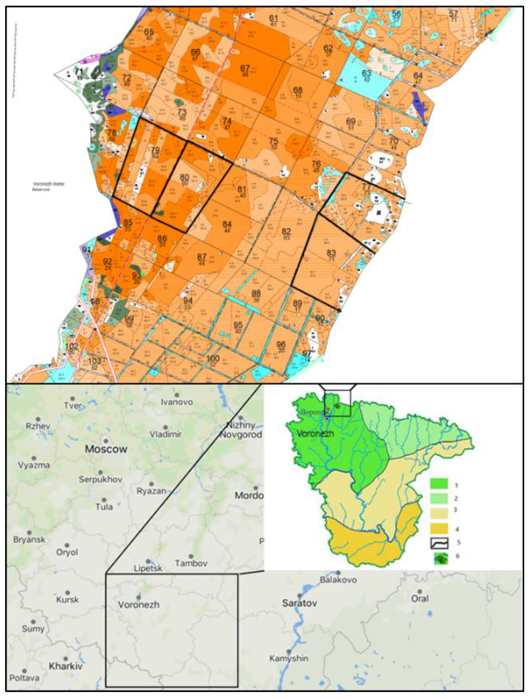
Figure 1. Location of sample plots 77, 79, 80, 83, and NF and climatic zoning in the Voronezh region: 1 - Western forest-steppe region; 2 - Eastern forest-steppe region; 3 - Northern steppe region; 4 - Southern steppe region; 5 - boundary of climatic regions; 6 - Left-bank forestry of the Voronezh region. Red dot on the map near Voronezh depicts location of the “Northern Forest” (NF) sample plot; upper part represents the map with sample plots 77, 79, 80, and 83.
Preprints 188408 g001
Figure 2. Samples 79/3 root (a) and 79/7 root (b) collected in the compartment 79 from the roots of a fresh windfall and from excavated roots, respectively.
Preprints 188408 g002
Table 1. Silvicultural and forest mensuration characteristics of Scots pine forest permanent sample plot stands where mycelial samples were collected in the summer of 2024.
Permanent sample plot Species composition Age, years Density Type of vegetation
77 Only Scots pine trees 70, 78 0.4-0.8 Fresh pine forest
79 40, 48, 55, 66 0.6-0.8 Fresh subor
80 Felling after forest fires. At the time of felling, the forest was approximately 120 years old At the moment of felling − 0.7 Fresh subor
83 83 0.6-0.8 Fresh subor
“Northern Forest” (NF) Almost all Scots pine trees, including occasional birch, aspen, and maple trees planting, average age 50 years 0.6-0.8 Fresh pine forest
Note: The permanent sample plots consist of heterogeneous stands that may differ in some or all silvicultural characteristics (height, density, age, composition, etc.), so their characteristics may vary.
Table 2. Brief characteristics of 11 identified taxa.
Sample Identified species Environmental group Mycelium collection site NCBI GenBank accession number
79/5 root Dichomitus squalens (P. Karst.) D.A. Reid xylosaprotroph dead wood of pine trees PX132320
79/8 root PX132322
79/4 root PX132312
79/6 root; NF 16 trunk Sistotrema sp. xylosaprotroph, forest litter saprotroph dead wood and plant detritus in the forest litter upper layers PX132321
83/3.2 root рoд Athelia Pers. xylosaprotroph dead wood and small woody plant residues PX132313
NF 4 forest litter Xenopolyscytalum pinea Crous forest litter saprotroph pine tree litter PX132315
NF 13 old windfallen tree Xylodon flaviporus (Berk. & M.A. Curtis ex Cooke) Riebesehl & E. Langer xylosaprotroph dead wood of pine and deciduous trees PX218715
NF 18 forest litter Trechispora sp. xylosaprotroph dead wood of pine and deciduous trees PX132316
NF 20 forest litter PX132317
NF 21 butt of a tree Sertulicium vernale Spirin &Volobuev xylosaprotroph dead wood of pine and deciduous trees PX218716
NF 22 root of old windfallen tree Gymnopilus penetrans (Fr.) Murrill facultative xylosaprotroph live and dead wood of pine and deciduous species PX132318
NF 23 butt of a deadtree Ganoderma applanatum (Pers.) Pat. facultative xylosaprotroph, parasite live and dead wood of pine and deciduous trees PX132319
K3, K7 root Trametes hirsuta saprotroph old stumps, deadwood, dying trunks of deciduous trees PX132323
NF 3 stem Trichaptum fuscoviolaceus saprotroph livie and dead wood of pine trees PX132314
Table 3. Taxonomy of identified species of wood-destroying fungi.
Phylum Ascomycota Basidiomycota
Class Leotiomycetes Agaricomycetes
Order Helotiales Agaricales Atheliales Canthare-llales Hymeno-chaetales Polyporales Sistotrematales Trechisporales
Family Hamatocanthoscyphaceae Strophariaceae Atheliaceae Hydnaceae Schizoporaceae Polyporaceae Sistotremastraceae Hydnodontaceae
Species Xenopolyscytalum pinea Crous Gymnopilus penetrans (Fr.) Murrill Athelia Pers. Sistotrema brinkmannii (Bres.) J. Erikss. Xylodon flaviporus (Berk. & M.A. Curtis ex Cooke) Riebesehl & E. Langer. 1. Dichomitus squalens (P. Karst.) D.A. Reid.
2. Ganoderma applanatum (Pers.) Pat.
3. Trametes hirsuta (Wulfen) Lloyd.
4. Trichaptum fuscoviolaceum (Ehrenb.) Ryvarden
Sertulicium vernale Spirin & Volobuev Trechispora P. Karst.
Disclaimer/Publisher’s Note: The statements, opinions and data contained in all publications are solely those of the individual author(s) and contributor(s) and not of MDPI and/or the editor(s). MDPI and/or the editor(s) disclaim responsibility for any injury to people or property resulting from any ideas, methods, instructions or products referred to in the content.
Copyright: This open access article is published under a Creative Commons CC BY 4.0 license, which permit the free download, distribution, and reuse, provided that the author and preprint are cited in any reuse.
Prerpints.org logo

Preprints.org is a free preprint server supported by MDPI in Basel, Switzerland.

Subscribe

Disclaimer

Terms of Use

Privacy Policy

Privacy Settings

© 2026 MDPI (Basel, Switzerland) unless otherwise stated