Preprint
Article

This version is not peer-reviewed.

Bacterial Diversity in Certain Captive Snake Species in Bulgaria: A One Health Challenge—Pilot Study

Submitted:

04 December 2025

Posted:

08 December 2025

You are already at the latest version

Abstract

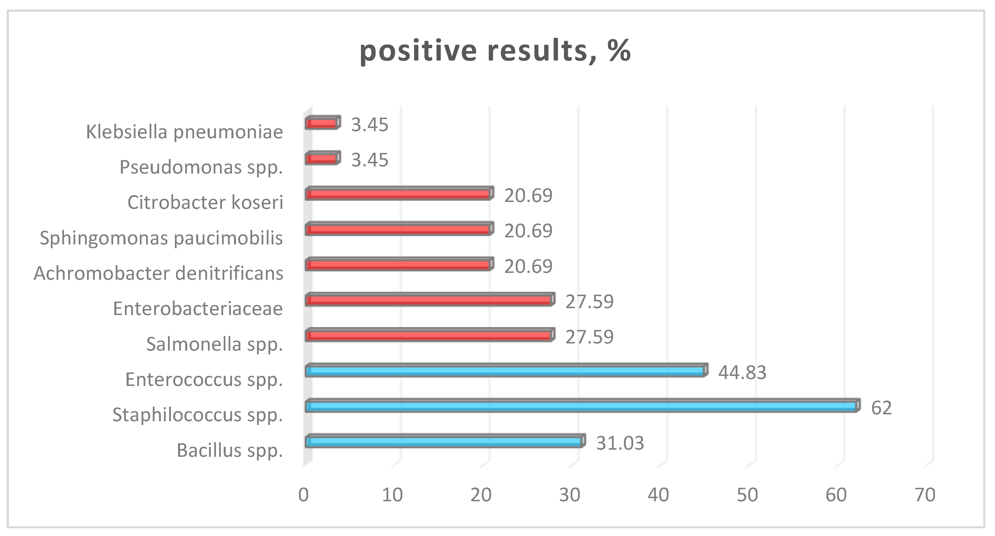
Snakes are becoming increasingly popular as companion animals in Bulgaria, yet their potential as reservoirs for zoonotic pathogens remains underexplored. This study investigates bacterial carriage in domesticated snakes and evaluates the public health risks associated with their handling. Materials and methods: Bacteriological analysis was performed on 29 snake samples from various species, including pythons, boas, corn snakes, and a venomous species. All individuals were raised in controlled environments and primarily fed frozen rodents. Standard microbiological techniques were employed for bacterial isolation and identification, and the results were confirmed using the Vitek 2 Compact System. Results: Salmonella enterica (group B) was isolated from 8 samples, indicating a notable prevalence of this pathogen among the examined snakes. Additionally, several other bacterial genera were identified, including Pseudomonas spp., Staphylococcus spp., Proteus spp., Enterobacteriaceae spp., Sphingomonas paucimobilis, Enterococcus spp., Bacillus spp., Achromobacter denitrificans, Citrobacter koseri, and Klebsiella pneumoniae. Conclusion: The study highlights the zoonotic risks associated with snake ownership, particularly through direct contact with the reptiles or exposure to contaminated environments. Food sources may act as vectors for microbial contamination. To mitigate the risk of bacterial transmission between snakes and humans, it is crucial to adopt rigorous hygiene practices, conduct regular health monitoring of the snakes, and implement proper protocols for feeding and cleaning.

Keywords: 
;  ;  ;  

1. Introduction

In recent decades, the growing interest in reptiles, particularly snakes, as pets has led to a significant increase in their popularity across various demographics, ranging from casual hobbyists to professional researchers. These animals are appreciated not only for their unique behaviors and diverse appearances in so-called designer morphs but also for their critical roles in ecological systems. However, the surge in reptile ownership brings with it important health considerations, especially concerning zoonotic diseases that can affect humans. Reptiles are known to harbor a variety of bacterial pathogens, often serving as asymptomatic carriers, thereby posing potential health risks to humans [1,2]. The ability of reptiles to disseminate pathogenic microorganisms without manifesting clinical signs complicates awareness and management of the health risks associated with their care. A deeper understanding of the oral and cloacal microbiota, particularly in snakes, is crucial for assessing the implications of these pathogens on both animal health and public safety. Research indicates that the snake’s diet and their associated flora significantly influence the composition of their microbiota. It has been suggested that the cloacal flora of their prey can transfer to snakes during feeding [2,3]. Despite the critical importance of this bacterial interplay, there is a notable scarcity of studies that thoroughly characterize the microbial diversity and distribution in reptiles [4]. Dominant species in the microbiota of healthy snakes include gram-positive bacteria such as Staphylococcus and Bacillus spp. In comparison, gram-negative pathogens such as Pseudomonas aeruginosa, Providencia rettgeri, and Stenotrophomonas maltophilia are often found in snakes suffering from stomatitis [5]. Notably, members of the Enterobacteriaceae family have also been implicated in respiratory diseases in humans [6]. Among the most concerning pathogens associated with reptiles, Salmonella spp. stands out, particularly due to its association with severe health outcomes in humans, notably in vulnerable populations [2,5]. While reptile-associated salmonellosis (RAS) represents a small percentage of overall salmonellosis cases in Europe and the USA, its clinical course can be markedly severe, especially in young children, the elderly, and pregnant women, contributing to numerous disease outbreaks globally [7,8]. The presence of opportunistic pathogens such as Pseudomonas spp., Staphylococcus spp., and Proteus spp. further emphasizes the need for careful management and awareness regarding the risks posed by these reptiles [9]. Therefore, as the trend of keeping reptiles continues to rise, it becomes increasingly critical to educate potential owners about the associated health risks. Implementing effective hygiene practices, proper handling techniques, and regular veterinary care are vital strategies for mitigating the risk of zoonotic infections. Consequently, promoting responsible ownership and facilitating the dissemination of accurate information will be essential for ensuring safe coexistence between humans and reptiles [10].

2. Materials and Methods

Fecal swab samples were collected from the cloaca of healthy adult snakes representing 29 different species (Table 1). The snakes were kept in 5 different private terrariums in Bulgaria. Due to the limited quantity of feces obtained during the initial examination, it was recommended to collect a larger sample after defecation for further testing, specifically for parasitological examination [11].
All samples were initially cultured in both liquid enrichment media (Tryptic Soy Broth and Selenite Broth HiMedia™) and on solid media using the four-quadrant streak plate method (Blood Agar Base with 5% defibrinated ovine blood and MacConkey agar, HiMedia™) [12,13]. The incubation process typically takes place at a controlled temperature of 37°C for 24 hours [14]. After this incubation, for more precisous and rapid identification, an automated biochemical profiling by Vitek 2 Compact system (bioMérieux, France) was used [15,16].

3. Results

A total of 29 samples were analyzed in this study to determine the prevalence of various bacterial species. The frequency of positive samples varied significantly among the different microorganisms (Table 2 andFigure 1). The highest prevalence was observed in Staphylococcus spp., while the lowest frequencies were reported for a group of less common gram-negative species, including Achromobacter denitrificans, Sphingomonas paucimobilis, Citrobacter koseri, and Klebsiella pneumoniae. On average, 30.94% of the samples tested positive for the studied bacterial species, with significant variability (SD ≈ 13.1%), indicating clear differences among the individual bacterial groups. To estimate the precision of the observed prevalence values, standard errors (SE) and 95% confidence intervals (CI) were calculated for each bacterial group. The prevalence of Staphylococcus spp. was the highest among all detected taxa (62.07%; 95% CI: 44.1–80.1), followed by Enterococcus spp. (44.83%; 95% CI: 26.6–63.0). Salmonella spp., a pathogen of significant zoonotic relevance, was detected in 27.59% of the samples (95% CI: 12.8–42.3). This finding confirms the relatively high level of asymptomatic carriage in snakes and underscores the potential public health risk associated with reptile handling. Other bacterial groups with comparable prevalence levels included Bacillus spp. (31.03%; 95% CI: 15.1–46.9), Enterobacteriaceae (27.59%; 95% CI: 12.8–42.3), and Pseudomonas spp. (27.59%; 95% CI: 12.8–42.3). Opportunistic pathogens such as A. denitrificans, S. paucimobilis, C. koseri, and K. pneumoniae were detected at similar frequencies (20.69%), each showing a 95% CI ranging from 6.7% to 34.6%. Although the confidence intervals reflect moderate variability due to the sample size (n = 29), the overall dataset demonstrates that Staphylococcus spp. dominate the microbial landscape in the studied snake population, while Salmonella spp. remain an important but less frequent component with notable epidemiological implications.

4. Discussion

The results of the present study show that a substantial proportion of the examined snakes (59.4%) were carriers of potentially pathogenic bacteria. These results align with other studies reporting complex polymicrobial microbiota in domestic snakes [17,18].
Our findings reveal a diverse spectrum of bacterial isolates: the most common genera were Staphylococcus spp. (62%), Enterococcus spp. (44.83%), and Bacillus spp. (31.03%), alongside Salmonella spp., members of Enterobacteriaceae, and Pseudomonas spp. (27.59%). This finding is consistent with international data indicating that between 30% and 80% of reptiles are chronic carriers of Salmonella spp. [7,19]. Historically, outbreaks of salmonellosis have been linked to a wide range of sources, including contaminated food, water, and contact with reptiles, particularly pet snakes and turtles [7,19]. These events highlight the importance of hygiene and public awareness among reptile owners and individuals in close contact with such animals.
The isolation of Salmonella enterica group B in 15.6% of the samples aligns with prevalence values reported from [2,20]. Several authors and health institutions have emphasized that contact with reptiles, including snakes, is associated with an increased risk of salmonellosis, especially in children, the elderly, and immunocompromised patients [21,22,23]. Our results, demonstrating the simultaneous presence of Salmonella spp., Pseudomonas spp., etc., confirm the view that snakes kept as pets or in zoos can act as a reservoir and mechanical vector of zoonotic pathogens, including multidrug-resistant strains [21,24,25]. However, not all isolated bacteria should be considered pathogens in the context of the snake host. Some of them probably represent normal or conditionally normal flora, adapted to the specific physiology and ecology of reptiles. A meta-analysis of the gut microbiota in reptiles reveals the presence of a limited “core” of bacterial taxa and a significant influence of both the environment and the host on the microbiome composition [26]. In this context, the bacteria isolated by us should be interpreted both as an indicator of bacterial contamination and as part of the natural microbiome profile of captive snakes.
Another factor that may influence bacterial colonization in snakes is related to food sources, specifically frozen rodents. Studies indicate that 8–10% of commercially supplied frozen rodent packages for reptile feeding contain Salmonella spp. [21]. Thus, frozen feeder rodents should be recognized as a relevant reservoir of pathogens capable of colonizing reptiles’ gastrointestinal tracts without causing clinical signs [19,21,27]. The isolation of Salmonella spp. (27.59%) and other members of the Enterobacteriaceae family, including C. koseri and K. pneumoniae, are crucial from a public health perspective. A recent systematic review and meta-analysis revealed that the overall prevalence of Salmonella in reptiles is high, often exceeding 50% in snakes, which confirms their role as a reservoir for the bacteria [19]. Additionally, a review by Pees et al. [7] highlights that Salmonella in reptiles is considered part of the normal intestinal microbiota, with continuous and intermittent shedding into the environment, posing significant zoonotic potential.
Citrobacter koseri and Klebsiella pneumoniae are well-described opportunistic pathogens in humans, associated with urinary tract infections, respiratory tract infections, neonatal meningitis, and nosocomial outbreaks. Their isolation from snakes raises the question of the transfer of antimicrobial-resistant strains from animals to humans, especially in the context of multidrug-resistant enterobacteria [18,21,25,28].
The isolation of A. denitrificans, S. paucimobilis, and other less common genera aligns with observations that the reptile microbiome contains a variety of opportunistic bacteria, each with different potential for pathogenicity [5,28,29]. Sphingomonas paucimobilis and related species have been identified as causative agents of nosocomial infections in immunocompromised patients. However, they are also present in the environment and can colonize animals without causing any clinical symptoms. Several studies have emphasized the role of snake oral microbiota as a potential reservoir for multidrug-resistant Gram-negative pathogens [28,30,31]. Recent high-throughput 16S rRNA sequencing studies of snake oral cavities similarly demonstrate a dominance of Gram-negative bacteria (e.g., Pseudomonas, Aeromonas, Enterobacteriaceae), while Gram-positive cocci, though less abundant, remain stable microbiome components [5,26].
The observed high frequencies of Staphylococcus spp. (62%) and Enterococcus spp. (44.83%) are consistent with existing literature indicating that in certain species, particularly those maintained in captivity, Gram-positive bacteria (including coriform bacteria and coagulase-negative staphylococci) can prevail in the oral cavity [30,32]. This dominance is likely associated with exposure to humans, as well as contact with feed, substrate, and surfaces within the artificial environment.
Even in the absence of venom, non-venomous snakes can pose significant risks to humans due to their potential to carry harmful pathogens. These pathogens can cause serious infections and diseases, underscoring that the danger from snakes is not limited to venomous snakes. Understanding the ecological roles and risks associated with both venomous and non-venomous species is crucial for addressing public health and safety concerns effectively.
Preventive measurement: Numerous studies and guidelines emphasize the crucial importance of maintaining good hygiene practices, especially in environments where there is a risk of exposure to certain animals or pathogens. Key recommendations from various sources highlight the necessity of thoroughly washing hands after any contact with animals or their habitats. This simple yet effective practice helps prevent the transmission of potential diseases. It is also advisable to avoid unnecessary contact with snakes, particularly in areas like the kitchen where food is prepared, as this can lead to contamination and health risks. Proper storage of pet food is equally important; keeping it in secure containers can prevent the introduction of pests and reduce the likelihood of interactions with animals. Furthermore, it is essential to keep children away from terrariums or habitats containing reptiles to mitigate potential dangers associated with these animals. Collectively, these guidelines stress the importance of vigilance and education to ensure both personal safety and public health [33,34].
Hygiene checklist for snake owners (to accompany counseling):
• Wash hands with soap and warm water after handling snakes, enclosures, equipment, or feeder rodents.
• Keep terraria and cleaning activities away from kitchens/food prep areas.
• Supervise children; avoid handling/cleaning by children <5 years, elderly, and immunocompromised persons.
• Use dedicated tools (brushes, buckets) for terraria; disinfect regularly.
• Store and thaw feeder rodents separately from human food; clean and disinfect surfaces afterward.
• Wear disposable gloves when cleaning enclosures or handling fecal material; avoid face touching.
• Seek veterinary advice for abnormal behavior or illness; discuss zoonotic risks at purchase/adoption.
Limitations and future directions: Limitations of the current analysis are the absence of antibiograms and molecular typing of the isolates, which would allow a more precise assessment of the zoonotic risk and the potential for the spread of resistant clones. Given the increasing number of data on multidrug-resistant strains of Salmonella, Citrobacter and Klebsiella isolated from both humans and animals, future studies should include a systematic analysis of antibiotic susceptibility and genetic determinants of resistance [7,21,25,26,35].

5. Conclusions

Even without venom, non-venomous snakes can be almost as dangerous as venomous ones because they can carry harmful pathogens to humans. Domestic snakes are increasingly recognized as a significant reservoir of pathogenic bacteria with well-documented zoonotic potential, particularly the genus Salmonella. These bacteria can cause serious gastrointestinal disease in humans, especially in vulnerable populations such as children and immunocompromised individuals.
One potential route of transmission is through food practices, particularly the feeding of frozen rodents that may contain these pathogens. Handling and preparing such food products without adequate hygiene practices can create opportunities for cross-contamination and subsequent infections.
To mitigate the risk of transmission and protect the health of both humans and animals, it is imperative to implement strict hygiene protocols. This includes regular cleaning and disinfection of snake habitats, proper hand washing after handling snakes or their food, and education of snake owners on safe handling practices.
Additionally, public health campaigns targeting pet owners could play a crucial role in raising awareness about the risks associated with pet snakes and the importance of preventive measures. By promoting a better understanding of the potential health effects and necessary precautions, we can help reduce the incidence of zoonotic infections associated with these fascinating reptiles.

Author Contributions

Conceptualization, Methodology, Investigation, Writing-original draft, Writing-review & editing: B. B-M.

Funding

This research received no external funding.

Institutional Review Board Statement

No ethical review and approval were required for the animal study, as we are presenting results of laboratory diagnostic tests. We have the animals' owners' consent to publish the results for scientific purposes

Informed Consent Statement

Owners' consent was obtained for sampling and use of anonymized data.

Data Availability Statement

Data supporting the findings are available from the corresponding author on reasonable request.

Conflicts of Interest

The author declares no conflict of interest.

Abbreviations

The following abbreviations are used in this manuscript:
RAS reptile-associated salmonellosis

References

  1. Warwick, C.; Arena, P.C.; Steedman, C. Health implications associated with exposure to farmed and wild sea turtles. JRSM short reports 2013, 4, 1–7. [Google Scholar] [CrossRef] [PubMed]
  2. Mermin, J.; Hutwagner, L.; Vugia, D.; Shallow, S.; Daily, P.; Bender, J. & Emerging Infections Program FoodNet Working Group. Reptiles, amphibians, and human Salmonella infection: a population-based, case-control study. Clinical infectious diseases 2004, 38 Suppl. S3, S253–S261. [Google Scholar]
  3. Whiley, H.; Gardner, M.G.; Ross, K. A review of Salmonella and squamates (lizards, snakes and amphisbians): implications for public health. Pathogens 2017, 6, 38. [Google Scholar] [CrossRef]
  4. Sodagari, H.R.; Habib, I.; Shahabi, M.P.; Dybing, N.A.; Wang, P.; Bruce, M. Public health challenges of salmonella and turtles. Veterinary Sciences 2020, 7, 56. [Google Scholar] [CrossRef]
  5. Lin, W.H.; Tsai, T.S. Comparisons of the oral microbiota from seven species of wild venomous snakes in Taiwan using the high-throughput amplicon sequencing of the full-length 16S rRNA gene. Biology 2023, 12, 1206. [Google Scholar] [CrossRef]
  6. Pasmans, F.; Martel, A.N.; Jacobson, E.R. Bacterial diseases of reptiles. Infectious diseases and Pathology of reptiles 2020, 705–794. [Google Scholar]
  7. Pees, M.; Brockmann, M.; Steiner, N.; Marschang, R.E. Salmonella in reptiles: Occurrence, interactions, shedding, and human infection risk. Frontiers in Cell and Developmental Biology 2023, 11, 1251036. [Google Scholar] [CrossRef]
  8. Meletiadis, A.; Biolatti, C.; Mugetti, D.; Zaccaria, T.; Cipriani, R.; Pitti, M.; Decastelli, L.; Cimino, F.; Dondo, A.; Maurella, C.; Bozzetta, E.; Acutis, P.L. Surveys on Exposure to Reptile-Associated Salmonellosis (RAS) in the Piedmont Region—Italy. Animals 2022, 12, 906. [Google Scholar] [CrossRef] [PubMed]
  9. JACOBSON ER. Bacterial diseases of reptiles. In Infectious diseases and pathology of reptiles; CRC Press, 2007; pp. 475–540. [Google Scholar]
  10. Pasmans, F.; Bogaerts, S.; Braeckman, J.; Cunningham, A.A.; Hellebuyck, T.; Griffiths, R.A.; Martel, A. Future of keeping pet reptiles and amphibians: towards integrating animal welfare, human health and environmental sustainability. Veterinary Record 2017, 181, 450–450. [Google Scholar] [CrossRef] [PubMed]
  11. Tagesu, A. Examination of feces. J. Int. Vet. Sci. Res 2018, 1, 45–50. [Google Scholar]
  12. Smith, J.; Thompson, R.; Walker, L. Enrichment media for pathogen detection in clinical laboratories. Journal of Microbial Science 2019, 15, 221–229. [Google Scholar]
  13. Sanders, E.R. Aseptic laboratory techniques: plating methods. Journal of Visualized Experiments (JoVE) 2012, (63), e3064. [Google Scholar]
  14. Mariam, S.H. Isolation and Characterization of Gram-Negative Bacterial Species from Pasteurized Dairy Products: Potential Risk to Consumer Health. J Food Qual. 2021, 17, 1‒10. [Google Scholar] [CrossRef]
  15. Boneva-Marutsova, B.; Marutsov, P.; Geisler, M.L.; Zhelev, G. Salmonellosis Outbreak in a Rottweiler Kennel Associated with Raw Meat-Based Diets. Animals 2025, 15, 3196. [Google Scholar] [CrossRef]
  16. Funke, G.; Funke-Kissling, P. Evaluation of the new VITEK 2 card for identification of clinically relevant gram-negative rods. Journal of clinical microbiology 2004, 42, 4067–4071. [Google Scholar] [CrossRef]
  17. Krishnankutty, S.P.; Muraleedharan, M.; Perumal, R.C.; Michael, S.; Benny, J.; Balan, B.; Zachariah, A. Next-generation sequencing analysis reveals high bacterial diversity in wild venomous and non-venomous snakes from India. Journal of Venomous Animals and Toxins Including Tropical Diseases 2018, 24, 41. [Google Scholar] [CrossRef] [PubMed]
  18. Su, H.Y.; Hussain, B.; Hsu, B.M.; Lee, K.H.; Mao, Y.C.; Chiang, L.C.; Chen, J.S. Bacterial community analysis identifies Klebsiella pneumoniae as a native symbiotic bacterium in the newborn Protobothrops mucrosquamatus. BMC microbiology 2023, 23, 213. [Google Scholar] [CrossRef]
  19. Muslin, C.; Salas-Brito, P.; Coello, D.; Morales-Jadán, D.; Viteri-Dávila, C.; Coral-Almeida, M. Salmonella prevalence and serovar distribution in reptiles: a systematic review and meta-analysis. Gut Pathogens 2025, 17, 52. [Google Scholar] [CrossRef]
  20. Chen, C.Y.; Chen, W.C.; Chin, S.C.; Lai, Y.H.; Tung, K.C.; Chiou, C.S.; Chang, C.C. Prevalence and antimicrobial susceptibility of salmonellae isolates from reptiles in Taiwan. Journal of Veterinary Diagnostic Investigation 2010, 22, 44–50. [Google Scholar] [CrossRef]
  21. Marin, C.; Martelli, F.; Rabie, A.; Davies, R. Commercial frozen mice used by owners to feed reptiles are highly externally contaminated with Salmonella Enteritidis PT8. Vector-Borne and Zoonotic Diseases 2018, 18, 453–457. [Google Scholar] [CrossRef] [PubMed]
  22. Varela, K.; Brown, J.A.; Lipton, B.; Dunn, J.; Stanek, D.; NASPHVCommittee, C.o.n.s.u.l.t.a.n.t.s.; Yager, C.M. A review of zoonotic disease threats to pet owners: a compendium of measures to prevent zoonotic diseases associated with non-traditional pets such as rodents and other small mammals, reptiles, amphibians, backyard poultry, and other selected animals. Vector-borne and zoonotic diseases 2022, 22, 303–360. [Google Scholar] [CrossRef]
  23. Waltenburg, M.A.; Perez, A.; Salah, Z.; Karp, B.E.; Whichard, J.; Tolar, B.; Nichols, M. Multistate reptile-and amphibian-associated salmonellosis outbreaks in humans, United States, 2009–2018. Zoonoses and Public Health 2022, 69, 925–937. [Google Scholar] [CrossRef]
  24. Ebani, V.V. Domestic reptiles as source of zoonotic bacteria: A mini review. Asian Pacific Journal of Tropical Medicine 2017, 10, 723–728. [Google Scholar] [CrossRef]
  25. Dégi, J.; Herman, V.; Radulov, I.; Morariu, F.; Florea, T.; Imre, K. Surveys on pet-reptile-associated multi-drug-resistant Salmonella spp. in the Timișoara metropolitan region—Western Romania. Antibiotics 2023, 12, 1203. [Google Scholar] [CrossRef] [PubMed]
  26. Hoffbeck, C.; Middleton, D.M.; Nelson, N.J.; Taylor, M.W. 16S rRNA gene-based meta-analysis of the reptile gut microbiota reveals environmental effects, host influences and a limited core microbiota. Molecular Ecology 2023, 32, 6044–6058. [Google Scholar] [CrossRef] [PubMed]
  27. Shivambu, N.; Shivambu, T.C.; Chimimba, C.T. Zoonotic pathogens associated with pet and feeder murid rodent species: a global systematic review. Vector-Borne and Zoonotic Diseases 2023, 23, 551–560. [Google Scholar] [CrossRef]
  28. Marques, I.; Pinto, A.R.; Martins, J.J.; Alvura, N.; Telinhos, P.; Mendes, P.; Saavedra, M.J. Assessing Potential Reservoir of Multidrug-Resistant Bacteria in the Oral Microbiota of Captive Burmese and Royal Pythons. Life 2025, 15, 442. [Google Scholar] [CrossRef] [PubMed]
  29. Artavia-León, A.; Romero-Guerrero, A.; Sancho-Blanco, C.; Rojas, N.; Umaña-Castro, R. Diversity of aerobic bacteria isolated from oral and cloacal cavities from free-living snake species in Costa Rica rainforest. International Scholarly Research Notices 2017, 2017, 8934285. [Google Scholar] [CrossRef]
  30. Chuang, P.C.; Tsai, Y.H.; Hsu, W.H.; Lin, C.C.; Chiou, C.S.; Hsu, T.C. Oral bacteria and their antibiotic susceptibilities in Taiwanese venomous snakes. Microorganisms 2022, 10, 951. [Google Scholar] [CrossRef]
  31. Young Yusty, S.; Prescilla-Ledezma, A. From culture to metagenomics: Advances in oral microbiota of venomous snakes. Microbiology Research 2025, 16, 233. [Google Scholar] [CrossRef]
  32. Dehghani, R.; Sharif, M.R.; Moniri, R.; Sharif, A.; Haddad Kashani, H. The identification of bacterial flora in oral cavity of snakes. Comparative Clinical Pathology 2016, 25, 279–283. [Google Scholar] [CrossRef]
  33. Centers for Disease Control and Prevention. Salmonella and reptiles and amphibians. Available online: https://www.cdc.gov.
  34. European Food Safety Authority. Public health risks associated with Salmonella in reptiles. EFSA Journal 2019, 17, e05656. [Google Scholar]
  35. Lamichhane, B.; Mawad, A.M.; Saleh, M.; Kelley, W.G.; Harrington, P.J.; Lovestad, C.W.; Helmy, Y.A. Salmonellosis: an overview of epidemiology, pathogenesis, and innovative approaches to mitigate the antimicrobial resistant infections. Antibiotics 2024, 13, 76. [Google Scholar] [CrossRef] [PubMed]
Figure 1. Isolated bacterial species (blue – Gram positive bacteria; red – Gram negative bacteria).
Preprints 188172 g001
Table 1. Types of snake species, included in the survey.
Species Family Common name N (29)
Python regius Pythonidae Ball python 3
Python curtus Pythonidae Sumatran short-tailed python 1
Python buvittatus Pythonidae Burmese python 2
Morelia spilota Pythonidae Carpet python 3
Acrantophis dumerili Boidae Dumerili’s boa 2
Boa constrictor Boidae Common boa 1
Boaedon fuliginosus Lamprophilidae African house snake 1
Pantherophis guttatus Colubridae Corn snake 15
Bothrops insularis Viperidae Golden lancehead 1
STATISTICS: Statistical analysis was performed using IBM SPSS Statistics v.26.
Table 2. Prevalence of bacterial species isolated from samples (n = 29).
Species Samples (n) Positive (n) % Positive SE 95% CI (lower–upper)
Salmonella enterica 29 8 27.59% 0.084 12.8% – 42.3%
Staphylococcus spp. 29 18 62.07% 0.091 44.1% – 80.1%
Enterococcus spp. 29 13 44.83% 0.092 26.6% – 63.0%
Bacillus spp 29 9 31.03% 0.086 15.1% – 46.9%
Enterobacteriaceae spp. 29 8 27.59% 0.084 12.8% – 42.3%
A. denitrificans 29 6 20.69% 0.076 6.7% – 34.6%
S. paucimobilis 29 6 20.69% 0.076 6.7% – 34.6%
C. koseri 29 6 20.69% 0.076 6.7% – 34.6%
Pseudomonas spp. 29 8 27.59% 0.084 12.8% – 42.3%
Klebsiella pneumoniae 29 6 20.69% 0.076 6.7% – 34.6%
Legend: n – total number of samples tested; Positive (n) – number of samples in which the microorganism was isolated; % Positive – percentage of positive samples; SE – standard error of the percentage; 95% CI – 95% confidence interval for the percentage (lower – upper bound).
Disclaimer/Publisher’s Note: The statements, opinions and data contained in all publications are solely those of the individual author(s) and contributor(s) and not of MDPI and/or the editor(s). MDPI and/or the editor(s) disclaim responsibility for any injury to people or property resulting from any ideas, methods, instructions or products referred to in the content.
Copyright: This open access article is published under a Creative Commons CC BY 4.0 license, which permit the free download, distribution, and reuse, provided that the author and preprint are cited in any reuse.
Prerpints.org logo

Preprints.org is a free preprint server supported by MDPI in Basel, Switzerland.

Subscribe

Disclaimer

Terms of Use

Privacy Policy

Privacy Settings

© 2026 MDPI (Basel, Switzerland) unless otherwise stated