Preprint
Article

This version is not peer-reviewed.

NGS-Based Genomic Characterization of ESBL/AmpC-Producing Extraintestinal Pathogenic Escherichia coli from Captive Wildlife in Tunisia

Submitted:

03 December 2025

Posted:

03 December 2025

You are already at the latest version

Abstract

Background/Objectives: Multidrug-resistant (MDR) Escherichia coli resistant to third-generation cephalosporins are a growing One Health concern, but data on extraintestinal pathogenic E. coli (ExPEC) from wildlife in North Africa remain scarce. We aimed to characterize ESBL/AmpC-producing ExPEC from captive wild mammals in Tunisia and to situate these isolates in a global genomic context. Methods: In 2018, 30 fecal samples from 14 captive wild mammals in a private farm were screened on cefotaxime agar. Four resistant E. coli were recovered from a llama, lion, hyena and tiger. Antimicrobial susceptibility testing and Illumina whole-genome sequencing were combined with in silico typing, resistome and virulome profiling, plasmid and mobile element analysis, human pathogenicity prediction and core-genome MLST-based minimum-spanning trees. Results: All isolates were MDR but remained susceptible to carbapenems, colistin and tigecycline. Two ST162/B1 isolates from the llama and tiger carried blaCMY-2, whereas two ST69/D isolates from the lion and hyena harbored blaCTX-M-15 and qnrS1. Genomes encoded 61–68 antimicrobial resistance genes and 114–131 virulence-associated genes, together with IncF-, IncI1- and IncY-type plasmids and IS26-rich insertion sequence profiles. PathogenFinder predicted a ≥0.93 probability of human pathogenicity for all isolates. cgMLST-based trees showed that Tunisian ST69 and ST162 clustered within internationally disseminated lineages containing human, animal and food isolates, rather than forming wildlife-restricted branches. Conclusions: Captive wild mammals in Tunisia can harbor high-risk ExPEC lineages combining ESBL/AmpC production, multidrug resistance and extensive virulence and mobility gene repertoires. These findings highlight captive wildlife as potential reservoirs and sentinels of clinically relevant E. coli and underscore the need for integrated WGS-based One Health surveillance at the human–animal–environment interface in North Africa.

Keywords: 
;  ;  ;  ;  ;  ;  ;  ;  ;  

1. Introduction

Antimicrobial resistance (AMR) in Gram-negative bacteria is now recognized as a major threat to human and animal health worldwide, compromising the efficacy of critically important antibiotics and increasing the burden of difficult-to-treat infections [1]. Among these pathogens, Escherichia coli plays a central role as both a commensal inhabitant of the intestinal microbiota and an opportunistic pathogen responsible for a wide range of extra-intestinal infections in humans and animals, including urinary tract infections, sepsis, and meningitis [2]. The emergence and dissemination of third-generation cephalosporin-resistant E. coli, frequently mediated by extended-spectrum β-lactamases (ESBLs) or plasmid-encoded AmpC enzymes, are of particular concern in clinical and community settings [3].
The ecology of AMR is complex and aligns closely with the One Health concept, which emphasizes the interconnectedness of human, animal, and environmental health. Resistant E. coli can circulate between humans, livestock, companion animals, and wildlife, facilitated by direct contact, contaminated food chains, shared water sources, and environmental reservoirs [4]. Wild animals, including those living in proximity to human activities or kept in captivity, may act as sentinels or reservoirs for multidrug-resistant (MDR) E. coli and other Enterobacterales, reflecting local antibiotic use and contamination pressure in surrounding ecosystems [5]. However, the contribution of wildlife to the broader AMR landscape remains incompletely characterized, especially in low- and middle-income countries.
Captive wild mammals in farms, zoos and wildlife parks occupy a unique ecological niche at the interface between the environment, domestic animals and humans. They can acquire MDR bacteria from contaminated feed, water, soil, fomites, caretakers or nearby livestock, and may in turn shed these organisms back into the environment. Nevertheless, there are still relatively few whole-genome sequencing (WGS) studies focusing on the resistome, virulome and mobile genetic elements of MDR E. coli from wildlife, particularly in North Africa and the Mediterranean region.
Tunisia has documented a high burden of ESBL-producing Enterobacterales in hospitals [6] and, to a lesser extent, in community and livestock settings [7,8], but data on AMR in wildlife are scarce.
Understanding possible cross-species transmission pathways and evaluating the pathogenic potential of multidrug-resistant E. coli prevalent in captive wild animals is crucial in the context of the One Health initiative.
Here, we combined phenotypic antimicrobial susceptibility testing with short-read WGS to investigate four third-generation cephalosporin-resistant E. coli isolates recovered from fecal samples of a llama, lion, hyena, and tiger housed in a private wildlife farm in northeastern Tunisia.
In order to identify the genomic characteristics of these wildlife-associated ESBL/AmpC-producing E. coli and to situate these isolates within the global phylogenetic context using cgMLST-based minimum-spanning trees, we employed a thorough in silico pipeline that integrated resistome and virulome profiling, plasmid and mobile genetic element analysis, CRISPR–Cas, and in silico pathogenicity prediction.

2. Results

2.1. Antimicrobial Susceptibility and Genomic Characteristics

Four third-generation cephalosporin-resistant E. coli isolates were recovered from fecal samples of llama (Ec1), lion (Ec2), hyena (Ec3), and tiger (Ec4). They were all multidrug resistant but remained susceptible to carbapenems, colistin, and tigecycline.
Illumina short-read sequencing and de novo assembly produced high-quality draft genomes for the four isolates (Ec1–Ec4). Genome sizes ranged from 4.92 to 5.03 Mb, with GC contents between 50.55% and 50.64%. The number of contigs varied from 68 to 97, with N50 values between 180,087 bp and 251,976 bp and L50 values of 8–9, indicating relatively contiguous assemblies (Table 1).
The RASTtk annotation projected 3–5 rRNA operons, 75–80 tRNA genes, and 4,735–5,016 coding sequences (CDS) per genome.
Each genome harbored 61–68 antimicrobial resistance genes (CARD), 114–131 virulence-associated genes (VFDB), 142–146 metal-resistance genes (BacMet), and 942–977 predicted transporters (TCDB) (Table 1).
Raw sequence reads for all four isolates have been deposited in the NCBI Sequence Read Archive (SRA) under BioProject accession PRJNA1367915.

2.2. Phylogenetic Background and In Silico Typing

In silico typing revealed two distinct clonal backgrounds among the wild-animal isolates. The llama (Ec1) and tiger (Ec4) strains belonged to ST162, phylogroup B1, with serotype O134:H19, whereas the lion (Ec2) and hyena (Ec3) carried ST69, phylogroup D, serotype O15:H18. Ribosomal MLST assigned rST 1544 to the ST162 isolates and rST 2135 to the ST69 isolates (Table 2).
When fimH32 in the ST162/B1 genomes (Ec1/Ec4) and fimH27 in the ST69/D genomes (Ec2/Ec3) were paired with fumC65 and fumC35 alleles, respectively, FimTyper produced unique fumC–fimH combinations that were consistent with lineages associated with extraintestinal pathogenic E. coli (ExPEC) (Table 2).
Using cgMLSTFinder with the 2,513-locus Escherichia/Shigella scheme, reliable core-genome sequence types were obtained for two isolates: Ec1 (cgST 207265; 94.3% loci called) and Ec3 (cgST 134012; 95.3% loci called). For Ec2 and Ec4, no cgST could be assigned because no alleles were called by the scheme, and these genomes were therefore excluded from further cgMLST-based comparisons.
Species identity as E. coli and the ESBL phenotype were independently confirmed from raw reads by KmerResistance/KmerFinder, which consistently identified E. coli reference genomes as the best species match and detected the same key β-lactamase genes as assembly-based tools.
CRISPRCasFinder detected a complete type I-E CRISPR–Cas locus with two huge CRISPR arrays in Ec3 and Ec4, however Ec1 and Ec2 only had short orphan CRISPR candidates with no discernible cas genes.

2.3. Resistome and Chromosomal Mutations

The four genomes carried a diverse set of acquired antimicrobial resistance genes. All isolates harbored blaTEM-1B together with the glycopeptide resistance gene vanG and multiple aminoglycoside-modifying enzymes, including aadA5, APH(6)-Id, and APH(3’’)-Ib, as well as sul2 and dfrA14/dfrA17 genes mediating sulfonamide and trimethoprim resistance. The tetracycline resistance gene tet(B) was present in the ST162 isolates (Ec1 and Ec4) (Table 2).
Regarding β-lactam resistance, the ST162/B1 strains (Ec1, Ec4) harbored the plasmid-mediated AmpC CMY-2, whereas the ST69/D strains (Ec2, Ec3) carried the ESBL CTX-M-15. These enzymes co-occurred with TEM-1B and additional narrow-spectrum β-lactamases (EC-18/EC-8). The ST69 isolates also contained the plasmid-mediated quinolone resistance gene qnrS1, while fluoroquinolone resistance in the ST162 isolates was mainly associated with chromosomal mutations (Table 2).
Consistent with the multidrug-resistant phenotype, all genomes exhibited multiple point mutations in antibiotic targets and regulatory loci. Fluoroquinolone resistance–associated substitutions gyrA S83L/D87N and parC S80I were found in the ST162 isolates, whereas ST69 genomes lacked parC mutations but retained wild-type quinolone target alleles. All four isolates carried the GlpT E448K substitution linked to fosfomycin resistance and several amino acid changes in PBP3 (FtsI; D350N, S357N) and cyaA (S352T in ST69) (Table 2).
Moreover, efflux and global regulator genes showed multiple variants, including alterations in AcrR and MarR within the AcrAB–TolC efflux system (Y137H and G103S) and mutations in oxidative stress regulators soxR and soxS (Table 2).

2.4. Virulence Gene Content, Plasmid Replicons and Mobile Genetic Elements

Virulence profiling revealed a rich repertoire of genes associated with adhesion, iron acquisition, serum resistance, and toxin production. All isolates carried type 1 fimbriae (fimH) and multiple additional adhesins, alongside genes for curli (csgA) and outer-membrane proteases (ompT). Iron acquisition systems included iroN, iucC/iutA, and sitA in ST162 isolates and fyuA and irp2 in the ST69 group. Several toxins or toxin-like factors were detected, such as astA and hlyE, together with complement and serum resistance factors iss, traT, and the tellurite resistance gene terC (Table 2).
PlasmidFinder identified multiple incompatibility groups. The ST162/B1 isolates (Ec1, Ec4) carried a complex plasmidome including IncFIB, IncFIC(FII), IncI1-I(Alpha), and small Col-type plasmids (ColpVC, Col(MG828)), whereas the ST69/D isolates (Ec2, Ec3) harbored IncY and Col(MG828) replicons.
Mobile genetic element analysis showed numerous insertion sequences and transposons, dominated by IS26, ISEc9, IS629, and related IS families, with additional elements such as ISKpn19, ISEc38, IS4, and ISSfl10 depending on the isolate.

2.5. In Silico Prediction of Human Pathogenic Potential

PathogenFinder classified all four genomes as likely human pathogens. Predicted probabilities of being human-pathogenic ranged from 0.928 to 0.935, based on 760–897 matched protein sequences covering 16–20% of each genome and mapping predominantly to protein families associated with known human-pathogenic E. coli lineages. The number of matched pathogenic protein families (750–890) greatly exceeded that of non-pathogenic families (7–12).

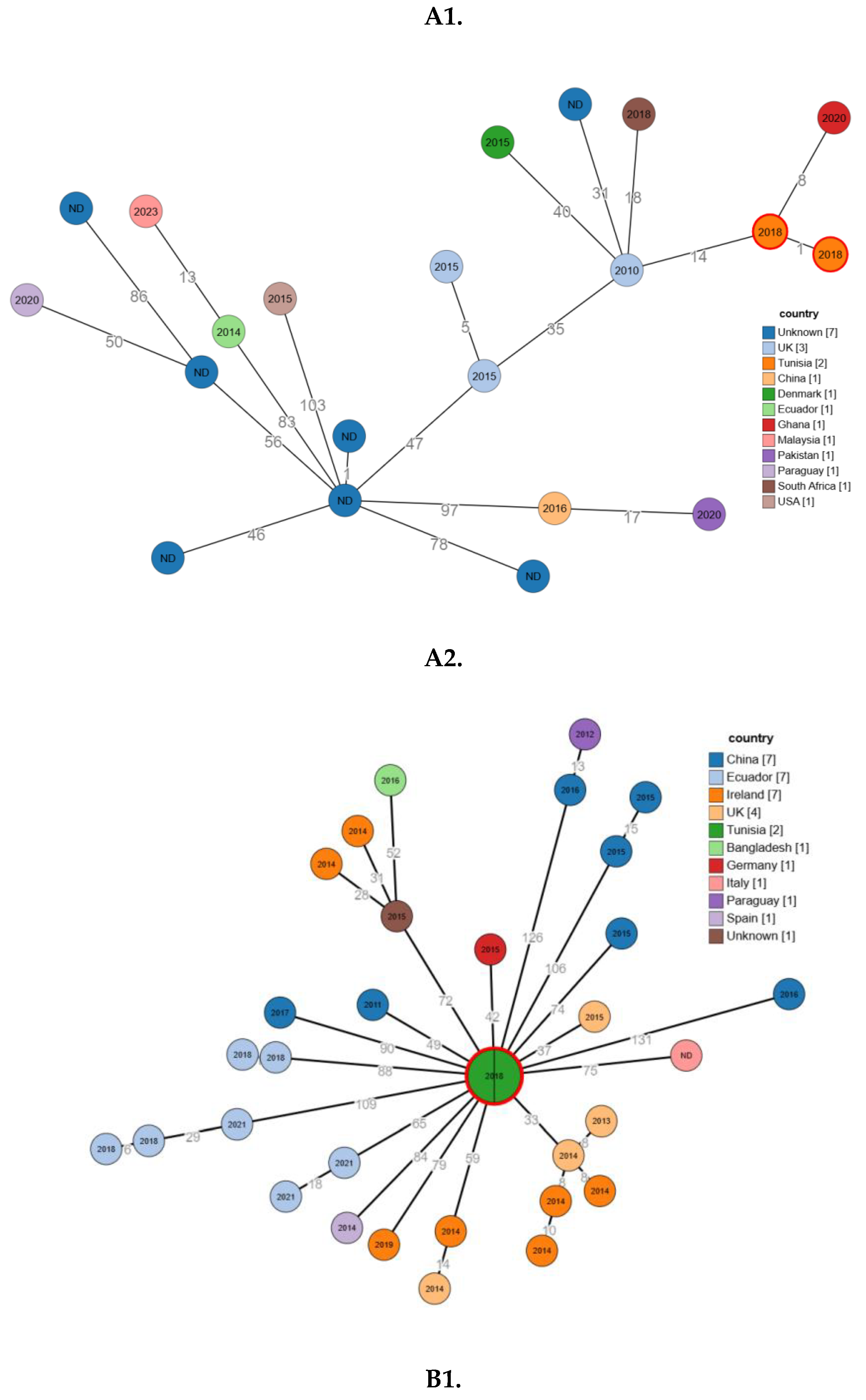
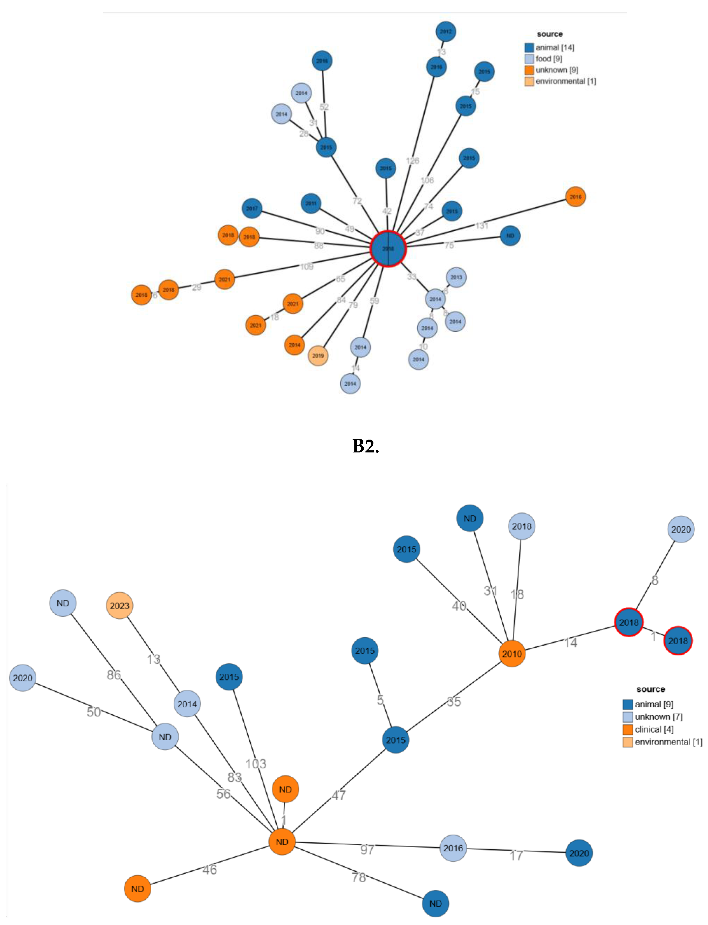
2.6. Phylogenetic Relatedness

The two Tunisian isolates, Ec1 and Ec3 (ST69), clustered together, differing by only one allele, and fell within an international sublineage that includes strains from Ghana, the UK, South Africa, and Denmark (Figure 1A). Their closest neighbor was a Ghanaian isolate from 2020 (8 alleles difference), and they were also closely related to a UK isolate from 2010 (14 alleles difference) (Figure A1).
The Tunisian isolates Ec2 and Ec4 (ST162) were indistinguishable by cgMLST and occupied a central position in a star-like cluster that includes isolates from the UK, Germany, China, Ecuador, Italy, and several other countries (Figure A2). Allelic distances between Ec2/Ec4 and these international neighbors ranged from a few dozen to around one hundred loci.
When node colors were mapped to the isolation source, the comparison set for ST162 contained 14/33 animal isolates, 9/33 food isolates, 9/33 isolates with an unknown source, and 1/33 environmental isolates. Ec2 and Ec4 corresponded to the central 2018 animal node and differed from the closest PubMLST isolates by 33–131 alleles (Figure 1B).
In the ST69 tree colored by source (Figure 2B), the dataset comprised 9/21 animal isolates, 7/21 isolates with unknown source, 4/21 clinical isolates, and one (over 21) environmental isolate. Ec1 and Ec3 differed by 1 allele from each other and by 14 alleles from the nearest 2010 clinical isolate.

3. Discussion

The recovery of four resistant to third-generation cephalosporin, multidrug-resistant E. coli isolates from the fecal microbiota of captive wild mammals (llama, lion, hyena, and tiger) adds to the growing evidence that wildlife, including zoo and farmed exotic animals, can act as reservoirs of clinically relevant AMR determinants.
Similar studies involving zoo mammals and free-ranging wildlife have identified multidrug-resistant (MDR) E. coli and other Enterobacterales in carnivores and ungulates. These findings indicate that such resistance is often present at low-to-moderate prevalence, yet the resistance profiles observed are concerning (9).
The preserved susceptibility of these isolates to carbapenems, colistin, and tigecycline suggests that, in this setting, resistance to key last-resort agents has not yet emerged or become established; nevertheless, the coexistence of third-generation cephalosporin resistance with multidrug resistance in wildlife-associated E. coli mirrors patterns seen in human and livestock compartments and fits into a One Health scenario where resistant clones and plasmids may circulate across species and environments [9].
From a genomic standpoint, the assemblies obtained are highly consistent with the known genomic architecture of E. coli, supporting the reliability of downstream in silico analyses.
The gene content falls squarely within the expected range for this species, indicating that the four genomes are broadly complete in functional terms. Beyond these basic metrics, the high numbers of annotated AMR genes (61–68 CARD hits) and virulence-associated loci (114–131 VFDB hits) underscore that these wildlife isolates are not innocuous commensals but are genetically equipped to withstand multiple antimicrobial classes and to express a broad virulence repertoire, as has been increasingly described for MDR E. coli from wild animals in other regions [10].
The detection of 142–146 metal-resistance genes (BacMet) and nearly 1,000 predicted transporters (TCDB) per genome is also noteworthy. Metal-resistance determinants and efflux systems are increasingly recognized as important components of the resistome: they can enhance bacterial fitness in contaminated environments and contribute to co-selection of antibiotic resistance when metals, biocides, and antibiotics co-occur [11,12].
The four wildlife isolates clustered into two well-known extraintestinal E. coli lineages, highlighting the integration of these animals into broader One Health transmission networks. The llama (Ec1) and tiger (Ec4) strains belonged to ST162, phylogroup B1, serotype O134:H19, whereas the lion (Ec2) and hyena (Ec3) carried ST69, phylogroup D, serotype O15:H18.
ST162 is a successful, globally disseminated lineage that has been reported in humans, livestock, companion animals, and wildlife, often in association with multidrug resistance and ESBL or AmpC production, including in fecal E. coli from wild mammals [9,13,14].
Similarly, ST69 is a pandemic ExPEC clone classically associated with urinary tract and bloodstream infections in humans and has also been detected in companion animals and environmental sources, underscoring its broad ecological range [15,16]. The detection of these two high-risk clonal lineages in carnivores and a camelid kept in a private wildlife farm suggests that wild or captive wild animals can act as sentinels or secondary reservoirs for globally circulating ExPEC lineages, likely reflecting spillovers from human, domestic animal, or environmental sources and fitting squarely within a One Health framework.
Higher-resolution genotyping confirmed the presence of two distinct genomic backgrounds. Ribosomal MLST assigned rST 1544 to the ST162/B1 strains and rST 2135 to the ST69/D strains, consistent with the ability of rMLST to resolve fine-scale clonal structure [17]. Using the 2,513-locus Escherichia/Shigella cgMLST scheme, reliable cgSTs were obtained for Ec1 (cgST 207265) and Ec3 (cgST 134012), confirming that they belong to distinct core-genome lineages. Although Ec2 and Ec4 could not be assigned cgSTs, species identity as E. coli and the main β-lactamase genes were independently confirmed from raw reads by KmerResistance/KmerFinder, supporting the robustness of the in silico typing pipeline.
CRISPR–Cas profiles indicated lineage-specific differences in adaptive immunity to mobile elements: Ec3 (ST69, hyena) and Ec4 (ST162, tiger) carried a complete type I-E CRISPR–Cas locus with two large CRISPR arrays, whereas Ec1 and Ec2 harbored only short orphan CRISPR candidates without cas genes. This heterogeneity mirrors previous observations that CRISPR–Cas systems in E. coli are variably maintained across phylogroups and ExPEC lineages and do not necessarily prevent acquisition of mobile resistance determinants [18]. In our isolates, both CRISPR-positive and CRISPR-poor genomes carried numerous resistance and virulence genes, suggesting that CRISPR–Cas variation more likely reflects differences in past phage and plasmid exposure than a simple barrier to horizontal gene transfer. Together, these phylogenetic and in silico typing data show that captive wild animals can harbor globally disseminated ExPEC-like clones with diverse genome architectures, underlining their relevance for One Health surveillance of antimicrobial resistance.
The fimbrial and fumC–fimH backgrounds further support the ExPEC potential of these isolates. FimTyper identified fimH32 in the ST162/B1 genomes (Ec1/Ec4) and fimH27 in the ST69/D genomes (Ec2/Ec3), in combination with fumC65 and fumC35, respectively. These fumC–fimH combinations have been reported in ExPEC-associated lineages, including ST162 and ST69 clones causing extraintestinal infections in humans and animals, and are frequently linked to multidrug-resistant or ESBL-producing strains [19,20]. The presence of such ExPEC-associated fimH alleles in wildlife isolates, together with their virulence gene profiles, suggests that these strains could, at least in principle, cause disease if transmitted to humans or domestic animals, reinforcing concerns about zoonotic and environmental circulation of ExPEC lineages.
The resistome of the four wildlife E. coli isolates was highly complex, combining multiple acquired AMR genes with target-site and regulatory mutations. The ubiquitous presence of blaTEM-1B, together with sulfonamide and trimethoprim determinants (sul2, dfrA14/dfrA17) and several aminoglycoside-modifying enzymes (aadA5, aph(6)-Id, aph (3’’)-Ib), mirrors the classical MDR backbone reported in ESBL/AmpC-producing E. coli from humans, livestock, and companion animals [21]. Detection of tet(B) in the ST162/B1 isolates (llama and tiger) is consistent with the strong ecological footprint of tetracycline use in food-producing animals and the environment, where tet genes are among the most widespread resistance determinants and frequently spill over into wildlife microbiota [22]. In this context, the identification of a similar MDR gene complement in wildlife-associated carnivores in Tunisia suggests that these animals are integrating AMR signals from anthropogenic sources (feed, water, human contact), reinforcing the One Health nature of this reservoir.
The β-lactam resistance profile is particularly concerning from a clinical and public-health perspective. The ST69/phylogroup D isolates (lion and hyena) carried blaCTX-M-15, one of the most prevalent ESBLs globally in E. coli from humans, livestock, and the environment, including Tunisia [23,24,25]. In contrast, the ST162/B1 strains (llama and tiger) harbored blaCMY-2, a plasmid-mediated AmpC enzyme widely reported in poultry, companion animals, and wildlife, and increasingly implicated in human infections [26]. Co-occurrence of these broad-spectrum cephalosporinases with blaTEM-1B and narrow-spectrum β-lactamases (blaEC-8/blaEC-18) illustrates the layered nature of β-lactam resistance in these isolates. Similar combinations of CTX-M-type or CMY-2 with TEM-1 have been described in ExPEC recovered from humans, poultry, and wild birds and are often plasmid-borne, facilitating horizontal transfer across host species [27].
The fluoroquinolone resistance determinants also show a “human-like” pattern that is worrisome in a wildlife setting. In the ST162 isolates, the double mutations gyrA S83L/D87N combined with parC S80I correspond to the canonical QRDR profile associated with high-level fluoroquinolone resistance in clinical E. coli [28]. By contrast, the ST69 isolates retained wild-type gyrA/parC but carried qnrS1, a plasmid-mediated quinolone resistance (PMQR) gene that confers low-level resistance and strongly promotes selection of QRDR mutants under fluoroquinolone exposure [29]. This pattern-PMQR alone in some lineages and full QRDR mutations in others-is in line with previous One Health studies showing stepwise evolution of fluoroquinolone resistance across humans, domestic animals, and wildlife, and again suggests that our wildlife-associated isolates participate in the same global resistance network.
Additional chromosomal mutations (GlpT E448K and cyaA S352T) may impair fosfomycin uptake, while PBP3 changes resemble cephalosporin-resistant variants. Variants in AcrR, MarR, and oxidative stress regulators (SoxR/S) likely promote efflux-mediated multidrug resistance. Together, these findings highlight captive wildlife as reservoirs and potential amplifiers of clinically relevant resistance traits.
The genomic profiles of the four E. coli isolates reveal a comprehensive set of virulence-associated traits, plasmids, and mobile genetic elements characteristic of extraintestinal pathogenic E. coli (ExPEC). All strains carried adhesins, including the type 1 fimbrial adhesin, multiple other fimbrial subunits, curli fiber genes (csgA), and the outer membrane protease ompT, which facilitate attachment to host tissues and biofilm formation [30]. Additionally, these isolates harbored a range of iron-acquisition systems, typical of virulent E. coli, reflecting their adaptation to iron-limited environments encountered in both animal hosts and humans. Several toxin and serum-resistance genes were also detected, contributing to enhanced survival in the bloodstream and tissues. Overall, each isolate possessed a suite of ExPEC-linked virulence factors, including adhesins, siderophores, toxins, and immune evasion genes, commonly found in pathogenic human strains [30]. Importantly, these findings align with previous studies of wildlife-derived E. coli, which often show a mixture of commensal and ExPEC traits, underscoring the complex ecology of these bacteria [31].
The predicted pathogenic potential of these isolates was very high, estimated at ≥92.8% probability of human pathogenicity for all genomes, with hundreds of proteins matching known pathogenic E. coli families and very few matching non-pathogenic ones [32]. In practical terms, this means the wildlife isolates encode many of the same protein families found in human disease strains, implying they could infect humans or livestock if transmitted. This genomic evidence is in line with their extensive virulence gene content and mirrors results from other studies where wildlife E. coli have zoonotic potential [10].
The ST162/B1 isolates carried a complex plasmidome comprising IncFIB, IncFIC(FII), IncI1, and small Col-type plasmids, while the ST69/D isolates exhibited a simpler profile with IncY and Col(MG828) plasmids. This variety of incompatibility groups and presence of common plasmid types are typical for E. coli from diverse sources [33]. Insertion sequences (IS) were abundant across all genomes, reflecting a high potential for horizontal gene mobilization. Notably, IS26 is known to drive genetic rearrangements and mobilize resistance and virulence genes across plasmids and chromosomes [33]. Together, the plasmidome and IS content indicate that these isolates are well-equipped to acquire and disseminate genes within microbial communities spanning human, domestic animal, and wildlife environments.
Comparing our results to other wildlife-derived E. coli studies reveal both similarities and notable differences by host species. As in our work, many free-ranging bird and mammal studies report low levels of clinical antimicrobial resistance but detect typical ExPEC virulence sets. For instance, wild bird surveys have repeatedly found pandemic human clones (ST131, ST69) and ESBL enzymes carrying ExPEC virulence genes, despite minimal direct exposure to antibiotics. Likewise, wild boars and other mammals often carry phylogroup B1 strains with IncF plasmids and siderophores, similar to our ST162 isolates. However, host ecology can bias the lineage distribution: scavenging birds frequently harbor E. coli lineages that overlap with human or livestock sources, whereas more isolated or herbivorous wildlife may carry strains more typical of the local environment. The fact that one of our isolates (ST69/D) came from a llama and displayed a human-linked virulence profile is a clear example of this effect.
These host-specific patterns underscore the importance of wildlife sampling in understanding pathogen transmission. When various species harbor distinct E. coli lineages, it indicates different exposure routes or transmission networks.
The cgMLST-based minimum-spanning trees showed that the Tunisian isolates are embedded within internationally disseminated ST69 and ST162 lineages rather than forming separate, wildlife-restricted branches. Ec1 and Ec3 (ST69) clustered tightly together and were most closely related to food and animal isolates from Ghana and the UK, while Ec2 and Ec4 (ST162) occupied a central position in a star-like network of predominantly animal and food isolates from Europe, Asia, and South America, separated from their nearest neighbors by only a few dozen to ~100 alleles. These patterns indicate that the wildlife isolates share recent common ancestry with strains circulating in human, livestock, and food compartments worldwide, supporting the view that ST69 and ST162 are part of a global gene pool that can move across species and geographic boundaries. The predominance of animal and food sources among closely related ST162 genomes, and the proximity of our ST69 isolates to clinical strains, further suggest that both food-production systems and human clinical settings may act as key reservoirs from which ExPEC lineages can spill over into captive wildlife.
From a One Health perspective, our findings carry important implications. The convergence of human-pathogen signatures in wildlife E. coli supports the concept that human, animal, and environmental health are tightly interwoven. Wildlife can serve as reservoirs and sentinels for zoonotic E. coli–they pick up genes circulating in human-impacted ecosystems and may in turn disseminate them across landscapes. The high burden of ExPEC virulence and mobile elements in these wildlife strains suggests that pathogenic E. coli genotypes can move across species barriers. This underscores the need for integrated surveillance: monitoring wildlife, domestic animals, and humans together can reveal the pathways by which pathogenic E. coli emerges and spread.

4. Materials and Methods

4.1. Bacterial Isolation and Antimicrobial Susceptibility Testing

Fourteen wild animals housed in a private wildlife farm in northeastern Tunisia were screened in 2018. A total of 30 fresh fecal droppings were sampled on the ground using sterile cotton swabs, including samples from tiger (n=4), lion (n=5), hyena (n=4), Watusi cattle (n=4), dwarf goat (n=7), and llama (n=6).
Samples were processed within 24 hours of collection after the swabs were promptly put into a Brain Heart Infusion (BHI) and brought to the lab in insulated cartons with cold packs.
After incubating BHI tubes at 37 °C for 18–24 hours, aliquots of the enriched cultures were streaked onto deoxycholate lactose agar (GDL) supplemented with cefotaxime (2 µg/mL) in order to select third-generation cephalosporin-resistant Gram-negative bacteria.
After incubation at 37 °C for 18–24 h, colonies with morphology compatible with Escherichia coli were subcultured and identified by conventional biochemical tests, including the API 20E system (bioMérieux, Marcy-l’Étoile, France).
Antimicrobial susceptibility testing was performed by the disk diffusion method on Mueller–Hinton agar using the following antibiotics: β-lactams (ampicillin, amoxicillin–clavulanic acid, ticarcillin, ticarcillin–clavulanic acid, ertapenem, imipenem, meropenem, cephalothin, cefoxitin, cefotaxime, ceftazidime, cefepime, aztreonam), aminoglycosides (tobramycin, netilmicin, amikacin), quinolones and fluoroquinolones (nalidixic acid, norfloxacin, ciprofloxacin), tetracyclines (tetracycline, minocycline, tigecycline) and other agents (fosfomycin, chloramphenicol, colistin, trimethoprim–sulfamethoxazole). Zone diameters were interpreted according to the European Committee on Antimicrobial Susceptibility Testing (EUCAST) guidelines.

4.2. DNA Extraction, Library Preparation and Whole-Genome Sequencing

Genomic DNA was extracted from overnight cultures of the E. coli isolates using the DNeasy Blood & Tissue Kit (Qiagen, Hilden, Germany), following the manufacturer’s instructions
A260/280 and A260/230 ratios were measured using a NanoDrop spectrophotometer (Thermo Fisher Scientific, Waltham, MA, USA) to determine the extracted DNA’s purity.
The Qubit dsDNA assay kit and a Qubit fluorometer (Thermo Fisher Scientific) were then used to quantify DNA concentrations fluorometrically, and the results were modified to meet input requirements for library construction.
Whole-genome sequencing (WGS) libraries were prepared using the Illumina DNA Prep kit (Illumina, San Diego, CA, USA) according to the manufacturer’s instructions. Indexed libraries were pooled equimolarly and sequenced on an Illumina iSeq 100.

4.3. Read Quality Control and De Novo Assembly

Raw reads were subjected to quality assessment using FastQC (Galaxy platform) and overall read quality and per-base sequence metrics were inspected to confirm the absence of major artifacts.
The Proksee web server, which offers a short-read assembler designed for bacterial genomes with default parameters, was then used to de novo assemble the Illumina reads.
For each isolate, the resulting draft genome was further evaluated based on assembly statistics (genome size, GC content, number of contigs, N50, and L50). Raw sequence reads for all four isolates have been deposited in the NCBI Sequence Read Archive (SRA).
Draft genomes were assembled using the Comprehensive Genome Analysis pipeline of BV-BRC, which selects an appropriate assembler and optimizes assembly parameters based on read characteristics. The BV-BRC assemblies and associated metrics were used as an independent quality control of the Proksee-based assemblies.

4.4. Genome Annotation and CRISPR Analysis

Functional annotation of the Proksee assemblies was primarily performed using Bakta, which predicts coding sequences, RNA genes, and functional attributes for bacterial genomes. Prokka was additionally run on the same assemblies to cross-check gene predictions and functional assignments, and discrepancies were manually inspected. As a complementary approach, genomes were annotated with the RASTtk pipeline within BV-BRC, providing independent counts of coding sequences, tRNAs, and rRNAs and assigning genes to subsystems and PATRIC protein families. Global genome features (AMR, virulence, metal-resistance genes, and transporters) were extracted from the BV-BRC reports.
CRISPRCasFinder (as implemented in the Proksee platform) was used with default parameters to identify putative CRISPR arrays and related cas genes.

4.5. In Silico Typing and Phylogenetic Context

Classical seven-locus MLST, O:H serotypes, and phylogroups were obtained by uploading assemblies to EnteroBase (Escherichia/Shigella database). Phylogroups were inferred using the in silico Clermont typing scheme implemented in EnteroBase.
To offer a reliable, high-resolution indicator of the genomic backdrop, ribosomal sequence types (rSTs) were obtained from the EnteroBase rMLST method.
To further describe the ExPEC-related lineages, CH clonotypes (fumC–fimH pairings) were assigned using CHTyper 1.0 with the same identity cutoff, and fimH alleles were identified using FimTyper 1.0 (Center for Genomic Epidemiology) with a minimum identity threshold of 95%.
Core-genome multilocus sequence typing (cgMLST) was used to further characterize genomic relatedness. The Escherichia/Shigella 2,513-locus scheme with default parameters was used to analyze draft genome assemblies uploaded to the cgMLSTFinder 1.2 tool (software version 1.0.1; CGE). Alleles were assigned at each locus, and a core-genome sequence type (cgST) was called based on the allele profile, treating loci without an assigned allele (including hypothetical novel alleles) as missing data.Species identity as E. coli and the presence of acquired resistance genes were additionally confirmed from raw reads using KmerResistance (CGE), with species determination based on maximum query coverage, a minimum identity threshold of 70%, and a depth-correction threshold of 10%. KmerFinder 3.2 (software version 3.0.2; database version 2022-07-11) was also used for k-mer–based species confirmation with default parameters.

4.6. Resistome, Virulome and Mobile Genetic Elements

ResFinder (CGE) was used to identify acquired antimicrobial resistance genes from assemblies. For both acquired resistance genes and chromosomal point mutations, a minimum identity threshold of 90% and a minimum alignment length of 60% of the reference gene were required.
The Proksee pipeline, which offered a supplemental annotation of resistance determinants, such as efflux pumps and regulatory genes, was used to combine the results with those from the Comprehensive Antibiotic Resistance Database (CARD).
Virulence-associated genes were predicted with VirulenceFinder 2.0 (software version 2.0.5, database version 2022-12-02; CGE), using a minimum identity threshold of 90% and a minimum gene length of 60% of the reference. Virulence factor annotations from VFDB, as reported by BV-BRC specialty-gene analysis, were used as an additional source of virulence-related information.
Plasmid replicon types were determined using PlasmidFinder 2.1 (software version 2.0.1, database version 2023-01-18; CGE), restricted to the Enterobacteriales database, with thresholds of 95% minimum identity and 60% minimum coverage. For plasmid sequence types, the pMLST 2.0 tool (software version 0.1.0, database version 2023-04-24; CGE) was applied to relevant incompatibility groups using default settings.
The MGE process (software version v1.0.3, database version v1.0.2; CGE) was used to characterize mobile genetic elements, and ISfinder was used to manually curate and rename insertion sequences in accordance with conventional nomenclature.

4.7. In Silico Prediction of Human Pathogenic Potential

The probability that each isolate could act as a human pathogen was estimated using the PathogenFinder web tool (CGE), with automatic model selection based on the taxonomic affiliation of the query genome and default thresholds (minimum identity 100%; Z-score threshold 25.37).
PathogenFinder, which compares genome-encoded protein families to reference sets from human-pathogenic and non-pathogenic bacteria and returns a probability score between 0 and 1 of being a human pathogen, the number of matched protein families and their classification as pathogenic or non-pathogenic, and the percentage of the genome covered by protein families associated with pathogenic bacteria, received the translated coding sequences for each genome assembly.

4.8. Identification of Closely Related Genomes

Genomes were retrieved from the Escherichia spp. PubMLST database (https://pubmlst.org/organisms/escherichia-spp).
Initially, the 15,512 full genomes (>4 Mb) available in the database were analyzed.
From these, genomes assigned to ST69 (n = 309) and ST162 (n = 102) were selected. To identify the closest genetically related isolates and to improve phylogenetic visualization, only genomes representing the most informative genomic distances were retained. This filtering step resulted in a final dataset of 21 ST69 and 33 ST162 genomes included in the comparative analysis (Supplementary Table). Core genome comparisons (2,513 loci; E. coli cgMLST v1.0) were performed using the GrapeTree tool implemented in PubMLST.

5. Conclusions

In this study, we performed phenotypic and whole-genome characterization of multidrug-resistant, third-generation cephalosporin-resistant E. coli isolates from captive wild mammals in Tunisia. Our findings revealed two prominent ExPEC lineages, ST162 and ST69, harboring CMY-2 and CTX-M-15 enzymes, respectively, alongside a broad arsenal of virulence and resistance genes, mobile genetic elements, and plasmids. The isolates demonstrated high predicted pathogenicity and shared genomic signatures with clinically relevant human strains. These results underscore the role of wildlife, even in captivity, as potential reservoirs and sentinels of antimicrobial resistance and zoonotic E. coli. Given the interconnectedness of ecosystems, humans, and animals, our findings reinforce the need for integrated One Health surveillance and containment strategies.

Author Contributions

Conceptualization, Z.H. and I.B.-B.B.; methodology, Z.H., H.K., S.F. and E.M.; investigation, Z.H., H.K., S.F. and E.M.; formal analysis, Z.H.; data curation, Z.H.; writing—original draft preparation, Z.H.; writing—review and editing, H.K., S.F., E.M., A.F., L.K. and I.B.-B.B.; supervision, I.B.-B.B.; project administration, L.K. and I.B.-B.B.; funding acquisition, L.K. and I.B.-B.B. All authors have read and agreed to the published version of the manuscript.

Funding

This research was funded by the Ministry of Higher Education and Scientific Research of Tunisia. The APC was funded by the Ministry of Higher Education and Scientific Research of Tunisia.

Institutional Review Board Statement

The animal study protocol was approved by the Animal Ethics Committee of the National School of Veterinary Medicine of Sidi Thabet, Tunisia (Comité d’Éthique en Expérimentation Animale, CEEA-ENMV; protocol code 90/2025/ENMV, approval date 14 April 2025).

Informed Consent Statement

Not applicable. The study did not involve humans.

Data Availability Statement

Whole-genome sequencing data generated in this study have been deposited in NCBI GenBank/SRA under BioProject number PRJNA1367915 and accession numbers SRR36138962– SRR36138961- SRR36138960- SRR36138959. Other data supporting the findings of this study are available from the corresponding author upon reasonable request.

Acknowledgments

Not applicable.

Conflicts of Interest

The authors declare no conflicts of interest.

Abbreviations

The following abbreviations are used in this manuscript:
AMR Antimicrobial resistance
BacMet Bacterial Metal Resistance Genes database
BHI Brain Heart Infusion
bp base pairs
BV-BRC Bacterial and Viral Bioinformatics Resource Center
CARD Comprehensive Antibiotic Resistance Database
CDS Coding DNA sequences
cgMLST Core-genome multilocus sequence typing
CRISPR Clustered regularly interspaced short palindromic repeats
EUCAST European Committee on Antimicrobial Susceptibility Testing
ExPEC Extraintestinal pathogenic Escherichia coli
GDL Deoxycholate lactose agar
MDR Multidrug resistant / multidrug resistance
MGE Mobile genetic element(s)
MLST Multilocus sequence typing
NGS Next-generation sequencing
PBP3 Penicillin-binding protein 3
PMQR Plasmid-mediated quinolone resistance
QRDR Quinolone resistance–determining region
rMLST Rbosomal multilocus sequence typing
rST Ribosomal sequence type
ST Sequence type
TCDB Transporter Classification Database
VFDB Virulence Factors of Pathogenic Bacteria database
WGS Whole-genome sequencing

References

  1. World Health Organization. WHO updates list of drug-resistant bacteria most threatening to human health [Internet]. Available from: https://www.who.int/news/item/17-05-2024-who-updates-list-of-drug-resistant-bacteria-most-threatening-to-human-health.
  2. Leimbach A, Hacker J, Dobrindt U. E. coli as an All-Rounder: The Thin Line Between Commensalism and Pathogenicity. In: Dobrindt U, Hacker JH, Svanborg C, editors. Between Pathogenicity and Commensalism [Internet]. Berlin, Heidelberg: Springer Berlin Heidelberg; 2013 [cited 2025 Nov 24]. p. 3–32. (Current Topics in Microbiology and Immunology; vol. 358). Available from: https://link.springer.com/10.1007/82_2012_303.
  3. Park, SH. Third-generation cephalosporin resistance in gram-negative bacteria in the community: a growing public health concern. Korean J Intern Med. 2014;29(1):27.
  4. Velazquez-Meza ME, Galarde-López M, Carrillo-Quiróz B, Alpuche-Aranda CM. Antimicrobial resistance: One Health approach. Vet World. 2022 Mar 28;743–9. [CrossRef]
  5. Torres RT, Cunha MV, Araujo D, Ferreira H, Fonseca C, Palmeira JD. A walk on the wild side: Wild ungulates as potential reservoirs of multi-drug resistant bacteria and genes, including Escherichia coli harbouring CTX-M beta-lactamases. Environ Pollut. 2022 Aug;306:119367. [CrossRef]
  6. Mahjoub Khachroub A, Souguir M, Châtre P, Elhouda Bouhlel N, Jaidane N, Drapeau A, et al. Carriage Rate of Enterobacterales Resistant to Extended-Spectrum Cephalosporins in the Tunisian Population. Pathogens. 2024 ;13(8):624. 26 July. [CrossRef]
  7. Hmidi I, Souguir M, Métayer V, Drapeau A, François P, Madec JY, et al. Characterization of Enterobacterales Resistant to Extended-Spectrum Cephalosporins Isolated from Meat in Tunisia. J Food Prot. 2025 Oct;88(11):100610. [CrossRef]
  8. Ncir S, Haenni M, Châtre P, Drapeau A, François P, Chaouch C, et al. Occurrence and persistence of multidrug-resistant Enterobacterales isolated from urban, industrial and surface water in Monastir, Tunisia. Sci Total Environ. 2024 May;926:171562.
  9. De Witte C, Vereecke N, Theuns S, De Ruyck C, Vercammen F, Bouts T, et al. Presence of Broad-Spectrum Beta-Lactamase-Producing Enterobacteriaceae in Zoo Mammals. Microorganisms. 2021 Apr 14;9(4):834. [CrossRef]
  10. Miltgen G, Martak D, Valot B, Kamus L, Garrigos T, Verchere G, et al. One Health compartmental analysis of ESBL-producing Escherichia coli on Reunion Island reveals partitioning between humans and livestock. J Antimicrob Chemother. 2022 Apr 27;77(5):1254–62. [CrossRef]
  11. Lagerstrom KM, Hadly EA. The under-investigated wild side of Escherichia coli : genetic diversity, pathogenicity and antimicrobial resistance in wild animals. Proc R Soc B Biol Sci. 2021 Apr 14;288(1948):rspb.2021.0399, 20210399.
  12. Pal C, Bengtsson-Palme J, Kristiansson E, Larsson DGJ. Co-occurrence of resistance genes to antibiotics, biocides and metals reveals novel insights into their co-selection potential. BMC Genomics. 2015 Dec;16(1):964. [CrossRef]
  13. Engin AB, Engin ED, Engin A. Effects of co-selection of antibiotic-resistance and metal-resistance genes on antibiotic-resistance potency of environmental bacteria and related ecological risk factors. Environ Toxicol Pharmacol. 2023 Mar;98:104081.
  14. Ewers C, De Jong A, Prenger-Berninghoff E, El Garch F, Leidner U, Tiwari SK, et al. Genomic Diversity and Virulence Potential of ESBL- and AmpC-β-Lactamase-Producing Escherichia coli Strains From Healthy Food Animals Across Europe. Front Microbiol. 2021 Apr 1;12:626774.
  15. Menezes J, Frosini SM, Weese S, Perreten V, Schwarz S, Amaral AJ, et al. Transmission dynamics of ESBL/AmpC and carbapenemase-producing Enterobacterales between companion animals and humans. Front Microbiol. 2024 Sept 3;15:1432240.
  16. Manges AR, Johnson JR. Food-Borne Origins of Escherichia coli Causing Extraintestinal Infections. Clin Infect Dis. 2012 Sept 1;55(5):712–9.
  17. Manges AR, Geum HM, Guo A, Edens TJ, Fibke CD, Pitout JDD. Global Extraintestinal Pathogenic Escherichia coli (ExPEC) Lineages. Clin Microbiol Rev. 2019 ;32(3):e00135-18. 19 June. [CrossRef]
  18. Jolley KA, Bliss CM, Bennett JS, Bratcher HB, Brehony C, Colles FM, et al. Ribosomal multilocus sequence typing: universal characterization of bacteria from domain to strain. Microbiology. 2012 Apr 1;158(4):1005–15.
  19. Mitić D, Bolt EL, Ivančić-Baće I. CRISPR-Cas adaptation in Escherichia coli. Biosci Rep. 2023 Mar 31;43(3):BSR20221198. [CrossRef]
  20. Zhao X, Zhao H, Zhou Z, Miao Y, Li R, Yang B, et al. Characterization of Extended-Spectrum β-Lactamase-Producing Escherichia coli Isolates That Cause Diarrhea in Sheep in Northwest China. Vignoli R, editor. Microbiol Spectr. 2022 Aug 31;10(4):e01595-22. [CrossRef]
  21. Russell CW, Fleming BA, Jost CA, Tran A, Stenquist AT, Wambaugh MA, et al. Context-Dependent Requirements for FimH and Other Canonical Virulence Factors in Gut Colonization by Extraintestinal Pathogenic Escherichia coli. Payne SM, editor. Infect Immun. 2018 Mar;86(3):e00746-17.
  22. Lagerstrom KM, Scales NC, Hadly EA. Impressive pan-genomic diversity of E. coli from a wild animal community near urban development reflects human impacts. iScience [Internet]. 2024 Mar 15 [cited 2025 Nov 25];27(3). Available from. [CrossRef]
  23. Perewari DO, Otokunefor K, Agbagwa OE. Tetracycline-Resistant Genes in Escherichia coli from Clinical and Nonclinical Sources in Rivers State, Nigeria. Genovese C, editor. Int J Microbiol. 2022 ;2022:1–5. 9 July.
  24. Ben Yahia H, Ben Sallem R, Tayh G, Klibi N, Ben Amor I, Gharsa H, et al. Detection of CTX-M-15 harboring Escherichia coli isolated from wild birds in Tunisia. BMC Microbiol. 2018 Dec;18(1):26.
  25. Harbaoui S, Ferjani S, Abbassi MS, Saidani M, Gargueh T, Ferjani M, et al. Genetic heterogeneity and predominance of blaCTX-M-15 in cefotaxime-resistant Enterobacteriaceae isolates colonizing hospitalized children in Tunisia. Lett Appl Microbiol. 2022 Dec 1;75(6):1460–74.
  26. Dziri R, Klibi N, Alonso CA, Jouini A, Ben Said L, Chairat S, et al. Detection of CTX-M-15-Producing Escherichia coli Isolates of Lineages ST131-B2 and ST167-A in Environmental Samples of a Tunisian Hospital. Microb Drug Resist. 2016 July;22(5):399–403.
  27. Awosile B, Fritzler J, Levent G, Rahman MdK, Ajulo S, Daniel I, et al. Genomic Characterization of Fecal Escherichia coli Isolates with Reduced Susceptibility to Beta-Lactam Antimicrobials from Wild Hogs and Coyotes. Pathogens. 2023 ;12(7):929. 11 July.
  28. Rodríguez-González MJ, Jiménez-Pearson MA, Duarte F, Poklepovich T, Campos J, Araya-Sánchez LN, et al. Multidrug-Resistant CTX-M and CMY-2 Producing Escherichia coli Isolated from Healthy Household Dogs from the Great Metropolitan Area, Costa Rica. Microb Drug Resist Larchmt N. 2020 Nov;26(11):1421–8.
  29. Komp Lindgren P, Karlsson Å, Hughes D. Mutation Rate and Evolution of Fluoroquinolone Resistance in Escherichia coli Isolates from Patients with Urinary Tract Infections. Antimicrob Agents Chemother. 2003 Oct;47(10):3222–32.
  30. Jacoby GA, Strahilevitz J, Hooper DC. Plasmid-mediated quinolone resistance. Microbiol Spectr. 2014 Oct;2(5).
  31. Author 1, A.B. Title of Thesis. Level of Thesis, Degree-Granting University, Location of University, Date of Completion.
  32. Title of Site. Available online: URL (accessed on Day Month Year).
Disclaimer/Publisher’s Note: The statements, opinions and dataontained in all publications are solely those of the individual author(s) and contributor(s) and not of MDPI and/or the editor(s). MDPI and/or the editor(s) disclaim responsibility for any injury to people or property resulting from any ideas, methods, instructions or products referred to in the content.
Figure 1. Core genome MLST–based minimum-spanning trees of E. coli ST69 and ST162 isolates from Tunisia and their closest relatives in the PubMLST database (A1) ST69 isolates colored according to country of isolation; (A2) ST162 isolates colored according to country of isolation; (B1) ST69 isolates colored according to source of isolation (animal, food, clinical, environmental, or unknown); (B2) ST162 isolates colored according to source of isolation. Trees were generated from allelic profiles of 2,513 core-genome loci using the cgMLST scheme implemented in PubMLST. Each node represents a unique allelic profile; node size is proportional to the number of identical isolates in the database. Tunisian isolates from this study (Ec1–Ec4) are highlighted/circled in red. Numbers on the branches indicate allelic differences between connected nodes, and numbers inside the nodes indicate the year of isolation (ND, no data).
Preprints 187997 g001aPreprints 187997 g001b
Table 1. General genomic features of third-generation cephalosporin-resistant Escherichia coli strains isolated from captive wild mammals in Tunisia.
Isolate ID Host Genome length (bp) GC (%) Contigs N50 (bp) L50 CDS tRNA rRNA AMR genes (CARD) Virulence genes (VFDB) Metal-resistance genes (BacMet) Transporters (TCDB)
Ec1 Llama 5017117 50,55 81 188611 9 4980 75 4 68 120 145 973
Ec2 Lion 4951530 50,64 73 251976 8 4794 80 3 62 114 144 945
Ec3 Hyena 4918706 50,62 68 180087 9 4735 76 3 61 114 142 942
Ec4 Tiger 5025665 50,55 97 193171 8 5016 75 5 68 131 146 977
ST, sequence type; GC, guanine–cytosine; bp, base pairs; N50, length of the shortest contig at 50% of the total genome length; L50, smallest number of contigs whose cumulative length accounts for 50% of the genome length; CDS, coding DNA sequences; tRNA, transfer RNA; rRNA, ribosomal RNA; AMR, antimicrobial resistance; CARD, Comprehensive Antibiotic Resistance Database; VFDB, Virulence Factors of Pathogenic Bacteria database; BacMet, Bacterial Metal Resistance Genes database; TCDB, Transporter Classification Database.
Table 2. Antimicrobial resistance genes, key chromosomal mutations, virulence factors, plasmid replicons and insertion sequences in third-generation cephalosporin-resistant Escherichia coli strains isolated from captive wild mammals in Tunisia.
Sample ID SRA Run accession Biosample accession Sequence Type Serotype Phylogroup Antibiotic Resistance Genes Chromosomal
Mutations
Virulence Genes Plasmids Insertion Sequences
Ec1 SRR36138962 SAMN53358001 ST162 O134:H19 B1 vanG, blaCMY-2, blaTEM-1B, aadA5, dfrA17, catA1, tet(B), blaEC-18, aph(6)-Id, sul2, aph(3’)-Ia, aph(3’’)-Ib gyrA (S83L, D87N), parC (S80I), GlpT (E448K), PBP3 (D350N, S357N), AcrAB-TolC with AcrR mutation, AcrAB-TolC with MarR mutations (Y137H, G103S), soxR and soxS mutations anr, astA, cib, csgA, cvaC, etsC, fdeC, fimH32, fumC65, gad, hlyE, hlyF, hra, iroN, iss, iucC, iutA, lpfA, mchF, nlpI, ompT, papC, sitA, terC, traJ, traT, yehA, yehB, yehC, yehD, yghJ ColpVC, IncFIB, IncFIC(FII), IncI1-I(Alpha), IncQ1 ISEc9, IS629, MITEEc1 (IS630), IS26
Ec2 SRR36138961 SRR36138961 ST69 O15:H18 D vanG, blaEC-8, qnrS1, blaCTX-M-15, blaTEM-1B, aph(6)-Id, aph (3’’)-Ib, sul2, dfrA14 GlpT (E448K),cyaA (S352T), PBP3 (D350N, S357N), AcrAB-TolC with
AcrR mutation, AcrAB-TolC
with MarR mutations (Y137H, G103S), soxS, soxR
csgA, fdeC, fimH27, fumC35, gad, hlyE, iss, lpfA, nlpI, ompT, sitA, terC, yehA, yehB, yehC, yehD, yghJ, AslA, chuA, eilA, fyuA, hha, irp2, kpsE, kpsMIII_K96 IncY ISEc9, ISKpn19, MITEEc1, ISEc46, ISEc38, IS4, ISSfl10, IS629, ISEc31, IS26
Ec3 SRR36138960 SAMN53358003 ST69 O15:H18 D vanG, blaEC-8, sul2, aph (3’’)-Ib, aph (6)-Id, blaTEM-1B, blaCTX-M-15, qnrS1, dfrA14 GlpT (E448K), cyaA (S352T), PBP3 (D350N, S357N), AcrAB-TolC with AcrR,
soxS, soxR, AcrAB-TolC with
MarR mutations (Y137H, G103S)
csgA, fdeC, fimH27, fumC35, gad, hlyE, iss, lpfA, nlpI, ompT, sitA, terC, yehA, yehB, yehC, yehD, yghJ, AslA, chuA, eilA, fyuA, hha, irp2, kpsE, kpsMIII_K96 Col(MG828), IncY ISEc9, MITEEc1, ISEc46, IS4, ISEc38, ISSfl10, IS629, ISEc31, IS26
Ec4 SRR36138959 SAMN53358004 ST162 O134:H19 B1 vanG, blaCMY-2, blaTEM-1B, blaEC-18, catA1, aadA5, tet(B), sul2, aph (6)-Id, aph (3’)-Ia, dfrA17 GlpT (E448K), gyrA (D87N, S83L), PBP3 (D350N, S357N), parC (S80I), AcrAB-TolC with AcrR mutation, soxR, soxS, AcrAB-TolC with MarR mutations (Y137H, G103S) anr, astA, cib, csgA, cvaC, etsC, fdeC, fimH32, fumC65, gad, hlyE, hlyF, hra, iroN, iss, iucC, iutA, lpfA, mchF, nlpI, ompT, papC, sitA, terC, traJ, traT, yehA, yehB, yehC, yehD, yghJ ColpVC, IncFIB, IncFIC(FII), IncI1-I(Alpha), IncQ1, Col(MG828) Tn2, ISEc9, MITEEc1, IS629, IS26
Disclaimer/Publisher’s Note: The statements, opinions and data contained in all publications are solely those of the individual author(s) and contributor(s) and not of MDPI and/or the editor(s). MDPI and/or the editor(s) disclaim responsibility for any injury to people or property resulting from any ideas, methods, instructions or products referred to in the content.
Copyright: This open access article is published under a Creative Commons CC BY 4.0 license, which permit the free download, distribution, and reuse, provided that the author and preprint are cited in any reuse.
Prerpints.org logo

Preprints.org is a free preprint server supported by MDPI in Basel, Switzerland.

Subscribe

Disclaimer

Terms of Use

Privacy Policy

Privacy Settings

© 2025 MDPI (Basel, Switzerland) unless otherwise stated