
Submitted:
01 December 2025
Posted:
03 December 2025
You are already at the latest version
Abstract
Male sterility is an important trait in heterosis breeding in a number of crops. The main advantage is low-cost seed production. Five types of male sterility have been discovered in melon, which are applied in seed production schemes. Bulgarian hybrid melon varieties were developed based on ms-4 type male sterility. The maternal parent component in nu-clear male sterility segregates into fertile to sterile plants 50%:50%. Therefore, fertile plants must be removed at the beginning of flowering period, which is a labor-intensive practice. This question provokes us to investigate for other ways to distinguish fertile from sterile plants. Fluorescence spectroscopy is a non-contact, fast-acting, selective, and non-destructive method of the sample being examined. The development of a method for evaluating melon seeds with its application is an important scientific and applied ad-vantage. To date, there is no data on a study. The aim of this study was to establish relia-ble parameters of fluorescence spectroscopy with the indicators of male sterility type ms-4. To establish the applicability of the method in primary screening for checking male steril-ity in the seed phase of melon, seeds from three breeding lines VK/1-5-5, K/15-6 and 11/9С were examined in March (sowing) and May (flowering) 2025, and June (sowing) to July (flowering) 2025. The results of the analysis reflect both a difference in signal intensity and shift in the emission maximum of the spectral distributions of the seeds from the different lines. Based on the generated clear and distinct differences in the spectral distribution in all seeds from the three breeding lines. It was established that fluorescence spectroscopy is applicable in screening melon seeds immediately before sowing to check their fertility or sterility.
Keywords:
emission wavelength
; spectral distribution
; breeding lines
; fertile and sterile plants
1. Introduction
Melon (Cucumis melo L.) is one of the important crops that is grown for its fruits, which are characterized by high taste qualities. According to FAO, annual melon production in Bulgaria amounts to 29,490 tons in 2023, and for the European Union it is 1,806,900 tons (1). The species is subdivided into several subspecies and varieties, which defines it as polymorphic in a number of characteristics [2]. The great variety of shapes, colors and sizes of the fruits is the most characteristic feature. In different regions of the world, melons are divided into specific types, for example, in France the Charentais type is common, in Spain – Piel de sapo, in the USA - Muskmelon, in Israel - Anans type, in China - Comonon, in Turkey Altinbas, in Bulgaria it is called delicatessen based on the Vidinski koravtsi variety, etc. Furthermore, the variety of flowering types is also large, which is typical of cucurbit crops in general. Seven types of flowering are known, but in melons, the most common are monoecious and andromonoecious [3]. The great diversity of flowering types is a prerequisite for developing breeding programs based on the heterosis method. So far, five types of male sterility have been identified, which are unique, and type ms-4 has also been discovered in Bulgaria [4,5,6]. The use of male sterility in heterosis breeding helps to obtain cheaper and more competitive seeds during seed production. The seed production scheme includes a maternal line with nuclear male sterility and an inbred pollinator line with normally developed male flowers. The most common seed production scheme for hybrid cultivar is in a 3:1 ratio, maternal component to pollinator. The five male sterility traits are inherited monogenically recessively, with the maternal component being in a heterozygous state. When maintaining the male-sterile line, the plants segregated to 50%:50% fertile to sterile. This requires that during seed production, all fertile plants be removed and only sterile ones remain in the field, allowing cross-pollination with the pollinator by bees. Removal of fertile plants is done manually during flowering. This work requires additional labor and it is often possible to miss some fertile plants, resulting in a percentage of non-hybrid seeds. This motivated us to test a new non-invasive method for distinguishing sterile from fertile plants at the seed stage, before sowing.
Technologies for rapid and non-invasive control using spectroscopic analyses have found increasingly widespread application in the field of agriculture in recent years.
Structural and conformational changes of PSP upon binding to apigenin were successfully investigated, and the molecular basis underlying their binding interaction was investigated using multispectroscopy techniques, including fluorescence spectrometry, ultraviolet spectroscopy, and FT-IR spectroscopy, in combination with molecular docking and MD simulation. The results of the study provide in-depth information on the nature of the interaction between PSP and apigenin, which may facilitate the development and potential application of flavonoid-PSP complexes as novel functional ingredients [7].
Fluorescence spectroscopy is a rapid, sensitive, specific, and non-invasive technique used to analyze fluorescent molecules (fluorophores) contained in samples. Fluorescence spectroscopy and multispectral imaging have been successfully applied in a preliminary study for fingerprinting seeds contaminated with aflatoxin [8].
Fluorescence spectroscopy and multispectral imaging were used on intact seeds to distinguish seeds heavily contaminated with AFB1 by inoculation of plants with fungal spores in the field from uncontaminated samples, although there was no apparent visual difference between these two batches of seeds. Both optical methods were used to increase the reliability of the results. [9].
The attenuated total reflection-Fourier transform infrared (ATR-FTIR) spectroscopy has been successfully applied to analyze the effects of applied voltage and discharge time on the germination of melon seeds, thereby accelerating seed germination at low temperatures. The study was conducted immediately before the germination of melon seeds at room temperature. It proves that plasma-assisted propagation is a viable strategy before planting melons [10].
The practical application of the optical fiber spectrometer—the AvaSpec-ULS2048CL-EVO model – for treated tomato and pepper seeds [11] and in combination with artificial intelligence for varietal grouping of leek seeds [12].
The aim of the study was to investigate the possibilities of applying fluorescence spectroscopy to optimize the durability in the melon breeding process of screening melon seeds immediately before sowing to check their fertility or sterility.
2. Materials and Methods
The study was carried out at the Department of Plant Breeding, Maritsa Vegetable Crops Research Institute (MVCRI), Plovdiv, Bulgaria during the periods from March (sowing) to May (flowering) 2025 and June (sowing) to July (flowering) 2025.
Plant Material
It was used seeds from melon breeding lines VK/1-5-5, K/15-6 and 11/9C for the following experiment. The three inbred lines carry the ms-4 gene, with the first two being the maternal parent component of the cultivar Hybrid 1 and Hybrid 15. From each genotype, 30 seeds per line were examined by fluorescence spectral analysis and immediately thereafter sown. Each seed was numbered before its emission wavelength profile was recorded and then sown with the same number. The seeds were sown in 0.5 liter pots filled with a mixture of peat and perlite in a 1:1 ratio by volume, with added mineral fertilizers. The plants were planted in soil after 30 days according to the 240 cm scheme between the centers of each pair of rows, 80 cm between the two rows within a pair, 45 cm between plants in the rows. Plant density was 0.72 plants/m2. The plants were grown in greenhouse, under optimal conditions for plant development, 20-25°C, 65-80% RH. After planting, the plants are allowed to develop until the flowering phase. Fertile and sterile plants were distinguished during flowering. The distinction was made by phenotyping the characteristic features of the flower buds (4, 6).
The data were processed using the jamovi software (Version 2.7) [Computer Software] (13).
Fluorescence Spectroscopy
Fluorescence analyses were performed with a fiber optic spectrometer, AvaSpec-ULS2048CL-EVO. It offers the latest optoelectronic technology, ensuring high signal sensitivity. AvaSpec-ULS0248CL-EVO works with USB 3.0 communication with 10 times higher speed compared to USB 2 and with a second communication port, which offers Gigabit Ethernet for network integration and long-distance communication.
In addition to high-speed communication options, the spectrometer hardware includes a fast microprocessor and 50 times more memory capacity, which will help to store a large package of spectra. The scheme of the experimental setup is presented in Figure 1.
AvaLight High Power LEDs with a wavelength of 285 nm were used as the excitation source in the circuit. It is high-power and has more demanding applications compared to conventional LED light sources. A key moment in its operation is the generation of a continuous or pulsed spectral signal at the output of the circuit at different wavelengths. It has an SMA-905 connector for connecting optical fibers and operates with a 5V / 1.6A power supply. Fluorescent signals were generated using AVASOFT-FULL software. It allows editable parameters for data collection per channel, such as detector integration time, automatic dark current correction, signal averaging, spline interpolation, and spectral smoothing. The software was chosen because of its ability to generate time series in which the output of user-defined functions, integrals, and peaks (intensity, wavelength) can be tracked simultaneously over time.
3. Results
A comparative analysis was performed between graphs in the following plant material samples:
- melon seeds of sterile plants from selection lines VK/1-5-5, K/15-6, and 11/9С (Figure 2a)
- melon seeds of fertile plants from selection lines VK/1-5-5, K/15-6, and 11/9С (Figure 2b)
- melon seeds of sterile and fertile plants from selection line VK/1-5-5 (Figure 3a)
- melon seeds of sterile and fertile plants from selection line K/15-6 (Figure 3b)
- melon seeds of sterile and fertile plants from selection line 11/9С (Figure 3c)
The results of the analysis of seeds from VK/1-5-5, K/15-6, and 11/9C presented in Figure 2 reflect both a difference in signal intensity and a shift in emission maximum of the spectral distributions of seeds from different lines in fertile and sterile plants. Fluorescence spectroscopy is applicable for varietal grouping of seeds from different melon breeding lines. Seed emission signals are unique for seeds from a particular line. The system is applicable for grouping available melon seeds of unknown origin in a non-invasive manner with high accuracy by monitoring the signal intensity. Clear and distinct differences in the spectral distribution are observed for the entire amount of seeds examined from the three breeding lines (Figure 3a, 3b, and 3c). This gives reason to assume that fluorescence spectroscopy can be applied for screening melon seeds to check their fertility or sterility.
In the analysis of seeds from the breeding line VK/1-5-5 presented in Figure 3a, a distinct difference in the emission signals of seeds from a sterile and fertile plant was observed in 98% of the plants (Figure 3a).
The results of the verified spectral analysis of seeds from the selection line VK/1-5-5 were compared with those reported from the breeding observation after flowering. A clear difference between sterile and fertile plants was observed, which was proportionally comparable with the results of the phenotype examination. This gives reason to assume that fluorescence spectroscopy is applicable in primary screening for checking male sterility of seeds from this line.
In the analysis of seeds from the K/15-6 selection line presented in Figure 3b, a distinct difference in the emission signals of seeds from a sterile and fertile plant was observed, they were clearly and distinctly distinguishable in 93% of the plants (Figure 3b).
The results of the verified spectral analysis of seeds from the selection line K/15-6 were compared with those reported from the breeding observation after flowering. A clear difference between sterile and fertile plants is observed, which is proportionally comparable with the results of the cytological examination. This gives reason to assume that fluorescence spectroscopy is applicable in primary screening for checking male sterility of seeds from this line.
In the analysis of seeds from breeding line 11/9C presented in Figure 5, the difference in the emission signals of seeds from sterile and fertile plants was clearly and distinctly distinguishable in 91% of the plants (Figure 3c).
The results of the verified spectral analysis of seeds from the selection line 11/9C were compared with those reported by the breeding observed after flowering. A clear difference between sterile and fertile plants is observed, which is proportionally comparable with the results of the cytological examination. This gives reason to assume that fluorescence spectroscopy is applicable in primary screening for checking male sterility of seeds from this line.
Fluorescence spectroscopy is applicable for primary screening to check male sterility in the seed stage of melons. Since a clear correlation has been established between the emission signal of seeds from sterile and fertile plants in approximately over 91% of the plants, which is proportionally comparable with the indicators of male sterility type ms-4, the method can be applied for the indication of seeds from seeds of sterile and fertile plants.
The three inbred lines were determined as fertile and sterile plants after the flowering phase. Monitoring was carried out based on specific flower characteristics to separate two groups of plants. The fertile flowers began to bloom first, and 3-4 days later the sterile ones. Fertile plants form normal male flowers at the base of the first internode, while in sterile plants the first male flower at the base of the first branch aborts. Later, the fertile male flowers produce abundant pollen in the stamens, which can be easily identified. The flowers of sterile male flowers are about 1/3 smaller than normal fertile ones, which can be seen with the naked eye. The stamens of sterile flowers do not form pollen and cannot pollinate and fertilize the female flowers. The female flowers of sterile and fertile plants do not differ, they are fertile and after the pollination and fertilization processes take place, they form normal fruits (Figure 4).
The results of the reading show that in line BK/1-5-5, 16 sterile plants and 14 fertile ones were observed (Table 1). The results of the reading showed that 16 sterile plants and 14 fertile ones were observed in line BK/1-5-5 (Table 1). In line K/15-6, the recorded fertile plants were 17 and the sterile ones were 13. In line 11/9 C, 14 fertile and 16 sterile, respectively. The reported proportions of fertile to sterile are close to 0.500, which is the theoretically expected value, i.e., the sgregation would be 1:1. The chi-square (χ2) goodness-of-fit analysis shows that the resulting segregation of male fertility and sterility does not differ from the theoretically expected one.
4. Discussion
An optoelectronic sensor for determining the level of physiological maturity of seeds has been successfully developed, allowing, by irradiating the seeds with two sources at certain wavelengths and recording the photoluminescent flux with appropriate receivers, to determine the stage of seed maturation. The maximum luminescence is less pronounced than in the excitation spectrum. The spectral luminescence characteristics of forage plant seeds were measured by scarification during the study [14]. The spectral characteristics of seeds increase due to the scarification of plants. It was found that in the studied seeds with repeated scarification, the observed qualitative changes in the excitation spectrum are associated with the appearance of a new maximum at a wavelength of 423 nm [15].
The excitation and photoluminescence spectra of seeds of agricultural plants, legumes [16] and tomatoes [17] were measured using a previously developed method. It was found that the typical excitation spectrum is in the range of 355–500 nm and has two maxima: the main one at 424 nm and the side one at 485 nm. The emission spectrum is in the range of 420–650 nm and has a maximum in the region of 500–520 nm [18]. With author’s participation, a portable fiber-optic spectrometer model AvaSpec-ULS2048CL-EVO is practically applied for the analysis of tomato and pepper seeds treated with growth-stimulating compounds. Faster logical variations of seeds treated with H2O2 and GA3 medium were found compared to those treated with SDW for 3 consecutive days. This approach represents a non-destructive rapid selection of optimal compounds that stimulate the growth of tomatoes and pepper compared to standard methods. In addition, the system is successfully applied for spectral analysis of tomato and pepper seeds treated with unknown compounds, thus making an inference about the content of a specific compound or compilation of compounds [11] In addition, with the participation of the authors, a portable fiber-optic spectrometer model AvaSpec-ULS2048CL-EVO in combination with machine learning was successfully applied to the analysis of seeds of different leek genotypes. The proposed approach, combining fluorescence spectroscopy and machine learning, can be practically applied to distinguish seeds of different varieties and breeding lines of leek. This allows the application of the system for the recognition of available leek seeds of unknown origin in a non-invasive manner with high accuracy by processing adapted spectral distributions for processing with machine learning techniques [12].
The application of fluorescence spectroscopy in primary screening of melon seeds for determining the indicators of male sterility type ms-4 allows for selection of seeds before sowing, thus saving the time until flowering. The method helps to collect a large database more quickly, which, when statistically processed, can give an accurate result for the indicators of sterility. Such an approach is particularly suitable for primary screening of seeds of genotypes for the purposes of breeding in melons.
The determination of sterile and fertile plants can be performed using molecular markers for three of the known types of sterility in melon – ms-1, ms-3 and ms-5 [19,20,21]. In our type of sterility ms-4, no genetic markers have been identified so far. This makes the present study useful for the selection of new lines with male sterility. On the other hand, the rapid and relatively easy distinction of sterile seeds is of great importance for the seed production of hybrid varieties on a male-sterile basis. The advantage of this method is in the scale and accuracy of the work performed, considering that about 50% of the seeds (fertile ones) must be removed. The areas required for seed production usually amount to several hectares, which under current conditions requires significant manual labor to carry out this manipulation.
5. Conclusions
The spectral distribution with specific emission signals of the studied melon seeds reflects the characteristic distribution of each of the studied breeding lines. The system is applicable for grouping available melon seeds of unknown origin in a non-invasive way with high accuracy by monitoring the signal intensity. These distributions are unique for the seeds of a specific variety or breeding line, which provides a basis for using the installation in recognizing available melon seeds of unknown origin in a non-invasive way with high accuracy.
It has been shown that a clear difference between sterile and fertile plants is observed, which is proportionally comparable with the results of the verified spectral analysis of seeds from a selection line compared with those reported by the breeding team observed after flowering. This gives reason to assume that fluorescence spectroscopy is applicable in primary screening for checking male sterility of seeds from this line.
Reliable fluorescence spectroscopy parameters (signal intensity level and local wavelength) with the indicators of male sterility type ms-4 have been established for the three breeding lines. The applicability of the method has been successfully proven in primary screening for male sterility in the seed phase of melon. The results of the analysis reflect both a difference in signal intensity and a shift in the emission maximum of the spectral distributions of the seeds from the different lines. Based on the generated clear and distinct differences in the spectral distribution in all seeds from the three breeding lines, it was established that fluorescence spectroscopy is applicable in screening melon seeds immediately before sowing to check their fertility or sterility. This method can be used both in the breeding and in the seed production of hybrid melon varieties based on male sterility.
Author Contributions
Conceptualization N.V. and V.S.; methodology, V.S. and N.V.; investigation, N.V. and V.S.; resources, N.V.; data curation, V.S.; writing—original draft preparation, V.S.; writing—review and editing, N.V. and V.S.; visualization, N.V. and V.S.; supervision, V.S.; project administration, N.V.; funding acquisition, N.V. and V.S. All authors have read and agreed to the published version of the manuscript.
Funding
This work has been carried out in the framework of the ZEMDKT 10 project financed by the Bulgarian Agricultural Academy.
Institutional Review Board Statement
Not applicable.
Informed Consent Statement
Not applicable.
Data Availability Statement
Data are contained within the article.
Conflicts of Interest
The authors declare no conflicts of interest. The funders had no role in the design of the study; in the collection, analyses, or interpretation of the data; in the writing of the manuscript; or in the decision to publish the results
References
- FAOSTAT, Value of Agricultural Production https://www.fao.org/faostat/en/#data/QCL.
- Pitrat M., Melon genetic resources: Phenotypic diversity and horticultural taxonomy. In: Grumet R., Katzir N., GarciaMas J., (eds). Genetics and genomics of cucurbitaceae. Springer International Publishing, Cham, Switz 2016, 25 - 60.
- Pitrat M. Melon (Cucumis melo L.). In: Prohens J., Nuez F., (eds). Springer New York: New York 2008.
- Lozanov P. Selection of male-sterile parental components to facilitate the production of hybrid melon seeds. Report of the first scientific conference on genetics and selection 1993 Razgrad (Bg).
- Lecouviour M., Pitrat M., Risser G. A Fifth Gene for Male Sterility in Cucumis melo. Cucurbit Genetics Cooperative Report 1990, 13, 34-35.
- Dogimont C. Gene List for Melon. Cucurbit Genetics Cooperative Report 2013, 33 - 34: 104 - 133.
- Liang, F.; Shi, Y.; Shi, J.; Cao, W. Exploring the binding mechanism of pumpkin seed protein and apigenin: Spectroscopic analysis, molecular docking and molecular dynamics simulation. Food Hydrocolloids 2023, 137, 108318. [CrossRef]
- ElMasry, G.; Mandour, N.; Al-Rejaie, S.; Belin, E.; Rousseau, D. Recent applications of multispectral imaging in seed phenotyping and quality monitoring—An overview. Sensors 2019, 19, 1090. [CrossRef]
- Bartolic, D.; Mutavdzic, D.; Carstensen, J.M. et al. Fluorescence spectroscopy and multispectral imaging for fingerprinting of aflatoxin-B1 contaminated (Zea mays L.) seeds: a preliminary study. Sci Rep, 2022, 12, 4849. [CrossRef]
- Fan, T.; Chen, Y.;Zhang, N. Nanosecond Pulsed Atmospheric-Pressure Plasma Enhanced the Germination of Melon (Cucumis melo L.) Seeds. Plasma Chem Plasma Process, 2023, 43, 1149–1167. [CrossRef]
- Slavova, V.; Masheva, V.; Uzundjalieva, K.; Tahsin, N. Analysis of germination of seeds of Capsicum annum, L. and Lycorersicum esculent, L. treated with growth-promoting compounds using fluorescence spectroscopy, Bulgarian Journal of Crop Science, 2022, 59, 34-40.
- Ropelewska, E.; Sabanci, K.; Slavova, V.; Genova, S. The classification of leek seeds based on fluorescence spectroscopic data using machine learning, European Food Research and Technology 2023, 249, 3217-3226. [CrossRef]
- The jamovi project jamovi. (Version 2.7) [Computer Software], 2025, Retrieved from https://www.jamovi.org.
- Belyakov, M.; Sokolova, E.; Listratenkova, V.; Ruzanova, N.; Kashko, L. Photoluminescent Control Ripeness of the Seeds of Plants. E3S Web of Conferences 273 2021, 01003 INTERAGROMASH 2021, 345-352.
- Belyakov, M. V., Bulatikova, V. O., Dimkova, V. V., Dymnikova, A. G. Photoluminescent characteristics wheat seeds of different ripeness. XXXIII International scientific and practical conference «International scientific review of the problems and prospects of modern science and education. Boston, USA, International scientific review, 2017, 4, 27- 31.
- Su, W.; Fennimore, S.; Slaughter, D. Fluorescence imaging for rapid monitoring of translocation behaviour of systemic markers in snap beans for automated crop/weed discrimination. Biosystems Engineering 2019, 186, 156-167. [CrossRef]
- Li, C., Wang, X., Meng, Z. Tomato seeds maturity detection system based on chlorophyll fluorescence. Proc. SPIE 10021 2019, Optical Design and Testing VII, 92-104.
- Boothe, J., Nykiforuk, C., Shen, Y., Zaplachinski, St., Szarka, St., Kuhlman, Ph., Murray, Douglas E., Moloney, M., Seed-based expression systems for plant molecular farming. Plant Biotechnology Journal 2010, 8, 3261-3268.
- Singh, M., Sharma, S. P., Sarao, N. K., Kaur, S., & Chhuneja, P. (2019). Molecular mapping of nuclear male-sterility gene ms-1 in muskmelon (Cucumis melo L.). The Journal of Horticultural Science and Biotechnology 2019, 95(2), 162–168.
- Park S, Crossby K, Huang R (2004) Identification and confirmation of RAPD and SCAR markers linked to the ms-3 gene controlling male sterility in melon (Cucumis melo L.) J Amer Soc Hort Sci 2004, 129, 819-825.
- Wang, L., Dai D., Wu X., Sheng Y., Ji, P., Li, D., Zhang, F., Di Wang, D. Genes Regulating the ABORTED MICROSPORES (AMS)-Mediated Male Sterility Networks in Melon (Cucumis melo L.). Horticultural Science and Technology 2021, 39, 645-659. [CrossRef]
Figure 1.
Fiber spectrometer -AvaSpec-ULS2048CL-EVO`s experimental setup used for the fluorescence spectroscopy.
Figure 1.
Fiber spectrometer -AvaSpec-ULS2048CL-EVO`s experimental setup used for the fluorescence spectroscopy.
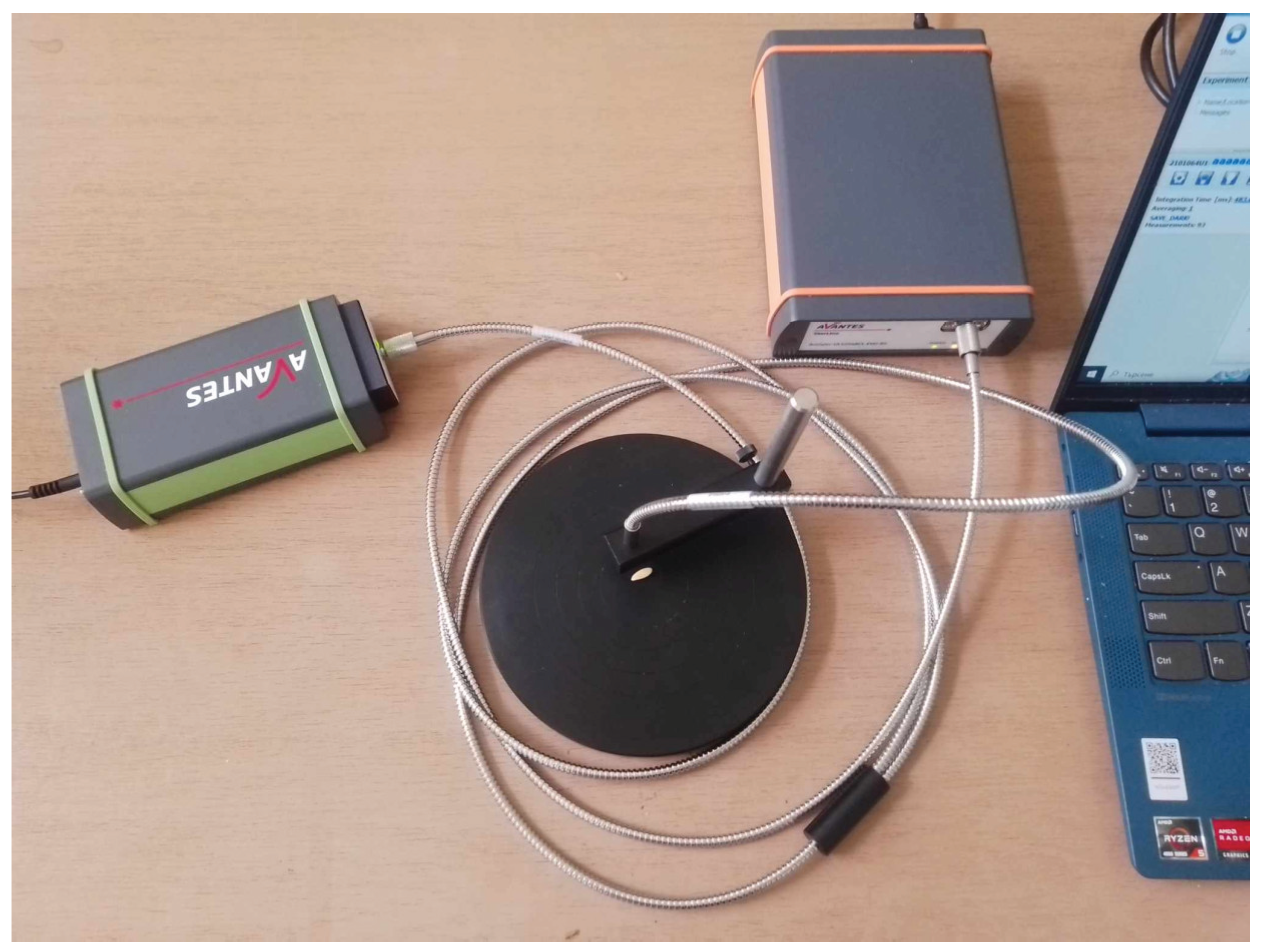
Figure 2.
Difference in emission wavelengths of melon seeds for breeding lines VK/1-5-5, K/15-6 and 11/9C (a sterile, b fertile).
Figure 2.
Difference in emission wavelengths of melon seeds for breeding lines VK/1-5-5, K/15-6 and 11/9C (a sterile, b fertile).
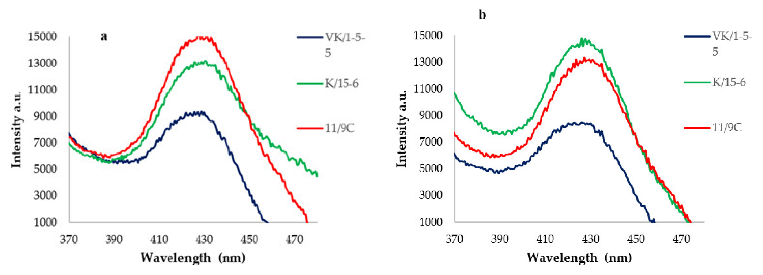
Figure 3.
Difference in emission wavelengths of melon seeds for sterile and fertile plants (breeding line VK/1-5-5a, breeding line K/15-6b and breeding line 11/9Cc).
Figure 3.
Difference in emission wavelengths of melon seeds for sterile and fertile plants (breeding line VK/1-5-5a, breeding line K/15-6b and breeding line 11/9Cc).
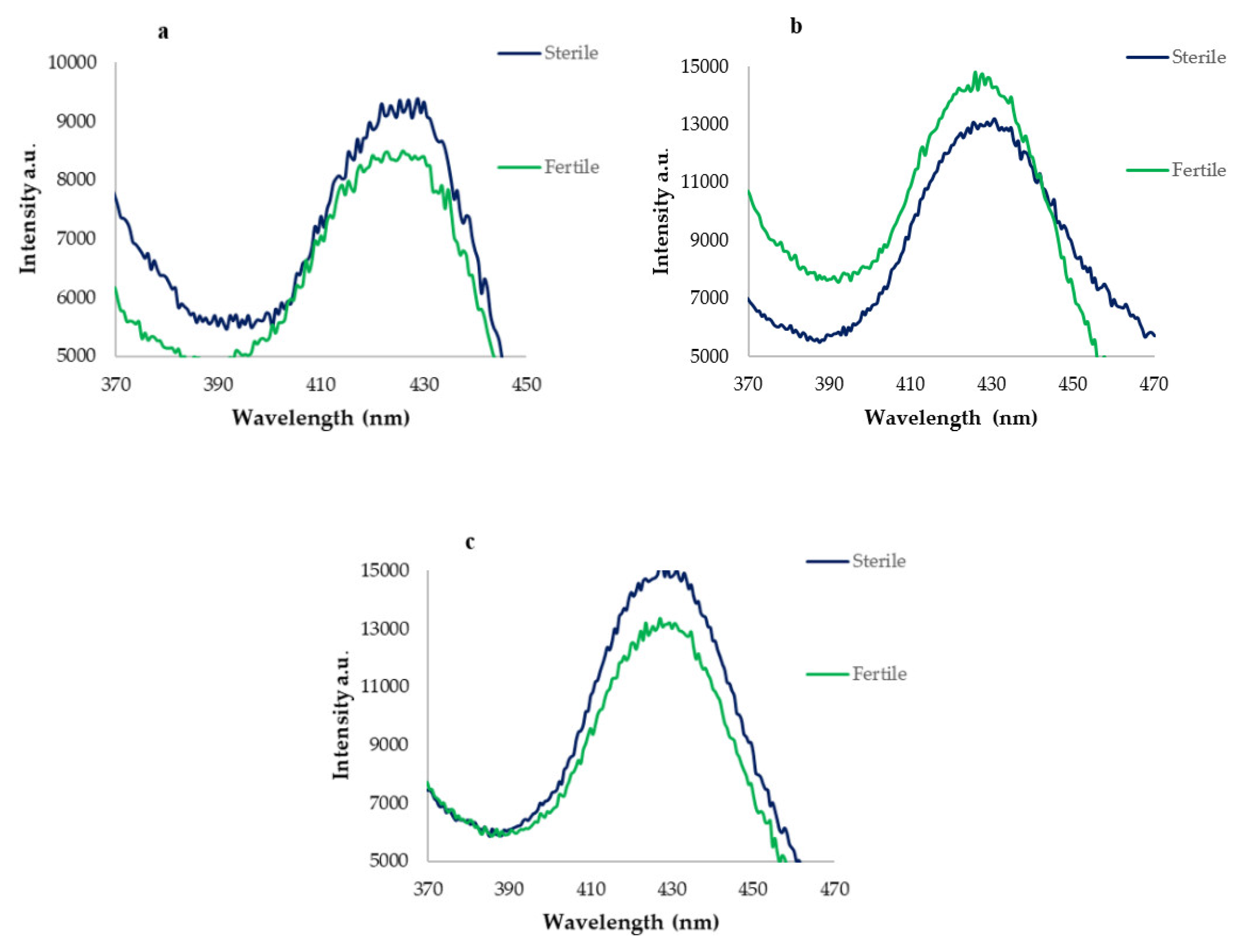
Figure 4.
Fertile and sterile flowers from studied melon lines; a – fertile male blossom before anthesis; b - fertile male blossom after anthesis; c – male sterile blossom after anthesis; d - male sterile blossom before anthesis; e – upper part - male sterile stamens, bottom part - male fertile stamens; f – pollen from fertile stamens; g – female blossom from sterile plant; h – ovary; i – fertile and sterile plants; j – sterile plant; k - upper part - male sterile flowers, bottom part - male fertile flowers; l - upper part - male sterile stamens, bottom part - male fertile stamens.
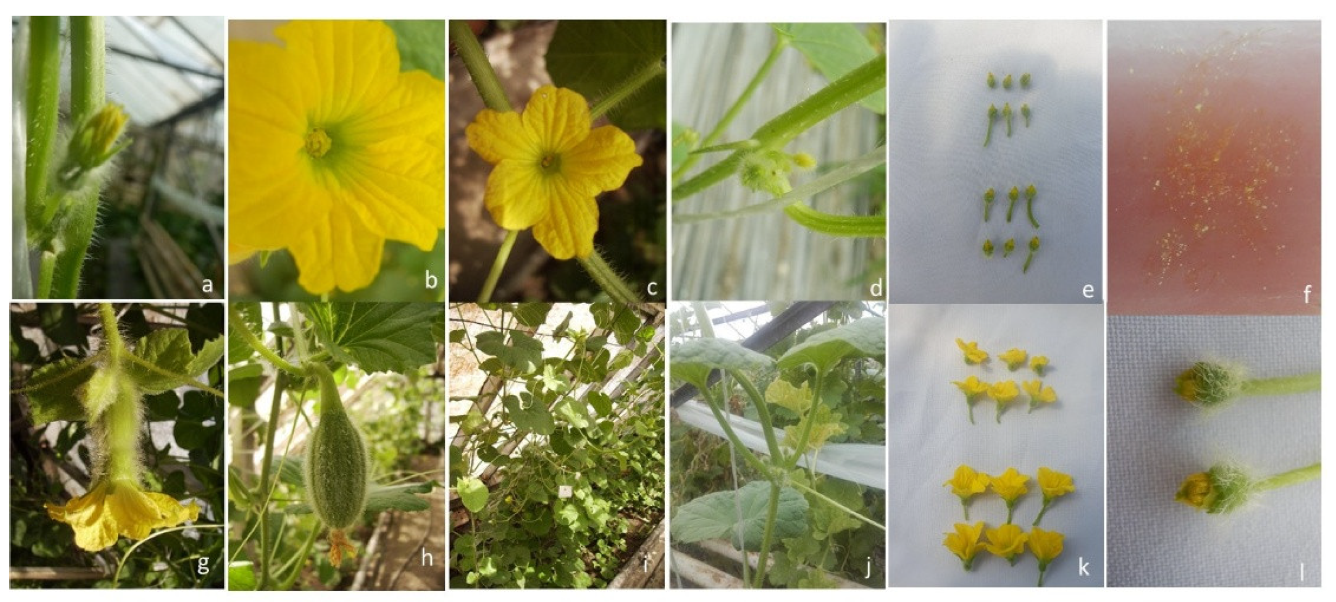

Table 1.
Proportions and χ2 Goodness of Fit of three male sterile melon lines.
| Level | Count | Proportion | χ2 | p | |
|---|---|---|---|---|---|
| VK/1-5-5 | |||||
| Fertile | Observed | 14 | 0.467 | 0.133 | 0.715 |
| Expected | 15.0 | 0.500 | |||
| Sterile | Observed | 16 | 0.533 | ||
| Expected | 15.0 | 0.500 | |||
| K/15-6 | |||||
| Fertile | Observed | 17 | 0.567 | 0.533 | 0.465 |
| Expected | 15.0 | 0.500 | |||
| Sterile | Observed | 13 | 0.433 | ||
| Expected | 15.0 | 0.500 | |||
| 11/9C | |||||
| Fertile | Observed | 14 | 0.467 | 0.133 | 0.715 |
| Expected | 15.0 | 0.500 | |||
| Sterile | Observed | 16 | 0.533 | ||
| Expected | 15.0 | 0.500 |
Disclaimer/Publisher’s Note: The statements, opinions and data contained in all publications are solely those of the individual author(s) and contributor(s) and not of MDPI and/or the editor(s). MDPI and/or the editor(s) disclaim responsibility for any injury to people or property resulting from any ideas, methods, instructions or products referred to in the content. |
© 2025 by the authors. Licensee MDPI, Basel, Switzerland. This article is an open access article distributed under the terms and conditions of the Creative Commons Attribution (CC BY) license (http://creativecommons.org/licenses/by/4.0/).
Copyright: This open access article is published under a Creative Commons CC BY 4.0 license, which permit the free download, distribution, and reuse, provided that the author and preprint are cited in any reuse.


