Preprint
Article

This version is not peer-reviewed.

Bacillus marisflavi XJ-04 Prevention of Watermelon Sclerotiniose and the Study of Biocontrol Effect

Submitted:

28 November 2025

Posted:

01 December 2025

You are already at the latest version

Abstract

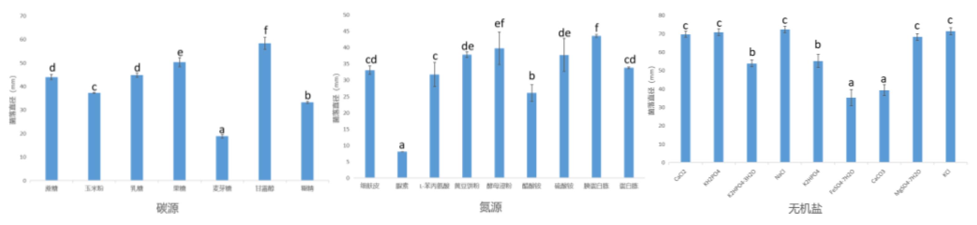
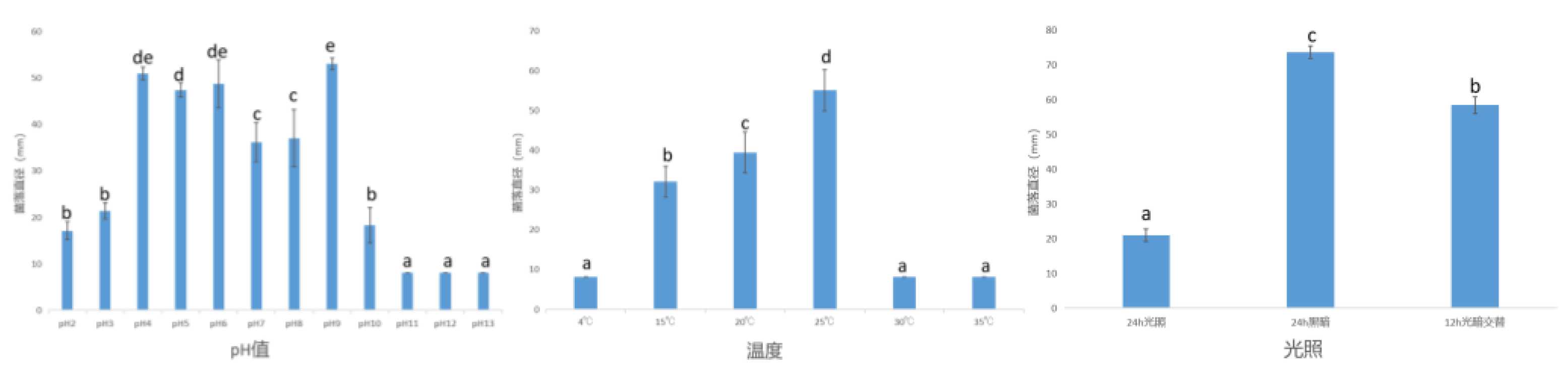
With the increase of watermelon cultivation area and continuous cropping, the harm of watermelon sclerotiniosis is becoming more and more serious. It has now risen to an important disease in watermelon production, which seriously affects the quality and yield of watermelon. The pathogen of watermelon Sclerotinia sclerotiorum is [Sclerotinia sclerotiorum(Lib.) De Bary], which is widely distributed in the world, causes plant sclerotia worldwide. The host range of Sclerotinia sclerotiorum is very wide, which not only harms watermelon, but also infects 75 families, 278 genera and 450 species of Cucurbitaceae, Leguminosae, Solanaceae and Cruciferae. To provide a new control method for the biological control of sclerotinia sclerotiorum in watermelon, the biocontrol bacteria in soil were isolated, screened and identified, and the bacteriostasis was studied. (1) The isolation, identification and biological characteristics of the pathogenic bacteria were determined. The fungus strain LY 24 was obtained from watermelon stem and vine infected with Sclerotinia sclerotiorum. The fungus strain LY 24 had good pathogenicity to watermelon plants by pathogenicity. The strain LY 24 was identified by morphology and molecular biology. The results showed that the best carbon source of strain LY 24 was mannitol, the best nitrogen source was tryptone, the best inorganic salt was NaCl, the best pH value was 9.The best growth temperature was 25, and the best light condition was whole darkness. (2) The isolation and identification of biocontrol bacteria in soil were carried out in the watermelon planting base around Changchun, such as the watermelon planting base in Jiutai District. Forty soil samples around the rhizosphere of watermelon plants were collected, and 300 strains of bacteria were isolated from the samples. The bacterial strains with significant antagonistic effect were obtained by plate confrontation method, and the strain XJ-04 with good antibacterial effect was selected as the research object. The inhibitory band width of strain XJ-04 was about 5.21 mm. The inhibitory rate of strain XJ-04 against Sclerotinia sclerotiorum was 70.12%. After morphological and molecular biological identification, strain XJ-04 was identified as Bacillus marisflavi. The physiological and biochemical characteristics of biocontrol strain XJ-04 were analyzed by detecting Gram reaction, contact enzyme reaction, methyl red reaction, V-P reaction and hydrogen sulfide reaction. (3) Studies on fermentation optimization of strain and fermentation broth stability. The optimum composition of culture medium was determined by orthogonal test, which was No.8 culture medium (sucrose, fine bran, K2HPO4·3H2O, pH 9). The inhibition rate was about 71.75%. The optimum addition amount of the above three components was determined by response surface methodology. When the addition amounts of sucrose, fine bran and K2HPO4·3H2O were about 21.08 g/L, 9.17 g/L and 9.77 g/L respectively, the highest inhibition rate was about 77. 78% by Design Expert software, which was about 7.66% higher than that before optimization, and 4% higher than that before optimization. The optimal fermentation conditions were 100 ml of liquid, 30 of temperature and 3d of fermentation time. The stability study showed that the bacteriostasis rate of fermentation broth treated by different temperature, pH, storage time and light had little change.

Keywords: 
;  ;  ;  ;  ;  

1. Introduction

Watermelon is an important economic crop. It is widely cultivated around the world, especially in Asia and America. With the increasing of market demand, watermelon becomes the fifth largest fruit in the world. The annual cultivation area of watermelon in China is over 200000 hectares, with a total output of 53.86 million tons.It ranks first in the world and has more than 60 % of the world ‘s total area.
The pathogen of watermelon Sclerotinia sclerotiorum is [Sclerotinia sclerotiorum(Lib.) De Bary], which is widely distributed in the world, causes plant sclerotia worldwide. S. sclerotiorum is a destructive plant pathogenic fungus with a wide host range, which can infect at least 75 families, 278 genera and more than 600 plants, including cruciferous, solanaceae, compositae and leguminosae and other important economic crops [1]. Plant sclerotiniose caused by S.sclerotiorum is distributed all over the world, and it occurs from seedling stage to flowering stage. In the past 10 years, the occurrence area of S.sclerotiorum in China was as high as 3.1 million hm2 per year, and the average annual actual yield loss was more than 170,000 tons, accounting for 55.60 % of the total loss of the 10 most important diseases and insect pests in rapeseed production in China [2].
At present, the control of crop diseases caused by S.sclerotiorum mainly depends on chemical fungicides. Although chemical fungicides can effectively control the growth of S.sclerotiorum mycelium, they have little effect on the survival of sclerotia in soil, and long-term use has caused great pressure on the ecological environment [3,4]. For example, Alternaria brassicola, which causes black spot disease in rape, has developed resistance to phenylpyrrole and dicarboxyimide reagents that are effective against S.sclerotiorum.The purpose of agricultural control measures is to reduce the number of sclerotia in soil or to create environmental conditions which are beneficial to the normal growth and metabolism of host crops but not to the growth of sclerotia or infection of host to control sclerotia [5].Breeding resistant (resistant) varieties is the most economical way to control S.sclerotiorum, which can reduce the cost of pesticide use and field management and improve the economic benefit of crops.Under the background of advocating green ecological civilization, biological control plays an increasingly important role in the prevention and control of S.sclerotiorum, and also meets the strategic requirements of the country to promote the ‘ double reduction ‘ planting mode. Compared with traditional chemical control, biological control has the advantages of strong selectivity, no drug resistance and environmental friendliness. It is an effective measure to protect the ecological environment and ensure the quality and safety of agricultural products [6].
The purpose of this study was to evaluate the inhibitory effect of a bacterial strain ( Bacillus marisflavi) isolated from soil on S.sclerotiorum, and to identify the biocontrol strain by morphological, molecular biological, physiological and biochemical identification. We also tested the optimization of fermentation conditions and fermentation medium, and studied the stability of the optimized fermentation broth and the determination of antibacterial effect. This study aims to deepen our understanding of its potential mechanism of action.

2. Materials and Methods

In this chapter, a pathogen was isolated and purified from the stems of watermelon plants infected with S.sclerotiorum. After pathogenicity determination, combined with morphological and molecular biological methods, it was identified as S.sclerotiorum. The Sclerotinia disease caused by Sclerotinia spp.is a widespread and serious disease in the world. S.sclerotiorum is a typical soil-borne pathogen with a wide range of hosts. It has been reported that S.sclerotiorum hosts more than 75 families, 278 genera and 500 crops. In this chapter, the biological characteristics of S.sclerotiorum were studied to provide a theoretical basis for the prevention and control of watermelon sclerotiniose.

2.1. Test Materials and Methods

2.1.1. Test Material

2.1.1.1. Test Strain

In the watermelon planting base of Jiutai District, Changchun City, watermelon plants with sclerotia were collected and isolated.

2.1.1.2 Test Medium

Potato dextrose agar ( PDA) medium : potato 200 g / L, glucose 20 g / L, agar powder 15 g / L.

2.1.1.3 Main Reagents

Glucose (Sinopharm Chemical Reagent Co., Ltd.), sucrose (Xilong Chemical Co., Ltd.), soluble starch (Shanghai Yuanye Biotechnology Co., Ltd.), dextrin (Shanghai Yuanye Biotechnology Co., Ltd.), maltose (Shanghai Yuanye Biotechnology Co., Ltd.), corn flour (Beijing Hongrun Baoshun Technology Co., Ltd.), lactose (Shanghai McLind Biotechnology Co., Ltd.), mannitol (Xilong Chemical Co., Ltd.), fructose (Xilong Chemical Co., Ltd.), peptone (Beijing Oboxing Biotechnology Co., Ltd.), yeast leaching powder (Beijing Oboxing Biotechnology Co., Ltd.), beef extract (Beijing Oboxing Biotechnology Co., Ltd.), tryptone (Beijing Oboxing Biotechnology Co., Ltd.), urea (Beijing Chemical Factory), potassium nitrate (Beijing Chemical Factory), Ammonium chloride (Beijing Chemical Factory), ammonium sulfate (Beijing Chemical Factory), ammonium acetate (Beijing Chemical Factory), L-phenylalanine (Beijing Chemical Factory), soybean cake powder (Beijing Hongrun Baoshun Technology Co., Ltd.), bran (Beijing Hongrun Baoshun Technology Co., Ltd.), diammonium hydrogen phosphate (Beijing Chemical Factory), CuSO4·5H2O (Beijing Chemical Factory), ZnSO4·7H2O (Beijing Chemical Factory), MgSO4·7H2O (Tianjin Chemical Reagent No. 3 Factory), FeSO4·7H2O (Tianjin Chemical Reagent No. 3 Factory), CaCO3 (Tianjin Fuchen Chemical Reagent Factory), K2HPO4·3H2O (Tianjin Fuchen Chemical Reagent Factory), KH2PO4 (Tianjin Fuchen Chemical Reagent Factory), NaCl (Tianjin Chemical Reagent No. 3 Factory), HCl (Beijing Chemical Factory), NaOH (Beijing Chemical Factory), KCl (Beijing Chemical Factory), Agar powder (Beijing Solaibao Technology Co., Ltd.), agarose sugar (Beijing Dingguo Changsheng Biotechnology Co., Ltd.), plant genomic DNA extraction kit (Jiangsu Kang for Century Biotechnology Co., Ltd.), dd H2O (Beijing Solaibao Technology Co., Ltd.), GelRed nucleic acid dye (Beijing Rangeko Technology Co., Ltd.), 2× Taq PCR premix (Beijing Solaibao Technology Co., Ltd.), DL2000 Maker (Beijing Rangeko Technology Co., Ltd.), 50× TAE Buffer (Shanghai Shengong Biology Co., Ltd.), KOH (Beijing Chemical Factory).

2.1.1.4 Main Instruments and Equipment

BIO-RAD Gel Imaging Analyzer (US Bio-Rad Company), TC-512 Gradient PCR Instrument (UK Techne Company), Manual Pipette (Germany Ebender Co., Ltd.), CT15E Table-type Micro High-speed Centrifuge (Hitachi China Co., Ltd.), YXQ-LS-75SII Vertical Pressure Steam Sterilizer (Shanghai Boxun Industrial Co., Ltd. Medical Equipment Factory), DYY-6C Electrophoresis Apparatus (Beijing Liuyi Instrument Factory), QL -866 Vortex Mixer (Kirin Medical Instrument Factory), ALC Electronic Balance (Germany Ekoller Co., Ltd.), SW=CJ-2D Super-clean Workbench (Jiangsu Sujing Group Co., Ltd.), subMIDI Horizontal Electrophoresis Tank (Beijing Baijing Biotechnology Co., Ltd.), DHG-9240 A Electrothermal Constant Temperature Blowing Drying Oven (Shanghai Shenxian Constant Temperature Equipment Factory), HZQ-X 100 Constant Temperature Oscillation Incubator (Taicang Experimental Equipment Factory), Thermostatic water bath (Shanghai Lancai Instrument Co., Ltd.).

2.1.2. Test Method

2.1.2.1 Isolation and Purification of Pathogenic Fungi

The pathogens were isolated from the stem of watermelon by tissue separation [7]. Select the stem of plant with typical symptoms, use a sterilizing knife to take 0.5 cm×0.5 cm tissue block at the interface of disease and health, put it into 3% sodium hypochlorite solution for disinfection for 1 min, then put it into 75% alcohol for disinfection twice for 1 min, then rinse it twice with sterile water, put it on sterile filter paper and blow off excess water, then transfer the tissue block to PDA plate, place five pieces for each dish evenly, carry out 25℃ constant-temperature dark culture, after the mycelium grows around the tissue block, take the mycelium and transfer it to new PDA plate, after purification, obtain the strain, and store it in 4℃ refrigerator for standby after numbering.

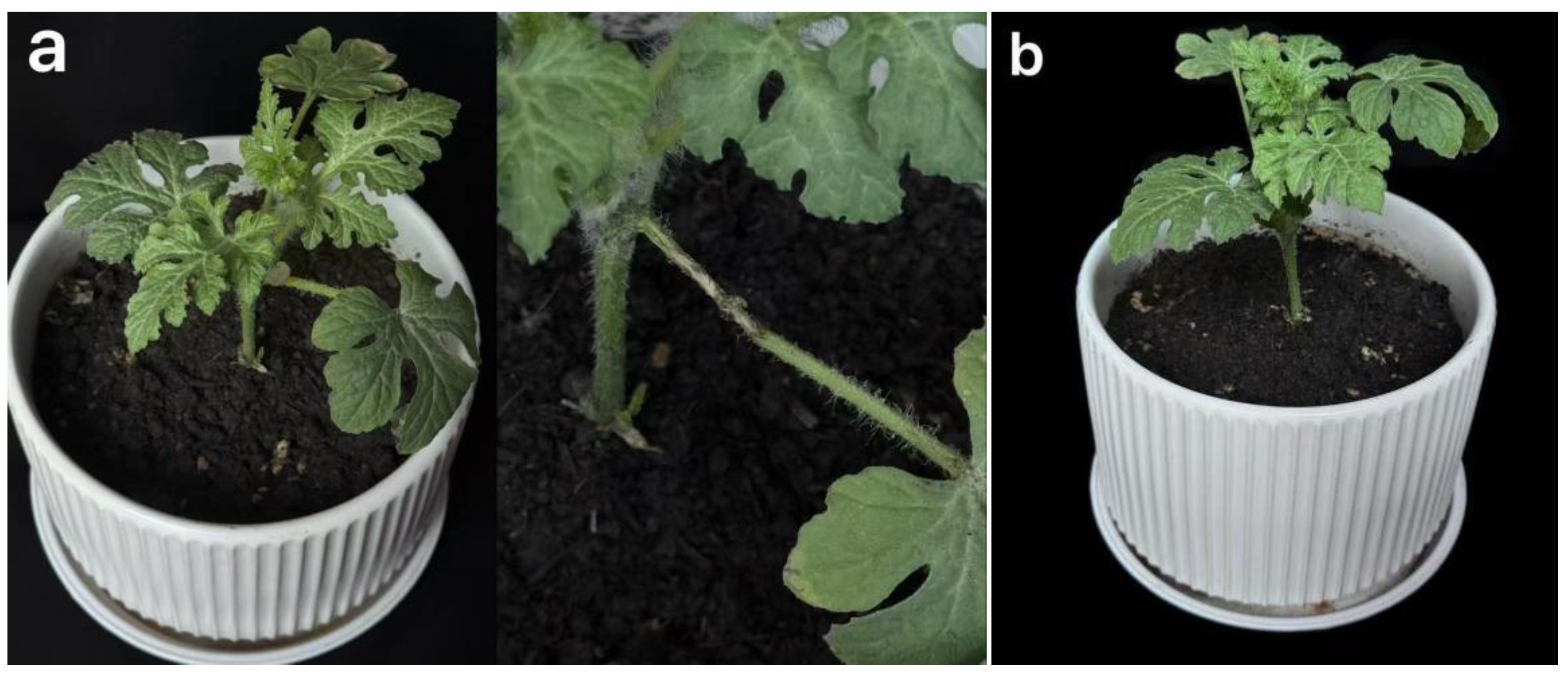
2.1.2.2 Pathogenicity Test

The pathogenicity of the isolated strain was determined by the PDA bacterial cake : The purified strain was inoculated on the PDA plate, cultured at 25 ℃ for 7 days, 8 mm bacterial cake was taken with a gun head, and healthy plants with better growth status were selected. The stems were washed with sterile water and dried, and the wounds were caused by sterile needles in the stems. The bacterial cake was inoculated and cultured at room temperature for 7 days. The incidence was observed regularly, and the non-inoculated bacterial cake was used as the blank control. After the plant was diseased, the diseased site was isolated and purified again, and the pathogenic bacteria were verified according to Koch ‘s rule.

2.1.2.3 Identification of Pathogens

1) Morphological identification : observe the colony diameter, color texture and the presence or absence of hyphae. Observe its microscopic morphology, drop KOH solution into the center of the slide, pick a small amount of mycelium in the center of the droplet, and observe the characteristics of the mycelium of the strain under a microscope after covering the slide.
2) Identification of molecular biology : DNA was extracted from fungi by CTAB method. PCR amplification was performed using fungal universal primers ITS1 (5’-TCCGTAGGTGAACCTGCGG-3’)/ITS4(5’-TCCTCCGCTTATTGATATATGC-3’) and β-tubulin gene fragment amplification primers Bt2a (5’-GGTAACCAAATCGGTGCTGCTTTC-3’)/Bt2b(5’-ACCCTCAGTGTAGTGACCCTTGGC-3’) [8]. Amplification procedure:94℃ pre-denaturation for 5 min ; denaturation 45 s, annealing at 53 ℃ for 30 s, extension at 72 ℃ for 45 s, a total of 35 cycles ; extension at 72 ℃ for 5 min [9]. The PCR amplification products were sequenced by Shanghai Shenggong Biotechnology Co., Ltd. The BLAST tool was used to analyze the homology of the obtained sequences in NCBI, and the sequences with the highest similarity were selected. The phylogenetic tree was constructed based on the neighbor-joining method using MEGA 7.0 software [10] to clarify the classification status of pathogens.

2.1.2.4 Biological Characteristics

Effects of carbon source, nitrogen source, inorganic salt, pH, temperature and light on colony growth.
1) The effect of carbon sources on colony growth : PDA medium was used as the basic medium, and the same amount of sucrose, corn flour, lactose, fructose, maltose, mannitol and dextrin were used to replace the glucose to prepare the medium containing different carbon sources. The bacterial cake with a diameter of 8 mm was connected to the center of the above medium, and the dark culture was carried out at 25 ℃ constant temperature incubator. When the colony diameter of the control group was 6 mm, the colony diameter was measured by the cross method and repeated three times.
2) The effect of nitrogen source on colony growth : PDA medium was used as the basic medium, and appropriate amount of soybean cake powder, L-phenylalanine, fine bran, tryptone, urea, peptone, yeast extract powder, ammonium sulfate and ammonium acetate were added to prepare the medium containing different nitrogen sources. The bacterial cake with a diameter of 8 mm was inoculated in the center of the above medium and cultured in a constant temperature incubator at 25 ℃ in dark. When the colony diameter of the control group reached 6 mm, the colony diameter was measured by cross-crossing method and repeated three times.
3) The effect of inorganic salts on colony growth : The PDA medium was used as the basic medium, and an appropriate amount of inorganic salts such as CaCl2, FeSO4·7H2O, NaCl, KH2PO4, CaCO3, KCl, K2HPO4, K2HPO4·3H2O and MgSO4·7H2O were added to prepare the medium containing different inorganic salts. The bacterial cakes with a diameter of 8 mm were inoculated in the center of the above medium and cultured in a constant temperature incubator at 25 ℃.When the colony diameter of the control group reached 6 mm, the colony diameter was measured by the cross-crossing method, which was repeated three times.
4) The effect of pH on colony growth : 8 mm bacterial cake was inoculated in the center of PDA medium with different pH ( 2,3,4,5,6,7,8,9,10,11,12 and 13), and cultured in 25 ℃ constant temperature incubator in dark. When the colony diameter of the control group reached 6 mm, the colony diameter was measured by cross-crossing method and repeated three times.
5) The effect of temperature on colony growth : The 8 mm mycelium was inoculated downward into the center of the PDA medium and cultured in a constant temperature incubator at 6 temperature gradients ( 4 ℃, 15 ℃, 20 ℃, 25 ℃, 30 ℃ and 35 ℃) in the dark. When the colony diameter of the control group reached 6 mm, the colony diameter was measured by the cross-crossing method and repeated three times.
6) The effect of light on colony growth : 8 mm bacterial cake was inoculated in the center of PDA medium and cultured in a 25 ℃ constant temperature incubator with three light conditions ( 24 h dark, 24 h light and 12 h light-dark alternation). When the colony diameter of the control group reached 6 mm, the colony diameter was measured by cross-crossing method and repeated three times..

3. Results

3.1. Isolation and Pathogenicity Determination of Pathogenic Fungi

3.1.1.1. Isolation and Purification of Pathogenic Fungi

The fungal strain isolated from the stem tissue of the diseased plant was numbered LY 24 ( Figure 3.1), and the strain was preserved in the Laboratory of Pathogen Biology, College of Plant Protection, Jilin Agricultural University.
Figure 3. 1. Stem symptoms of watermelon sclerotiniose.
Preprints 187202 g001

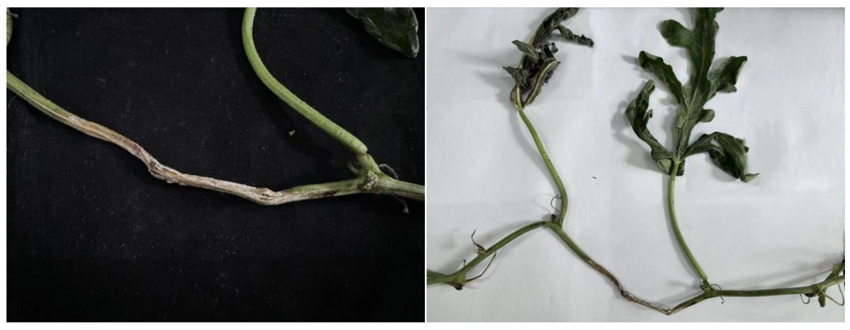
3.1.1.2. Pathogenicity Test

Strain LY 24 was tested for pathogenicity and showed typical sclerotinia symptoms seven days after inoculation (Figure 3.2 left). The control group was not affected (Figure 3.2 right).
Figure 3. 2. Pathogenicity test results of watermelon sclerotiniose pathogen LY 24 on watermelon plants. a: LY 24 inoculated plants; b: Watering plants with clean water
Preprints 187202 g002
Isolate and purify the pathogen at the inoculated site, number LY 25 (Figure 3.3 right), observe the colony, which is basically the same as that of LY 24 (Figure 3.3 left), and preliminarily determine that LY 24 is the pathogen of watermelon sclerotia.
Figure 3. 3. Comparison of colony morphology of watermelon Sclerotinia sclerotiorum pathogens LY 24 and LY 25. a: Strain LY 24 colony; b: Strain LY 25 colony
Preprints 187202 g003

3.1.2. Identification of Pathogens

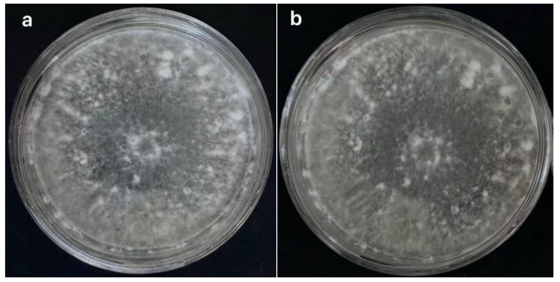
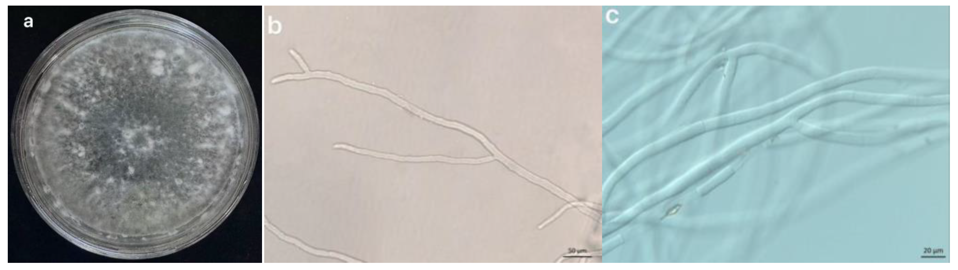
3.1.2.1 Morphological Identification

The mycelium of LY 24 pathogen was white on PDA medium, and it was round and evenly expanded on the plate. At the beginning, the mycelium was sparse, and then the mycelium was thickened, and the mycelium gathered to form sclerotia. The sclerotia is white at the beginning, gradually becomes brown, and usually becomes black after 15 days. The shape of the sclerotia is mostly spherical, and some are rat feces or irregular, with different sizes ( Figure 3.4-a). The mycelium has a diaphragm, the diaphragm is densely distributed, the diaphragm distance is irregular, and the branch is slightly constricted. The diameter of the mycelium is 4.2 ~ 7.0μm ( Figure 3.4-b, c), which is consistent with the morphological characteristics of S.sclerotiorum, and can be preliminarily identified as S.sclerotiorum.
Figure 3. 4. Macrostructure and microstructure of the pathogen LY 24 of watermelon Sclerotinia sclerotiorum. a: Colony morphology diagram; b,c: Mycelium morphology diagram
Preprints 187202 g004

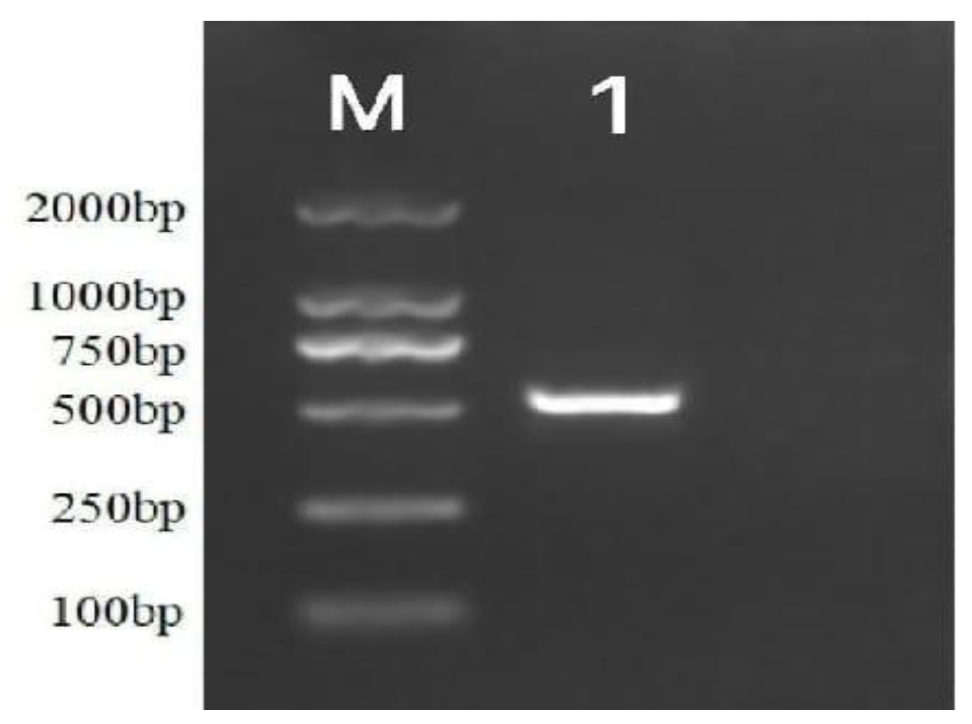
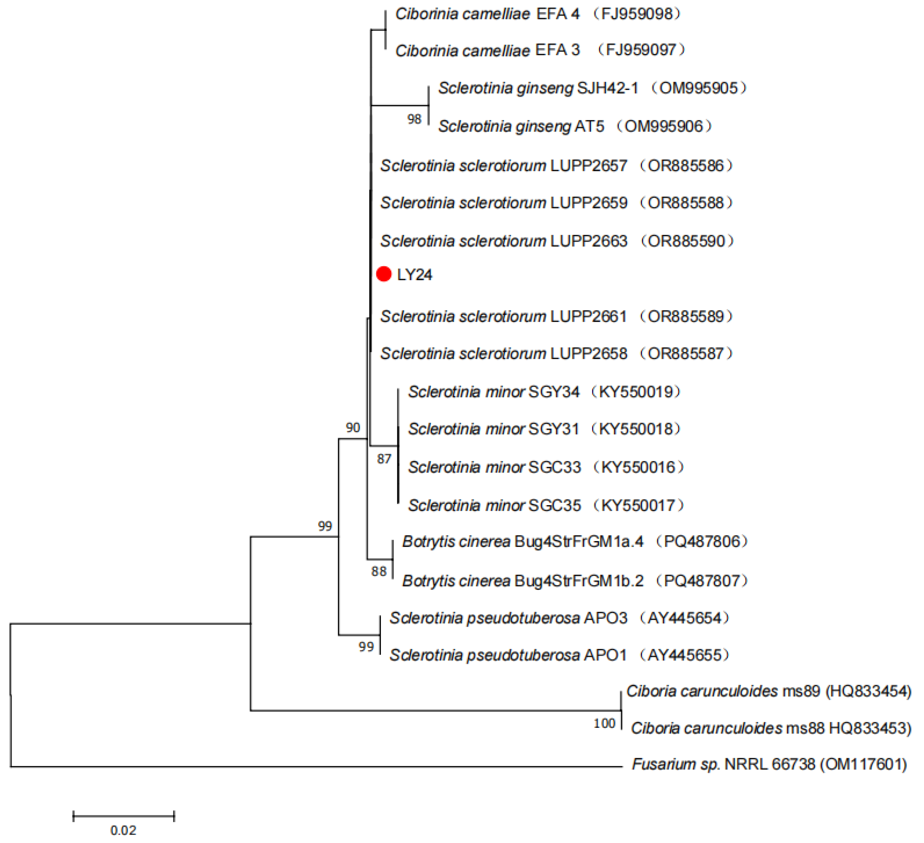
3.1.2.2 Molecular Biological Identification

After PCR amplification, the product electrophoresis test was qualified, and Shanghai Sangon Biotechnology Co., Ltd.was entrusted for sequencing. The length of the ITS gene sequence fragment of LY 24 is about 504 bp, and the electrophoresis picture is shown in Figure 3.5. The preliminary sequence alignment on NCBI showed that the strain LY 24 had 99 % homology with S.sclerotiorum.
Figure 3. 5. Electrophoretic band of PCR product of pathogen strain LY 24 of watermelon Sclerotinia sclerotiorum. M: DL2000Maker; 1: ITS.
Preprints 187202 g005
Based on the ITS gene sequence, the phylogenetic tree was constructed using Mega 7.0 ( Figure 3.6), which showed that LY 24 and S.sclerotiorum were clustered into one branch, and the strain LY 24 was verified as S.sclerotiorum from molecular biology.
Figure 3. 6. The phylogenetic tree of the pathogen strain LY 24 of watermelon Sclerotinia sclerotiorum was constructed by Mega 7.0 based on ITS gene.
Preprints 187202 g006

3.1.3. Biological Characteristics

3.1.3.1 Effects of Medium Components on Mycelial Growth of Pathogenic Fungi

The mycelial growth of LY 24 strain under seven carbon sources was tested. The results showed that the three lowest colony diameters were : maltose ( 18.76 mm) < dextrin ( 32.87 mm) < corn flour ( 37.14 mm) ; the three highest colony diameters were : mannitol ( 58.24 mm) > fructose ( 50.21 mm) > lactose ( 44.69 mm). Among them, the mycelial growth was the best when the carbon source was mannitol, and the mycelial growth was the worst when the carbon source was maltose. The mycelial growth of LY 24 strain under 9 nitrogen sources was tested. The results showed that the three species with the lowest colony diameter were : urea ( 8.00 mm) < ammonium acetate ( 25.99 mm) fine bran < ( 32.99 mm) ; the three highest colony diameters were : tryptone ( 43.48 mm) > yeast extract powder ( 39.69 mm) > soybean cake powder ( 37.74 mm). Among them, the mycelial growth was the best when the nitrogen source was tryptone, and the mycelial growth was the worst when the nitrogen source was urea. The mycelial growth of LY 24 strain under 9 inorganic salts was tested. The results showed that the three species with the lowest colony diameter were : FeSO4·7H2O ( 35.14 mm) < CaCO3 ( 39.25 mm) < K2HPO4·3H2O ( 53.74 mm) ; the three highest colony diameters were : NaCl ( 72.25 mm) > KCl ( 71.34 mm) > KH2PO4 ( 70.74 mm). Among them, when the inorganic salt is NaCl, the mycelium growth is the best, and when the inorganic salt is FeSO4·7H2O, the mycelium growth is the worst. The results are shown in Figure 3.7.
Figure 3. 7. Mycelial growth of watermelon Sclerotinia sclerotiorum pathogen strain LY 24 under different carbon sources, nitrogen sources and inorganic salts.
Preprints 187202 g007

3.1.3.2 Effects of Culture Conditions on Mycelial Growth of Pathogens

The mycelial growth of LY 24 strain under 12 pH conditions was tested. The results showed that the three lowest colony diameters were : pH11 ( pH12, pH13) ( 8.00 mm) < pH2 ( 17.03 mm) < pH10 ( 18.18 mm) ; the three highest colony diameters were : pH9 ( 52.90 mm) > pH4 ( 50.87 mm) > pH6 ( 48.63 mm). Among them, pH9 was the best for mycelial growth, and pH11, pH12, and pH13 did not grow. The mycelial growth of LY 24 strain at 6 temperatures was tested. The results showed that the three species with the lowest colony diameter were : 4 ℃ ( 8.00 mm) = 30 ℃ ( 8.00 mm) = 35 ℃ ( 8.00 mm) ; the three highest colony diameters were : 25 ℃ ( 54.93 mm) > 20 ℃ ( 39.33 mm) > 15 ℃ ( 31.98 mm). Among them, 25 ℃ was the best for mycelium growth, and mycelium did not grow at 4 ℃, 30 ℃, and 35 ℃. The mycelial growth of LY 24 strain under three light conditions was tested. The results showed that the mycelial growth was the best in full dark and the worst in full light. The results are shown in Figure 3.8.
Figure 3. 8. Mycelial growth of Sclerotinia sclerotiorum pathogen LY 24 under different pH, temperature and light conditions.
Preprints 187202 g008

3.1.4. Summary

1) A fungal strain LY 24 was isolated from the stems of watermelon plants infected with Sclerotinia sclerotiorum. It was determined to be pathogenic to watermelon plants by pathogenicity test. Through morphological and molecular biological identification, LY 24 was identified as S.sclerotiorum.
2) After measuring its biological characteristics, the best carbon source for the growth of LY 24 strain was mannitol, the best nitrogen source was tryptone, the best inorganic salt was NaCl, the best pH value was 9, the best temperature was 25 °C, and the best light condition was full darkness.

3.2. Isolation, Screening and Identification of Biocontrol Bacteria

In this chapter, biocontrol bacteria were isolated from the rhizosphere soil of healthy watermelon plants. S.sclerotiorum was used as the target fungi, and the strains with good biocontrol effect were screened out for morphological, molecular biological and physiological and biochemical identification. Finally, its taxonomic status was clarified.

3.2. Test Materials and Methods

3.2.1. Test Material

3.2.1.1 Target Strain

The strain of S.sclerotiorum was preserved in the Laboratory of Plant Fungi Diseases, College of Plant Protection, Jilin Agricultural University.

3.2.1.2 Soil Samples and Culture Medium

Soil samples were collected from watermelon planting bases in the surrounding areas of Changchun, such as the watermelon planting base in Jiutai District, and 40 soil samples around the rhizosphere of watermelon plants were collected.
Potato glucose agar medium ( PDA), Listeria chromogenic medium medium ( LA), potato glucose water medium ( PDB), Luria-Bertani medium ( LB), glucose peptone water medium ( MR-VP), lead acetate medium ( LAM).

3.2.1.3 Main Reagents

Glucose (Sinopharm Group Chemical Reagent Co., Ltd.), agar powder (Beijing Solaibao Technology Co., Ltd.), agarose sugar (Beijing Dingguo Changsheng Biotechnology Co., Ltd.), yeast leaching powder (Beijing Oboxing Biotechnology Co., Ltd.), tryptone (Beijing Oboxing Biotechnology Co., Ltd.), peptone (Beijing Oboxing Biotechnology Co., Ltd.), dipotassium hydrogen phosphate (Tianjin Kemio Chemical Reagent Co., Ltd.), beef paste (Beijing Oboxing Biotechnology Co., Ltd.), sodium thiosulfate (Shandong Keyuan Biotechnology Co., Ltd.), sodium chloride (Tianjin Chemical Reagent No. 3 Factory), plant genomic DNA extraction kit (Jiangsu Kangwei Century Biotechnology Co., Ltd.), bacterial genomic DNA rapid extraction kit (Biotechnology Shanghai Co., Ltd.), Gram staining kit (Beijing Solaibao Technology Co., Ltd.), Methyl Red (Shandong Keyuan Biochemical Co., Ltd.), VP kit (Guangdong Huankai Microorganism Technology Co., Ltd.), 3%H2O2 (Beijing Solaibao Technology Co., Ltd.), dd H2O (Beijing Solaibao Technology Co., Ltd.), GelRed nucleic acid dye (Beijing Lanjieke Technology Co., Ltd.), 2× Taq PCR premix (Beijing Solaibao Technology Co., Ltd.), DL2000 Maker (Beijing Lanjieke Technology Co., Ltd.), 50× TAE Buffer (Biotechnology (Shanghai) Co., Ltd.), KOH (Beijing Chemical Factory).

3.2.1.4 Main Instruments and Equipment

Same as 2.1.1.4.

3.2.2. Test Method

3.2.2.1 Collection of Soil Samples and Separation and Purification of Biocontrol Bacteria

The rhizosphere soil of healthy watermelon plants was taken by five-point sampling method [11]. The soil samples were numbered, dried in the shade, passed through a 60-mesh sieve, and stored in a refrigerator at 4 °C. The soil suspension was prepared with sterile water and continuously diluted 10-1 ~ 10-7 times, and then coated on LA medium. After 2 d ~ 3 d, single colonies were picked for purification and preservation [12].

3.2.2.2 Screening of Biocontrol Bacteria

1) Preliminary screening : Using the plate confrontation method, the bacteria to be tested and the watermelon Sclerotinia sclerotiorum fungus cake were selected and inoculated on the PDA medium for confrontation culture [13]. Three groups of replicates were set up. According to the growth of pathogenic bacteria in the control group, the existence of bacteriostatic zone was observed and its width was measured.
2) Re-screening : The strains with good antagonistic effect in the preliminary screening were selected and numbered. The bacteria were picked with a gun head and placed in 150 mL LB medium, and cultured at 28 ° C and 180 rpm for 2 days. The supernatant was centrifuged at 12000 rpm for 5 min and filtered with a 0.45 μm filter to obtain the fermentation broth.
Inhibition of S.sclerotiorum mycelium : The above fermentation broth was uniformly mixed with PDA medium at 1 : 5, poured into the plate, and inoculated with S.sclerotiorum after standing. Each group was repeated three times, and sterile water was mixed with PDA medium at 1 : 5 as the control group. Cultured at 28 ℃, the diameter was measured after 3 days, and the inhibition rate of the fermentation broth was calculated by the following formula [14]:
Inhibitory   rates = Colony   diameter   of   control   group - Treatment   group   colony   diameter Colony   diameter   of   control   group - The   diameter   of   the   fungus   cake × 100 %
SPSS software was used to analyze the inhibitory rate.
3) The effect on sclerotia germination of S.sclerotiorum : 50 sclerotia of S.sclerotiorum with basically the same size were washed with sterile water and dried, and then treated with 75 % ethanol for 3 min. Finally, the sclerotia were placed on the filter paper of the bacteria, and the water on the surface was absorbed. 25 sclerotia were put into the fermentation broth of biocontrol strains and soaked for 30 min. Another 25 sclerotia were placed in LB liquid medium and soaked for 30 min as the control group. The above sclerotia were dried and placed on a PDA plate, 5 sclerotia per dish, and cultured in a 25 ℃ incubator. The germination of sclerotia in the two groups was observed at 1 d, 3 d and 5 d, respectively, and the germination inhibition rate was calculated. Each treatment was repeated three times.
Inhibitory   rates = Control   sclerotium   germination   number - The   number   of   treated   sclerotia   germination Control   sclerotium   germination   number × 100 %

3.2.2.3 Identification of Biocontrol Bacteria

1) Morphological identification : Observe the size, color, shape, transparency, uplift, edge and other characteristics of the colony.
2) Physiological and biochemical identification : The specific operation method of physiological and biochemical test refers to the specific reference ‘General bacterial identification method ‘ [15] ‘ Berger bacterial identification manual ‘ [16] ‘ Common bacterial system identification manual ‘ [17].
Gram staining test : take a piece of glass slide, take the bacterial liquid smear, dry, fixed. When fixed, it can be fixed by flame 1-2 times, not overheated, and it is advisable that the glass slide is not hot. Initial dyeing : after adding crystal violet dyeing solution, dyeing for 1 min, washing. Mordant dyeing : Dyeing for 1 min after adding iodine solution and washing with water. Decolorization : Drop the decolorization solution, shake the slide, decolor for 20-60 s according to the thickness of the smear, wash with water, and absorb the water. Re-staining : After the saffron staining solution was added dropwise, it was stained for 1 min and washed with water. After the filter paper was dried with excess water or dried in the air, microscopic examination was performed.
Contact enzyme test : The strain was inoculated on the plate of NA medium and cultured at 28 ℃ for 2 days. The colony was scraped with a sterilized pick needle and placed in a test tube containing 3 % H2O2 solution. The reaction results were observed immediately. If a large number of bubbles appeared immediately after the addition of H2O2, it was a positive result, indicating that the bacteria had catalase. If there is no bubble, it is a negative result, indicating that the bacteria lack catalase.
Methyl red ( MR) test : 0.1 g methyl red was dissolved in 300 mL 95 % alcohol, and then 200 mL distilled water was added to prepare methyl red reagent. The tested strains were inoculated in glucose peptone water medium, cultured at 35 ℃ for 2-5 d, and 5 drops of methyl red reagent were added. The results were observed immediately and the red was positive. Orange red is weakly positive ; yellow is negative.
V-P test : The strains to be tested were inoculated in glucose peptone water medium and cultured at 35 ℃ for 24-48 h. Then 10 drops ( about 0.5 mL) of A solution ( 1-naphthol 0.6 g, 95 % ethanol 10 mL) and 4 drops ( about 0.2 mL) of B solution ( potassium hydroxide 4 g, deionized water 10 mL) in the kit were added dropwise, and the test tube was fully shaken to observe the results. The positive reaction appeared red immediately or within a few minutes. If the reaction was negative, it should be placed at 35 ℃ for 4 h and observed again.
Hydrogen sulfide test : 10.0 g peptone, 3.0 g beef extract, 5.0 g sodium chloride, 2.5 g sodium thiosulfate and 12.0 g agar were used to prepare lead acetate medium. The strains to be tested were inoculated into the above medium and cultured at 35 ℃ for 24-48 h. The results were observed. If the medium became black, it was positive, and the medium did not change color, it was negative.
3) Identification of molecular biology : The strain was inoculated into LB medium, cultured overnight at 28 ℃, 180 r / min in a constant temperature shaker, and 1 mL of bacterial solution was taken into a sterile centrifuge tube. Bacterial genomic DNA was extracted step by step using the bacterial genomic DNA rapid extraction kit ( Bioengineering Shanghai Co., Ltd.) according to the instructions. Bacterial strain DNA was amplified by PCR using bacterial universal primers 16S rDNA universal primers 27F (5’-AGTTTGATCMTGGCTCAG-3’) and 1492R (5’-GGTTACCTTGTTACGACTT-3’). Amplification procedure : 94 ℃ pre-denaturation for 5 min ; denaturation 45 s, annealing at 53 ℃ for 30 s, extension at 72 ℃ for 45 s, a total of 35 cycles ; extend at 72 ℃ for 5 min. After the amplification, the PCR products were detected by agarose gel electrophoresis, and then the PCR products were sent to Shanghai Shenggong Bioengineering Co., Ltd.for DNA sequencing. The obtained sequences were blasted in NCBI database, and the related gene sequences were downloaded in Genbank. The data were analyzed by MEGA 11.0 software, and the phylogenetic tree was constructed.

3.2.3. Results and Analysis3.2.3. Isolation and Screening of Soil Bacteria

3.2.3.1 Isolation of Bacterial Strains

In this experiment, 300 different bacterial strains were isolated from 40 soil samples, and different forms of bacteria were isolated and purified by plate streaking technique.

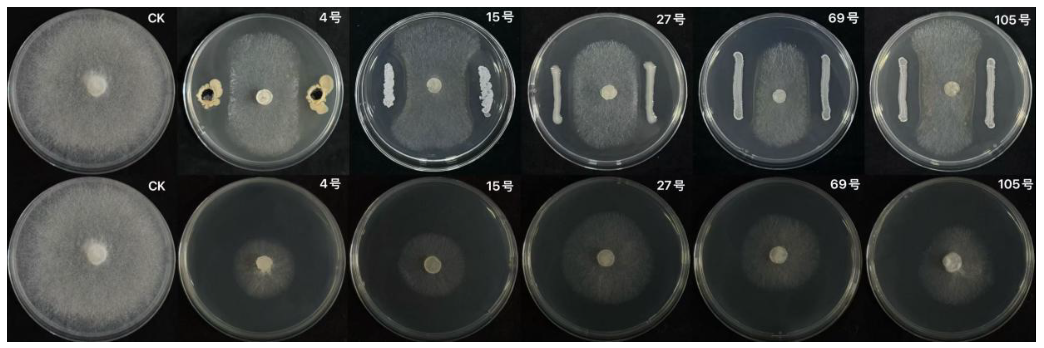
3.2.3.2 Screening of Antagonistic Bacteria Strains

After preliminary screening, five strains of XJ-04, LY-15, SY-27, XG-69 and JH-105 were found to have obvious bacteriostatic zones when they were confronted with S.sclerotiorum, and the antagonistic effect was good. Among them, the inhibition zone of XJ-04 and LY-15 strains was more than 4.8 mm, which was significantly better than other strains. The results are shown in Figure 4.1.
The results of re-screening test showed that the fermentation broth of SY-27 strain, XG-69 strain and JH-105 strain had weak inhibitory effect on the pathogen of watermelon sclerotiniose, and the inhibition rate was only 50.32 %, 59.63 % and 67.60 %. The fermentation broth of strain XJ-04 and strain LY-15 had a good inhibitory effect on the pathogen of S.sclerotiorum, and the inhibition rate reached more than 70.00 %.
Based on the results of preliminary screening and rescreening, XJ-04 strain was the most effective soil bacteria against S.sclerotiorum. After preliminary identification, XJ-04 strain was identified as Bacillus marisflavi. By querying the articles, it was found that there were few studies on the biological control of XJ-04 strain. Most of them were studies on the biological characteristics of the strain, and the articles on the inhibitory effect of the strain on the pathogen of watermelon sclerotiniosis were even rarer. Therefore, XJ-04 strain was selected as the research object for subsequent experiments. The inhibition zone width of strain XJ-04 was about 5.21 mm, and the inhibition rate of fermentation broth was about 70.12 %. The results are shown in Table 1.
The bacteriostatic effect of the initial screening and rescreening of soil bacteria is shown in Figure 4.1:
Figure 4. 1. The antibacterial effect of 5 strains of soil bacteria in primary screening ( upper) and rescreening ( lower). A: CK; B: XJ-04; C: LY-15; D: SY-27; E: XG-69; F: JH-105
Preprints 187202 g009
After treatment with the fermentation supernatant of strain XJ-04, the germination of sclerotia was significantly inhibited compared with the control group. The number of sclerotia germinated within 3 days was 15 less than that of the control group, and the inhibition rate of sclerotia germination was 60 %. The results are shown in Table 3-2.
Table 3-2. Effect of fermentation broth of strain XJ-04 on sclerotia germination.
Treatment Number of sclerotia germination in 3d Inhibition rate(%)
Fermentation supernatant 10 60
CK 25 /

3.2.2. Identification of Biocontrol Fungi

3.2.2.1 Morphological Identification

The colony of strain XJ-04 in NA medium was light yellow, round, 0.8cm in size, smooth, neat edge, wet, convex and transparent. Gram-positive bacteria, bacilli, arranged in a single, length of about 1.5-2.0 μm, width of about 0.8 μm, with spores, mesophytic or terminal, as shown in Figure 4.2.
Figure 4. 2. Colony morphology of strain XJ-04. a: Colony front; b: The back of the colony.
Preprints 187202 g010

3.2.2.2 Physiological and Biochemical Identification
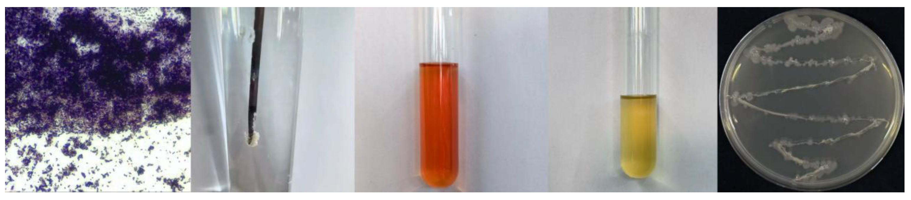
The results of physiological and biochemical tests showed that strain XJ-04 was an aerobic bacterium. The physiological and biochemical tests such as Gram reaction, contact enzyme reaction and methyl red reaction were positive, and the physiological and biochemical tests such as V-P reaction and hydrogen sulfide reaction were negative. According to the morphological characteristics and physiological and biochemical characteristics of strain XJ-04, it can be preliminarily identified as Bacillus marisflavi. The results are shown in Table 3-3 and Figure 4.3.
Figure 4. 3. Physiological and Biochemical Characteristics of Strain XJ-04.
Preprints 187202 g011
Table 3-3. Some physiological and biochemical characteristics of strain XJ-04.
Item Result
Gram reaction +
Contact enzyme reaction +
Methyl red reaction +
V-P Reaction -
Hydrogen sulfide reaction -
Note: “+” positive,”-” negative.

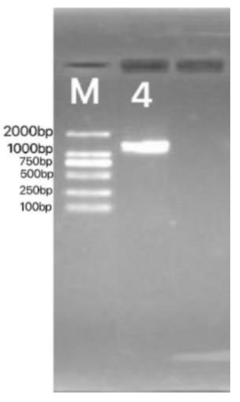
3.2.2.3 Molecular Biological Identification
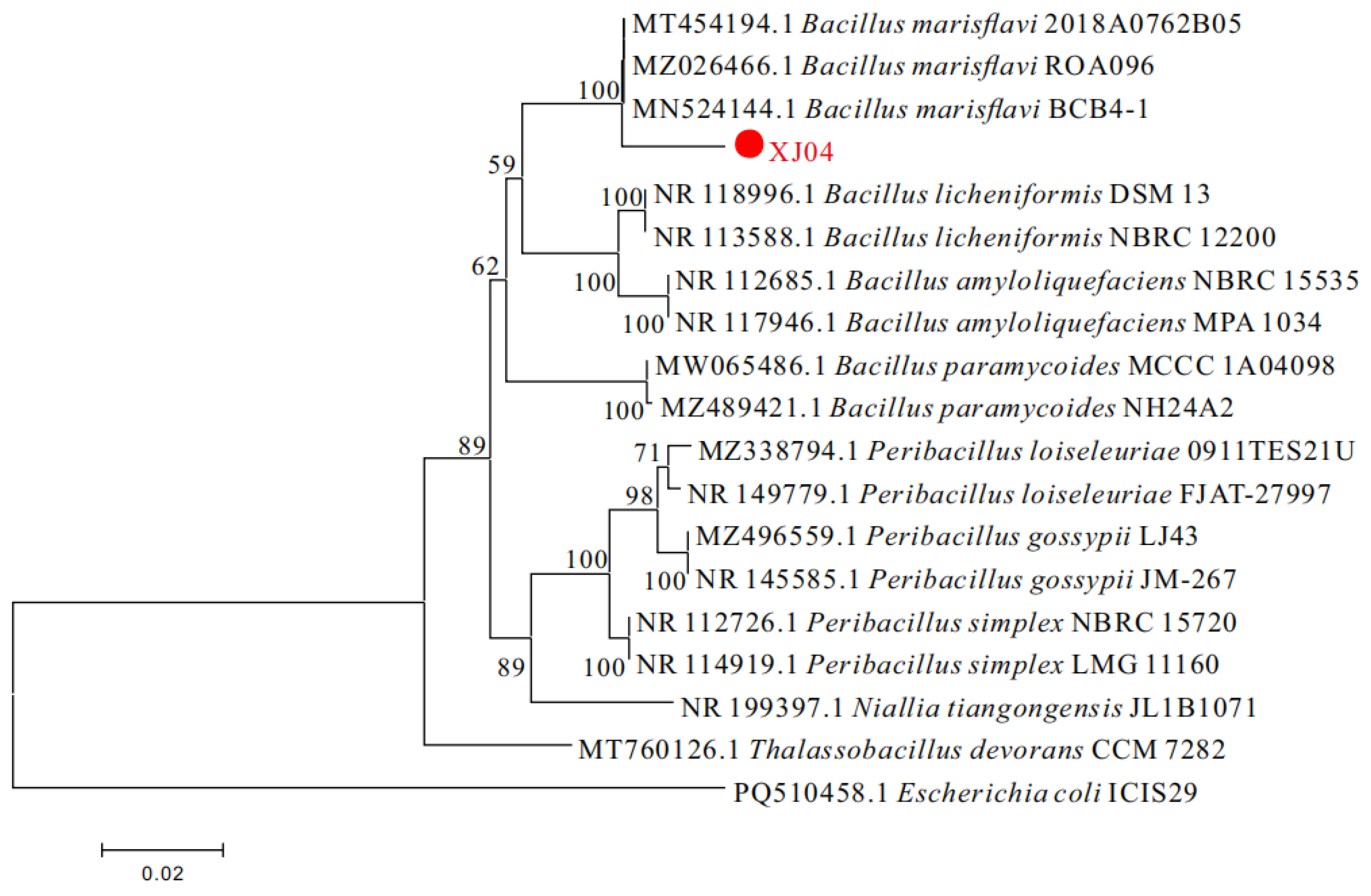
Bacterial strain DNA was amplified by PCR using bacterial universal primers 16S rDNA universal primers 27F (5’-AGTTTGATCMTGGCTCAG-3’) and 1492R (5’-GGTTACCTTGTTACGACTT-3’). Amplification procedure : 94 °C pre-denaturation for 5 min ; denaturation 45 s, annealing at 53 ℃ for 30 s, extension at 72 °C for 45 s, a total of 35 cycles ; extend at 72 ℃ for 5 min. After the amplification, the PCR products were detected by agarose gel electrophoresis as shown in Figure 3.4, and then the PCR products were sent to Shanghai Shenggong Bioengineering Co., Ltd.for DNA sequencing. After preliminary comparison on NCBI, it was found that strain XJ-04 had the highest homology with Bacillus marisflavi, reaching 97.87 %. The results are shown in Figure 4.4.
Figure 4. 4. Electrophoretic band of PCR product of strain XJ-04. M: DL2000Maker; 4: 16S rDNA.
Preprints 187202 g012
The obtained sequences were blasted in NCBI database, and the related gene sequences were downloaded in Genbank. The data were analyzed by MEGA 11.0 software, and the phylogenetic tree was constructed. The results showed that the strain XJ-04 and Bacillus marisflavi were clustered into a branch. The strain XJ-04 was verified to be Bacillus marisflavi by molecular biology. The results are shown in Figure 3.5.
Figure 4. 5. The phylogenetic tree of strain XJ-04 was constructed by Mega 11.0 based on 16S rDNA sequence.
Preprints 187202 g013

3.2.3. Summary

1) 300 strains of biocontrol bacteria were isolated from the rhizosphere soil of watermelon plants. Using watermelon Sclerotinia sclerotiorum as the target bacteria, XJ-04 strain with better biocontrol effect was finally selected through plate confrontation screening and rescreening. The inhibition bandwidth of the bacteria in the primary screening was 5.21 mm, and the inhibition rate of the secondary screening fermentation broth was 70.12 %. It was finally identified as Bacillus marisflavi by morphological identification, physiological and biochemical tests and molecular biology identification.

3.3. Optimization of Fermentation Conditions and Stability of Fermentation Broth of Strain XJ-04

The nature and concentration of fermentation medium components affect the supply of nutrients and microbial cell metabolism, thus affecting the inhibitory effect on target bacteria. Among them, carbon source, nitrogen source and trace elements usually play a leading role in affecting fermentation productivity, and stability determines whether the biocontrol bacteria can be applied to actual production [18,19]. In this chapter, the inhibition rate of the fermentation broth of strain XJ-04 against S.sclerotiorum was used as the determination index. Firstly, the best combination of carbon, nitrogen source and inorganic salt components was screened on the basis of LB medium. Then, the addition amount of each component was optimized by response surface test to determine the optimum liquid loading, fermentation time and fermentation temperature. Finally, the optimum fermentation conditions were determined. At the same time, the thermal stability, light stability, acid-base stability, storage stability and passage stability of the fermentation broth of strain XJ-04 were also determined, which could finally exert the highest antibacterial ability of the biocontrol strain.

3.3.1. Test Materials and Methods

3.3.1.1 Test Material3.3.1.1.1 Test Strain

Strain XJ-04, S.sclerotiorum.

3.3.1.1.2 Test reagent and culture medium

1) Carbon source test medium : carbon source ( glucose, sucrose, soluble starch, dextrin, maltose, corn flour, lactose, mannitol, fructose) 5 g / L, tryptone 10 g / L, NaCl 10 g / L;
2) Nitrogen source test medium : yeast extract powder 5 g / L, nitrogen source ( peptone, yeast extract powder, beef extract, urea, potassium nitrate, ammonium chloride, soybean powder, bran, diammonium hydrogen phosphate) 10 g / L, NaCl 10 g / L;
3) Inorganic salt test medium : yeast extract 5 g / L, tryptone 10 g / L, inorganic salt ( CuSO4·5H2O / ZnSO4·7H2O / MgSO4·7H2O / FeSO4·7H2O / CaCO3 / K2HPO4·3H2O / KH2PO4) 10 g / L;
4) pH test medium : yeast extract 5 g / L, tryptone 10 g / L, NaCl 10 g / L The pH values of the sterilized medium were adjusted to 5.0,6.0,7.0,8.0,9.0, respectively.
4.1.1.3. Main instruments and equipment:
See 2.1.1.4

3.3.1.2. Test Method3.3.1.2.1 Screening of Optimal Components in Fermentation Culture

Two 8 mm bacterial cakes of strain XJ-04 were taken with a blue tip and inoculated into the above 30 kinds of carbon sources, nitrogen sources, inorganic salts and pH test media, respectively. Three replicates were set and cultured at 28 ℃ and 180 rpm for 2 days. The inhibition rate of the fermentation filtrate of strain XJ-04 fermented with different components to S.sclerotiorum was measured and calculated according to the method of 3.1.2.2, and the significance was analyzed by SPSS software.

3.3.1.2.2. Orthogonal Test

Carbon source, nitrogen source, inorganic salt and pH were used as four factors, and the optimal three components of each factor obtained in 4.1.2.1 were three levels. The optimal combination of additive components was obtained by L9 ( 34) orthogonal test design ( Table 4-1) and range analysis.
Table 4-1. Factors and levels of orthogonal design.
Horizontal
Level
Factor Factor
A Carbon source B nitrogen source C Inorganic salt D pH
1 Maltose Urea K2HPO4·3H2O 7
2 Mannitol Fine bran FeSO4·7H2O 8
3 Sucrose Diammonium phosphate CaCO3 9

3.3.1.2.3. Optimization of the Addition Amount of Fermentation Medium Components

Based on the optimal combination of components obtained by orthogonal test, the addition amount of carbon source, nitrogen source and inorganic salt was optimized by response surface test, and a set of optimal liquid medium for fermentation of strain XJ-04 was selected.
Taking the inhibition rate as the response index, the effects of sucrose addition ( 5 g / L, 10 g / L, 15 g / L, 20 g / L, 25 g / L), fine bran addition ( 5 g / L, 10 g / L, 15 g / L, 20 g / L, 25 g / L) and K2HPO4·3H2O addition ( 2.5 g / L, 5 g / L, 7.5 g / L, 10 g / L, 12.5 g / L) on the inhibition rate were investigated by single factor rotation method. The initial fermentation conditions were 28 °C, 180 r / min, 2 d, pH 6.0.
According to the results of single factor test, the range of response surface test parameters for the addition of fermentation medium components of strain XJ-04 was determined [20,21]. The response surface test of 3 factors and 3 levels [22] was carried out by Box-Behnken test design and analysis method, and the optimal process parameters of strain XJ-04 fermentation were obtained [23,24]. Design Expert 8.0.6 was used to analyze the data obtained [25,26].

3.3.1.2.4. Optimization of fermentation conditions of medium

The liquid volume was set to 50 mL, 75 mL, 100 mL, 125 mL and 150 mL ( 250 mL conical flask), the fermentation time was 1 d, 2 d, 3 d, 4 d and 5 d, and the fermentation temperature was 15 °C, 20 °C, 25 °C, 30 °C and 35 °C. The bacteriostatic rate of the fermentation broth was calculated, and the optimal medium fermentation conditions were selected.

3.3.1.2.5. Preliminary Study on the Stability of Fermentation Broth of Strain XJ-04

(1)
Heat treatment : The prepared fermentation broth of strain XJ-04 was treated at 20 ℃, 40 ℃, 60 ℃, 80 ℃, 100 ℃ and 120 ℃ for 30 min, 60 min and 90 min, respectively;
(2)
Acid-base treatment : The fermentation broth was adjusted to pH 2.0,4.0,6.0,8.0,10.0 and 12.0 with 0.1 mol / L NaOH and 0.1 mol / L HCL, respectively [27]. The fermentation broth was placed at room temperature for 30 min, 60 min and 90 min, then adjusted to the original pH and filtered with a filter of 0.22 μm in size;
(3)
Light treatment : The fermentation broth was irradiated for 1 h, 2 h, 3 h, 4 h, 5 h and 6 h under 30 W ultraviolet lamp and fluorescent lamp at a vertical distance of 30 cm;
(4)
Storage treatment : The fermentation broth of the strain was placed at room temperature and 4 °C for 10 d, 20 d and 30 d;
(5)
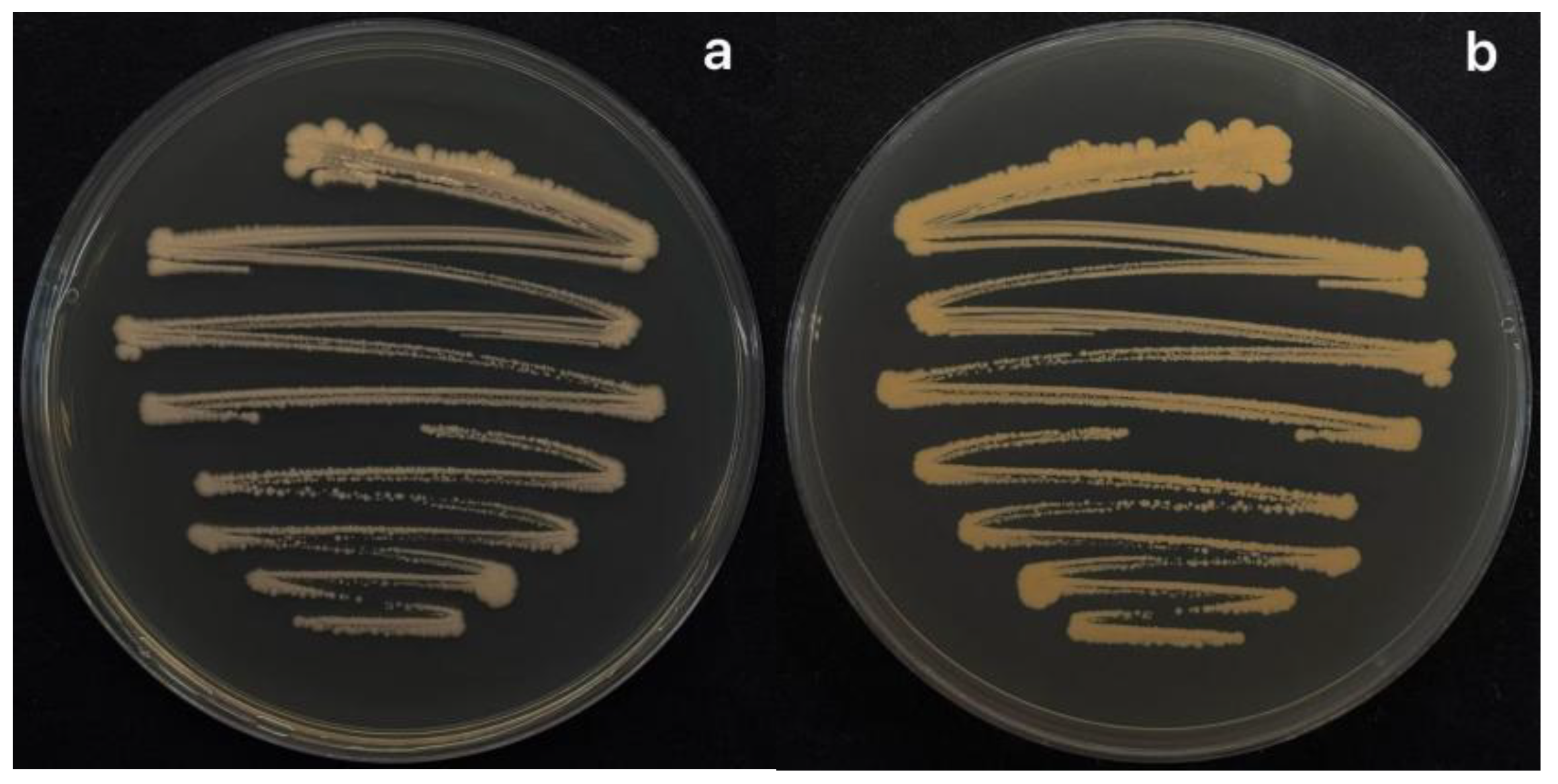
Passage treatment : The initial strain XJ-04 was subcultured, and then the colony was transferred to the plate every 5 days to obtain 1-5 generations of strains;
The fermentation filtrate of the strain obtained by the above treatment was mixed with PDA at a ratio of 1 : 5 to make a plate, and the inhibition rate of S.sclerotiorum was calculated. SPSS software was used to analyze and verify its stability.

3.3.2. Results and analysis

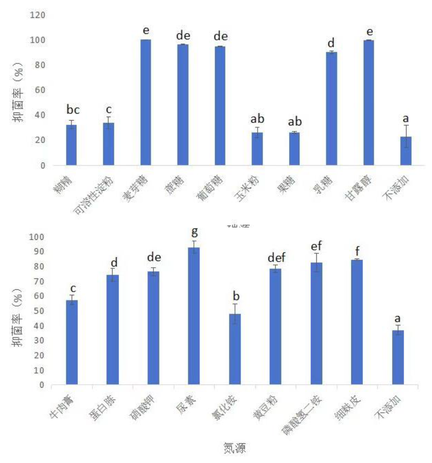
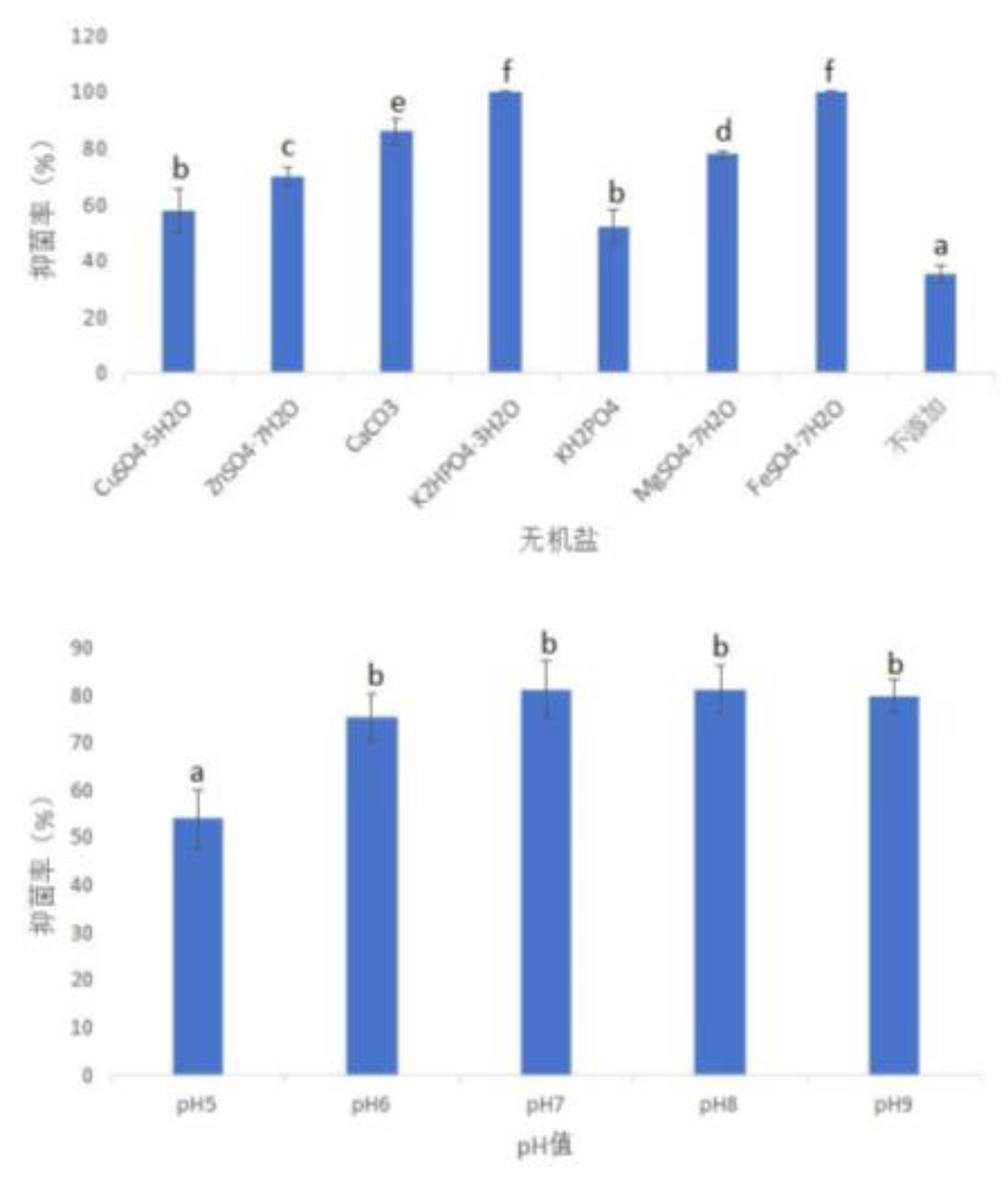
3.3.2.1. Optimization of Fermentation Medium Composition3.3.2.1.1. Single Factor Test

After fermentation with different carbon sources, nitrogen sources, inorganic salts and pH media, the bacteriostatic effect of the fermentation filtrate of strain XJ-04 was different.
Firstly, nine kinds of carbon sources and no additional carbon sources were screened. The results showed that the three carbon sources with the lowest inhibition rate were : no carbon source ( 22.92 %) < corn flour ( 26.01 %) < fructose ( 26.11 %). The three carbon sources with the highest bacteriostatic rate were : maltose ( 100 %) > mannitol ( 99.68 %) > sucrose ( 96.45 %). After fermentation with medium supplemented with maltose, mannitol or sucrose, the antibacterial effect was significantly better than other carbon sources at the P < 0.05 level, and the results are shown in Figure 5.1.
At the same time, 9 nitrogen sources and no additional nitrogen sources were screened. The three nitrogen sources with the lowest bacteriostatic rate were : no nitrogen source ( 36.97 %) < ammonium chloride ( 47.74 %) < beef paste ( 57.24 %) ; the three nitrogen sources with the highest bacteriostatic rate were : urea ( 92.74 %) > fine bran ( 84.43 %) > diammonium hydrogen phosphate ( 82.45 %). After fermentation with urea, fine bran or diammonium hydrogen phosphate, the bacteriostatic rate was significantly higher than that without additional nitrogen source or other nitrogen sources. The results are shown in Figure 5.1.
Figure 5. 1. Inhibition rate of fermentation with different carbon and nitrogen sources.
Preprints 187202 g014
Note: Using SPSS for data analysis, different lowercase letters indicate significant differences at the P < 0.05 level.
After that, seven kinds of inorganic salts and no inorganic salts were screened. The three kinds of inorganic salts with the lowest antibacterial rate were : no inorganic salts ( 34.97 %) < KH2PO4 ( 51.98 %) < CuSO4·5H2O ( 57.77 %) ; the three inorganic salts with the highest inhibition rate were K2HPO4·3H2O ( 100 %) > FeSO4·7H2O ( 99.80 %) > CaCO3 ( 85.89 %). The bacteriostatic rate of the medium added with K2HPO4·3H2O, FeSO4·7H2O or CaCO3 was significantly higher than that of the medium without inorganic salt or other inorganic salt components. The results are shown in Figure 5.2.
Finally, in the screening of five pH values, the antibacterial effect of the fermentation broth was the best when the pH values were 7,8 and 9, and the results are shown in Figure 5.2.
Figure 5. 2. Inhibition rate of fermentation with different inorganic salt and pH.
Preprints 187202 g015
Note: Using SPSS for data analysis, different lowercase letters indicate significant differences at the P < 0.05 level.

3.3.2.1.2. Intuitive Analysis of Orthogonal Test Results of Inhibition Rate of Fermentation Broth

The data obtained by orthogonal test and the results of range analysis showed that ( Table 4-3), the range ( R) of factor D was 24.6, that is, the size of pH value was the primary factor affecting the inhibition rate. Among the components of the medium, the range ( R) of factor C was the largest ( 4.37), so the bacteriostatic rate was most affected by factor C. The range of factor A and factor B were 2.91 and 0.2, respectively. The importance of the influence on the inhibition rate was pH value, inorganic salt, carbon source and nitrogen source. Because diammonium hydrogen phosphate and urea itself has a good inhibitory effect on S.sclerotiorum, so we late fermentation medium optimization selection combination for A3B2C1D3 No.8, namely when the carbon source for sucrose, nitrogen source for fine bran, inorganic salt for K2HPO4·3H2O, pH 9 antibacterial rate is the highest, about 71.75 %, the results are shown in Table 4-2.
Table 4-2. Results of orthogonal test.
Test number Test factor Factor
Inhibition rate(%)

A Carbon source

B nitrogen source

C Inorganic salt
D pH
1 (A1B1C1D1) Maltose Urea K2HPO4·3H2O 7 98.77±2.1362d
2 (A1B2C3D2) Maltose Fine bran CaCO3 8 64.42±2.4606a
3 (A1B3C2D3) Maltose Diammonium hydrogen phosphate FeSO4·7H2O 9 100.00±0.0000d
4 (A2B1C3D3) Mannitol Urea CaCO3 9 99.79±0.1050d
5 (A2B2C2D1) Mannitol Fine bran FeSO4·7H2O 7 47.31±6.3369a
6 (A2B3C1D2) Mannitol Diammonium hydrogen phosphate K2HPO4·3H2O 8 65.59±1.4816b
7 (A3B1C2D2) Sucrose Urea FeSO4·7H2O 8 67.73±2.9556b
8 (A3B2C1D3) Sucrose Fine bran K2HPO4·3H2O 9 71.75±4.5160c
9 (A3B3C3D1) Sucrose Diammonium hydrogen phosphate CaCO3 7 100.00±0.0000d
Note: Using SPSS for data analysis, different lowercase letters indicate significant differences at the P<0.05 level.
Table 4-3. Range analysis results of various factors.
Indicators
Index
Test factor Factor
A Carbon source B Nitrogen source C Inorganic salt D
pH
K1 244.19 262.29 232.11 246.08
K2 208.69 164.48 211.04 197.74
K3 235.48 261.59 245.21 271.54
k1 81.40 87.43 77.37 82.03
k2 69.56 54.83 70.35 65.91
k3 78.49 87.20 81.74 90.51
R 2.91 0.23 4.37 24.6
Note: K1, K2 and K3 are the sum of all levels of each factor; k1, k2 and k3 are the average values of the level of each factor; R value of are the range of k1, k2 and k3.

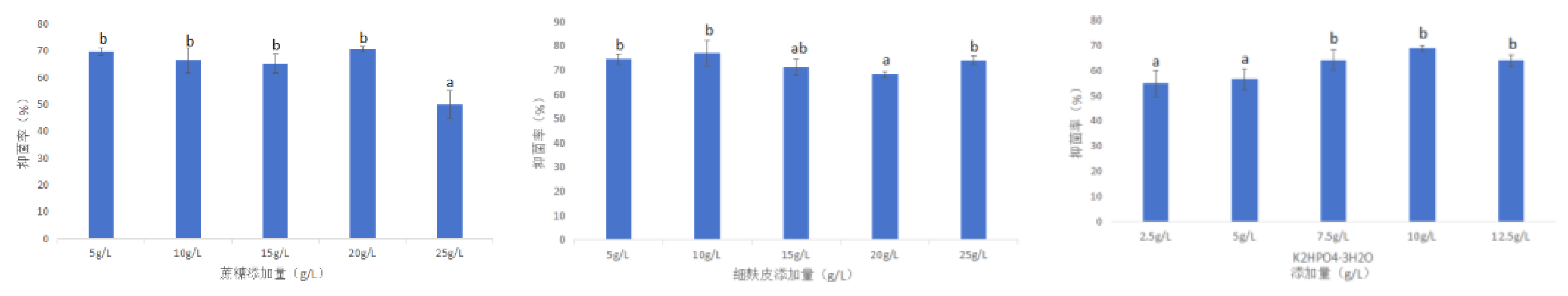
3.3.2.2. Optimization of Fermentation Medium Composition Addition Amount3.3.2.2.1. Single Factor Test

The results showed that the inhibitory effect of strain XJ-04 on S.sclerotiorum was different after fermentation in medium supplemented with 5 g / L ~ 25 g / L sucrose. When the addition amount of sucrose was 20 g / L, the inhibition rate of S.sclerotiorum was the highest ( about 70.41 %). Too little or excessive addition of sucrose would reduce the antibacterial effect of strain XJ-04. The inhibitory effect of 5 g / L ~ 25 g / L fine bran addition on the fermentation filtrate was also significantly different. When the addition amount of fine bran was 10 g / L, the inhibition rate of S.sclerotiorum was the highest ( about 76.95 %). When the addition amount of K2HPO4·3H2O was 2.5 g / L ~ 12.5 g / L, the antibacterial rate of the fermentation broth was also significantly different. When the addition amount of K2HPO4·3H2O was 10 g / L, the inhibition rate of watermelon sclerotiniosis was the highest ( about 68.78 %). Therefore, the optimum addition of sucrose, fine bran and K2HPO4·3H2O in liquid fermentation medium were 20 g / L, 10 g / L and 10 g / L, respectively. The results are shown in Figure 5.3.
Figure 5. 3. The effects of the addition of sucrose, fine bran and K2HPO4·3H2O on the bacteriostatic rate of fermentation broth were studied.
Preprints 187202 g016
Note: Using SPSS for data analysis, different lowercase letters indicate significant differences at the P<0.05 level.

3.3.2.2.2. Response Surface Optimization Experiment

The establishment and significance test of the modelThe No.8 medium was selected, the inhibition rate ( Y) was used as the response value, the amount of sucrose added ( X1), the amount of fine bran added ( X2), and the amount of K2HPO4·3H2O added ( X3) were used as the investigation factors [28,29].At the level of-1, 0, and 1 representative variables ( Table 4-4), the Design Expert 8.0.6 was used to design the response surface optimization test of 3 factors and 3 levels to determine the optimal addition combination. The response surface optimization test data results are shown in Table 4-5.
Table 4-4. Variables and level of Box-Behnken design.
Horizontal
Level
Factor
X1Sucrose addition ( g / L) X2The amount of fine bran added ( g / L) X3The addition of K2HPO4·3H2O ( g / L)
-1 15 5 7. 5
0 20 10 10
1 25 15 12.5
Table 4-5. The results of response surface optimization test for strain XJ-04 fermentation.
Code Factors and levels YInhibition rate(%)
X1 (g / L) X2 (g / L) X3 (g /L)
1 15.00 5.00 10.00 74.87
2 25.00 5.00 10.00 76.16
3 15.00 15.00 10.00 73.96
4 25.00 15.00 10.00 74.57
5 15.00 10.00 7.50 75.60
6 25.00 10.00 7.50 76.64
7 15.00 10.00 12.50 75.69
8 25.00 10.00 12.50 76.42
9 20.00 5.00 7.50 76.48
10 20.00 15.00 7.50 74.05
11 20.00 5.00 12.50 75.02
12 20.00 15.00 12.50 75.85
13 20.00 10.00 10.00 77.85
14 20.00 10.00 10.00 77.92
15 20.00 10.00 10.00 78.12
16 20.00 10.00 10.00 77.87
17 20.00 10.00 10.00 77.75
Use Design expert V8.0.6.1 software to fit the scores in Table 3-4, and get the regression equation: Y = 77.9 + 0.46X1 - 0.51X2 + 0.026X3 - 0.17X1X2 - 0.078X1X3 + 0.82X2X3 - 1.14X12 - 1.87X22 - 0.68X32. It can be concluded from the equation that in this model, the inhibition rate of fermentation broth was most affected by the addition amount of nitrogen source fine bran, followed by the addition amount of sucrose, and was least affected by the addition amount of K2HPO4·3H2O.
Analysis of variance and significance of the above inhibition rate results showed that the F value of the established model was 116.70, and P < 0.0001, indicating that the model was significant. X12, X22, X32 are extremely significant items, X1X2, X1X3, X2X3 are not significant items. The F value of the mismatch term is 2.34, P ( = 0.2149) > 0.05, which means that the mismatch term is not significant relative to the error term, indicating that the model has high reliability. The results are shown in Table 4-6.
Table 4-6. Variance analysis of response surface experiments results.
Source Sum of Squares Degrees of freedom Mean Square F Value P-value Prob > F Significance
Model 30.86 9 3.43 116.70 <0.0001 **
X1 1.68 1 1.68 57.31 0.0001
X2 2.10 1 2.10 71.52 <0.0001
X3 0.0055 1 0.0055 0.19 0.6779
X1X2 0.12 1 0.12 3.93 0.0877
X1X3 0.024 1 0.024 0.82 0.3959
X2X3 2.66 1 2.66 90.43 <0.0001
X12 5.45 1 5.45 185.36 <0.0001 **
X22 14.80 1 14.80 503.71 <0.0001 **
X32 1.93 1 1.93 65.73 <0.0001
Residual 0.21 7 0.029
Lack of Fit 0.13 3 0.044 2.34 0.2149
Pure Error 0.075 4 0.019
Cor Total 31.06 16
Note: Using Design Expert software for data analysis, “*”indicated significant impact on the results (0.01<P<0.05); “**”indicated that the impact on the results was extremely significant (P<0.01).

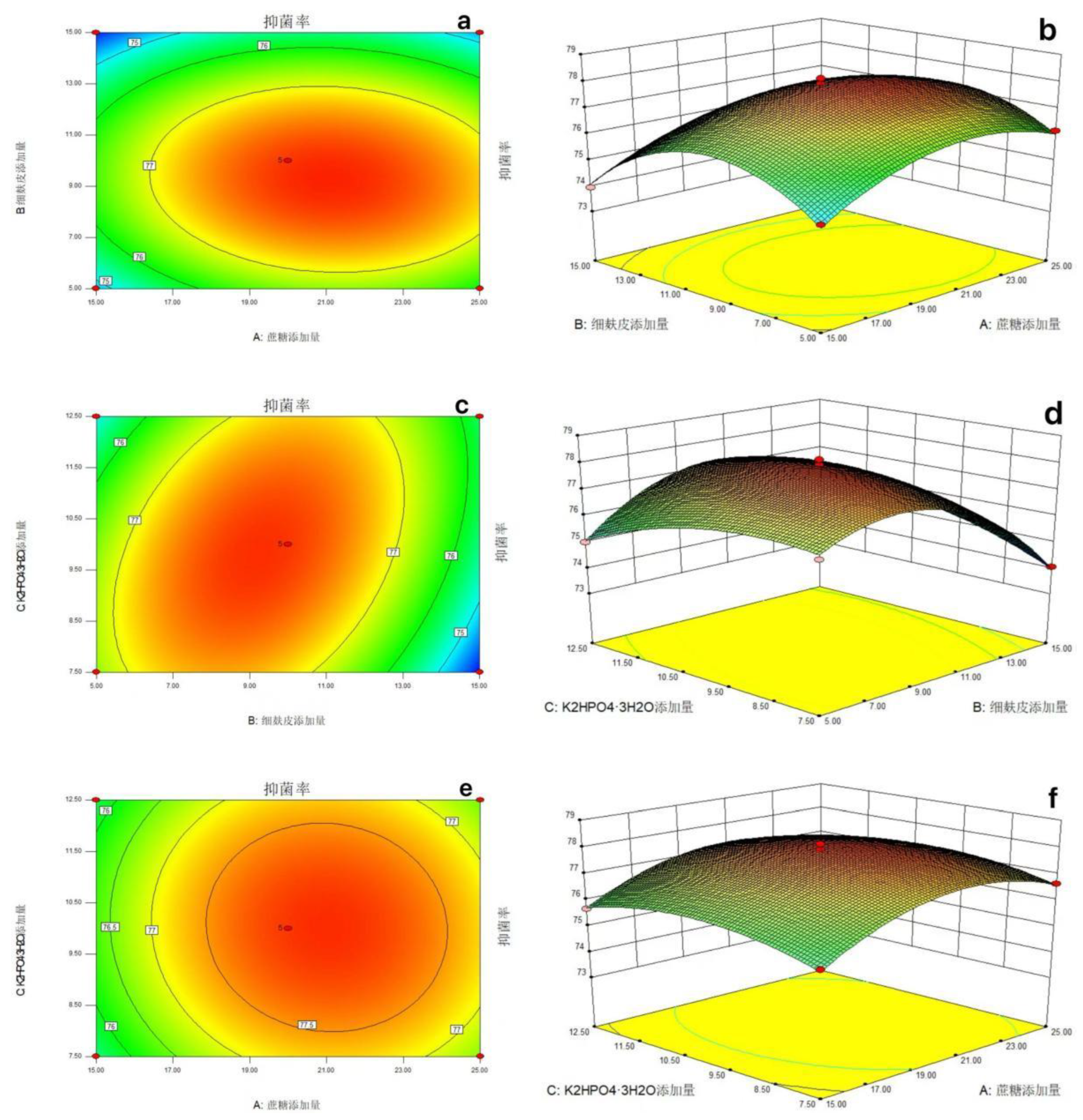
3.3.2.2.3. Response Surface Curve Analysis

The response surface curve and contour line of the interaction effect of sucrose, fine bran and K2HPO4·3H2O addition on the inhibition rate were shown in Figure 5.4.
Figure 5.4-a, b is the interaction between the amount of sucrose added and the amount of fine bran added when the amount of K2HPO4·3H2O added is at the central level, that is, 10 g / L, indicating that with the increase of the amount of sucrose added and the amount of fine bran added, the bacteriostatic rate shows a trend of increasing first and then decreasing. Figure 5.4-c, d is the interaction between the amount of fine bran and the amount of K2HPO4·3H2O added when the amount of sucrose added is at the central level, that is, 20 g / L. It shows that with the amount of fine bran added and the amount of K2HPO4·3H2O added, the bacteriostatic rate shows a trend of increasing first and then decreasing. Figure 5.4-e, f shows the interaction between sucrose addition and K2HPO4·3H2O addition at the central level of fine bran addition ( 10 g / L). The results showed that the inhibition rate increased first and then decreased with the increase of sucrose addition and K2HPO4·3H2O addition.
Figure 5. 4. Contour and 3D surface maps of interactions between factors.
Preprints 187202 g017
The software was used to predict the maximum response value. The sucrose addition amount of each factor combination in the model was about 21.08 g / L, the fine bran addition amount was about 9.17 g / L, the K2HPO4·3H2O addition amount was 9.77 g / L, and the inhibition rate was about 77.99 %. According to the optimal composition combination given by the model, the experiment was carried out, and the bacteriostatic rate was 77.78 %, which was similar to the predicted value. It was proved that the model was effective, which was 7.66 % higher than the bacteriostatic rate of 70.12 % fermented by PDB medium before optimization. According to the best combination of added components given by the model, the experiment on the effect of sclerotia germination was carried out again. The method was the same as 3.1.2.2, and the inhibition rate of sclerotia germination was 64 %, which was 4 % higher than that before optimization. The results are shown in Figure 5.5 and Table 4-7.
Figure 5. 5. Comparison of inhibition rate between initial (a) and optimized (b) fermentation.
Preprints 187202 g018
Table 4-7. The effect of optimized fermentation broth of strain XJ-04 on sclerotia germination.
Treatment Number of sclerotia germination in 3d Inhibition rate(%)
Fermentation supernatant 9 64
CK 25 /

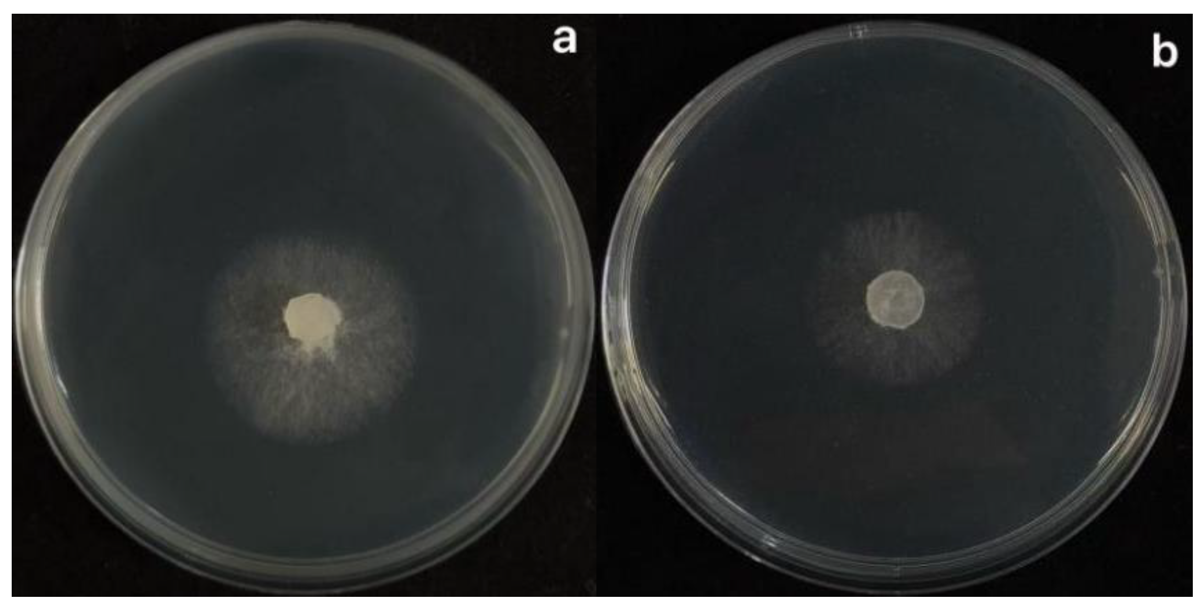
3.3.2.3. Fermentation Condition Optimization

By studying the antibacterial effect of different fermentation time, different liquid volume and different fermentation temperature of XJ-04 strain fermentation broth on S.sclerotiorum, it was found that the antibacterial rate showed a trend of increasing first and then decreasing. When the fermentation time was 3 d, the antibacterial rate was the highest, about 76.16 %, which was significantly better than 1 d, 2 d, 4 d and 5 d. When the medium volume was 100 mL ( 250 mL conical flask), the bacteriostatic rate was the highest, about 76.43 %, which was significantly better than 50 mL, 75 mL, 125 mL and 150 mL. When the fermentation temperature was 30 ℃, the bacteriostatic rate was the highest, about 78.35 %, which was significantly better than other temperatures. The results showed that the XJ-04 strain had the highest antibacterial rate when the fermentation temperature was 30 ℃, the fermentation time was 3d, and the medium volume was 100 mL. The results are shown in Figure 5.6.
Figure 5. 6. After different fermentation time, liquid volume and temperature treatment, the inhibition rate of fermentation broth was determined.
Preprints 187202 g019
Note: Using SPSS for data analysis, Different lowercase letters indicate significant differences at the P<0.05 level.

3.3.2.4. Study on the Stability of Fermentation Broth of Strain XJ-04

(1)
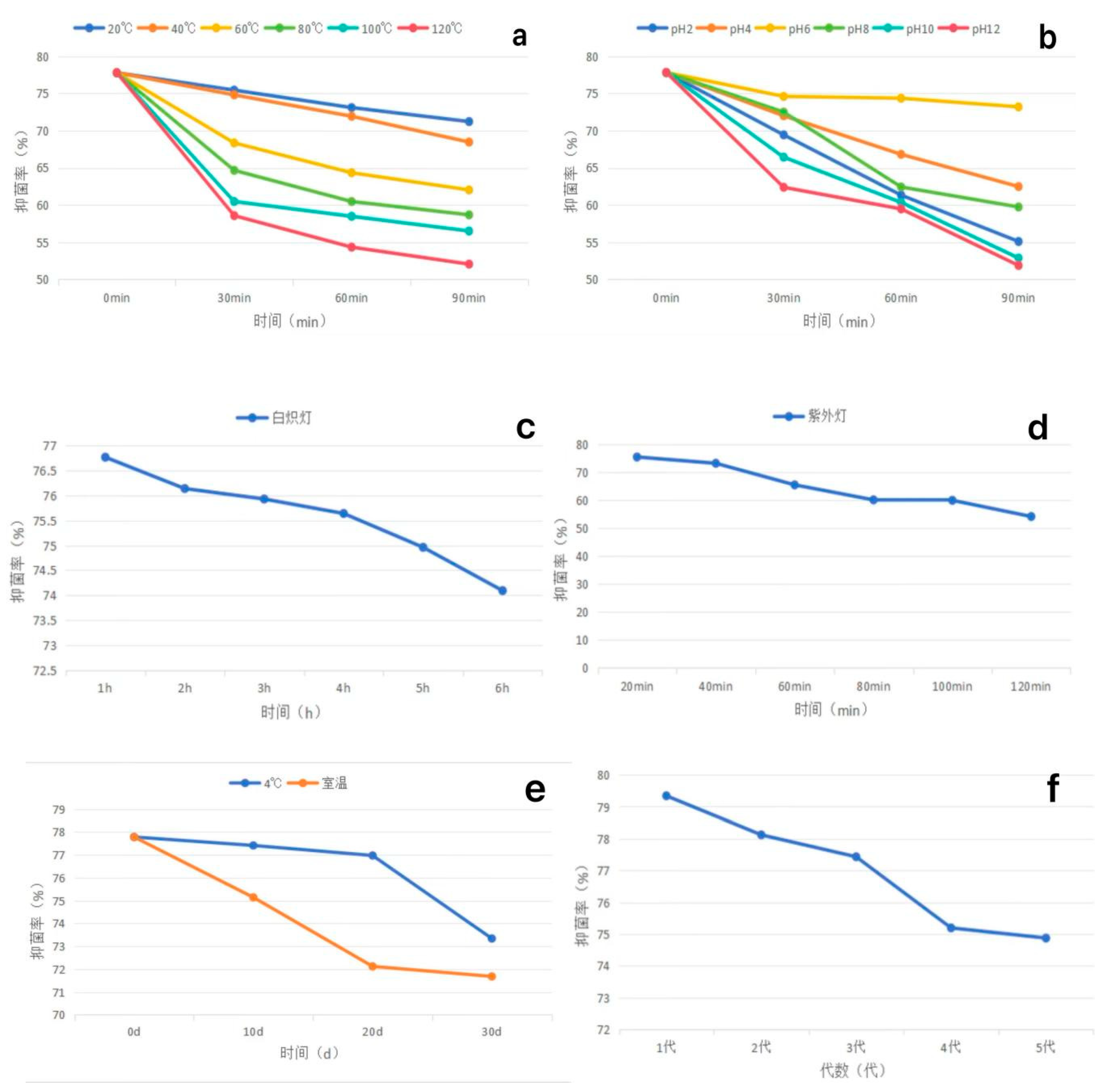
Thermal stability : The fermentation broth of strain XJ-04 was treated at 20 ℃, 40 ℃, 60 ℃, 80 ℃ water bath for 30 min, 60 min and 90 min, and the bacteriostatic rate decreased by less than 15 %. Treatment at 100 ℃ and 120 ℃ for 60 min and 90 min had a great influence on the inhibition rate, and the inhibition rate decreased by nearly 20 %. Among them, there was no significant difference in the bacteriostatic rate between the 20 ℃ treatment for 30 min and the 40 ℃ treatment for 30 min and the control group ( without water bath heat treatment). It shows that the fermentation broth has good heat resistance within 80 ℃. The results are shown in Figure 5.7-a.
(2)
Acid-base stability : After the fermentation broth was treated with acid and alkali, compared with the original environment ( pH 6), the acid or alkali environment had a greater impact on the antibacterial effect of the fermentation broth. When the pH value was in the range of 2~10, the inhibition rate could still reach more than 60 % after treatment for 60 min. However, the inhibition rate was less than 60 % in the environment of pH 12, and the environment of acid and alkali had a great influence on the inhibition rate. The results are shown in Figure 5.7-b.
(3)
Light stability : Incandescent lamp irradiation had no effect on the fermentation broth, and the antibacterial rate did not change significantly after 6 h of irradiation. There was no significant difference in the bacteriostatic rate within 40 min of ultraviolet light irradiation. When the irradiation time was more than 60 min, the bacteriostatic rate was significantly different from that of the control group, but the bacteriostatic rate remained above 50 %. The results showed that the fermentation broth of XJ-04 strain had good light stability, and the results were shown in Figure 5.7-c and d.
(4)
Storage stability : There was no significant difference in the inhibition rate within 10-20 days of storage at 4 °C. The bacteriostatic rate decreased by 4.44 % at 30 d. There was no significant difference in the inhibition rate after 10 days of storage at room temperature. The inhibition rate decreased by 5.66 % after 20 days of storage and decreased by 6.10 % after 30 days of storage. The results are shown in Figure 5.7-e.
(5)
Passage stability : The stability test of the fermentation filtrate was carried out. The results showed that the antibacterial activity of the fermentation broth of different generations of XJ-04 strain was small. The antibacterial rate of the fermentation broth of the fifth generation of XJ-04 strain remained above 70 %, and the antibacterial rate was as high as 74.87 %, indicating that the strain had good passage stability. The results are shown in Figure 5.7-f.
Figure 5. 7. The inhibition rate of fermentation broth of different generations of strains was changed after treatment with different temperature, pH value, light and storage time. a: Temperature; b: pH; c: Ultraviolet Lamp; d: Incandescent Lamp; e: Storage time; f: Generations
Preprints 187202 g020
Note: Using SPSS for data analysis, different lowercase letters indicate significant differences at the P < 0.05 level.

3.3.3. Summary

(1)
The inhibition rate was used as the main consideration index. The addition components of the fermentation medium of strain XJ-04 were optimized by single factor test and orthogonal test. The No.8 medium ( sucrose, fine bran, K2HPO4·3H2O, pH 9) was the best component combination, and the inhibition rate was about 77.78 %.
(2)
The optimal addition parameters of each component of the fermentation medium were determined by the response surface test method and the maximum response value was predicted. It was predicted that when the sucrose addition amount was 21.08 g / L, the fine bran addition amount was 9.17 g / L and the K2HPO4·3H2O addition amount was 9.77 g / L, the inhibition rate was up to about 77.99 %. The bacteriostatic rate was 77.78 %, which was similar to the predicted value, and was about 7.66 % higher than the bacteriostatic rate before optimization. The experiment on the effect of sclerotia germination was carried out, and the inhibition rate of sclerotia germination was 64 %, which was 4 % higher than that before optimization.
(3)
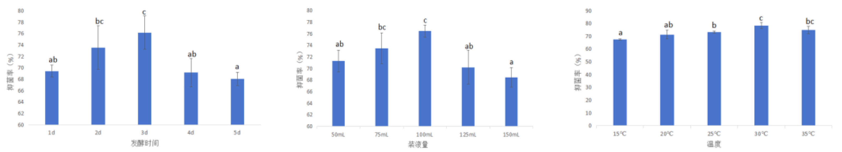
The optimum fermentation conditions of strain XJ-04 were determined as follows : fermentation time was 3 d, liquid volume was 100 mL, fermentation temperature was 30 ℃.
(4)
The fermentation broth of strain XJ-04 had good thermal stability, acid-base stability, light stability and storage stability. At the same time, strain XJ-04 had good passage stability.

3.4. Study on the Growth-Promoting Effect of Strain XJ-04 Fermentation Broth on Watermelon and In Vitro Culture of Leaves

In this chapter, the effects of strain XJ-04 fermentation broth on watermelon were determined by watermelon seed germination and seedling plant height, root length, base diameter, fresh weight and dry weight. The potential of biocontrol application of strain XJ-04 was further confirmed by leaf culture in vitro, aiming to provide a better theoretical and practical basis for the better use of biocontrol technology to control watermelon sclerotiniose.

3.4.1. Test Materials and Methods

3.4.1.1. Test Material3.4.1.1.1. Test Strains and Plant Materials

Strain XJ-04, S.sclerotiorum.
Tested watermelon seeds : Jilin Shengshi Seed Industry Co., Ltd.

3.4.1.1.2. Main Reagents and Equipment

Sodium hypochlorite, CT15E desktop micro high-speed centrifuge.

3.4.1.2. Test Method3.4.1.2.1. Determination of Growth-Promoting Effect of Fermentation Broth of Strain XJ-04

A total of 350 watermelon seeds with full particles and uniform size were selected and divided into 7 groups after disinfection and placed in petri dishes. According to the ratio of strain XJ-04 fermentation broth : water = 1 : 5,1 : 10,1 : 20,1 : 50,1 : 75,1 : 100, the seeds were soaked in petri dishes for 10 h, and the seeds soaked in sterile water were used as the control. The seeds were cultured in darkness at 25 ℃, and the seeds were planted in the seedling pot after germination. The number of seed germination was recorded from 1 d to 7 d, and the seed germination rate and germination potential were calculated according to the following formula. After 30 d, the plant height, root length, base diameter, fresh weight and dry weight were measured.
Germinating   energy = The   number   of   seeds   germinated   at   the   peak   of   germination Total   number   of   kernels × 100 %
Percentage   of   germination = Number   of   germinated   seeds Total   number   of   kernels × 100 %

3.4.1.2.2. Leaf Culture In Vitro

Watermelon sclerotinia can occur in the aboveground part of watermelon during the whole growth period. Watermelon stem, petiole, tendril, flower and fruit can be damaged, mainly causing leaf and stem death and fruit decay. Watermelon seeds were disinfected with 1 % sodium hypochlorite solution, soaked in darkness at 25 ℃ for 12 h, and germinated. When the buds grew to about 1 ~ 2 cm, they were sown in a seedling pot. After growing for a period of time, the watermelon leaves with good growth and consistent size were selected and cut off. First, they were washed with tap water, and then soaked in 75 % alcohol and sterile water for 2 min. They were fully washed and placed in a sterile ultra-clean bench to dry. The sterile fermentation filtrate of strain XJ-04 and sterile water were configured in different proportions to 1 : 1,1 : 5,1 : 10,1 : 15 and 1 : 20, and the leaves were placed in a prepared sterile tray. A layer of wet cotton was laid on the bottom of the tray, and 100 μL of sterile filtrate with different dilutions was sucked by a pipette gun and beaten on the surface of watermelon leaves. The same amount of sterile water was placed on the surface of the leaves as a blank control. The liquid on the surface of the leaves was dried and inoculated with S.sclerotiorum, and 3 replicates were set. Placed in a 25 ℃ constant temperature light incubator. After 7 days, the incidence of the leaves was observed, the lesion diameter was measured and the inhibition rate was calculated [30,31].

3.4.2. Results and Analysis

3.4.2.1. Effects of Fermentation Broth of Strain XJ-04 on Seed Germination and Seedling Growth of Watermelon

The fermentation broth of strain XJ-04 was diluted into different proportions with sterile water, and the seeds were soaked to determine its growth-promoting effect. After the watermelon seeds were treated with the fermentation broth of strain XJ-04, the effects of different proportions of fermentation broth on the germination of watermelon seeds were different. With the increase of fermentation broth concentration, the promotion effect on seed germination increased first and then decreased. The ratio of fermentation broth to water was 1 : 50, and the germination potential was 96 %, which was higher than 85 % of the control group. The results are shown in Table 5-1.
Table 5-1. Effects of fermentation broth of strain XJ-04 on seed germination of watermelon.
Code Fermentation broth : Water Germination potential(%) Germination rate(%)
A 1:5 60 87
B 1:10 72 90
C 1:25 67 82
D 1:50 89 96
E 1:75 87 90
F 1:100 70 92
G CK 65 85
After 30 days, the biomass of watermelon seedlings was counted. The results showed that the fermentation broth of strain XJ-04 promoted the plant height, root length, base diameter, fresh weight and dry weight of watermelon seedlings at low concentration and inhibited at high concentration. When the ratio of fermentation broth to water was 1:50, the plant height and root length of watermelon seedlings were significantly higher than those of other groups. When the dilution ratio of fermentation broth was 1:25, the basal diameter of watermelon seedlings was significantly wider than that of the control group, which was 5.31 % wider than that of the control group. When the dilution ratio of the fermentation broth was 1:50 and 1:75, the fresh weight of watermelon seedlings was significantly higher than that of the control group, and the fresh weight of the plant was the highest when the dilution ratio was 1:50, which was 53.57 % higher than that of the control group. When the dilution ratio was 1:75, the dry weight of the plant was the largest, followed by the dilution ratio of 1:50, which increased by 0.1g and 0.07g respectively compared with the control group. Through the analysis of the results, it can be concluded that this group of treatments has a significant effect on the growth of watermelon seedlings. When the concentration of the fermentation broth was high, it had a certain inhibitory effect on seed germination and seedling growth, which may be due to the presence of antibacterial substances in the fermentation filtrate to affect seed growth. The results are shown in Table 5-2 and Figure 6.1.
Figure 6. 1. Effects of fermentation broth of strain XJ-04 on growth of watermelon seedlings Fermentation broth : Water = 1:100;1:75;1:50;1:25;1:10;1:5;CK.
Preprints 187202 g021
Table 5-2. Effects of fermentation broth of strain XJ-04 on growth of watermelon seedlings.
Fermentation broth : Water Plant height(mm) Root length(mm) Stem diameter(mm) Fresh weight(g) Dry weight(g)
1:100 (A) 135.50±3.88c 46.72±3.34ab 3.47±0.19a 1.28±0.10ab 0.11±0.02b
1:75 (B) 130.19±6.03bc 49.25±1.19bc 3.47±0.25a 1.71±0.10d 0.16±0.03c
1:50 (C) 137.67±2.91c 53.37±1.43c 3.46±0.08a 1.72±0.09d 0.13±0.03bc
1:25 (D) 136.96±8.56c 50.09±1.51bc 3.57±0.09a 1.45±0.07bc 0.08±0.02ab
1:10 (E) 121.33±2.17ab 47.88±2.49ab 3.49±0.29a 1.55±0.15cd 0.10±0.02ab
1:5 (F) 114.80±13.03a 45.91±3.83ab 3.42±0.23a 1.17±0.12a 0.09±0.04ab
CK (G) 116.44±8.00a 43.77±2.57a 3.39±0.23a 1.12±0.19a 0.06±0.02a
Note: Using SPSS for data analysis, different lowercase letters indicate significant differences at the P<0.05 level.

3.4.2.2. Leaf In Vitro Culture Experiment
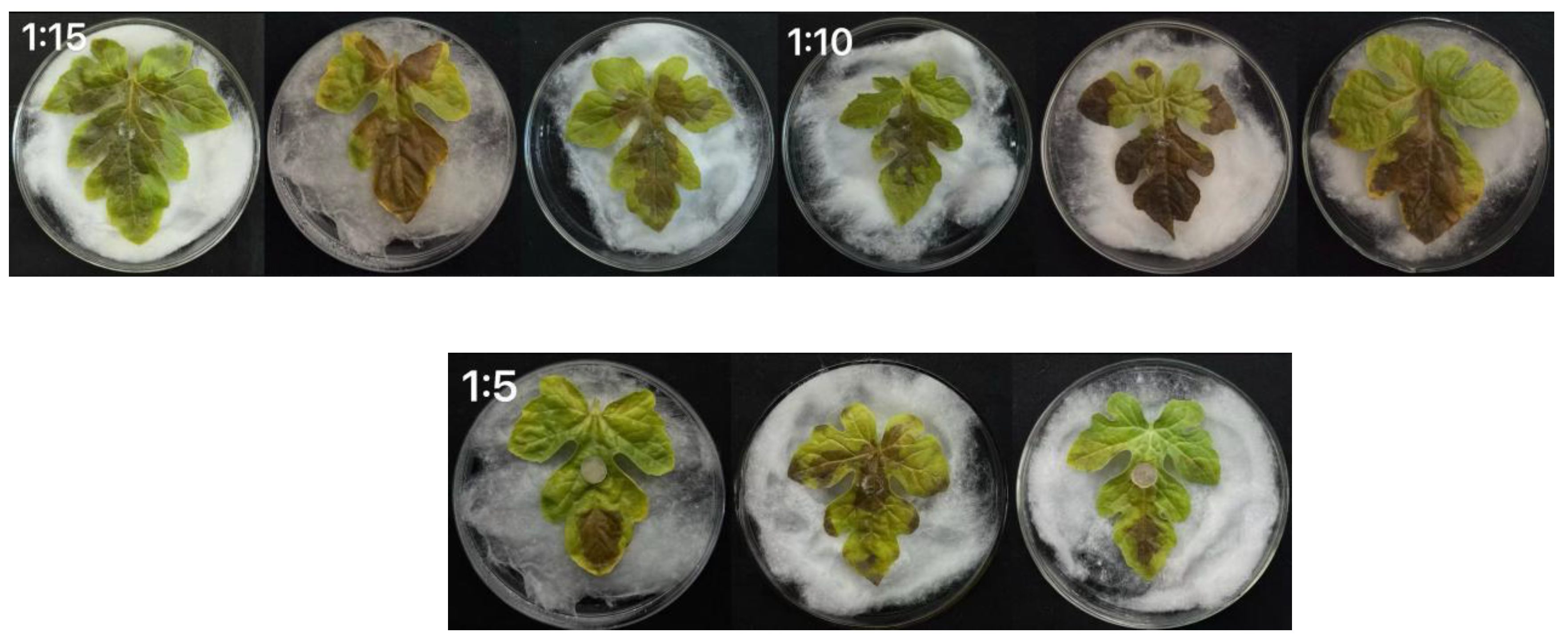
The healthy and disease-free watermelon leaves with good growth and consistent size were collected, and the leaves were cultured in vitro with the fermentation broth of strain XJ-04. The watermelon leaves in the control group showed dark brown lesions, and the lesion area was large. When the fermentation broth : water was 1:5 and 1:10, the bacteriostatic effect was the most obvious. Compared with other groups, the lesion of the control group was significantly reduced. When the fermentation broth : water is 1:15 and 1:20, the fermentation broth can also inhibit the size of the lesion to a certain extent, and the greater the concentration of the fermentation broth, the smaller the lesion. Through the experiment, the disease spots of watermelon leaves treated with the fermentation filtrate of strain XJ-04 were smaller than those without treatment, indicating that the fermentation broth of strain XJ-04 had a good control effect on watermelon sclerotiniosis. The results are shown in Table 5-3 and Figure 6.2.
Figure 6. 2. Antifungal effect of fermentation broth of strain XJ-04 on detached leaves of watermelon Sclerotinia sclerotiorum.
Preprints 187202 g022
Table 5-3. The lesion diameter of watermelon sclerotiniose after the leaves were treated with the fermentation broth of strain XJ-04.
Code Fermentation broth : Water Lesion diameter(mm)
A 0:1 54.26±1.5245e
B 1:20 49.61±0.5823d
C 1:15 44.66±0.8426c
D 1:10 33.84±1.6038b
E 1:5 20.07±1.0758a
Note: Using SPSS for data analysis, Different lowercase letters indicate significant differences at the P<0.05 level.

3.4.2.3. Summary

(1)
When the ratio of strain XJ-04 fermentation broth to water was 1:50, the germination rate and germination potential of watermelon seeds and the plant height and root length of watermelon seedlings were significantly improved. When the dilution ratio of the fermentation broth was 1:25, the basal diameter of the seedlings was significantly widened. When the dilution ratio of the fermentation broth was 1:50 and 1:75, the fresh weight of the seedlings was significantly increased.
(2)
The in vitro experiment of watermelon leaves showed that the fermentation broth of strain XJ-04 had a good control effect on watermelon sclerotiniosis.

Author Contributions

Conceptualization, Lingyue Zhu.; methodology, Lingyue Zhu.; software, Lingyue Zhu.; validation, Lingyue Zhu..; formal analysis, Lingyue Zhu.; investigation, Lingyue Zhu.; resources, Xiaomei Wang; data curation, Lingyue Zhu; writing—original draft preparation, Lingyue Zhu.; writing—review and editing, Xiaomei Wang.; visualization, Xiaomei Wang; supervision, Xiaomei Wang; project administration, Xiaomei Wang; funding acquisition, Xiaomei Wang. All authors have read and agreed to the published version of the manuscript.

Funding

“This research was funded by the National KeyResearch and Development Project,grant number 2016YFD0201004:2018YFD0201206:2016YFD0200708. Jilin province modern agricultural industry technology system construction demonstration project,grant number JLARS-2025-120206”. and “The APC was funded by Lingyue Zhu”.

Institutional Review Board Statement

Not applicable.

Informed Consent Statement

Not applicable.

Data Availability Statement

The raw data supporting the conclusions of this article will be made available by the authors on request.

Conflicts of Interest

The authors declare no conflicts of interest.

Abbreviations

The following abbreviations are used in this manuscript:

References

  1. BOLTON M D, THOMMA B P, NELSON B D. Sclerotinia sclerotiorum(Lib.) de Bary: biology and molecu- lar traits of a cosmopolitan pathogen[J]. Molecular Plant Pathology, 2006, 7(1): 1. [CrossRef]
  2. Yang Qingpo, Liu Wancai, Huang Chong, et al.. Statistics and Analysis of Major Diseases and Insect Pests in Rape in Recent Ten Years [J]. Plant Protection, 2018,44: 24. [CrossRef]
  3. SHARMA P, MEENA P D, VERMA P R, et al.Sclerotinia sclerotiorum (Lib) de Bary causing sclerotinia rot in oilseed Brassicas: a review[J]. Journal of Oilseed Brassica, 2015, 6(S): 1.
  4. WILLBUR J, MCCAGHEY M, KABBAGE M, et al.An overview of the Sclerotinia sclerotiorum pathosystem in soybean: impact, fungal biology, and current management strategies[J]. Tropical Plant Pathology, 2019, 44(1):3. [CrossRef]
  5. Derbyshire, M.C.; Denton-Giles, M. The control of sclerotinia stem rot on oilseed rape (Brassicanapus): current practices and future opportunities. Plant Pathology. 2016, 65, 859–887. [Google Scholar] [CrossRef]
  6. Liu Yun-hui. Existing Problems, Application Status and Development of Biological Control Technology [J]. Journal of Seed Industry, 2019,06: 14-16.
  7. Fangzhongda. Methods of Plant Disease Research, 3rd Edition [M]. China Agriculture Press, 2007: 46-50.
  8. Samson, R.A.; Visagie, C.M.; Houbraken, J.J.; et al. Phylogeny, identification and nomenclature of the genus Aspergillus[J]. Studies in Mycology, 2014, 78, 141–173. [Google Scholar] [CrossRef]
  9. Visagie, C.M.; Houbraken, J.; Frisvad, J.C.; et al. Identification and nomenclature of the genus Penicillium[J]. Studies in Mycology, 2014, 78, 343–371. [Google Scholar] [CrossRef]
  10. Houbraken, J.; Visagie, C.M.; Meijer, M.; et al. A taxonomic and phylogenetic revision of Penicillium section Aspergilloides[J]. Studies in Mycology, 2014, 78, 373–451. [Google Scholar] [CrossRef] [PubMed]
  11. Amein, T.; Omer, Z.; Welch, C. Application and evalution of Pseudomonas strains for biocontrol of wheat seedling blight[J]. Crop Protection, 2008, 27, 532–536. [Google Scholar] [CrossRef]
  12. Zhou Xiangping, Shu Cuihua, Teng Kai, et al.. Identification of Endophytic Bacillus Starch Xe01 and Optimization of Fermentation Conditions [J]. Chinese Tobacco Science, 2020,: 58-67.
  13. Zhang Kecheng, Ran Longxian, et al. Isolation and Identification of Antagonistic Actinomycetes from Apple Rotting Bacteria [J]. Hebei Forest Fruit Research. 2008, 23, 5.
  14. Yasmin Z, Shamsi S. In vitro screening of fungicides and plant extracts against Colletotrichum gloeosporioides(Penz.) Rauwolfia serpentina(L.) Sacc. the causal agent of anthracnose disease of Sacc. the causal agent of anthracnose disease of Benth Ex Kurz[J]. Benth Ex Kurz[J]. Journal of The Asiatic Society of Bengal, 2019, 45(1): 35-43.
  15. Bacterial Classification Group., Institute of Microbiology, Chinese Academy of Agricultural Sciences Common Identification Methods of General Bacteria [M]. Science Press, 1978.
  16. R.E. Buchanan, N.E. R.E. Buchanan, N.E. Gibbons et al.. Berger Bacteria Identification Manual (8th Edition) [M]. Science Press, 1984.
  17. Dongxiuzhu, Cai Miaoying. Manual for Identification of Common Bacterial Systems [M]. Science Press, 2001.
  18. Hajjaj, H.; Niederberger, P.; Duboc, P. Lovastatin biosynthesis by Aspergillus terreus in a chemically defined medium[J]. Applied and Environmental Microbiology, 2001, 67, 2596–2602. [Google Scholar] [CrossRef] [PubMed]
  19. López, J.C.; Pérez, J.S.; Sevilla, J.F.; et al. Fermentation optimization for the production of lovastatin by Aspergillus terreus: use of response surface methodology[J]. Journal of Chemical Technology & Biotechnology Biotechnology, 2010, 79, 1119–1126. [Google Scholar]
  20. Song Xiao-feng, former Zengyan, Li Peng-xu, et al. Optimization of Extraction and Purification of Total Flavonoids from Hawthorn Leaves by Box-Behnken Design and Study on Their Antibacterial Activity [J]. Henan Science, 2018,36: 6.
  21. Wei, W.; Yuan, T.; Wang, K.; et al. Statistical optimization of cellulase production by the brown rot fungi, Fomitopsis palustris, and its application in the enzymatic hydrolysis of LHW-pretreated woody biomass[J]. Process Biochemistry, 2012, 47, 2552–2556. [Google Scholar]
  22. Guowei, S.; Chunju, B.; He, C.; et al. Fermentation optimization of goat milk with Lactobacillus acidophilus and Bifidobacterium bifidum by Box-Behnken design[J]. Acta Scientiarum Polonorum Technologia Alimentaria, 2016, 15, 151–159. [Google Scholar]
  23. Dertli, E.; Toker, O.S.; Durak, M.Z.; et al. Development of a fermented ice-cream as influenced by in situ exopolysaccharide production: Rheological, molecular, microstructural and sensory characterization[J]. Carbohydrate Polymers, 2016, 136, 145–151. [Google Scholar] [CrossRef] [PubMed]
  24. Chen Xi, Li Guolin, Chen Mengyu, et al.. Optimization of fermentation conditions of blueberry vinegar by response surface methodology [J]. Brewing in China 2018, 37, 67–71.
  25. Wang Qiang, Huang Meigui, Xie Yuejie, et al.. Optimization of Extraction Process of Hydroxytyrosol from Olive Pomace by Response Surface Methodology [J]. Food Industry Technology, 2018,39: 145-151.
  26. Morita, K.; Nishijima, Y.; Kana, T. Chemical nature of a desmutagenic factor from burdock (Arctium lappa Linne)[J]. Agricultural and Biological Chemistry, 1985, 49, 925–932. [Google Scholar] [CrossRef]
  27. Wang Wei. Biological Characteristics of Sclerotinia Sclerotiorum and Screening of Control Agents [D]. Sichuan Agricultural University, 2012.
  28. Chen Xi, Li Guolin, Chen Mengyu, et al.. Optimization of fermentation conditions of blueberry vinegar by response surface methodology [J]. Brewing in China, 2018,37: 67-71.
  29. Wang Qiang, Huang Meigui, Xie Yuejie, et al.. Optimization of Extraction Process of Hydroxytyrosol from Olive Pomace by Response Surface Methodology [J]. Food Industry Technology, 2018,39: 145-151.
  30. Kulandaivelu, V.; Radha, P. Trichoderma–azotobacter Biofilm-based Formulation Enhance Natural Plant Defense Enzyme Activities in Wheat and Cotton Seedlings[J]. National Academy Science Letters, 2023, 47, 61–64. [Google Scholar]
  31. Emily R, T M M. Data on Plant Defense Enzyme Activity Associated with Three Endophytes Against Cornus Florida Erysiphe Pulchra Powdery Mildew[J]. Data in Brief, 2023, 48: 109220-109220.
Table 1. Inhibitory effect of five soil bacteria on S.sclerotiorum.
No.
Code
Inhibitory zone(Mm)
Inhibition zone
Rescreening inhibition rate(%)
Inhibition rate of secondary screening
XJ-04 5.21±0.2450c 70.12±0.6710d
LY-15 4.93±0.1652c 69.32±3.2216d
SY-27 3.76±0.4328a 50.32±0.6727a
XG-69 4.55±0.2658b 59.63±3.9452b
JH-105 4.68±0.1747ab 64.60±0.8278c
Disclaimer/Publisher’s Note: The statements, opinions and data contained in all publications are solely those of the individual author(s) and contributor(s) and not of MDPI and/or the editor(s). MDPI and/or the editor(s) disclaim responsibility for any injury to people or property resulting from any ideas, methods, instructions or products referred to in the content.
Copyright: This open access article is published under a Creative Commons CC BY 4.0 license, which permit the free download, distribution, and reuse, provided that the author and preprint are cited in any reuse.
Prerpints.org logo

Preprints.org is a free preprint server supported by MDPI in Basel, Switzerland.

Subscribe

Disclaimer

Terms of Use

Privacy Policy

Privacy Settings

© 2025 MDPI (Basel, Switzerland) unless otherwise stated