Preprint
Article

This version is not peer-reviewed.

Insight into the Cervical Microbiota through 16s rRNA Gene Sequencing: A Greek Pilot Study

Submitted:

27 November 2025

Posted:

28 November 2025

You are already at the latest version

Abstract

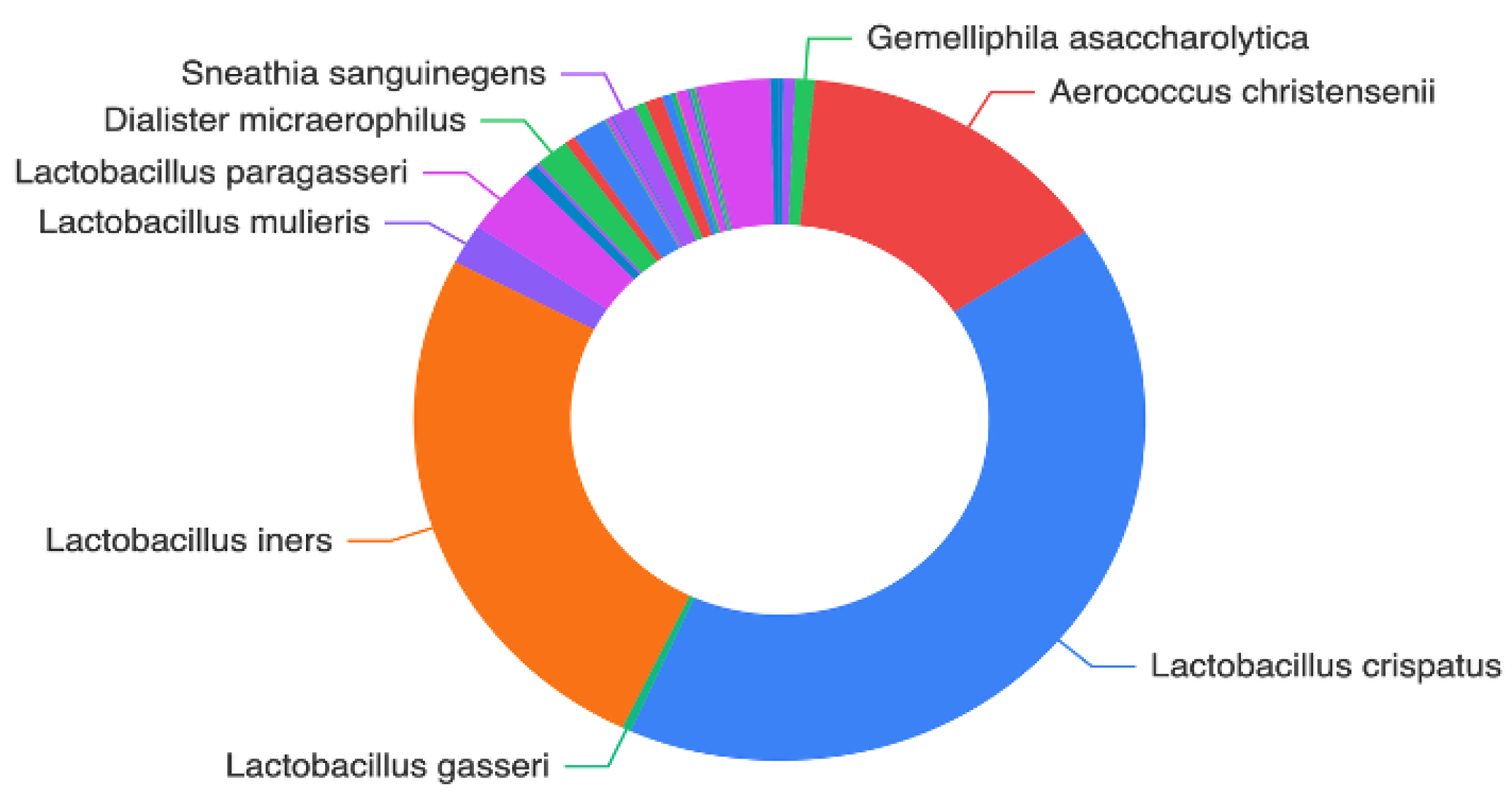
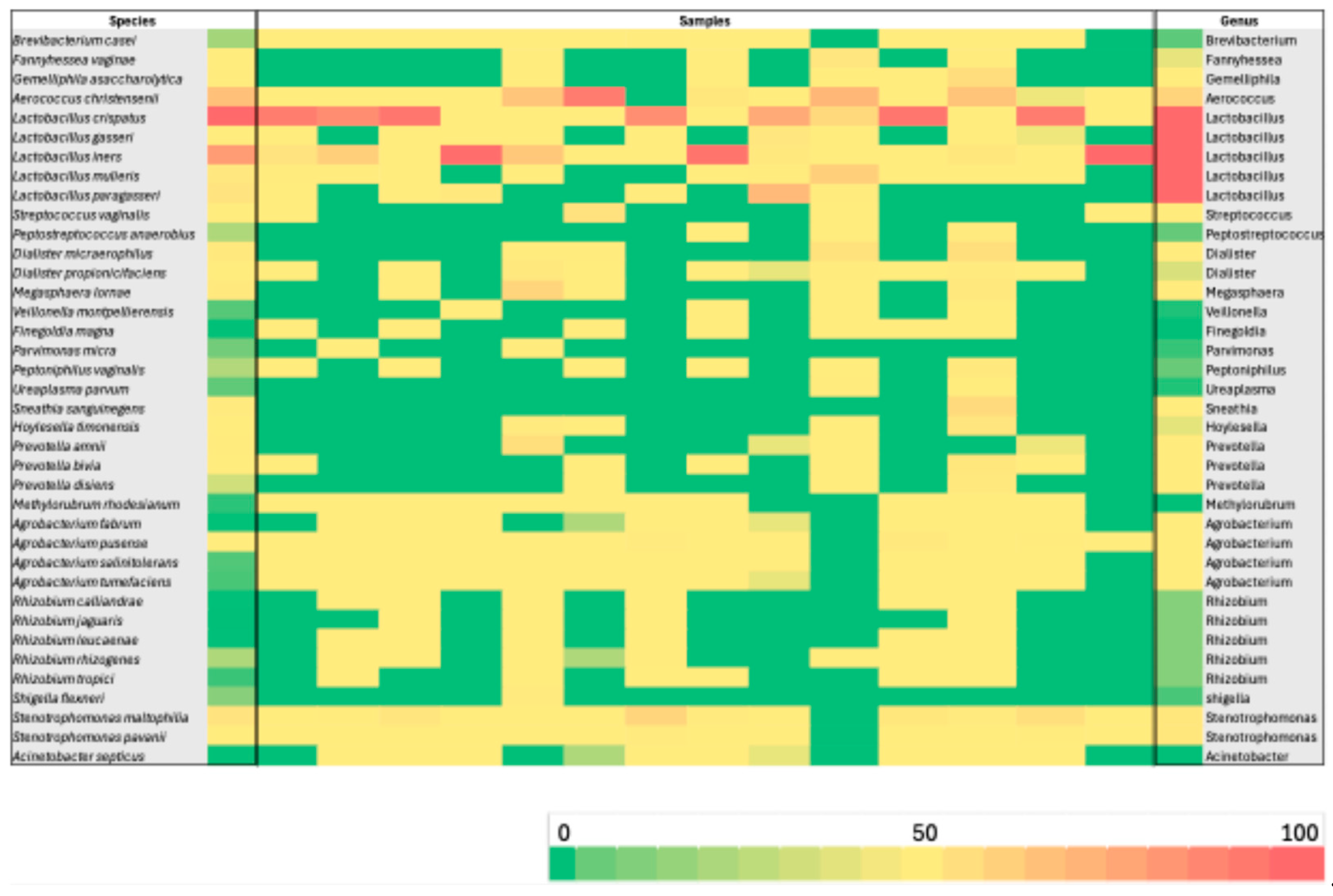
Background/Objectives: The vaginal microbiota (VM) represents a highly diverse microbial ecosystem shaped by the distinctive mucosal environment and immunological characteristics of the female genital tract. Recent evidence emphasizes that alterations in cervical microbial composition may contribute to high-risk gynecological conditions. In this context, the present study sought to comprehensively characterize the cervical microbiota of a well-defined cohort of Greek women. The primary aim was to evaluate the functional microbial landscape, with a focus on identifying bacterial signatures and potential microbial pathways that may influence cervical physiology, protection, and disease susceptibility. Methods: Microbial genomic DNA of 60 samples was extracted using the Magcore Bacterial automated Kit and was subjected to 16S rRNA sequencing using the Nanopore MinION™ enabling a comprehensive analysis of the microbial community. Results: More than 75% of the total microbial community of the cervical samples were represented by the species: Lactobacillus iners and Lactobacillus crispatu and Aerococcus christensenii while the species Stenotrophomonas maltophilia, S. pavanii, Acinetobacter septicus, Rhizobium rhizogenes, R. tropici, R. jaguaris, Prevotella amnii, P. disiens, Brevibacterium casei, Fannyhessea vaginae, Gemelliphila asaccharolytica, flexneri were detected in lower abundances. Conclusions: These findings highlight the predominant protective role of Lactobacillus species while emphasizing the potential contributions of low-abundance or environmentally derived bacteria whose functional implications require further investigation. Broader population studies are essential to establish microbial signatures as diagnostic markers or therapeutic targets for optimizing cervical health.

Keywords: 
;  ;  ;  

1. Introduction

The microbial communities that inhabit the human body play a crucial role in determining health and disease. Among these, the microbiota residing along the female reproductive tract have received increasing scientific attention, with the cervical microbiome emerging as a distinct and biologically important niche. The vaginal microbiota (VM) is characterized by a heterogeneous variety of microorganisms that are commonly found in cervicovaginal samples of female patients in both pathological and non-pathological conditions. Although contiguous with the vaginal environment, the cervix microbial communities reflect its unique mucosal physiology and immunological landscape. Emerging evidence suggests that shifts in cervical microbial composition may influence the persistence of high-risk human papillomavirus (HPV) infection and the progression to cervical intraepithelial neoplasia (CIN) or cancer [1,2,3,4].
Cervical microbial communities in reproductive-age women are often described through community state profiles analogous to those in the vagina, with a dominance of Lactobacillus species in healthy states (e.g., L. crispatus, L. gasseri, L. iners, L. jensenii) and more diverse anaerobe-rich communities in dysbiotic states [4,5].
Lactobacillus-rich cervical communities are generally associated with a lower pH, improved mucosal barrier function, and reduced risk of HPV persistence and lesion development, while depletion of Lactobacilli and overrepresentation of anaerobic bacteria—such as Gardnerella, Prevotella, Sneathia, and Streptococcus—have been linked to HPV infection and cervical disease severity[4,6]. Among these, Sneathia has emerged as one of the most prominent non-Lactobacillus genera linked to pathogenic outcomes; epidemiological reviews and meta-analyses frequently report its enrichment in high- grade cervical lesions and its strong association with HPV persistence, suggesting a potential co-carcinogenic or disease-modifying role within the dysbiotic cervical microenvironment [7].Lactic acid production, bacteriocin secretion, and modulation of local immune signaling are among the proposed mechanisms through which Lactobacillus species may exert protective effects, although the precise molecular and host–microbe interactions remain under investigation [2,4].
Importantly, the cervical microbiome appears dynamic. Longitudinal and cross-sectional studies show that community composition may shift in response to hormonal fluctuations, sexual behavior, physiological changes, or infection. Such transitions between Lactobacillus-dominated and more diverse states may influence HPV acquisition, persistence, and disease progression [3,8]. Yet, the ecological and host factors that drive these shifts—whether through immune modulation, metabolic changes, or other pathways—are not fully understood.
Much of our current insight into cervical microbial communities comes from 16S rRNA gene amplicon sequencing, allowing taxonomic characterization across large cohorts [1]. More recently, multi-omics approaches (including shotgun metagenomics and metabolomics) have provided deeper insight into microbial function, diversity, and host–microbe interactions. For example, multi-omics work has linked specific bacteria (e.g., Lacticaseibacillus iners, Prevotella bivia) to metabolic pathways associated with CIN and HPV status [1]. Metabolomic profiling has also revealed key metabolite changes in cervicovaginal fluid (e.g., succinic acid) that correlate with microbial shifts and disease severity in HPV-positive women [9].
Despite these advances, metatranscriptomic data on the cervical microbiome remain limited. Functional profiling of microbial gene expression could clarify how microbial communities modulate their activity in response to infection or host signals, and how these changes contribute to disease progression. Integrative, high-resolution studies combining community composition, gene expression, and host response may provide mechanistic understanding of how cervical microbes influence HPV persistence, cervical inflammation, and neoplastic transformation. Such work has the potential to uncover microbiome-based biomarkers for early detection of cervical disease and therapeutic strategies (e.g., probiotics or microbiome modulation) aimed at maintaining or restoring protective microbial states.
The purpose of this research is to evaluate the cervical microbiota profiles on non-pregnant reproductive-age women. Through comprehensive integrative analyses, we aim to elucidate the functional landscape of the cervical microbiota and uncover key microbial activities that may contribute to cervical health and disease.

2. Materials and Methods

2.1. Cervical Samples

Sixty independent cervical samples were collected from 60 women with written consent. Additionally, women who had taken antibiotics within the last three months were excluded. Cervical samples were stored at room temperature until DNA extraction in accordance with the manufacturer’s instructions. DNA was extracted using the Magcore Bacterial automated Kit following the protocol recommended by the supplier.

2.2. DNA Amplification, Barcoding and Library Preparation and 16S rRNA Sequencing

The concentration of each concentrated was confirmed with an Invitrogen Qubit 4 fluorometer (Thermo Fisher Scientific, Waltham, MA, USA), 16S rRNA gene amplicon barcoding PCR and library preparation (ONT MinION 16S V1–V9 library preparation).
The extracted gDNA was then prepared for prokaryotic metagenome sequencing using the 16S Barcoding Kit and 16S Barcoding Kit 0–24 (SQK-RAB204 and SQK-16S024, Oxford Nanopore Technologies, Oxford, UK), according to the manufacturer’s protocol, using 10 ng of the extracted gDNA per sample. The PCR reaction was performed on the full 16S hypervariable region (V1-V9) by injecting each multiplexing barcode included in the 16S Barcoding Kit 0–24 into 10 ng of each extracted DNA under the following conditions: initial 30 s denaturation at 98°C (Stage 1), 25 cycles of 10 s denaturation at 98°C, 30 s annealing at 55°C, 90 s extension at 65°C (Stage 2), and 5 min final extension at 65°C (Stage 3), with NEBNext® Ultra™ II Q5® Master Mix (New England Biolabs, Ipswich, MA, USA) as the PCR polymerase reagent mixture. The 16S V1–V9 amplicons were subsequently purified using Agencourt AMPure XP (Beckman Coulter, Brea, CA, USA) magnetic beads with a PCR reaction mix to magnetic bead ratio of 5:3 and washed twice with freshly prepared 70% ethanol. The final elution of purified DNA was performed by adding 12 μL of 10 mM Tris–HCl pH 8.0 with 50 mM NaCl, incubating for 2 min at room temperature, and recovering 10 μL of the elute from each tube. The concentration of each purified 16S V1–V9 amplicon was confirmed with an Invitrogen Qubit 4 fluorometer (Thermo Fisher Scientific, Waltham, MA, USA), and the samples were pooled with a total concentration of 100 fmol in 10 μL of the final library.
Basecalling was carried out using the Guppy agent (version 6.3.7) embedded within the EPI2ME platform (version 5.2.13, ONT), converting FAST5 files into FASTQ format. Barcode sequences were removed, and only reads with a q-score of at least 9 were retained. The resulting FASTQ files were further analyzed with Minimap2. These same files were then uploaded to EPI2ME and processed through the ‘16S’ workflow (EPI2ME-16S), with the minimum accuracy threshold set to 77%. Within EPI2ME, sequencing data are handled via cloud-based computational resources, which perform demultiplexing, quality filtering, and taxonomic classification using the BLAST algorithm against the Reference Sequence (RefSeq) database[10], an open-access, curated, and annotated repository of nucleotide sequences maintained by the National Center for Biotechnology Information (NCBI). Reads shorter than 1,000 bp or longer than 1,850 bp were excluded from further analysis.

2.3. Bioinformatics and Statistical Analysis

The reliability of the subsampled read numbers was verified. Prior to subjecting the full dataset to taxonomic analysis, three random samples from the ONT sequencing were subsampled to 30,000 reads and 100,000 reads per sample to ensure adequate read depth was achieved. The results showed negligible differences of less than 0.1% in all taxonomic levels, showing that the read depth of 30,000 reads per sample was sufficient for detecting minor constituents of the 16S. For the main taxonomic analysis, 50,000 reads per sample were used to further ensure adequate read depth. For diversity analysis, the alpha rarefaction curves of various alpha diversity parameters were checked to verify the plateau of the curves. The statistical analysis was performed in GraphPad Prism 10 for mac, while data visualization and graphing were performed in SRplot [11].

3. Results

3.1. Clinical and Demographic Characteristic of Samples

All analyzed specimens consisted of cervical samples collected from women of reproductive age (18–39 years). The study population displayed homogeneous demographic characteristics, with no reported gynecological disorders or significant clinical comorbidities.

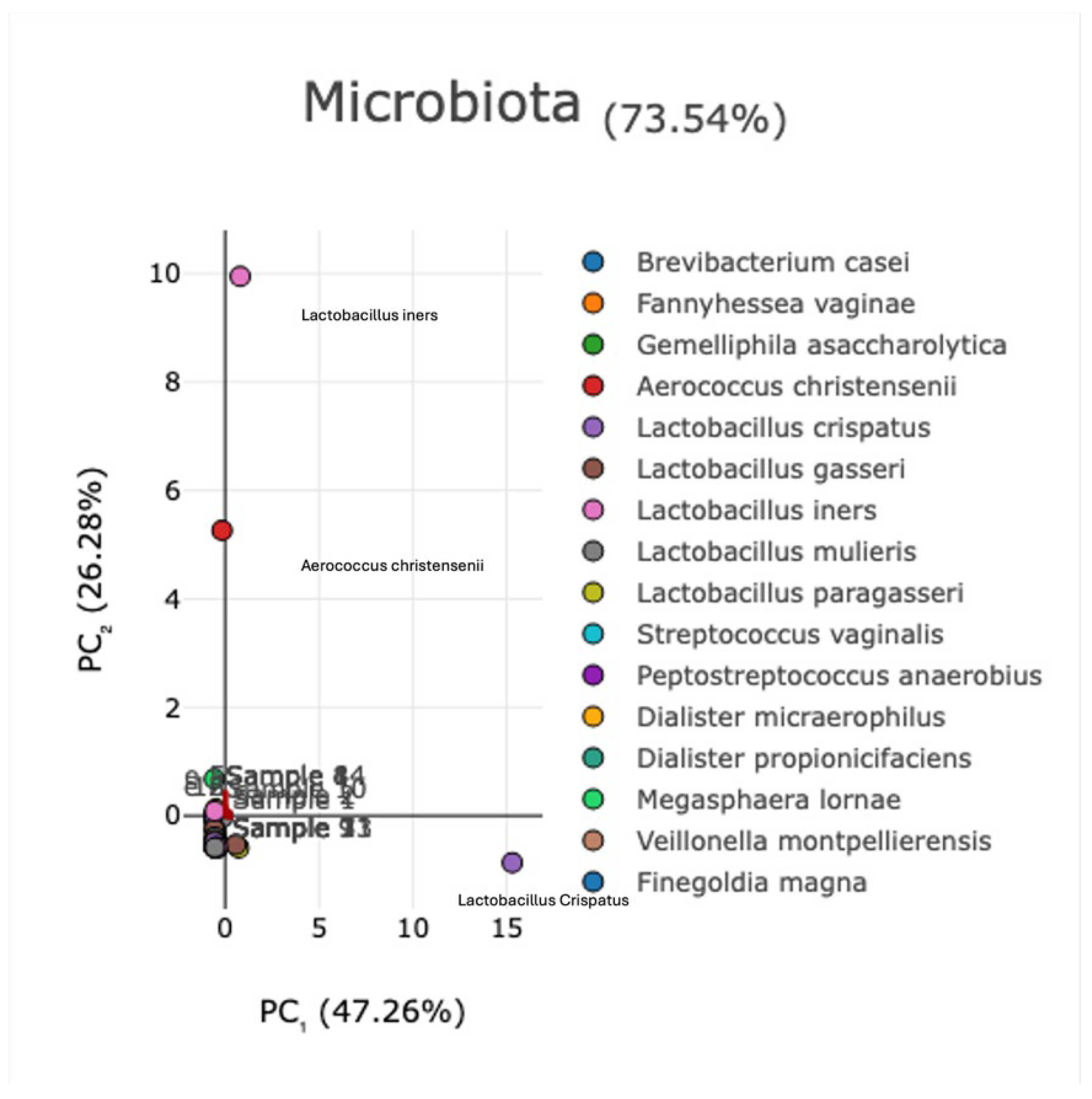
3.2. Evaluation of Microbial Diversity

To assess the microbial diversity within our study population, a principal component analysis (PCA) was conducted. The first two principal components (PC1 and PC2) accounted for 47.26% and 26.28% of the total variance, respectively, explaining 73.54% of the overall microbiota variation (Figure 1). According to the PCA model, Lactobacillus spp. and Aerococcus spp. were the predominant species in the population.
Figure 2 illustrates the distribution of the detected species across all cerviral samples. Lactobacillus spp. (notably L. iners and L. crispatus) and Aerococcus spp. (particularly/ A. christensenii) were the most abundant species, together collectively accounting for more than 75% of the total microbial community. Additional species relative frequently identified included Stenotrophomonas maltophilia, S. pavanii, Acinetobacter septicus, Rhizobium rhizogenes, R. tropici, R. jaguaris, Prevotella amnii, P. disiens, Brevibacterium casei, Fannyhessea vaginae, Gemelliphila asaccharolytica, flexneri (Figure 2).
Consistent with these findings, the heatmap analysis (Figure 3) revealed a similar microbial distribution pattern, illustrating the relative abundances of species across the study population. The most dominant genera were Lactobacillus and Aerococcus, whereas Agrobacterium, Stenotrophomonas, Prevotella, Streptococcus, Sneathia, Megasphaera, Dialister, and Fannyhessea were also detected at relatively high frequencies.

4. Discussion

The composition of the cervical microbiota is not static but fluctuates over time in response to hormonal status, age, sexual behavior, and environmental factors [12,13]. In our study which included women of reproductive age, several Lactobacillus species — particularly L. crispatus, L. iners, L. paragasseri (formerly L. gasseri), and L. mulieris (formerly L. jensenii) were reported which were recurrently associated with healthy microbial states and protection against infection and inflammation [14].
Among these species, L. crispatus is considered the hallmark of a stable vaginal microbiome. It exerts its protective role primarily through the abundant production of lactic acid (both D- and L-isomers) and hydrogen peroxide, maintaining a low pH and directly inhibiting the proliferation of opportunistic microorganisms [15]. In the context of in vitro fertilization (IVF), a moderate and balanced abundance of Lactobacillus crispatus (L. crispatus ) appears to be favorable for pregnancy success, supporting a healthier reproductive environment [16]. In our study, L. crispatus was consistently identified across all cervical samples analyzed, confirming its dominant and stabilizing role within the cervical microbiota, with relative abundance up to 91.07%. Lactobacillus iners was also detected in every sample, exhibiting highly variable abundance levels (up to 99.62%), reflecting its metabolic adaptability and potential involvement in transitional microbial states. At the same time, Lactobacillus iners (L. iners) displays a metabolically flexible phenotype that enables persistence under fluctuating environmental conditions, yet its dominance has been associated with transitional or less stable vaginal microbiota configurations and an increased risk of bacterial vaginosis and sexually transmitted infections, like Chlamydia trachomatis, human immunodeficiency virus (HIV), Neisseria gonorrhoeae and HSV-2 [17]. In addition, L. iners tends to inhibit unpredictable behavior during pregnancy. Several studies have suggested that its dominance, as opposed to the more protective L. crispatus, may raise the risk of preterm birth, although the findings are inconsistent [17]. Overall, L. iners is classified as an ‘intermediate’ bacterium that does not provide consistent protection throughout pregnancy. Compared to other Lactobacillus species, L. iners possesses more complex nutritional requirements and a Gram-variable morphology. Moreover, the genome of L. iners encodes inerolysin, a pore-forming toxin homologous to vaginolysin from Gardnerella vaginalis. This feature implies that L. iners may encompass distinct clonal variants—some contributing to vaginal homeostasis, while others are linked to dysbiosis and disease [14].
In addition to Lactobacillus spp. dominance, Aerococcus christensenii (A. christensenii ) was also detected in several cervical samples, with relative abundances up to 87.32%, indicating substantial inter-individual variability. Previous studies have suggested that A. christensenii is a commensal species of the vaginal and cervical microbiota, often coexisting with Lactobacillus spp. in eubiotic states [14]. However, its presence at higher proportions has also been associated with transitional microbial profiles and, in some cases, with mild inflammatory responses or subclinical dysbiosis [18,19]. Recent findings reinforce its potential clinical relevance, as A. christensenii was found to increase in abundance among patients experiencing recurrent bacterial vaginosis following metronidazole therapy, particularly in those who relapsed [20]. Moreover, Norenhag (2024) reported higher levels of A. christensenii in women with cervical dysplasia compared to healthy controls, alongside increased microbial diversity and reduced Lactobacillus dominance, suggesting a potential link between A. christensenii and early cervical epithelial alterations [21]. Furthermore, A. christensenii has genes related with pathogenicity, bloodstream invasion, and antibiotic resistance, which can lead to several complications, like chorioamnionitis and bacteremia. Recent findings demonstrate A. christensenii can survive in the blood and cause infection [22]. Lin et al. also highlighted the need of recognizing this microorganism as a possible pathogen in pregnancy and including it into clinical evaluation of reproductive tract infections[22]. In our study, the coexistence of A. christensenii with L. crispatus and L. iners in most of the cervical samples may therefore represent a balanced microbial state, where Aerococcus species possibly contribute to mucosal defense under eubiotic conditions, but could also participate in transitional or dysbiotic shifts under altered host or environmental contexts [18,19,20].
In our analysis, Lactobacillus gasseri (L. gasseri) was detected in low relative abundances (up to 4.77%), indicating its limited yet consistent presence within the cervical microbiota. The recently reclassified L. paragasseri, previously grouped within L. gasseri, has been identified in both vaginal and cervical samples and may contribute to epithelial protection via lactic acid production and cell adhesion, although its precise physiological function remains to be elucidated [23,24,25]. Similarly, Lactobacillus mulieris was identified in our samples with relative abundances ranging up to 22.94%. This species, a close phylogenetic relative of Lactobacillus jensenii (L. jensenii) has been increasingly recognized as a commensal member of the vaginal niche, potentially supporting microbial stability and mucosal defense through the production of lactic acid and competitive exclusion of opportunistic pathogens [26,27]. One such species, L. jensenii, releases biosurfactants that disrupt biofilms of pathogens such as Enterobacter aerogenes and Escherichia coli [28]. In addition, a number of unexpected or low-abundance bacterial species such as :Stenotrophomonas maltophilia, Stenotrophomonas pavanii, Dialister micraerophilus, Dialister propionicifaciens, Acinetobacter septicus, Rhizobium rhizogenes, Rhizobium tropici, Rhizobium jaguaris, Prevotella amnii, Prevotella disiens, Brevibacterium casei, Fannyhessea vaginae, Gemelliphila asaccharolytica, Agrobacterium Pusense, Agrobacterium salinitolerans, Agrobacterium tumefaciens have been reported in cervical microbiome studies, though the functional significance of many remains uncertain. For example, Stenotrophomonas maltophilia (a member of Proteobacteria) has been detected in higher abundance in cervical intraepithelial neoplasia (CIN) samples compared with healthy controls [29]. In addition, the presence of Stenotrophomonas maltophilia may be associated with persistent or recurrent vaginal discharge. This implies that even if the more prevalent causes (like candidiasis) have been treated, the presence of this pathogen may still induce or prolong symptoms [30]. Multivariate analyses in such studies also identified Rhizobium genera (family Rhizobiaceae) as independently associated with CIN [29]. Environmental or plant-related genera such as Agrobacterium and Rhizobium may reflect transient colonization, possible contamination, or low-biomass bacterial populations rather than stable, functionally active members of the cervical niche.
On the other hand, several anaerobic bacteria more strongly linked to cervical dysbiosis are well-supported by clinical studies. Dialister micraerophilus (D. micraerophilus) (or closely related Dialister spp.) and Prevotella amnii / Prevotella disiens are often enriched in states of vaginal or cervical dysbiosis [31]. Additionally, recent findings position D. micraerophilus as an important contributor to the metabolic and ecological landscape of BV-associated cervical dysbiosis, complementing the established pathogenic roles of F. vaginae and other anaerobic taxa [32]. Fannyhessea vaginae (formerly Atopobium vaginae) is well recognized as a hallmark taxon of dysbiotic vaginal communities and has been consistently associated with adverse cervicovaginal conditions [33].
As a well-established BV-associated bacterium, F. vaginae plays a critical role in polymicrobial biofilm formation on the vaginal epithelium, where it engages in reciprocal transcriptomic interactions with Gardnerella spp. and Prevotella bivia[34]. Beyond its established involvement in BV, increasing evidence indicates that Fannyhessea contributes to broader cervicovaginal pathophysiology. In patients with persistent high-risk HPV infection and high-grade cervical intraepithelial neoplasia (CIN), members of the Fannyhessea genus mediate distinct metabolomic shifts associated with Lactobacillus depletion, epithelial barrier disruption, and mucosal immune dysregulation [35]. Furthermore, systematic reviews of cervical carcinogenesis describe non-Lactobacillus–dominant states enriched in F. vaginae as potential cofactors that may sustain chronic inflammation, modulate local immune responses, and promote viral persistence, thereby contributing to an environment permissive to neoplastic progression[36]. Other less common species such as Brevibacterium casei, Gemelliphila asaccharolytica, Stenotrophomonas pavanii, Acinetobacter septicus, Rhizobium tropici / jaguaris, and Agrobacterium tumefaciens / pusense / salinitolerans — are rarely reported in cervical microbiome cohorts. Their detection may represent very low-abundance, transient exposure rather than established colonization. The biological roles of these species in the cervix are thus currently unknown. Indeed, environmental genera such as Stenotrophomonas and Rhizobium have been observed in some CIN-associated cervical microbiome studies, however most of the well-characterized dysbiotic bacteria in this niche remain anaerobes such as Dialister, Prevotella, and Fannyhessea. Although usually at low relative abundances, Acinetobacter septicus (A. septicus ) was found in a subset of cervical and endometrial samples, indicating its status as a transitory or low-biomass component of the female reproductive tract microbiota. According to previous reports, A. septicus is an opportunistic environmental microbe that is sometimes found in uterine or vaginal samples, especially in research using high-resolution metagenomic techniques [37,38]. Higher A. septicus signal intensity has been seen in pregnancies complicated by inflammation-associated preterm birth, where it appeared as part of a wider shift toward diverse, low-Lactobacillus communities, even though it is typically interpreted as a commensal or incidental taxon. Moreover, sporadic clinical observations—most notably bacteremia cases in obstetric wards—suggest that A. septicus may gain transient pathogenic potential under disrupted mucosal or iatrogenic conditions [37].
In a small percentage of vaginal samples, Rhizobium rhizogenes (R. rhizogenes) was occasionally found, usually at very low relative abundances, which is consistent with its classification as an environmental or low-biomass taxon within the reproductive tract. Recent high-resolution metagenomic studies have revealed that R. rhizogenes is present in the vaginal microbiota of pregnant people, especially in cohorts at risk for inflammation-driven preterm birth, even though it is mainly recognized as a plant-associated organism [39]. In these investigations, increased R. rhizogenes signal emerged in transitional community states marked by decreased Lactobacillus dominance and increased ecological diversity and was interpreted as a sign of wider microbial instability rather than direct pathogenicity.
Additional analyses of host–microbiome interactions likewise place R. rhizogenes among low-abundance taxa associated with heightened immune activation in pregnancy-related dysbiosis [19].
Rhizobium tropici is classified as a primarily environmental and plant-associated taxon rather than a stable component of the human reproductive tract microbiome because it was only occasionally and at very low abundances found in all the cervical samples that were examined. Despite being a well-characterized symbiotic nitrogen-fixing species in legumes, R. tropici's appearance in human metagenomic datasets has typically been interpreted as incidental, reflecting fluctuations in the low-biomass community or temporary environmental contamination rather than actual colonization [40].
As a recently identified environmental species with no known function in the human reproductive system, Rhizobium jaguaris (R. jaguaris) only occasionally and consistently appeared at trace-level abundances in the examined samples. R. jaguaris was first identified from legume-associated root nodules, but it has not been linked to human colonization or pathogenicity. Its infrequent discovery in cervical metagenomic datasets is typically interpreted as a low-biomass signal or temporary environmental carryover rather than a significant microbial presence [41].
Prevotella amnii (P. amnii )is a known member of the larger anaerobic community linked to both stable and transitional states of the female reproductive tract microbiome. Since its initial isolation from amniotic fluid, P. amnii has been associated with a variety of cervicovaginal profiles and is more common in communities with subtle inflammatory signatures and increased diversity[19] . Previous research shows that increased P. amnii levels frequently may be involved in low-grade mucosal inflammation or early dysbiotic microbiome, especially in ecosystems linked to bacterial vaginosis [42,43].
According to our results, the predominance of Lactobacillus species in cervical samples is indicative of a balanced and eubiotic microbial environment. Their synergistic activities — acidification of the vaginal milieu, inhibition of pathogenic colonization, modulation of host immunity, and reinforcement of epithelial barrier function — highlight their crucial contribution to maintaining cervical–vaginal homeostasis and protecting against dysbiosis and infection. In addition, more targeted and high-resolution studies (e.g., using metagenomics and metatranscriptomics) throughout different populations are needed to clarify whether environmental or other species play any functional role in cervical health or disease.

5. Conclusions

This study provides a comprehensive characterization of the cervical microbiota in non-pregnant, reproductive-age women, revealing a microbial landscape dominated by Lactobacillus species-particularly L. crispatus and L.iners and supported by variable contributions from Aerococcus christensenii and several low-abundance taxa. The predominance of Lactobacilli reflects a generally eubiotic cervical environment, while the presence of transitional or dysbiosis-associated anaerobic genera (Dialister, Prevotella, and Fannyhessea) highlights the inherent ecological variability of the cervical niche. These findings reinforce the central role of Lactobacillus-driven mucosal protection and underline the complexity introduced by low-biomass or environmentally derived species whose functional relevance remains uncertain. Future research across diverse population and clinical contexts will be essential to determine whether specific microbial community patterns can serve as biomarkers or therapeutics targets for improving cervical health.

Author Contributions

For research articles with several authors, a short paragraph specifying their individual contributions must be provided. The following statements should be used. Conceptualization D.H. ;methodology, D.H.,S.L.,D.V; formal analysis, S.L.; investigation, S.L.,D.V, K.L; resources, D.H. data curation, D.H.,D.V.,Z.S.,S.L;.K.L.; writing—original draft preparation, D.V.,S.L.,A.T.,I.M.,Z.S..; writing—review and editing, P.H.,D.H.,Z.S.,A.T.,K.L; visualization, S.L.,D.V.; supervision, D.H.; All authors have read and agreed to the published version of the manuscript..

Funding

This research received no external funding

Institutional Review Board Statement

This study was conducted according to the guidelines of the Declaration of Helsinki, and it was approved by the ethics committee of the University GeneralHospital “ATTIKON” Ethical Committee with the protocol number 1235 (15 April 2020).

Informed Consent Statement

Informed consent was obtained from all subjects involved in the study.

Data Availability Statement

The original contributions presented in this study are included in the article.The data of the current study are available from the corresponding author upon reasonable request.

Conflicts of Interest

The authors declare no conflicts of interest.

References

  1. Pu, X.; Wang, X.; Wang, J.; Gu, Z.; Zhu, H.; Li, C. Microbial and Metabolic Profiles Associated with HPV Infection and Cervical Intraepithelial Neoplasia: A Multi-Omics Study. Microbiol Spectr 2025, 13. [Google Scholar] [CrossRef]
  2. Curty, G.; de Carvalho, P.S.; Soares, M.A. The Role of the Cervicovaginal Microbiome on the Genesis and as a Biomarker of Premalignant Cervical Intraepithelial Neoplasia and Invasive Cervical Cancer. Int J Mol Sci 2019, 21, 222. [Google Scholar] [CrossRef]
  3. Hu, M.; Yang, W.; Yan, R.; Chi, J.; Xia, Q.; Yang, Y.; Wang, Y.; Sun, L.; Li, P. Co-Evolution of Vaginal Microbiome and Cervical Cancer. J Transl Med 2024, 22, 559. [Google Scholar] [CrossRef]
  4. Shen, J.; Sun, H.; Chu, J.; Gong, X.; Liu, X. Cervicovaginal Microbiota: A Promising Direction for Prevention and Treatment in Cervical Cancer. Infect Agent Cancer 2024, 19, 13. [Google Scholar] [CrossRef] [PubMed]
  5. Peng, Y.; Tang, Q.; Wu, S.; Zhao, C. Associations of Atopobium, Garderella, Megasphaera, Prevotella, Sneathia, and Streptococcus with Human Papillomavirus Infection, Cervical Intraepithelial Neoplasia, and Cancer: A Systematic Review and Meta-Analysis. BMC Infect Dis 2025, 25, 708. [Google Scholar] [CrossRef]
  6. Mazinani, S.; Aghazadeh, M.; Poortahmasebi, V.; Arafi, V.; Hasani, A. Cervical Cancer Pathology and Vaginal and Gut Microbiota: Conception of the Association. Lett Appl Microbiol 2025, 78. [Google Scholar] [CrossRef] [PubMed]
  7. Mancilla, V.; Jimenez, N.R.; Bishop, N.S.; Flores, M.; Herbst-Kralovetz, M.M. The Vaginal Microbiota, Human Papillomavirus Infection, and Cervical Carcinogenesis: A Systematic Review in the Latina Population. J Epidemiol Glob Health 2024, 14, 480–497. [Google Scholar] [CrossRef] [PubMed]
  8. Wen, Q.; Wang, S.; Min, Y.; Liu, X.; Fang, J.; Lang, J.; Chen, M. Associations of the Gut, Cervical, and Vaginal Microbiota with Cervical Cancer: A Systematic Review and Meta-Analysis. BMC Womens Health 2025, 25, 65. [Google Scholar] [CrossRef]
  9. Shen, S.; Zhao, S.; Shan, J.; Ren, Q. Metabolomic and Microbiota Profiles in Cervicovaginal Lavage Fluid of Women with High-Risk Human Papillomavirus Infection. Sci Rep 2025, 15, 796. [Google Scholar] [CrossRef]
  10. Pruitt, K.D.; Tatusova, T.; Maglott, D.R. NCBI Reference Sequences (RefSeq): A Curated Non-Redundant Sequence Database of Genomes, Transcripts and Proteins. Nucleic Acids Res 2007, 35, D61–D65. [Google Scholar] [CrossRef]
  11. Tang, D.; Chen, M.; Huang, X.; Zhang, G.; Zeng, L.; Zhang, G.; Wu, S.; Wang, Y. SRplot: A Free Online Platform for Data Visualization and Graphing. PLoS One 2023, 18, e0294236. [Google Scholar] [CrossRef] [PubMed]
  12. Ravel, J.; Brotman, R.M. Translating the Vaginal Microbiome: Gaps and Challenges. Genome Med 2016, 8, 35. [Google Scholar] [CrossRef] [PubMed]
  13. France, M.T.; Ma, B.; Gajer, P.; Brown, S.; Humphrys, M.S.; Holm, J.B.; Waetjen, L.E.; Brotman, R.M.; Ravel, J. VALENCIA: A Nearest Centroid Classification Method for Vaginal Microbial Communities Based on Composition. Microbiome 2020, 8, 166. [Google Scholar] [CrossRef] [PubMed]
  14. Petrova, M.I.; Reid, G.; Vaneechoutte, M.; Lebeer, S. Lactobacillus Iners : Friend or Foe? Trends Microbiol 2017, 25, 182–191. [Google Scholar] [CrossRef]
  15. Galicia-Campos, E.; García-Villaraco, A.; Montero-Palmero, Ma.B.; Gutiérrez-Mañero, F.J.; Ramos-Solano, B. Bacillus G7 Improves Adaptation to Salt Stress in Olea Europaea L. Plantlets, Enhancing Water Use Efficiency and Preventing Oxidative Stress. Sci Rep 2023, 13, 22507. [Google Scholar] [CrossRef]
  16. Wang, T.; Li, P.; Bai, X.; Tian, S.; Yang, M.; Leng, D.; Kui, H.; Zhang, S.; Yan, X.; Zheng, Q.; et al. Vaginal Microbiota Are Associated with in Vitro Fertilization during Female Infertility. iMeta 2024, 3. [Google Scholar] [CrossRef]
  17. Zheng, N.; Guo, R.; Wang, J.; Zhou, W.; Ling, Z. Contribution of Lactobacillus Iners to Vaginal Health and Diseases: A Systematic Review. Front Cell Infect Microbiol 2021, 11. [Google Scholar] [CrossRef]
  18. Shen-Gunther, J.; Xia, Q.; Cai, H.; Wang, Y. Cervicovaginal Microbiome and HPV: A Standardized Approach to 16S/ITS NGS and Microbial Community Profiling for Viral Association. Int J Mol Sci 2025, 26, 8090. [Google Scholar] [CrossRef]
  19. Gao, J.; Peng, Y.; Jiang, N.; Shi, Y.; Ying, C. High-Throughput Sequencing-Based Analysis of Changes in the Vaginal Microbiome during the Disease Course of Patients with Bacterial Vaginosis: A Case–Control Study. Biology (Basel) 2022, 11, 1797. [Google Scholar] [CrossRef]
  20. Mollin, A.; Katta, M.; Sobel, J.D.; Akins, R.A. Association of Key Species of Vaginal Bacteria of Recurrent Bacterial Vaginosis Patients before and after Oral Metronidazole Therapy with Short- and Long-Term Clinical Outcomes. PLoS One 2022, 17, e0272012. [Google Scholar] [CrossRef]
  21. Johanna Norenhag Vaginal Microbiota Composition and Function. Digital Comprehensive Summaries of Uppsala Dissertations from the Faculty of Medicine 2070 2024.
  22. Lin, Y.; He, J.; Zhang, Q.; Li, Y.; Ke, J.; Lin, C.; Yao, B.; Zhang, C.; Tan, N. Aerococcus Christensenii: An Emerging Pathogen Associated with Infections and Bacteremia in Pregnancy—Genomic Insights and Pathogenicity Evaluation. Funct Integr Genomics 2025, 25, 229. [Google Scholar] [CrossRef] [PubMed]
  23. Mehra, Y.; Viswanathan, P. High-Quality Whole-Genome Sequence Analysis of Lactobacillus Paragasseri UBLG-36 Reveals Oxalate-Degrading Potential of the Strain. PLoS One 2021, 16, e0260116. [Google Scholar] [CrossRef] [PubMed]
  24. Ene, A.; Stegman, N.; Wolfe, A.; Putonti, C. Genomic Insights into Lactobacillus Gasseri and Lactobacillus Paragasseri. PeerJ 2022, 10, e13479. [Google Scholar] [CrossRef]
  25. Zhao, F.; Hu, X.; Ying, C. Advances in Research on the Relationship between Vaginal Microbiota and Adverse Pregnancy Outcomes and Gynecological Diseases. Microorganisms 2023, 11, 991. [Google Scholar] [CrossRef]
  26. Putonti, C.; Shapiro, J.W.; Ene, A.; Tsibere, O.; Wolfe, A.J. Comparative Genomic Study of Lactobacillus Jensenii and the Newly Defined Lactobacillus Mulieris Species Identifies Species-Specific Functionality. mSphere 2020, 5. [Google Scholar] [CrossRef]
  27. Gajer, P.; Brotman, R.M.; Bai, G.; Sakamoto, J.; Schütte, U.M.E.; Zhong, X.; Koenig, S.S.K.; Fu, L.; Ma, Z. (Sam); Zhou, X.; et al. Temporal Dynamics of the Human Vaginal Microbiota. Sci Transl Med 2012, 4. [Google Scholar] [CrossRef]
  28. Morais, I.M.C.; Cordeiro, A.L.; Teixeira, G.S.; Domingues, V.S.; Nardi, R.M.D.; Monteiro, A.S.; Alves, R.J.; Siqueira, E.P.; Santos, V.L. Biological and Physicochemical Properties of Biosurfactants Produced by Lactobacillus Jensenii P6A and Lactobacillus Gasseri P65. Microb Cell Fact 2017, 16, 155. [Google Scholar] [CrossRef]
  29. Lin, S.; Zhang, B.; Lin, Y.; Lin, Y.; Zuo, X. Dysbiosis of Cervical and Vaginal Microbiota Associated With Cervical Intraepithelial Neoplasia. Front Cell Infect Microbiol 2022, 12. [Google Scholar] [CrossRef]
  30. Marselinus Edwin Widyanto Daniwijaya, A.A.C.P.S.D.A.S.T.N. Identification of Streptococcus Intermedius and Stenotrophomonas Maltophilia in Recurrent Leucorrhoea: A Case Report. Journal of Clinical Microbiology and Infectious Diseases (JCMID) 2021, 1, 38–41. [Google Scholar]
  31. Liu, Y.; Wang, S.; Liu, J.; Su, M.; Diao, X.; Liang, X.; Zhang, J.; Wang, Q.; Zhan, Y. Characteristics of Vaginal Microbiota in Various Cervical Intraepithelial Neoplasia: A Cross-Sectional Study. J Transl Med 2023, 21, 816. [Google Scholar] [CrossRef] [PubMed]
  32. Lee, E.M.; Srinivasan, S.; Purvine, S.O.; Fiedler, T.L.; Leiser, O.P.; Proll, S.C.; Minot, S.S.; Djukovic, D.; Raftery, D.; Johnston, C.; et al. Syntrophic Bacterial and Host–Microbe Interactions in Bacterial Vaginosis. ISME J 2025, 19. [Google Scholar] [CrossRef] [PubMed]
  33. Ma, Y.; Li, Y.; Liu, Y.; Cao, L.; Han, X.; Gao, S.; Zhang, C. Vaginal Microbiome Dysbiosis Is Associated with the Different Cervical Disease Status. Journal of Microbiology 2023, 61, 423–432. [Google Scholar] [CrossRef] [PubMed]
  34. Sousa, L.G. V.; Novak, J.; França, A.; Muzny, C.A.; Cerca, N. Gardnerella Vaginalis, Fannyhessea Vaginae, and Prevotella Bivia Strongly Influence Each Other’s Transcriptome in Triple-Species Biofilms. Microb Ecol 2024, 87, 117. [Google Scholar] [CrossRef]
  35. Yang, Q.; Dai, W.; Wu, D.; Xu, R.; Li, C.; Wu, R.; Du, H. Inferred Bi-Directional Interactions between Vaginal Microbiota, Metabolome and Persistent HPV Infection Accompanied by High-Grade Cervical Intraepithelial Neoplasia. BMC Microbiol 2025, 25, 404. [Google Scholar] [CrossRef]
  36. Bautista, J.; Altamirano-Colina, A.; López-Cortés, A. The Vaginal Microbiome in HPV Persistence and Cervical Cancer Progression. Front Cell Infect Microbiol 2025, 15. [Google Scholar] [CrossRef]
  37. Horii, T.; Tamai, K.; Mitsui, M.; Notake, S.; Yanagisawa, H. Blood Stream Infections Caused by Acinetobacter Ursingii in an Obstetrics Ward. Infection, Genetics and Evolution 2011, 11, 52–56. [Google Scholar] [CrossRef]
  38. Bednarska-Czerwińska, A.; Morawiec, E.; Zmarzły, N.; Szapski, M.; Jendrysek, J.; Pecyna, A.; Zapletał-Pudełko, K.; Małysiak, W.; Sirek, T.; Ossowski, P.; et al. Dynamics of Microbiome Changes in the Endometrium and Uterine Cervix during Embryo Implantation: A Comparative Analysis. Medical Science Monitor 2023, 29. [Google Scholar] [CrossRef]
  39. Cavanagh, M.; Amabebe, E.; Kulkarni, N.S.; Papageorgiou, M.D.; Walker, H.; Wyles, M.D.; Anumba, D.O. Vaginal Host Immune-Microbiome-Metabolite Interactions Associated with Spontaneous Preterm Birth in a Predominantly White Cohort. NPJ Biofilms Microbiomes 2025, 11, 52. [Google Scholar] [CrossRef]
  40. Silva, J.G. da; Ferreira, E.P. de B.; Damin, V.; Nascente, A.S. Response of the Common Bean to Liquid Fertilizer and Rhizobium Tropici Inoculation. Semin Cienc Agrar 2020, 41, 2967–2976. [Google Scholar] [CrossRef]
  41. Rincón-Rosales, R.; Villalobos-Escobedo, J.M.; Rogel, M.A.; Martinez, J.; Ormeño-Orrillo, E.; Martínez-Romero, E. Rhizobium Calliandrae Sp. Nov., Rhizobium Mayense Sp. Nov. and Rhizobium Jaguaris Sp. Nov., Rhizobial Species Nodulating the Medicinal Legume Calliandra Grandiflora. Int J Syst Evol Microbiol 2013, 63, 3423–3429. [Google Scholar] [CrossRef]
  42. Carter, K.A.; Balkus, J.E.; Anzala, O.; Kimani, J.; Hoffman, N.G.; Fiedler, T.L.; Mochache, V.; Fredricks, D.N.; McClelland, R.S.; Srinivasan, S. Associations Between Vaginal Bacteria and Bacterial Vaginosis Signs and Symptoms: A Comparative Study of Kenyan and American Women. Front Cell Infect Microbiol 2022, 12. [Google Scholar] [CrossRef]
  43. George, S.D.; Van Gerwen, O.T.; Dong, C.; Sousa, L.G. V.; Cerca, N.; Elnaggar, J.H.; Taylor, C.M.; Muzny, C.A. The Role of Prevotella Species in Female Genital Tract Infections. Pathogens 2024, 13, 364. [Google Scholar] [CrossRef]
Figure 1. PCA plot based on the relative abundance data.
Preprints 187098 g001
Figure 2. Doughnut chart of the data based on species.
Preprints 187098 g002
Figure 3. Heat representing map the most abundant species and genus in the population.
Preprints 187098 g003
Disclaimer/Publisher’s Note: The statements, opinions and data contained in all publications are solely those of the individual author(s) and contributor(s) and not of MDPI and/or the editor(s). MDPI and/or the editor(s) disclaim responsibility for any injury to people or property resulting from any ideas, methods, instructions or products referred to in the content.
Copyright: This open access article is published under a Creative Commons CC BY 4.0 license, which permit the free download, distribution, and reuse, provided that the author and preprint are cited in any reuse.
Prerpints.org logo

Preprints.org is a free preprint server supported by MDPI in Basel, Switzerland.

Subscribe

Disclaimer

Terms of Use

Privacy Policy

Privacy Settings

© 2025 MDPI (Basel, Switzerland) unless otherwise stated