

Submitted:
26 November 2025
Posted:
27 November 2025
You are already at the latest version
Abstract
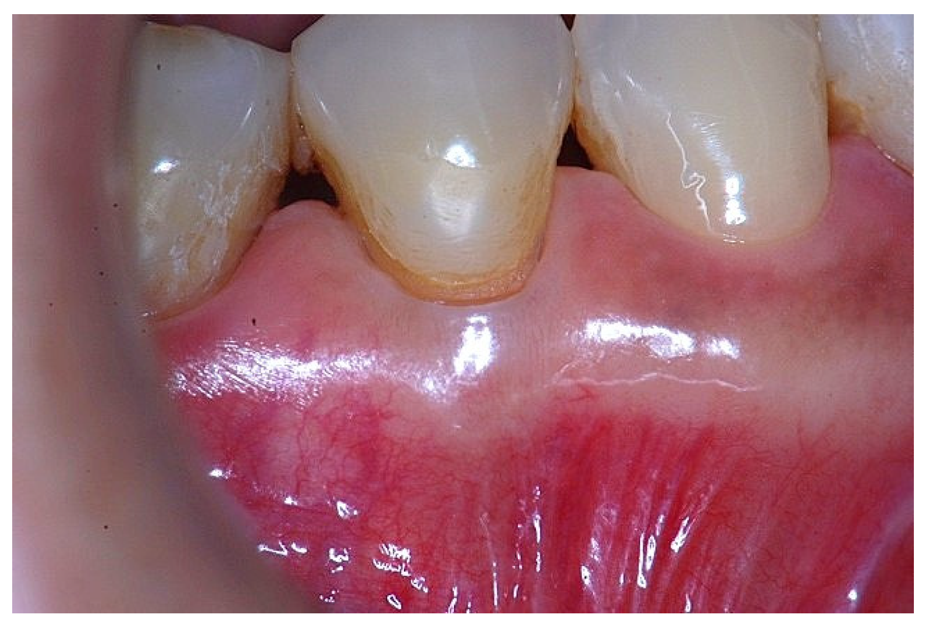
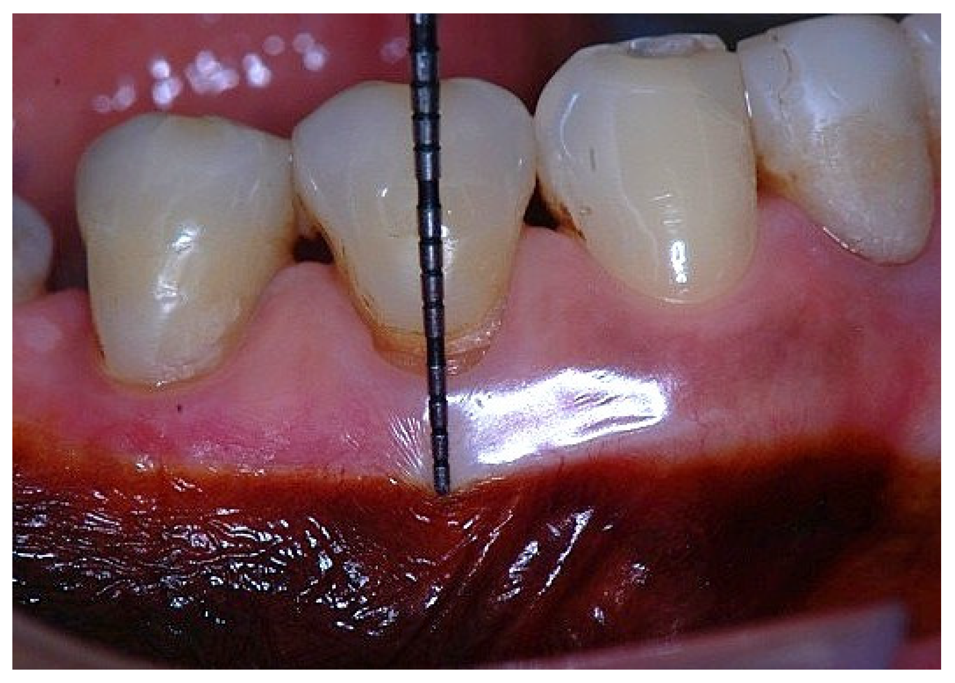
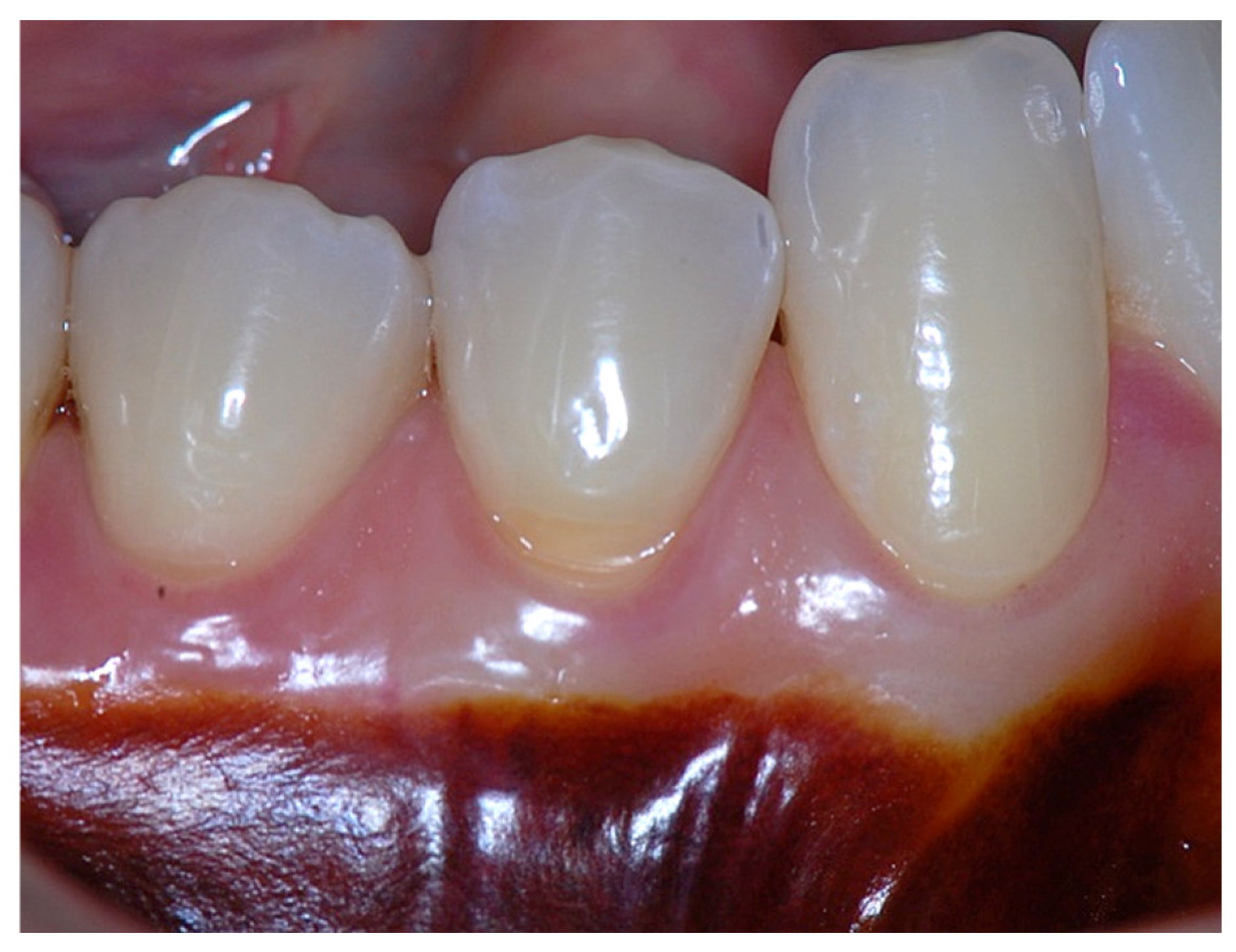
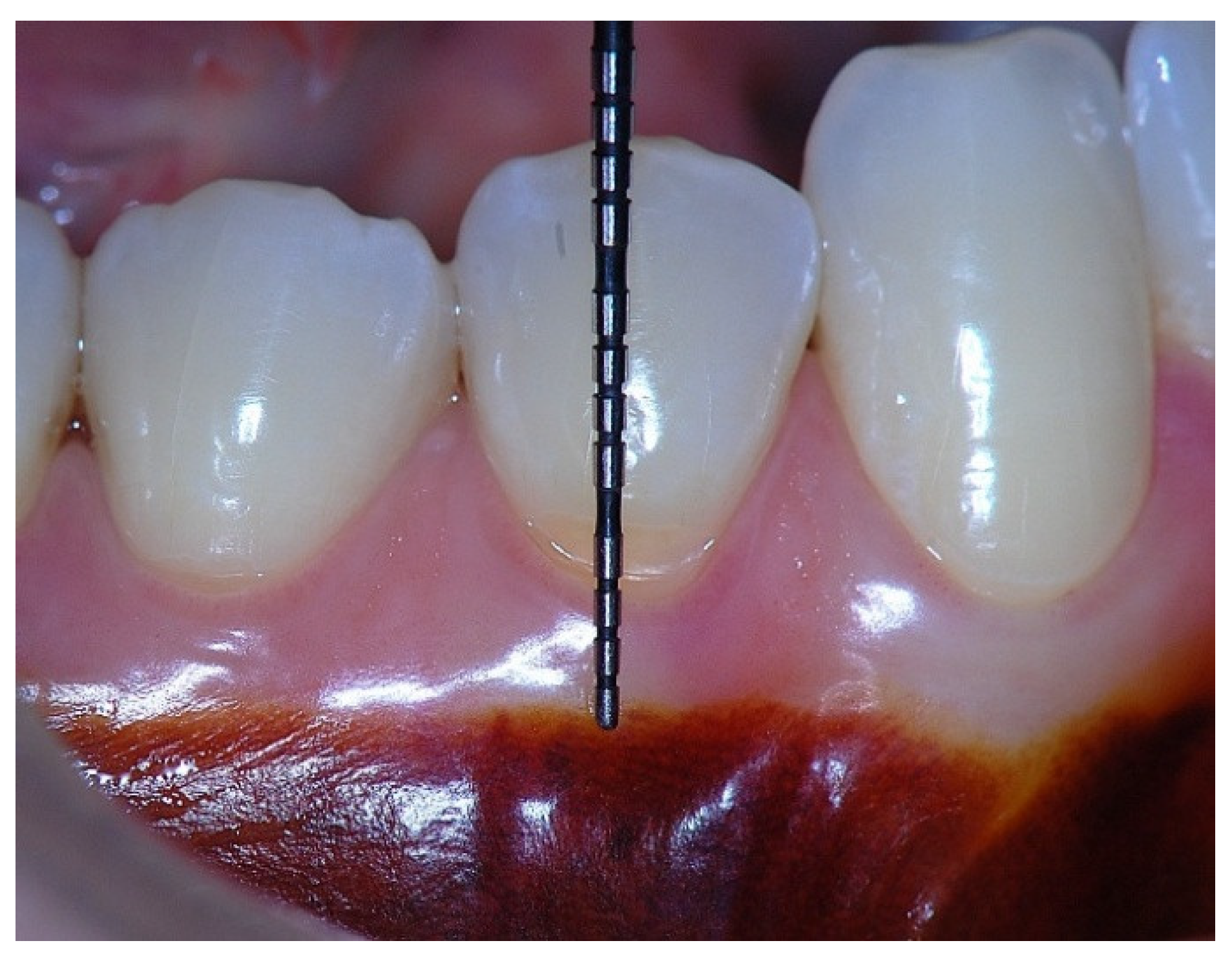
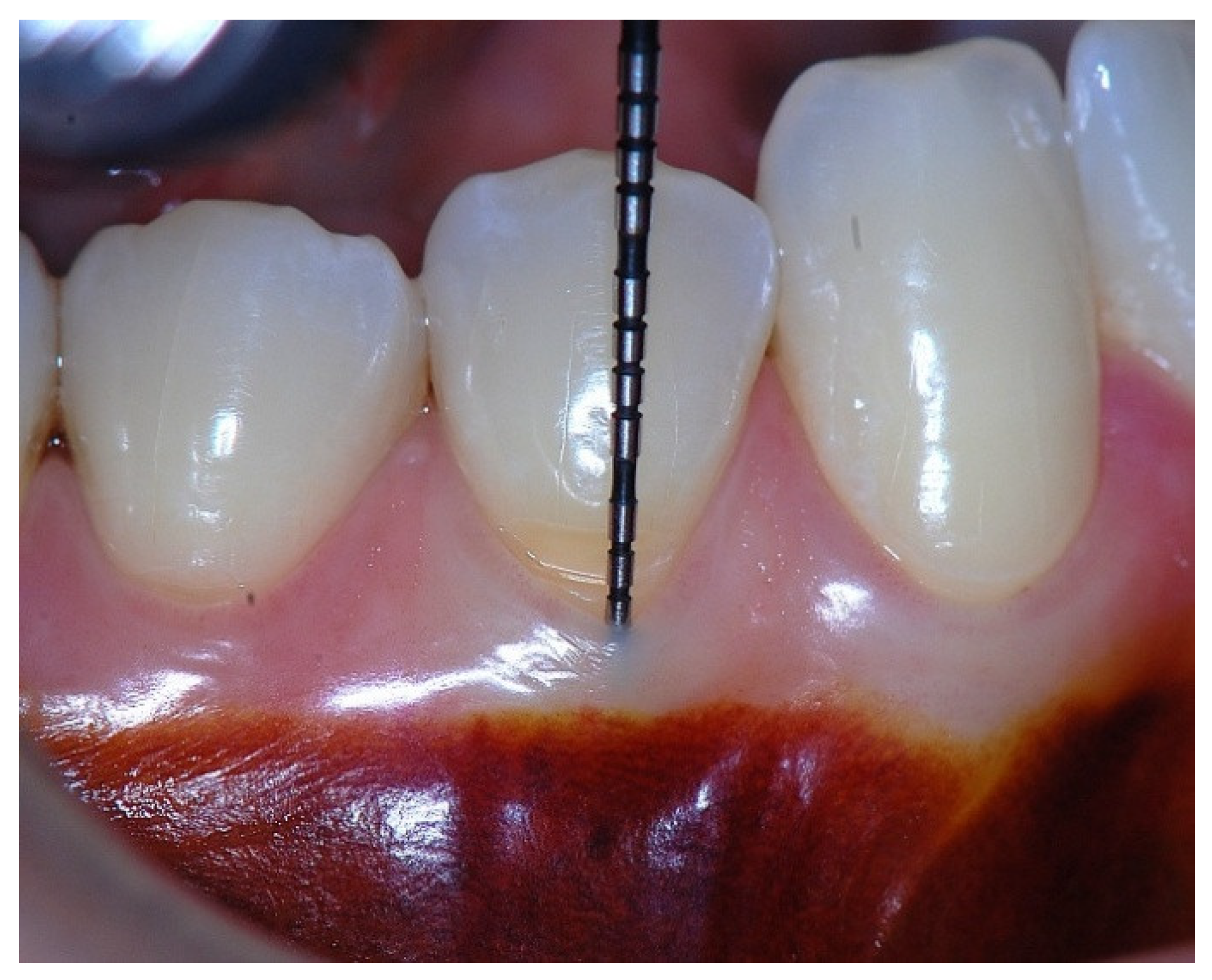
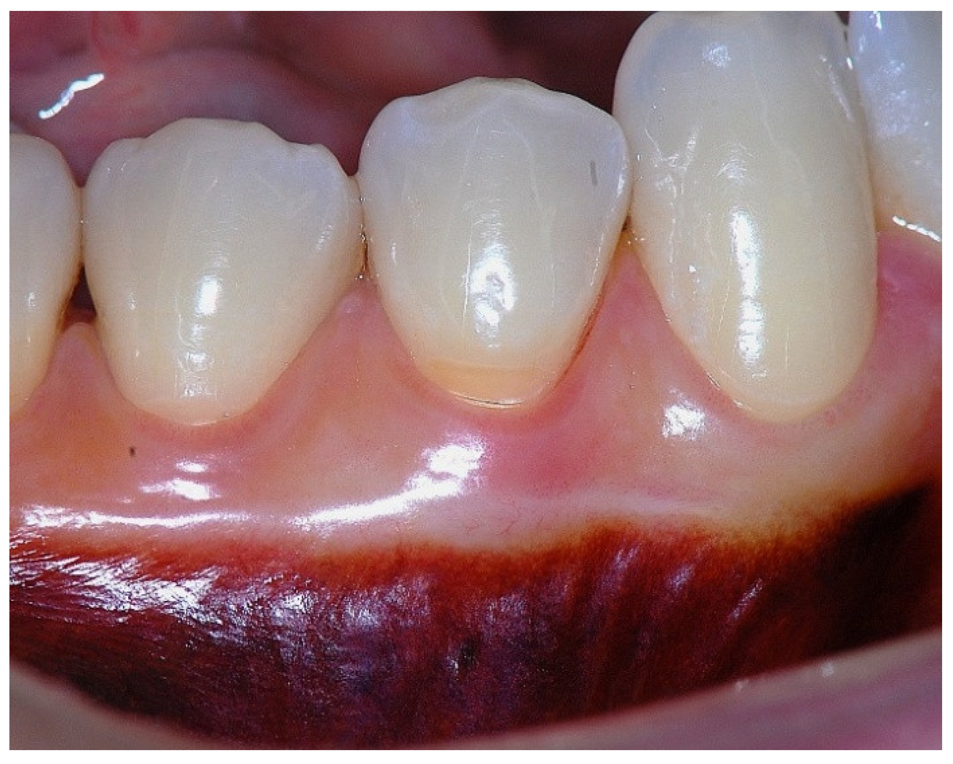
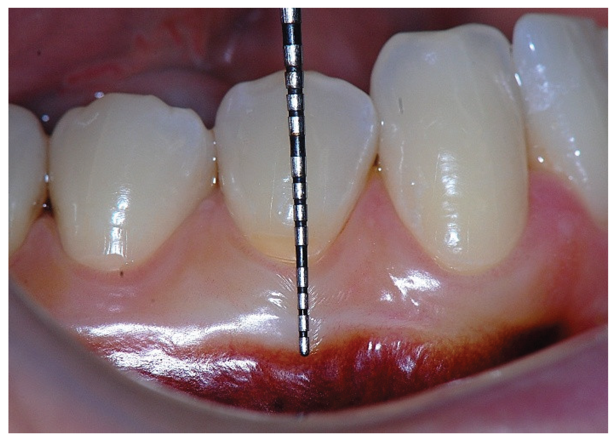
Background: The attached gingiva is the portion of the gingiva that is firmly bound to the underlying alveolar bone and cementum. Its firm attachment to the periosteum and connective tissue renders it immobile during physiologic movements. Functionally, the attached gingiva increases the resistance of the periodontium to mechanical and external injuries, contributes to the stabilization of the gingival margin, and aids in the dissipation of physiological forces exerted by the muscular fibers of the alveolar mucosa onto the gingival tissues. Histologically, it is characterized by keratinized epithelium supported by densely packed collagen fibers, which provide firm anchorage to the underlying bone and cementum and contribute to the structural integrity of the dento-gingival unit. Anatomically, the attached gingiva lies apical to the junctional epithelium and coronal to the alveolar mucosa. Clinically, its width is typically determined by subtracting the sulcus depth from the total width of keratinized tissue, a method that inherently includes the junctional epithelium within the measurement. Although some studies indicate that periodontal stability can be maintained with minimal attached gingiva under optimal plaque control, substantial evidence suggests that its presence remains fundamental for preserving gingival architecture, resisting mechanical trauma, and maintaining periodontal health. Practical Application: From a clinical standpoint, it has been suggested that an adequate width of attached gingiva is necessary to protect the periodontium. Nevertheless, situations may arise in which its dimension is reduced or even absent. It has been suggested that a minimum width of 1 mm of attached gingiva is required to maintain periodontal health and provide protection to the periodontium. However, when only this minimal dimension is present, the attachment is predominantly derived from the junctional epithelium, and such a fragile adherence may not be sufficient to protect the dento-gingival unit against mechanical trauma. Consequently, a width greater than 1mm of attached gingiva should be recommended to ensure adequate mechanical protection and to support long-term stability of the gingival margin. Methods: This narrative review addresses classical and contemporary literature on the morphology, histology, function, and clinical relevance of the attached gingiva. Studies evaluating the width of keratinized and attached tissues, and their influence on periodontal stability, inflammation, and mucogingival conditions were examined to clarify the biological significance of adequate tissue dimensions. Results: When minimal attached gingiva is present, reliance on the junctional epithelium may offer insufficient protection against mechanical trauma. Evidence suggests that a width greater than 1 mm of attached gingiva provides improved mechanical defense, supports the stability of the gingival margin, and contributes to the preservation of periodontal health. A tissue-based categorization of the attached gingiva is proposed to assist clinicians in assessing mucogingival conditions and in determining the need for surgical augmentation. Conclusion: This review consolidates current concepts regarding the dimensions required for the protective and resistance functions of the attached gingiva. The proposed categorization offers a practical framework to guide clinical decision-making, enabling practitioners to accurately evaluate attached gingiva quality and determine when surgical intervention is indicated for managing mucogingival deficiencies.
Keywords:
1. Introduction
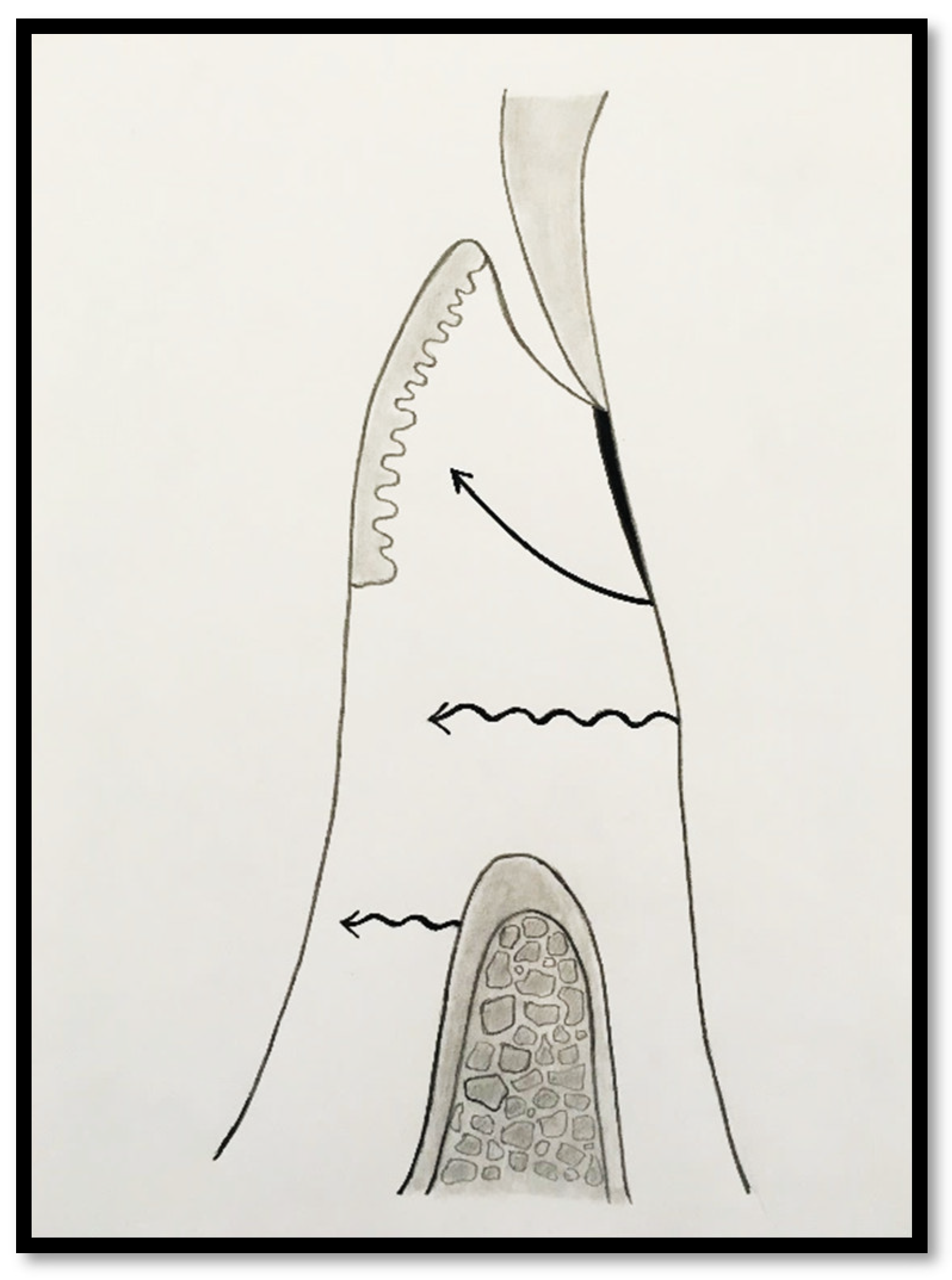
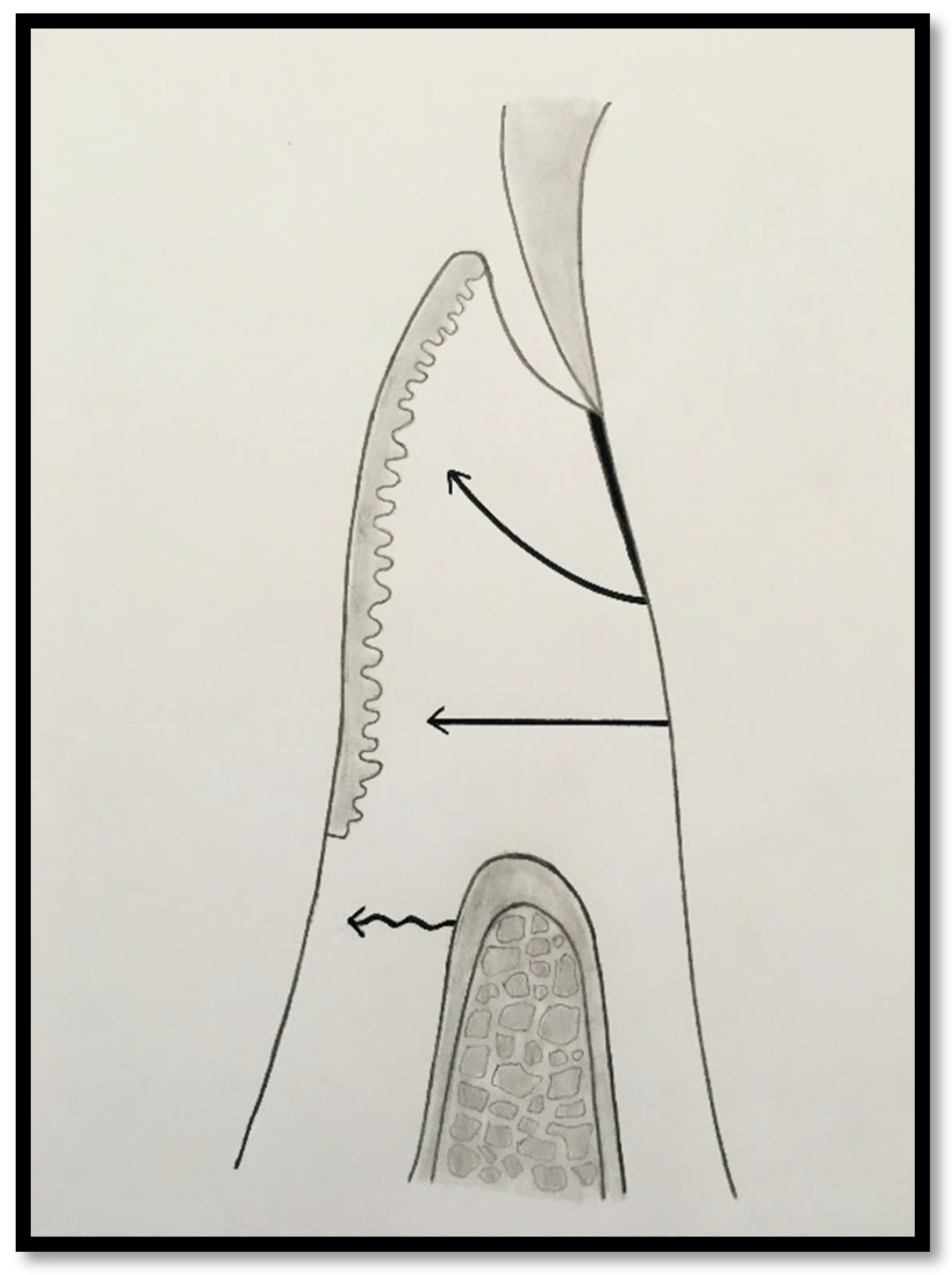
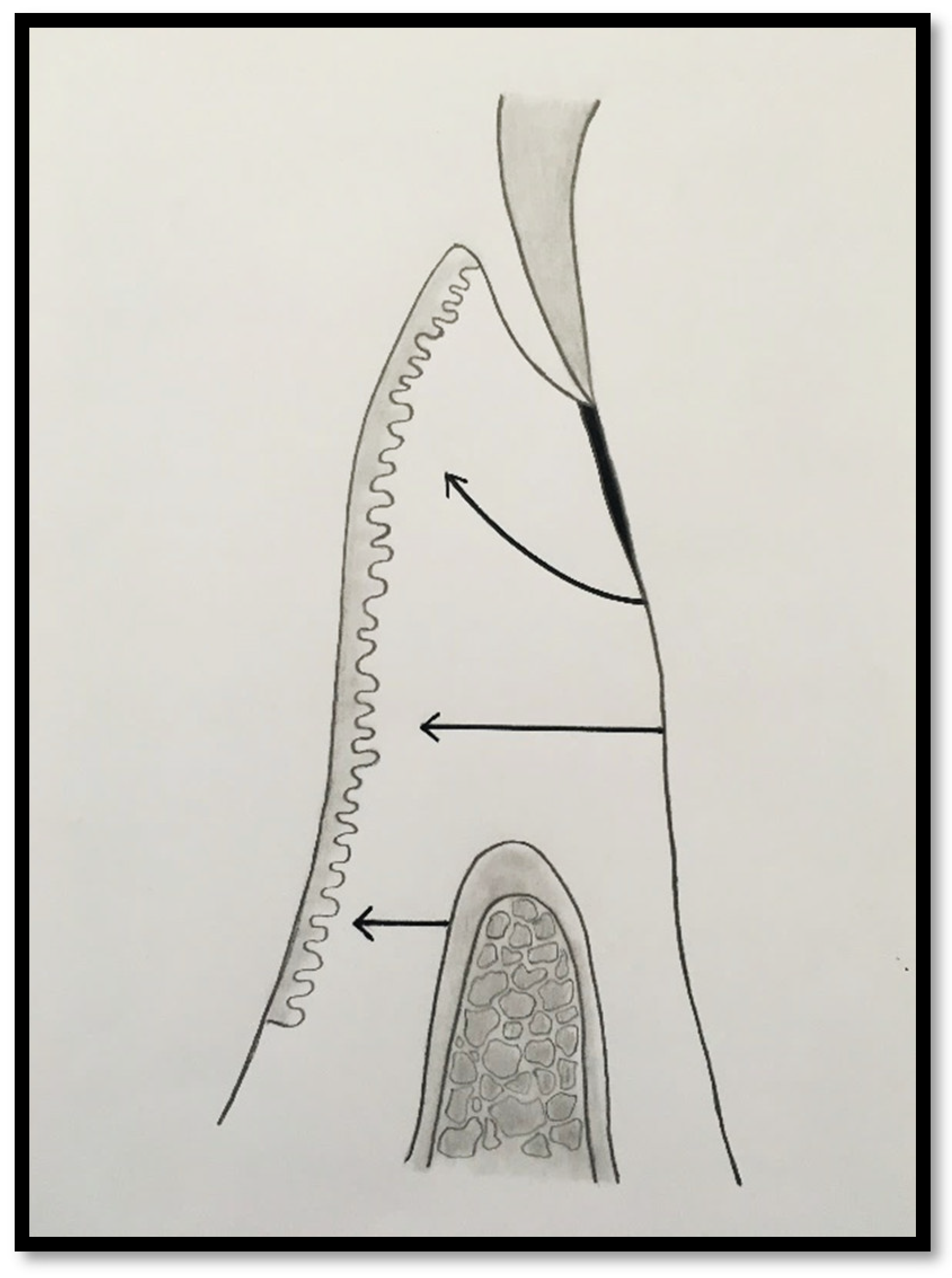
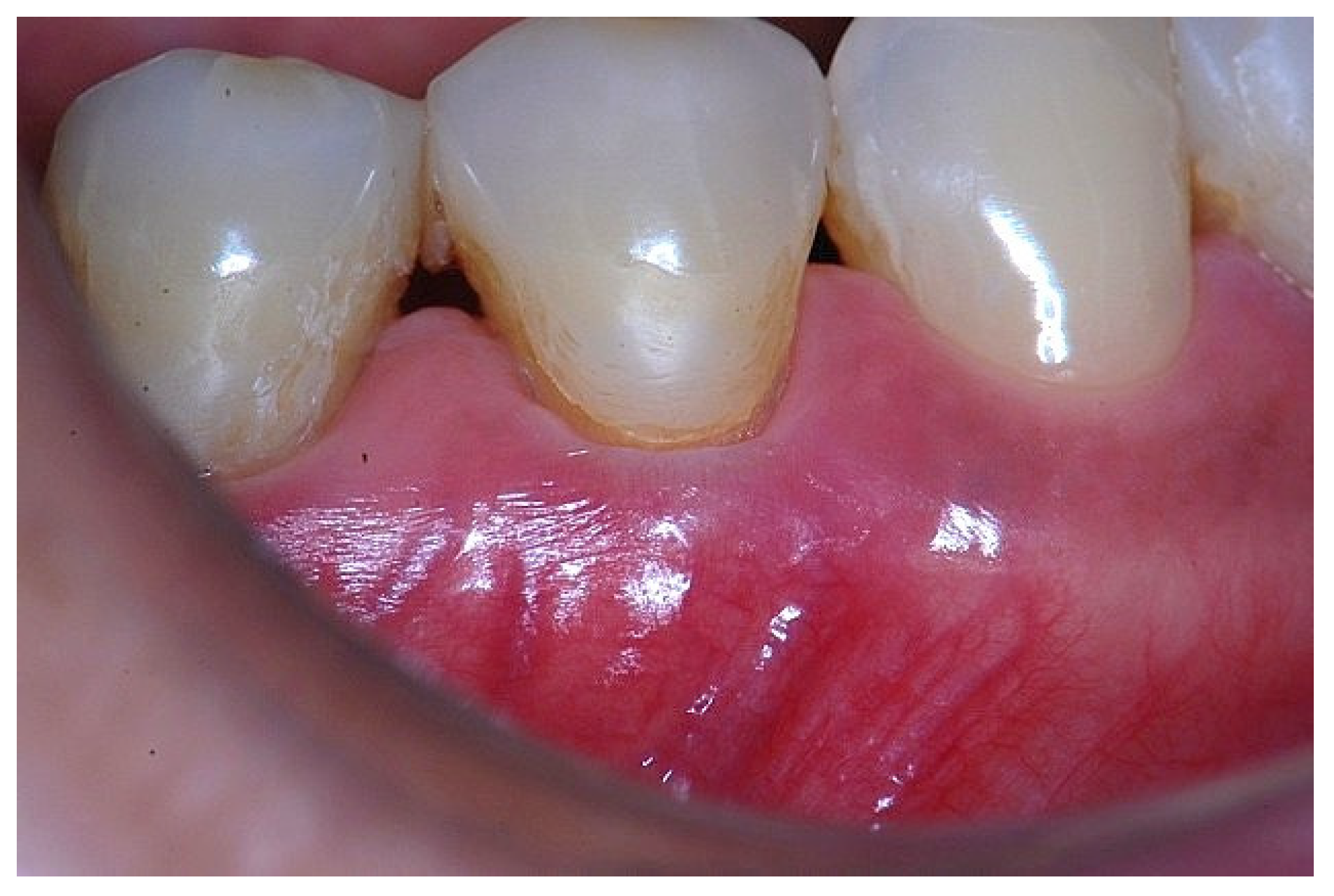
2. Material and Methods
3. Clinical Presentation
4. Discussion
5. Conclusions
6. Clinical Relevance
7. Future Perspectives
Author Contributions Statement
References
- Glossary of Periodontal Terms 2001, 4th edition, American Academy of Periodontology.
- Cho, MI. Garant PR. Development and general structure of the periodontium. Periodontology 2000;24:9-27.
- Newman MG, Takei HH, Klokkevold PR, Carranza FA. Carranza’s Clinical Periodontology, 11th Edition, Elsevier Saunders;2012:9-57.
- Bowers, G.M. A Study of the Width of Attached Gingiva. J. Periodontol. 1963, 34, 201–209. [Google Scholar] [CrossRef]
- Carnio, J.; Camargo, P.M.; Passanezi, E. Increasing the Apico-Coronal Dimension of Attached Gingiva Using the Modified Apically Repositioned Flap Technique: A Case Series With a 6-Month Follow-Up. J. Periodontol. 2007, 78, 1825–1830. [Google Scholar] [CrossRef]
- Wennstrom J, Lindhe J, Nyman S. Role of keratinized gingiva for gingival health. Clinical and histologic study of normal and regenerated gingival tissue in dogs. J Clin Periodontol 1981;8:311-328.
- Ericsson I, Lindhe J. Recession in sites with inadequate width of keratinized gingiva. J Clin Periodontol 1984;11:95-103.
- Wennström, J.L. Mucogingival Therapy. Ann. Periodontol. 1996, 1, 671–701. [Google Scholar] [CrossRef]
- Pini Prato, GP. Mucogingival deformities. Ann Periodontol 1999;4;98-101.
- Maynard JG, Jr. , Wilson RDK. Physiologic dimension of the periodontium significant to the restorative dentist. J Periodontol 1979;50:170-174.
- Stetler, K.J.; Bissada, N.F. Significance of the Width of Keratinized Gingiva on the Periodontal Status of Teeth with Submarginal Restorations. J. Periodontol. 1987, 58, 696–700. [Google Scholar] [CrossRef]
- Nevins, M. Attached gingiva--mucogingival therapy and restorative dentistry. . 1986, 6, 9–27. [Google Scholar]
- Lang, N.P.; Löe, H. The Relationship Between the Width of Keratinized Gingiva and Gingival Health. J. Periodontol. 1972, 43, 623–627. [Google Scholar] [CrossRef]
- Scheyer, E.T.; Sanz, M.; Dibart, S.; Greenwell, H.; John, V.; Kim, D.M.; Langer, L.; Neiva, R.; Rasperini, G. Periodontal Soft Tissue Non–Root Coverage Procedures: A Consensus Report From the AAP Regeneration Workshop. J. Periodontol. 2015, 86, S73–S76. [Google Scholar] [CrossRef] [PubMed]
- Wennström, JL. Lack of association between width of attached gingiva and development of soft tissue recession: A 5-year longitudinal study. J Clin Periodontol 1987;14:181-184.
- Freedman, AL. The clinical significance of the width of keratinized gingiva. J Periodontol. 1977;48:595–596.
- Schmidt, J.C.; Sahrmann, P.; Weiger, R.; Schmidlin, P.R.; Walter, C. Biologic width dimensions – a systematic review. J. Clin. Periodontol. 2013, 40, 493–504. [Google Scholar] [CrossRef] [PubMed]
- Gargiulo, A.W.; Wentz, F.M.; Orban, B. Dimensions and Relations of the Dentogingival Junction in Humans. J. Periodontol. 1961, 32, 261–267. [Google Scholar] [CrossRef]
- Passanezi E, Sant’Ana ACP, Rezende MLR, Greghi SLA, Janson WA. Distancias biologicas periodontais: principios para a reconstrucao periodontal, estetica e protetica. 1o Edicao, Artes Medicas, 2011.
- Magnusson, I.; Runstad, L.; Nyman, S.; Lindhe, J. A long junctional epithelium - A locus minoris resistentiae in plaque infection? J. Clin. Periodontol. 1983, 10, 333–340. [Google Scholar] [CrossRef]
- Wennström, J.; Lindhe, J. Plaque-induced gingival inflammation in the absence of attached gingiva in dogs. J. Clin. Periodontol. 1983, 10, 266–276. [Google Scholar] [CrossRef]
- Miyasato, M.; Crigger, M.; Egelberg, J. Gingival condition in areas of minimal and appreciable width of keratinized gingival. J. Clin. Periodontol. 1977, 4, 200–209. [Google Scholar] [CrossRef]
- Kennedy, J.E.; Bird, W.C.; Palcanis, K.G.; Dorfman, H.S. A longitudinal evaluation of varying widths of attached gingiva. J. Clin. Periodontol. 1985, 12, 667–675. [Google Scholar] [CrossRef] [PubMed]
- Schroeder, H.E.; Listgarten, M.A. The gingival tissues: the architecture of periodontal protection. Periodontology 2000 1997, 13, 91–120. [Google Scholar] [CrossRef] [PubMed]
- E Schroeder, H.; A Listgarten, M. Fine structure of the developing epithelial attachment of human teeth. . 1971, 2, 1–134. [Google Scholar]
- Waerhaug, J. Healing of the dento-epithelial junction following the use of dental floss. J. Clin. Periodontol. 1981, 8, 144–150. [Google Scholar] [CrossRef]
- Armitage, G.C. Manual periodontal probing in supportive periodontal treatment. Periodontology 2000 1996, 12, 33–39. [Google Scholar] [CrossRef]
- Phatale, S.; Marawar, P.; Byakod, G.; Lagdive, S.B.; Kalburge, J.V. Effect of retraction materials on gingival health: A histopathological study. J. Indian Soc. Periodontol. 2010, 14, 35–9. [Google Scholar] [CrossRef]
- Schroeder, HE. The periodontium. Handbook of microscopic anatomy. Vol V/5. Berlin:Springer, 1986.
- Lindhe J, Lang NP, Karring T. Clinical Periodontology and Implant Dentistry. 6th Edition, Oxford, Wiley Blackwell, 2015.
- Newman MG, Takei HH, Klokkevold PR, Carranza FA. Carranza’s Clinical Periodontology. 12th Edition, St.Louis, Elsevier Saunders, 2015.
- Jepsen, S.; Caton, J.G.; Albandar, J.M.; Bissada, N.F.; Bouchard, P.; Cortellini, P.; Demirel, K.; de Sanctis, M.; Ercoli, C.; Fan, J.; et al. Periodontal manifestations of systemic diseases and developmental and acquired conditions: Consensus report of workgroup 3 of the 2017 World Workshop on the Classification of Periodontal and Peri-Implant Diseases and Conditions. J. Periodontol. 2018, 89, S237–S248. [Google Scholar] [CrossRef]
- Hempton TJ, Dominici JT. Contemporary crown-lengthening therapy: a review. J Am Dent Assoc 2010;141:647-655.
- Marzadori, M.; Stefanini, M.; Sangiorgi, M.; Mounssif, I.; Monaco, C.; Zucchelli, G. Crown lengthening and restorative procedures in the esthetic zone. Periodontology 2000 2018, 77, 84–92. [Google Scholar] [CrossRef]
- Hassel, TM. Tissues and cells of the periodontium. Periodontol 2000 1993;3:9-28.
- Proceedings of the 1996 Word Workshop in Periodontics. Ann Periodontol 1996;1:1-947.
- Agudio, G.; Nieri, M.; Rotundo, R.; Cortellini, P.; Prato, G.P. Free Gingival Grafts to Increase Keratinized Tissue: A Retrospective Long-Term Evaluation (10 to 25 years) of Outcomes. J. Periodontol. 2008, 79, 587–594. [Google Scholar] [CrossRef] [PubMed]
Disclaimer/Publisher’s Note: The statements, opinions and data contained in all publications are solely those of the individual author(s) and contributor(s) and not of MDPI and/or the editor(s). MDPI and/or the editor(s) disclaim responsibility for any injury to people or property resulting from any ideas, methods, instructions or products referred to in the content. |
© 2025 by the authors. Licensee MDPI, Basel, Switzerland. This article is an open access article distributed under the terms and conditions of the Creative Commons Attribution (CC BY) license (https://creativecommons.org/licenses/by/4.0/).