
Submitted:
19 January 2026
Posted:
21 January 2026
You are already at the latest version
Abstract
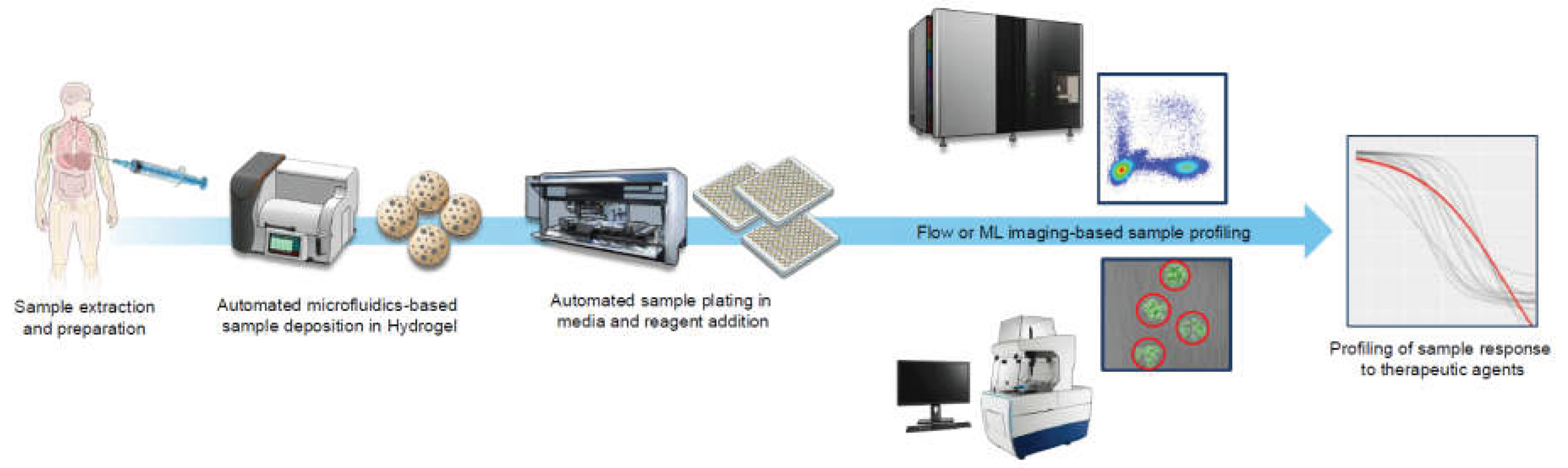
Breast cancer–associated malignant pleural effusion (MPE) is a common and debilitating manifestation of advanced disease, yet current management is largely limited to indwelling pleural catheters and chemical pleurodesis and offers only transient palliation without addressing the underlying tumor biology. We propose that integrating patient-derived organoid modeling of pleural tumor cells with characterization via technologies like next-generation sequencing could shift MPE care from symptom management toward precision intervention. Organoid-based drug testing enables ex vivo evaluation of local therapeutic agents, including intrapleural chemotherapy, immune modulators, and bispecific antibodies, while paired genomic profiling may reveal actionable resistance pathways unique to pleural metastases. Together, these approaches could identify rational, localized combination therapies that improve local control, reduce effusion recurrence, and ultimately extend survival. By coupling functional and molecular analyses directly to the pleural compartment, we envision a translational framework that redefines breast MPE from a purely palliative condition to one amenable to mechanism-driven, patient-tailored therapy.
Keywords:
1. Introduction
1.1. Standard-of-Care
1.2. Alternatives to Standard-of-Care
1.3. Functional Precision Medicine in Cancer
1.4. Modernization and Automation
1.5. Modern Approaches to Breast MPE
1.6. Discussion
Author Contributions
Acknowledgments
Conflicts of Interest
References
- Filho, A.M.; Laversanne, M.; Ferlay, J.; Colombet, M.; Piñeros, M.; Znaor, A.; et al. The GLOBOCAN 2022 cancer estimates: Data sources, methods, and a snapshot of the cancer burden worldwide. Int J Cancer 2025, 156(7), 1336–1346. [Google Scholar] [CrossRef]
- Winters, S.; Martin, C.; Murphy, D.; Shokar, N.K. Breast cancer epidemiology, prevention, and screening. Prog Mol Biol Transl Sci. 2017, 151, 1–32. [Google Scholar]
- Vrancken Peeters, N.J.M.C.; Kerklaan, R.; Vlooswijk, C.; Bijlsma, R.M.; Kaal, S.E.J.; Tromp, J.M.; et al. Long-term health-related quality of life among adolescent and young adult breast cancer survivors. Qual Life Res. 2025, 34(5), 1483–1500. [Google Scholar] [CrossRef]
- Cohn, J.G.; Locke, S.C.; Herring, K.W.; Dent, S.F.; LeBlanc, T.W. Palliative care use and end-of-life care quality in HR+/HER2- metastatic breast cancer. Breast Cancer Res Treat [Internet]. Available online. 16 Aug 2025. [CrossRef]
- Han, Y.-M.; Dong, Y.; Wang, H.-L.; Li, X.-M.; Zhang, X.; Wei, X.-Y.; Qian, F.-W.; Li, Z.-G. Prognostic significance of malignant pleural effusions in patients with advanced luminal B breast cancer. BMC Womens Health 2024, 24(1), 562. [Google Scholar]
- Soni, A.; Ren, Z.; Hameed, O.; Chanda, D.; Morgan, C.J.; Siegal, G.P.; et al. Breast cancer subtypes predispose the site of distant metastases. Am J Clin Pathol. 2015, 143(4), 471–478. [Google Scholar] [CrossRef]
- Piggott, L.M.; Hayes, C.; Greene, J.; Fitzgerald, D. Malignant pleural disease. Breathe (Sheff) [Internet]. Available online. 1 Dec 2023, 19. [CrossRef]
- Zamboni, M.M.; da Silva, CTJr; Baretta, R.; Cunha, E.T.; Cardoso, G.P. Important prognostic factors for survival in patients with malignant pleural effusion. BMC Pulm Med. 2015, 15(1), 29. [Google Scholar] [CrossRef]
- Fentiman, I.S.; Millis, R.; Sexton, S.; Hayward, J.L. Pleural effusion in breast cancer: A review of 105 cases. Cancer 1981, 47(8), 2087–2092. [Google Scholar] [CrossRef] [PubMed]
- Wang, Y.; Zhou, T.; Zhao, S.; Li, N.; Sun, S.; Li, M. A novel clinical prognostic model for breast cancer patients with malignant pleural effusion: Avoiding chemotherapy in low-risk groups? Cancer Manag Res. 2023, 15, 409–422. [Google Scholar] [CrossRef]
- Munavvar, M.; Bodtger, U.; Carus, A.; Cordovilla, R.; Naik, S.; Salud, A.; et al. Current trends in treating malignant pleural effusion: Evidence, guidelines, and best practice recommendations. JCO Oncol Pract. 2025, 21(6), 759–765. [Google Scholar] [CrossRef]
- Zaza, C.; Baine, N. Cancer pain and psychosocial factors: A critical review of the literature. J Pain Symptom Manage 2002, 24(5), 526–542. [Google Scholar] [CrossRef]
- Guo, Y.-Q.; Ju, Q.-M.; You, M.; Liu, Y.; Yusuf, A.; Soon, L.K. Depression, anxiety and stress among metastatic breast cancer patients on chemotherapy in China. BMC Nurs. 2023, 22(1), 33. [Google Scholar] [CrossRef]
- Economidou, F.; Margaritopoulos, G.; Antoniou, K.M.; Siafakas, N.M. The angiogenetic pathway in malignant pleural effusions: Pathogenetic and therapeutic implications. Exp Ther Med. 2010, 1(1), 3–7. [Google Scholar] [CrossRef] [PubMed]
- Jovanovic, D. Etiopathogenesis of malignant pleural effusion. AME Med J. 2021, 6, 28–28. [Google Scholar] [CrossRef]
- Donnenberg, V.S.; Luketich, J.D.; Sultan, I.; Lister, J.; Bartlett, D.L.; Ghosh, S.; et al. A maladaptive pleural environment suppresses preexisting anti-tumor activity of pleural infiltrating T cells. Front Immunol. 2023, 14, 1157697. [Google Scholar] [CrossRef]
- Sivabalah, K.; Balata, H.; Craig, C.; Alsaaty, A.; Conroy, K.; Ong, W.H.; et al. The 2023 British Thoracic Society guideline for pleural disease update on malignant pleural effusion. JoR 2024, 4(4), 210–222. [Google Scholar]
- Shafiq, M.; Frick, K.D.; Lee, H.; Yarmus, L.; Feller-Kopman, D.J. Management of malignant pleural effusion. J Bronchology Interv Pulmonol. 2015, 22(3), 215–225. [Google Scholar] [CrossRef]
- Siefen, A.-C.; Eilers, L.; Baltin, C.T.; Kron, F. Cost comparison of treatment alternatives for pleural effusion and ascites from a payer perspective: Are there cost savings from indwelling catheters? J Palliat Med. 2023, 26(11), 1510–1520. [Google Scholar] [CrossRef]
- Duong, V.; Hargreaves, B.; Muruganandan, S. Management of malignant pleural effusion in 2024: A definitive and unified global approach. JCO Oncol Pract. 2025, 21(6), 739–741. [Google Scholar] [CrossRef]
- Peel, A.M.; Mishra, E.K. The psychosocial impact of Indwelling Pleural Catheters: A scoping review. Cureus 2023, 15(7), e41689. [Google Scholar] [CrossRef] [PubMed]
- Iqbal, B.; Bedawi, E.; Rahman, N.M. Pro: Indwelling pleural catheters cause harm to patients. Breathe (Sheff) 2024, 20(3), 240034. [Google Scholar] [CrossRef] [PubMed]
- Sidhu, C.; Wright, G.; Peddle-McIntyre, C.J.; Tan, A.L.; Lee, Y.C.G. Management of malignant pleural effusion and trapped lung: A survey of respiratory physicians and thoracic surgeons in Australasia. Intern Med J. 2024, 54(7), 1119–1125. [Google Scholar]
- Mei, F.; Tamburrini, M.; Gonnelli, F.; Morandi, L.; Bonifazi, M.; Sediari, M.; et al. Management of malignant pleural effusion in Italian clinical practice: A nationwide survey. BMC Pulm Med. 2023, 23(1), 252. [Google Scholar] [CrossRef] [PubMed]
- Sarkar, R.R.; Courtney, P.T.; Bachand, K.; Sheridan, P.E.; Riviere, P.J.; Guss, Z.D.; et al. Quality of care at safety-net hospitals and the impact on pay-for-performance reimbursement. Cancer 2020, 126(20), 4584–4592. [Google Scholar] [CrossRef] [PubMed]
- Thomas, R.; Francis, R.; Davies, H.E.; Lee, Y.C.G. Interventional therapies for malignant pleural effusions: The present and the future: Interventions for MPE. Respirology 2014, 19(6), 809–822. [Google Scholar]
- Gonnelli, F.; Hassan, W.; Bonifazi, M.; Pinelli, V.; Bedawi, E.O.; Porcel, J.M.; et al. Malignant pleural effusion: Current understanding and therapeutic approach. Respir Res. 2024, 25(1), 47. [Google Scholar] [CrossRef]
- Orlandi, R.; Cara, A.; Cassina, E.M.; Degiovanni, S.; Libretti, L.; Pirondini, E.; et al. Malignant pleural effusion: Diagnosis and treatment-up-to-date perspective. Curr Oncol. 2024, 31(11), 6867–6878. [Google Scholar] [CrossRef] [PubMed]
- Boshuizen, R.C.; Thomas, R.; Lee, Y.C.G. Advantages of indwelling pleural catheters for management of malignant pleural effusions. Curr Respir Care Rep. 2013, 2(2), 93–99. [Google Scholar] [CrossRef]
- Bhatnagar, R.; Kahan, B.C.; Morley, A.J.; Keenan, E.K.; Miller, R.F.; Rahman, N.M.; et al. The efficacy of indwelling pleural catheter placement versus placement plus talc sclerosant in patients with malignant pleural effusions managed exclusively as outpatients (IPC-PLUS): Study protocol for a randomised controlled trial. Trials 2015, 16, 48. [Google Scholar] [CrossRef]
- Semenova, Y.; Burkitbayev, Z.; Kalibekov, N.; Digay, A.; Zhaxybayev, B.; Shatkovskaya, O.; et al. The evolving role of chemotherapy in the management of pleural malignancies: Current evidence and future directions. Cancers (Basel) [Internet]. Available online. 25 Jun 2025. [CrossRef]
- Mitchell, M.A.; Deschner, E.; Dhaliwal, I.; Robinson, M.; Li, P.; Kwok, C.; et al. Patient perspectives on the use of indwelling pleural catheters in malignant pleural effusions. Thorax 2023, 78(11), 1111–1117. [Google Scholar] [CrossRef]
- Wang, S.; Zhang, R.; Wan, C.; Qin, J.; Hu, X.; Shen, Y.; et al. Incidence of complications from indwelling pleural catheter for pleural effusion: A meta-analysis. Clin Transl Sci. 2023, 16(1), 104–117. [Google Scholar] [CrossRef]
- Zhang, J.; Liang, J.; Kadwani, O.; Agoramoorthy, L.; Montalvo, S.; Radcliffe, G.; et al. S138 Malignant pleural effusions: Evaluating the psychosocial impact of indwelling pleural catheters on patients (MY-IPC) – an interim analysis. In Bridge over troubled waters’ – Managing the exudative effusion [Internet]; BMJ Publishing Group Ltd and British Thoracic Society, 2023. [Google Scholar] [CrossRef]
- Kathamuthu, V.; Balakrishnan, R.; Rajendran, S.; Rathinam, P. The safety and efficacy of chemical pleurodesis agents in patients with malignant pleural effusion admitted in tertiary care hospital. Journal of Association of Pulmonologist of Tamil Nadu 2025, 8(1), 17–22. [Google Scholar] [CrossRef]
- Kwok, C.; Thavorn, K.; Amjadi, K.; Aaron, S.D.; Kendzerska, T. Mortality after treatment of malignant pleural effusions with indwelling pleural catheters versus chemical pleurodesis: A population-based study. Respir Res. 2024, 25(1), 409. [Google Scholar] [CrossRef]
- Baiu, I.; Yevudza, E.; Shrager, J.B. Talc pleurodesis: A medical, medicolegal, and socioeconomic review. Ann Thorac Surg. 2020, 109(4), 1294–1301. [Google Scholar] [CrossRef] [PubMed]
- Zhang, W.; Zhao, Y.-L.; Li, S.-J.; Zhao, Y.-N.; Guo, N.-N.; Liu, B. Complications of thoracoscopic talc insufflation for the treatment of malignant pleural effusions: A meta-analysis. J Cardiothorac Surg. 2021, 16(1), 125. [Google Scholar] [CrossRef]
- Chinese Thoracic Society; Chinese Medical Association. Chinese expert consensus on treatment of malignant pleural effusion (2023 Edition). Zhonghua Jie He He Hu Xi Za Zhi 2023, 46(12), 1189–1203. [Google Scholar]
- Xu, Y.; Cui, Y.; Jiang, L.; Yu, Y.; Si, W.; Zhu, X. Thoracic perfusion of antiangiogenic agents combined with chemotherapy for treating malignant pleural effusion in non-small cell lung cancer: A network meta-analysis. BMJ Open. 2024, 14(12), e080703. [Google Scholar] [CrossRef] [PubMed]
- Wang, C.-Q.; Huang, X.-R.; He, M.; Zheng, X.-T.; Jiang, H.; Chen, Q.; et al. Intrapleural administration with Rh-endostatin and chemical irritants in the control of malignant pleural effusion: A systematic review and meta-analysis. Front Oncol. 2021, 11, 649999. [Google Scholar] [CrossRef]
- Fan, Y.; Zou, Q.; Li, X.; Qi, X.; Dong, J.; Liu, J.; et al. Analysis of the Efficacy of Endostar Thoracic Perfusion and DDP Intravenous Chemotherapy for Malignant Pleural Effusion of Breast Cancer. Pract J Cancer 2018, 33(7), 1175–1177. [Google Scholar]
- Biaoxue, R.; Xiguang, C.; Hua, L.; Wenlong, G.; Shuanying, Y. Thoracic perfusion of recombinant human endostatin (Endostar) combined with chemotherapeutic agents versus chemotherapeutic agents alone for treating malignant pleural effusions: A systematic evaluation and meta-analysis. BMC Cancer 2016, 16(1), 888. [Google Scholar] [CrossRef]
- Hao, Y.; Gkasti, A.; Managh, A.J.; Dagher, J.; Sifis, A.; Tiron, L.; et al. Hyperthermic intrathoracic chemotherapy modulates the immune microenvironment of pleural mesothelioma and improves the impact of dual immune checkpoint inhibition. Cancer Immunol Res. 2025, 13(2), 185–199. [Google Scholar] [CrossRef]
- Khosrawipour, C.; Nicpoń, J.; Kiełbowicz, Z.; Prządka, P.; Liszka, B.; Zielinski, K.; et al. First in vivo applicational data of foam-based intrathoracic chemotherapy (FBiTC) in a swine model. Pharmaceuticals 2023, 17, 45. [Google Scholar] [CrossRef]
- Sebastian, M.; Kiewe, P.; Schuette, W.; Brust, D.; Peschel, C.; Schneller, F.; et al. Treatment of malignant pleural effusion with the trifunctional antibody catumaxomab (Removab) (anti-EpCAM x Anti-CD3), results of a phase 1/2 study. J Immunother. 2009, 32(2), 195–202. [Google Scholar] [CrossRef] [PubMed]
- Ammouri, L.; Prommer, E.E. Palliative treatment of malignant ascites: Profile of catumaxomab. Biologics 2010, 4, 103–110. [Google Scholar] [PubMed]
- Aggarwal, C.; Haas, A.R.; Metzger, S.; Aguilar, L.K.; Aguilar-Cordova, E.; Manzanera, A.G.; et al. Phase I study of intrapleural gene-mediated cytotoxic immunotherapy in patients with malignant pleural effusion. Mol Ther. 2018, 26(5), 1198–1205. [Google Scholar] [CrossRef]
- Park, H.; Lewis, C.; Dadgar, N.; Sherry, C.; Evans, S.; Ziobert, S.; et al. Intra-pleural and intra-peritoneal tocilizumab therapy for managing malignant pleural effusions and ascites: The Regional Immuno-Oncology Trial (RIOT)−2 study protocol. Surg Oncol Insight 2024, 1(2), 100045. [Google Scholar] [CrossRef]
- Donnenberg, A.D.; Luketich, J.D.; Dhupar, R.; Donnenberg, V.S. Treatment of malignant pleural effusions: The case for localized immunotherapy. J Immunother Cancer 2019, 7(1), 110. [Google Scholar] [CrossRef]
- Li, X.; Wu, G.; Chen, C.; Zhao, Y.; Zhu, S.; Song, X.; et al. Intrapleural injection of anti-PD1 antibody: A novel management of malignant pleural effusion. Front Immunol. 2021, 12, 760683. [Google Scholar] [CrossRef] [PubMed]
- Wang, P.; Zhang, C.; Hao, P.; Wang, S.; Zhu, R.; Li, J.; et al. The Observation of Clinical Efficacy and Safety of De-Platinum-Based Pleural Perfusion in the Treatment of Malignant Pleural Effusion and Its Correlation with the Expression of VEGF in Pleural Fluid. Journal of Cancer Therapy 2024, 15(12), 432–445. [Google Scholar] [CrossRef]
- Kroesen, B.J.; Nieken, J.; Sleijfer, D.T.; Molema, G.; de Vries, E.G.; Groen, H.J.; et al. Approaches to lung cancer treatment using the CD3 x EGP-2-directed bispecific monoclonal antibody BIS-1. Cancer Immunol Immunother. 1997, 45(3–4), 203–206. [Google Scholar] [CrossRef]
- Cai, J.; Zhang, F.; Song, Z.; Jin, J.; Lv, D.; Pang, W.; et al. 1371P An anti-EpCAM x CD3 bispecific antibody, M701, for the treatment of malignant pleural effusion in NSCLC patients: Intermediate results of a prospective multicenter phase Ib trial. Ann Oncol. 2024, 35, S862. [Google Scholar] [CrossRef]
- He, D.; Ding, R.; Wen, Q.; Chen, L. Novel therapies for malignant pleural effusion: Anti-angiogenic therapy and immunotherapy (Review). Int J Oncol. 2021, 58(3), 359–370. [Google Scholar] [CrossRef]
- Murthy, V.; Katzman, D.; Sterman, D.H. Intrapleural immunotherapy: An update on emerging treatment strategies for pleural malignancy. Clin Respir J. 2019, 13(5), 272–279. [Google Scholar] [CrossRef]
- Wong, T.; Fuld, A.D.; Feller-Kopman, D.J. Malignant pleural effusions in the era of immunotherapy and antiangiogenic therapy. Semin Respir Crit Care Med. 2023, 44(4), 447–453. [Google Scholar] [CrossRef]
- Wang, D.-X.; Zhu, M.; Guo, D.-H.; Gu, J.; Xia, L.; Huang, X.-W.; et al. Safety of Endostar in combination with chemotherapy in patients with cancer. Indian J Cancer 2024, 61(4), 694–702. [Google Scholar] [CrossRef]
- Penz, E.; Watt, K.N.; Hergott, C.A.; Rahman, N.M.; Psallidas, I. Management of malignant pleural effusion: Challenges and solutions. Cancer Manag Res. 2017, 9, 229–241. [Google Scholar] [CrossRef] [PubMed]
- Honkala, A.; Malhotra, S.V.; Kummar, S.; Junttila, M.R. Harnessing the predictive power of preclinical models for oncology drug development. Nat Rev Drug Discov. 2022, 21(2), 99–114. [Google Scholar] [CrossRef] [PubMed]
- Mak, I.W.; Evaniew, N.; Ghert, M. Lost in translation: Animal models and clinical trials in cancer treatment. Am J Transl Res. 2014, 6(2), 114–118. [Google Scholar]
- NIH animal model funding. Available online: https://grants.nih.gov/news-events/nih-extramural-nexus-news/2025/07/nih-funding-announcements-to-align-with-nih-initiative-to-prioritize-human-based-research.
- Letai, A. Functional precision cancer medicine—Moving beyond pure genomics. News@nat,Com 2017, 23, 1028–1035. [Google Scholar] [CrossRef] [PubMed]
- Napoli, G.C.; Figg, W.D.; Chau, C.H. Functional drug screening in the era of precision medicine. Front Med (Lausanne) 2022, 9, 912641. [Google Scholar] [CrossRef]
- Morand du Puch, C.B.; Vanderstraete, M.; Giraud, S.; Lautrette, C.; Christou, N.; Mathonnet, M. Benefits of functional assays in personalized cancer medicine: More than just a proof-of-concept. Theranostics 2021, 11(19), 9538–9556. [Google Scholar] [CrossRef]
- Kornblith, P.L. Role of tissue culture in prediction of malignancy. Neurosurgery 1978, 25 (Supplement 1), 346–376. [Google Scholar] [CrossRef]
- Su, Y. Cancer Chemosensitivity Testing: Review. J Cancer Ther. 2014, 05(07), 672–679. [Google Scholar] [CrossRef]
- Letai, A.; Bhola, P.; Welm, A.L. Functional precision oncology: Testing tumors with drugs to identify vulnerabilities and novel combinations. Cancer Cell. 2022, 40(1), 26–35. [Google Scholar] [CrossRef]
- Crystal, A.S.; Shaw, A.T.; Sequist, L.V.; Friboulet, L.; Niederst, M.J.; Lockerman, E.L.; et al. Patient-derived models of acquired resistance can identify effective drug combinations for cancer. Science 2014, 346(6216), 1480–1486. [Google Scholar] [CrossRef]
- Friedman, A.A.; Letai, A.; Fisher, D.E.; Flaherty, K.T. Precision medicine for cancer with next-generation functional diagnostics. Nat Rev Cancer 2015, 15(12), 747–756. [Google Scholar] [CrossRef] [PubMed]
- Knight, E.; Przyborski, S. Advances in 3D cell culture technologies enabling tissue-like structures to be created in vitro. J Anat. 2015, 227(6), 746–756. [Google Scholar] [CrossRef]
- Petersen, O.W.; Rønnov-Jessen, L.; Howlett, A.R.; Bissell, M.J. Interaction with basement membrane serves to rapidly distinguish growth and differentiation pattern of normal and malignant human breast epithelial cells. Proc Natl Acad Sci U S A 1992, 89(19), 9064–9068. [Google Scholar] [CrossRef]
- Fuchs, E.; Tumbar, T.; Guasch, G. Socializing with the neighbors: Stem cells and their niche. Cell. 2004, 116(6), 769–778. [Google Scholar] [CrossRef] [PubMed]
- de la Puente, P.; Muz, B.; Gilson, R.C.; Azab, F.; Luderer, M.; King, J. 3D tissue-engineered bone marrow as a novel model to study pathophysiology and drug resistance in multiple myeloma. Biomaterials 2015, 73, 70–84. [Google Scholar] [CrossRef] [PubMed]
- Fan, H.; Demirci, U.; Chen, P. Emerging organoid models: Leaping forward in cancer research. J Hematol Oncol. 2019, 12(1), 142. [Google Scholar] [CrossRef]
- Santo, V.E.; Rebelo, S.P.; Estrada, M.F.; Alves, P.M.; Boghaert, E.; Brito, C. Drug screening in 3D in vitro tumor models: Overcoming current pitfalls of efficacy read-outs. Biotechnol J. 2017, 12(1), 1600505. [Google Scholar] [CrossRef]
- Kleinman, H.K.; Philp, D.; Hoffman, M.P. Role of the extracellular matrix in morphogenesis. Curr Opin Biotechnol. 2003, 14(5), 526–532. [Google Scholar] [CrossRef]
- Riedl, A.; Schlederer, M.; Pudelko, K.; Stadler, M.; Walter, S.; Unterleuthner, D.; et al. Comparison of cancer cells in 2D vs 3D culture reveals differences in AKT-mTOR-S6K signaling and drug responses. J Cell Sci. 2017, 130(1), 203–218. [Google Scholar] [PubMed]
- Tong, L.; Cui, W.; Zhang, B.; Fonseca, P.; Zhao, Q.; Zhang, P.; et al. Patient-derived organoids in precision cancer medicine. Med (N Y) 2024, 5(11), 1351–1377. [Google Scholar] [CrossRef]
- Taurin, S.; Alzahrani, R.; Aloraibi, S.; Ashi, L.; Alharmi, R.; Hassani, N. Patient-derived tumor organoids: A preclinical platform for personalized cancer therapy. Transl Oncol. 2025, 51(102226), 102226. [Google Scholar] [CrossRef] [PubMed]
- Zhao, Z.; Chen, X.; Dowbaj, A.M.; Sljukic, A.; Bratlie, K.; Lin, L.; et al. Organoids. Nat Rev Methods Primers [Internet]. 2022, 2. [CrossRef]
- Wensink, G.E.; Elias, S.G.; Mullenders, J.; Koopman, M.; Boj, S.F.; Kranenburg, O.W.; et al. Patient-derived organoids as a predictive biomarker for treatment response in cancer patients. NPJ Precis Oncol. 2021, 5(1), 30. [Google Scholar] [CrossRef]
- Jiang, S.; Zhao, H.; Zhang, W.; Wang, J.; Liu, Y.; Cao, Y.; et al. An automated organoid platform with inter-organoid homogeneity and inter-patient heterogeneity. Cell Rep Med. 2020, 1(9), 100161. [Google Scholar] [CrossRef]
- Yang, C.; Yang, L.; Feng, Y.; Song, X.; Bai, S.; Zhang, S.; et al. Modeling methods of different tumor organoids and their application in tumor drug resistance research. Canc Drug Resist. 2025, 8, 32. [Google Scholar] [CrossRef] [PubMed]
- Yang, H.; Li, J.; Wang, Z.; Khutsishvili, D.; Tang, J.; Zhu, Y.; et al. Bridging the organoid translational gap: Integrating standardization and micropatterning for drug screening in clinical and pharmaceutical medicine. Life Med. 2024, 3(2), lnae016. [Google Scholar] [CrossRef]
- Aisenbrey, E.A.; Murphy, W.L. Synthetic alternatives to Matrigel. Nat Rev Mater. 2020, 5(7), 539–551. [Google Scholar] [CrossRef]
- Li, K.; He, Y.; Jin, X.; Jin, K.; Qian, J. Reproducible extracellular matrices for tumor organoid culture: Challenges and opportunities. J Transl Med. 2025, 23(1), 497. [Google Scholar] [CrossRef]
- Lumibao, J.C.; Okhovat, S.R.; Peck, K.L.; Lin, X.; Lande, K.; Yomtoubian, S.; et al. The effect of extracellular matrix on the precision medicine utility of pancreatic cancer patient-derived organoids. JCI Insight [Internet] 2024, 9(1). [Google Scholar] [CrossRef]
- Driehuis, E.; Kretzschmar, K.; Clevers, H. Establishment of patient-derived cancer organoids for drug-screening applications. Nat Protoc. 2020, 15(10), 3380–3409. [Google Scholar] [CrossRef]
- Foo, M.A.; You, M.; Chan, S.L.; Sethi, G.; Bonney, G.K.; Yong, W.-P.; et al. Clinical translation of patient-derived tumour organoids- bottlenecks and strategies. Biomark Res. 2022, 10(1), 10. [Google Scholar] [CrossRef]
- Xiang, D.; He, A.; Zhou, R.; Wang, Y.; Xiao, X.; Gong, T.; et al. Building consensus on the application of organoid-based drug sensitivity testing in cancer precision medicine and drug development. Theranostics 2024, 14(8), 3300–3316. [Google Scholar] [CrossRef]
- Brugge, J.; Chang, K.-C.; Silvestri, F.; Olipant, M.; Martinez-Gakidis, M.A.; Orgill, D.; et al. Breast organoid suspension cultures maintain long-term estrogen receptor expression and responsiveness [Internet]. Res. Sq. 2024. Available online: https://www.researchsquare.com/article/rs-4463390/v1.
- Önder, C.E.; Ziegler, T.; Becker, R.; Brucker, S.; Hartkopf, A.; Engler, T.; et al. Advancing cancer therapy predictions with patient-derived organoid models of metastatic breast cancer. Cancers (Basel) [Internet]. 2023, 15. [CrossRef]
- Laberiano-Fernandez, C.; Gan, Q.; Wang, S.M.; Tamegnon, A.; Wistuba, I.; Yoon, E.; et al. Exploratory pilot study to characterize the immune landscapes of malignant pleural effusions and their corresponding primary tumors from patients with breast carcinoma and lung adenocarcinoma. J Am Soc Cytopathol. 2024, 13(3), 161–173. [Google Scholar] [CrossRef]
- Pan, B.; Zhao, D.; Liu, Y.; Li, N.; Song, C.; Li, N.; et al. Breast cancer organoids from malignant pleural effusion-derived tumor cells as an individualized medicine platform. In Vitro Cell Dev Biol Anim. 2021, 57(5), 510–518. [Google Scholar] [CrossRef]
- Choi, W.; Kim, Y.H.; Woo, S.M.; Yu, Y.; Lee, M.R.; Lee, W.J.; et al. Establishment of patient-derived organoids using ascitic or pleural fluid from cancer patients. Cancer Research and Treatment: Official Journal of Korean Cancer Association 2023, 55(4), 1077–1086. [Google Scholar] [CrossRef]
- NIH establishes nation’s first dedicated organoid development center to reduce reliance on animal modeling [Internet]. National Institutes of Health (NIH). Available online: https://www.nih.gov/news-events/news-releases/nih-establishes-nations-first-dedicated-organoid-development-center-reduce-reliance-animal-modeling (accessed on 8 October 2025).
- Yang, S.-R.; Mooney, K.L.; Libiran, P.; Jones, C.D.; Joshi, R.; Lau, H.D.; et al. Targeted deep sequencing of cell-free DNA in serous body cavity fluids with malignant, suspicious, and benign cytology. Cancer Cytopathol. 2020, 128(1), 43–56. [Google Scholar] [CrossRef] [PubMed]
- Liu, Y.; Gan, Y.; AiErken, N.; Chen, W.; Zhang, S.; Ouyang, J.; et al. Combining organoid models with next-generation sequencing to reveal tumor heterogeneity and predict therapeutic response in breast cancer. J Oncol. 2022, 2022, 9390912. [Google Scholar] [CrossRef] [PubMed]
- Wang, Z.; Boretto, M.; Millen, R.; Natesh, N.; Reckzeh, E.S.; Hsu, C.; et al. Rapid tissue prototyping with micro-organospheres. Stem Cell Reports 2022, 17(9), 1959–1975. [Google Scholar] [CrossRef] [PubMed]
- Ding, S.; Hsu, C.; Wang, Z.; Natesh, N.R.; Millen, R.; Negrete, M.; et al. Patient-derived micro-organospheres enable clinical precision oncology. Cell Stem Cell. 2022, 29(6), 905–917.e6. [Google Scholar] [CrossRef]
- Chakraborty, R.; Fagan-Solis, K.; DeVilla, J.; Recaldin, T.; Bscheider, M.; Gjorevski, N.; et al. Patient-derived skin MicroOrganoSpheres and clinical response to immunotherapy. J Clin Oncol. 2023, 41((16_) suppl, 2588–2588. [Google Scholar] [CrossRef]
- Köhler, B.C.; Gobits, R.; Schleußner, N.; Oliver, G.R.; Schoenberg, M.R.; SSW, F; et al. 821P Functional precision medicine using microorganospheres for treatment response prediction in advanced colorectal cancer. Annals of Oncology 2025, 36, S555–6. [Google Scholar] [CrossRef]
- Gobits, R.; Schleußner, N.; Oliver, G.; Koomen, M.; Suen, S.; Paolucci, F.; et al. Predicting colorectal cancer patient response to neoadjuvant chemotherapy using the MicroOrganoSphere (MOS) platform. J Clin Oncol [Internet] 2025, 43(16_suppl), 8047. [Google Scholar] [CrossRef]
- Xi, R.; Wang, X.; Baro, N.; Raman, R.; Steele, S.; Helman, E.; et al. Patient-derived MicroOrganoSpheres (MOS) and precision clinical decision-making for patients with multiple myeloma. J Clin Oncol. 2023, 41(16_suppl), 8047. [Google Scholar] [CrossRef]
- Xi, R.; Wang, X.; Moseley, R.; Raman, R.; Zhang, R.; Jaibbar, S.; et al. Abstract 3412: Patient-derived MicroOrganoSpheres (MOS) enable precision clinical decision-making for multiple myeloma patients. Cancer Res. 2023, 83(7_Supplement), 3412–3412. [Google Scholar] [CrossRef]
- Graham, D.; Rupprecht, G.; Watlington, W.; Cushman, J.; Montalvo, A.; Womack, S.; et al. Pilot study of a micro-organosphere drug screen platform to lead care in advanced breast cancer (MODEL-ABC). J Clin Oncol. 2023, 41(16_suppl), 1107. [Google Scholar] [CrossRef]
- Ding, S.; Natesh, N.R.; Spiller, K.; Xi, R.; Nelson, D.; Gjorevski, N.; et al. Micro-organospheres retain patient tumor microenvironment for precision immuno-oncology. J Clin Oncol. 2022, 40((16_) suppl, 2573–2573. [Google Scholar] [CrossRef]
- Hanahan, D. Hallmarks of cancer: New dimensions. Cancer Discov. 2022, 12(1), 31–46. [Google Scholar] [CrossRef]
- Aliazis, K.; Christofides, A.; Shah, R.; Yeo, Y.Y.; Jiang, S.; Charest, A.; et al. The tumor microenvironment’s role in the response to immune checkpoint blockade. Nat Cancer 2025, 6(6), 924–937. [Google Scholar] [CrossRef] [PubMed]
- Wang, Q.; Shao, X.; Zhang, Y.; Zhu, M.; Wang, F.X.C.; Mu, J.; et al. Role of tumor microenvironment in cancer progression and therapeutic strategy. Cancer Med. 2023, 12(10), 11149–11165. [Google Scholar] [CrossRef]
- Thorel, L.; Perréard, M.; Florent, R.; Divoux, J.; Coffy, S.; Vincent, A.; et al. Patient-derived tumor organoids: A new avenue for preclinical research and precision medicine in oncology. Exp Mol Med. 2024, 56(7), 1531–1551. [Google Scholar] [CrossRef] [PubMed]
- Zhou, C.; Wu, Y.; Wang, Z.; Liu, Y.; Yu, J.; Wang, W.; et al. Standardization of organoid culture in cancer research. Cancer Med. 2023, 12(13), 14375–14386. [Google Scholar] [CrossRef] [PubMed]
- Huang, S.; Mei, Z.; Wan, A.; Zhao, M.; Qi, X. Application and prospect of organoid technology in breast cancer. Front Immunol. 2024, 15, 1413858. [Google Scholar] [CrossRef]
- Tzeng, Y.-D.T.; Hsiao, J.-H.; Tseng, L.-M.; Hou, M.-F.; Li, C.-J. Breast cancer organoids derived from patients: A platform for tailored drug screening. Biochem Pharmacol. 2023, 217(115803), 115803. [Google Scholar] [CrossRef]
- Chakraborty, R.; Fagan-Solis, K.; DeVilla, J.; Recaldin, T.; Bscheider, M.; Gjorevski, N.; et al. Patient-derived skin MicroOrganoSpheres and clinical response to immunotherapy. J Clin Oncol. 2023, 41((16_) suppl, 2588–2588. [Google Scholar] [CrossRef]
Disclaimer/Publisher’s Note: The statements, opinions and data contained in all publications are solely those of the individual author(s) and contributor(s) and not of MDPI and/or the editor(s). MDPI and/or the editor(s) disclaim responsibility for any injury to people or property resulting from any ideas, methods, instructions or products referred to in the content. |
© 2026 by the authors. Licensee MDPI, Basel, Switzerland. This article is an open access article distributed under the terms and conditions of the Creative Commons Attribution (CC BY) license.


