
Submitted:
18 November 2025
Posted:
19 November 2025
You are already at the latest version
Abstract
A new iodine-dextrin-lithium complex (IDLC) was synthesized and structurally characterized as a hybrid supramolecular system combining antiseptic, stabilizing, and biocompatible components. The compound integrates iodine as the primary antimicrobial agent, lithium as a coordination and stabilization element, and dextrin as a biodegradable polysaccharide matrix enabling sustained release. Physicochemical analyses confirmed the formation of a uniform, thermally stable complex. Biological evaluation revealed strong bactericidal activity, with minimum bactericidal concentrations (MBC) ranging from 1.95 to 15.63 µg ml-1 against both Gram-positive and Gram-negative pathogens, including multidrug-resistant Staphylococcus aureus and Acinetobacter baumannii. The complex also demonstrated inhibitory effects in virus-infected MDCK cell cultures. Cytotoxicity studies showed CC50 = 0.23-0.48 mg ml-1 on human MNCs and more than 10 mg ml-1 on MDCK cells, confirming low toxicity. The results of the study on the cytotoxicity of IDLC suggest a cytotoxic effect on tumor cell lines such as HepG2, HeLa, AGS, K562, and H9, as well as normal cell lines MeT-5A. Therefore, it can be estimated that IDLC has the potential to be a new drug with antitumor activity against these cell lines. The research confirms that iodine can be effectively stabilized within a dextrin-lithium framework to yield a biologically active, thermally resistant complex, suitable for pharmaceutical use.
Keywords:
dextrin
; iodine
; polymer-iodine complex
; antimicrobial activity
; cytotoxicity
1. Introduction
The objective of modern pharmaceutical manufacturing is the development of not necessarily structurally intricate molecules, but more functionally flexible molecules - complex formulations comprising various bioactive constituents that exert targeted therapeutic effects through synergistic interactions. In the formulation of complex drug products, it is essential to consider both the individual pharmacological properties of each component and their conjoint synergistic potential [1].
Iodine exerts its antimicrobial activity predominantly via oxidative mechanisms, targeting vital cellular structures such as membrane lipids and proteins, resulting in damage to microbial membranes, protein denaturation, and metabolic inhibition [2]. The oxidative disruption of cell wall integrity by iodine leads to rapid microbial lysis and renders pathogens unable to mount effective resistance mechanisms. Iodine demonstrates efficacy against a diverse array of pathogenic microorganisms, including multidrug-resistant strains [3,4].
Lithium is frequently used as a stabilizing agent in pharmaceutical preparations due to its ability to reduce the degradation rate of active substances and extend their duration of action. In addition, lithium exhibits the ability to modulate cellular processes and acts as a membrane stabilizer, impairing the normal functioning of bacterial cells [5,6,7,8]. Its ability to interfere with microbial transport systems and enzymatic pathways positions lithium as a potentiator of antimicrobial action within combinatory systems.
Dextrin, a polysaccharide derivative utilized as a pharmaceutical adjuvant, serves multiple functions within the formulation. It enhances the solubility and bioavailability of APIs and acts as a colloidal stabilizer in aqueous formulations. Dextrin effectively encapsulates labile components such as iodine, by preventing premature breakdown in biological systems, maintaining the chemical stability and pharmacological availability of the formulation [9,10,11,12].
The rational selection of iodine, lithium, and dextrin as components of the multicomponent therapeutic system is predicated not only on their individual bioactivities but also on their capacity for synergistic integration. Iodine serves as the principal antiseptic, while lithium enhances its activity and stabilizes the complex, and dextrin improves release and mitigates irritant effects.
Equally critical to the development of such a complex is the characterization of its physicochemical properties. These parameters determine the viability of the complex as a stable, efficacious therapeutic agent suitable for translational application across multiple delivery platforms.
2. Materials and Methods
2.1. Reagents
The reagents used for the present work were of analytical grade, obtained from commercial sources, and used without further purification. Potato starch (JSC “Rogoznitsky Starch Plant”. Belarus, 98%), dextrin (Sigma-Aldrich, Germany), lithium iodide (Sigma-Aldrich, Germany, ≥ 98%), iodine (G.Amphray Lab. India), magnesium iodide (Sigma-Aldrich, Germany, 98%), hydrochloric acid, lithium chloride (Sigma-Aldrich, Germany, ≥ 99%), purified water by water purification system UltraClear TWF, SG Wasseraufbereitungund Regenerierstation GmbH, Germany), sodium thiosulfate, silver nitrate, nitric acid (fixanal, Uralkhiminvest, Russia), standard solutions for pH-meter (Reagecon Diagnostics Ltd, Ireland).
2.2. Test-Systems
The following museum test strains were used in the study: from the WHO list of strains: Staphylococcus aureus ATCC 6538-P is a sensitive reference strain for determining antimicrobial activity, obtained from the Republican Collection of Microorganisms (RCM), Astana, Kazakhstan; Staphylococcus aureus ATCC BAA-39 is a resistant test strain obtained from the American Collection of Type Cultures (ATCC), USA; Escherichia coli ATCC 8739 is a reference straina strain for determining antimicrobial activity obtained from the American Collection of Type Cultures (ATCC), USA; Escherichia coli ATCC BAA-196 is a resistant test strain for determining antimicrobial activity, obtained from the American Collection of Type Cultures (ATCC), USA; Pseudomonas aeruginosa ATCC 9027 is a reference strain for determining antimicrobial activity, obtained from the American Collection of Type Cultures (ATCC), USA; Pseudomonas aeruginosa TA2 is a resistant strain from clinical study; Acinetobacter baumanii ATCC BAA-1790 is a resistant test strain for determining antimicrobial activity obtained from the American Collection of Type Cultures (ATCC), USA.
To conduct safety and efficacy studies of the new active compound in vitro, the following research objects were used:
Human blood - the work used whole peripheral blood from healthy (absence of acute diseases and severe chronic diseases) donors of both sexes as a source of human immunocompetent cells.
Cell lines - H9 (human T cell lymphoma, ATCC-HTB-176, USA), K-562 (human erythroblastoid leukemia, ATCC-CCL-243, USA), MCF-7 (human breast adenocarcinoma, ATCC-HTB-22, USA), HeLa (human cervical adenocarcinoma, ATCC-CCL-2, USA), P31Cis (cisplatin-resistant mesothelioma, provided by K. Grankvist, Umea University), MeT-5A (SV40 - immortalized human mesothelial cell line, ATCC-CRL-9444, USA) were used for the antitumor effect stydying.
Cell cultures - MDCK kidney cells of the Madin Darby Canine Kidney dog, obtained from the Laboratory of Cellular Biotechnology of the Research Institute of Problems of Biological Safety of the National Research Library of the Ministry of Education and Science of the Republic of Kazakhstan; RD cells of human embryonic rhabdomyosarcoma were obtained from the collection of the Museum of cell Cultures of the National Center for Infectious and Parasitic Diseases, Republic of Bulgaria, Sofia;
Viruses are avian influenza virus strain A /FPV/Waybrige/78/H7N7, obtained from the Laboratory of Virus Ecology at the Institute of Virology and Microbiology of the Science Committee of the Ministry of Education and Science of the Republic of Kazakhstan. The virus was propagated in MDCK cell culture for 72 hours at 37 °C. The titer of the virus in the culture fluid was 107 TCD50/ml.
2.3. Preparation of the Iodine-Containing Complex
2.3.1. Preparation of Dextrin Solution
A starch solution was prepared to obtain dextrin. A flat-bottomed glass flask is filled with demineralized, distilled water in a certain amount. Next, starch powder measured on an electronic technical scale is added to the flask. The solution is thoroughly mixed for 30 minutes. Then pour in a working solution of hydrochloric acid, which is 1/3 of the calculated volume. Then the starch solution (with a molecular weight of 7846 kDa) is transferred to a glass reactor. Hydrolysis is carried out for 25 minutes at a temperature not lower than 88 ° C with constant stirring. At this stage, the temperature control of the process is carried out using a combined Testo 925 measuring device. When the required state is reached, a visual change in the solution to a more transparent one is observed.
2.3.2. Preparation of a Solution of Lithium Triiodide, Magnesium Triiodide
A glass flask with a volume of 500 ml is filled with distilled water, then a measured amount of lithium iodide is filled in. After its dissolution, iodine is added after 10-15 minutes [13]. The contents of the flask are thoroughly mixed. In another flask, the same actions were performed for the magnesium triiodide solution.
2.3.3. Preparation of Lithium Chloride Solution
The required amount of lithium chloride was weighed in an analytical balance with an accuracy of ± 0.001 g. It was then dissolved with distilled water and mixed until completely dissolved.
2.3.4. Mixing Solutions
The prepared solutions of the components were combined into one flask with constant stirring to avoid local concentration differences. The mixture was poured into a mold, which was pre-washed and dried.
2.3.5. Crystallization
The crystallizer with the solution was placed in a desiccator with calcium chloride to ensure slow evaporation of water. The system was left alone until the crystals were completely formed.
2.4. Quantum-Chemical Calculations
Geometry optimization and formation energy calculations of the studied structures were performed using the DFT method with the B3PW91 functional and 6-31G** basis set. All computations were carried out on a Fujitsu PRIMERGY BX920 S1 supercomputer with a peak performance of 10.9 TFLOPS. The formation energy of the complex was calculated according to the following expression:
where, Е1 total - total energy of complex;
∆Е = Е1 total - (Е2 total+ Е3 total+ Е4 total), (1)
Е2 total - total energy of dextrin helix with polypeptide;
Е3 total - total energy of Li+ ion;
Е4 total - total energy of I3- or I2Cl- ion.
2.5. UV-Vis Spectroscopy
UV-vis spectra of the samples were collected using a LAMBDA-35 UV-Vis spectrophotometer (PerkinElmer, USA). The samples were dissolved in a water solution (1 mg/mL), and the solvent was used as a reference. The scanning range was 190-1100 nm [14].
2.6. FT-IR
The samples were processed via the ZnS pellet method, and their FTIR spectra were collected using an FT-IR spectrometer (Nicolet 6700 FTIR spectrophotometer, Thermo Scientific, USA). The test conditions were as follows: wavenumber range, 4000-400 cm-1; scan number, 32; and resolution, 4 cm-1.
2.7. 1H NMR
2.8. XRD
The samples were analyzed using an XRD diffractometer, SmartLab (Rigaku Ultima IV, Japan), under the following conditions: Cu Kα radiation (λ = 1.54059 Å), a one-dimensional detector (D/teX Ultra, Rigaku) with a Kβ filter, and step-scan measurements conducted within a 2θ range of 5-90°, with a step width (Δ2θ) of 0.1° and a scanning speed of 5°/min. Phase identification and investigation of the crystalline structure were performed using the PDXL: Integrated X-Ray Powder Diffraction Software and the international database ICDD PDF-2.
2.9. Thermogravimetric (TG) analysis
Thermal stability analysis of the samples was performed using a TG/DSC STA 449 F1 Jupiter (NETZSCH, Germany) under the following conditions: nitrogen as the carrier gas, a flow rate of 50 mL/min, a temperature range of 30-600 °C, and a heating rate of 10 °C/min [18].
2.10. Capillary Electrophoresis
Capillary electrophoresis was carried out using an Agilent 1600 system with a diode-array detector. Analyte concentrations in sample solutions were adjusted to 5-200 mg/L and filtered through 0.2 µm membranes. Qualitative identification was based on migration time comparison with reference standards, while quantitative determination relied on peak area analysis against certified standards [19].
2.11. SEM-Analysis
SEM analysis was conducted using a Hitachi TM4000Plus tabletop scanning electron microscope equipped with an energy-dispersive X-ray spectroscopy (EDX) module. Imaging was performed at a magnification of x500 under an accelerating voltage of 15 kV.
2.12. Antimicrobial Activity Screening
To determine the MICs of each antimicrobial agent individually, a broth microdilution assay was performed using the method of two-fold serial dilutions according to the CLSI (Clinical and Laboratory Standards Institute) protocol. The bacterial suspension was adjusted to a turbidity of 0.5 McFarland units, corresponding to approximately 1.5×10⁸ CFU/ml. For preparation of the working bacterial suspensions, the stock inoculum was diluted 100-fold in isotonic saline to obtain a final concentration of approximately 1.5×10⁶ CFU/ml.
Microtiter plates were prepared stepwise as follows:
- 100 µL of appropriate liquid growth medium was added to the required wells;
- 100 µL of a pre-prepared 4x stock solution of the corresponding antibiotic was added to the first wells of each row (A1, B1, C1, D1, E1, F1, H1), followed by two-fold serial dilutions across the row to create horizontal serial dilutions of the antibiotic;
- 100 µL of pre-prepared 2x dilutions of the APS compound were added vertically to generate serial dilutions along the columns;
- 20 µL of the working inoculum solution was added to each well containing 200 µL of the antibiotic and compound mixture. As a result, the final bacterial concentration in each well after inoculation was approximately 1.5×10⁵ CFU/mL.
Control wells included a growth control (medium with inoculum, without test compounds) and a sterility control (medium only, without inoculum).
2.13. Cytotoxic Effect
2.13.1. Mononuclear Cells
Peripheral whole blood from healthy male and female donors (free from acute and severe chronic diseases) was used as a source of human immunocompetent cells. Mononuclear cells (MNCs) were isolated by density gradient centrifugation using Histopaque (Sigma, USA) at a 1:1 ratio, followed by centrifugation at 3000 rpm for 20 minutes at 4 °C. The mononuclear cell fraction was washed twice and resuspended in RPMI-1640 culture medium (Sigma, USA). Cell viability was determined by trypan blue exclusion, and only samples with >90% viable cells were used for further experiments [20,21].
2.13.2. Madin-Darby Canine Kidney Cells
MDCK (Madin-Darby Canine Kidney) cells, derived from canine kidney epithelium, were obtained from the Cell Biotechnology Laboratory of the Research Institute for Biological Safety Problems, National Center for Biotechnology. The cells were seeded into 96-well plates at a density of 2 × 10⁵ cells/ml and incubated at 37 °C in a humidified atmosphere containing 5% CO2.
In vitro cytotoxicity of the compound was evaluated using the MTT assay. Compound was added once to the wells containing the cell suspension at final concentrations of 20.0, 10.0, 5.0, 2.5, 1.25, 0.63, 0.31, 0.16, 0.08, and 0.04 mg/ml. The exposure duration was 72 hours. Cytotoxic effects were assessed based on cell viability in comparison to untreated controls [20,21].
3. Results and Discussion
3.1. Quantum-Chemical Calculations
Studying the structure of a compound using the quantum chemical method before the start of synthesis allows not only to predict chemical reactions, but also to optimize the choice of synthesis conditions, such as temperature, catalysts, solvents, and other factors. Special attention is paid to the active centers of the molecule - atoms or their groups, which are key participants in chemical transformations.
The active centers of the iodine-containing complexes were modeled using quantum-chemical calculations employing density functional theory (DFT). The molecular structure of new compund that were coded as IDLC, incorporating two dextrin rings (a), and the appearance of the obtained samples (b) are shown in Figure 1.
In the molecular structure images, atoms are represented with the following color scheme: grey for carbon, blue for nitrogen, red for oxygen, violet for iodine, pink for lithium, yellow for the magnesium ion, and small white spheres for hydrogen.
In complex III, the Li⁺ ion engages in non-covalent interactions with iodine and chlorine atoms, characterized by interatomic distances of 3.02 Å (Li-I) and 2.25 Å (Li-Cl), respectively. The lithium cation is additionally coordinated by an oxygen atom from the dextrin helix (Li-O = 2.22 Å), suggesting a chelating interaction. Molecular iodine within the complex demonstrates ambident behavior, functioning as an electron acceptor toward the chloride anion (I-Cl = 2.22 Å) and as an electron donor toward the lithium cation, as evidenced by the I-I bond length of 2.82 Å. The calculated complexation energy is ΔE = -843.27 kcal/mol.
The calculation results show that molecular iodine and triiodide can be embedded in two dextrin rings, which helps to stabilize their structure and improve their interactions with biological objects. In particular, the interaction of iodine atoms with carbon and oxygen atoms of the dextrin ring through Vandervaals forces maintains the stability of the complex and its ability to effectively act as an anti-infective agent.
3.2. Physicochemical Properties of the Iodine-Containing Complex (IDLC)
IDLC are solutions of a complex iodine compound with polydentate ligands, represented by carbohydrate and polymer associates. The active substance of the sample is an iodine-polymer complex, which serves as a matrix from which the active molecule iodine is gradually released. The colour, percentage yield and melting points (M.P) or temperature of decomposition (d) of the IDLC are presented in Table 1. The complex was dark grey in color (Figure 1b). The solubility of the complexes in water was determined by the Flask method (to determine values above 0.01 g/L) according to the OECD guidelines for the testing of chemicals (water solubility). According to the CE results, the average amount of iodide ions in IDLC is 43.25 mg/l. The lithium ions in IDLC amount to 6.78 mg/l (Table 2).
3.3. UV-Vis Spectral Analysis
Figure 2 presents the spectral characteristics of IDLC. The peaks at 194.58 nm and 223.19 nm correspond to the σ→σ∗ transitions, which are characteristic of ionic bonds such as Li⁺I⁻ and Li⁺Cl⁻. The lithium-chlorine bonds (Li⁺Cl⁻) contribute to the spectrum, causing additional absorption. The peak at 285.39 nm is associated with an n→π∗ transition occurring in iodine (I₂) molecules. The peak at 339.72 nm is attributed to π→π∗ transitions taking place in the iodine molecular system or iodide complexes. The long-wavelength peak at 450.30 nm corresponds to molecular resonance of iodine or to the formation of its complexes.
3.4. TG Analysis
The thermal behaviour of IDLC, which contains dextrin as a matrix component, was evaluated using thermogravimetric analysis (TGA) and differential scanning calorimetry (DSC), as presented in Figure 3. The melting point of the complex, observed at 169.6 °C on the DSC curve, is in good agreement with the value determined using a Gallen kamp variable heating device (Table 4), thereby confirming the consistency of the data.
Thermal decomposition of the IDLC complex occurs in two main stages. Stage I takes place within the temperature range of 28-150 °C, corresponding to the release of physically adsorbed and structurally bound water molecules. This is accompanied by a mass loss of 8.13%, and the DSC curve displays a minor endothermic peak near 97.0 °C, which is consistent with the dehydration process. Stage II begins above 169.6 °C and is associated with the thermal degradation of the organic and iodinated components of the complex. A significant mass loss of 53.39 % is recorded during this stage, indicating the breakdown of the dextrin-based polymeric framework and iodine-containing moieties. The DSC thermogram exhibits two prominent thermal events: an endothermic peak at 169.6 °C, attributed to melting, and a second event at 316.0 °C, which likely corresponds to further decomposition or structural rearrangement. After heating to 500 °C, the residual ash content was found to be 38.48 %, suggesting nearly complete decomposition of the compound.
According to literature, native dextrin typically shows thermal degradation in the range of 250-350 °C, with no distinct melting point due to its amorphous and polymeric nature [22]. In the IDLC complex, the appearance of a sharp melting peak at 169.6 °C indicates a change in the thermal behaviour of dextrin caused by specific interactions with lithium polyiodide species and structural reorganization within the complex. This shift suggests the formation of a more ordered supramolecular structure compared to free dextrin.
Overall, TGA/DSC analysis of IDLC demonstrates a well-defined thermal degradation profile and nearly complete mass loss, indicating high chemical purity and thermal stability of the compound under the experimental conditions.
1H NMR Characterization
The ¹H and ¹³C NMR spectra of the IDLC sample confirm the preservation of the polysaccharide backbone and reveal specific interactions between dextrin and iodine species, likely mediated by lithium ions. In the ¹H NMR spectrum (Figure S1), a distinct signal at 5.239 ppm corresponds to the anomeric proton (H1) of α-1,4-glycosidic linkages, characteristic of glucosidic bonds in starch-derived oligosaccharides. Multiplets in the range of 3.1-3.7 ppm (at 3.108, 3.259, 3.479, 3.588, and 3.612 ppm) are assigned to protons at positions C2-C6 of the glucose units, while a signal at 3.689 ppm corresponds to methylene (CH₂) groups at branching points, typically associated with α-1,6-linkages [3]. The broadness of these signals reflects polymer heterogeneity and possible supramolecular interactions. The ¹³C NMR spectrum (Figure S2) displays resonances for anomeric carbon atoms (C1) at 95.746 ppm (α-form) and 99.580 ppm (β-form), ring carbons (C2-C5) in the range of 68-78 ppm (e.g., 69.277, 71.137, 71.528, 72.673, 73.331, and 76.679 ppm), and the C6 carbon at 60.425 ppm, corresponding to the primary hydroxymethyl group [15]. The observed chemical shift deviations and signal broadening, compared to native dextrin, are attributed to weak ion-dipole or coordination interactions between polyiodide species and hydroxyl groups of the dextrin chain, potentially stabilized by lithium cations [16]. These spectral features support the formation of a structured iodine-polymer complex, as illustrated in Figure 4, and are consistent with earlier studies on similar iodine-polysaccharide systems [17].
XRD Analysis
The XRD pattern of the synthesized sample reveals a combination of crystalline and amorphous features, reflecting the hybrid nature of the iodine-polysaccharide complex (Figure 4).
A broad peak centered around 2θ ≈ 20° corresponds to the amorphous halo of the polysaccharide backbone, which is typical for non-crystalline carbohydrate matrices such as dextrin. The slight shift and increased intensity of this peak compared to pure dextrin suggest partial structural ordering or intermolecular interaction due to iodine incorporation. Distinct crystalline reflections are observed at 2θ = 30.5°, 37.5°, 44.5°, 50.5°, and 55.5°, which match the reference diffraction peaks for lithium iodide (LiI) and confirm its crystalline presence in the sample. These peaks are sharp and well-defined, indicating high crystallinity of the LiI phase. Additional signals at 2θ ≈ 35-37° and 50-56° can be attributed to LiI-I₂ and LiI-Cl-I associate phases, suggesting the formation of mixed halide-iodine interactions within the structure. The signal labeled as I₂//Cl-I corresponds to a possible co-crystallization or complexation between molecular iodine and chloride/iodide species, stabilized by the Li⁺ cation.
The presence of combined reflections from LiI, elemental iodine, and polysaccharide-associated iodine phases indicates the formation of a semiorganic hybrid material, where iodine is partially complexed with the carbohydrate matrix and partially present in crystalline inorganic domains. This supports the proposed structure of a multicomponent iodine coordination complex, where iodine species exist both in bound and free (crystalline) forms.
SEM-Analysis
Quantitative elemental analysis of IDLC was performed using a JEOL JSM-IT200(LA) scanning electron microscope equipped with an energy-efficient X-ray detector (EDX). The distribution of the elements was investigated in several directions, for greater reliability. The data obtained using EDX analysis consists of spectra showing peaks corresponding to the elements that make up the true composition of the analyzed samples. Elemental mapping of the sample and image analysis are also possible (Figure 5), as shown in Figure 6.
The results of EDX IDLC showed that the mass percentage of carbon, oxygen, nitrogen, potassium and iodine is 34,62 ± 0,15; 47,68 ± 0,33; 1,55 ± 0,05; 0,30 ± 0,02 and 10.15 ± 0.16%, respectively. (Table 3). The mass percentages of the constituent elements were compared at all the analyzed points, and it was found that the results were consistent with each other, indicating an equal distribution of the constituent elements in the compound sample. The analysis results confirm the proposed molecular formula of the C₁₂₄H₂₄₈O₁₂₂NI₄K₂ complex.
Antimicrobial Activity Screening
In the course of this study, a screening of IDLC was carried out against several strains of microorganisms, including Staphylococcus aureus, Escherichia coli, Acinetobacter baumannii, and Pseudomonas aeruginosa. The main objective of the work was to determine the minimum bactericidal concentration (MBC) for each of the tested strains and to evaluate their antimicrobial activity. The studies were conducted in accordance with CLSI standards and included both Gram-positive and Gram-negative microorganisms responsible for acute and chronic infectious diseases, characterized by varying levels of resistance to antibiotic drugs.
Antimicrobial activity was evaluated using six reference strains obtained from the American Type Culture Collection (ATCC) and one clinical isolate, namely: Staphylococcus aureus ATCC 6538-P, Staphylococcus aureus ATCC BAA-39, Escherichia coli ATCC 8739, Escherichia coli ATCC BAA-196, Acinetobacter baumannii ATCC BAA-1790, Pseudomonas aeruginosa ATCC 9027, and Pseudomonas aeruginosa TA2. The results of the in vitro screening are presented in Table 4.
IDLC demonstrated the following antimicrobial activity results: MBC 1,95 mcg/ml against S. aureus ATCC BAA-39; 3,91 mcg/ml against P.aeruginosa TA2 and A.baumannii ATCC BAA-1790; 7,81 mcg/ml against E. coli ATCC 196 and P.aeruginosa ATCC 9027; 15,63 mcg/ml against S. aureus ATCC 6538-P and E. coli ATCC 8739. The compound IDLC also exhibits significant activity, especially against the multi-resistant strain S. aureus ATCC BAA-39, with an MBC recorded at a concentration of 1.95 μg/mL; for A. baumannii ATCC BAA-1790 and P. aeruginosa TA2, the MBC is 3.91 μg/mL; it inhibits the growth of Gram-negative bacteria at a concentration of 7.81 μg/mL and has an MBC of 15.63 μg/mL against S. aureus ATCC 6538-P and E. coli ATCC 8739, which makes it promising for further research.
3.8. Cytotoxicity Study
The study of cytotoxicity is an integral part of the process of developing new anti-infective drugs, as it allows us to assess the safety of potential drugs for living cells.
At the stage of development of new active pharmaceutical substances, it is extremely important to determine their effect on cells in order to avoid potential side effects. Cytotoxicity assessment makes it possible to establish safe doses and regimens of drugs, as well as to prevent possible risks in their clinical use.
Cytotoxicity studies of the IDLC have been conducted on different types of cells, such as human peripheral blood MNC cells and MDCK cells (dog kidney mnestocytes). Detection of cytotoxicity for various living cells contributes to a more comprehensive assessment of the drug's safety. Each cell type represents a specific model reflecting different aspects of interaction with the drug, which helps to obtain a more accurate and complete picture of its toxicity. As part of this task, studies have been conducted to study the cytotoxic effect of IDLC on human peripheral blood MNC culture. A quantitative assessment of the cytotoxic effect of the studied substances was carried out using an MTT test. Cytotoxicity assessment and determination of cytotoxic concentration (CC50) of IDLC substances after 24 and 48 hours of exposure to MNC culture in vitro were performed using GraphPad Prism 6 (Table 5).
As a result of the study, curves of the effect dependence on the studied concentrations of IDLC substances on MNC culture were obtained using the nonlinear regression method (Figure 7). After processing the entire concentration range, the CC50 value was calculated in the program, which for IDLC was 0.23 mg/ml at 24-hour exposure. With 48-hour exposure to CC50 IDLC = 0.48 mg/ml.
According to the recommendations [19,20], the maximum tolerable (maximum non-toxic) concentration is ¼ CC50, respectively, the maximum non-toxic concentration at 24-hour exposure to MNC for substance IDLC is 0.06 mg/ml, at 48-hour exposure for IDLC - 0.12 mg/ml. Effective doses, as a rule, should be less than these concentrations.
Studies of the cytotoxicity of IDLC substances in human peripheral blood MNC culture have shown that the complex exhibits varying degrees of toxicity depending on the concentration and time of exposure.
As part of this task, a study of the safety of substances was also conducted on MDCK cell culture, since studying the safety of IDLC on different cell types can help to obtain a more complete picture of toxicity.
The cytotoxicity of the substances in vitro was determined using the MTT test. The MDCK cell culture was seeded into 96-well dies at a concentration of 2 x 10⁵ cells per 1 ml. The dies were cultured in a thermostat at 37 °C and 5% CO2. The results are presented at the Figure 8.
Based on the results obtained to determine the safety of the new complex in in vitro experiments on MDCK cell culture, it was found that IDLC is a low-toxic compound with CC50 concentrations ranging from 10.0 to 20.0 mg/ml.
The data obtained are important for the further development and optimization of the dosage of the studied complex as an API, ensuring a balance between the effectiveness and safety of the drug.
3.9. Assesment of Antitumor Activity of IDLC
In order to assess the potential antitumor activity of the new IDLC complex, the cytotoxic effect on HeLa, HepG2, AGS, K-562, H9 and normal MeT-5A cell lines was studied.
When exposed of IDLC at a concentration of 5 mg/ml, toxicity is observed against HepG2 and K562 tumor lines (28.7% and 11.8% of viable cells, respectively) and especially AGS (3.9% of viable cells), as well as the normal MeT-5A line (34.9% of viable cells). In relation to the H9 tumor line, the toxic effect is noted at concentrations of 5 and 2.5 mg/ml (9.8% and 38.1% of viable cells, respectively) (Table 6).
After processing the entire concentration range in the program, the CC50 value for IDLC was calculated for the HeLa cell line = 1.201 mg/ml (C.I. 0.049 to 29.44), AGS = 1.765 mg/ml (C.I. from 1,600 to 1,947), K562 = 3.533 mg/ml, H9 = 2.003 mg/ml (C.I. from 1,709 to 2,349) and normal MeT-5A cells = 1.370 mg/ml (C.I. from 1,133 to 1,656).
Thus, the results of studying the cytotoxicity of the substance IDLC indicate the presence of a cytotoxic effect against the tumor cell lines HepG2, HeLa, AGS, K562, H9 and the normal cell line MeT-5A. It can be estimetd IDLC as a new potential drug with a antitumor activity.
4. Conclusions
Comprehensive physicochemical, spectroscopic, and biological analyses confirm the successful synthesis and stability of a novel hybrid supramolecular system: the iodine-dextrin-lithium complex (IDLC). Theoretical modeling using DFT calculations demonstrated a highly favorable formation energy (ΔE = -843.27 kcal/mol), indicating strong thermodynamic stability and efficient coordination between Li+, polyiodide species, and dextrin hydroxyl groups. The optimized geometry reveals chelating interactions (Li-O = 2.22 Å, Li-I = 3.02 Å), which ensure the spatial fixation of iodine within the polysaccharide matrix and support controlled release alongside enhanced structural order.
Spectroscopic investigations (UV-Vis, FTIR, 1H/13C NMR) and XRD analyses confirmed the formation of a semi-crystalline supramolecular structure, where iodine species exist in both bound and crystalline states. Characteristic absorption bands at 194-450 nm, chemical shift deviations in NMR spectra, and diffraction peaks corresponding to LiI and LiI-I2 phases collectively indicate complexation between iodine, lithium, and dextrin via noncovalent and coordination interactions. Thermal analysis revealed a two-stage degradation process, with a melting point of 169.6 °C and a total mass loss of approximately 61.5%, confirming the high purity and improved thermal stability of the complex compared to native dextrin.
Biological testing showed that the IDLC exhibits pronounced antibacterial activity, with minimum bactericidal concentrations (MBCs) ranging from 1.95 to 15.63 µg/ml. The strongest effects were observed against S. aureus (MBC = 1.95 µg/ml) and A. baumannii (MBC = 3.91 µg/ml), including multidrug-resistant strains. These findings confirm that lithium inclusion significantly enhances the oxidative bactericidal mechanism of iodine by stabilizing active molecular species and promoting their penetration into microbial membranes.
Cytotoxicity assays revealed moderate, concentration-dependent effects on human mononuclear cells (CC50 = 0.23-0.48 mg/ml) and low toxicity toward MDCK cells (CC50 = 10-20 mg/ml), indicating a favorable therapeutic index. The calculated maximum non-toxic concentrations (0.06-0.12 mg/ml for MNCs) further delineate a broad safety margin for future pharmacological evaluation.
The findings of the study on the toxicity of IDLC suggest a negative impact on various cancer cell lines, including HepG2, HeLa, AGS, K562, and H9, as well as healthy cell lines such as MeT-5A. Therefore, it is logical to conclude that IDLC may have the potential to be a novel drug with anti-cancer effects against these cell types.
The integration of iodine, lithium, and dextrin into a single supramolecular system has yielded a chemically stable, thermally resistant, and biologically active complex. From a pharmaceutical perspective, the IDLC demonstrates promising characteristics as a biocompatible antiseptic and stabilizing platform suitable for development into next-generation iodine-based pharmaceutical formulations with enhanced efficacy against multiresistant pathogens.
Supplementary Materials
The following supporting information can be downloaded at the website of this paper posted on Preprints.org, Figure S1: Figure S1 ¹H NMR spectra of the IDLC sample; Figure S2: ¹³C NMR spectra of the IDLC sample.
Author Contributions
Conceptualization, D.S., S.T. and A.S.; methodology, A.J. and T.B.; validation, B.D., D.A. and Y.D.; formal analysis, A.K., K.S.; investigation, A.J., T.S., T.B., A.Z.; writing—original draft preparation, D.S. and A.S.; writing—review and editing, T.S.; visualization, A.S. and D.S.; supervision, A.S.; project administration, T.S. ans A.S. All authors have read and agreed to the published version of the manuscript.
Funding
This research is funded by a grant №BR24992760 and №AP19576946 from the Ministry of Science and Higher Education of the Republic of Kazakhstan .
Institutional Review Board Statement
Not applicable.
Informed Consent Statement
Not applicable.
Data Availability Statement
The data used to support the findings of this study are available from the corresponding author upon reasonable request.
Conflicts of Interest
The authors declare that they have no conflicts of interest.
Abbreviations
The following abbreviations are used in this manuscript
- API active pharmaceutical ingredient
- CC50 Half cytotoxixity concntration
- DSC Differential scanning calorimetry
- IDLC iodine-dextrin-lithium complex
- MBC minimum bactericidal concentrations
- MDCK Madin-Darby Canine Kidney
- MNC Mononuclear cells
- MTT 3-(4,5-dimethylthiazol-2-yl)-2,5-diphenyltetrazolium bromide)
- XRD X-ray diffraction analysis
References
- Lekhan, A.; Turner, R.J. Exploring Antimicrobial Interactions between Metal Ions and Quaternary Ammonium Compounds toward Synergistic Metallo-Antimicrobial Formulations. Microbiol. Spectr. 2024, 12, e0104724. [Google Scholar] [CrossRef] [PubMed]
- Babalska, Z.Ł.; Korbecka-Paczkowska, M.; Karpiński, T.M. Wound Antiseptics and European Guidelines for Antiseptic Application in Wound Treatment. Pharmaceuticals 2021, 14, 1253. [Google Scholar] [CrossRef]
- Vergara, D.; Loza-Rodríguez, N.; Acevedo, F.; Bustamante, M.; López, O. Povidone-Iodine Loaded Bigels: Characterization and Effect as a Hand Antiseptic Agent. J. Drug Deliv. Sci. Technol. 2022, 72, 103427. [Google Scholar] [CrossRef]
- Smerdely, P.; Lim, A.; Boyages, S.C.; Waite, K.; Wu, D.; Roberts, V.; Leslie, G.; Arnold, J.; John, E.; Eastman, C.J. Topical Iodine-Containing Antiseptics and Neonatal Hypothyroidism in Very-Low-Birthweight Infants. Lancet 1989, 2, 661–664. [Google Scholar] [CrossRef]
- Li, J.; Sun, D.; Wu, J.; Liu, F.; Xu, Y.; Wang, Y.; Shui, X.; Li, Q.; Zhao, B. Lithium Enhanced Plasmid-Mediated Conjugative Transfer of Antimicrobial Resistance Genes in Escherichia coli: Different Concentrations and Mechanisms. Aquat. Toxicol. 2025, 279, 107263. [Google Scholar] [CrossRef] [PubMed]
- Chernova, A.; Pukhniarskaia, D.; Biryukov, M.; Plotnikov, E. Influence of Lithium Salt on Escherichia coli Growth and Viability. Ind. Biotechnol. 2022, 18, 32–37. [Google Scholar] [CrossRef]
- Vaidya, M.Y.; McBain, A.J.; Butler, J.A.; et al. Antimicrobial Efficacy and Synergy of Metal Ions against Enterococcus faecium, Klebsiella pneumoniae and Acinetobacter baumannii in Planktonic and Biofilm Phenotypes. Sci. Rep. 2017, 7, 5911. [Google Scholar] [CrossRef] [PubMed]
- Plotnikov, E.; Pukhnyarskaya, D.; Chernova, A. Lithium and Microorganisms: Biological Effects and Mechanisms. Curr. Pharm. Biotechnol. 2023, 24, 1623–1629. [Google Scholar] [CrossRef] [PubMed]
- Pedotti, S.; Ferreri, L.; Migliore, R.; Leotta, C.G.; Pitari, G.M.; D’Antona, N.; Petralia, S.; Aleo, D.; Sgarlata, C.; Consoli, G.M.L. A Novel Cationic β-Cyclodextrin Decorated with a Choline-Like Pendant Exhibits Iodophor, Mucoadhesive and Bactericidal Properties. Carbohydr. Polym. 2024, 328, 121698. [Google Scholar] [CrossRef] [PubMed]
- Saffarionpour, S.; Diosady, L.L. Cyclodextrins and Their Potential Applications for Delivering Vitamins, Iron, and Iodine for Improving Micronutrient Status. Drug Deliv. Transl. Res. 2025, 15, 26–65. [Google Scholar] [CrossRef] [PubMed]
- Hu, Y.; Yu, B.; Wang, L.; McClements, D.J.; Li, C. Study of Dextrin Addition on the Formation and Physicochemical Properties of Whey Protein-Stabilized Emulsion: Effect of Dextrin Molecular Dimension. Food Hydrocoll. 2022, 128, 107569. [Google Scholar] [CrossRef]
- Zisi, A.P.; Exindari, M.K.; Siska, E.K.; Koliakos, G.G. Iodine-Lithium-Alpha-Dextrin (ILαD) against Staphylococcus aureus Skin Infections: A Comparative Study of In-Vitro Bactericidal Activity and Cytotoxicity between ILαD and Povidone-Iodine. J. Hosp. Infect. 2018, 98, 134–140. [Google Scholar] [CrossRef] [PubMed]
- GOST 4159-79. Reagents. Iodine. Specifications; Standards Publishing House: Moscow, Russia, 1979; 16 pp.
- Kireev, S.V.; Simanovsky, I.G.; Shnyrev, S.L. Investigation of the Absorption Spectra of Molecular Iodine in Liquid Media in the UV and Visible Regions. In Scientific Session of MEPhI; 2007; Volume 4, pp. 49. [CrossRef]
- Brown, G.D.; Bauer, J.; Osborn, H.M.; Kuemmerle, R. A Solution NMR Approach to Determine the Chemical Structures of Carbohydrates Using the Hydroxyl Groups as Starting Points. ACS Omega 2018, 3, 17957–17975. [Google Scholar] [CrossRef] [PubMed]
- Bodach, A.; Ortmeyer, J.; Herrmann, B.; Felderhoff, M. Amino–Organolithium Compounds and Their Aggregation for the Synthesis of Amino–Organoaluminium Compounds. Eur. J. Inorg. Chem. 2021, 23, 2248–2256. [Google Scholar] [CrossRef]
- Cardoso, M.V.C.; Sabadini, E. Before and Beyond the Micellization of n-Alkyl Glycosides: A Water-¹H NMR Relaxation Study. Langmuir 2013, 29, 15778–15786. [Google Scholar] [CrossRef] [PubMed]
- Sabitov, A.; Turganbay, S.; Kerimkulova, A.; Doszhanov, Y.; Saurykova, K.; Atamanov, M.; Zhumazhanov, A.; Bolatova, D. 1-Carboxy-2-phenylethan-1-aminium Iodide 2-Azaniumyl-3-phenylpropanoate Crystals: Properties and Its Biochar-Based Application for Iodine Enrichment of Parsley. Appl. Sci. 2025, 15, 10752. [Google Scholar] [CrossRef]
- European Pharmacopoeia; 7th ed. ; Council of Europe: Strasbourg, France, 2011; Volume 1, pp. 77–82.
- Pracher, L.; Zeitlinger, M. Preclinical and Clinical Studies in the Drug Development Process of European Medicines Agency-Approved Non-HIV Antiviral Agents: A Narrative Review. Clin. Microbiol. Infect. 2025, 31, 931–940. [Google Scholar] [CrossRef]
- OECD. Guideline for the Testing of Chemicals No. 487 – In Vitro Mammalian Cell Micronucleus Test; OECD: Paris, France, 2016. [Google Scholar]
- Turganbay, S.; Ilin, A.; Sabitov, A.; Askarova, D.; Jumagaziyeva, A.; Iskakbayeva, Z.; Seisembekova, A.; Bukeyeva, T.; Azembayev, A. Dextrin/Polyvinyl Alcohol/Iodine Complexes: Preparation, Characterization, and Antibacterial, Cytotoxic Activity. Eng. Sci. 2025, 35, 1595. [Google Scholar] [CrossRef]
Figure 1.
Molecular structure of new compound IDLC, incorporating two dextrin rings (a), and the appearance of the obtained samples (b).
Figure 1.
Molecular structure of new compound IDLC, incorporating two dextrin rings (a), and the appearance of the obtained samples (b).
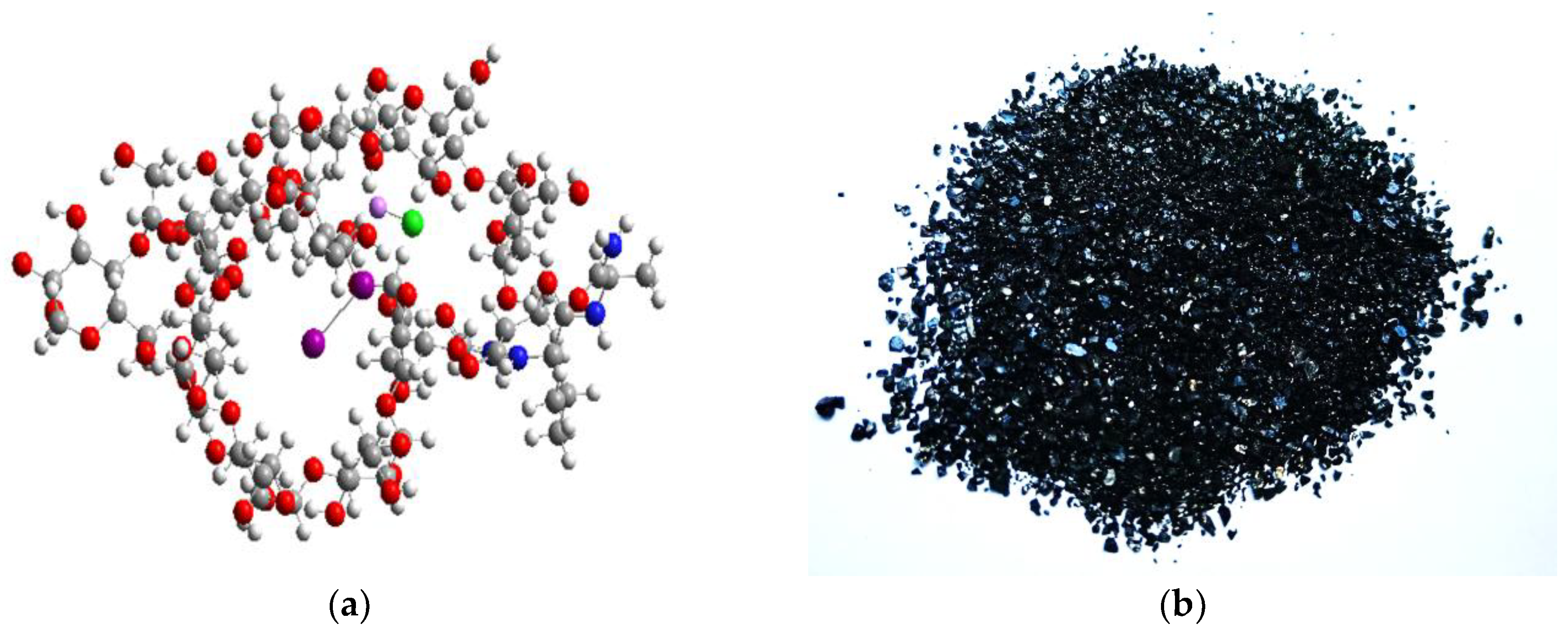
Figure 2.
UV-vis spectrum of IDLC (0,025% solution).
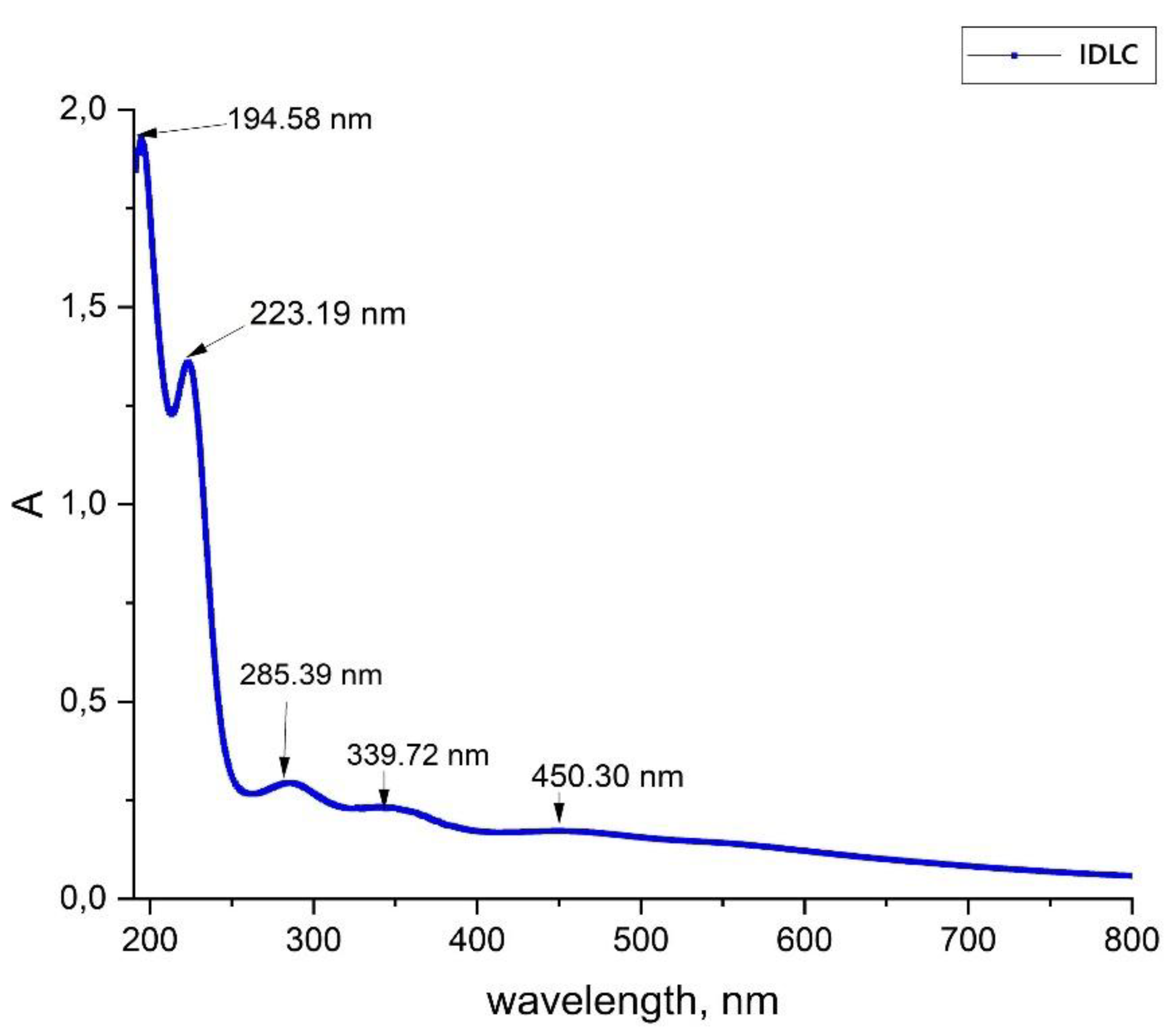
Figure 3.
DSC and TGA curves of IDLC.
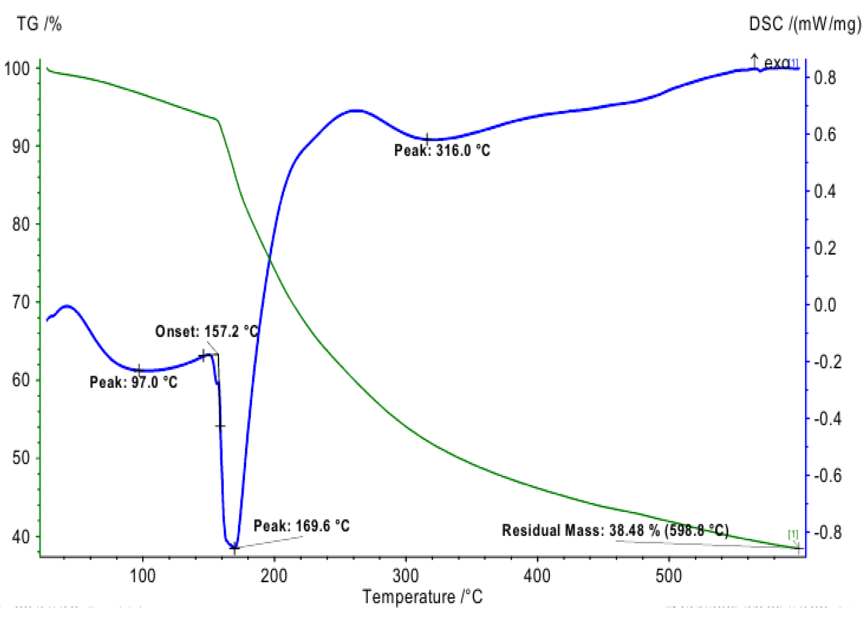
Figure 4.
XRD pattern of the IDLC.
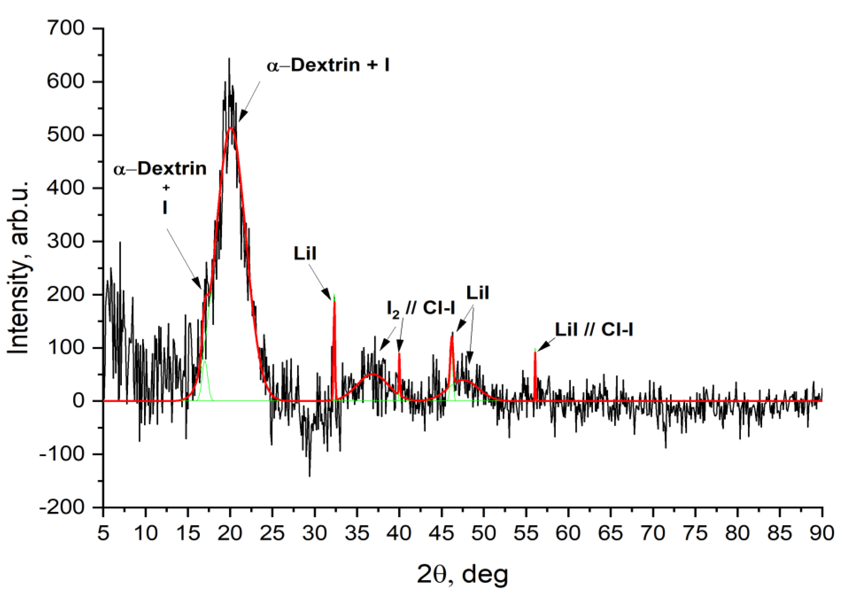
Figure 5.
SEM images of the IDLC.
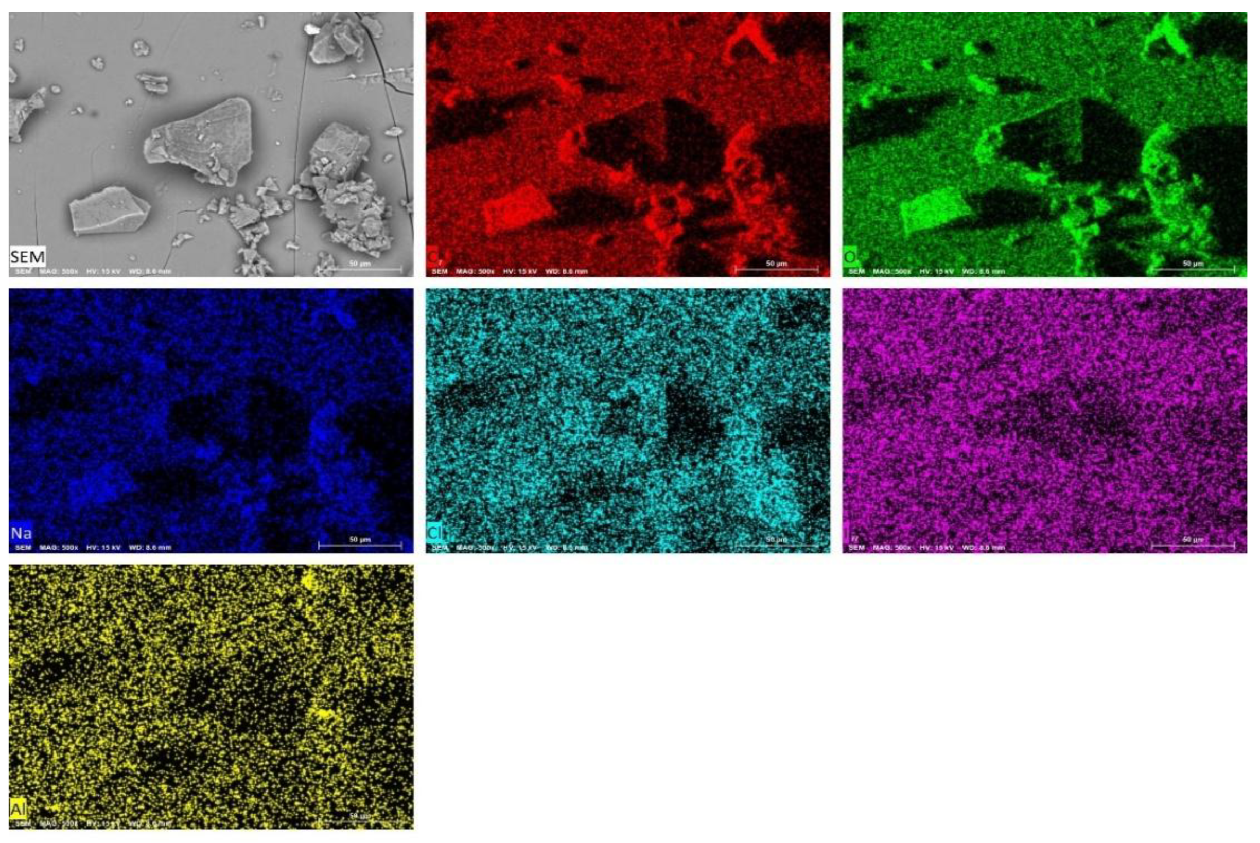
Figure 6.
EDX spectrum of the IDLC.
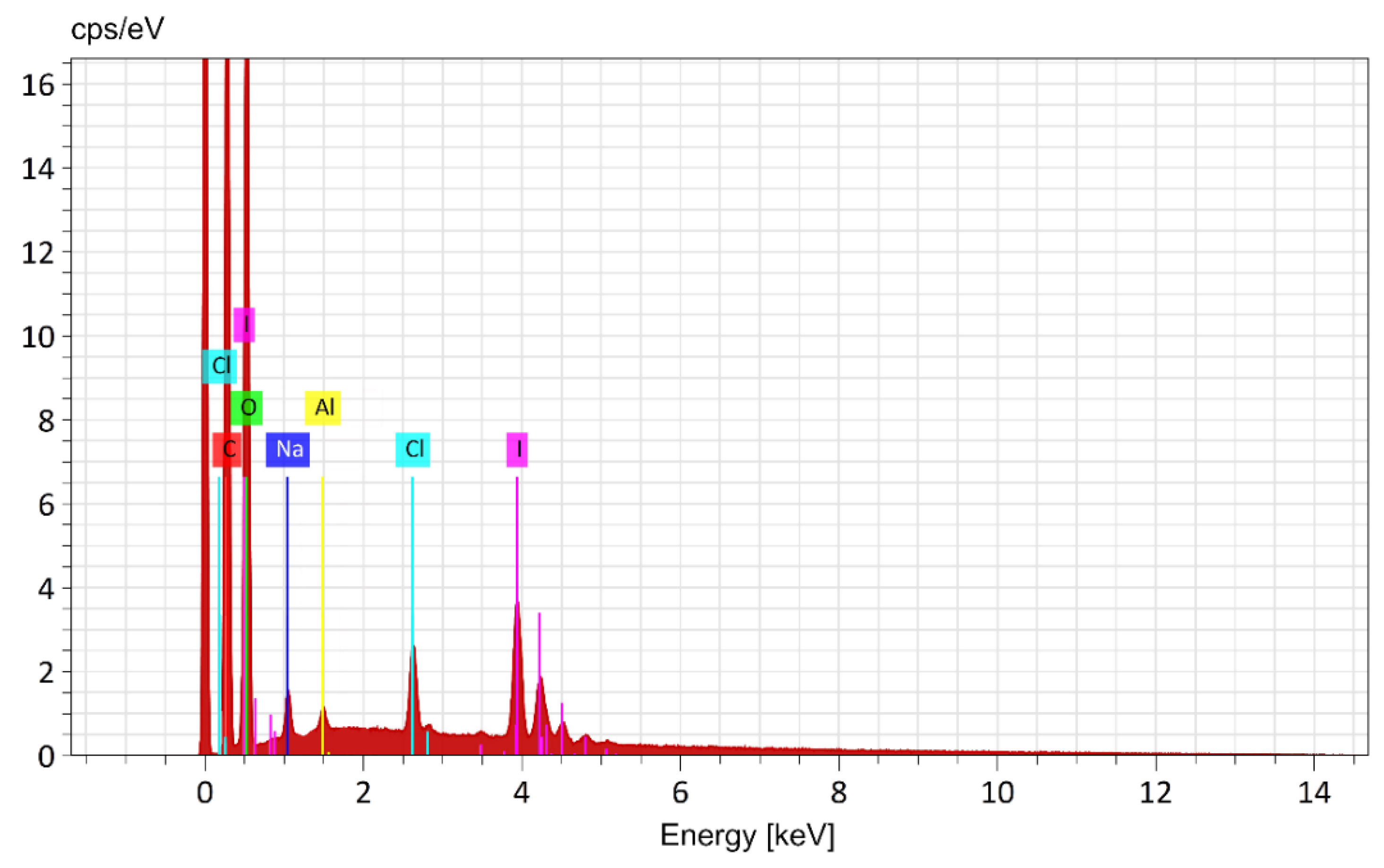
Figure 7.
Dose-effect curve for IDLC on MNC culture after 24- and 48-hour exposure.
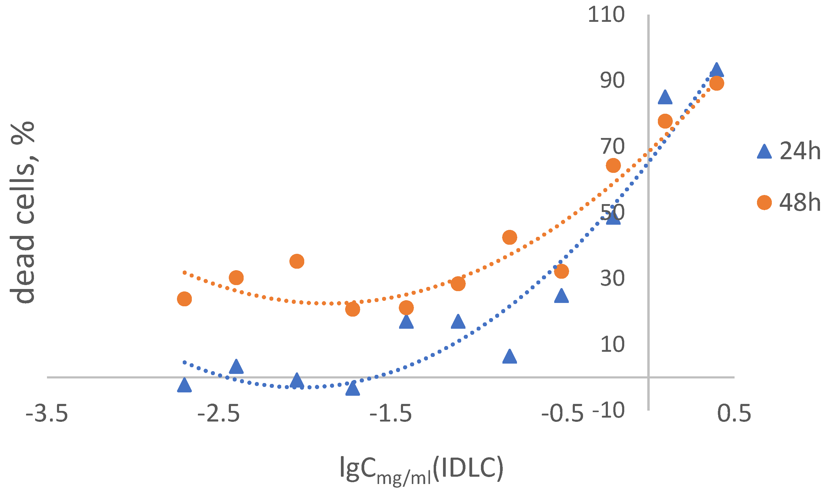
Figure 8.
Cytotoxic effect of IDLC on MDCK cell culture.
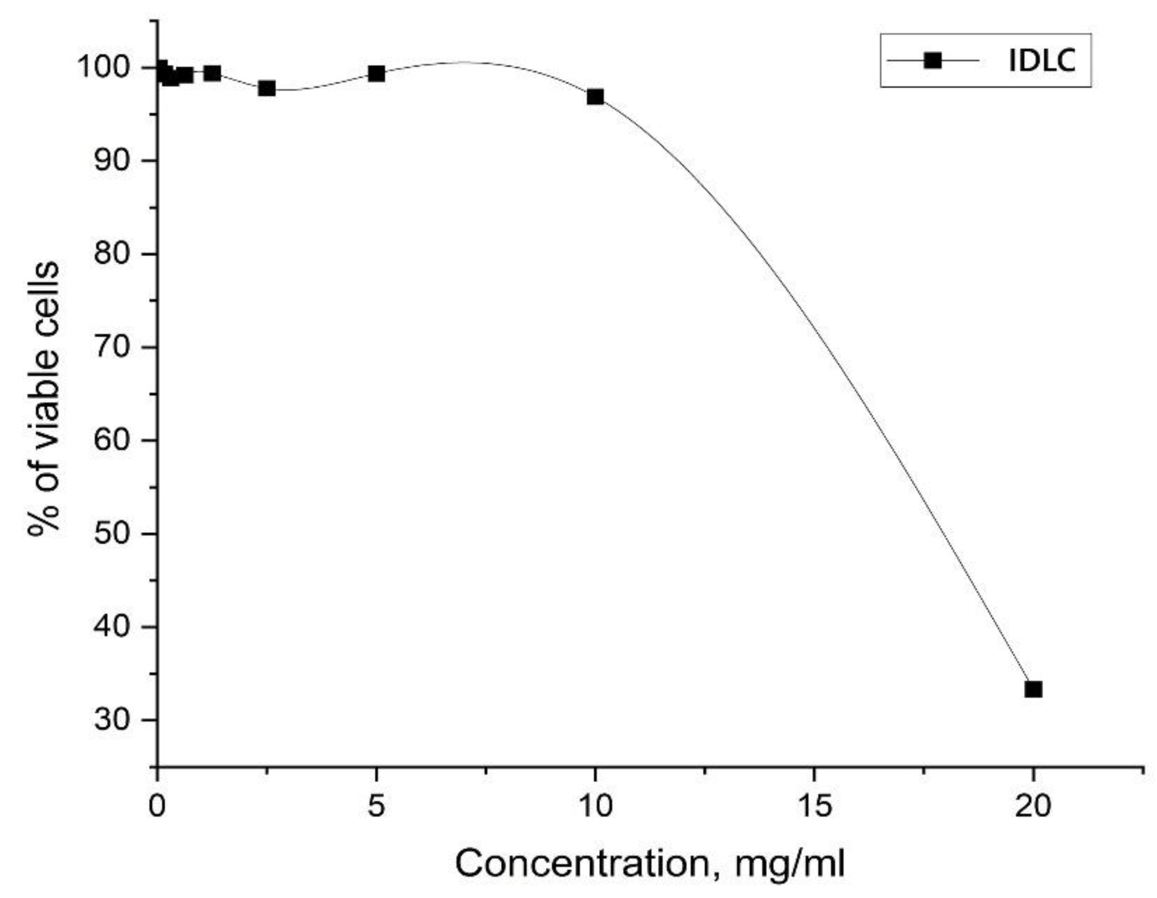

Table 1.
Physicochemical properties of the dextrin/PVA/iodine complexes.
| Compound | Solubility in water | рН | Melting point, 0С | Quantitative determination of iodine, g/kg |
|---|---|---|---|---|
| IDLC | 1 g / 20 ml, soluble | 4,83 ± 0,01 | 146-148± 1 | 50,71 ± 0,01 |
Table 2.
Quantitative determination of iodide ions and cations by the CE method.
| Compound | I-, mg/l (CE) | Li+, mg/l (CE) | ||
| exp. | theo. | exp. | theo. | |
| IDLC | 43,25 ± 0,08 | 47,5 | 6,78 ± 0,01 | 8,2 |
Table 3.
EDX analysis data of IDLC.
| Element | Mass % (theory) C124H248O122NI4K2. |
Mass % (detected) |
Line type |
|---|---|---|---|
| C | 34,70 | 34.62 ± 0.15 | K Series |
| O | 45,52 | 47.68 ± 0.33 | K Series |
| К | 1,82 | 1.55 ± 0.05 | K Series |
| N | 0,33 | 0,30 ± 0.02 | - |
| I | 11,84 | 10.15 ± 0.16 | L Series |
| H* | 5.78 | - | - |
| Totals | 100 | 94.3 |
* The number of H is calculated from the difference in the mass of the sample and the elements determined in the experiment.
Table 4.
Results of antimicrobial activity screening of the IDLC.
| Compound name |
Name of the test strains | ||||||
| S. aureus ATCC 6538-P | S. aureus ATCC BAA-39 | E. coli ATCC 8739 | E. coli ATCC 196 | P.aeruginosa ATCC 9027 | P.aeruginosa TA2 | A.baumannii ATCC BAA-1790 | |
| Value of the minimum bactericidal concentration (MBC), mcg/ml in terms of substance | |||||||
| IDLC | 15,63 | 1,95 | 15,63 | 7,81 | 7,81 | 3,91 | 3,91 |
Table 5.
Cytotoxic effect of IDLC on human peripheral blood MNC culture after 24- and 48-hour exposure.
Table 5.
Cytotoxic effect of IDLC on human peripheral blood MNC culture after 24- and 48-hour exposure.
| Concentration of IDLC, mg/ml | % of viable cells (Mean ± SD) | |
|---|---|---|
| 24 h | 48 h | |
| Negative control | 100 ± 13,4 | 100 ± 13,0 |
| 2,5 | 6,7 ± 4,7 | 10,8 ± 1,8 |
| 1,25 | 15,0 ± 4,5 | 22,3 ± 29,8 |
| 0,625 | 51,5 ± 15,5 | 35,8 ± 5,5 |
| 0,312 | 75,2 ± 22,2 | 57,9 ± 14,2 |
| 0,156 | 83,6 ± 9,7 | 59,6 ± 4,8 |
| 0,078 | 83,0 ± 12,5 | 71,6 ± 13,1 |
| 0,039 | 83,0 ± 22,4 | 78,9 ± 17,1 |
| 0,019 | 103,3 ± 22,6 | 79,4 ± 15,9 |
| 0,009 | 100,8 ± 23,0 | 64,8 ± 6,6 |
| 0,004 | 96,6 ± 15,9 | 69,8 ± 7,5 |
| 0,002 | 122,3 ± 29,0 | 76,2 ± 15,5 |
| CC50 | 0,23 mg/ml | 0,48 mg/ml |
Table 6.
Cytotoxicity of IDLC on tumor and normal cell lines in vitro.
| Concentration, mg/mL | % of viable cells (Mean ± SD) | |||||
|---|---|---|---|---|---|---|
| HepG2 | HeLa | AGS | К562 | Н9 | MeT-5A | |
| Negative control | 100 ± 6,5 | 100 ± 3,7 | 100 ± 3,8 | 100,0 ± 3,0 | 100,0 ± 2,3 | 100,0 ± 4,6 |
| 5 | 28,7 ± 2,5 | 50,6 ± 2,7 | 3,9 ± 0,6 | 11,8 ± 2,4 | 9,8 ± 0,5 | 34,9 ± 2,8 |
| 2,5 | 74,9 ± 9,7 | 102,4 ± 3,3 | 28,0 ± 0,4 | 108,2 ± 4,6 | 38,1 ± 4,1 | 50,0 ± 0,8 |
| 1,25 | 158,6 ± 8,6 | 182,9 ± 5,3 | 88,9 ± 0,6 | 98,6 ± 13,9 | 92,3 ± 9,8 | 81,7 ± 3,0 |
| 0,625 | 154,4 ± 2,7 | 142,1 ± 4,3 | 101,6 ± 3,3 | 97,5 ± 4,1 | 95,4 ± 4,7 | 123,1 ± 3,0 |
| 0,312 | 130,2 ± 11,4 | 136,0 ± 2,9 | 101,6 ± 3,3 | 103,3 ± 3,6 | 95,1 ± 8,1 | 117,8 ± 6,9 |
| 0,156 | 129,0 ± 6,4 | 129,9 ± 1,4 | 111,7 ± 4,0 | 101,2 ± 5,9 | 103,8 ± 10,5 | 110,9 ± 1,9 |
| 0,08 | 124,0 ± 4,7 | 116,1 ± 1,7 | 102,4 ± 0,7 | 95,8 ± 11,9 | 87,4 ± 6,9 | 114,5 ± 5,4 |
| 0,04 | 125,9 ± 8,4 | 136,5 ± 1,7 | 106,3 ± 3,6 | 89,9 ± 8,2 | 98,0 ± 17,7 | 113,1 ± 4,3 |
| 0,02 | 149,0 ± 31,2 | 113,5 ± 9,3 | 98,8 ± 7,0 | 85,6 ± 3,7 | 95,7 ± 4,0 | 99,3 ± 16,9 |
| CC50, mg/mL | ~2,440 | 1,201 | 1,765 | 3,533 | 2,003 | 1,370 |
Disclaimer/Publisher’s Note: The statements, opinions and data contained in all publications are solely those of the individual author(s) and contributor(s) and not of MDPI and/or the editor(s). MDPI and/or the editor(s) disclaim responsibility for any injury to people or property resulting from any ideas, methods, instructions or products referred to in the content. |
© 2025 by the authors. Licensee MDPI, Basel, Switzerland. This article is an open access article distributed under the terms and conditions of the Creative Commons Attribution (CC BY) license (http://creativecommons.org/licenses/by/4.0/).
Copyright: This open access article is published under a Creative Commons CC BY 4.0 license, which permit the free download, distribution, and reuse, provided that the author and preprint are cited in any reuse.


