Preprint
Review

This version is not peer-reviewed.

Antibiotic Resistance and Virulence Mechanisms in Klebsiella pneumoniae: Understanding for Better Interventions

Submitted:

12 November 2025

Posted:

13 November 2025

You are already at the latest version

Abstract

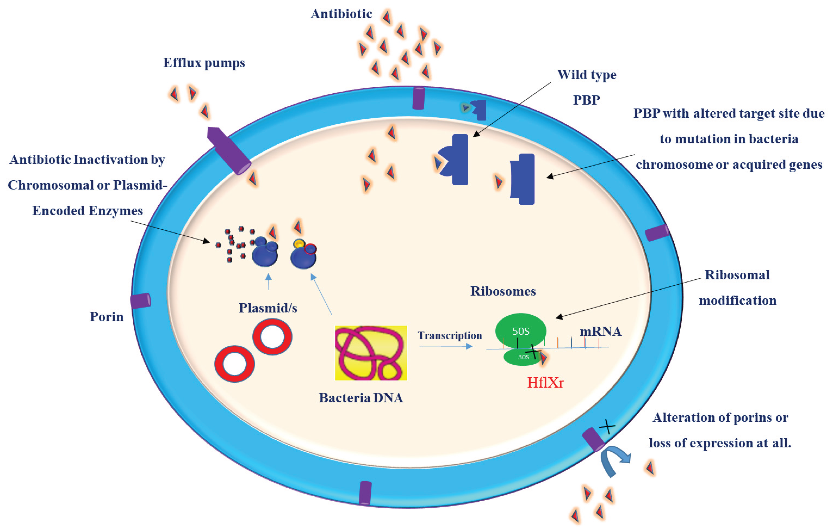
Klebsiella pneumoniae is a prominent pathogen implicated in a wide range of infections, including pneumonia, urinary tract infections, and septicemia. Its ability to acquire and disseminate antibiotic resistance, coupled with the rising prevalence of hypervirulent strains, represents a significant public health threat. Understanding the molecular basis of drug resistance can guide the design and development of effective treatment strategies. Antimicrobial resistance (AMR) in these bacteria is a complicated process and cannot be attributed to only a single resistance mechanism. K. pneumoniae develops resistance to antibiotics through a variety of mechanisms, ranging from single molecular mechanisms to complex interactions, where molecular synergy exacerbates resistance. This review summarizes the current understanding of the molecular mechanisms that contribute to the drug resistance and virulence of this pathogen. Key antibiotic resistance mechanisms include drug inactivation via B-lactamases and carbapenemases, membrane remodeling, efflux pump systems, such as AcrAB-TolC and OqxAB, and biofilm formation facilitated by quorum sensing. Additionally, the role of ribosomal changes in resistance was highlighted. This review also examines the mechanisms of virulence, emphasizing fimbriae, iron acquisition systems, and immune evasion strategies. Understanding these mechanisms of drug resistance and virulence is crucial for remodeling existing antibiotics and developing new therapeutic strategies.

Keywords: 
;  ;  ;  ;  ;  ;  

1. Introduction

Klebsiella pneumoniae (K. pneumoniae) is a ubiquitous, rod-shaped, capsulated, facultative anaerobic, non-motile, gram-negative bacterium. The genome size of this bacterium ranges from 5.1 to 5.6 Mb [1,2]. Genetic variation among different strains of this species can be attributed to genome rearrangements, which often result from the inversion of chromosomes, mobile genetic elements, plasmids, and the presence of genes specific to strains. K. pneumoniae was initially described in 1882 by Carl Friedlander as a bacterium recovered from the lungs of patients who died of pneumonia and is known as the Friedlander bacterium [3]. Later, the Klebsiella genus was named by German microbiologist Edwin Klebs, assigning the current name K. pneumoniae [4]. K. pneumoniae is found in different environments, including the soil, water, and the gastrointestinal and respiratory tracts of humans and animals. K. pneumoniae can colonize healthcare environments and medical devices. This bacterium is a great public health challenge because of its increasing pathogenicity, virulence, and emergence of antimicrobial resistance. The association of K. pneumoniae with invasive procedures and its ability to form biofilms increases the risk of infection and complicates the treatment options [5,6]. Historically, this bacterium has been associated with infections in immunocompromised individuals. However, the emergence of antibiotic-resistant strains and increased virulence has presented dual risks in healthy and immunocompetent individuals [3,7,8,9]. The increasing number of infections and the emergence of MDR strains of this bacterium have attracted attention.
The prevalence of multidrug-resistant (MDR) K. pneumoniae strains is increasing. While various molecular mechanisms contribute to resistance, the production of extended-spectrum beta-lactamases (ESBLs) and carbapenemases is the primary driver of drug resistance in this pathogen. Another concern associated with this bacterium is its pathogenicity, which results from the complex interaction of various virulence factors that enable colonization and immune evasion, and facilitate systemic spread [10]. These virulence factors include lipopolysaccharides, iron uptake systems, capsules, exopolysaccharides associated with mucoviscosity, and adhesins [11]. Reports indicate that K. pneumoniae isolates are increasingly displaying MDR characteristics, with a rapid increase in global resistance rates for carbapenems [12]. The dual challenges of increased virulence and drug resistance to currently available antibiotics highlight the urgent need for innovative treatment options such as promising phage therapy [13,14].

1.2. Antimicrobial Resistance in K. pneumoniae

Antimicrobial resistance (AMR) has been recognized as a significant public health crisis. AMR is characterized by the ability of microorganisms to resist medications intended to kill them [15]. The World Health Organization (WHO) has identified AMR as a critical threat to public health. If there are no new antibiotics or novel therapeutic options, it is estimated that by 2050, infections related to AMR could lead to the death of 10 million people [16]. K. pneumoniae rapidly develops resistance to antibiotics. By 2024, carbapenem-resistant K. pneumoniae had risen to the top position on the WHO bacterial priority pathogen list (BPPL), highlighting an alarming increase in resistance among these pathogens[17]. The WHO has prioritized Klebsiella pneumoniae as a critical pathogen due to its ability to acquire AMR genes, rendering it resistant to multiple classes of antibiotics. Current studies reveal that more than half of K. pneumoniae isolates are resistant to key antibiotics like third-generation cephalosporins and carbapenems, underscoring the urgent need for enhanced surveillance, novel treatment strategies, and effective antimicrobial stewardship [18,19,20].

2. Molecular Mechanisms of Drug Resistance in K. pneumoniae

The emergence of multidrug-resistant (MDR) strains of Klebsiella pneumoniae constitutes a significant public health challenge due to the pathogen’s multifaceted and dynamic resistance mechanisms. Unlike simpler models of antimicrobial resistance (AMR), resistance in K. pneumoniae arises from an intricate interplay of molecular strategies, which collectively amplify the bacterium’s ability to withstand diverse antibiotic classes (Figure 1). This complexity arises not only from individual resistance determinants but also from synergistic interactions between mechanisms, underlining the pathogen’s adaptive versatility (Figure 1) [19,20]. A primary mode of resistance involves modification of antibiotic targets, thereby reducing drug affinity and efficacy. Complementarily, K. pneumoniae frequently expresses a spectrum of drug-inactivating enzymes, such as β-lactamases, which hydrolyze antibiotic molecules, rendering them ineffective [21,22,23]. The genetic basis of these enzymatic factors is notably versatile, with resistance genes encoded both chromosomally and on mobile plasmids. The latter route facilitates horizontal gene transfer (HGT), which has been pivotal in disseminating resistance traits across clinical isolates.
Plasmid acquisition is particularly consequential, as it enables the concurrent carriage of multiple resistance genes, fostering the development of extensively drug-resistant (XDR) strains. For instance, co-localization of genes such as blaNDM and fluoroquinolone resistance determinants within single plasmids has been repeatedly documented in nosocomial isolates, highlighting the role of plasmids as reservoirs for resistance cassettes that complicate treatment strategies [24].
Efflux pump systems, primarily chromosomally encoded in K. pneumoniae, provide an additional layer of resistance by decreasing intracellular drug concentrations through active extrusion. These pumps are tightly regulated at the genetic level. They can synergize with plasmid-borne resistance genes to enhance resistance phenotypes, demonstrating a dual genetic control framework that underscores the pathogen’s adaptability under antibiotic selective pressure. This synergy between chromosomal and plasmid-mediated mechanisms exemplifies the evolutionary plasticity of K. pneumoniae, which is further accelerated by mobile genetic elements facilitating rapid genetic recombination and adaptation [25,26]. The presence of multiple resistance genes on a single plasmid has been documented, contributing to the development of extensively drug-resistant (XDR) strains. For example, studies have shown the presence of carbapenemase and fluoroquinolone resistance genes, as well as blaAmpC and fluoroquinolone resistance genes, within plasmids acquired by bacteria that frequently lead to hospital-acquired infections. Efflux pump systems, which are chromosomally encoded, play a significant role in antibiotic resistance by altering the permeability of the cell membrane to drugs. These pumps are a key mechanism contributing to the drug resistance observed in K. pneumoniae [27].

2.1. Drug Inactivation and Target Modification

K. pneumoniae can resist antibiotics by hydrolyzing enzymes, such as B-lactamases, AmpC B-lactamases, and carbapenemases. These Enzymes hydrolyze critical parts of the antibiotics. The primary resistance mechanism involves the production of β-lactamases, enzymes that hydrolyze and inactivate β-lactam antibiotics [28]. B-lactams are a widely used class of antibiotics. After the discovery of benzylpenicillin in the 1920s by Alexander Fleming, numerous penicillin derivatives and related B-lactam classes, including monobactams, cephalosporins, cephamycins, and carbapenems, have been developed. The development was aimed either at expanding the spectrum of activity to cover new species or to combat emerged resistance [29]. B-lactams are commonly used to treat infections caused by K. pneumoniae. B -Lactams work by inhibiting penicillin-binding protein (PBPs) (Figure 1), enzymes responsible for cell wall synthesis [30,31].
Without proper peptidoglycan cross-linking, cells experience an uncontrolled influx, leading to swelling and lysis. Even minor disruptions in cross-linking can trigger structural instability, causing membrane blebbing and eventual rupture. Therefore, the precise architecture of peptidoglycan is essential for bacterial survival, acting as both a structural component and an osmotic barrier [32,33]. A key target in the peptidoglycan synthesis pathway is the PBPs, transpeptidases responsible for catalyzing the final cross-linking step in the peptidoglycan assembly. B-lactams mimic the D-Ala-D-Ala terminus of peptidoglycan precursors and act as suicide substrates [34]. B-Lactams irreversibly bind to PBPs and inhibit transpeptidase activity. This disruption prevented peptidoglycan cross-linking. This weakens the bacterial cell wall and leads to cell death through osmotic lysis. The effectiveness of B-lactams is attributed to their covalent interactions with PBPs across all bacterial species [30]. Bacteria develop resistance to β-lactam antibiotics by modifying penicillin-binding proteins (PBPs) at the molecular level, which reduces the binding affinity of these antibiotics. In K. pneumoniae, this resistance primarily arises from mutations in the genes encoding PBPs, leading to structural changes that decrease their ability to bind β-lactams effectively [30,31]. The loss or structural modification of outer membrane proteins (OMPs) is another mechanism that confers resistance to K. pneumoniae. The outer membrane acts as a barrier that antibiotic molecules must cross before binding to penicillin-binding proteins (PBPs). Antibiotics must pass through the porins, specifically OmpK35 and OmpK36. However, the expression levels of these porins are frequently modified, leading to a reduction in membrane permeability. This decrease limits the influx of antibiotics into the bacterial cell, thereby reducing their intracellular concentrations and effectiveness. As a result, such alterations in porin abundance directly contribute to the development of antibiotic resistance by preventing sufficient drug accumulation within the bacteria [35,36]. Together, these multilayered defenses create a formidable barrier against treatment, underscoring the urgent need for integrated molecular surveillance and novel therapeutic strategies to manage emerging multidrug-resistant K. pneumoniae infections.

2.1.1. B-Lactamases

B-lactamases are enzymes that are capable of hydrolyzing B-lactam antibiotics. The dominant mechanism of resistance in K. pneumoniae involves the production of an extended spectrum of B-lactamases (ESBLs) and AmpC B-lactamases, which are often found on mobile genetic elements, allowing for rapid spread among bacterial populations [37,38]. B-lactamases are categorized into ESBLs, cephalosporinases (AmpC), and carbapenemases [39]. The production of these enzymes in K. pneumoniae confers resistance to carbapenems, penicillins, and cephalosporins. ESBLs include cefotaximase (CTX), Temoneria (TEM), sulfhydryl variable (SHV), and oxacillinase (OXA). AmpC confers resistance to first-to third-generation cephalosporins and enzyme inhibitors. AmpC can be either chromosomal- or plasmid-mediated. Currently, more than 40 genetic variants of AMPC have the potential for rapid plasmid-mediated spread among strains [20]. Production of carbapenemase enzymes is another mechanism of resistance in K. pneumoniae, leading to the development of carbapenem-resistant K. pneumoniae (CRKP). These enzymes reduce the sensitivity to carbapenem-class antibiotics [39].
Temoneria (TEM)-1 is the first reported B-lactamase. TEM was described in 1965, followed by the identification of sulphydryl variable (SHV)-1 B-Lactamase [40]. These enzymes help bacteria survive penicillin treatment. Subsequently, derivatives of TEM and SHV-type B-lactamase, classified as ESBL, were detected. These variants demonstrate hydrolysis of oxyimino B-lactam antibiotics due to minor molecular changes in their active sites [41,42].
Notably, this type of resistance was observed in K. pneumoniae shortly after the introduction of third-generation cephalosporins in 1982 [40]. Historically, SHVs or TEMs have been the dominant B-lactamases. However, there has been a shift, with CTX-M being the most commonly detected ESBL worldwide. The distribution of ESBL-producing K. pneumoniae, which encodes CTX-M, has increased significantly over the years [43]. Outbreaks of ESBL-producing K. pneumoniae in hospitals have posed a threat for several years, dating back to the first report in the 1980s in France [40]. The production of diverse B-lactamases in K. pneumoniae, including ESBLs, AmpC cephalosporinases, and carbapenemases, constitutes the primary mechanism driving resistance to critical β-lactam antibiotics (Table 1). The evolving landscape of these enzymes, from the early TEM and SHV variants to the widespread CTX-M types, highlights the dynamic adaptation of K. pneumoniae to antimicrobial pressure. The rapid dissemination of these genes via mobile genetic elements exacerbates the challenge of treating infections caused by multidrug-resistant strains, underscoring the urgent need for continuous surveillance, stewardship, and the development of novel therapeutic strategies.

2.1.2. Carbapenemases

Carbapenemases are bacterial enzymes that break down carbapenem antibiotics, causing significant treatment challenges. Among these, the "Big Five" carbapenemases K. pneumoniae carbapenemase (KPC) (class A), Imipenemase (IMP), Verona integron-encoded metallo-β-lactamase (VIM), New Delhi metallo-β-lactamase (NDM) (class B metallo-β-lactamases), and OXA-48-like oxacillinases (class D) are of particular clinical concern due to their widespread distribution and their role in conferring resistance in Gram-negative bacteria such as K. pneumoniae [39]. Carbapenemases are B-lactamases with versatile hydrolytic activity. Many of these enzymes can hydrolyze nearly all B-lactams, making them largely resistant to the currently available B-lactam antibiotics. Carbapenemases are categorized into A, B, and D molecular classes of B-lactamases. Molecular classes A and D use serine-dependent hydrolytic mechanisms, whereas class B comprises metallo-B-lactamases that incorporate zinc into their active site [39]. The term carbapenem-hydrolyzing enzymes is preferable over “carbapenemases," as carbapenems represent only one part of the substrate spectrum. The mechanism of action of Carbapenem-hydrolyzing enzymes acts through the action of catalytic sites, such as serine or zinc. According to Amber’s classification of B-lactamases, carbapenem-hydrolyzing enzymes are classified into molecular classes A, B, and D [44]. Each class comprises multiple enzyme families and their respective subfamilies. Among these enzymes, IMP, VIM, KPC, NDM, and OXA are particularly significant in clinical settings [45,46]. These enzymes are encoded both chromosomally and on plasmid genes, resulting in variability among carbapenemases. Plasmid-encoded carbapenemase genes can be horizontally transferred between related bacterial genera, facilitating the rapid spread of carbapenemase-producing bacteria [24]. Carbapenemases are enzymes that hydrolyze carbapenem antibiotics, posing significant challenges to the effective treatment of infections caused by Gram-negative bacteria such as K. pneumoniae. The “Big Five” carbapenemases are especially concerning due to their widespread presence and broad-spectrum hydrolytic activity against almost all β-lactam antibiotics (Table 1). These enzymes employ serine-dependent (classes A and D) or zinc-dependent (class B) catalytic mechanisms, reflecting their molecular diversity. Because their substrate range extends beyond carbapenems, the term carbapenem-hydrolyzing enzymes more accurately describes them. Their genetic encoding on both chromosomes and plasmids facilitates horizontal gene transfer, accelerating the spread of carbapenem resistance among bacterial populations and complicating infection control efforts globally.

2.1.2.1. Serine Carbapenemases (Molecular Class A Carbapenemases)

Serine carbapenemases constitute a group of B-lactamases (functional group 2f) that utilize serine residues for their hydrolytic activity [46,47]. These enzymes effectively degrade a wide range of β-lactam antibiotics, including carbapenems, using serine residues in their active site. These enzymes effectively degrade antibiotics, including cephalosporins, carbapenems, penicillins, and monobactams. Serine carbapenemases are not inhibited by metal-chelating agents such as ethylenediaminetetraacetic Acid (EDTA) [46,47]. These enzymes are commonly identified in K. pneumoniae clinical isolates of K. pneumoniae. The well-characterized groups of serine carbapenemases include KPC (K. pneumoniae carbapenemase), IMI/NMC (Imipenem-hydrolyzing B-Lactamase/Non-Metallo Carbapenemase), SME (Serratia marcescens enzyme), and GES (Guiana ESBL). In addition, SHV-38 and SFC-1 (Serratia fonticola carbapenemase-1) represent emerging variants. SHV-38, SFC-1, IMI/NMC, and SME are typically chromosomal, whereas KPC and GES are often plasmid-mediated, facilitating rapid horizontal transmission among bacterial populations [24,39].

2.1.2.2. Molecular Class B Carbapenemases /Metallo- B-Lactamases (MBLs)

MBLs are zinc-dependent enzymes that hydrolyze carbapenems and nearly all B-lactams [48,49]. MBLs can be categorized into three subclasses: B1, B2, and B3. Each subclass has unique characteristics. Class B1 MBLs, including NDM, VIM, IMP, GIM, and SIM, are the most clinically significant and require two zinc ions for catalysis [45,46]. These enzymes are frequently plasmid-mediated, enabling rapid K. pneumoniae. Class B2 MBLs are active mono-zinc proteins [50]. CphA from Aeromonas species utilizes only one zinc ion (Zn+2) for catalysis and demonstrates a high specificity for carbapenems. Notably, the Zn1 site in B2 MBLs is non-catalytic and can inhibit activity when occupied. Class B3 MBLs, which include L1 from Stenotrophomonas maltophilia, AIM-1 from P. aeruginosa, and BcII from Bacillus cereus, also require two zinc ions but are structurally and phylogenetically distinct from B1 and B2 enzymes. B3 MBLs possess active site motifs, such as HHH/DHH, and show greater sequence diversity [51]. In contrast to B1 MBLs, B3 MBLs are often encoded chromosomally. MBLs can evade inhibition by B-lactamase inhibitors such as clavulanate, and MBLs can be inhibited by chelating agents such as EDTA [52,53]. Unlike serine β-lactamases, MBLs are not inhibited by traditional β-lactamase inhibitors, but can be targeted by metal chelators such as EDTA, highlighting the need for novel therapeutic approaches to overcome MBL-mediated resistance.

2.1.2.3. Molecular Class D Carbapenemases (OXA Carbapenemase)

Class D carbapenemases, commonly referred to as oxacillinase (OXA) carbapenemases, are serine-dependent enzymes that hydrolyze carbapenems and B-lactam antibiotics [54]. OXA is a carbapenem-hydrolyzing enzyme classified into a family of molecular class D serine-dependent enzymes. OXA was first reported in 2003 in Turkey [55]. These enzymes have a wide genetic diversity and are encoded by plasmids. Class D carbapenemases utilize a serine residue for their catalytic activity and exhibit resistance to inhibition by EDTA, clavulanate, and tazobactam, owing to their serine-based mechanism. Class D carbapenemases have been identified predominantly in K. pneumoniae. The OXA group consisted of more than 489 variants, 37 of which were classified as clinically significant carbapenemases. Among these, OXA-48, including its variants (OXA-181 and OXA-232), is particularly noteworthy and often associated with outbreaks in healthcare environments [56,57]. In contrast to plasmid-mediated carbapenemases (e.g., KPC), OXA enzymes are primarily encoded chromosomally, although plasmid-borne variants have been reported. Their capacity to circumvent conventional inhibitors and propagate through horizontal gene transfer highlights their significance in the context of multidrug-resistant infection [24].
The five most significant carbapenemases, known as "the Big Five," are KPC, IMP, VIM, NDM, and OXA [58]. These enzymes are commonly found in K. pneumoniae and other pathogenic bacteria. Their ability to spread via plasmids and chromosomes, especially variants like OXA-48, makes them a critical factor in the rise of multidrug-resistant K. pneumoniae infections, and underscores the challenge in managing last-resort carbapenem therapy.

2.1.2.4. K. pneumoniae Carbapenemase (KPC)

This is the most clinically reported type of molecular class A carbapenemase. KPC was first identified in North Carolina, USA, in 1996 [59]. KPC is now globally distributed, with reports from all continents and regions [60,61,62,63,64,65,66,67]. Reports have identified over 150 different blaKPC-coding genes [68]. The most prevalent KPC variants are KPC-2 and KPC-3. Newer variants that confer resistance to ceftazidime/avibactam (CZA), such as KPC-80, KPC-97, KPC-81, and KPC-96, have also been reported [69]. This variation is caused by the accumulation of mutations in active site loops, such as the Ω-loop and Lys270-loop [68]. Although KPC has been repeatedly reported in K. pneumoniae, it has also been isolated from other Enterobacterales, including Enterobacter spp., Klebsiella oxytoca, E. coli, Serratia marcescens, Citrobacter freundii, Salmonella enterica, Proteus mirabilis, and P. aeruginosa [70,71,72,73]. The widespread emergence and diversification of KPC enzymes significantly threaten the effectiveness of current β-lactam and carbapenem therapies, complicating infection management worldwide. Urgent development of novel inhibitors and robust surveillance systems is essential to control the spread and impact of KPC-producing pathogens.

2.1.2.5. Imipenemase (IMP)

IMPs are carbapenemases classified as a family of molecular class B MBLs that utilize zinc ions (Zn²⁺) for their catalytic effect, allowing them to confer resistance to carbapenems, particularly imipenem and meropenem [74]. IMP was first identified in Japan as IMP-1, which has since diversified into 88 variants, with the majority being found in Asia [74,75,76]. IMP are increasingly identified in K. pneumoniae, significantly contributing to its multidrug resistance. In K. pneumoniae, IMP carbapenemases are typically encoded on plasmids, enabling rapid horizontal gene transfer within and between bacterial populations [77]. These enzymes inactivate nearly all B-lactams, except monobactams, such as aztreonam, rendering carbapenems ineffective. The IMP-43 and IMP-44 variants demonstrated increased catalytic efficiency against carbapenems compared to the IMP-7 and IMP-11 variants. This highlights the growing threat posed by IMP carbapenemase [78]. The global dissemination of IMP-producing pathogens, facilitated by plasmid-mediated horizontal gene transfer, exacerbates resistance and complicates treatment [74,79]. The presence of IMP enzymes in K. pneumoniae undermines the effectiveness of carbapenems, often regarded as last-resort antibiotics for severe infections caused by this pathogen. This enzymatic resistance severely restricts treatment options and is linked to increased morbidity and mortality in infected patients. Therefore, monitoring and controlling the spread of IMP-producing K. pneumoniae is essential for infection prevention and effective antimicrobial stewardship.

2.1.2.6. Verona Integron-Encoded Metallo-B-Lactamases (VIMs)

VIMs are carbapenem-hydrolyzing enzymes classified into a family of molecular class B MBLs that utilize zinc ions (Zn²⁺) for their catalytic activity [80,81]. VIMs are encoded by integron blaVIM genes found in plasmids. These enzymes are among the most widespread metallo-B-lactamases (MBLs) worldwide and demonstrate a wide range of hydrolytic activities against cephamycins, penicillins, cephalosporins, and carbapenems. VIMs are not effective against monobactams such as aztreonam. VIM was first reported in 1999 in Verona [82], since it has been reported in regions including the Far East, USA, Europe, and South America [83]. Variants of VIM, VIM-1, and VIM-2 were most frequently reported. These carbapenemases are primarily associated with K. pneumoniae. The global spread of VIM-producing bacteria is facilitated by mobile genetic elements within the class 1 integrons. This enables horizontal gene transfer across various bacterial species. Recent studies have highlighted the emergence of novel variants, such as VIM-84 in Asia and Europe, which further complicate the management of multidrug-resistant infections [84,85].

2.1.2.7. New Delhi Metallo-B-Lactamases (NDM)

NDM are hydrolytic enzyme classified into a family of molecular class B carbapenemases (MBLs). NDM was first reported in 2008 in a Swedish patient of Indian descent, who had been treated in New Delhi, India [86,87]. The first NDM variant was isolated from K. pneumoniae as NDM-1. Since then, NDM has been disseminated globally. These enzymes hydrolyze almost all B-lactams and carbapenems. These enzymes do not hydrolyze or inactivate monobactams such as aztreonam. NDM carbapenemases are plasmid-mediated and can be shared among bacterial species via horizontal gene transfer. Although aztreonam is still effective against NDM-producing strains, the coexistence of other resistance mechanisms such as ESBLs may reduce its efficacy in some cases [88].
Table 1. Summary of β-Lactamase and Carbapenemase Classes and Key Features.
Enzyme Class Molecular Class Representative Enzymes Substrate Specificity Inhibition Ref
Serine-B-lactamases (Class A) A KPC,TEM, SHV, CTX-M Penicillins, cephalosporins, carbapenems (KPC) Inhibited by clavulanic acid, sulbactam [46,47]
Metallo-B-lactamases (Class B) B IMP, NDM, VIM, SPM, GIM Broad spectrum including carbapenems, not monobactams Inhibited by metal chelators (e.g., EDTA) [45,46]
Serine-B-lactamases (Class C) C AmpC-type enzymes Cephalosporins, penicillins Not inhibited by clavulanic acid [39,142]
Oxacillinases (Class D) D OXA-48-like carbapenemases, OXA-23, OXA-24/40, OXA-58, OXA-1, OXA-10 Penicillins, cephalosporins, carbapenems (OXA-48 group) Variable inhibition by clavulanic acid [54]
Extended-Spectrum-B-Lactamases (ESBLs) Mostly Class A CTX-M, SHV variants, TEM variants Expanded activity against third-generation cephalosporins Inhibited by clavulanic acid [41,42]
Carbapenemases (Functional Group) Classes A, B, D KPC (A), IMP, VIM, NDM (B), OXA-48-like (D) Hydrolyze carbapenems and other β-lactams Varies by class (see above) [45,46,59,74,80,81]

2.1.3. Resistance to Colistin, Aminoglycosides, and Fluoroquinolones

In addition to its primary resistance mechanisms, K.pneumonia has several secondary pathways that enhance its multidrug-resistant profile and create significant challenges for effective treatment. One notable mechanism is the presence of plasmid-borne mcr genes [89], which pose a critical threat by conferring resistance to colistin, a last-resort antibiotic. These genes code for phosphoethanolamine transferase enzymes that chemically modify the lipid A component of the bacterial outer membrane lipopolysaccharide. This modification decreases colistin binding, reducing its bactericidal effectiveness. The plasmid location of mcr genes enables horizontal gene transfer among bacterial populations, promoting the rapid dissemination of colistin resistance in both clinical and environmental K. pneumoniae isolates.
Aminoglycoside resistance in K. pneumoniae is primarily driven by aminoglycoside-modifying enzymes, which include acetyltransferases (such as aac(3)-II and aac(6')-Ib), nucleotidyltransferases, and phosphotransferases [19]. These enzymes inactivate aminoglycosides through processes like acetylation, adenylation, or phosphorylation, effectively neutralizing the antibiotic. The genes for these enzymes are often located on transferable genetic elements, such as plasmids and transposons, facilitating their spread among bacterial populations. Furthermore, 16S rRNA methyltransferases, like ArmA and RmtB, provide high-level resistance by methylating the ribosomal target of aminoglycosides, which prevents the drugs from binding effectively.
Fluoroquinolone resistance mainly results from point mutations in the quinolone resistance-determining regions (QRDRs) of the genes encoding DNA gyrase (gyrA) and topoisomerase IV (parC) [90,91]. These mutations modify the target enzymes, preventing fluoroquinolones from effectively binding and inhibiting their activity. Resistance is further increased by efflux pumps, such as AcrAB-TolC and OqxAB, which actively remove fluoroquinolones from the bacterial cell, reducing intracellular drug concentrations. Additionally, plasmid-mediated quinolone resistance (PMQR) genes can work in conjunction with chromosomal mutations and efflux mechanisms to elevate resistance levels [91]. Collectively, these mechanisms;-plasmid-mediated colistin resistance, enzymatic drug modification by aminoglycoside-modifying enzymes, and target alteration plus efflux in fluoroquinolone resistance contribute substantially to the multidrug-resistant threat posed by K. pneumoniae. Understanding and monitoring these pathways are crucial for developing effective therapeutic strategies and containment policies to combat infections caused by this formidable pathogen.

2.2. Membrane Remodeling

The cell envelope is crucial for the adaptation and survival of the bacteria. The cell envelope of gram-negative bacteria possesses three important layers: the outer membrane (OM), peptidoglycan, and inner membrane [33]. Porin proteins exist in the outer membrane. They are called outer membrane porins and act as protein channels [35]. These porins allow the transport of small nutrients, antibiotics, iron, and hydrophilic molecules across the outer membrane. Porins also serve as bacteriocins and phage receptor proteins, and in association with lipopolysaccharide and peptidoglycan, maintain the integrity of the cell. Earlier studies have indicated a dual role for these proteins in virulence and drug resistance [35]. Extensive studies related to porin characterization have been conducted on the E. coli laboratory strain K-12. OmpC and OmpF porins are well known and have been characterized in this strain. In E. coli, the port of OmpF is narrower than that of OmpC, which makes it easier for molecules to pass through the latter. In K. pneumoniae two well-known active major outer membrane porins, OmpK35 and OmpK36, have been identified in K. pneumoniae. OmpK35 and OmpK36 were homologous to OmpC and OmpF, respectively. OmpK35 and OmpK36 allow antibiotics to penetrate the K. pneumoniae cell wall. This experiment was initially shown in various studies that cloned the ectopic expression of OmpK35 or OmpK36 in deficient strains lacking both OmpK35 and OmpK36. These studies indicate that cephalosporins pass through OmpK35 more efficiently than through OmpK36. These studies also showed that OmpK35 is not expressed in media with high osmolality. This agrees with the theory that OmpF is preferentially expressed in a medium with low osmolality, whereas OmpC is highly abundant at high osmolality [35]. Most K. pneumoniae strains that produce ESBL express only OmpK36, whereas the majority of non-ESBL-producing K. pneumoniae were found to express both OmpK35 and OmpK36 [35]. ESBL-producing K. pneumoniae typically lacks one of these porins, making it more likely to develop resistance by losing the expression of the remaining porins. Both clinical and experimental evidence show that the loss of porins in K. pneumoniae that produces ESBLs increases resistance to oxyimino cephalosporins and cefoxitin and decreases susceptibility to fluoroquinolones [92,93].
Coordinated OMP membrane folding and insertion are facilitated by the B-barrel assembly machinery (BAM) complex [94]. The BAM complex of gram-negative bacteria is composed of the outer membrane protein BamA, also known as Omp85 or YaeT, and lipoproteins, such as BamB (YfgL), BamC (NIpB), BamD (YfiO), and BamE (SmpA) [95]. In the BAM complex, BamA is the core molecular component that plays a crucial role in assembling outer membrane proteins (OMPs). Other lipoproteins, including BamA, BamC, BamD, and BamE, directly or indirectly interact with BamA anchored to the periplasmic side of the outer membrane [96]. The absence of BamA causes misfolding of the outer membrane of the B-barrel, preventing its proper insertion into the bacterial outer membrane. This misfolding damages the outer membrane structure, ultimately leading to bacterial cell death [96]. The assembly of porins into the outer membrane of bacteria at the molecular level, combined with molecular knowledge of the signals that determine which porin-encoding genes are activated in response to specific stimuli, such as antibiotics [35,97]. Targeting molecules such as BAM on the bacterial outer membrane surface could serve as promising sites for drug development.

2.3. Efflux Pump Systems

Efflux pumps are transport proteins localized in the bacterial membrane. Efflux pumps regulate the internal cellular environment by fluxing toxic substances, including antibiotics. Most efflux pump systems are activated by stimuli (i.e., substrate-induced activation). This mechanism reduces the concentration of antibiotics within the bacteria and contributes to the development of drug resistance. Multidrug efflux pumps contribute to increased resistance to various antimicrobial agents, including antibiotics, dyes, antiseptics, and detergents. In K. pneumoniae, several multidrug efflux pumps have been identified in K. pneumoniae, including AcrAB, AxAB, EefAB, KexD, KmrA, KdeA, and CepA [98]. Among these, the dominant efflux pump systems are AcrAB-ToIC and OqxAB from the RND family. AcrAB, OqxAB, EefAB, and KexD belong to the RND family. The RND-type efflux pump system is composed of three parts: outer membrane, inner membrane, and periplasmic protein. The RND-type efflux pump, along with its function in intrinsic resistance, is also thought to play a role in bacterial colonization of the intestinal tract by releasing virulence factors [98]. The AcrAB-ToIC system is also known for fluxing B-lactam antibiotics, macrolides, fluoroquinolones, tetracycline, and amido-alcohol antibiotics. The regulatory genes acrR and ramR associated with this system often contain mutations that favor overexpression of the AcrAB-ToIC efflux pump [99]. This efflux plays an important role in the drug resistance observed in K. pneumoniae. Autosomal transfer of OqxAB to a plasmid can increase the expression of the efflux pump, which further increases drug resistance.

2.4. Quorum Sensing and Biofilm Formation

A biofilm is a collection of bacteria embedded in self-produced molecules known as an extracellular polymeric matrix. Biofilm formation is a multistage process that includes the attachment of free bacteria to the surface or other cells, followed by the formation of microcolonies, maturation, and dispersion, which allows the biofilm to spread and colonize new surfaces [100]. Biofilms are formed when many bacterial cells aggregate in extracellular mucus composed of a polysaccharide matrix, lipids, and proteins secreted by bacteria. Biofilms may be comprised of a single bacterial species or a mixture of other microorganisms [101]. Bacteria form biofilms in response to external pressure, including high osmotic pressure, low PH, oxidative stress, and antimicrobial exposure. The mechanism by which bacterial cells communicate their population density is known as quorum sensing (QS), also called density sensing. This process regulates various biological behaviors in bacteria by adjusting gene expression based on the population density. As the density of the bacterial population increases, bacterial cells begin to produce signaling molecules that trigger various cellular mechanisms, such as the expression of virulence factors, antibiotic tolerance, biofilm formation, and the development of drug resistance [102]. This coordination allows bacteria to activate this mechanism only in the presence of maximum environmental stress; conditions that threaten their survival and require a collective adaptive response [98,103,104].
In K. pneumoniae, QS is essential for regulating biofilm formation, the expression of virulence factors, and antimicrobial resistance. This bacterium primarily utilizes acyl-homoserine lactones (AHLs) as QS signaling molecules, which coordinate group behaviors crucial for its survival and pathogenicity. QS enhances the production of extracellular polymeric substances that comprise the biofilm matrix, facilitating strong biofilm development on both living tissues and medical devices [105]. Biofilm formation in K. pneumoniae is strongly linked to its ability to evade host immune responses and resist antibiotic treatment, contributing to persistent infections such as pneumonia, urinary tract infections, and bloodstream infections. The biofilm matrix acts as a physical barrier that limits antibiotic penetration and facilitates horizontal gene transfer of resistance genes, including those encoding carbapenemases [106]. Moreover, QS-regulated biofilm formation increases the expression of capsular polysaccharides and fimbrial adhesins in K. pneumoniae, both of which are key virulence determinants that promote adherence and immune evasion. Disrupting QS signaling pathways in K. pneumoniae has shown promise as a therapeutic strategy to inhibit biofilm formation and reduce virulence, underscoring the impact of QS on its pathogenic potential [106]. During critical stress conditions such as nutrient limitation, immune attack, oxidative stress, desiccation, or antibiotic exposure, bacteria require strong defenses. The bacterial capsule, a protective polysaccharide layer, helps shield cells from these threats by preventing phagocytosis, reducing water loss, and limiting antibiotic entry. Quorum sensing allows bacteria to detect when their population and stress levels are sufficient to trigger the energy-demanding production of capsules, ensuring coordinated formation that enhances survival and virulence under high-risk conditions. The activity of the bacterial community is regulated by the expression of autoinducers. Autoinducers vary among different bacteria. For instance, gram-negative bacteria, including K. pneumoniae, rely on acyl-homoserine lactones, which are small molecules. Gram-positive bacteria use auto-inducing peptides that activate the kinase activities of their receptors [103].

3. Molecular Mechanisms of Virulence and Pathogenicity in K. pneumoniae

Although K. pneumoniae is historically known to cause infection in immunocompromised individuals, there are increasing reports of virulent strains capable of causing disease in healthy individuals [3,7,8,9,11,18,66,107]. The pathogenicity of K. pneumoniae depends on the presence and type of its virulence factors of K. pneumoniae. Infections caused by K. pneumoniae include respiratory tract infection (RTI), urinary tract infection (UTI), bloodstream infection (BSI), and wound site infections. K. pneumoniae strains are traditionally known to have low virulence and a high degree of antibiotic resistance; therefore, these infections are difficult to treat. Hypervirulent strains are highly pathogenic and can cause community-acquired infections. Hypervirulent strains mainly cause meningitis and liver abscesses [18,107,108]. Although hypervirulent strains commonly cause community-acquired infections, studies have reported that hospital outbreaks are associated with hypervirulent K. pneumoniae. Although the convergence of MDR and hypervirulence is emerging, hypervirulent strains are more susceptible to antibiotic treatment than classical strains. Well-known and characterized virulence factors in K. pneumoniae are fimbriae, capsules, siderophores, iron uptake systems, and the ability to metabolize allantoin [109,110].

3.1. Fimbriae

Gram-negative bacteria and some gram-positive bacteria have fimbriae that help them adhere to the surface and other cells [111]. Although the clusters were under-characterized, from genome analysis of K. pneumoniae there estimated ten types of fimbriae. Seven gene clusters were identified and named kpa to kpg [112]. The operon responsible for the expression of E. coli common pilus (ECP), a well-studied and characterized E. coli fimbriae, has been reported in K. pneumoniae. The well-known and characterized fimbriae in K. pneumoniae are types 1 and 3, which serve as structural and adhesive molecules. K. pneumoniae virulence is enhanced by the presence of fimbriae. Fimbriae facilitate adherence to surfaces and host tissues. Type 1 and type 3 fimbriae are membrane-anchored and divided into adhesive and structural subunits. The subunits contain specific genes in the form of gene clusters, fim (fimABCDEFGHIK) and mrk (mrkABCDF), in type 1 and type 3 fimbriae, respectively, [11,113]. Adhesive subunits in the extremities are involved in virulence. K. pneumoniae uses the fimH gene to express the FimH adhesive subunit, which is important for recognizing and adhering to the mannosylated regions of glycoproteins in the urothelium (uroplaquin Ia). FimH gene-expressing K. pneumoniae is considered uropathogenic, as the urothelium is an uroplasmic-rich tissue [11,114]. The mrkA and mrkD genes of type 3 fimbriae express the adhesive subunits of type 3 fimbriae. K. pneumoniae fimbriae in this class are known to mediate biofilm formation, which was demonstrated in gene knockout studies by comparing the wild-type and mrk-deficient mutant strains. Studies have also shown the co-presence of type 1 and type 3 fimbriae, which increases virulence, and mrkD adhesins with FimH. Studies have reported that the prevalence of mrkD is 100% among biofilm-producing K. pneumoniae isolates [11,115]. Understanding the genetic regulation and interplay of fimbrial gene clusters offers valuable insight for developing new strategies to prevent and treat biofilm-associated infections.

3.2. Iron Acquisition Systems

Iron is an essential metabolic cofactor for both eukaryotes and prokaryotes. There is natural competition for this metal between the host cells and bacterial pathogens. Iron is vital to organismal growth and development [116]. Iron acts as a cofactor for essential enzymes involved in oxidative metabolism and is a crucial component of oxygen transport proteins. Although iron is abundant in nature, its presence in the oxidized state (Fe3+) reduces the free iron available for microbial use. The concentration of free iron in the natural environment is approximately 1 × 10⁻¹⁸ mo/L, which is very low compared with the 1 × 10⁻⁶ mo/L required by most microbes [117].
The ability of many pathogens to proliferate is restricted by iron availability. The iron concentration required for bacterial growth is higher than that required for its free availability in mammalian hosts [116]. Studies conducted in a siderophore mutant E. coli strain were found to acquire 0.05 µM, increasing the iron concentration from 2 µM to improve growth in this strain [118]. This creates selective pressure that promotes the evolution of systems that can overcome iron sequestration in hosts. Although mammalian cells defend against this mechanism by sequestering Fe3+ in binding proteins and ferritin [119], K. pneumoniae has a mechanism to overcome the limited iron in the host environment. In response to host defence mechanisms that limit iron access, K. pneumoniae secretes siderophore proteins to extract Fe3+ from tissues, cells, and proteins. Siderophores are small molecules known for their strong ability to bind iron, which is crucial for the life forms that require this metal. K. pneumoniae secreted siderophores have a strong affinity for iron chelates and transport iron to bacteria, which is essential for their development and metabolism. Since these siderophores have a greater affinity for absorbing iron than the host, this is one of the pathogenicity factors [119]. The ligand-gated porins in these bacteria actively acquire ferric siderophores and other iron-containing molecules such as heme. K. pneumoniae produces important siderophores such as enterobactin, which is encoded by the gene cluster entABCDEF. The Enterobactin entB gene is the most frequently reported [120,121]. Another prevalent siderophore in K. pneumoniae is yersiniabactin, which is found in nearly all hypervirulent strains. The ybtAEPQSTUX gene cluster, irp1, and irp2 are responsible for the expression of yersiniabactin [122]. The frequently reported genes include irp1, irp2, ybtA, and ybtS. Another important siderophore found in hypervirulent K. pneumoniae, aerobactin, is rare in the classical K. pneumoniae strains. Aerobactins are expressed in the iucABCD gene cluster [123]. The prevalence of these siderophores is high in hypervirulent K. pneumoniae [124], and they can serve as virulence markers. The ability of K. pneumoniae to produce diverse siderophores allows it to overcome host iron sequestration. This underscores the importance of iron acquisition systems as key virulence factors. These systems also represent potential targets for therapeutic intervention in infections caused by this pathogen.

3.3. Capsule

The capsule is the most widely studied virulence factor in K. pneumoniae. The capsule contains an extracellular polysaccharide matrix. Capsules enhance cellular fitness by protecting against harmful stimuli, such as antibiotics, and facilitate competitive interactions with other bacterial species. Capsules are also known to increase virulence, especially in human hosts [104]. K. pneumoniae uses its capsules to evade immune phagocytosis and to protect itself from biocidal molecules. The capsular polysaccharide (CPS) is a crucial virulence factor that contributes to the hypervirulent phenotype of K. pneumoniae. Variations in the composition and structure of CPS affect the virulence of these bacteria. Although the host immune defense system consists of an innate immune system that provides first-line defense against invading foreign antigens, K. pneumoniae CPS has immune evasion mechanisms that enhance survival, colonization, and dissemination. Factors, such as lipopolysaccharides (LPS) and CPS, contribute to survival and pathogenicity [125,126]. CPS (K antigen) and LPS (O antigen) are key components of the outer membrane of K. pneumoniae. These molecules play an important role in host interactions. K. pneumoniae contains LPS that are anchored to its membrane. LPS is composed of three structural components: the Lipid A oligosaccharide core and the terminal side chain of the O antigen. Lipid A anchors the bacterial cell membrane and is synthesized by the Ipx gene cluster [125,127]. The host immune system recognizes LPS predominantly through lipid A, which binds strongly to Toll-like receptor 4 (TLR4). This recognition leads to the activation of innate immune responses. However, the modification of this molecule hinders K. pneumoniae by immune cells [125]. The core oligosaccharide connects lipid A to O antigen. The waa locus contains genes that encode the core oligosaccharides. The O antigen is attached to the core oligosaccharide via the WaaL ligase, which is encoded by the waaL gene. The O antigen is composed of a polymer of repeating units of oligosaccharides located in the outer part of LPS. The wb gene cluster regulates the synthesis, assembly, and transfer of diverse O antigens. Among the nine K. pneumoniae identified, the differences arose from variations in the sequence and composition of the sugar monomers. Diverse repeats of O antigens contribute to the structural variations in LPS [128]. LPS is important for both the virulence and drug resistance of these bacteria. K. pneumoniae develops resistance to polymyxin by altering the LPS structure, as polymyxin primarily targets the negatively charged lipid A [129].
K. pneumoniae produces an acidic CPS that plays a crucial role in its survival within the host. Historically, K. pneumoniae has been categorized based on its (K antigen) serotyping, with 79 capsule types currently identified [130]. Differences in the capsule types of K. pneumoniae are due to variations in the nucleotide sequences and the number of genes. Studies have proposed a new classification system for CPS that relies on the sequencing of conserved wzi/wzc genes located in the cps locus [131,132,133]. The CPS of K. pneumoniae is recognized as a key virulence factor, owing to its ability to inhibit phagocytosis. The severity of infection associated with this bacterium is also linked to the presence of strains possessing specific K antigens or CPS types. For instance, the CPS of K1 and K2 expressing K. pneumoniae strains is associated with pyogenic liver abscesses, and they were found to be resistant to serum complement [125,134]. Understanding the genetic regulation and diversity of CPS and LPS highlights their roles in pathogenicity and antimicrobial resistance. This knowledge emphasizes their potential as targets for therapeutic intervention.

4. Novel Therapeutic Strategies

Recent experimental and preclinical studies have highlighted promising novel therapeutic strategies against multidrug-resistant K. pneumoniae. Phage therapy, which employs bacteriophages specific to K. pneumoniae, has shown remarkable efficacy in disrupting biofilms and reducing bacterial loads in infected animal models (Table 2). For example, phage cocktails that combine multiple bacteriophages have significantly increased survival rates in mice infected with drug-resistant strains, while also lowering bacterial burden in tissues and blood [135,136]. Additionally, individual phages targeting specific K. pneumoniae serotypes have demonstrated therapeutic potential in surgical site infection models, improving survival rates and decreasing inflammation. Importantly, combining phages with antibiotics like meropenem or imipenem has enhanced bacterial clearance and reduced the development of resistance [137,138,139].
Anti-biofilm agents complement these therapies by inhibiting biofilm formation or promoting biofilm dispersal, thereby restoring antibiotic susceptibility and aiding immune system clearance. Antimicrobial peptides (AMPs), which are naturally occurring small cationic peptides, disrupt bacterial membranes and modulate host immunity [15,140]. Studies indicate that AMPs can work synergistically with conventional antibiotics against K. pneumoniae, leading to reduced dosages and diminished resistance emergence. Furthermore, nanoparticle-based approaches enable targeted delivery of antimicrobials and provide intrinsic antibacterial activity through the generation of reactive oxygen species and membrane disruption, thereby enhancing drug penetration and overcoming resistance barriers [141]. Collectively, these strategies form a multifaceted arsenal against MDR K. pneumoniae, offering promising advances for more effective clinical interventions. Continued research and clinical validation of these approaches are vital to address the growing threat posed by drug-resistant K. pneumoniae infections.

5. Conclusion

The molecular basis of K. pneumoniae virulence and mechanisms of drug resistance are multifaceted and complex. Understanding the molecular mechanisms involved can help to predict novel therapeutic targets. The production of B-lactamases and carbapenemases renders last-resort drugs ineffective. Genetic variations in these enzymes complicate treatment options. Different enzymes exhibit varying resistance profiles and are often associated with specific geographic regions. Additionally, certain virulence factors play dual roles in influencing virulence and antibiotic resistance. For example, the ability to form biofilms enhances resistance mechanisms and promotes colonization and infection. K. pneumoniae uses lipid A for virulence and resistance. Moreover, genes associated with virulence, such as those encoding adhesins and siderophores, help the bacterium evade the host immune system and persist in the presence of antimicrobial agents. The increasing prevalence of MDR and hypervirulent strains highlights the need for novel therapeutic strategies. Future research should focus on exploring novel therapeutic strategies, including developing anti-biofilm agents, combining traditional antibiotics with natural compounds, and advancing therapies such as phage therapy, antimicrobial peptides, and nanoparticles to combat resistant and hypervirulent bacterial strains. Such innovative approaches show significant potential for effectively tackling resistant K. pneumoniae infections and enhancing patient outcomes.

Disclosure

The author(s) report no conflicts of interest in this work.

References

  1. Kumar, V., et al., Comparative genomics of Klebsiella pneumoniae strains with different antibiotic resistance profiles. Antimicrobial agents and chemotherapy, 2011. 55(9): p. 4267-4276. [CrossRef]
  2. Bai, J., et al., Insights into the evolution of gene organization and multidrug resistance from Klebsiella pneumoniae plasmid pKF3-140. Gene, 2013. 519(1): p. 60-66. [CrossRef]
  3. Paczosa, M.K. and J. Mecsas, Klebsiella pneumoniae: going on the offense with a strong defense. Microbiology and molecular biology reviews, 2016. 80(3): p. 629-661. [CrossRef]
  4. Rogers, K. Klebsiella (https://www.britannica.com/science/Klebsiella). 2022.
  5. Bengoechea, J.A. and J. Sa Pessoa, Klebsiella pneumoniae infection biology: living to counteract host defences. FEMS microbiology reviews, 2019. 43(2): p. 123-144.
  6. Murray, C.J., et al., Global burden of bacterial antimicrobial resistance in 2019: a systematic analysis. The lancet, 2022. 399(10325): p. 629-655. [CrossRef]
  7. Lam, M.M.C., et al., Population genomics of hypervirulent Klebsiella pneumoniae clonal-group 23 reveals early emergence and rapid global dissemination. Nature Communications, 2018. 9(1): p. 2703. [CrossRef]
  8. Hala, S., et al., The emergence of highly resistant and hypervirulent Klebsiella pneumoniae CC14 clone in a tertiary hospital over 8 years. Genome Med, 2024. 16(1): p. 58. [CrossRef]
  9. WHO, Antimicrobial Resistance, Hypervirulent Klebsiella pneumoniae - Global situation (https://www.who.int/emergencies/disease-outbreak-news/item/2024-DON527). 2024.
  10. Riwu, K.H.P., et al., A review: Virulence factors of Klebsiella pneumonia as emerging infection on the food chain. Vet World, 2022. 15(9): p. 2172-2179. [CrossRef]
  11. Monteiro, A.d.S.S., S.M. Cordeiro, and J.N. Reis, Virulence Factors in Klebsiella pneumoniae: A Literature Review. Indian Journal of Microbiology, 2024. 64(2): p. 389-401. [CrossRef]
  12. Wang, G., et al., The characteristic of virulence, biofilm and antibiotic resistance of Klebsiella pneumoniae. International journal of environmental research and public health, 2020. 17(17): p. 6278. [CrossRef]
  13. Abedon, S.T., et al., Phage therapy: past, present and future. 2017, Frontiers Media SA. p. 981. [CrossRef]
  14. Townsend, E.M., et al., Isolation and characterization of Klebsiella phages for phage therapy. Therapy, Applications, and Research, 2021. 2(1): p. 26-42. [CrossRef]
  15. Abebe, A.A. and A.G. Birhanu, Methicillin Resistant Staphylococcus aureus: Molecular Mechanisms Underlying Drug Resistance Development and Novel Strategies to Combat. Infect Drug Resist, 2023. 16: p. 7641-7662. [CrossRef]
  16. Willy, C., et al., Phage Therapy in Germany-Update 2023. Viruses, 2023. 15(2).
  17. WHO, WHO bacterial priority pathogens list, 2024(https://www.who.int/publications/i/item/9789240093461), in WHO. 2024.
  18. WHO, Antimicrobial Resistance, Hypervirulent Klebsiella pneumoniae - Global situation (https://www.who.int/emergencies/disease-outbreak-news/item/2024-DON527) searched in september 2024. . 2024.
  19. Li, Y., S. Kumar, and L. Zhang, Mechanisms of Antibiotic Resistance and Developments in Therapeutic Strategies to Combat Klebsiella pneumoniae Infection. Infect Drug Resist, 2024. 17: p. 1107-1119. [CrossRef]
  20. Li, Y., et al., Characteristics of antibiotic resistance mechanisms and genes of Klebsiella pneumoniae. Open Med (Wars), 2023. 18(1): p. 20230707. [CrossRef]
  21. Li, J., et al., Mechanisms of Antimicrobial Resistance in Klebsiella: Advances in Detection Methods and Clinical Implications. Infection and Drug Resistance, 2025: p. 1339-1354. [CrossRef]
  22. Moya, C. and S. Maicas. Antimicrobial resistance in Klebsiella pneumoniae strains: mechanisms and outbreaks. in Proceedings. 2020. MDPI.
  23. Navon-Venezia, S., K. Kondratyeva, and A. Carattoli, Klebsiella pneumoniae: a major worldwide source and shuttle for antibiotic resistance. FEMS microbiology reviews, 2017. 41(3): p. 252-275. [CrossRef]
  24. Castañeda-Barba, S., E.M. Top, and T. Stalder, Plasmids, a molecular cornerstone of antimicrobial resistance in the One Health era. Nature Reviews Microbiology, 2024. 22(1): p. 18-32. [CrossRef]
  25. Li, Y., et al., Characteristics of antibiotic resistance mechanisms and genes of Klebsiella pneumoniae. Open Medicine, 2023. 18(1): p. 20230707. [CrossRef]
  26. Blair, J.M., et al., Molecular mechanisms of antibiotic resistance. Nature reviews microbiology, 2015. 13(1): p. 42-51.
  27. Gaurav, A., et al., Role of bacterial efflux pumps in antibiotic resistance, virulence, and strategies to discover novel efflux pump inhibitors. Microbiology, 2023. 169(5): p. 001333. [CrossRef]
  28. Queenan, A.M. and K. Bush, Carbapenemases: the versatile β-lactamases. Clinical microbiology reviews, 2007. 20(3): p. 440-458. [CrossRef]
  29. Bush, K. and P.A. Bradford, β-Lactams and β-Lactamase Inhibitors: An Overview. Cold Spring Harb Perspect Med, 2016. 6(8).
  30. Dabhi, M., et al., Penicillin-binding proteins: the master builders and breakers of bacterial cell walls and its interaction with β-lactam antibiotics. Journal of Proteins and Proteomics, 2024. 15(2): p. 215-232. [CrossRef]
  31. Sethuvel, D.P.M., et al., β-Lactam Resistance in ESKAPE Pathogens Mediated Through Modifications in Penicillin-Binding Proteins: An Overview. Infect Dis Ther, 2023. 12(3): p. 829-841. [CrossRef]
  32. Heijenoort, J.v., Formation of the glycan chains in the synthesis of bacterial peptidoglycan. Glycobiology, 2001. 11(3): p. 25R-36R. [CrossRef]
  33. Garde, S., P.K. Chodisetti, and M. Reddy, Peptidoglycan: structure, synthesis, and regulation. EcoSal Plus, 2021. 9(2). [CrossRef]
  34. Kim, D., et al., Structural Insights for β-Lactam Antibiotics. Biomol Ther (Seoul), 2023. 31(2): p. 141-147.
  35. Tsai, Y.K., et al., Klebsiella pneumoniae outer membrane porins OmpK35 and OmpK36 play roles in both antimicrobial resistance and virulence. Antimicrob Agents Chemother, 2011. 55(4): p. 1485-93. [CrossRef]
  36. Sugawara, E., S. Kojima, and H. Nikaido, Klebsiella pneumoniae Major Porins OmpK35 and OmpK36 Allow More Efficient Diffusion of β-Lactams than Their Escherichia coli Homologs OmpF and OmpC. J Bacteriol, 2016. 198(23): p. 3200-3208. [CrossRef]
  37. Raouf, F.E.A., et al., Extended-spectrum beta-lactamases among Klebsiella pneumoniae from Iraqi patients with community-acquired pneumonia. Rev Assoc Med Bras (1992), 2022. 68(6): p. 833-837. [CrossRef]
  38. Soltani, E., et al., Virulence characterization of Klebsiella pneumoniae and its relation with ESBL and AmpC beta-lactamase associated resistance. Iran J Microbiol, 2020. 12(2): p. 98-106. [CrossRef]
  39. Queenan, A.M. and K. Bush, Carbapenemases: the versatile beta-lactamases. Clin Microbiol Rev, 2007. 20(3): p. 440-58, table of contents.
  40. De, J.M., et al., Review-Understanding β-lactamase Producing Klebsiella pneumoniae. Antimicrobial Resistance [Internet]. Rijeka: IntechOpen, 2015.
  41. Livermore, D.M., et al., CTX-M: changing the face of ESBLs in Europe. Journal of Antimicrobial Chemotherapy, 2007. 59(2): p. 165-174. [CrossRef]
  42. Rawat, D. and D. Nair, Extended-spectrum β-lactamases in Gram Negative Bacteria. Journal of global infectious diseases, 2010. 2(3): p. 263-274. [CrossRef]
  43. Lewis, J.S., et al., First report of the emergence of CTX-M-type extended-spectrum β-lactamases (ESBLs) as the predominant ESBL isolated in a US health care system. Antimicrobial agents and chemotherapy, 2007. 51(11): p. 4015-4021. [CrossRef]
  44. Antunes, N.T., et al., Class D β-lactamases: are they all carbapenemases? Antimicrobial agents and chemotherapy, 2014. 58(4): p. 2119-2125. [CrossRef]
  45. Han, R., et al., Dissemination of carbapenemases (KPC, NDM, OXA-48, IMP, and VIM) among carbapenem-resistant Enterobacteriaceae isolated from adult and children patients in China. Frontiers in cellular and infection microbiology, 2020. 10: p. 314. [CrossRef]
  46. Hammoudi Halat, D. and C. Ayoub Moubareck, The current burden of carbapenemases: review of significant properties and dissemination among gram-negative bacteria. Antibiotics, 2020. 9(4): p. 186. [CrossRef]
  47. Chen, C., et al., Structure and mechanism-guided design of dual serine/metallo-carbapenemase inhibitors. Journal of Medicinal Chemistry, 2022. 65(8): p. 5954-5974. [CrossRef]
  48. Boyd, S.E., et al., Metallo-β-lactamases: structure, function, epidemiology, treatment options, and the development pipeline. Antimicrobial agents and chemotherapy, 2020. 64(10): p. 10.1128/aac. 00397-20. [CrossRef]
  49. Palzkill, T., Metallo-β-lactamase structure and function. Annals of the New York Academy of Sciences, 2013. 1277(1): p. 91-104.
  50. Ju, L.-C., et al., The continuing challenge of metallo-β-lactamase inhibition: mechanism matters. Trends in pharmacological sciences, 2018. 39(7): p. 635-647. [CrossRef]
  51. Wilson, L.A., et al., Kinetic and structural characterization of the first B3 metallo-β-lactamase with an active-site glutamic acid. Antimicrobial agents and chemotherapy, 2021. 65(10): p. 10.1128/aac. 00936-21. [CrossRef]
  52. Krco, S., et al., Structure, function, and evolution of metallo-β-lactamases from the B3 subgroup—emerging targets to combat antibiotic resistance. Frontiers in Chemistry, 2023. 11: p. 1196073. [CrossRef]
  53. Selleck, C., et al., AIM-1: An Antibiotic-Degrading Metallohydrolase That Displays Mechanistic Flexibility. Chemistry–A European Journal, 2016. 22(49): p. 17704-17714.
  54. Yoon, E.-J. and S.H. Jeong, Class D β-lactamases. Journal of Antimicrobial Chemotherapy, 2021. 76(4): p. 836-864.
  55. Antunes, N.T. and J.F. Fisher, Acquired class D β-lactamases. Antibiotics, 2014. 3(3): p. 398-434. [CrossRef]
  56. Poirel, L., A. Potron, and P. Nordmann, OXA-48-like carbapenemases: the phantom menace. Journal of Antimicrobial Chemotherapy, 2012. 67(7): p. 1597-1606. [CrossRef]
  57. Stewart, A., et al., Treatment of infections by OXA-48-producing Enterobacteriaceae. Antimicrobial agents and chemotherapy, 2018. 62(11): p. 10.1128/aac. 01195-18. [CrossRef]
  58. Johnson, C.L., et al., Multiplex detection of the big five carbapenemase genes using solid-phase recombinase polymerase amplification. Analyst, 2024. 149(5): p. 1527-1536. [CrossRef]
  59. Chen, L., et al., Carbapenemase-producing Klebsiella pneumoniae: molecular and genetic decoding. Trends Microbiol, 2014. 22(12): p. 686-96. [CrossRef]
  60. Brink, A.J., et al., Emergence of New Delhi metallo-beta-lactamase (NDM-1) and Klebsiella pneumoniae carbapenemase (KPC-2) in South Africa. Journal of clinical microbiology, 2012. 50(2): p. 525-527. [CrossRef]
  61. Lowe, M., et al., Klebsiella pneumoniae ST307 with blaoxa-181, South Africa, 2014–2016. Emerging infectious diseases, 2019. 25(4): p. 739.
  62. Budia-Silva, M., et al., International and regional spread of carbapenem-resistant Klebsiella pneumoniae in Europe. Nature communications, 2024. 15(1): p. 5092. [CrossRef]
  63. Ramos-Castañeda, J.A., et al., Mortality due to KPC carbapenemase-producing Klebsiella pneumoniae infections: systematic review and meta-analysis: mortality due to KPC Klebsiella pneumoniae infections. Journal of Infection, 2018. 76(5): p. 438-448.
  64. Shankar, C., et al., KPC-2 producing ST101 Klebsiella pneumoniae from bloodstream infection in India. Journal of medical microbiology, 2018. 67(7): p. 927-930. [CrossRef]
  65. Remya, P., M. Shanthi, and U. Sekar, Prevalence of blaKPC and its occurrence with other beta-lactamases in Klebsiella pneumoniae. Journal of Laboratory Physicians, 2018. 10(04): p. 387-391. [CrossRef]
  66. Xu, M., et al., High prevalence of KPC-2-producing hypervirulent Klebsiella pneumoniae causing meningitis in Eastern China. Infection and drug resistance, 2019: p. 641-653. [CrossRef]
  67. Awoke, T., et al., Detection of bla KPC and bla NDM carbapenemase genes among Klebsiella pneumoniae isolates in Addis Ababa, Ethiopia: Dominance of bla NDM. PLoS One, 2022. 17(4): p. e0267657. [CrossRef]
  68. Ding, L., et al., Klebsiella pneumoniae carbapenemase variants: the new threat to global public health. Clin Microbiol Rev, 2023. 36(4): p. e0000823. [CrossRef]
  69. Sanz, M.B., et al., KPC-2 allelic variants in Klebsiella pneumoniae isolates resistant to ceftazidime-avibactam from Argentina: bla KPC-80, bla KPC-81, bla KPC-96 and bla KPC-97. Microbiology Spectrum, 2024. 12(3): p. e04111-23.
  70. Robledo, I.E., E.E. Aquino, and G.J. Vázquez, Detection of the KPC gene in Escherichia coli, Klebsiella pneumoniae, Pseudomonas aeruginosa, and Acinetobacter baumannii during a PCR-based nosocomial surveillance study in Puerto Rico. Antimicrob Agents Chemother, 2011. 55(6): p. 2968-70. [CrossRef]
  71. Cai, J.C., et al., Emergence of Serratia marcescens, Klebsiella pneumoniae, and Escherichia coli Isolates possessing the plasmid-mediated carbapenem-hydrolyzing beta-lactamase KPC-2 in intensive care units of a Chinese hospital. Antimicrob Agents Chemother, 2008. 52(6): p. 2014-8. [CrossRef]
  72. Validi, M., et al., Identification of Klebsiella pneumoniae carbapenemase-producing Klebsiella oxytoca in clinical isolates in Tehran Hospitals, Iran by chromogenic medium and molecular methods. Osong Public Health and Research Perspectives, 2016. 7(5): p. 301-306. [CrossRef]
  73. Kazmierczak, K.M., et al., Global dissemination of bla KPC into bacterial species beyond Klebsiella pneumoniae and in vitro susceptibility to ceftazidime-avibactam and aztreonam-avibactam. Antimicrobial agents and chemotherapy, 2016. 60(8): p. 4490-4500. [CrossRef]
  74. Pongchaikul, P. and P. Mongkolsuk, Comprehensive analysis of imipenemase (IMP)-type metallo-β-lactamase: a global distribution threatening asia. Antibiotics, 2022. 11(2): p. 236. [CrossRef]
  75. Lowe, C.F., et al., The brief case: IMP, the uncommonly common carbapenemase. 2020, American Society for Microbiology 1752 N St., NW, Washington, DC. [CrossRef]
  76. Cheng, Z., et al., Carbapenem use is driving the evolution of imipenemase 1 variants. Antimicrobial Agents and Chemotherapy, 2021. 65(4): p. 10.1128/aac. 01714-20. [CrossRef]
  77. Li, J., et al., Mechanisms of Antimicrobial Resistance in Klebsiella: Advances in Detection Methods and Clinical Implications. Infect Drug Resist, 2025. 18: p. 1339-1354. [CrossRef]
  78. Tada, T., et al., IMP-43 and IMP-44 metallo-β-lactamases with increased carbapenemase activities in multidrug-resistant Pseudomonas aeruginosa. Antimicrob Agents Chemother, 2013. 57(9): p. 4427-32. [CrossRef]
  79. Tada, T., et al., IMP-43 and IMP-44 metallo-β-lactamases with increased carbapenemase activities in multidrug-resistant Pseudomonas aeruginosa. Antimicrobial agents and chemotherapy, 2013. 57(9): p. 4427-4432. [CrossRef]
  80. Bahr, G., L.J. Gonzalez, and A.J. Vila, Metallo-β-lactamases in the age of multidrug resistance: from structure and mechanism to evolution, dissemination, and inhibitor design. Chemical reviews, 2021. 121(13): p. 7957-8094. [CrossRef]
  81. Rondinelli, M.A., Variations in carbapenem resistance associated with the Verona integron-encoded metallo-beta-lactamase across the order Enterobacterales. 2022, Queen's University (Canada).
  82. Lombardi, G., et al., Nosocomial infections caused by multidrug-resistant isolates of Pseudomonas putida producing VIM-1 metallo-β-lactamase. Journal of clinical microbiology, 2002. 40(11): p. 4051-4055. [CrossRef]
  83. Makena, A., et al., Comparison of Verona integron-borne metallo-β-lactamase (VIM) variants reveals differences in stability and inhibition profiles. Antimicrobial agents and chemotherapy, 2016. 60(3): p. 1377-1384. [CrossRef]
  84. Zhao, L., et al., High prevalence of carbapenem-resistant Pseudomonas aeruginosa and identification of a novel VIM-type metallo-β-lactamase, VIM-92, in clinical isolates from northern China. Frontiers in Microbiology, 2025. 16: p. 1543509. [CrossRef]
  85. Matsumura, Y., et al., Genomic epidemiology of global VIM-producing Enterobacteriaceae. J Antimicrob Chemother, 2017. 72(8): p. 2249-2258. [CrossRef]
  86. Wu, W., et al., NDM Metallo-β-Lactamases and Their Bacterial Producers in Health Care Settings. Clin Microbiol Rev, 2019. 32(2). [CrossRef]
  87. Halaby, T., et al., A case of New Delhi metallo-β-lactamase 1 (NDM-1)-producing Klebsiella pneumoniae with putative secondary transmission from the Balkan region in the Netherlands. Antimicrob Agents Chemother, 2012. 56(5): p. 2790-1. [CrossRef]
  88. Al-Agamy, M.H., et al., Cooccurrence of NDM-1, ESBL, RmtC, AAC(6')-Ib, and QnrB in Clonally Related Klebsiella pneumoniae Isolates Together with Coexistence of CMY-4 and AAC(6')-Ib in Enterobacter cloacae Isolates from Saudi Arabia. Biomed Res Int, 2019. 2019: p. 6736897. [CrossRef]
  89. Janssen, A.B., et al., Evolution of Colistin Resistance in the Klebsiella pneumoniae Complex Follows Multiple Evolutionary Trajectories with Variable Effects on Fitness and Virulence Characteristics. Antimicrob Agents Chemother, 2020. 65(1). [CrossRef]
  90. Minarini, L.A. and A.L. Darini, Mutations in the quinolone resistance-determining regions of gyrA and parC in Enterobacteriaceae isolates from Brazil. Braz J Microbiol, 2012. 43(4): p. 1309-14.
  91. Kherroubi, L., J. Bacon, and K.M. Rahman, Navigating fluoroquinolone resistance in Gram-negative bacteria: a comprehensive evaluation. JAC Antimicrob Resist, 2024. 6(4): p. dlae127. [CrossRef]
  92. García-Sureda, L., et al., OmpK26, a novel porin associated with carbapenem resistance in Klebsiella pneumoniae. Antimicrobial agents and chemotherapy, 2011. 55(10): p. 4742-4747. [CrossRef]
  93. Jacoby, G.A., D.M. Mills, and N. Chow, Role of β-lactamases and porins in resistance to ertapenem and other β-lactams in Klebsiella pneumoniae. Antimicrobial agents and chemotherapy, 2004. 48(8): p. 3203-3206.
  94. Fairman, J.W., N. Noinaj, and S.K. Buchanan, The structural biology of β-barrel membrane proteins: a summary of recent reports. Current opinion in structural biology, 2011. 21(4): p. 523-531. [CrossRef]
  95. Wu, T., et al., Identification of a multicomponent complex required for outer membrane biogenesis in Escherichia coli. Cell, 2005. 121(2): p. 235-245. [CrossRef]
  96. Hagan, C.L., T.J. Silhavy, and D. Kahne, β-Barrel membrane protein assembly by the Bam complex. Annual review of biochemistry, 2011. 80(1): p. 189-210. [CrossRef]
  97. Rosas, N.C. and T. Lithgow, Targeting bacterial outer-membrane remodelling to impact antimicrobial drug resistance. Trends in Microbiology, 2022. 30(6): p. 544-552. [CrossRef]
  98. Ni, R.T., et al., The role of RND-type efflux pumps in multidrug-resistant mutants of Klebsiella pneumoniae. Scientific Reports, 2020. 10(1): p. 10876. [CrossRef]
  99. Li, Y., S. Kumar, and L. Zhang, Mechanisms of antibiotic resistance and developments in therapeutic strategies to combat Klebsiella pneumoniae infection. Infection and Drug Resistance, 2024: p. 1107-1119. [CrossRef]
  100. Assefa, M. and A. Amare, Biofilm-Associated Multi-Drug Resistance in Hospital-Acquired Infections: A Review. Infect Drug Resist, 2022. 15: p. 5061-5068. [CrossRef]
  101. Zhao, A., J. Sun, and Y. Liu, Understanding bacterial biofilms: From definition to treatment strategies. Front Cell Infect Microbiol, 2023. 13: p. 1137947. [CrossRef]
  102. Raju, D.V., et al., Effect of bacterial quorum sensing and mechanism of antimicrobial resistance. Biocatalysis and Agricultural Biotechnology, 2022. 43: p. 102409. [CrossRef]
  103. Papenfort, K. and B.L. Bassler, Quorum sensing signal–response systems in Gram-negative bacteria. Nature Reviews Microbiology, 2016. 14(9): p. 576-588. [CrossRef]
  104. Kim, K.H., S. Aulakh, and M. Paetzel, The bacterial outer membrane β-barrel assembly machinery. Protein Sci, 2012. 21(6): p. 751-68.
  105. Balestrino, D., et al., Characterization of type 2 quorum sensing in Klebsiella pneumoniae and relationship with biofilm formation. J Bacteriol, 2005. 187(8): p. 2870-80. [CrossRef]
  106. Li, Y. and M. Ni, Regulation of biofilm formation in Klebsiella pneumoniae. Front Microbiol, 2023. 14: p. 1238482. [CrossRef]
  107. Zhu, J., et al., Virulence Factors in Hypervirulent Klebsiella pneumoniae. Front Microbiol, 2021. 12: p. 642484. [CrossRef]
  108. Weber-Dąbrowska, B., et al., Characteristics of Environmental Klebsiella pneumoniae and Klebsiella oxytoca Bacteriophages and Their Therapeutic Applications. Pharmaceutics, 2023. 15(2). [CrossRef]
  109. Paczosa, M.K., High-Throughput Identification and Characterization of Klebsiella pneumoniae Virulence Determinants in the Lungs. 2017, Tufts University-Graduate School of Biomedical Sciences.
  110. Klebba, P.E., et al., Iron acquisition systems of gram-negative bacterial pathogens define TonB-dependent pathways to novel antibiotics. Chemical reviews, 2021. 121(9): p. 5193-5239. [CrossRef]
  111. Jin, X. and J.S. Marshall, Mechanics of biofilms formed of bacteria with fimbriae appendages. PLoS One, 2020. 15(12): p. e0243280. [CrossRef]
  112. Wu, C.-C., et al., Regulation of the Klebsiella pneumoniae Kpc fimbriae by the site-specific recombinase KpcI. Microbiology, 2010. 156(7): p. 1983-1992. [CrossRef]
  113. Wu, C.C., et al., Regulation of the Klebsiella pneumoniae Kpc fimbriae by the site-specific recombinase KpcI. Microbiology (Reading), 2010. 156(Pt 7): p. 1983-1992. [CrossRef]
  114. Caneiras, C., et al., Community- and Hospital-Acquired Klebsiella pneumoniae Urinary Tract Infections in Portugal: Virulence and Antibiotic Resistance. Microorganisms, 2019. 7(5). [CrossRef]
  115. Adeolu, M., et al., Genome-based phylogeny and taxonomy of the 'Enterobacteriales': proposal for Enterobacterales ord. nov. divided into the families Enterobacteriaceae, Erwiniaceae fam. nov., Pectobacteriaceae fam. nov., Yersiniaceae fam. nov., Hafniaceae fam. nov., Morganellaceae fam. nov., and Budviciaceae fam. nov. Int J Syst Evol Microbiol, 2016. 66(12): p. 5575-5599.
  116. Andrews, S.C., A.K. Robinson, and F. Rodríguez-Quiñones, Bacterial iron homeostasis. FEMS Microbiol Rev, 2003. 27(2-3): p. 215-37. [CrossRef]
  117. Raymond, K.N., E.A. Dertz, and S.S. Kim, Enterobactin: an archetype for microbial iron transport. Proceedings of the national academy of sciences, 2003. 100(7): p. 3584-3588. [CrossRef]
  118. Hartmann, A. and V. Braun, Iron uptake and iron limited growth of Escherichia coli K-12. Arch Microbiol, 1981. 130(5): p. 353-6. [CrossRef]
  119. Parrow, N.L., R.E. Fleming, and M.F. Minnick, Sequestration and scavenging of iron in infection. Infect Immun, 2013. 81(10): p. 3503-14. [CrossRef]
  120. Cheng, J., et al., Genetic diversity and molecular epidemiology of outbreaks of Klebsiella pneumoniae mastitis on two large Chinese dairy farms. Journal of Dairy Science, 2021. 104(1): p. 762-775. [CrossRef]
  121. Chen, T., et al., Effects of iron on the growth, biofilm formation and virulence of Klebsiella pneumoniae causing liver abscess. BMC microbiology, 2020. 20: p. 1-7. [CrossRef]
  122. Zhu, Z., et al., Emergence and genomics of OXA-232-producing Klebsiella pneumoniae in a hospital in Yancheng, China. Journal of Global Antimicrobial Resistance, 2021. 26: p. 194-198. [CrossRef]
  123. Bailey, D.C., et al., Structural and functional delineation of aerobactin biosynthesis in hypervirulent Klebsiella pneumoniae. Journal of Biological Chemistry, 2018. 293(20): p. 7841-7852. [CrossRef]
  124. Remya, P., M. Shanthi, and U. Sekar, Characterisation of virulence genes associated with pathogenicity in Klebsiella pneumoniae. Indian journal of medical microbiology, 2019. 37(2): p. 210-218. [CrossRef]
  125. Cortés, G., et al., Molecular analysis of the contribution of the capsular polysaccharide and the lipopolysaccharide O side chain to the virulence of Klebsiella pneumoniae in a murine model of pneumonia. Infection and immunity, 2002. 70(5): p. 2583-2590. [CrossRef]
  126. Xu, L., et al., Klebsiella pneumoniae capsular polysaccharide: Mechanism in regulation of synthesis, virulence, and pathogenicity. Virulence, 2024. 15(1): p. 2439509. [CrossRef]
  127. Raetz, C.R., et al., Discovery of new biosynthetic pathways: the lipid A story. Journal of lipid research, 2009. 50: p. S103-S108. [CrossRef]
  128. Whitfield, C., Biosynthesis of lipopolysaccharide O antigens. Trends in microbiology, 1995. 3(5): p. 178-185. [CrossRef]
  129. Nang, S.C., et al., Polymyxin resistance in Klebsiella pneumoniae: multifaceted mechanisms utilized in the presence and absence of the plasmid-encoded phosphoethanolamine transferase gene mcr-1. J Antimicrob Chemother, 2019. 74(11): p. 3190-3198. [CrossRef]
  130. Pan, Y.-J., et al., Genetic analysis of capsular polysaccharide synthesis gene clusters in 79 capsular types of Klebsiella spp. Scientific reports, 2015. 5(1): p. 15573. [CrossRef]
  131. Brisse, S., et al., wzi Gene sequencing, a rapid method for determination of capsular type for Klebsiella strains. Journal of clinical microbiology, 2013. 51(12): p. 4073-4078. [CrossRef]
  132. Pan, Y.-J., et al., Identification of capsular types in carbapenem-resistant Klebsiella pneumoniae strains by wzc sequencing and implications for capsule depolymerase treatment. Antimicrobial agents and chemotherapy, 2015. 59(2): p. 1038-1047. [CrossRef]
  133. Pan, Y.-J., et al., Capsular polysaccharide synthesis regions in Klebsiella pneumoniae serotype K57 and a new capsular serotype. Journal of clinical microbiology, 2008. 46(7): p. 2231-2240. [CrossRef]
  134. Siu, L.K., et al., Klebsiella pneumoniae liver abscess: a new invasive syndrome. The Lancet infectious diseases, 2012. 12(11): p. 881-887.
  135. Hung, C.H., et al., Experimental phage therapy in treating Klebsiella pneumoniae-mediated liver abscesses and bacteremia in mice. Antimicrob Agents Chemother, 2011. 55(4): p. 1358-65. [CrossRef]
  136. Cano, E.J., et al., Phage Therapy for Limb-threatening Prosthetic Knee Klebsiella pneumoniae Infection: Case Report and In Vitro Characterization of Anti-biofilm Activity. Clin Infect Dis, 2021. 73(1): p. e144-e151. [CrossRef]
  137. Rubalskii, E., et al., Bacteriophage therapy for critical infections related to cardiothoracic surgery. Antibiotics, 2020. 9(5): p. 232. [CrossRef]
  138. Eskenazi, A., et al., Combination of pre-adapted bacteriophage therapy and antibiotics for treatment of fracture-related infection due to pandrug-resistant Klebsiella pneumoniae. Nature communications, 2022. 13(1): p. 302. [CrossRef]
  139. Doub, J.B., et al., Salphage: salvage bacteriophage therapy for recalcitrant MRSA prosthetic joint infection. Antibiotics, 2022. 11(5): p. 616.
  140. Asma, S.T., et al., An Overview of Biofilm Formation-Combating Strategies and Mechanisms of Action of Antibiofilm Agents. Life (Basel), 2022. 12(8). [CrossRef]
  141. Chatupheeraphat, C., et al., Synergistic effect and antibiofilm activity of the antimicrobial peptide K11 with conventional antibiotics against multidrug-resistant and extensively drug-resistant Klebsiella pneumoniae. Front Cell Infect Microbiol, 2023. 13: p. 1153868. [CrossRef]
  142. Mitra, S., et al., Evaluation of co-transfer of plasmid-mediated fluoroquinolone resistance genes and bla(NDM) gene in Enterobacteriaceae causing neonatal septicaemia. Antimicrob Resist Infect Control, 2019. 8: p. 46. [CrossRef]
  143. Martins, W., et al., Effective phage cocktail to combat the rising incidence of extensively drug-resistant Klebsiella pneumoniae sequence type 16. Emerg Microbes Infect, 2022. 11(1): p. 1015-1023. [CrossRef]
  144. Broncano-Lavado, A., et al., Advances in Bacteriophage Therapy against Relevant MultiDrug-Resistant Pathogens. Antibiotics (Basel), 2021. 10(6). [CrossRef]
  145. Gan, L., et al., Bacteriophage Effectively Rescues Pneumonia Caused by Prevalent Multidrug-Resistant Klebsiella pneumoniae in the Early Stage. Microbiol Spectr, 2022. 10(5): p. e0235822. [CrossRef]
  146. Chadha, P., O.P. Katare, and S. Chhibber, In vivo efficacy of single phage versus phage cocktail in resolving burn wound infection in BALB/c mice. Microb Pathog, 2016. 99: p. 68-77. [CrossRef]
  147. Li, Z., et al., Promising treatments for refractory pneumonia caused by multidrug-resistant Klebsiella pneumoniae. Journal of Drug Delivery Science and Technology, 2023. 87: p. 104874. [CrossRef]
  148. Liang, B., et al., Effective of phage cocktail against Klebsiella pneumoniae infection of murine mammary glands. Microbial Pathogenesis, 2023. 182: p. 106218. [CrossRef]
  149. Kou, X., X. Yang, and R. Zheng, Challenges and opportunities of phage therapy for Klebsiella pneumoniae infections. Applied and Environmental Microbiology, 2024. 90(10): p. e01353-24. [CrossRef]
Figure 1. Generalized illustration of mechanisms involved in antimicrobial resistance.
Preprints 184848 g001
Table 2. Successful phage therapies against MDR K. pneumoniae.
Study / Report Model / Patient Phage Therapy Details Outcome / Success Summary Ref.
φNK5 phage in a mouse model of K. pneumoniae liver infection Mouse model (liver abscess) Single dose φNK5, intragastric or intraperitoneal Protected mice from death, cleared bacteria, reduced liver damage [135]
Personalized phage therapy for prosthetic knee infection Human patient with prosthetic knee infection De novo isolated phages φ2 and φ4, used alone Infection controlled, clinical improvement, tolerated well [136]
Phage cocktail Katrice-16 against MDR K. pneumoniae ST16 In vitro and preclinical Cocktail of 8 lytic phages High in vitro activity against MDR K. pneumoniae, potential for human use [143]
Treated pneumonia caused by MDR K. pneumoniae A human patient with pneumonia Increasing doses of nebulized phages over 16 days, combined with antibiotics initially Clinical improvement, bacterial load reduction, and discharge from hospital [144]
Dual-phage cocktail in mice for K. pneumoniae infection Mouse model Dual-phage cocktail Improved survival rates compared to single phage therapy
Treated pneumonia caused by MDR K. pneumoniae Murine pneumonia model Phages pKp11 and pKp383 targeting ST11 and ST383 MDR K. pneumoniae Effective treatment of pneumonia [145]
Phage cocktail therapy for burn wound infections (includes K. pneumoniae) Animal model Phage cocktail Remarkable therapeutic efficacy and tolerance [146]
Phage therapy in refractory pneumonia caused by MDR K. pneumoniae Clinical case reports Phage therapy alone or combined with antibiotics Promising treatment outcomes in refractory pneumonia [147]
Phage cocktails reduce inflammation in mouse mammary gland infection Mouse model Phage cocktail Reduced bacterial load and inflammatory factors [148]
Phage therapy for carbapenem-resistant K. pneumoniae in a trauma patient Human patient Phage cocktail targeting K. pneumoniae Avoided amputation, clinical improvement [144]
Phage therapy for K. pneumoniae infections in burn wounds Animal model Phage cocktail Improved survival and infection control [149]
Disclaimer/Publisher’s Note: The statements, opinions and data contained in all publications are solely those of the individual author(s) and contributor(s) and not of MDPI and/or the editor(s). MDPI and/or the editor(s) disclaim responsibility for any injury to people or property resulting from any ideas, methods, instructions or products referred to in the content.
Copyright: This open access article is published under a Creative Commons CC BY 4.0 license, which permit the free download, distribution, and reuse, provided that the author and preprint are cited in any reuse.
Prerpints.org logo

Preprints.org is a free preprint server supported by MDPI in Basel, Switzerland.

Subscribe

Disclaimer

Terms of Use

Privacy Policy

Privacy Settings

© 2025 MDPI (Basel, Switzerland) unless otherwise stated