Preprint
Article

This version is not peer-reviewed.

Marginal and Internal Fit of Zirconia Crowns with Varying Yttria Content and Finish Line Configurations: An In Vitro Study

A peer-reviewed article of this preprint also exists.

Submitted:

15 October 2025

Posted:

20 October 2025

You are already at the latest version

Abstract

Aim: This in vitro study aimed to evaluate the marginal and internal fit of three monolithic CAD/CAM zirconia ceramics with different Y-TZP contents, prepared with chamfer and rounded shoulder finish lines. Methods. Sixty zirconia crowns were fabricated and equally divided into three material groups, each further subdivided into chamfer and rounded shoulder designs. Marginal and internal gaps were assessed using the silicone replica technique under a stereomicroscope by a single operator. Statistical analysis was performed with three-way ANOVA and Tukey’s post hoc test (p < 0.05). Results: The occlusal region exhibited the largest gap values, while the axial region showed the smallest across all groups. Mean marginal and internal gaps were 33.79 µm for chamfer and 43.37 µm for rounded shoulder finish lines. Zirconia with higher Y-TZP content demonstrated significantly greater gap values than those with lower percentages (p < 0.05). Significant interactions were found among finish line design, material type, and measurement region (P < 0.05), with rounded shoulder margins showing larger gaps (p = 0.001). Conclusions: Y-TZP content significantly affects marginal and internal adaptation, with higher percentages associated with increased gap values. Both finish line types produced clinically acceptable fits, although chamfer margins provided superior adaptation.

Keywords: 
;  ;  ;  ;  

1. Introduction

Aesthetic expectations in restorative dentistry have led to the development of advanced ceramic materials that combine visual appeal with mechanical reliability. Zirconia, distinguished by its high strength, fracture toughness, and biocompatibility, has become a widely preferred material among full-ceramic options [1,2]. However, its limited translucency has necessitated the application of veneering porcelains to achieve natural-looking results. This bilayer configuration often results in technical complications, particularly chipping or fracture of the veneering layer. To overcome these issues, monolithic zirconia restorations with improved translucency have been introduced through CAD-CAM technologies, eliminating the need for veneering. Moreover, the emergence of polychromatic, multilayered monolithic zirconia systems that replicate the natural color gradient of teeth has further enhanced their clinical applicability and esthetic performance [3].
All dental restorations must exhibit an acceptable marginal fit. Insufficient marginal fit leads to cement dissolution and subsequent microleakage [4]. The design of the finish line plays a crucial role in marginal fit [5]. Ideally designed finish lines result in a more compatible marginal fit and, consequently, less microleakage. Reduced microleakage leads to decreased plaque retention and a reduction in recurrent caries formation [6]. Furthermore, internal marginal fit plays a primary role in the retention and stabilization of the restoration [7]. This aspect is closely related to the uniformity and thickness of the cement film within the internal gap, which directly influences both the mechanical stability and longevity of the crown [8].
The superior mechanical properties of zirconia allow for a minimal finish line width, thereby enabling greater preservation of cervical dentin. Common finish line designs for ceramic crowns include rounded shoulder and chamfer [9]. The “rounded shoulder” finishing line, typically characterized by a rounded margin, is designed to enhance material thickness at the restoration margin while simultaneously providing anti-rotational stability. Additionally, the “chamfer finishing line” involves the use of a bur with a geometrically defined chamfer-shaped internal contour to form the chamfer margin [10].
Various methodologies have been utilized to assess the marginal adaptation of dental restorations. Notably, the silicone replica technique has been extensively adopted in the literature owing to its cost-effectiveness and demonstrated reliability. Moreover, this method facilitates the intraoral quantification of marginal discrepancies. Nonetheless, inherent limitations include the risk of deformation or tearing of the impression materials during prosthesis removal, as well as the constraint of the analysis being limited to two dimensions (2D) [11].
Current evidence regarding the clinical performance of recently developed monolithic zirconia restorations is still limited. The aim of the present study is to evaluate and compare the marginal and internal fit values of multilayer monolithic zirconia restorations fabricated using various finish line designs and zirconia materials. This study tests two null hypothesis: (1) the finish line design would not affect the marginal and internal fit of monolithic zirconia crowns; (2) the type of zirconia material would not affect the marginal and internal fit of monolithic zirconia crowns.

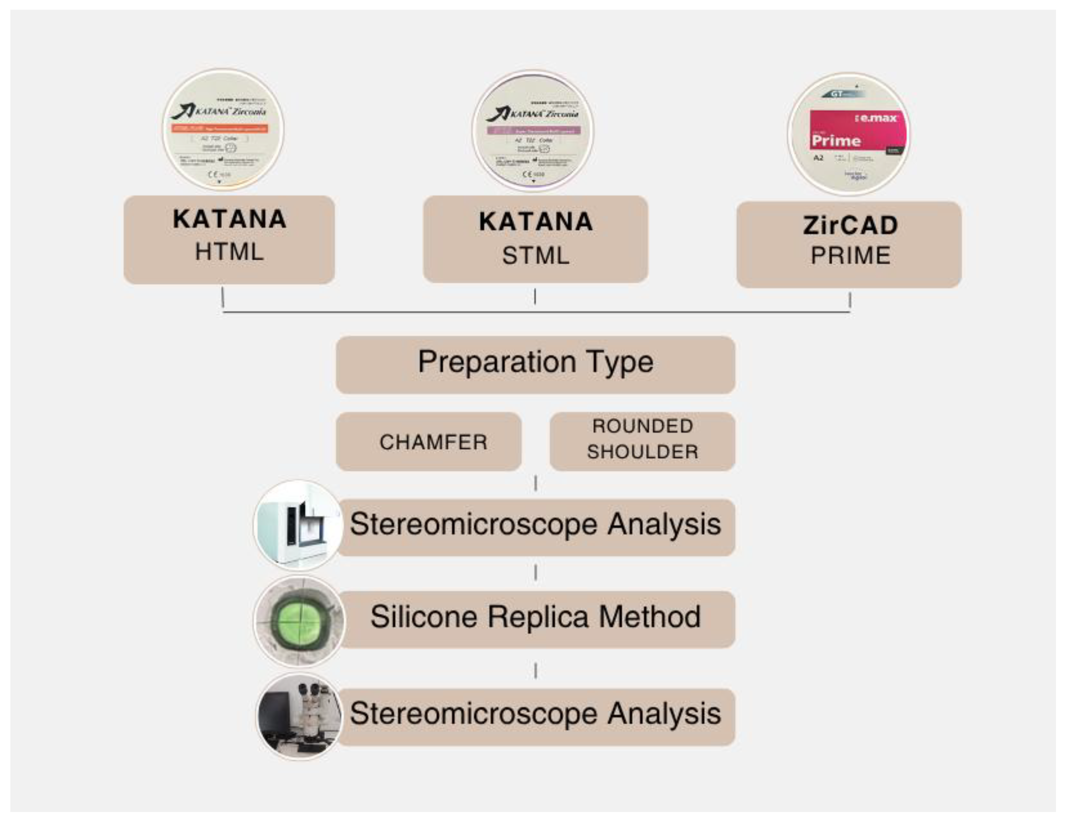
2. Materials and Methods

The study design of this in vitro study was shown in Figure 1. In the present in vitro study, the marginal and internal fit of three different monolithic CAD/CAM zirconia ceramics, applied to chamfer and rounded shoulder finish lines, was evaluated using the silicone replica technique.
In this study three different multilayer zirconia materials: KATANA High Translucent Multilayer Zirconia (HTML) (Kuraray Noritake Dental Inc., Tokyo, Japan; lot number: EMYJI), KATANA Super Translucent Multilayer Zirconia (STML) (Kuraray Noritake Dental Inc., Tokyo, Japan; lot number: EMBUD) and IPS e.max ZirCAD Prime (ZirCAD) (Ivoclar Vivadent, Schaan, Liechtenstein; lot number: Z061CN) with two different finish line designs: chamfer (C) and rounded shoulder (RS) were used to fabricate the restorations.

2.1. Sample Size Calculation

A priori power analysis was conducted using G*Power version 3.1.9.4 to determine the required sample size for three-way ANOVA. With a total sample size of 60 (n=10), significance level p < 0.05, and effect size f = 0.50, the achieved statistical power was 0.93.

2.2. Sample Preparation

Elastomeric molds (ANA-4 G; Frasaco GmbH, Tettnang, Germany) of the maxillary and mandibular arches were utilized for the fabrication of diagnostic casts. A typodont mandibular first molar (Frasaco GmbH, Tettnang, Germany) was placed into a mandibular elastomeric mold and cast using gypsum 4 (Extra Hard Dental Plaster, Cerestone; PERA Dental) to create mandibular and maxillary models. These models were then mounted on a semi-adjustable articulator (Stratos 100; Ivoclar AG, Liechtenstein). The mandibular right first molar was selected as the test abutment tooth and was prepared with an occlusal reduction of 1.0 mm and a taper of 6 to 10 degrees by a prosthodontist (D.G.D.). They were further subdivided into two subgroups (n=10) according to the type of finish line design: RS, C. C finish line was created by a chamfer bur with a 1 mm-wide while RS finish line was created by a rounded shoulder with a 90° internal angle. The finish lines were prepared supragingivally. The rough surfaces of the prepared teeth were smoothed using a polishing bur.
The maxillary and mandibular casts were scanned using a lab scanner (Dental Wings 7Series, Straumann, Basel, Switzerland) and crowns were designed with a (designed with a) CAD program (Exocad DentalDB 2.4 Plovdiv, Darmstadt, Germany) with a 50 μm cement thickness setting according to the manufacturer’s recommendation.
Restorations were fabricated using a 5-axis milling machine (Redon Hybrid, Redon Group, Istanbul, Türkiye). Three different types of zirconia, namely HTML, STML and ZirCAD, were manufactured. Sintering of zirconia crown restorations was done using sintering furnace (Protherm Furnaces, Czechoslovakia) at 1540ºC to obtain the original size, strength, and color, and the intaglio surface were facing upward according to manufacturer recommendations. The cooling protocol for zirconia crowns was done according to a built-in program in the sintering furnace.

2.3. Evaluation of Marginal and İnternal Fit

The replica technique as described by Molin and Karlsson was used to measure the marginal and internal fit [12]. In this in vitro study, teeth from the Frasaco master model, created with two different finish line design, were used. Each crown was filled with light-body silicone (Zhermack Hydrorise Extra Light Body Fast Set 100ml, Dentsply Sirona, Badia Polesine, Italy) and placed on the master model with finger pressure in accordance with the clinical procedure. After the light-body silicone was set, the crowns were removed, with the thin silicone remaining on the master model. Subsequently, a heavy-body silicone (Zhermack Elite HD+, Dentsply Sirona, Badia Polesine, Italy) was put onto the thin silicone. After setting, the silicone replica was removed from the master model. A total of 60 replicas were made, one for each crown. The replicas were sectioned at the center of each crown in both the bucco-lingual and mesio-distal directions. These divided parts were numbered from 1 to 4. The surfaces were examined individually. To utilize the smooth surfaces, the buccal section of part 1, the distal section of part 2, the lingual section of part 3, and the mesial section of part 4 were included in the study. The silicone replicas were then carefully separated from the inner surface of the crowns. This procedure was repeated for each stage for a total of 60 restorations. Each replica was carefully examined, and those showing surface tears were excluded from the study, with replication procedures repeated as necessary. The created silicone replicas were divided into four equal parts in both the buccolingual and mesiodistal directions using a No. 11 scalpel (Plasmed Sterilance Medical Inc, Suzhou, China). These divided parts were numbered from 1 to 4. The surfaces were examined individually. To utilize the smooth surfaces, the buccal section of part 1, the distal section of part 2, the lingual section of part 3, and the mesial section of part 4 were included in the study. The sections placed on the slides were evaluated under 40x magnification using a stereomicroscope (Leica DM4000 B, Wetzlar, Germany) in conjunction with the imaging program (Leica Qwin Plus ,Leica Microsystems Imaging Solutions Ltd, Cambridge, United States of America). Using the Leica Qwin Plus program, measurements were taken from 13 points on each section: 1 point from the marginal area, 5 points from the axial area, 2 points from the axio-occlusal area, and 5 points from the occlusal area, all in micrometers (µm). A total of 52 measurements were taken for each tooth, resulting in 3120 measurements across 60 teeth. As a result, 4 sections were evaluated for each restoration, and for each section, 4 regions were examined. The average of the multiple measurements was taken to calculate the average for the occlusal, axio-occlusal, axial, and marginal surfaces of each tooth, resulting in a single value for each point. For each restoration, 20 occlusal averages, 8 axio-occlusal averages, 20 axial averages, and 4 marginal averages were obtained, yielding 4 different values for each tooth.

2.4. Statistical Analysis

A three-way analysis of variance (ANOVA) was performed to assess the main and interaction effects of finish line design, material type, and measurement region on the gap values. The interaction terms included finish line × material, finish line × measurement region, material × measurement region, and the three-way interaction among all variables. Tukey’s post-hoc test was applied for pairwise comparisons when significant differences were found. Statistical analysis was performed using IBM SPSS Statistics for Windows, Version 21.0 (Armonk, NY: IBM Corp.), with the level of significance set at p < 0.053.

3. Results

Descriptive statistics for each finish line design are summarized in Table 1, Table 2 and Table 3. For the C finish line, all material types (HTML, STML, ZirCAD) showed the highest mean values in the occlusal region and the lowest in the axial region, with overall means of 36.700 µm, 32.452 µm, and 31.799 µm, respectively. The general mean for the C group was 33.792, with the occlusal region reaching 43.562 µm and the axial region 21.460 µm. In the RS finish line group, HTML, STML, and ZirCAD materials similarly exhibited their highest means in either the occlusal or marginal regions and their lowest in the axial region. Overall mean values were 51.901 µm, 41.519 µm, and 36.694 µm, respectively. The general mean for the RS group was 43.371 µm, with occlusal and axial means of 54.444 µm and 24.990 µm. When all measurements were considered collectively, occlusal regions consistently presented the highest values, while axial regions showed the lowest across all material types. General mean values were 43.939 µm for HTML, 37.224 µm for STML, and 34.246 µm for ZirCAD. The overall average across all groups and regions was 38.582 µm.
According to the results of the three-way ANOVA (Table 4), the mean measurement values demonstrated statistically significant differences based on finish line type, material type, and measurement region (p < 0.05). A significant interaction was observed between finish line type and material type (F = 4.505, p = 0.012), as well as between finish line type and measurement region (F = 2.939, p = 0.034), and between material type and measurement region (F = 4.034, p = 0.001). However, interaction among finish line type, material type, and measurement region was not statistically significant (F=1.117, p = 0.354). When finish line types were compared, the RS finish line (mean = 43.371) exhibited significantly higher measurement values than the C type (mean = 33.792) (p = 0.001).
Tukey’s post-hoc multiple comparison test (Table 5) was performed to determine which specific groups differed significantly. The results showed that the HTML material had a significantly higher mean value of 43.939 µm compared to both STML, with a mean of 37.224 µm, and ZirCAD , with a mean of 34.246 µm (p < 0.05). In contrast, the difference between STML and ZirCAD was not statistically significant (p > 0.05). Regarding the measurement regions, the occlusal region had the highest mean value at 49.003 µm and was significantly greater than both the axio-occlusal region, which had a mean of 37.756 µm, and the axial region, with a mean of 23.225 µm (p < 0.05). Similarly, the marginal region, with a mean of 44.343 µm, was significantly higher than the axio-occlusal and axial regions (p < 0.05). However, no statistically significant difference was found between the occlusal and marginal region means (p > 0.05).
For both the C and RS finish line types, the mean value of the HTML material was greater than that of the STML material, while the STML mean was also higher than that of the ZirCAD material. In both C and RS finish line types, the mean value of the occlusal region was higher than that of the marginal region, which was higher than the axio-occlusal region; additionally, the axio-occlusal region exhibited higher mean values than the axial region. The difference between the occlusal and marginal region means decreased when transitioning from the C to the RS finish line. In the HTML material group, the regional means were ranked from highest to lowest as follows: occlusal, axio-occlusal, marginal, and axial regions. In the STML material group, the ranking was occlusal, marginal, axio-occlusal, and axial. For ZirCAD, the order was marginal, occlusal, axio-occlusal, and axial.

4. Discussion

In this study, the marginal and internal gap values of monolithic crowns fabricated using different finish line designs and zirconia materials were compared. For this purpose, three different monolithic zirconia materials were included in the study. Based on the findings of the study, it was concluded that the finish line design had an effect on both marginal and internal fit, leading the rejection of the first null hypothesis. Additionally, it was found that the type of zirconia material also had an effect on the marginal and internal fit, resulting in the rejection of the second null hypothesis.
The success of dental restorations relies on four fundamental factors: marginal adaptation, biocompatibility, aesthetics, and mechanical strength. Of these properties, marginal adaptation plays a crucial role in fixed dental restorations. Inadequate marginal adaptation can lead to cement dissolution, plaque accumulation, secondary caries, and root canal infection [6,13]. Conversely, proper marginal adaptation minimizes the risk of microleakage and microcracks, thereby enhancing the longevity of the restoration [9]. In addition, internal fit plays a critical role in long-term clinical success, as insufficient fit may result in increased cement thickness, compromised retention, occlusal discrepancies, and reduced fracture resistance [8].
There is no consensus on clinically acceptable marginal and internal gap values for fixed dental restorations [5]. Clinically acceptable marginal gap has been reported as ≤120 µm and internal gap as ≤300 µm [14]. Although a universally accepted standard for clinically acceptable marginal and internal gap values in fixed dental restorations has not been established, numerous studies have proposed reference ranges based on clinical performance and long-term success [5]. For an optimal marginal seal, the space between the prepared tooth and the indirect restoration should be filled with a luting agent. It is generally recommended that this gap be maintained between 50 µm and 200 µm to ensure adequate bonding strength between the tooth and the restoration [15]. When the gap is less than 40 µm, the thin cement layer may compromise the bonding efficacy. Conversely, if the gap exceeds 150 µm, the cement may become exposed to the oral environment, increasing the risk of degradation and marginal leakage [16]. A frequently cited threshold value is 120 µm, as reported by McLean and von Fraunhofer [17] in their study involving 1,000 crowns. In accordance with these findings, several authors have considered a marginal gap of ≤120 µm and an internal gap of ≤300 µm to be within clinically acceptable limits for fixed restorations [14]. In our study, the average marginal and internal gap values of the restorations were 38.582 µm, which falls within the acceptable range.
The cervical finish line design of a tooth preparation is a critical factor that directly affects the marginal volume and contour of the restorative material, as well as the seating and marginal adaptation of the restoration [18]. However, there is no consensus in the current literature regarding the optimal finish line configuration [19]. While Haggag et al. [20] recommended a deep chamfer (C) finish line for monolithic restorations, Yu et al. [21], in their meta-analysis, reported that the rounded shoulder (RS) finish line provided better marginal adaptation compared to the chamfer, whereas the chamfer demonstrated superior internal fit compared to the shoulder finish line. Conversely, Yadav et al. [22] found that chamfer finish lines resulted in better marginal adaptation than shoulder finish lines in zirconia and hybrid restorations. Some studies have found no significant difference between C and RS designs. For instance, Angerame et al. [23] reported that both rounded 90-degree shoulder and minimally invasive chamfer preparations yielded satisfactory results without significant differences.
In the present study, the mean marginal gap was measured as 33.792 µm for the chamfer finish line and 43.371 µm for the rounded shoulder, with the chamfer showing statistically significantly better results. The superior marginal and internal fit observed with the chamfer finish line compared to the rounded shoulder may be attributed to several factors. The chamfer finish line is more easily recognized by CAD/CAM systems, allowing for higher accuracy during digital scanning and milling [24]. Additionally, the geometry of milling burs is generally more compatible with chamfer designs, facilitating better adaptation at the finish line. Moreover, the simpler and more defined form of the chamfer margin provides a clearer seating area for the restoration, reducing the risk of excessive material accumulation or misfit. High-strength ceramics such as zirconia have also been shown to exhibit better adaptation with chamfer finish lines [22]. Furthermore, chamfer preparations are considered more clinically feasible and reproducible, thereby minimizing operator-dependent variability [21]. Intrinsic characteristics of CAD/CAM systems, such as production processes and milling accuracy, also influence the precision of finish line reproduction. The combination of these factors may explain the statistically superior outcomes observed with the chamfer design in this study.
These findings align with the recommendations by Mansuco et al. [10], who emphasized the advantage of preparation designs lacking internal angles to improve restoration seating accuracy. Supporting this, Rizonaki et al. [8] evaluated lithium disilicate CAD crowns fabricated with three different finish lines (chamfer, rounded shoulder, and feather-edge), reporting that crowns with a rounded shoulder finish line exhibited the poorest internal fit. This result is consistent with our findings, and importantly, all three finish line types were within clinically acceptable limits.
However, the discrepancy between our results and those of Yu et al. [21], who found better marginal adaptation with the rounded shoulder finish line, may be attributed to differences in ceramic materials, measurement methods, or the specific CAD/CAM systems used. Operator experience and preparation standardization could also have contributed to these differences. Further research is needed to elucidate the discrepancies observed in previous findings.
In this study, restorations fabricated using Katana HTML material exhibited higher marginal and internal gap values compared to those made from STML and ZirCAD. Katana HTML is composed entirely of 3Y-TZP zirconia, which is characterized by high flexural strength and hardness [25]. However, the increased hardness may present challenges during CAD/CAM milling, especially in achieving accurate marginal adaptation [26]. In restorations with complex margin geometries, it may be difficult to remove sufficient material during milling, or microstructural fractures may occur. Additionally, 3Y-TZP-based materials undergo more substantial shrinkage during sintering, potentially compromising the dimensional stability and negatively affecting the marginal and internal fit of the restorations [2,27].
In contrast, multilayer zirconia materials such as STML and ZirCAD contain a higher yttria content (approximately 4Y–5Y), which, while slightly reducing mechanical strength, improves machinability and enhances translucency [1,3]. Their lower hardness and larger grain size enable more precise milling, particularly in critical marginal areas. Moreover, the layered structure of these materials helps to distribute internal stresses more uniformly during sintering, contributing to improved dimensional stability [28,29,30].
Consistent with our findings, a study by Tabata et al.[15], identified the type of restorative material as a statistically significant factor affecting marginal discrepancy.. Monolithic zirconia restorations are subjected to a crystallization process prior to cementation [31]. While Kim et al. [32] reported no significant change in internal fit following crystallization, they observed an increase in marginal gap values, suggesting that the crystallization process may affect marginal and internal adaptation differently.
Similarly, Yıldırım et al.[33] reported higher marginal discrepancy values for the ceramic group (IPS e.max CAD) compared to the composite resin group (Lava Ultimate). In another study, Goujat et al.[34] evaluated the internal fit of four different CAD/CAM ceramic materials (Vita Enamic, Lava Ultimate, IPS e.max CAD, and Cerasmart) and found a negative correlation between flexural strength and internal discrepancy, indicating that materials with higher flexural strength may exhibit better internal adaptation.
In conclusion, although Katana HTML offers superior mechanical properties, its high hardness and limitations during the manufacturing process may adversely affect the marginal and internal fit of restorations. Therefore, when selecting a material, factors such as machinability and sintering behavior should be considered alongside mechanical strength.
Various methods have been employed to evaluate the marginal and internal adaptation of dental restorations [35]. Among these, the silicone replica technique has gained prominence due to its cost-effectiveness, reliability, and widespread use in the literature [12,36,37,38,39]. Its non-destructive nature and applicability under both in vitro and in vivo conditions have established it as a well-accepted and standardized method [40]. Furthermore, it enables the intraoral assessment of marginal discrepancies [4]. Nevertheless, the technique presents certain limitations, such as the potential deformation or tearing of the impression material during the removal of the prosthesis, and the inherent restriction to two-dimensional (2D) analysis [11].
Several studies have reported varying numbers of marginal and internal gap measurements in in vitro analyses [41,42]. In 2022, Guacheta et al. [42] conducted eight marginal and three internal measurements to evaluate the fit quality of ceramic laminate veneers (CLVs) fabricated using a conventional waxing technique, in comparison with those produced by 3D printing. Subsequently, in 2023, Al-Dwairi et al. [41] performed two marginal and six internal measurements to assess the fit of CLVs fabricated through both direct and indirect digitization techniques.
To mitigate these limitations and enhance measurement precision, multiple reference points were utilized on each replica in the present study. Specifically, measurements were obtained from 13 distinct locations: one from the marginal area, five from the axial area, two from the axio-occlusal area, and five from the occlusal area. Accordingly, a total of 52 measurements were recorded per group. This methodological approach was adopted to increase the reliability of the data and to provide a more comprehensive evaluation of crown adaptation.
Numerous studies have demonstrated that gap measurements can vary considerably based on the specific location being assessed [22,38,41,42]. For example, Cunali et al. [38] evaluated both marginal and internal adaptation using the silicone replica technique alongside micro-CT and reported clinically acceptable discrepancies at four distinct measurement points. Similarly, Yadav et al. [22] presented findings that align with these observations. In the present study, the largest gap values were recorded in the occlusal and marginal areas, whereas the axial surfaces exhibited the smallest gaps. One potential explanation for the greater occlusal discrepancies is the loss of fine detail and rounding of edges, which may result from the scanner’s resolution limitations. Supporting this hypothesis, another study assessing gap widths in all-ceramic restorations produced via three different CAD-CAM systems found a consistent increase in gap size from the margin toward the center of the restoration. Such inconsistencies in measurement could stem from factors including the precision of digital scanning and data processing, as well as the design and condition of the milling tools employed during fabrication [43,44].
This study, which provides information on the physical properties of the increasingly used new generation monolithic blocks, has some limitations. One limitation is that the in-vitro conditions used in the study do not mimic the oral environment. In-vivo studies would provide more comprehensive information on the longevity of restorations, potential complications, and changes in the supporting teeth and surrounding tissues. Therefore, it is necessary to evaluate the marginal and internal fit of new generation monolithic zirconia materials under in-vivo conditions. Also, the dimensional stability changes of the silicone material used in the silicone replica technique may negatively affect the marginal gap measurements. Additionally, as noted in other studies, the silicone replica technique, unlike direct marginal gap measurement techniques, limits the areas where measurements can be taken [43]. Therefore, studies that use different evaluation methods for marginal and internal fit are needed.

5. Conclusions

Based on the findings of this in vitro study, the following conclusions were drawn:
The amount of Y-TZP in the material composition influences the marginal and internal fit, with multilayer monolithic zirconia restorations demonstrating marginal and internal fit values within clinically acceptable limits.
Finish line design affects the marginal and internal fit of multilayer zirconia restorations, with the C design showing superior fit. Both designs had marginal and internal gap values within clinically acceptable limits.

Author Contributions

Conceptualization, D.G.D. and O.S.Y.; methodology, D.G.D.; software, D.G.D.; validation, D.G.D., and O.S.Y.; formal analysis, D.G.D.; investigation, D.G.D.; resources, D.G.D.; data curation, D.G.; writing—original draft preparation, D.G.D.; writing—review and editing, D.G.D.; visualization, D.G.D.; supervision, O.S.Y.; project administration, D.G.D.; funding acquisition, O.S.Y. All authors have read and agreed to the published version of the manuscript.

Funding

This research was funded by the Scientific Research Projects Coordination Unit of Gazi University (grant no. TDH-2023-8909).

Institutional Review Board Statement

Not applicable.

Informed Consent Statement

Not applicable.

Data Availability Statement

Data presented in this study are available on request from the corresponding author.

Conflicts of Interest

The authors declare no conflicts of interest.

Abbreviations

The following abbreviations are used in this manuscript:
HTML KATANA High Translucent Multilayer Zirconia
STML KATANA Super Translucent Multilayer Zirconia
ZirCAD IPS e.max ZirCAD Prime
R Rounded Shoulder
C Chamfer
µm Micrometer
ANOVA A three-way analysis of variance
Y-TZP Yttria-stabilized tetragonal zirconia polycrystal
2D Two Dimensions

References

  1. P.F. Cesar, R.B. de Paula Miranda, K.F. Santos, S.S. Scherrer, Y. Zhang, Recent advances in dental zirconia: 15 years of material and processing evolution, Dent. Mater. (2024). [CrossRef]
  2. Y. Zhang, B.R. Lawn, Evaluating dental zirconia, Dent. Mater. 35(1) (2019) 15–23. [CrossRef]
  3. S. Kongkiatkamon, D. Rokaya, S. Kengtanyakich, C. Peampring, Current classification of zirconia in dentistry: An updated review, PeerJ 11 (2023) e15669. [CrossRef]
  4. M. Hasanzade, M. Moharrami, M. Alikhasi, How adjustment could affect internal and marginal adaptation of CAD/CAM crowns made with different materials, J. Adv. Prosthodont. 12(6) (2020) 344. [CrossRef]
  5. M.R. Baig, A.A. Akbar, M. Embaireeg, Effect of finish line design on the fit accuracy of CAD/CAM monolithic polymer-infiltrated ceramic-network fixed dental prostheses: an in vitro study, Polymers 13(24) (2021) 4311. [CrossRef]
  6. S. Suzuki, Y. Katsuta, K. Ueda, F. Watanabe, Marginal and internal fit of three-unit zirconia fixed dental prostheses: effects of prosthesis design, cement space, and zirconia type, J. Prosthodont. Res. 64(4) (2020) 460–467. [CrossRef]
  7. C.-C. Peng, K.-H. Chung, H.-T. Yau, Assessment of the internal fit and marginal integrity of interim crowns made by different manufacturing methods, J. Prosthet. Dent. 123(3) (2020) 514–522. [CrossRef]
  8. M. Rizonaki, W. Jacquet, P. Bottenberg, L. Depla, M. Boone, P.J. De Coster, Evaluation of marginal and internal fit of lithium disilicate CAD-CAM crowns with different finish lines by using a micro-CT technique, J. Prosthet. Dent. 127(6) (2022) 890–898. [CrossRef]
  9. J. Lyu, X. Cao, X. Yang, J. Tan, X. Liu, Effect of finish line designs on the dimensional accuracy of monolithic zirconia crowns fabricated with a material jetting technique, J. Prosthet. Dent. (2025). [CrossRef]
  10. [10] E. Mancuso, T. Gasperini, T. Maravic, C. Mazzitelli, U. Josic, A. Forte, et al., The influence of finishing line and luting material selection on the seating accuracy of CAD/CAM indirect composite restorations, J. Dent. 148 (2024) 105231. [CrossRef]
  11. [11] P. Fasih, S. Tavakolizadeh, M.S. Monfared, A. Sofi-Mahmudi, A. Yari, Marginal fit of monolithic versus layered zirconia crowns assessed with 2 marginal gap methods, J. Prosthet. Dent. 130(2) (2023) 250.e1–250.e7. [CrossRef]
  12. [12] M. Molin, S. Karlsson, The fit of gold inlays and three ceramic inlay systems: A clinical and in vitro study, Acta Odontol. Scand. 51(4) (1993) 201–206. [CrossRef]
  13. [13] J. Abduo, G. Ho, A. Centorame, S. Chohan, C. Park, R. Abdouni, et al., Marginal accuracy of monolithic and veneered zirconia crowns fabricated by conventional and digital workflows, J. Prosthodont. 32(8) (2023) 706–713. [CrossRef]
  14. [14] H. Zhu, Y. Zhou, J. Jiang, Y. Wang, F. He, Accuracy and margin quality of advanced 3D-printed monolithic zirconia crowns, J. Prosthet. Dent. (2023). [CrossRef]
  15. [15] L.F. Tabata, T.A. de Lima Silva, A.C. de Paula Silveira, A.P.D. Ribeiro, Marginal and internal fit of CAD-CAM composite resin and ceramic crowns before and after internal adjustment, J. Prosthet. Dent. 123(3) (2020) 500–505. [CrossRef]
  16. [16] A.C. de Paula Silveira, S.B. Chaves, L.A. Hilgert, A.P.D. Ribeiro, Marginal and internal fit of CAD-CAM-fabricated composite resin and ceramic crowns scanned by 2 intraoral cameras, J. Prosthet. Dent. 117(3) (2017) 386–392. [CrossRef]
  17. [17] J.W. McLean, Evolution of dental ceramics in the twentieth century, J. Prosthet. Dent. 85(1) (2001) 61–66. [CrossRef]
  18. [18] A. Baldi, A. Comba, D. Rozzi, G.K.R. Pereira, L.F. Valandro, R.M. Tempesta, et al., Does partial adhesive preparation design and finish line depth influence trueness and precision of intraoral scanners?, J. Prosthet. Dent. 129(4) (2023) 637.e1–637.e9. [CrossRef]
  19. [19] S. Sirous, A. Navadeh, S. Ebrahimgol, F. Atri, Effect of preparation design on marginal adaptation and fracture strength of ceramic occlusal veneers: A systematic review, Clin. Exp. Dent. Res. 8(6) (2022) 1391–1403. [CrossRef]
  20. [20] K. Haggag, M. Abbas, H. Ramadan, Effect of thermo-mechanical aging on the marginal fit of two types of monolithic zirconia crowns with two finish line designs, Al-Azhar Dent. J. Girls 5(1) (2018) 121–128.
  21. [21] H. Yu, Y.-h. Chen, H. Cheng, T. Sawase, Finish-line designs for ceramic crowns: A systematic review and meta-analysis, J. Prosthet. Dent. 122(1) (2019) 22–30.e5. [CrossRef]
  22. [22] P. Yadav, V. Sharma, J. Paliwal, K.K. Meena, R. Madaan, B. Gurjar, An in vitro comparison of zirconia and hybrid ceramic crowns with heavy chamfer and shoulder finish lines, Cureus 15(1) (2023). [CrossRef]
  23. [23] D. Angerame, M. De Biasi, M. Agostinetto, A. Franzò, G. Marchesi, Influence of preparation designs on marginal adaptation and failure load of full-coverage occlusal veneers after thermomechanical aging simulation, J. Esthet. Restor. Dent. 31(3) (2019) 280–289. [CrossRef]
  24. [24] R. Euán, O. Figueras-Álvarez, J. Cabratosa-Termes, R. Oliver-Parra, Marginal adaptation of zirconium dioxide copings: Influence of the CAD/CAM system and the finish line design, J. Prosthet. Dent. 112(2) (2014) 155–162. [CrossRef]
  25. [25] M. Bruhnke, Y. Awwad, W.-D. Müller, F. Beuer, F. Schmidt, Mechanical properties of new generations of monolithic, multi-layered zirconia, Materials 16(1) (2022) 276. [CrossRef]
  26. [26] S.E. Elsaka, Influence of surface treatment on the bond strength of resin cements to monolithic zirconia, J. Adhes. Dent. 18(5) (2016). [CrossRef]
  27. Y. Zhang, B.R. Lawn, Novel zirconia materials in dentistry, J. Dent. Res. 97(2) (2018) 140–147. [CrossRef]
  28. M. Michailova, A. Elsayed, G. Fabel, D. Edelhoff, I.-M. Zylla, B. Stawarczyk, Comparison between novel strength-gradient and color-gradient multilayered zirconia using conventional and high-speed sintering, J. Mech. Behav. Biomed. Mater. 111 (2020) 103977. [CrossRef]
  29. C.-M. Kang, W.-C. Hsu, M.-S. Chen, H.-Y. Wu, Y. Mine, T.-Y. Peng, Fracture characteristics and translucency of multilayer monolithic zirconia crowns of various thicknesses, J. Dent. 145 (2024) 105023. [CrossRef]
  30. N. Güntekin, B. Kızılırmak, A.R. Tunçdemir, Comparison of mechanical and optical properties of multilayer zirconia after high-speed and repeated sintering, Materials 18(7) (2025) 1493. [CrossRef]
  31. R. Giordano II, Ceramics overview, Br. Dent. J. 232(9) (2022) 658–663. [CrossRef]
  32. J.-H. Kim, S. Oh, S.-H. Uhm, Effect of the crystallization process on the marginal and internal gaps of lithium disilicate CAD/CAM crowns, Biomed. Res. Int. 2016(1) (2016) 8635483. [CrossRef]
  33. G. Yildirim, I.H. Uzun, A. Keles, Evaluation of marginal and internal adaptation of hybrid and nanoceramic systems with microcomputed tomography: An in vitro study, J. Prosthet. Dent. 118(2) (2017) 200–207. [CrossRef]
  34. G. Goujat, H. Abouelleil, P. Colon, C. Jeannin, N. Pradelle, D. Seux, et al., Mechanical properties and internal fit of 4 CAD-CAM block materials, J. Prosthet. Dent. 119(3) (2018) 384–389. [CrossRef]
  35. K. Son, S. Lee, S.H. Kang, J. Park, K.-B. Lee, M. Jeon, et al., A comparison study of marginal and internal fit assessment methods for fixed dental prostheses, J. Clin. Med. 8(6) (2019) 785. [CrossRef]
  36. P. Boitelle, L. Tapie, B. Mawussi, O. Fromentin, Evaluation of the marginal fit of CAD-CAM zirconia copings: Comparison of 2D and 3D measurement methods, J. Prosthet. Dent. 119(1) (2018) 75–81. [CrossRef]
  37. K. Ueda, F. Watanabe, Y. Katsuta, M. Seto, D. Ueno, K. Hiroyasu, et al., Marginal and internal fit of three-unit fixed dental prostheses fabricated from translucent multicolored zirconia: Framework versus complete contour design, J. Prosthet. Dent. 125 (2021) 340.e1–340.e6. [CrossRef]
  38. R.S. Cunali, R.C. Saab, G.M. Correr, L.F. Cunha, B.P. Ornaghi, A.V. Ritter, et al., Marginal and internal adaptation of zirconia crowns: a comparative study of assessment methods, Braz. Dent. J. 28 (2017) 467–473. [CrossRef]
  39. T.M. Doan, T.N. Nguyen, V.-K. Pham, N. Chotprasert, C.T.B. Vu, Comparative analysis of the fit quality of monolithic zirconia veneers produced through traditional and digital workflows using silicone replica technique: an in vitro study, BMC Oral Health 24 (2024) 1566. [CrossRef]
  40. A. Arezoobakhsh, S.S. Shayegh, A.J. Ghomi, S.M.R. Hakimaneh, Comparison of marginal and internal fit of 3-unit zirconia frameworks fabricated with CAD-CAM technology using direct and indirect digital scans, J. Prosthet. Dent. 123 (2020) 105–112. [CrossRef]
  41. Z.N. Al-Dwairi, M. Al-Sardi, B.J. Goodacre, C.J. Goodacre, K.Q. Al Hamad, M. Özcan, et al., Evaluation of marginal and internal fit of ceramic laminate veneers fabricated with five intraoral scanners and indirect digitization, Materials 16 (2023) 2181. [CrossRef]
  42. L. Guachetá, C.D. Stevens, J.A. Tamayo Cardona, R. Murgueitio, Comparison of marginal and internal fit of pressed lithium disilicate veneers fabricated via a manual waxing technique versus a 3D printed technique, J. Esthet. Restor. Dent. 34 (2022) 715–720. [CrossRef]
  43. N. Sakornwimon, C. Leevailoj, Clinical marginal fit of zirconia crowns and patients’ preferences for impression techniques using intraoral digital scanner versus polyvinyl siloxane material, J. Prosthet. Dent. 118 (2017) 386–391. [CrossRef]
  44. Z.N. Al-Dwairi, R.M. Alkhatatbeh, N.Z. Baba, C.J. Goodacre, A comparison of the marginal and internal fit of porcelain laminate veneers fabricated by pressing and CAD-CAM milling and cemented with 2 different resin cements, J. Prosthet. Dent. 121 (2019) 470–476. [CrossRef]
Figure 1. Schematic representation of the study design.
Preprints 181041 g001
Table 1. Descriptive Statistics for the C Finish Line Type.
Material Type Measured Region Mean±SD1
HTML Occlusal Mean 51.117± 19.01
Axio-occlusal Mean 40.085±13.215
Axial Mean 23.317± 3.306
Marginal Mean 34.426±10.245
Total 37.237±15.928
STML Occlusal Mean 42.913±6.140
Axio-occlusal Mean 30.795±5.516
Axial Mean 19.750±2.950
Marginal Mean 35.901±8.060
Total 32.340±10.290
ZirCAD Occlusal Mean 36.654±6.266
Axio-occlusal Mean 28.287±2.433
Axial Mean 21.311±3.184
Marginal Mean 40.944±6.300
Total 31.799±8.988
Overall Occlusal Mean 43.562±13.131
Axio-occlusal Mean 33.056±9.598
Axial Mean 21.460±3.383
Marginal Mean 37.091±8.551
Total 33.792±12.262
1SD: Standard Deviation.
Table 2. Descriptive Statistics for the RS Finish Line Type.
Material Type Measured Region Mean±SD1
HTML Occlusal Mean 66.663±14.895
Axio-occlusal Mean 55.164±12.050
Axial Mean 29.179±13.233
Marginal Mean 56.598±10.364
Total 51.901±18.616
STML Occlusal Mean 53.664±20.243
Axio-occlusal Mean 37.166±12.458
Axial Mean 20.283±4.331
Marginal Mean 54.962±15.504
Total 41.519±19.870
ZirCAD Occlusal Mean 43.004±10.589
Axio-occlusal Mean 35.036±6.548
Axial Mean 25.508±2.950
Marginal Mean 43.227±5.370
Total 36.694±9.918
Overall Occlusal Mean 54.444±18.101
Axio-occlusal Mean 42.456±13.815
Axial Mean 24.990±8.755
Marginal Mean 51.596±12.392
Total 43.371±17.771
1SD: Standard Deviation.
Table 3. Descriptive Statistics for All Measurements.
Material Type Measured Region Mean±SD1
HTML Occlusal Mean 58.890±18.440
Axio-occlusal Mean 47.625±14.538
Axial Mean 26.249±9.857
Marginal Mean 45.512±15.164
Total 44.569±18.729
STML Occlusal Mean 48.289±15.569
Axio-occlusal Mean 33.980±9.931
Axial Mean 20.017±3.617
Marginal Mean 45.432±15.500
Total 36.929±16.387
ZirCAD Occlusal Mean 39.829±9.073
Axio-occlusal Mean 31.662±5.925
Axial Mean 23.409±3.683
Marginal Mean 42.085±5.816
Total 34.246±9.722
Overall Occlusal Mean 49.003±16.610
Axio-occlusal Mean 37.756±12.711
Axial Mean 23.225±6.817
Marginal Mean 44.343±12.842
Total 38.582±15.973
1SD: Standard Deviation.
Table 4. Three-Way ANOVA Test Results.
Variable df F p-value
Model 23 15.510 <0.001
Model intercept 1 3.355.358 <0.001
Finish Line Type 1 51.712 <0.001
Material Type 2 21.554 <0.001
Measured Region 3 71.062 <0.001
Finish Line Type * Material Type 2 4.505 0.012
Finish Line Type * Measured Region 3 2.939 0.034
Material Type * Measured Region 6 4.034 0.001
Finish Line Type * Material Type * Region 6 1.117 0.354
df: Degrees of freedom, F: ANOVA test statistic.
Table 5. Tukey Test Results for Multiple Comparisons.
Group Group2 Mean p-value
Material Type HTML 43.939±18.714a     <0.001
STML 37.224±16.489b
ZirCAD 34.246±9.722bc
Overall 38.582±15.973
Measured Region Occlusal 49.003±16.610a     <0.001
Axio-occlusal 37.756±12.711b
Axial 23.225±6.817c
Marginal 44.343±12.842a
Overall 38.582±15.973
Disclaimer/Publisher’s Note: The statements, opinions and data contained in all publications are solely those of the individual author(s) and contributor(s) and not of MDPI and/or the editor(s). MDPI and/or the editor(s) disclaim responsibility for any injury to people or property resulting from any ideas, methods, instructions or products referred to in the content.
Copyright: This open access article is published under a Creative Commons CC BY 4.0 license, which permit the free download, distribution, and reuse, provided that the author and preprint are cited in any reuse.
Prerpints.org logo

Preprints.org is a free preprint server supported by MDPI in Basel, Switzerland.

Subscribe

Disclaimer

Terms of Use

Privacy Policy

Privacy Settings

© 2025 MDPI (Basel, Switzerland) unless otherwise stated