
Submitted:
14 October 2025
Posted:
15 October 2025
You are already at the latest version
Abstract
The sensory system plays a critical role in the development since it allows us to correct and process the internal-external interaction, through the detection and encoding of the external stimuli. Dysfunctions of this system lead to the sensory integration, thus to the sensory processing disorders (SPDs). PSD affects about 5%–13% of 4-6-year-old children affecting not just their ability to respond to sensory information from the en-vironment, but also their social interaction and regulation of emotions, as well as the motor actions, learning, attention, communication, and sleep. SPDs have been extensively studied under different aspects, from molecular to behavioural ones, but the pathological mechanisms still remain under debate. Moreover, while there are many studies investigating the behavioural consequences of SPDs, which are used for diagnostic purposes, more are needed to find biomarkers. Indeed, since SPDs are often misdiagnosed with behaviour-related dysfunction, e.g., autism spectrum disorder, it is urgent to develop a new diagnostic tool, therefore targeting therapies. SPDs are misdiagnosed with attention deficit hyperactivity disorder (ADHD). Nevertheless, despite their similarity in behavioural symptoms and consequences, they have different causes, involve different neuronal circuits, and therefore have different treatments. In this narrative review, we aim to provide an overview of the literature on molecular and neuroimaging findings about SPDs. We will concentrate on the interaction between SPDs and ADHD, highlight the similarities and the differences, to more deeply understand the differences in pathological mechanisms and, in turn to highlight unique possible biomarkers, diagnostic and therapeutic tools.
Keywords:
SPD
; fMRI
; ADHD
; brain structure
; brain oscillations
; sensory dysfunctions
; sensory system
1. Introduction
Several inputs to our sensory systems influence how we interact with surroundings in daily life, e.g., listening to a person talk while watching their lip and arms movements and smelling specific odours or parfum, all at once. Our brain integrates several streams of inputs to create a single percept, and this process is called multisensory integration. We can detect, identify, and distinguish between exterior things more accurately and advantageously through this process than we could with a single stream of data (Marshall et al., 2022).
The intricacy of the stimulus determines whether attention is focused early or late in multisensory integration. Simple stimuli allow for early processing through bottom-up attentional processing; more complex stimuli need top-down processing because incoming stimuli must be compared to prior information via feedback loops to sensory cortices (2-3). By modulating the gain and stimulus saliency between sensory modalities, attentional multisensory integration processing is regulated both top-down and bottom-up (4). While signals are naturally integrated in sensory regions and superior colliculi, higher-order association areas require top-down attention to correctly integrate information (5).
Dysfunctions of this system lead to difficulties in daily life. These dysfunctions are present in different physiological and pathological conditions, e.g., in old people (Völter et al., 2021), depression, injuries of peripheral inputs (Wall and Wang, 2002), autism spectrum like disorder (ASD), attention deficit hyperactivity disorder (ADHD) and sensory processing disorders (SPDs).
ADHD is a neurodevelopmental disorder characterized by persistent and pervasive patterns of inattention, hyperactivity, and impulsivity, frequently associated with unusual responses to sensory stimulation (Little et al., 2023). In this review, we examine the relationship between SPDs and ADHD, highlighting particularly the role of sensory dysfunction as a shared feature. While sensory abnormalities are increasingly recognized as part of the ADHD phenotype, they are also central to the clinical presentation of SPDs, raising important questions about their neurobiological overlap. Despite growing interest, the precise neuronal mechanisms underlying sensory dysregulation in both conditions remain incompletely understood. This gap in knowledge contributes to a significant clinical challenge: the frequent misdiagnosis or conflation of SPDs and ADHD, especially in pediatric populations. Both disorders can manifest with similar behavioral profiles—such as distractibility, emotional dysregulation, and motor restlessness—and exert comparable impacts on daily functioning across developmental stages. By exploring converging and diverging neurofunctional pathways, this review aims to clarify the distinctions and intersections between SPDs and ADHD, ultimately contributing to more accurate diagnosis and targeted interventions. Moreover, since the ADHD-SPDs association is still unclear, the clinical guidelines for ADHD do not include the assessment of the sensory processing pattern (Jurek et al., 2025). Nevertheless, because therapeutic interventions depend on an accurate diagnosis, correct identification is essential to ensure appropriate treatment and avoid exacerbating the pathological condition. Therefore, there is an urgent need to identify specific biomarkers and to develop new, targeted diagnostic tools and therapies for both SPDs and ADHD.
Considering that sensory discrimination deficits—a hallmark of SPD—frequently co-occur with attentional dysfunctions in ADHD (Mark et al., 2023; Little et al., 2023), the investigation of shared neural mechanisms becomes particularly relevant, though still not fully elucidated. Although the existing literature on the neurobiological overlap between SPDs and ADHD remains limited, this narrative review aims to bridge that gap by examining potential similarities in their underlying neuronal mechanisms. By synthesizing current findings and theoretical models, the review offers an integrated perspective that seeks to deepen our understanding of the complex and often intertwined relationship between these two conditions. This approach not only highlights shared patterns of sensory dysregulation and attentional control but also underscores the need for more nuanced diagnostic frameworks and targeted interventions.
2. Materials and Methods
2.1. Study Experimental Design
This narrative review included all types of peer-reviewed English-language publications, i.e., case reports, original research articles, and reviews (narrative, systematic and meta-analysis reviews), conducted in both animal models and humans. The search was performed in PubMed between September 2024 and September 2025, using the following keywords: “ADHD”, “Attention-deficit hyperactivity disorder”, “SPD”, “Sensory processing disorder”, “ADHD” AND “brain”, “SPD” AND “brain”, “ADHD” AND “fMRI”, “SPD” AND “fMRI”, “ADHD” AND “ERP”, “SPD” AND “ERP”, “ADHD” AND “SEP”, “SPD” AND “SEP”, “ADHD” AND “brain structure”, and “SPD” AND “brain structure”. Only studies investigating the neuronal functions of ADHD and/or SPD were considered, while those focusing on the effect of pharmacological interventions on ADHD and/or SPD on neuronal activity were excluded. This selection strategy was adopted to align with the primary objective of the review, i.e., to provide an overview of the shared neuronal mechanisms underlying ADHD and SPD.
3. Results
Recent research suggested that ADHD symptoms may arise from selective disruptions in automatic processing, and this could be due to atypical sensory processing, which in turns impairs the ability to automate behaviours and process information rapidly and efficiently without conscious effort (Ullman and Pullman, 2015; Fabio, 2020).
The presence of sensory deficits in both SPDs and ADHD, indicates a strong correlation between the two pathological conditions. In detail, up to 64% of children with ADHD present atypical sensory responses, including both over- and under-responsivity (Cascio et al., 2010), in inattentive and hyperactive/impulsive subtypes (Yochman et al., 2004; Engel-Yeger and Ziv-On, 2011). Conversely, about 40% of children with SPD also meet criteria for ADHD (Koziol and Budding, 2012). This bidirectional association has led to the hypothesis that SPD may potentially represent a diagnostic marker of neuropsychiatric disorders such as ADHD (Passarello et al., 2022), but this is an open debate in literature. Interestingly, it is well known the importance of the it has been proposed the critical importance of the early sensory processing in the manage and defined the cortical and subcortical systems, allowing correct sensory process in the future (Happel et al., 2022). Indeed, the sensory pathways have hierarchical cortical-subcortical organization that permits the accurate processing of sensory information (Happel et al., 2022).
To elucidate how these neuronal pathways are involved in the sensory deficits, it is crucial to employ physiological measures such as somatosensory evoked potentials, sympathetic indices of nervous system activity (e.g., electrodermal reactivity), electroencephalography (EEG), event-related potentials (ERP), and functional magnetic resonance imaging (fMRI). Moreover, EEG and fMRI are nowadays important and valuable diagnostic tools, since allow the detection of abnormal activity in comparison to control healthy subjects (Passarello et al., 2022), both in children and in adulthood. Nevertheless, it is critical to notice that each of used technique has advantage and disadvantages, as temporal and spatial resolution. Indeed, the ERP has higher temporal resolution (milliseconds) in comparison of EEG and fMRI, while fMRI has higher spatial resolution in comparison to ERP and EEG. Moreover, for example, fMRI is not always easy to use with children. The following sections summarize the main physiological findings reported in studies applying these neuronal physiological methodologies. Importantly, the information from different techniques could be integrated with each other, and it is also possible to perform simultaneous EEG-fMRI studies (e.g., Lenartowicz et al., 2016).
3.1. Shared Brain Networks
Despite the ADHD, but not SPDs, symptoms are the result of abnormalities in large-scale brain networks, the two conditions shared common brain circuits (Koziol et al., 2012). The executive functions, e.g., attention, and sensory gating, as well as reward processing, and emotions (Fettes et al., 2017) are encoded by fronto-striatal circuits and they are impairment in both ADHD and SPDs conditions (Cupertino et al., 2020; Cubillo et al., 2012). Cupertino and colleagues (2020) reported not just the role of the fronto-striatal circuitry but also the critical importance of white-matter in the pathophysiology of ADHD. Indeed, they reported the reduction in the volume in frontal lobes, striatum, and their interconnecting white-matter, in both children, and adults (Cupertino et al., 2020; see Section “Brain structure” section for details in this study).
The fronto-striato-thalamic circuit represents a distributed network linking the caudate, putamen, thalamus, supplementary motor area, lateral prefrontal cortex, and parietal lobe. The interpretation of sensory processing deficits within this system requires the analysis of the specific affected hubs, and their role in shaping the function of the circuit. Cortese et al. (2012) conducted a meta-analysis of neuroimaging studies in ADHD, integrating structural and functional findings. They identified consistent abnormalities in fronto-striatal, cerebellar, and parietal regions across imaging modalities. These alterations were linked to core ADHD symptoms such as inattention, impulsivity, and hyperactivity. Moreover, a second fronto-striatal circuit seems to be involved in the pathological process of ADHD, i.e. the circuit involving the nucleus accumbens and orbitofrontal cortex (Zaher et al., 2025). In particular, the nucleus accumbens has been shown to be a central player in the neurobiology of ADHD symptoms, because its involvement in the dopaminergic system, so in reward processing and motivation, which is often impaired in ADHD (Luman et al., 2005). Nevertheless, nowadays there is no scientific evidence of the nucleus accumbens involvement in SPDs pathology. Similarly, while the dorsal and ventral medial prefrontal cortex are involved in ADHD, there is no study about the correlation between SPDs and their dysfunctions (Dubljević et al., 2025). Contrary, amygdala and insula seems to be involved in both SPDs and ADHD. Amygdala in both the evaluation and response to emotionally salient stimuli and it is essential for the sensory processing (Zald, 2003; Turner and Herkenham, 1991). For example, it has been delineated a circuit through which the amygdala exerts top-down influence on early sensory processing via projections to the locus coeruleus, in relation to the olfactory system. The olfactory bulb does not receive any direct projections from the amygdala, therefore it influences on early sensory processing through the locus coeruleus. These results could be potentially shed light in the mechanisms of the sensory abnormalities (Fast and McGann, 2017). Concerning ADHD, contrasting data are present in the literature, with evidence of its abnormal activation in emotion processing and facial recognition (Brotman et al., 2010; Herpertz et al., 2008; Marsh et al., 2008). Adra and colleagues (2021) have reported in ADHD adults, the positive correlation between both sensory craving and under-responsivity with the volume of the amygdala volume (Adra et al., 2021). However, studies directly examining the role of the amygdala in modulating sensory processing within the broader neural networks of ADHD have not been extensively investigated. Similarly, despite the established role of the amygdala in sensory processing, research on SPSDs remains considerably more limited than in ADHD, with no studies to date, to our knowledge, directly investigating amygdala-mediated modulation of sensory input within the neural circuits underlying these disorders.
Similarly, the insula cortex plays a pivotal role in the emotional valence of the stimulus, being also a critical hub of the social brain circuit. Indeed, here the social touch is encoded (Grandi, 2016; Morrison et al., 2011; Olausson et al., 2002; Björnsdotter et al., 2010). Importantly, it is connected with the amygdala (Stein et al., 2017; Mufson et al., 1981; Mesulam and Mufson, 1982). There are evidence of the involvement of the insula in ADHD. For example, Lopez-Larson (2012) have reported bilateral reduction in anterior insular gray matter volumes in youths with ADHD, in relation to attention and inhibitory capacity in ADHD. Fateh and colleagues (2022) reported that ADHD patients have abnormal insular dynamic functional connectivity among distinct brain regions, i.e., left frontal_mid gyrus, left postcentral gyrus, right of cerebellum crus, thalamus and precuneus (Fateh et tal., 2022). Nevertheless this study do not taken into account the sensory processing in ADHD. Therefore the results could be considered with regard, for example, to cognitive and executive functioning, working memory, learning problems, anxiety-related symptoms, to social-related functions.
Given the social value of the social touch, its sensory component and its processing in the insular cortex, an interesting review is that of Smirni and colleagues (2019) that evaluated the social touch in neurodevelopmental disorders (Smirni et al., 2019).
Nevertheless, to our knowledge, studies directly assessing insula-mediated modulation of sensory input within these neural circuits remain limited. This gap underscores the need for further research to elucidate how insular dysfunction contributes to the atypical sensory processing and network-level abnormalities characteristic of ADHD. Research on SPSDs is even less developed, with no studies to date directly investigating insula modulation of sensory processing in these conditions. This gap underscores the need for further research to elucidate how insular dysfunction contributes to atypical sensory processing and network-level abnormalities across neurodevelopmental disorders.
3.2. Brain Oscillations
Neural oscillations are rhythmic patterns of neural activity originating from single neurons or from neuronal networks, measured by EEG. At the single-cell level, they are as fluctuations in the resting potential or as rhythmic action potentials able to induce post-synaptic oscillatory activity. At the network level, synchronized neuronal firing produces macroscopic oscillations, which can be detected through electroencephalography. This oscillatory activity occurs across multiple frequency intervals, conventionally categorized as delta (2–4 Hz), theta (4–8 Hz), alpha (8–13 Hz), beta (13–40 Hz), and gamma (60–90 Hz). Each frequency band has been associated with distinct functions, both in physiological and pathological states (Schnitzler et al., 2005; Pevzner et al., 2016). For example, it has been reported that motor cortex is one of the main locations of genesis of beta waves, leading to the hypothesis of its involvement in the motor functions (Jasper et al., 1949). Theta oscillations are associated with different cognitive and physiological functions, as sleep. In line with this association, theta waves are abnormal in sleep-related diseases, as in the narcolepsy with cataplexy condition (Vassalli et al., 2013 and 2017). Theta waves seems to be involved also in other functions. For example, their activity in the midfrontal area seems to be associated with cognitive functions (Cavanagh and Frank, 2014), being involved in error detection and mental vigilance (Arnau et al., 2017), visual working memory (Onton et al., 2005, de Vries et al., 2018), and in the dynamic adjustment of behavioral performance (Jiang et al., 2018). In the posterior area, theta activity seems to be associated to the target presentation and goal-directed attention, with a typical lateralization to the contralateral hemisphere in healthy adults (Kawasaki and Yamaguchi, 2012, Harris et al., 2017). Despite this evidence, the frontal-posterior theta modulation relationship in children is still object of research.
EEG of children with both SPD and ADHD have showed a reduced midline frontal theta activity (Guo et al., 2020), as well as a greater elevated theta power difference between a task state and the resting state than typically developing children (Mann et al., 1992). In addition, the theta event-related synchronization (ERS) in ADHD children seems to be more increased than that in healthy children during visual working memory tasks (Lenartowicz et al., 2014). Since this evidence, theta activity is considered a marker of attention abilities captured in real-time and seems to be abnormal in ADHD in comparison to healthy human subjects. (Anguera et al., 2017).
Another brain activity that seems to be abnormal in ADHD is the alpha activity, despite the results are not consistent. Alpha waves range from 8 and 13 Hz and it has critical role in cognitive, psychomotor, psycho-emotional and physiological functions. Nowadays, it still lacks a robust consensus about the definition and the exact functions, neither in the indices that should be used to characterize it in physiological and pathological conditions. For example, it has been proposed that alpha peak frequency could be an endophenotypic marker (Bazanova and Vernon, 2014).
In addition, the posterior alpha modulation was attenuated in covert spatial attention in both ADHD adults (ter Huurne et al., 2013) and children (Vollebregt et al., 2016; ter Huurne et al., 2013). Interestingly, seems to be a lateralization since the weakened alpha modulation in children with ADHD in comparison to healthy control seems to be in one hemisphere, in the situations in which attention is directed by social cues. In addition, the change in alpha activity seems to be correlated with inattentive symptoms (Guo et al., 2019).
Another brain oscillation implicated in ADHD is the mu rhythm. Ter Huurne and colleagues (2017) reported that sensorimotor regions in adults with ADHD fail to exhibit task-related increases in mu rhythm, indicating impaired inhibition of task-irrelevant sensorimotor areas. This aberrant modulation of the mu rhythm may reflect dysregulated motor network activity, contributing to the characteristic hyperactivity and motor disinhibition in ADHD (ter Huurne et al., 2017). Despite converging evidence suggesting spatial attention deficits in ADHD, findings regarding the relationship between mu rhythm dynamics and attentional control remain inconsistent, highlighting the need for further investigation into the functional connectivity of sensorimotor and attentional networks in this disorder.
3.3. Brain Structure
The study of brain structure, that is, the physical and morphological organization of the brain, provides essential evidence for distinguishing between physiological and non-physiological states. Such analyses help clarify whether and how deficits in certain functions during pathological states relate to different brain structures compared to physiological conditions. In this paragraph, we present evidence about the difference between SPD and ADHD brains, in terms of structures and anatomical features. While the Magnetic Resonance Imaging (MRI) is most frequently used technique for the study of the brain structure, T1-weighted anatomical imaging is used to highlight the brain macrostructure and diffusion-tensor imaging is used to assess the brain microstructures (Alexander et al., 2007).
A substantial body of research has established that individuals with ADHD exhibit notable structural brain differences compared to neurotypical controls. Neuroanatomical investigations have consistently reported reductions in total gray matter volume, as well as decreased volumes in the basal ganglia and cerebellum. Additionally, abnormalities in cortical thickness have been observed within the cerebellum, alongside diminished volume and cortical thickness in both the frontal and temporal lobes. Further volumetric alterations have been identified in limbic regions such as the amygdala and insula. These brain areas are critically involved in executive functioning and emotional regulation, highlighting the neurobiological underpinnings of the behavioral symptoms commonly associated with ADHD. Nevertheless, these findings are not consistent across all studies, maybe because the presence of comorbid disorders in the studied ADHD samples (Villemonteix et al., 2015; Plessen et al., 2006). Surprisingly, most studies on neuroanatomical correlates of ADHD did not investigate or report on the presence of comorbidities, and the few studies in ADHD-only samples were less likely to find volumetric abnormalities in the frontal cortex than studies that included comorbid individuals (Steven and Haney-Caron, 2012), as well as no volumetric abnormalities in the amygdala (Plessen et al., 2006). Another structural difference between ADHD and healthy subjects is the cortical development. Indeed, it has been reported that the maturation to progress in a similar manner regionally in both children with and without ADHD, with primary sensory areas attaining peak cortical thickness before polymodal, high-order association areas. However, there was a marked delay in ADHD in attaining peak thickness throughout most of the cerebrum. The delay was most prominent in prefrontal regions important for control of cognitive processes including attention and motor planning (Shaw et al., 2007).
Mark and colleagues (2023) found disrupted white matter microstructure in males with both SPD and ADHD. In detail, they identified decreased Neurite Density Index (NDI), in 1) projection tracts, having a role in higher-order sensory functions including multisensory integration; 2) the retrolenticular limb of the internal capsule, having the somatosensory and auditory radiations; and 3) the posterior limb of the internal capsule and the cerebral peduncles, containing corticobulbar and corticospinal projection fibers (Mark et al., 2023).
In addition, commissural fibers of the splenium of the corpus callosum also exhibit low NDI in males with both SPD and ADHD. Chang et al. (2015) observed that white matter in NDI and ODI increases with age during childhood in healthy subjects (Chang et al., 2015). Therefore, the delay and/or disruption in its maturation suggests an association between the degree of sensory tract pathology and the emergence of comorbid ADHD (Mark et al., 2023). In addition, since the differences in males and females, the study of Mark and colleagues (2023) highlighted also that hypotheses about the mechanism of ADHD in females with SPD require further studies, since the small sample size of females with both ADHD and SPD compared to boys (Mark et al., 2023).
Adra and colleagues (2021) examined the relationships between Sensory Over-Responsivity and intracranial volumes in adults with ADHD, focusing on the basal ganglia, including the caudate nucleus, putamen, globus pallidus, nucleus accumbens, subthalamic nucleus, and substantia nigra. These regions are hypothesized to play a role in SPD, as they are involved in sensory processing (Koziol et al., 2011; LeDoux, 2003). They reported a positive correlation between the amygdala and Sensory Craving and SUR subscales, and between the striatum and Sensory Under-Responsivity; a significant negative correlation between the posterior Ventral Diencephalon and Sensory Over-Responsivity; and a marginally negative correlation between the posterior Ventral Diencephalon and the Sensory Over-Responsivity subscale (Adra et al., 2021). Since Sensory Over-Responsivity may be linked to dysfunction of the dopaminergic network, the evidence from Adra and colleagues (2021) supports this hypothesis (Schneider et al., 2019).
Cupertino and colleagues (2020) reported the reduction of the volume in frontal lobes, striatum, and their interconnecting white-matter, in both children, and adults with ADHD (Cupertino et al., 2020). They used a data-driven multivariate approach, analysing two independent cohorts, i.e. the Dutch NeuroIMAGE cohort of 17.2 years children and the Brazilian IMpACT cohort of adult ADHD subjects. The used methods were the tensor-based morphometry (TBM) and the independent component analysis (ICA), that allow them to optimize sensitivity to the detection of local differences in both grey- and white-matter tissue and their spatial covariation, in a whole-brain multivariate analysis. Interestingly, their results indicate that this reduction in volume of the bilateral fronto-striatal white matter, adjacent to the orbitofrontal cortex, is age-independent, i.e. it is present both in childhood and in adulthood. These results are of critical importance since demonstrates that even ADHD has a different clinical profile across the lifespan, also in terms of comorbidity profile (Franke et al., 2018; Gillberg et al., 2004), these changes do not reflect the brain related modifications.
Diffusion Tensor Imaging (DTI) is an advanced neuroimaging technique that quantitatively assess the integrity of white matter fiber tracts, which play a critical role in neural connectivity and communication across brain regions. In individuals with ADHD, several studies have reported both increases and decreases in fractional anisotropy (FA)—a key DTI-derived metric reflecting fiber density, axonal diameter, and myelination—across multiple white matter clusters. Notably, these alterations have been observed in regions such as the cingulum bundle, the corpus callosum, and the left inferior fronto-occipital fasciculus, all of which are implicated in attention regulation, executive functioning, and interhemispheric communication. Complementary findings from tract-based spatial statistics (TBSS) analyses further support the presence of widespread white matter abnormalities in ADHD, often characterized by reduced FA values, suggesting compromised structural connectivity. In a particularly informative study, Cha et al. (2015) employed tractography—a method that reconstructs white matter pathways from DTI data—to examine the connectivity profiles of individuals with ADHD. Their results revealed significant disruptions in prefrontal and striatal circuits, highlighting altered structural links between regions involved in cognitive control, reward processing, and behavioral regulation when compared to neurotypical controls.
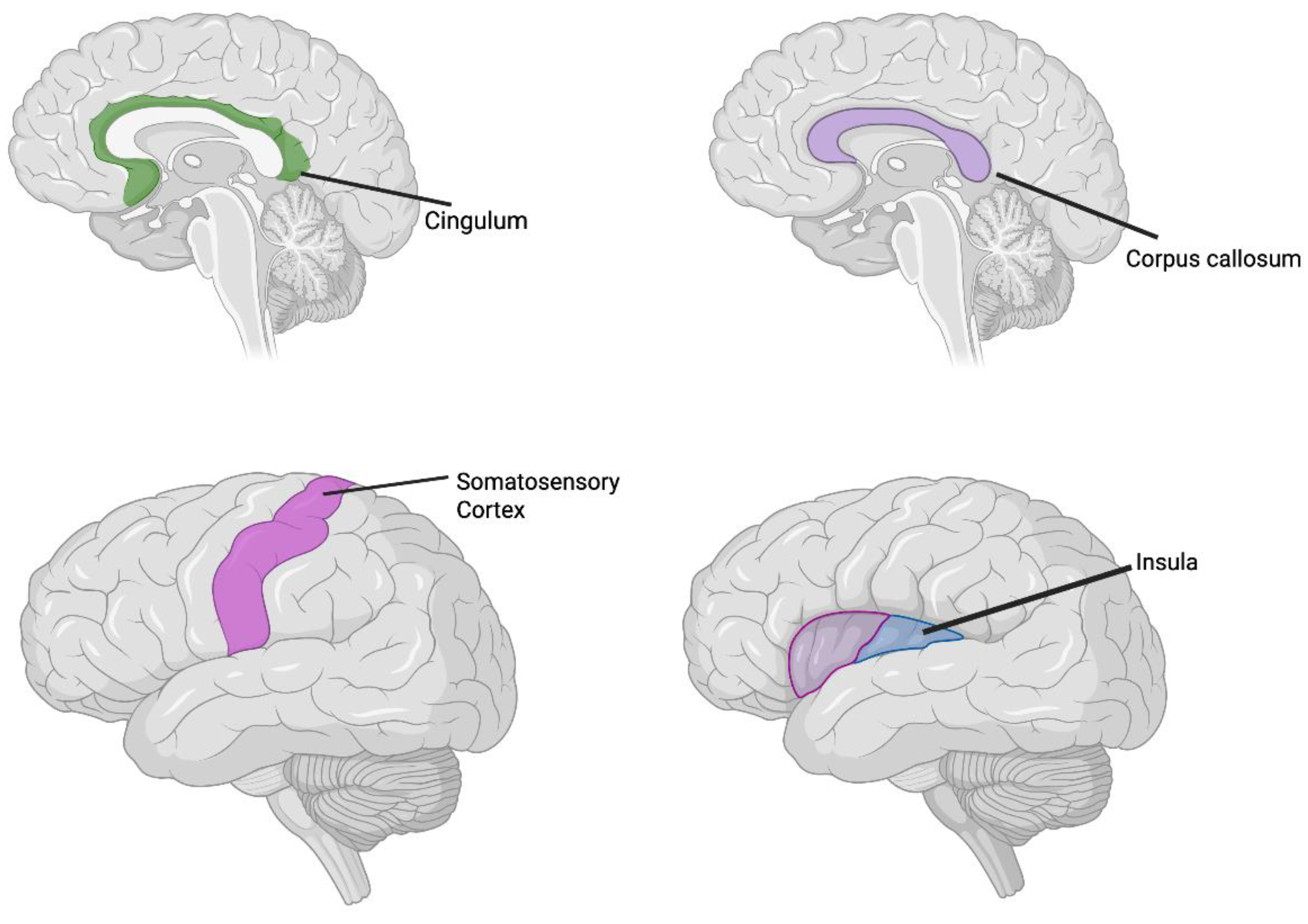
Figure 1.
Common brain alteration in ADHD and SPDs (Created in BioRender. Corbo, D. (2025) https://BioRender.com/lgqx0dz.
Figure 1.
Common brain alteration in ADHD and SPDs (Created in BioRender. Corbo, D. (2025) https://BioRender.com/lgqx0dz.
4. Discussion
Understanding the neural substrates underlying different sensory processing features may be valuable for identifying biomarkers across ADHD subgroups. An important consideration is that brain structure and function cannot be treated as independent domains, as they are intrinsically interrelated (Sáenz et al., 2019). Recognizing this interdependence is essential for future investigations, as it may provide a more comprehensive framework for identifying reliable structural and functional markers of ADHD. Such an integrative approach could ultimately improve diagnostic precision and enhance our understanding of the neurobiological mechanisms underlying the disorder.
Although ADHD and SPDs are clinically distinct, emerging evidence suggests partial neurobiological overlap, particularly in regions involved in sensory modulation, emotional regulation, and executive control. Both conditions exhibit atypical activity in the prefrontal cortex, insula, and somatosensory areas, as well as disrupted white matter connectivity, especially in tracts like the cingulum and corpus callosum. These shared features may underlie common behavioral manifestations such as distractibility, emotional lability, and sensory over-responsivity.
Beyond fMRI, two other physiological measurements commonly employed are event-related potentials (ERPs) and somatosensory evoked potentials (SEPs). The P3 component of ERPs is considered a marker of ADHD, typically exhibiting reduced amplitude and prolonged latency (Güven et al., 2020). Other studies have reported that ERP components associated with target selection are delayed in adults with ADHD (Cross-Villasana et al., 2015) and suppressed during visual search tasks in children with ADHD (Wang et al., 2016). While ERPs are not used diagnostically for SPDs, they may help discriminate ADHD from SPD, facilitating more accurate diagnosis.
Regarding SEPs, evidence suggests that children with ADHD exhibit greater impairments in tactile processing compared to typically developing peers (Hern et al., 1992), with females showing higher levels of tactile defensiveness than males. Tactile defensiveness is defined as hypersensitive responses to routine tactile stimulation, characterized by behaviors such as rubbing, scratching, negative facial expressions, withdrawal, or avoidance (Ayres, 1962; Baranek and Berkson, 1994). Interestingly, SEP results indicate that these behaviors may be correlated with impairments in central processing of somatosensory information (Parush et al., 2007). In Parush and colleagues’ study (2007), SEPs were obtained by delivering brief rectangular electrical stimuli to the median nerve at the wrist, a procedure activating the primary somatosensory pathway associated with cutaneous sensation. Stimulus intensity was adjusted to the minimal current required to elicit a thumb twitch (0.2 ms pulses at approximately 2 Hz) (Parush et al., 2007).However, while there are common mechanisms between ADHD and SPDs, key differences remain. ADHD is primarily characterized by dysfunction in fronto-striatal circuits and dopaminergic pathways, which are central to attention regulation and impulse control. In contrast, SPDs show more pronounced alterations in thalamocortical and parietal sensory integration networks, reflecting their core deficit in processing and responding to sensory input. Additionally, neurotransmitter profiles differ, with ADHD linked to dopamine and norepinephrine dysregulation, while SPDs may involve atypical glutamatergic or GABAergic signaling [Miller et al. 2009, Cortese et al. 2012].To the best of our knowledge, this is the first narrative review to specifically address the neurobiological relationship between ADHD and Sensory Processing Disorders. This perspective offers a novel contribution to the field by integrating current findings and highlighting potential overlaps. However, a key limitation remains the scarcity of available studies, which underscores the need for further research to clarify the mechanisms underlying these conditions and their interactions.
5. Conclusions
Taken together, these findings underscore the critical role of integrative neural mechanisms in shaping sensory processing in ADHD and potentially in SPDs. By combining structural, functional, and electrophysiological measures, future research can move toward a more precise characterization of neurobiological subtypes, identify reliable biomarkers, and ultimately inform targeted interventions. Advancing this integrative approach has the potential to transform both diagnosis and treatment, bridging the gap between neural circuitry dysfunction and observable sensory and cognitive deficits in neurodevelopmental disorders.
Author Contributions
Conceptualization, L.C.G. and D.C.; methodology, L.C.G. and D.C.; writing—original draft preparation, L.C.G. and D.C.; writing—review and editing, L.C.G and D.C.
Funding
This research received no external funding.
Conflicts of Interest
The authors declare no conflicts of interest.
References
- Ayres, A.J. Tactile functions: their relation to hyperactive and perceptual motor behaviour. Am J Occup Ther. 1964, 18, 83–95. [Google Scholar]
- Adra, N.; Cao, A.; Makris, N.; Valera, E.M. Sensory Modulation Disorder and its Neural Circuitry in Adults with ADHD: A Pilot Study. Brain Imaging Behav. 2021, 15, 930–940. [Google Scholar] [CrossRef]
- Albajara Sáenz, A.; Villemonteix, T.; Massat, I. Structural and functional neuroimaging in attention-deficit/hyperactivity disorder. Dev Med Child Neurol. 2019, 61, 399–405. [Google Scholar] [CrossRef] [PubMed]
- Arnsten, A.F.; Rubia, K. Neurobiological circuits regulating attention, cognitive control, motivation, and emotion: disruptions in neurodevelopmental psychiatric disorders. J Am Acad Child Adolesc Psychiatry. 2012, 51, 356–367. [Google Scholar] [CrossRef] [PubMed]
- Baddeley, A. Working memory. Science. 1992, 255, 556–559. [Google Scholar] [CrossRef]
- Baddeley, A. Is working memory still working? Eur. Psychol. 2002, 7, 85–97. [Google Scholar] [CrossRef]
- Baranek, G.T.; Berkson, G. Tactile defensiveness in children with developmental disabilities: responsiveness and habituation. J Autism Dev Disord. 1994, 24, 457–471. [Google Scholar] [CrossRef]
- Bijlenga, D.; Tjon-Ka-Jie, J.Y.M.; Schuijers, F.; et al. Atypical sensory profiles as core features of adult ADHD, irrespective of autistic symptoms. Eur Psychiatry. 2017, 43, 51–57. [Google Scholar] [CrossRef]
- Björnsdotter, M.; Morrison, I.; Olausson, H. Feeling good: on the role of C fiber mediated touch in interoception. Exp Brain Res. 2010, 207, 149–155. [Google Scholar] [CrossRef] [PubMed]
- Brotman, M.A.; Rich, B.A.; Guyer, A.E.; Lunsford, J.R.; Horsey, S.E.; Reising, M.M.; Leibenluft, E. Amygdala activation during emotion processing of neutral faces in children with severe mood dysregulation versus ADHD or bipolar disorder. American Journal of Psychiatry. 2010, 167, 61–69. [Google Scholar] [CrossRef]
- Cascio, C.J. Somatosensory processing in neurodevelopmental disorders. J. Neurodev. Disord. 2010, 2, 62–69. [Google Scholar] [CrossRef] [PubMed]
- Cha, J.; Fekete, T.; Siciliano, F.; et al. Neural correlates of aggression in medication-naive children with ADHD: multivariate analysis of morphometry and tractography. Neuropsychopharmacology. 2015, 40, 1717–1725. [Google Scholar] [CrossRef] [PubMed]
- Choi, I.; Lee, J.Y.; Lee, S.H. Bottom-up and top-down modulation of multisensory integration. Curr Opin Neurobiol. 2018, 52, 115–122. [Google Scholar] [CrossRef]
- Cortese, S. The neurobiology and genetics of Attention-Deficit/Hyperactivity Disorder (ADHD): what every clinician should know. Eur J Paediatr Neurol. 2012, 16, 422–433. [Google Scholar] [CrossRef]
- Cubillo, A.; Halari, R.; Smith, A.; Taylor, E.; Rubia, K. A review of fronto-striatal and fronto-cortical brain abnormalities in children and adults with attention deficit hyperactivity disorder (ADHD) and new evidence for dysfunction in adults with ADHD during motivation and attention. Cortex. 2012, 48, 194–215. [Google Scholar] [CrossRef]
- Cupertino, R.B.; Soheili-Nezhad, S.; Grevet, E.H.; Bandeira, C.E.; Picon, F.A.; Tavares, M.E.A.; Naaijen, J.; van Rooij, D.; Akkermans, S.; Vitola, E.S.; Zwiers, M.P.; Rovaris, D.L.; Hoekstra, P.J.; Breda, V.; Oosterlaan, J.; Hartman, C.A.; Beckmann, C.F.; Buitelaar, J.K.; Franke, B.; Bau, C.H.D.; Sprooten, E. Reduced fronto-striatal volume in attention-deficit/hyperactivity disorder in two cohorts across the lifespan. Neuroimage Clin. 2020, 28, 102403. [Google Scholar] [CrossRef]
- Zald, D.H. The human amygdala and the emotional evaluation of sensory stimuli. Brain Research Reviews. 2003, 41, 88–123. [Google Scholar] [CrossRef] [PubMed]
- Dhamala, M.; Assisi, C.G.; Jirsa, V.K.; et al. Multisensory integration for timing engages different brain networks. Neuroimage. 2007, 34, 764–773. [Google Scholar] [CrossRef]
- Fast, C.D.; McGann, J.P. Amygdalar Gating of Early Sensory Processing through Interactions with Locus Coeruleus. J Neurosci. 2017, 37, 3085–3101. [Google Scholar] [CrossRef]
- Fateh, A.A.; Huang, W.; Mo, T.; Wang, X.; Luo, Y.; Yang, B.; Smahi, A.; Fang, D.; Zhang, L.; Meng, X.; Zeng, H. Abnormal Insular Dynamic Functional Connectivity and Its Relation to Social Dysfunctioning in Children With Attention Deficit/Hyperactivity Disorder. Front. Neurosci. 2022, 16, 890596. [Google Scholar] [CrossRef]
- Franke, B.; Michelini, G.; Asherson, P.; Banaschewski, T.; Bilbow, A.; Buitelaar, J.K.; Cormand, B.; Faraone, S.V.; Ginsberg, Y.; Haavik, J.; Kuntsi, J.; Larsson, H.; Lesch, K.P.; Ramos-Quiroga, J.A.; Réthelyi, J.M.; Ribases, M.; Reif, A. Live fast, die young? A review on the developmental trajectories of ADHD across the lifespan. Eur. Neuropsychopharmacol. 2018, 28, 1059–1088. [Google Scholar] [CrossRef] [PubMed]
- Frodl, T.; Stauber, J.; Schaaff, N.; Koutsouleris, N.; Scheuerecker, J.; Ewers, M.; et al. Amygdala reduction in patients with ADHD compared with major depression and healthy volunteers. Acta Psychiatr Scand. 2010, 121, 111–118. [Google Scholar] [CrossRef]
- Ghanizadeh, A. Sensory processing problems in children with ADHD, a systematic review. Psychiatry Investig. 2011, 8, 89–94. [Google Scholar] [CrossRef]
- Gillberg, C.; Gillberg, I.C.; Rasmussen, P.; Kadesjö, B.; Söderström, H.; Råstam, M.; Johnson, M.; Rothenberger, A.; Niklasson, L. Co-existing disorders in ADHD - implications for diagnosis and intervention. Eur. Child Adolesc. Psychiatry. 2004, 13, I80–I92. [Google Scholar] [CrossRef]
- Grandi, L.C. From Sweeping to the Caress: Similarities and Discrepancies between Human and Non-Human Primates’ Pleasant Touch. Front. Psychol. 2016, 7, 1371. [Google Scholar] [CrossRef] [PubMed]
- Guo, J.; Luo, X.; Li, B.; Chang, Q.; Sun, L.; Song, Y. Abnormal modulation of theta oscillations in children with attention-deficit/hyperactivity disorder. Neuroimage Clin. 2020, 27, 102314. [Google Scholar] [CrossRef] [PubMed]
- Guo, J.L.; Luo, X.S.; Wang, E.C.; Li, B.K.; Chang, Q.Y.; Sun, L.; Song, Y. Abnormal alpha modulation in response to human eye gaze predicts inattention severity in children with ADHD. Dev. Cogn. Neurosci. 2019, 38, 100671. [Google Scholar] [CrossRef]
- Güven, A.; et al. Combining functional near-infrared spectroscopy and EEG measurements for the diagnosis of attention-deficit hyperactivity disorder. Neural Comput. Appl. 2020, 32, 8367–8380. [Google Scholar] [CrossRef]
- Happel Max, F.K.; Hechavarria Julio, C.; Livia, H. Editorial: Cortical-Subcortical Loops in Sensory Processing. Frontiers in Neural Circuits. 2022, 16, 851612. [Google Scholar] [CrossRef]
- Hern, K.L.; Hynd, G.W. Clinical differentiation of the attention deficit disorder subtypes: do sensorimotor deficits characterize children with ADD/WO? Arch Clin Neuropsychol. 1992, 7, 77–83. [Google Scholar] [CrossRef]
- Herpertz, S.C.; Huebner, T.; Marx, I.; Vloet, T.D.; Fink, G.R.; Stoecker, T.; Herpertz-Dahlmann, B. Emotional processing in male adolescents with childhood-onset conduct disorder. Journal of Child Psychology and Psychiatry. 2008, 49, 781–791. [Google Scholar] [CrossRef] [PubMed]
- Wall, J.T.; Xu, J.; Wang, X. Human brain plasticity: an emerging view of the multiple substrates and mechanisms that cause cortical changes and related sensory dysfunctions after injuries of sensory inputs from the body. Brain Research Reviews. 2002, 39, 181–215. [Google Scholar] [CrossRef]
- Jasper, H.; Penfield, W. Electrocorticograms in man: Effect of voluntary movement upon the electrical activity of the precentral gyrus. Archiv Für Psychiatrie Und Nervenkrankheiten. 1949, 183, 163–174. [Google Scholar] [CrossRef]
- Koziol, L.F.; Budding, D.E.; Chidekel, D. Sensory integration, sensory processing, and sensory modulation disorders: putative functional neuroanatomic underpinnings. Cerebellum. 2011, 10, 770–792. [Google Scholar] [CrossRef]
- Koziol, L.F.; Budding, D. ADHD and Sensory Processing Disorders: Placing the Diagnostic Issues in Context. Applied Neuropsychology: Child. 2012, 1, 137–144. [Google Scholar] [CrossRef] [PubMed]
- LeDoux, J. The emotional brain, fear, and the amygdala. Cell Mol Neurobiol. 2003, 23, 727–738. [Google Scholar] [CrossRef]
- Leekam, S.R.; Nieto, C.; Libby, S.J.; Wing, L.; Gould, J. Describing the sensory abnormalities of children and adults with autism. J. Autism Dev. Disord. 2007, 37, 894–910. [Google Scholar] [CrossRef]
- Lenartowicz, A.; Lu, S.; Rodriguez, C.; Lau, E.P.; Walshaw, P.D.; McCracken, J.T.; Cohen, M.S.; Loo, S.K. Alpha desynchronization and fronto-parietal connectivity during spatial working memory encoding deficits in ADHD: A simultaneous EEG-fMRI study. Neuroimage Clin. 2016, 11, 210–223. [Google Scholar] [CrossRef]
- Lopez-Larson, M.P.; King, J.B.; Terry, J.; McGlade, E.C.; Yurgelun-Todd, D. Reduced insular volume in attention deficit hyperactivity disorder. Psychiatry Res. 2012, 204, 32–39. [Google Scholar] [CrossRef]
- Jurek, L.; Duchier, A.; Gauld, C.; Hénaault, L.; Giroudon, C.; Fourneret, P.; Cortese, S.; Nourredine, M. Sensory Processing in Individuals With Attention-Deficit/Hyperactivity Disorder Compared With Control Populations: A Systematic Review and Meta-analysis. Journal of the American Academy of Child & Adolescent Psychiatry. [CrossRef]
- Luman, M.; Oosterlaan, J.; Sergeant, J. The impact of reinforcement contingencies on AD/HD: A review and theoretical appraisal. Clin. Psychol. Rev. 2005, 25, 183–213. [Google Scholar] [CrossRef]
- MacAluso, E.; Noppeney, U.; Talsma, D.; et al. The curious incident of attention in multisensory integration: bottom-up vs. top-down. Multisens Res. 2016, 29, 557–583. [Google Scholar] [CrossRef]
- Marsh, A.A.; Finger, E.C.; Mitchell, D.G.V.; Reid, M.E.; Sims, C.; Kosson, D.S.; Blair, R.J.R. Reduced amygdala response to fearful expressions in children and adolescents with callous-unemotional traits and disruptive behavior disorders. American Journal of Psychiatry. 2008, 165, 712–720. [Google Scholar] [CrossRef]
- Marshall, A.C.; Gentsch-Ebrahimzadeh, A.; Schütz-Bosbach, S. From the inside out: Interoceptive feedback facilitates the integration of visceral signals for efficient sensory processing. NeuroImage. 2022, 251, 119011. [Google Scholar] [CrossRef]
- Mason, D.J.; Humphreys, G.W.; Kent, L. Insights into the control of attentional set in ADHD using the attentional blink paradigm. J Child Psychol Psychiatry. 2005, 46, 1345–1353. [Google Scholar] [CrossRef] [PubMed]
- Mesulam, M.M.; Mufson, E.J. Insula of the old world monkey. III: Efferent cortical output and comments on function. The Journal of Comparative Neurology. 1982, 212, 38–52. [Google Scholar] [CrossRef] [PubMed]
- Miller, L.J.; Nielsen, D.M.; Schoen, S.A.; Brett-Green, B.A. Perspectives on sensory processing disorder: a call for translational research. Front Integr Neurosci. 2009, 3, 22. [Google Scholar] [CrossRef]
- Morrison, I.; Björnsdotter, M.; Olausson, H. Vicarious responses to social touch in posterior insular cortex are tuned to pleasant caressing speeds. J Neurosci. 2011, 31, 9554–9562. [Google Scholar] [CrossRef] [PubMed]
- Mufson, E.J.; Mesulam, M.M.; Pandya, D.N. Insular interconnections with the amygdala in the rhesus monkey. Neuroscience. 1981, 6, 1231–1248. [Google Scholar] [CrossRef]
- Norman, L.J.; Carlisi, C.; Lukito, S.; et al. Structural and functional brain abnormalities in attention-deficit/hyperactivity disorder and obsessive-compulsive disorder: a comparative meta-analysis. JAMA Psychiatry. 2016, 73, 815–825. [Google Scholar] [CrossRef]
- Olausson, H.; Lamarre, Y.; Backlund, H.; Morin, C.; Wallin, B.G.; Starck, G.; Ekholm, S.; Strigo, I.; Worsley, K.; Vallbo, Å.B.; Bushnell, M.C. Unmyelinated tactile afferents signal touch and project to insular cortex. Nat Neurosci. 2002, 5, 900–904. [Google Scholar] [CrossRef]
- Dubljević, O.; Pavković, Ž.; Srbovan, M.; Potrebić, M.; Stanojlović, M.; Pešić, V. Attention-deficit/hyperactivity disorder-related psychomotor activity and altered neuronal activity in the medial prefrontal cortex and striatum in the A53T mouse model of Parkinson's disease and other synucleinopathies: Findings from an “endophenotype” approach. Progress in Neuro-Psychopharmacology and Biological Psychiatry. 2025, 137, 111273. [Google Scholar] [CrossRef]
- Shaw, P.; Eckstrand, K.; Sharp, W.; Blumenthal, J.; Lerch, J.P.; Greenstein, D.; Clasen, L.; Evans, A.; Giedd, J.; Rapoport, J.L. Attention-deficit/hyperactivity disorder is characterized by a delay in cortical maturation. Proc. Natl. Acad. Sci. U.S.A. 2007, 104, 19649–19654. [Google Scholar] [CrossRef]
- Parush, S.; Sohmer, H.; Steinberg, A.; Kaitz, M. Somatosensory function in boys with ADHD and tactile defensiveness. Physiol Behav. 2007, 90, 553–558. [Google Scholar] [CrossRef]
- Passarello, N.; Tarantino, V.; Chirico, A.; Menghini, D.; Costanzo, F.; Sorrentino, P.; Fucà, E.; Gigliotta, O.; Alivernini, F.; Oliveri, M.; Lucidi, F.; Vicari, S.; Mandolesi, L.; Turriziani, P. Sensory Processing Disorders in Children and Adolescents: Taking Stock of Assessment and Novel Therapeutic Tools. Brain Sci. 2022, 12, 1478. [Google Scholar] [CrossRef]
- Perlov, E.; Philipsen, A.; van Elst, L.T.; Ebert, D.; Henning, J.; Maier, S.; et al. Hippocampus and amygdala morphology in adults with attention-deficit hyperactivity disorder. J Psychiatry Neurosci. 2008, 33, 509–515. [Google Scholar] [CrossRef]
- Pevzner, A.; Izadi, A.; Lee, D.J.; Shahlaie, K.; Gurkoff, G.G. Making waves in the brain: what are oscillations, and why modulating them makes sense for brain injury. Front Syst Neurosci. 2016, 10, 30. [Google Scholar] [CrossRef]
- Piccardi, E.S.; Begum Ali, J.; Jones, E.J.H.; et al. Behavioural and neural markers of tactile sensory processing in infants at elevated likelihood of autism spectrum disorder and/or attention deficit hyperactivity disorder. J Neurodevelop Disord. 2021, 13, 1. [Google Scholar] [CrossRef]
- Plessen, K.J.; Bansal, R.; Zhu, H.; Whiteman, R.; Amat, J.; Quackenbush, G.A.; et al. Hippocampus and amygdala morphology in attention-deficit/hyperactivity disorder. Arch Gen Psychiatry. 2006, 63, 795–807. [Google Scholar] [CrossRef] [PubMed]
- Polanczyk, G.; De Lima, M.S.; Horta, B.L.; Biederman, J.; Rohde, L.A. The worldwide prevalence of ADHD: a systematic review and metaregression analysis. Am J Psychiatry. 2007, 164, 942–948. [Google Scholar] [CrossRef] [PubMed]
- Rach, S.; Diederich, A.; Colonius, H. On quantifying multisensory interaction effects in reaction time and detection rate. Psychol Res. 2011, 75, 77–94. [Google Scholar] [CrossRef]
- Schneider, M.L.; Moore, C.F.; Ahlers, E.O.; Barnhart, T.E.; Christian, B.T.; DeJesus, O.T.; Converse, A.K. PET Measures of Dl, D2, and DAT Binding Are Associated with Heightened Tactile Responsivity in Rhesus Macaques: Implications for Sensory Processing Disorder. Front Integr Neurosci. 2019, 13, 29. [Google Scholar] [CrossRef]
- Schnitzler, A.; Gross, J. Normal and pathological oscillatory communication in the brain. Nat Rev Neurosci. 2005, 6, 285–296. [Google Scholar] [CrossRef]
- Schulze, M.; Aslan, B.; Stöcker, T.; Stirnberg, R.; Lux, S.; Philipsen, A. Disentangling early versus late audiovisual integration in adult ADHD: a combined behavioural and resting-state connectivity study. J Psychiatry Neurosci. 2021, 46, E528–E537. [Google Scholar] [CrossRef]
- Smirni, D.; Smirni, P.; Carotenuto, M.; Parisi, L.; Quatrosi, G.; Roccella, M. Noli Me Tangere: Social Touch, Tactile Defensiveness, and Communication in Neurodevelopmental Disorders. Brain Sci. 2019, 9, 368. [Google Scholar] [CrossRef] [PubMed]
- Stein, J.L.; Wiedholz, L.M.; Bassett, D.S.; et al. A validated network of effective amygdala connectivity. Neuroimage. 2007, 36, 736–745. [Google Scholar] [CrossRef] [PubMed]
- Stevens, M.C.; Haney-Caron, E. Comparison of brain volume abnormalities between ADHD and conduct disorder in adolescence. J Psychiatry Neurosci. 2012, 37, 389–398. [Google Scholar] [CrossRef] [PubMed]
- ter Huurne, N.; Lozano-Soldevilla, D.; Onnink, M.; Kan, C.; Buitelaar, J.; Jensen, O. Diminished modulation of preparatory sensorimotor mu rhythm predicts attention-deficit/hyperactivity disorder severity. Psychological Medicine. 2017, 47, 1947–1956. [Google Scholar] [CrossRef]
- ter Huurne, N.; Onnink, M.; Kan, C.; Franke, B.; Buitelaar, J.; Jensen, O. Behavioral consequences of aberrant alpha lateralization in attention-deficit/hyperactivity disorder. Biol. Psychiatry. 2013, 74, 227–233. [Google Scholar] [CrossRef]
- Turner, B.H.; Herkenham, M. Thalamoamygdaloid projections in the rat: a test of the amygdala’s role in sensory processing. J Comp Neurol. 1991, 313, 295–325. [Google Scholar] [CrossRef]
- Vassalli, A.; Dellepiane, J.M.; Emmenegger, Y.; Jimenez, S.; Vandi, S.; Plazzi, G.; Franken, P.; Tafti, M. Electroencephalogram paroxysmal θ characterizes cataplexy in mice and children. Brain. 2013, 136, 1592–1608. [Google Scholar] [CrossRef]
- Vassalli, A.; Franken, P. Hypocretin (orexin) is critical in sustaining theta/gamma-rich waking behaviors that drive sleep need. Proc Natl Acad Sci U S A. 2017, 114, E5464–E5473. [Google Scholar] [CrossRef]
- Villemonteix, T.; De Brito, S.A.; Kavec, M.; Baleriaux, D.; Metens, T.; Slama, H.; et al. Grey matter volumes in treatment naive vs. chronically treated children with attention deficit/hyperactivity disorder: A combined approach. Eur Neuropsychopharmacol. 2015, 25, 1118–1127. [Google Scholar] [CrossRef]
- Vollebregt, M.A.; Zumer, J.M.; ter Huurne, N.; Buitelaar, J.K.; Jensen, O. Posterior alpha oscillations reflect attentional problems in boys with attention deficit hyperactivity disorder. Clin. Neurophysiol. 2016, 127, 2182–2191. [Google Scholar] [CrossRef] [PubMed]
- Völter, C.; Thomas, J.P.; Maetzler, W.; Guthoff, R.; Grunwald, M.; Hummel, T. Sensory Dysfunction in Old Age. Dtsch Arztebl Int. 2021, 118, 512–520. [Google Scholar] [CrossRef]
- Willcutt, E.G. The prevalence of DSM-IV attention-deficit/hyperactivity disorder: a meta-analytic review. Neurotherapeutics. 2012, 9, 490–499. [Google Scholar] [CrossRef]
- Wing, L. The handicaps of autistic children: A comparative study. Journal of Child Psychology and Psychiatry. 1969, 10, 1–40. [Google Scholar] [CrossRef] [PubMed]
- Chang, Y.S.; Owen, J.P.; Pojman, N.J.; Thieu, T.; Bukshpun, P.; Wakahiro, M.L.; Berman, J.I.; Roberts, T.P.; Nagarajan, S.S.; Sherr, E.H. White matter changes of neurite density and fiber orientation dispersion during human brain maturation. PLoS One. 2015, 10, e0123656. [Google Scholar] [CrossRef] [PubMed]
- Yochman, A.; Parush, S.; Ornoy, A. Responses of preschool children with and without ADHD to sensory events in daily life. Am J Occup Ther. 2004, 58, 294–302. [Google Scholar] [CrossRef]
- Zaher, A.; Leonards, J.; Reif, A.; et al. Functional connectivity of the nucleus accumbens predicts clinical course in medication adherent and non-adherent adult ADHD. Sci Rep. 2025, 15, 19663. [Google Scholar] [CrossRef]
Disclaimer/Publisher’s Note: The statements, opinions and data contained in all publications are solely those of the individual author(s) and contributor(s) and not of MDPI and/or the editor(s). MDPI and/or the editor(s) disclaim responsibility for any injury to people or property resulting from any ideas, methods, instructions or products referred to in the content. |
© 2025 by the authors. Licensee MDPI, Basel, Switzerland. This article is an open access article distributed under the terms and conditions of the Creative Commons Attribution (CC BY) license (http://creativecommons.org/licenses/by/4.0/).
Copyright: This open access article is published under a Creative Commons CC BY 4.0 license, which permit the free download, distribution, and reuse, provided that the author and preprint are cited in any reuse.


