Preprint
Article

This version is not peer-reviewed.

Elevated Antibacterial Activity with Decreased Cytotoxicity of a Polygalacturonic + Caprylic Acid Wound Ointment Compared with Hypochlorous Acid in a Three-Dimensional Wound Biofilm Model

Submitted:

22 September 2025

Posted:

24 September 2025

You are already at the latest version

Abstract

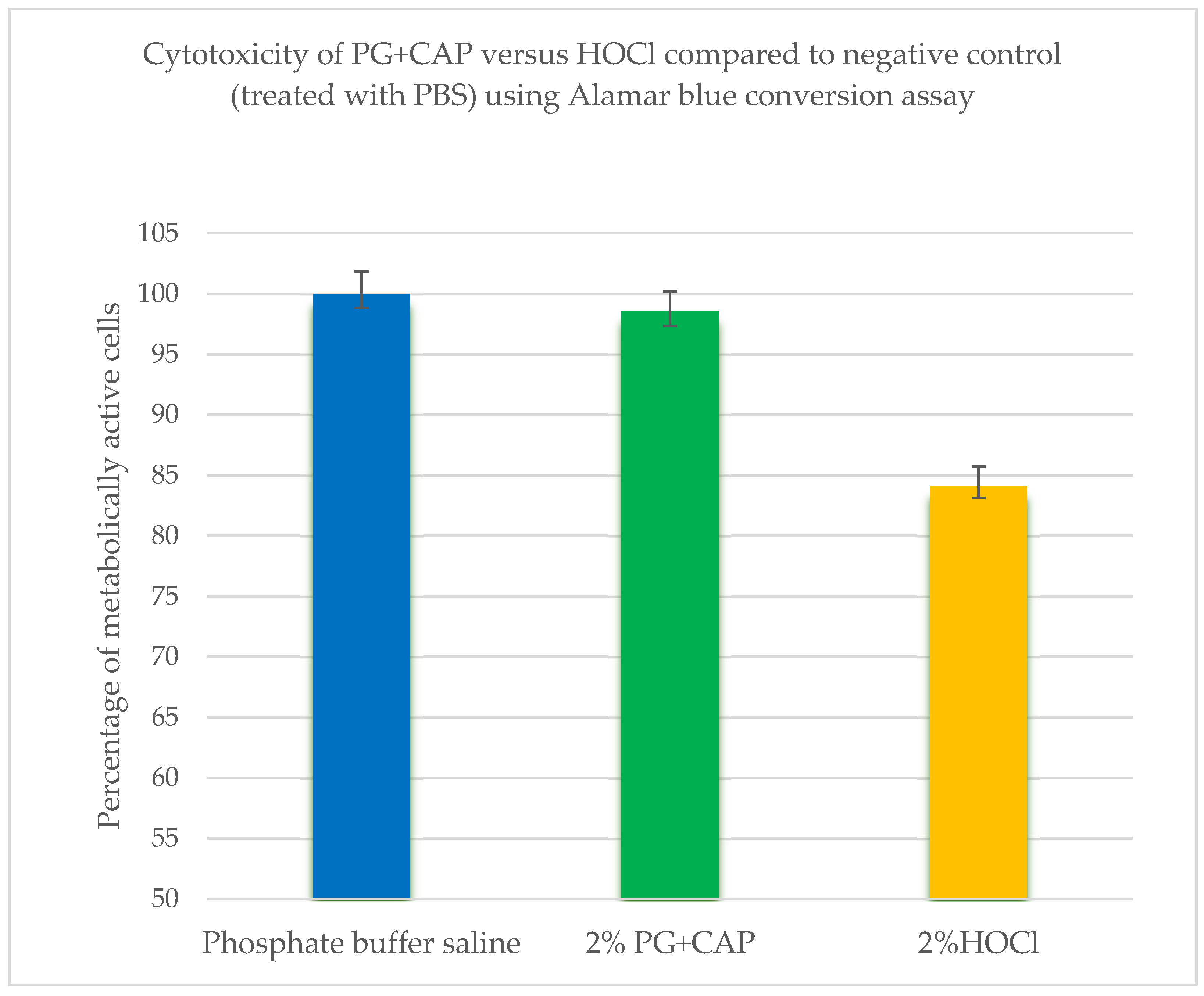
Introduction: Natural, plant-based agents are alternatives to antibiotics and antiseptics in treating bacterially colonized wounds and potentially offer an antimicrobial activity without cytotoxicity. In this study, we used a three-dimensional fibrin-gel wound biofilm (FGWB) model to be more representative of the wound biofilm environment. We compared a common antiseptic wound care agent, hypochlorous acid (HOCl), with a combination of two plant-based agents; polygalacturonic acid (PG) and caprylic acid (CAP) for bacterial biofilm eradication and assessed their cytotoxicities towards fibroblast cells. Material and Methods: The efficacy PG+CAP ointment was compared to HOCl irrigant solution in biofilm eradication using the FGWB against clinical resistant bacterial isolates of MRSA, MDR Pseudomonas aeruginosa, CRE Escherichia coli, and Streptococcus pyogenes. Trypan blue exclusion and Alamar blue conversion assays were used for cytotoxicity assays. Results: PG+CAP produced a significantly greater reduction of viable organisms than HOCl for all tested bacterial isolates in the FGWB model (P≤ 0.05). Also, cytotoxicity tests showed that, PG+CAP was comparable to the non-antimicrobial negative control and was less cytotoxic than HOCl (P≤0.05). Conclusion: PG+CAP was highly effective against biofilms of highly resistant bacterial isolates in the FGWB model, and less cytotoxic than HOCl. PG+CAP merits further in vivo study.

Keywords: 
;  ;  ;  ;  ;  ;  ;  ;  

1. Introduction

Prolonged inflammation due to bacterial infection plays a significant role in the development of chronic wound biofilms due to delayed healing [1]. Treating bacterial chronic wound biofilms is a significant problem and is one of the main reasons for development of resistant bacterial isolates when antibiotic ointments are used [2]. In contrast to antibiotics, antiseptics which can be bactericidal or bacteriostatic, are used to reduce the presence of bacteria in wounds without inducing antibiotic-resistant bacteria [2,3,4]. However, the use of antiseptics during wound-healing has been associated with cytotoxicity [4]. Cytotoxicity also produces inflammation in wounds and contributes to delayed wound healing by degrading newly deposited granulation tissues [4,5,6]. Natural, plant-based agents, which can eradicate bacterial biofilms without leading to antibiotic resistance or cytotoxicity, are alternatives to antiseptics in the treatment of bacterially contaminated wounds that potentially offer an improved balance of antimicrobial activity without cytotoxicity [7].
Biofilm-producing bacteria are of great importance from public health perspective because they cause 80% of the infections that occur in the human body [8]. Chronic wounds represent a significant economic burden, including $25 billion dollars annually in the United States alone [9,10]. Bacterial biofilms comprise microbial communities surrounded by extracellular matrices of polymeric macromolecules [2]. Bacteria within the biofilm become less susceptible to antimicrobial agents through physical barriers to access created by the biofilm extracellular matrix as well as by phenotypic adaptations including altering metabolism, genetic regulation, and protein expression [3,11]. Many traditional biofilm models have been used to study the efficacy of antimicrobial agents against pathogenic bacterial biofilms [12], but these traditional models are not representative of the three-dimensional matrix of microbes found in wound biofilms. Therefore, In the current study, we used a three-dimensional fibrin-gel wound biofilm (FGWB) model to be more representative of the three-dimensional extracellular matrix in which bacterial wound biofilms actually are enmeshed.
In vitro cytotoxicity assays are indicators of inflammatory reactions by damaged cells in wound beds [13,14]. Many in vitro methods can be used to investigate the cytotoxic effects of antimicrobial agents on tissue cells. We used the Trypan blue assay to distinguish living cells having intact viable membranes from lysed or disrupted cells that release inflammatory cytokines [15]. This is a coarse measure of whether contact with chemical agents kills or severely damages fibroblast membrane integrity causing release of inflammatory cytokines that recruit inflammatory cells to wound beds [15]. We also used the Alamar blue conversion assay was used to assess whether contact with chemical agents alters cellular metabolic activity [16,17,18]. Altered metabolic activity is more sensitive than trypan blue assay indicates whether contact with chemical agents impairs the ability of fibroblasts to perform their wound repair functions.
In the current study, we compared a widely used antiseptic agent in wound care, hypochlorous acid (HOCl), to a novel combination of two natural plant-based agents; polygalacturonic acid (PG) and caprylic acid (CAP). Previous studies have shown that PG+CAP is highly effective against pathogenic bacterial isolates in biofilms both in vitro and in vivo [19,20]. Hypochlorous acid, a widely used, commercially available, antimicrobial wound irrigant, has demonstrated fast acting inhibition of bacterial wound infection through disruptive reactions with different bacterial lipids, nucleotides, and proteins [21, 22). Although the mechanism of action of HOCl against microorganisms is not completely understood, many studies have reported that HOCl rapidly kills microbes through these oxidative chemical reactions [23,24,25,26,27,28]. Here we report studies of the comparative efficacies of PG+CAP and HOCl in eradicating bacterial biofilms using three-dimensional FGWB as well as their relative cytotoxicities towards fibroblasts cells which are essential for dermal wound repair and healing.

2. Materials and Methods

Antibacterial Agents:
We compared the efficacy of PG+CAP ointment with that of HOCl (PG: Sigma-Aldrich Inc, St. Louis, MO, USA, catalog #P3889-100G, CAS #25990-10-7; CAP: Sigma-Aldrich, catalog #03907, CAS #124-07-2; HOCl: Aqua Science Inc, Columbus, OH, USA, catalog #01S.06E, CAS #7790-92-3). PG+CAP ointment was prepared in a laboratory as previously described by Gerges et al [19], and the HOCl wound solution was used at a concentration of 400 ppm as directed by the manufacturer. An aqueous gel containing 2-hydroxyethylcellulose (Fisher Scientific/Janssen Pharmaceuticals [now Johnson & Johnson Innovative Medicine], Belgium, catalog #G33-500, CAS #56-81-5) was used as the ointment base for PG+CAP ointment.
Three-Dimensional FGWB Model:
A quantitative, in vitro, three-dimensional FGWB model adapted from the model of Besser and Stuermer [29] and prepared according to the specifications of Truong et al [30] and Gerges et al [31] was used in the current study. Briefly, 20 mg/mL fibrinogen (Fisher Scientific, catalog #34-157-61GM) was slowly dissolved in phosphate-buffered saline (PBS) at 37° C; 5 units/mL thrombin (Sigma-Aldrich, catalog #T7009-100UN; CAS #9002-04-4) was dissolved in PBS; and 125mM calcium chloride (Sigma-Aldrich, catalog #C3306-100G, CAS #10035-04-8) was dissolved in deionized water.
Biofilm Eradication Assay:
Biofilm eradication testing was conducted using highly virulent clinical bacterial isolates of MRSA (MDA #120), multidrug-resistant (MDR) Pseudomonas aeruginosa (MDA #118), carbapenem-resistant Enterobacterales (CRE) Escherichia coli (MB #9245), and Streptococcus pyogenes (MB #3175) as representative hospital-acquired infection pathogens from cancer patients. The organisms were grown from glycerol stock on trypticase soy agar + 5% sheep blood (Remel, Lenexa, Kansas, USA, reference #R01202). Each organism was inoculated into Muller Hinton broth (Fisher Scientific, BBL Mueller Hinton II Broth Cation Adjusted, Sparks, MD, USA, catalog #B12322) and diluted to 0.5 McFarland. Additional dilutions were made to obtain ~104 colony-forming units/mL (CFUs/mL). The efficacy of PG+CAP versus HOCl was determined using the FGWB model as described by Gerges et al [31]. Six replicates of FGWB were used for each of PG+CAP, HOCl, and negative control against each organism. Regrowth experiments were conducted as described by Gerges et al 2025 [31] to ensure that eradication was complete on FGWB disks from which no viable colonies were recovered following exposure to PG+CAP or HOCl.
Cytotoxicity Tests:
1. 
Culturing of Fibroblasts cells:
Fibroblast NCTC clone 929 (L cell, L-929, derivative of Strain L) was used in the current study. Cell culture was performed according to fibroblast protocols as described by Gomes et al 2011 [32].and as follows; the vial of fibroblasts was thawed by gentle agitation in a 37° C water bath for approximately 2 minutes, under strict aseptic conditions. The vial content was transferred to a centrifuge tube containing 9.0 mL complete culture medium and spun at 400 × g for 10 minutes. The complete medium consisted of ATCC-formulated Eagle Minimum Essential Medium (Sigma-Aldrich Inc, Millipore Sigma, USA, catalog #M4655) plus horse serum (ATCC 30-2040) at a final concentration of 10%. The cells pellet was resuspended with 4 mL of complete medium, then 1 mL of this solution was added to 10 mL of complete medium in a T-25-cm2 tissue culture flask and incubated at 37° C + 5% CO2. The fibroblasts were refreshed every 48 hours by replacing the old medium with fresh medium, and fibroblasts were observed daily under inverted microscope for 60-70% confluence.
2. 
Subculturing procedure:
Briefly, the old culture medium was removed and discarded, and the cell layer was rinsed with Hanks balanced salt solution (Sigma-Aldrich Inc, Millipore Sigma, USA, catalog #H6648) to remove all traces of serum that contained trypsin inhibitor. Two milliliters of trypsin-EDTA solution 0.25% (gibco reference #2500-056) were added to the flask, and the cells were observed under an inverted microscope until the cell layer was dispersed. Then, 8.0 mL of complete growth medium was added, and cells were aspirated by gently pipetting. Appropriate aliquots of the cell suspension were added to a new culture T-flask and incubated at 37° C + 5% CO2.
3. 
Cytotoxicity techniques:
PG+CAP ointment was compared with HOCl irrigant solution for cytotoxicities using the Trypan blue exclusion assay as described by Strober [15] and the Alamar blue conversion assay as described by O’Brien et al [16]. Briefly, for the Trypan blue exclusion assay; 1 mL of culture medium containing 20,000 cells/mL was added in each well of a 24-well tissue culture plate, then an equal quantity of tested antimicrobial agent was added to corresponding wells and incubated for 24 hours at 37° C + 5% CO2. All content of each well was collected in small tubes and centrifuged at 1160 rpm for 7 minutes. Supernatant was discarded, and 1 mL of medium (without serum) was added to each tube and mixed gently, and then 50 µL of the cell suspension was added in a cryovial, with an equal quantity of Trypan blue dye 0.4% (gibco reference #1520-061), mixed gently up and down, and incubated for 1-2 minutes at room temperature as recommended [32]. Then, 10 µL was added into a hemocytometer to count unstained cells (live cells) and stained blue cells (dead cells) separately. The percentage of viable cells were calculated as follow:
% of viable cells = total number of viable cells × 100/total number of viable and dead cells.
For the Alamar blue conversion assay; every well of 96-well tissue culture plates was seeded with 100 µL of the suspension containing 3,200 cells/well, incubated at 37° C + 5% CO2, and observed daily for 60-70% confluence (~72 hours). The old medium was then aspirated and washed with Hanks balanced salt solution. Antimicrobial agent was added to each corresponding well and incubated for 24 hours at 37° C + 5% CO2. The medium containing ointments was replaced with only Hanks balanced salt solution, and then 10% volume (10 µL) of Alamar blue dye (Alamar blue cell viability reagent, Thermo Fisher Scientific/Invitrogen, reference #DAL 1025) was added and incubated for 4 hours at 37° C + 5% CO2. The plate was read at 570 nm using a spectrophotometer, and the absorbance for each tested agent was compared with that of cells treated with phosphate-buffered saline (negative control). PG+CAP ointment and HOCl irrigant were used at 2% concentrations.
Statistical Analysis:
To compare the efficacies of the treatments in biofilm eradication, colony-forming unit/milliliter (CFU/mL) values for each bacterial isolate were analyzed across the three groups; PG+CAP, HOCl, and negative control, using the Kruskal–Wallis test. When this test detected a significant difference, pairwise comparisons were carried out using the Wilcoxon rank-sum test. To control the overall Type I error from multiple comparisons, p-values were adjusted using the Holm-Bonferroni method. The reduction in CFU/mL relative to the negative control, expressed as log₁₀, was also computed for both the PG+CAP and HOCl treatment groups. For cytotoxicities evaluation, viable cell counts following treatment with PG+CAP or HOCL were compared individually against the negative control using the Wilcoxon rank-sum test. All tests were two-sided, with a significance level set as P ≤ 0.05. Data analyses were conducted using SAS version 9.4 (SAS Institute Inc., Cary, NC, USA).

3. Results

Figure 1 presents the median recovered viable colonies (CFUs/mL.) of tested infectious bacterial biofilms of MRSA, MDR P. aeruginosa, CRE E. coli, and St. pyogenes after three-hours of exposure to PG+CAP or HOCl in a FGWB model. Table 1 presents the log10 reductions in CFUs/mL. After three-hours of exposure, relative to the negative control, PG+CAP reduced viable MRSA CFUs/mL by 7.9 log10, and HOCl reduced MRSA CFUs/mL by 4.2 log10. PG+CAP and HOCl reduced viable MDR P. aeruginosa CFUs/mL by 8.28 log10 and 2.32 log10, respectively, CRE E. coli CFUs/mL by 4.26 log10 and 1.69 log10, respectively, and St. pyogenes by 5.12 log10 and 3.0 log10, respectively. The antibacterial superiority of PG+CAP relative to HOCl was statistically significant (P ≤ 0.05) against all tested bacterial isolates.
Table 2 shows results of the Trypan blue exclusion cytotoxicity assay. The mean number of viable fibroblast cells after treatment with PG+CAP was statistically similar to that of the negative control cells after treatment with phosphate-buffered saline (P = 0.26). Treatment with HOCl led to significantly reduced cell viability of fibroblasts cells compared with PG+CAP and negative control (P ≤ 0.05).
Figure 2 and Table 3 present the results of the Alamar blue conversion metabolic activity assay. Fibroblasts cells treated with PG+CAP had similar metabolic activity to that observed in the negative control that treated with phosphate-buffered saline (P = 0.083), whereas the metabolic activity of fibroblasts cells was significantly reduced after treatment with HOCl compared with negative control fibroblasts cells and fibroblasts cells treated with PG+CAP (P ≤ 0.05).
Treated cells with phosphate buffer saline (PBS) were used as negative control. *P-value is from the Wilcoxon rank-sum test comparing the cells viability between the corresponding treatment and PG+CAP. **P-value is from the Wilcoxon rank-sum test comparing the cells viability between the corresponding treatment and negative control.
Results are expressed as the absorbance relative to control as measured by a spectrophotometer at 570 nm. Higher metabolic activity indicates lower cytotoxicity. Treated cells with phosphate buffer saline were used as negative control. *P-value is from the Wilcoxon rank-sum test comparing the metabolic activity between the corresponding treatment and PG+CAP. **P-value is from the Wilcoxon rank-sum test comparing the metabolic activity between the corresponding treatment and control.

4. Discussion

Acute inflammation plays an important role in normal wound healing. The acute response to dermal wounds includes recruitment of neutrophils, macrophages, and lymphocytes which clear damaged tissue and colonizing bacteria through phagocytosis, secretion of reactive oxidative moieties (such as HOCl) and secretion of proteases [33,34]. Subsequently a key part of normal wound healing as inflammation subsides is the transition to the proliferation and remodeling phases in which cytokine-recruited fibroblasts and keratinocytes take over key roles [35]. Fibroblasts play key roles in generating new granulation tissue over which keratinocytes can proliferate to re-epithelialize the wound [36].
Chronic non-healing wounds occur when the inflammatory phase becomes entrenched preventing transition to the proliferation phase [37]. Aberrant inflammation associated with chronic non-healing wounds frequently contains bacteria that are able to survive and evade immune reactions or antibiotics by forming protective biofilms [38]. Furthermore, sustained tissue damage by chemotherapeutic agents can further impede successful transition [1]. Hence, therapies to restore normal healing of chronic wounds need to be able to eradicate bacteria in wound biofilms. In the current study, we allowed bacteria to form mature protective biofilms enmeshing in both bacterial extracellular matrix as well as fibrin, an abundant protein present in inflammatory wound exudate [39]. Our experiments showed a single treatment of PG+CAP for three hours was highly effective in eradicating bacteria in the mature fibrin gel biofilms but did not fully eradicate all bacteria. MRSA and MDR P. aeruginosa biofilms were fully eradicated; however, CRE E. coli and St. Pyogenes had reduced viable bacterial concentrations (over 4 Log10 reductions) but were not fully eradicated in a single treatment. The three-hours exposure was selected as a representative duration over which the applied ointment would not be expected to become excessively diluted by diffusion from the wound bed or dilution by wound exudate. The tested bacteria are common resistant dermal wound pathogens associated with chronic non-healing wounds [40,41]. The PG+CAP results suggest multiple continued applications of PG+CAP would be required to sustain minimal bacterial bioburden in chronic wounds allowing granulation tissue formation to proceed without excessive inflammatory disruption. HOCl was significantly less effective than PG+CAP in reducing bacterial bioburden in fibrin gel biofilms against all tested bacteria. This suggests once biofilm matures, a robust bacterial population could survive HOCl irrigation and continue inducing inflammatory responses.
Effective chronic wound therapies further need to optimize eradication of bacterial biofilms with minimizing cytotoxic effects on proliferative cells so as not to produce inflammation that is counterproductive to chronic wound healing and closure. The results of our experiments show that PG+CAP was comparable to control and significantly superior to HOCl in minimizing cytotoxic reaction against fibroblasts. Fibroblasts play a key role in wound healing proliferative and remodeling phases and especially in production of new granulation tissue. Thus, repetitive applications of HOCl, although not necessarily damaging to tissue, might impair critical healing activities of fibroblasts.
HOCl exerts its antibacterial activity primarily by oxidative chemical reactions [42]. It may therefore be limited in the three-dimensional fibrin gel wound biofilms by being consumed through non-specific oxidative reactions with extracellular matrix molecules. Bacterial extracellular matrix and fibrin are rich in oxidizable primary and side chains that can rapidly consume the HOCl before is able to react with resident bacteria [43]. Our finding of limited HOCl antibacterial efficacy against bacteria embedded in complex matrices like wound biofilms has similarly been reported by other authors [42,44]. PG+CAP synergistically works by CAP disrupting bacterial membranes with PG providing optimal pH and emulsifying CAP for optimal bioavailability [7,45]. CAP has been shown to provide an optimal lipid chain length for selectively minimizing disruption of human cell membranes while killing bacteria. Longer chain fatty acids produce progressively increase human cell membrane disruption [46]. In addition, PG has been shown to inhibit destructive wound matrix metalloproteases (MMP-2 and MMP-9) produced during inflammation responses while maintaining wound hydration [47]. PG is derived from pectin in tree fruits such as apple and is readily available at low cost [48]. CAP can be extracted from coconut and is also readily available at low cost [49,50]. Thus PG+CAP is expected to be a practical plant-based wound treatment technology.
Our study has several limitations. It was entirely in vitro; thus in vivo verification is required. A pilot six-weeks clinical trial did demonstrate safety and effectiveness of PG+CAP in improving chronic wounds in humans [20], but additional in vivo study is required. The models used in this study attempted to replicate many real complexities of chronic wound biofilm extracellular matrixes; however, the full complexity of biofilms in wound beds may not have been entirely replicated. Also, wounds frequently are colonized by multiple bacterial species while our models were single species. Nevertheless, our results showed that HOCl may have limited effectiveness in chronic wounds heavily colonized with mature bacterial biofilms and that PG+CAP is a promising alternative with superior antibiofilm and safety profiles that merits further study.

Author Contributions

BG: JR, and IR designed the study. BG, Y-LT performed bacterial biofilm eradication and cytotoxicity tests. YJ performed the statistical analysis. BG, JR, and IR reviewed and analyzed data and contributed conclusions. BG, and JR wrote the manuscript. All authors read, commented on, and approved it. IR reviewed the final manuscript and approved it.

Funding

This research received no external funding.

Institutional Review Board Statement

Not applicable, because this study is not a clinical study.

Informed Consent Statement

Not applicable, because this study is not a clinical study.

Data Availability Statement

Data is available on request.

Disclosure

A part of this work was originally presented at the Infectious Diseases Society of America ID Week Meeting in October 2024, as abstract # 1823168, and poster # P1069 (Bahgat Gerges, PhD; Joel Rosenblatt, PhD; Y-Lan Truong, BS; Ying Jiang, MS; Issam Raad, MD. Novel antimicrobials and treatment of resistant bacterial infection).

Acknowledgments

We thank Ms. Salli Saxton for her help in submitting this manuscript for publication. Many thanks to Erica Goodoff (Research Medical Library, The University of Texas MD Anderson Cancer Center) for editing the manuscript.

Conflicts of Interest

Issam Raad and Joel Rosenblatt are co-inventors of the polygalacturonic acid + caprylic acid technology, which is owned by The University of Texas MD Anderson Cancer Center. The other authors have no competing interests. All authors approve the submission.

References

  1. Qian, L.W.; Fourcaudot, A.B.; Yamane, K.; You, T.; Chan, R.K.; Leung, K.P. Exacerbated and prolonged inflammation impairs wound healing and increases scarring. Wound Repair Regen. 2016, 24, 26–34. [Google Scholar] [CrossRef] [PubMed]
  2. Zhao, A.; Sun, J.; Liu, Y. Understanding bacterial biofilms: From definition to treatment strategies. Front Cell Infect Microbiol. 2023, 6, 1137947. [Google Scholar] [CrossRef]
  3. Donlan, R.M.; Costerton, J.W. Biofilms: Survival mechanisms of clinically relevant microorganisms. Clin Microbiol Rev. 2002, 15, 167–193. [Google Scholar] [CrossRef] [PubMed]
  4. Daeschlein, G. Antimicrobial and antiseptic strategies in wound management. Int Wound J. 2013, 10 (s1), 9–14. [Google Scholar] [CrossRef]
  5. Falanga, V. Classifications for wound bed preparation and stimulation of chronic wounds. Wound Repair Regen. 2000, 8, 347–52. [Google Scholar] [CrossRef]
  6. Miquel, S.; Lagrafeuille, R.; Souweine, B.; Forestier, C. Antibiofilm activity as a health issue. Front Microbiol. 2016, 7, 592. [Google Scholar] [CrossRef]
  7. Rosenblatt, J.; Reitzel, R.A.; Vargas-Cruz, N.; Chaftari, A.M.; Hachem, R.; Raad, I. Caprylic and polygalacturonic acid combinations for eradication of microbial organisms embedded in biofilm. Front Microbiol. 2017, 8, 1999–2017. [Google Scholar] [CrossRef]
  8. Davies, D. Understanding biofilm resistance to antibacterial agents. Nat Rev Drug Discov. 2003, 2, 114–122. [Google Scholar] [CrossRef]
  9. Lazarus, G.; Valle, M.F.; Malas, M.; Qazi, U.; Merutha, N.M.; Doggett, D.; Fawole, O.A.; Bass, E.B.; Zenilman, J. Chronic venous leg ulcer treatment: Future research needs. Wound Repair Regen. 2014, 22, 34–42. [Google Scholar] [CrossRef] [PubMed]
  10. Olsson, M.; Järbrink, K.; Divakar, U.; Bajpai, R.; Upton, Z.; Schmidtchen, A.; Car, J. The humanistic and economic burden of chronic wounds: A systematic review. Wound Repair Regen. 2019, 27, 114–125. [Google Scholar] [CrossRef]
  11. Stoodley, P.; Hall-Stoodley, L. Evolving concepts in biofilm infections. Cell Microbiol. 2009, 11, 1034–1043. [Google Scholar] [CrossRef]
  12. Ganesh, K.; Sinha, M.; Mathew-Steiner, S.; Das, A.; Roy, S.; Sen, C.K. Chronic wound biofilm model. Adv Wound Care (New Rochelle). 2015, 4, 382–388. [Google Scholar] [CrossRef]
  13. Black, J. Biological Performance of Materials: Fundamentals of Biocompatibility. 2006, 4th ed. CRC Press, Boca Raton, FL, p520.
  14. Piccinini, F.; Tesei, A.; Arienti, C.; Bevilacqua, A. Cell counting and viability assessment of 2D and 3D cell cultures: expected reliability of the trypan blue assay. Biological procedures online. 2017, 19, 8. [Google Scholar] [CrossRef]
  15. Strober, W. Trypan blue exclusion test of cell viability. Curr Protoc Immunol. 2015, 21, A.3B.1–A.3B.2. [Google Scholar] [CrossRef] [PubMed]
  16. O’Brien, J.; Wilson, I.; Orton, T.; Pognan, F. Investigation of the Alamar Blue (resazurin) fluorescent dye for the assessment of mammalian cell cytotoxicity. Eur J Biochem. 2000, 267, 5421–5426. [Google Scholar] [CrossRef]
  17. Al-Nasiry, S.; Geusens, N.; Hanssens, M.; Luyten, C.; Pijnenborg, R. The use of Alamar Blue assay for quantitative analysis of viability, migration, and invasion of choriocarcinoma cells. Human Reproduction. 2007, 22, 1304–1309. [Google Scholar] [CrossRef] [PubMed]
  18. Longhin, E.M.; El Yamani, N.; Rundén-Pran, E.; Dusinska, M. The Alamar blue assay in the context of safety testing of nanomaterials. Front. Toxicol. 2022, 4, 981701. [Google Scholar] [CrossRef] [PubMed]
  19. Gerges, B.Z.; Rosenblatt, J.; Truong, Y.L.; Reitzel, R.A.; Hachem, R.; Raad, I. Enhanced biofilm eradication and reduced cytotoxicity of a novel polygalacturonic and caprylic acid wound ointment compared with common antiseptic ointments. BioMed Res Int 2710, 2710484. eCollection. [Google Scholar] [CrossRef]
  20. Hachem, R.Y.; Hakim, C.; Dagher, H.; Samaha, R.; Hammoudeh, D.; Hamerschlak, N.; Nasr, J.; Rosenblatt, J.; Jiang, Y.; Chaftari, A.M.; Ghanem, O.; Ibrahim, A.; Bizri, A.R.; Raad, I. Novel polygalacturonic and caprylic Acid (PG+CAP) antimicrobial wound ointment is effective in managing microbially contaminated chronic wounds in a pilot prospective randomized clinical study. Open Forum Infect Dis 2023, 10, ofad500.434. [Google Scholar] [CrossRef]
  21. Goemans, C.V.; Collet, J.F. Stress-induced chaperones: a first line of defense against the powerful oxidant hypochlorous acid. F1000Res. 2019, 8, 1678–1678. [Google Scholar] [CrossRef]
  22. Palau, M.; Muñoz, E.; Lujan, E.; Larrosa, N.; Gomis, X.; Len, E.M.O.; Almirante, B.; Colominas, A.J.S.; Gavald, J. In Vitro and in vivo antimicrobial activity of hypochlorous acid against drug-resistant and biofilm-producing strains. Microbiol Spectr. 2022, 10. [Google Scholar] [CrossRef]
  23. Wang, L.; Bassiri, M.; Najafi, R.; Najafi, K.; Yang, J.; Khosrovi, B.; Hwong, W.; Barati, E.; Belisle, B.; Celeri, C.; Robson, M.C. Hypochlorous acid as a potential wound care agent: part I. Stabilized hypochlorous acid: a component of the inorganic armamentarium of innate immunity. J Burns Wounds 2007, 6, e5. [Google Scholar] [PubMed] [PubMed Central]
  24. Armstrong, D.G.; Bohn, G.; Glat, P.; Kavros, S.J.; Kirsner, R.; Snyder, R.; Tettelbach, W. Expert recommendations for the use of hypochlorous solution: science and clinical application. Ostomy Wound Manage. 2015, 61, S2–S19. [Google Scholar] [PubMed]
  25. Chen, C.J.; Chen, C.C.; Ding, S.J. Effectiveness of hypochlorous acid to reduce the biofilms on titanium alloy surfaces in vitro. Int J Mol Sci. 2016, 17, 1161. [Google Scholar] [CrossRef]
  26. Hu, H.; Sleiman, J.; Johani, K.; Vickery, K. Hypochlorous acid versus povidone-iodine containing irrigants: which antiseptic is more effective for breast implant pocket irrigation? Aesthet Surg J. 2018, 38, 723–727. [Google Scholar] [CrossRef] [PubMed]
  27. Joachim, D. Wound cleansing: benefits of hypochlorous acid. J of Wound Care. 2020, 29 (Sup10a), S4–S8. [Google Scholar] [CrossRef]
  28. Yash, S.R.; Flurin, L.; Abdelrhman, M.; Kerryl, E.G.; Haluk, B.; Robin, P. In vitro antibacterial activity of hydrogen peroxide and hypochlorous acid, including that generated by electrochemical scaffolds. Antimicrob Agents Chemother. 2021, 65, e01966-20. [Google Scholar] [CrossRef]
  29. Besser, M.; Stuermer, E.K. Efficiency of antiseptics in a novel three-dimensional human plasma biofilm model (hpBIOM). NPJ Biofilms and Microbiomes. 2019, 10, 4792. [Google Scholar] [CrossRef]
  30. Truong, Y.L.; Rosenblatt, J.S.; Raad, I. Nitroglycerin inhibition of thrombin-catalyzed gelation of fibrinogen. J Pharm & Clin Toxicol 2022, 10,1168. [Google Scholar] [CrossRef]
  31. Gerges, Z.G.; Rosenblatt, J.; Truong, Y.L.; Jiang, Y.; Raad, I. The antifungal activity of a polygalacturonic and caprylic acid ointment in an in vitro, three-dimensional wound biofilm model. J Fungi. 2025, 11, 178. [Google Scholar] [CrossRef]
  32. Gomes, D.; Figueiral, M.H.; Fernandes, M.H.R.; Scully, C. Cytotoxicity of denture adhesives. Clin Oral Investig. 2011, 15, 885–93. [Google Scholar] [CrossRef]
  33. Martin, P.; Leibovich, S.J. Inflammatory cells during wound repair: the good, the bad and the ugly. Trends Cell Biol. 2005, 15, 599–607. [Google Scholar] [CrossRef]
  34. Koh, T.J.; DiPietro, L.A. Inflammation, and wound healing: The role of the macrophage. Expert Rev Mol Med. 2013, 13, e23. [Google Scholar] [CrossRef]
  35. Landén, N.X.; Li, D.; Ståhle, M. Transition from inflammation to proliferation: a critical step during wound healing. Cell Mol Life Sci. 2016, 73, 3861–3885. [Google Scholar] [CrossRef] [PubMed]
  36. Cialdai, F.; Risaliti, C.; Monici, M. Role of fibroblasts in wound healing and tissue remodeling on Earth and in space. Front Bioeng Biotechnol. 2022, 4, 958381. [Google Scholar] [CrossRef]
  37. Menke, N.B.; Ward, K.R.; Witten, T.M.; Bonchev, D.G.; Diegelmann, R.F. Impaired wound healing. Clinics in dermatology. 2007, 1, 19–25. [Google Scholar] [CrossRef]
  38. Velnar, T.; Bailey, T.; Smrkolj, V. The wound healing process: An overview of the cellular and molecular mechanisms. J Int Med Res. 2009, 37, 1528–1542. [Google Scholar] [CrossRef] [PubMed]
  39. Bayer, I.S. Advances in fibrin-based materials in wound repair: a review. Molecules. 2022, 14, 4504. [Google Scholar] [CrossRef]
  40. Rahim, K.; Saleha, S.; Zhu, X.; Huo, L.; Basit, A.; Franco, O.L. Bacterial contribution in chronicity of wounds. Microb Ecol. 2017, 73, 710–721. [Google Scholar] [CrossRef] [PubMed]
  41. Sachdeva, C.; Satyamoorthy, K.; Murali, T.S. Microbial interplay in skin and chronic wounds. Current Clinical Microbiology Reports. 2022, 9, 21–31. [Google Scholar] [CrossRef]
  42. Boecker, D.; Zhang, Z.; Breves, R.; Herth, F.; Kramer, A.; Bulitta, C. Antimicrobial efficacy, mode of action and in vivo use of hypochlorous acid (HOCl) for prevention or therapeutic support of infections. GMS Hyg Infect Control 2023, 27, Doc07. eCollection 2023. [Google Scholar] [CrossRef]
  43. Folkes, L.K.; Candeias, L.P.; Wardman, P. Kinetics, and mechanisms of hypochlorous acid reactions. Arch Biochem Biophys. 1995, 20, 120–126. [Google Scholar] [CrossRef]
  44. Rembe, J.D.; Huelsboemer, L.; Plattfaut, I.; Besser, M.; Stuermer, E.K. Antimicrobial hypochlorous wound irrigation solutions demonstrate lower anti-biofilm efficacy against bacterial biofilm in a complex in-vitro human plasma biofilm model (hpBIOM) than common wound antimicrobials. Front Microbiol. 2020, 9, 564513. [Google Scholar] [CrossRef]
  45. Kim, S.A.; Rhee, M.S. Marked synergistic bactericidal effects and mode of action of medium-chain fatty acids in combination with organic acids against Escherichia coli O157:H7. Appl Environ Microbiol. 2013, 79, 6552–6560. [Google Scholar] [CrossRef] [PubMed]
  46. Lima, T.M.; Kanunfre, C.C.; Pompeia, C.; Verlengia, R.; Curi, R. Ranking the toxicity of fatty acids on Jurkat and Raji cells by flow cytometric analysis. Toxicol In Vitro. 2002, 16, 741–747. [Google Scholar] [CrossRef]
  47. Gerges, B.Z.; Rosenblatt, J.; Truong, Y.L.; Raad, I. Polygalacturonic acid partially inhibits matrix metalloproteinases and dehydration in wounds. Wounds. 2024, 36, 216–220. [Google Scholar] [CrossRef]
  48. Barrett, A.J.; Northcote, D.H. Apple fruit pectic substances. Biochem J. 1965, 94, 617–627. [Google Scholar] [CrossRef]
  49. Ng, Y.J.; Tham, P.E.; Khoo, K.S.; Cheng, C.K.; Chew, K.W.; Show, P.L. A comprehensive review on the techniques for coconut oil extraction and its application. Bioprocess Biosyst Eng. 2021, 44, 1807–1818. [Google Scholar] [CrossRef] [PubMed]
  50. Gervajio, G.C. Fatty acids and derivatives from coconut oil. Bailey’s industrial oil and fat products. 2005, 8, 1-56. [CrossRef]
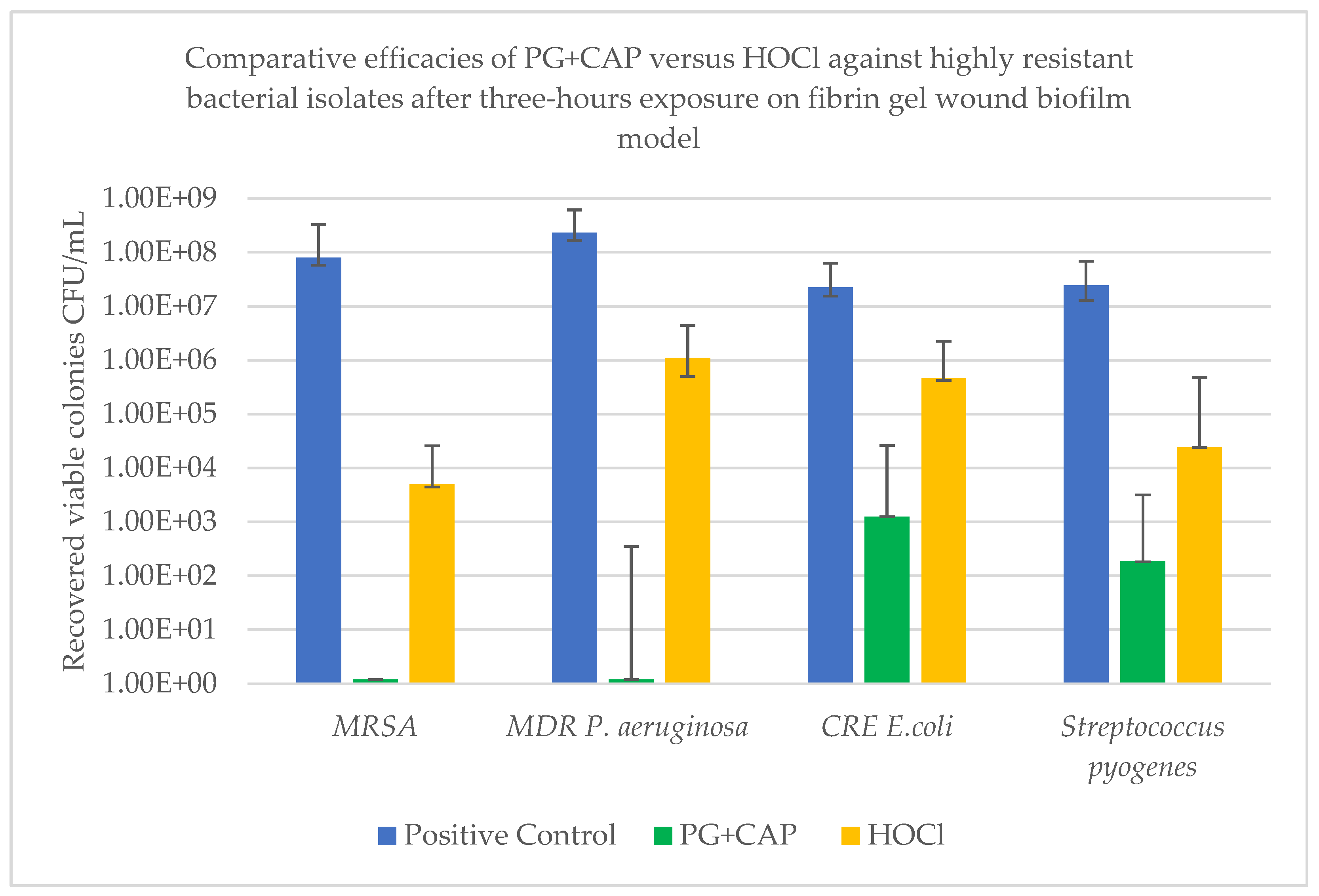
Figure 1. Efficacies of PG+CAP and HOCl against tested bacterial biofilms after three-hours exposure using fibrin gel wound biofilm model. Values are presented as the median number of recovered viable colony-forming units/mL (CFUs/mL) for six replicates. Bars indicate the median numbers of recovered viable colonies, and error bars indicate the ranges. Abbreviations: MRSA, methicillin resistant Staphylococcus aureus; MDR, multi-drug resistant; CRE, carbapenem-resistant Enterobacterales.
Preprints 177782 g001
Figure 2. In vitro cytotoxicity assessment of cell metabolic viability of L-929 fibroblasts after exposure to 2% PG+CAP ointment and 2% of HOCl irrigant for 24 hours, using the Alamar blue conversion metabolic assay. Treated cells with phosphate buffer saline were used as negative control. Results are expressed as a percentage of absorbance relative to negative control as measured by a spectrophotometer at 570 nm. Higher metabolic activity indicates lower cytotoxicity.
Preprints 177782 g002
Table 1. Log10 reduction of colony-forming units/mL (CFUs/mL) for tested bacterial isolates after three-hours of exposure to polygalacturonic acid + caprylic acid or hypochlorous acid in a fibrin-gel wound biofilm model.
Efficacy variable Tested organism
MRSA MDR
Pseudomonas aeruginosa
CRE
Escherichia coli
Streptococcus pyogenes
Log10 median viable colonies for negative control 8.00E+07 2.31E+08 2.25E+07 2.43E+07
Log10 reduction of CFU with PG+CAP relative to negative control 7.9 8.28 4.26 5.12
Log10 reduction of CFU with HOCl relative to negative control 4.2 2.32 1.69 3.0
P value for PG+CAP versus HOCl 0.0028 0.0043 0.005 0.019
Abbreviations: MRSA, methicillin resistant Staphylococcus aureus; MDR, multi-drug resistant; CRE, carbapenem-resistant Enterobacterales. Statistical comparisons between PG+CAP and negative control, as well as between HOCl and negative control, were statistically significant for all tested bacterial isolates (P ≤ 0.05).
Table 2. In vitro cytotoxicity assessment of cell viability of L-929 fibroblasts after exposure to 2% polygalacturonic acid + caprylic acid (PG+CAP) ointment and 2% hypochlorous acid (HOCl) irrigant for 24 hours, using the Trypan blue exclusion method.

Treatment
Mean ± SD cells
% Viable
P value* P value**
Live cells Dead cells
Negative control 2.69 × 10⁶ ± 7.59 × 10⁴ 6.0 × 10⁴ ± 8.16 × 10³ 97.82 0.26 -
2% PG+CAP 2.65 × 10⁶ ± 3.77 × 10⁴ 6.5 × 10⁴ ± 5.77 × 10³ 97.61 - 0.26
2% HOCl 2.46 × 10⁶ ± 4.83 × 10⁴ 4.43 × 10⁵ ± 1.50 × 10⁴ 84.76 <0.001 <0.001
Table 3. In vitro cytotoxicity assessment of cell metabolic activity of L-929 fibroblasts after exposure to 2% polygalacturonic acid and caprylic acid (PG+CAP) ointment or 2% hypochlorous acid (HOCl) irrigant for 24 hours, using the Alamar blue conversion metabolic assay.
Treatment Median reading Interquartile range P value* P value**
Negative control 1.124 1.121 - 1.129 0.083 -
2% PG+CAP 1.111 1.108 - 1.131 - 0.083
2% HOCl 0.942 0.848 - 1.00 0.014 0.014
Disclaimer/Publisher’s Note: The statements, opinions and data contained in all publications are solely those of the individual author(s) and contributor(s) and not of MDPI and/or the editor(s). MDPI and/or the editor(s) disclaim responsibility for any injury to people or property resulting from any ideas, methods, instructions or products referred to in the content.
Copyright: This open access article is published under a Creative Commons CC BY 4.0 license, which permit the free download, distribution, and reuse, provided that the author and preprint are cited in any reuse.
Prerpints.org logo

Preprints.org is a free preprint server supported by MDPI in Basel, Switzerland.

Subscribe

Disclaimer

Terms of Use

Privacy Policy

Privacy Settings

© 2025 MDPI (Basel, Switzerland) unless otherwise stated