Submitted:
17 September 2025
Posted:
19 September 2025
You are already at the latest version
Abstract
Diabetes mellitus (DM) is a disease that affects over 537 million people worldwide and results in 6.7 million deaths annually. Conventional treatment of this disease focuses on lifestyle changes and drug administration. However, very few people can adhere to a healthier lifestyle, and drugs are difficult to access, especially in low- and middle-income countries. An alternative as an adjuvant to the treatment of DM is the phenolic compounds from plants with reported anti-diabetic effects. However, the bioavailability of these compounds is very low since they are affected by the gastrointestinal tract and xenobiotic metabolism. To improve the availability of these compounds, an emerging technology such as encapsulation is being used since it has been reported that the encapsulation of phenolic compounds improves both their bioaccessibility and bioavailability, as well as their bioactivity. In this review, we will focus on compiling the most up-to-date information on the different encapsulation processes of phenolic compounds and the antidiabetic effect of encapsulated phenolic compounds using the databases PubMed, Scopus, Web of Science, and Google Scholar. We will discuss the mechanisms, pathways, and receptors involved in the modulation of DM, especially those related to inflammation, oxidative stress, and insulin resistance.
Keywords:
antidiabetic
; diabetes mellitus
; encapsulated
; insulin resistance
; microencapsulated
; polyphenols
1. Introduction
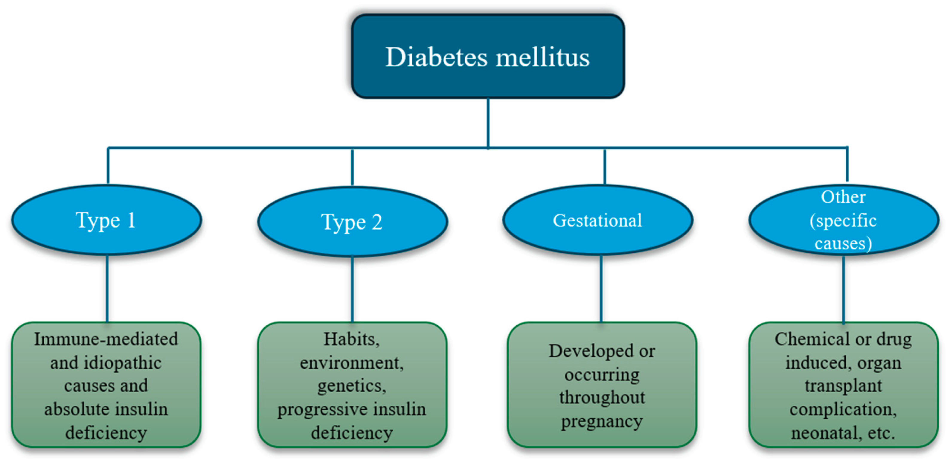
Diabetes mellitus (DM) is considered a group of metabolic disorders; this includes not only the inappropriate utilization of glucose as the main energy source but also impaired glucogenesis, gluconeogenesis, and insulin secretion and signalization. According to the American Diabetes Association (ADA), there are four types of diabetes mellitus categorized in Figure 1 [1,2]. When DM is not well-treated, several complications can develop, such as fatty liver, renal disease, metabolic syndrome, infertility, and neurological diseases, among others [3,4].
Figure 1.
Classification of diabetes mellitus according to the ADA.
Worldwide, it is estimated that more than 422 million people live with DM, and incidence has been increasing steadily, especially in low and middle-income regions. This disease is considered a global health emergency, with projections expecting that by 2030, cases will reach 643 million and 783 million by 2045. In countries such as the United States, the incidence of DM in the young population continues to rise, with estimates of 18,200 cases of type 1 DM and 5,300 cases of type 2 DM. Nonetheless, worldwide, it is considered that 98 % of diagnoses are for type 2 DM, and the rest are other types [1,5,6].
Considering type 2 DM (T2DM) is the most common diagnosis, most investigations have been aimed at it to understand psychopathology, complications, and treatments. But firstly, a correct diagnosis is necessary, considering for non-pregnant individuals the following tests: plasma A1C, fasting plasma glucose (FPG) value, 2-h glucose (2-h PG) value during a 75-g oral glucose tolerance test (OGTT), or random glucose value. It is also crucial to consider symptoms of hyperglycemia, such as polyuria, polydipsia, unexplained weight loss, and increased appetite [1]. Contrary to Type 1 DM, T2DM can be prevented and well managed by modifying risk habits and conditions. T2DM is closely linked to obesity and, therefore, to inflammation and oxidative stress. These three factors are commonly found in all non-communicable diseases [7,8].
Treatment and prevention of DM include exercise, a correct diet, drugs, and insulin in specific cases. Managing obesity and being overweight in diabetic patients also plays an important role. Access to treatment is key for individuals with DM to increase life quality and expectancy [9,10,11]. Nowadays, including other therapies and adjuvants to control or reduce oxidative stress and inflammation related to obesity and diabetes is a promising strategy. In this regard, several natural compounds, supplements, extracts, and other nutraceuticals have been studied to determine effective and safe doses for individuals with diabetes and prediabetes [12,13,14,15].
Several compounds, especially polyphenols, have been studied in vivo and in vitro as agents for prevention, management, and co-adjuvants in DM, regarding their proven and potential health benefits. Some activities attributed to polyphenols in this matter are antioxidants, anti-inflammatory, hypoglycemic, antiadipogenic, anti-gluconeogenesis, modulation of glucagon, insulin, and others [3,16,17,18,19].
Polyphenolic compounds are a heterogeneous class of phytochemicals characterized by multiple phenolic structural units, which are widely distributed in various plant-derived foods. Polyphenols are classified into two primary categories: flavonoids and non-flavonoids. Flavonoids include subcategories, such as anthocyanins, flavanols, flavanones, flavonols, and isoflavones, each characterized by distinct structural variations. In contrast, non-flavonoids comprise phenolic acids, xanthones, stilbenes, lignans, and tannins [20].
Polyphenols are abundantly present in fruits, vegetables, cereals, and other natural plant matrices, being integral to the sensory profiles and serving as biochemical markers for several foods and processing methods. For instance, in wines, polyphenols contribute to the complexity of flavor, color, and astringency while also providing insights into the authenticity and geographical origin of the product [21]. Furthermore, these compounds are pivotal to human health, with numerous studies elucidating their potential therapeutic benefits and prophylactic effects by several mechanisms against various pathologies [22]. Their bioactive properties have been associated with the modulation of oxidative stress [23], mitigating oxidative stress at the cellular level via scavenging and neutralizing reactive oxygen species [24,25,26]. For instance, it has been reported that dill (Anethum graveolens L.) extract exhibits antidiarrheal, anti-inflammatory, and antioxidant properties associated with its polyphenols [23]. Pumpkin pulp has been reported as a natural reservoir of polyphenolic compounds, showing antioxidant and antimicrobial properties, making it an attractive candidate for developing products to promote health [27]. In another study, it was reported that there are anti-inflammatory and antioxidant properties in several types of beans [28]. Similarly, a combination of vitamin C, resveratrol, and astaxanthin showed anti-inflammatory and antioxidant properties [29]. In other studies, resveratrol, in combination with urapidil, has neuroprotective effects by downregulating neurodegeneration [30].
On the other hand, a recent study suggests that tannic acid is a versatile candidate for preventing doxorubicin (DOX)-induced hepatotoxicity, potentially through the preservation of cellular physiology, viability, and, notably, redox homeostasis [31]. Another beneficial effect of polyphenols is the modulation of intestinal microbiota. A study showed how epigallocatechin-3-gallate has been shown to elevate the production of short-chain fatty acids, enhance amino acid metabolism, and downregulate pathways associated with intestinal inflammation. Additionally, this compound modulates the gut microbiota and mitigates Clostridioides difficile infection, offering novel insights into potential therapeutic interventions [32]. The study by Zhao et al. [33] suggests that polyphenols can modulate the gut microbiota, significantly impacting the production of microbial metabolites like isovaleric acid and isobutyric acid.
In the case of metabolic diseases, polyphenols, such as catechins, proanthocyanidins, hydroxybenzoic acids, and lignans, have been associated with a minor risk of developing type 2 diabetes [34]. A study conducted by Liao et al. [35] showed that Chinese jujube polyphenols exhibit significant hypoglycemic and antioxidative effects in rats with T2DM, thereby ameliorating glucose metabolism disorders and oxidative damage. Aqueous extracts of cinnamon and clove demonstrated greater potency than acarbose in inhibiting alpha-glucosidase activity and exhibited the highest antioxidant activity. The polyphenol content strongly correlated with antioxidant capacity, suggesting that these spices hold potential for the prevention and treatment of DM [36]. A study on the low-temperature aqueous extract of sea mustard (Undaria pinnatifida) at 50 °C indicates potential antihyperglycemic effects. This effect is mediated by modulation of glucose uptake via specific glucose transporters, suggesting the extract's capacity to mitigate postprandial hyperglycemia [37]. Also, Liu et al. [38] indicate that continuous administration of theaflavins (100 mg/kg) significantly suppressed blood glucose levels, reduced insulin resistance, and decreased the expression of oxidative stress markers and inflammatory cytokines in Goto-Kakizaki rats. Furthermore, consuming theaflavins facilitated the restoration of intestinal microbial community structure by reducing the abundance of pathogenic bacteria and increasing the prevalence of beneficial microorganisms.
In a study designed to evaluate the effects of a six-year nutritional and lifestyle intervention on oxidative and inflammatory markers in individuals aged 55 and older, who are at high risk of cardiovascular diseases [39], results showed that increased polyphenol intake was associated with a greater reduction in body mass index among participants. This suggests that polyphenols may play a role in promoting weight loss or maintaining healthier body weight, which is crucial for reducing the risk of obesity-related diseases such as T2DM, and at the same time, the study suggests that adherence to a low-calorie Mediterranean diet with increased polyphenol intake could contribute to positive health outcomes in a synergistic way.
These studies prove that polyphenols confer numerous health benefits and contribute to disease prevention and management. However, one of their primary limitations is the low bioavailability of these compounds [40]. In this sense, bioavailability is enhanced by 1) the physiological dose, 2) reduced particle size and thermal treatment, which facilitate compound release from the matrix, and 3) the presence of lipids, with minimal proteins and indigestible carbohydrates in the matrix [40]. For these reasons, it is particularly pertinent to explore strategies aimed at enhancing the bioavailability of these compounds to maximize the potential health benefits associated with their consumption.
Micro- and nano-encapsulated PC delivery systems have been developed for this purpose. The wall material and micro- or nanoparticle preparation method should be selected considering the PCs' biological properties, physicochemical characteristics, and purity to be encapsulated [41,42]. The present review is focused on the technologies used to encapsulate PCs that could serve as nutraceuticals for the prevention of T2DM or adjuncts in the therapeutic management of this chronic disease. The mechanism involved in the PCs-induced antidiabetic, anti-inflammatory, and antioxidant effects is also discussed.
2. Polyphenols
Most of the bioactive properties of polyphenols depend on their bioavailability, as non-bioavailable polyphenols can still exert preventive properties in the onset of colorectal cancer and modulate the intestinal microbiota. In vivo, many polyphenols that exhibit promising in vitro bioactivity suffer from low intestinal absorption and rapid elimination, resulting in limited systemic exposure.
A critical factor contributing to the variability in polyphenol bioavailability is the interindividual differences in absorption, distribution, metabolism, and excretion (ADME). These differences may stem from genetic polymorphisms affecting intestinal enzymes or xenobiotic transporters [43,44]. Additionally, variations in dietary habits, physiological conditions, and the permeability of biological barriers between healthy individuals and those with compromised health may further influence polyphenol bioavailability [45,46].
Enhancing polyphenol bioavailability, particularly in the context of dietary intake, is critically influenced by the interaction between nutritional lipids and polyphenols. Dietary fats, notably, have been recognized for their ability to enhance the solubility of polyphenols, potentially increasing their bioavailability [47]. Hydrophobic polyphenols, such as curcumin, demonstrate improved bioavailability when co-administered with dietary lipids [40]. This evidence suggests that integrating dietary fats with polyphenol consumption could optimize their absorption and subsequent physiological effects.
Furthermore, macronutrients such as carbohydrates and fats have been identified as intestinal absorption enhancers, which can modulate the time required to reach peak plasma concentrations of polyphenols [47]. The gut microbiota also plays a pivotal role in the metabolism and bioavailability of polyphenols. It has been demonstrated that gut bacteria can generate bioactive metabolites from polyphenols, thereby modulating various physiological processes and enhancing overall bioavailability [48,49,50,51,52].
The interaction between polyphenols and gut microbiota is bidirectional. Gut microbiota can convert polyphenols into simpler, more absorbable forms [53,54]. Conversely, polyphenols can influence gut microbiota composition, promote the growth of beneficial bacteria while suppressing pathogenic strains, thereby supporting a balanced gut microbiome [55]. Moreover, the complexation of polyphenols with proteins has improved their bioaccessibility and bioavailability. This mechanism protects polyphenols during gastrointestinal transit, enabling them to reach the colon, where they undergo further metabolism by gut microbiota, thus amplifying their health benefits [56].
4. Microencapsulation
Microencapsulation is one of the most promising technologies for directed therapeutic treatments used in the last years [57]. It is a process in which bioactive compounds like phenolics are trapped in an encapsulating material to create particles with a semipermeable membrane [58]. These bioactive agents can be encapsulated in their solid, liquid, and gaseous forms to obtain microcapsules with a size between 1 and 1000 μm [59]. Furthermore, the morphologies depend on the microencapsulation method, obtaining microspheres, microcapsules, and microparticles [60].
Microencapsulation of phenolic compounds protects them from environmental factors (light, humidity, temperature, and oxygen), provides controlled release over time, improves bioaccessibility, and increases shelf life and ease of storage [61]. There are several conventional and emerging microencapsulation techniques. Generally, the most used are physical methods, such as spray drying, freeze-drying, and extrusion, and physicochemical methods, such as liposomes, coacervation, ionic gelation, and co-crystallization [60,62]. Some microencapsulation examples of phenolic compounds are summarized in Table 1, and their basis/foundation is described later.
4.1. Physical Methods
4.1.1. Spray Drying
Spray drying is one of the best technologies for microencapsulating phenolic compounds. This technique consists of the atomization of a liquid mixture integrated by the core material (phenolic compounds) and the wall (encapsulating agent) in a stream of hot air, generating water evaporation and obtaining dry microparticles with a size between 1 y 100 µm [63,64]. This method offers several advantages, including simplicity, flexibility, low cost, easy scaling, high stability of the final product, and high encapsulation efficiency. Additionally, it is suitable for heat-sensitive compounds due to its short exposure times at high temperatures [65,66]. However, the selection of coat materials is important since it can affect the properties of the microparticles. Among the main encapsulating agents, carbohydrates, gums, pectin, proteins, and mixtures stand out [65,67].
4.1.2. Freeze-Drying
Freeze-drying is the most efficient technique for the encapsulation of bioactive compounds; it is the most popular drying process used for compounds that are heat sensitive. The method is based on the freezing and later sublimation of water from the solid/frozen state directly to the gaseous state, applying a vacuum. Exposure to low temperatures causes lyophilized products to retain their initial nutraceutical properties; however, the microencapsulation efficiency of this technology depends on the used wall materials, among them polymers, sugars such as maltodextrin, mannose, and trehalose, milk, polyols, and others [58,68,69].
4.1.3. Extrusion
Extrusion is a physical method in which phenolic compounds are encapsulated in hydrocolloid materials. Extrusion using natural polymers is a technique that improves bioactive compounds' stability, limits the use of high temperatures and organic solvents, and is also low-cost [70]. Generally, extrusion microencapsulation includes three processes: (1) melt injection, (2) melt-extrusion, and (3) centrifugal extrusion (coextrusion) [71]. This process is used to produce microcapsules by forcing a stream of shell material to surround the core material, a process based on the forced pass of a solution containing phenolic compounds through nozzles using droplet-generating equipment [58,59].
4.2. Physico-Chemical Methods
4.2.1. Liposomes
Liposomes, also called lipid vesicles, are spherical microscopic structures that consist of one or more phospholipid bilayers trapping an aqueous compartment in which lipophilic and hydrophilic agents can be dissolved in the lipid membrane and in the nucleus, respectively. The size of these lipid vesicles can vary from a few nanometers to several micrometers [72,73]. Due to their size, amphiphilic character, and biocompatibility, liposomes have been used as delivery vehicles for different phenolic compounds. Its application as a carrier system for phenolic compounds depends strictly on the physicochemical properties of its membranes, its size, the nature of its components, surface charge, and lipid organization [73,74].
4.2.2. Coacervation
Coacervation: it is a technique that consists of the phase separation of a colloidal system in the liquid-liquid phase of a polymer or a mixture of these with opposite charges in an aqueous solution caused by electrostatic interactions, hydrogen bonds, hydrophobic interactions, and enzymatic cross-linking agents (altering ionic strength, pH, or temperature). The process includes three basic steps: the formation of three immiscible phases, the deposition of the coating, and finally, the solidification of the coating [58,75,76]. Coacervation can be simple or complex, depending on the number of polymers used. Simple coacervation employs a simple polymer that absorbs at the interface between the colloidal solution and the solvent. Complex coacervation uses two or more polymer solutions for the formation of walls around an active core [58,76]. Proteins and polysaccharides are generally used as covering materials [77,78].
4.2.3. Co-Crystallization
Co-crystallization: is a method that uses sucrose as a matrix to incorporate bioactive compounds, it includes the preparation of a supersaturated sucrose solution, the addition of central materials, uniform mixing, and heating the mixture up to crystallization temperature [79]. This is a drying process where core materials in liquid form are directly converted into dry powder without the need for an additional drying step. Co-crystallization improves solubility, humectability, uniformity, dispersibility, hydration, anti-agglomeration, stability, and fluidity of the encapsulated bioactive compound [80].
4.2.4. Ionic Gelation
Ionic gelation: is a physicochemical method for the encapsulation of phenolic compounds. This method can be done through atomization, electrostatic deposition, or drop procedures. The fundamental consists of trapping an active substance and releasing it through gel phase changes (pH, mechanical wear, enzymes, and osmosis). Encapsulation starts with an aqueous polymeric solution, with low molecular mass ions that interact with polyelectrolytes of opposite charges, reacting and forming an insoluble gel [81,82]. Ion gelation is a simple procedure that does not require specialized equipment, uses relatively low temperatures and slow agitation, does not use organic solvents, and is low-cost, allowing the encapsulation of compounds that would degrade under other conditions. However, a disadvantage of this method is the low retention of hydrophilic compounds; hence, it is important to apply strategies like emulsion systems and cover material to enhance the encapsulation efficiency [83].
5. Nanoencapsulation
One emergent technology used to entrap polyphenolic compounds is nanotechnology; this science involves the design of nanoscale systems (particle size 1-100 nm). The size allows it to pass through the tissue and reach the sites of interest, since it increases the surface-volume ratio; therefore it is used in different disciplines such as biology, chemistry, and medicine [91,92,93]. The main objective of nanoencapsulation is protect an active ingredient (gas, solid, or liquid) with a matrix or shell, to form different types of nanoparticles, such as nanosphere, nanocapsules, nanoemulsion, nanoliposome, and nanoniosome, using different nanoencapsulation techniques such as deprotonation, ionic crosslinking, pH-regulated self-aggregation, polyelectrolyte complexation, ionic gelation, and hydrophobic modification, coacervation, nanoprecipitation, emulsification, layer-by-layer, sonication, desolvatation, reverse-phase evaporation, supercritical fluid, electrospray, nano spray drying [94,95,96,97]. Depending on the characteristics mentioned above and the encapsulation technique, the polyphenolic compound can be found dissolved within the nanoparticle, dispersed, trapped, or adsorbed [98]. On the other hand, there are many wall materials, some of the most commonly used being chitosan, gold (chloroauric acid), silver, mesoporous silica, hyaluronic acid, sodium alginate, polylactides (PLA), albumin, gelatin, poly(lactide)-poly(ethylene glycol) (PLA-PEG), poly(lactide-co-glycolide)-poly(ethylene glycol) (PLGA-PEG), polyglycolides (PGA), lecithin, polyglutamic acid, wheat protein, β-Lactoglobulin, among others [93,94,95]. All these materials must meet certain safety requirements, such as being non-toxic, easily degradable, and having physicochemical properties compatible with the polyphenol for better release [95].
In this sense, the wall material, polyphenol, and the nanoencapsulation technique are considered in the design of nanoformulations (Table 2) to improve the release of polyphenolic compounds in the specific targets of action. It has been shown that polyphenols influence different non-communicable diseases such as diabetes. For this reason various researchers have dedicated efforts to developing nanoformulations loaded with these compounds, due to the great advantages offered by nanoencapsulation, such as increasing its effectiveness by having a smaller size, improve solubility, in addition to protecting the compound from the degradation process caused by environmental factors such as light, changes in pH, temperature and radiation, and lastly and most importantly, a better bioavailability of the compounds is achieved. This could reduce the negative effects and help achieve greater specificity of the active compound or polyphenol to enhance its therapeutic action [91,99,100].
6. Current Evidence Regarding the Efficacy of Encapsulated Polyphenols
6.1. In Vitro
As mentioned above, diabetes mellitus is one of the most prevalent diseases worldwide, which is why many studies have focused on investigating this disease and how to improve its symptoms. In vitro analysis is one of the most widely used techniques to determine the beneficial effect of encapsulated polyphenols against diabetes. One of the more commonly used in vitro studies is simulated digestion, as phenolic compounds have a major bioavailability problem. To mention some examples, Verônica Cardoso de Souza et al. [123] studied Bauhinia forficata, a plant rich in polyphenols that is mainly used for its hypoglycemic activity, which is related to its antioxidant and anti-inflammatory potential, performed nanoencapsulation of infusion and decoction of B. forficata leaves using spray drive using maltodextrin and colloidal silicon dioxide as wall material, reporting that the nanoencapsulated flavonoid compounds were bioaccessible after the gastric phase (49.38 % and 64.17 % of polyphenols and 64.08 % and 36.61 % of flavonoids) and duodenal (52.68 % and 79.06 % of polyphenols and 13.24 % and 139.03 % of flavonoids), with a variation of 52.27 % to 70.55 % of the antioxidant activity maintained, by the ORAC method, after gastric digestion and still 25 % after duodenal, concluding that nanoencapsulation is a very viable technique for the conservation of bioactive compounds.
On the other hand, another of the most widely used in vitro techniques is the inhibition of enzymes related to carbohydrate metabolism. A clear example is the research carried out by Kerbab et al. [124], which studied the effect of the shrub Halimium halimifolium as an antidiabetic agent, finding that the phenolic compounds of this shrub have great antioxidant capacity and antidiabetic potential by inhibiting the enzymes α-amylase and α-glucosidase (IC50 = 0.82 mg/mL and 25.01 μg/mL, respectively); likewise, the authors performed microencapsulation of the compounds to optimize stability, handling, and delivery of bioactive compounds, using microencapsulation through spray drying and cellulose acetate phthalate as wall material. Likewise, Zorzenon et al. [125] evaluated maltodextrin microcapsules containing ethanolic extract of Stevia, by means of driver spray encapsulation, analyzed physicochemical parameters, antidiabetic activity (through inhibition of α-amylase), cytotoxicity, bioaccessibility of the compounds by in vitro digestion, as well as the structure of the microcapsules by scanning electron microscopy, the microcapsules showed greater solubility (∼35%), lower moisture content (∼29%) and maltodextrin DE10 had higher efficiency as an encapsulating agent (87%) compared to DE19 (76%) and showed well-defined spherical structures. Microencapsulation preserved the phenolic compound content and antioxidant activity present in the extract (7.2% and 87.5%, respectively). De Silva et al. [126] evaluated nanoencapsulated compounds of Bael fruit, using nanoencapsulation by ionic gelation using alginate as wall material; the authors report that with this nanoencapsulation, the compounds were more stable and that it enhanced the antidiabetic, antioxidant, and anti-inflammatory effects by having a slower and more controlled release profile with respect to non-encapsulated compounds.
Another of the routes studied for the determination of the antidiabetic effect of polyphenols is the glucose transporters GLUT-4. Chauhan et al. [127] studied chitosan-encapsulated nanocurcumin and the impact it has on the translocation of this glucose transporter, reporting that chitosan-nanocurcumin capsules caused an increase in the translocation of GLUT-4 to the cell surface in L6 skeletal muscle cells. The effect was associated with an increase in the phosphorylation of AKT (Ser-473) and its subsequent target GSK-3β (Ser-9).
6.2. In Vivo
In recent years, there has been an interest in studying the in vivo effect of encapsulated polyphenols, namely phenolic acids or flavonoids, and the polyphenolic-rich extracts from natural sources, as antidiabetic agents. According to Pandey and Dvorakova [128], the most common in vivo model for testing antidiabetic drugs is the induction of diabetes in rodents using streptozotocin (STZ). This drug has been used in several doses (10-150 mg/kg) and is usually administered via oral or intraperitoneal. The mode of action of STZ to induce diabetes is to selectively damage the pancreatic β-cells present in the Islets of Langerhans through several mechanisms, so the pancreas stops producing insulin, consequently inducing type I diabetes in a single dose [129]. Type I diabetes is, therefore, the most studied in rodent models. Inducing type II diabetes in vivo in rats or mice is more laborious. Several strategies are used together with the STZ administration, for example, using high-fat diets, nicotinamide, or rodents genetically susceptible to developing diabetes [128]. Even though these models show some disadvantages, such as high cost and variability, they are still relevant to studying diabetes and potential antidiabetic drugs.
Regarding the use of individual polyphenols as antidiabetic agents Panwar, Raghuwanshi, Srivastava, Sharma, and Pruthi [130] evaluated the antihyperglycemic effect of chitosan-encapsulated ferulic acid in diabetic Wistar rats. These authors reported that encapsulated ferulic acid significantly (compared with the diabetic group) reduced the levels of blood glucose and increased the secretion of insulin, as well as restoration of the pancreatic islets of Langerhans. Additionally, they observed a reduction in total cholesterol and triglycerides, which are biochemical markers of hyperlipidemia caused by diabetes complications. Nanoparticles of the flavonoid hesperidin were evaluated in a nicotinamide + STZ-induced diabetic model in male albino rats. After administering encapsulated hesperidin, the rats showed significantly lower plasma glucose concentration and increased insulin levels than the diabetic control group. Furthermore, the pancreatic islets of Langerhans were restored in rats treated with the nanoparticles of hesperidin. In contrast, rats treated with metformin still showed degeneration in the pancreatic cell clusters caused by the STZ. Other research evaluating the antidiabetic effect of diverse individual polyphenols can be reviewed in Table 3. It is important to mention that most in vivo studies have reported that encapsulating phenolic compounds improves their antidiabetic effects compared to free phenolics.
Encapsulated phenolic extracts from several herbs and plants are also investigated in models of rodents for their antidiabetic properties. The advantage of studying plant extracts over individual polyphenols could provide information regarding the synergistic effect that several compounds found in an extract might exert, therefore potentiating their biological effect [131]. In this sense, a poly-herbal (Justicia glabr, Adhatoda zeylanica, Andrographis paniculata, Gymnema sylvestre, Andrographis alata, and Syzygium cumini) ethanolic extract encapsulated with chitosan (particle size 62.6 ± 2.15 nm) was administered to diabetic rats for 30 days. After the experimental period, the rats exhibited significantly lower glucose concentrations and HbA1c levels, along with increased insulin and liver glycogen levels compared to the diabetic control group [132].
Furthermore, encapsulated (maltodextrin + whey protein) extracts from coffee parchment containing chlorogenic acid significantly reduced the glucose and HOMA-IR levels in obese male Wistar rats. Rats treated with the encapsulated extracts exhibited better biochemical parameters, since the TC, TG, and AST and ALT levels were significantly lowered; furthermore, TC and TG in the liver were also diminished [146]. An encapsulated anthocyanin-rich extract from the fruit of Vaccinium meridionale was administered to obese C57BL/6 mice, which significantly reduced the glucose and TC levels, when compared with the obese control group [147]. Other research evaluating the antidiabetic effect of polyphenolic-containing extracts is mentioned in Table 4.
It has been stated that polyphenols reduce ROS levels, inflammation, and oxidative stress in pancreatic-damaged cells, which helps to restore and maintain the correct functionality of β-cells and the regulation of insulin secretion. Other proposed antidiabetic mechanisms of polyphenols are related to the inhibition of: 1) digestive enzymes, 2) dipeptidyl-peptidase IV, 3) glycation of proteins, and 4) diabetic-related complications, among others [148]. The evidence suggests that encapsulated polyphenols, both individually and in extract, show promising attributes to be considered in the management of diabetes.
7. ADMET Analysis of Polyphenols with Antidiabetic Properties
Some of the chemical characteristics of a potential drug agent can be used to evaluate the drug-likeness of a molecule; this is called Lipinski’s rule of 5 [152]; these are 1) molecular weight below 500, 2) the molecule has no more than 5 hydrogen bond donors, 3) the molecule has no more than 10 hydrogen bond acceptors, and 4) the partition coefficient (Log p) is under 5. These characteristics can help us predict the passive absorption of a molecule. Here, we summarize the polyphenols with antidiabetic properties and their potential bioavailability using the rule of 5.

Table 5.
Lipinski’s rule of 5 evaluation of polyphenols with antidiabetic properties.
| Molecule | Class of compound | PubChem CID | Chemical Formula | Molecular Weight | H Bond donors | H Bond acceptor | Log p* | Lipinski Rule of 5 | |
| Cyanidin 3-glucoside | Anthocyanin | 197081 | C21H21ClO11 | 484.8 | 8 | 11 | -1.5 | No | |
| Curcumin | Curcuminoids | 969516 | C21H20O6 | 368.4 | 2 | 6 | 3.2 | Yes | |
| (+)-Catechin | Flavanol | 9064 | C15H14O6 | 290.27 | 5 | 6 | 1.4 | Yes | |
| (-)-Epicatechin | Flavanol | 72276 | C15H14O6 | 290.27 | 5 | 6 | 1.8 | Yes | |
| Liquiritin | Flavanone | 503737 | C21H22O9 | 418.4 | 5 | 9 | 0.4 | Yes | |
| Naringenin | Flavanone | 439246 | C15H12O5 | 272.25 | 3 | 5 | 2.2 | Yes | |
| Chrysin | Flavone | 5281607 | C15H10O4 | 254.24 | 2 | 4 | 2.5 | Yes | |
| Hesperidin | Flavone | 10621 | C28H34O15 | 610.6 | 8 | 15 | -1.1 | No | |
| Luteolin | Flavone | 5280445 | C15H10O6 | 286.24 | 4 | 6 | 2.0 | Yes | |
| Myricetin | Flavonol | 5281672 | C15H10O8 | 318.23 | 6 | 8 | 1.6 | No | |
| Quercetin | Flavonol | 5280343 | C15H10O7 | 302.23 | 5 | 7 | 1.5 | Yes | |
| Mangiferin | Glucosylxanthone | 5281647 | C19H18O11 | 422.3 | 8 | 11 | -0.4 | No | |
| Benzoic acid | Hydroxybenzoic acid | 243 | C7H6O2 | 122.12 | 1 | 2 | 1.87 | Yes | |
| Hydroxybenzoic acid | Hydroxybenzoic acid | 135 | C7H6O3 | 138.12 | 2 | 3 | 1.58 | yes | |
| Gallic acid | Hydroxybenzoic acid | 370 | C7H6O5 | 170.12 | 4 | 5 | 0.7 | Yes | |
| Ferulic acid | Hydroxycinnamic acid | 445858 | C10H10O4 | 194.18 | 2 | 4 | 1.5 | Yes | |
| Cinnamic acid | Hydroxycinnamic acid | 444539 | C9H8O2 | 148.16 | 1 | 2 | 2.1 | Yes | |
| Caffeic acid | Hydroxycinnamic acid | 689043 | C9H8O4 | 180.16 | 3 | 4 | 1.2 | Yes | |
| Coumaric acid | Hydroxycinnamic acid | 637542 | C9H8O3 | 164.16 | 2 | 3 | 1.5 | Yes | |
| Rosmarinic acid | Hydroxycinnamic acid | 5281792 | C18H16O8 | 360.3 | 5 | 8 | 2.4 | Yes | |
| Resveratrol | Stilbene | 445154 | C14H12O3 | 228.24 | 3 | 3 | 3.1 | Yes | |
8. Conclusions
Diabetes mellitus is a global health emergency affecting millions of individuals; it impacts society at an economic level, but also affects life expectancy and life quality. Diagnosing and treatment of prediabetes and DM are crucial. Still, limited access to medications and health care in middle and low-income populations influences treatment adherence and increases the risk of developing other health complications. In this regard, research aiming to create safe and effective alternatives obtained from natural sources represents a promising strategy. Research and technology have made it possible to protect compounds such as polyphenols by encapsulating them in different materials and with other methods. Choosing the appropriate and safe polyphenol dosages to achieve the antidiabetic effect is important. As discussed in this paper, selecting the proper encapsulation material, specific delivery, and polyphenols (isolated, mixed, or in addition to other bioactive compounds) must also be a priority to ensure bioavailability and nutraceutical properties.
Author Contributions
RCPL and EPGG designed the work; RCPL, FICT, NO, CAER, LACA, NYLL, MJBM, JBH, and EPGG prepared the review article; EPGG supervised the manuscript work. All authors have read and agreed to the published version of the manuscript.
Funding
Not applicable.
Institutional Review Board Statement
Not applicable
Data Availability Statement
All the data collected can be found in this document.
Conflicts of Interest
The authors declare no conflicts of interest.
Abbreviations
The following abbreviations are used in this manuscript:
| ADA | American Diabetes Association |
| ADME | Absorption, distribution, metabolism, and excretion |
| ALP | Alkaline phosphate |
| ALT | Alanine aminotransferase |
| AST | Aspartate aminotransferase |
| BUN | Blood urea nitrogen |
| BW | Body weight |
| DOAJ | Directory of open access journals |
| DM | Diabetes mellitus |
| FPG | Fasting plasma glucose |
| GLUT4 | Insulin-regulated glucose transporter |
| Hb1A1c | Glycosylated hemoglobin |
| HDL | High density lipoproteins |
| HOMA-B | Homeostasis model assessment of β-cell function |
| HOMA-IR | Homeostasis model assessment-insulin resistance |
| IC50 | Inhibitory Concentration 50 |
| LUV | Unilamellar vesicles |
| LD | Linear dichroism |
| LDL | Low density lipoproteins |
| LMPH | Longzhua mushroom polysaccharide hydrogel |
| MDPI | Multidisciplinary Digital Publishing Institute |
| MLV | Multilamellar vesicles |
| NLCs | Nanostructured Lipid Carriers |
| NSC | N-succinylated chitosan |
| OGTT | Oral glucose tolerance test |
| PGA | polyglycolides |
| PLA | Polylactides |
| PLA-PEG | poly(lactide)-poly(ethylene glycol) |
| PLGA | DL-polylactide/glycolide copolymer |
| PLGA-PEG | poly(lactide-co-glycolide)-poly(ethylene glycol) |
| SLNs | Solid Lipid Nanoparticles |
| STZ | streptozotocin |
| TC | Total cholesterol |
| TG | Triglycerides |
| T2DM | Type 2 Diabetes mellitus |
| 2-hPG | 2-h Plasma glucose |
References
- Committee, A.D.A.P.P. 2. Diagnosis and Classification of Diabetes: Standards of Care in Diabetes—2024. 2023, 47, S20–S42. [Google Scholar] [CrossRef]
- Popoviciu, M.S.; Paduraru, L.; Nutas, R.M.; Ujoc, A.M.; Yahya, G.; Metwally, K.; Cavalu, S. Diabetes Mellitus Secondary to Endocrine Diseases: An Update of Diagnostic and Treatment Particularities. 2023, 24, 12676.
- Sharma, P.; Hajam, Y.A.; Kumar, R.; Rai, S. Complementary and alternative medicine for the treatment of diabetes and associated complications: A review on therapeutic role of polyphenols. 2022; 2, 100188. [Google Scholar] [CrossRef]
- Vlacho, B.; Rossell-Rusiñol, J.; Granado-Casas, M.; Mauricio, D.; Julve, J. Overview on chronic complications of diabetes mellitus. In Chronic Complications of Diabetes Mellitus; Elsevier: 2024; pp. 1-10.
- International Diabetes Federation. Diabetes Atlas. Available online: https://diabetesatlas.org/atlas-reports (accessed on.
- World Health Organization. Diabetes. Available online: https://www.who.int/health-topics/diabetes#tab=tab_1 (accessed on 1 August 2024).
- Chandrasekaran, P.; Weiskirchen, R. The Role of Obesity in Type 2 Diabetes Mellitus—An Overview. 2024, 25, 1882.
- Rohm, T.V.; Meier, D.T.; Olefsky, J.M.; Donath, M.Y. Inflammation in obesity, diabetes, and related disorders. 2022, 55, 31-55.
- Li, Y.; Schoufour, J.; Wang, D.D.; Dhana, K.; Pan, A.; Liu, X.; Song, M.; Liu, G.; Shin, H.J.; Sun, Q. Healthy lifestyle and life expectancy free of cancer, cardiovascular disease, and type 2 diabetes: prospective cohort study. 2020, 368.
- Lingvay, I.; Sumithran, P.; Cohen, R.V.; le Roux, C.W. Obesity management as a primary treatment goal for type 2 diabetes: time to reframe the conversation. 2022, 399, 394-405.
- Magkos, F.; Hjorth, M.F.; Astrup, A. Diet and exercise in the prevention and treatment of type 2 diabetes mellitus. 2020, 16, 545-555.
- Akbari, B.; Baghaei-Yazdi, N.; Bahmaie, M.; Mahdavi Abhari, F. The role of plant-derived natural antioxidants in reduction of oxidative stress. 2022, 48, 611-633.
- Pasupuleti, V.R.; Arigela, C.S.; Gan, S.H.; Salam, S.K.N.; Krishnan, K.T.; Rahman, N.A.; Jeffree, M.S. A review on oxidative stress, diabetic complications, and the roles of honey polyphenols. 2020, 2020, 8878172.
- Pérez-Torres, I.; Castrejón-Téllez, V.; Soto, M.E.; Rubio-Ruiz, M.E.; Manzano-Pech, L.; Guarner-Lans, V. Oxidative stress, plant natural antioxidants, and obesity. 2021, 22, 1786.
- Unuofin, J.O.; Lebelo, S.L. Antioxidant effects and mechanisms of medicinal plants and their bioactive compounds for the prevention and treatment of type 2 diabetes: an updated review. 2020, 2020, 1356893.
- Arkoub-Djermoune, L.; Boulekbache-Makhlouf, L.; Zeghichi-Hamri, S.; Bellili, S.; Boukhalfa, F.; Madani, K. Influence of the Thermal Processing on the Physico-Chemical Properties and the Antioxidant Activity of A Solanaceae Vegetable: Eggplant. 2016; 39, 181–191. [Google Scholar] [CrossRef]
- Aryal, D.; Joshi, S.; Thapa, N.K.; Chaudhary, P.; Basaula, S.; Joshi, U.; Bhandari, D.; Rogers, H.M.; Bhattarai, S.; Sharma, K.R.; et al. Dietary phenolic compounds as promising therapeutic agents for diabetes and its complications: A comprehensive review. 2024; 12, 3025–3045. [Google Scholar] [CrossRef]
- Naz, R.; Saqib, F.; Awadallah, S.; Wahid, M.; Latif, M.F.; Iqbal, I.; Mubarak, M.S. Food Polyphenols and Type II Diabetes Mellitus: Pharmacology and Mechanisms. 2023, 28, 3996.
- Wang, Y.; Alkhalidy, H.; Liu, D. The Emerging Role of Polyphenols in the Management of Type 2 Diabetes. 2021, 26, 703.
- Durazzo, A.; Lucarini, M.; Souto, E.B.; Cicala, C.; Caiazzo, E.; Izzo, A.A.; Novellino, E.; Santini, A. Polyphenols: A concise overview on the chemistry, occurrence, and human health. 2019; 33, 2221–2243. [Google Scholar] [CrossRef]
- Sánchez Arribas, A.; Moreno, M.; Moreno, G.A.; Bermejo, E.; Zapardiel, A.; Chicharro, M. Characterization of White Wines by Electrochemical Indexes Obtained Using Carbon Nanotube-modified Electrodes. 2018; 30, 1461–1471. [Google Scholar] [CrossRef]
- Barbosa, S.; Pardo-Mates, N.; Hidalgo-Serrano, M.; Saurina, J.; Puignou, L.; Núñez, O. Detection and Quantitation of Frauds in the Authentication of Cranberry-Based Extracts by UHPLC-HRMS (Orbitrap) Polyphenolic Profiling and Multivariate Calibration Methods. 2018; 66, 9353–9365. [Google Scholar] [CrossRef]
- Brinsi, C.; Jedidi, S.; Sammari, H.; Selmi, H.; Sebai, H. Antidiarrheal, anti-inflammatory and antioxidant effects of Anethum graveolens L. fruit extract on castor oil-induced diarrhea in rats. 2024; e14892. [Google Scholar] [CrossRef]
- Arslaner, A.; Türkoğlu, Z. A potential antiviral and food-derived healty ingredient: Resveratrol. 2021; 7. [Google Scholar] [CrossRef]
- Santos Sánchez, N.; Salas-Coronado, R.; Villanueva, C.; Hernandez-Carlos, B. Antioxidant Compounds and Their Antioxidant Mechanism. 2019.
- Xuan Hoan, N.; Anh, L.; Quan, D.; Cuong, D.; Thai Ha, H.; Minh, N.; Hieu, D.; Thuat, N.; Pham, T.; Tuyen, D. Functional-Antioxidant Food. 2021.
- Pinna, N.; Ben Abbou, S.; Ianni, F.; Angeles Flores, G.; Pietercelie, A.; Perretti, G.; Blasi, F.; Angelini, P.; Cossignani, L. Phenolic compounds from pumpkin pulp: Extraction optimization and biological properties. 2024; 23, 101628. [Google Scholar] [CrossRef]
- Upadhyaya, B.; Moreau, R.; Majumder, K. Antioxidant and Anti-Inflammatory Capacities of Three Dry Bean Varieties after Cooking and In Vitro Gastrointestinal Digestion. 2024. [Google Scholar] [CrossRef]
- Marzagalli, M.; Battaglia, S.; Raimondi, M.; Fontana, F.; Cozzi, M.; Ranieri, F.R.; Sacchi, R.; Curti, V.; Limonta, P. Anti-Inflammatory and Antioxidant Properties of a New Mixture of Vitamin C, Collagen Peptides, Resveratrol, and Astaxanthin in Tenocytes: Molecular Basis for Future Applications in Tendinopathies. 2024; 2024, 5273198. [Google Scholar] [CrossRef]
- Çetin, R.; Bahadir, S.; Basar, İ.; Aslanoglu, B.; Atlas, B.; Kaya, S.; Güzel, B.C.; Turan, Y. Neuroprotective effects of the combined treatment of resveratrol and urapidil in experimental cerebral ischemia-reperfusion injury in rats. 2024; 39, e395329. [Google Scholar] [CrossRef]
- Özturk, N.; Ceylan, H.; Demir, Y. The hepatoprotective potential of tannic acid against doxorubicin-induced hepatotoxicity: Insights into its antioxidative, anti-inflammatory, and antiapoptotic mechanisms. 2024; 38, e23798. [Google Scholar] [CrossRef]
- Wu, Z.; Shen, J.; Xu, Q.; Xiang, Q.; Chen, Y.; Lv, L.; Zheng, B.; Wang, Q.; Wang, S.; Li, L. Epigallocatechin-3-Gallate Improves Intestinal Gut Microbiota Homeostasis and Ameliorates Clostridioides difficile Infection. 2022; 14. [Google Scholar] [CrossRef]
- Zhao, X.; Guo, J.; Wang, Y.; Yi, X. High-tannin food enhances spatial memory and scatter-hoarding in rodents via the microbiota-gut-brain axis. 2024; 12, 140. [Google Scholar] [CrossRef]
- Tresserra-Rimbau, A.; Castro-Barquero, S.; Vitelli-Storelli, F.; Becerra-Tomas, N.; Vázquez-Ruiz, Z.; Díaz-López, A.; Corella, D.; Castañer, O.; Romaguera, D.; Vioque, J.; et al. Associations between Dietary Polyphenols and Type 2 Diabetes in a Cross-Sectional Analysis of the PREDIMED-Plus Trial: Role of Body Mass Index and Sex. 2019; 8. [Google Scholar] [CrossRef]
- Liao, M.; Wang, X. Ameliorating effect of Chinese jujube polyphenol on blood glucose oxidative stress in type 2 diabetic rats. 2024; 38, 108804. [Google Scholar] [CrossRef]
- Choockong, C.; Itharat, A.; Pipatrattanaseree, W.; Ninlaor, T.; Piwngam, K.; Intharit, N.; Sukkhum, S.; Davies, N.M. The most commonly used spices in Thai traditional medicine: in vitro evaluation of anti-hyperglycemic, antioxidant, polyphenol content, and nitric oxide production inhibitory activities. 2024; 19, 13–28. [Google Scholar] [CrossRef]
- Lee, S.M.; Park, S.Y.; Kim, J.Y. Comparative evaluation of the antihyperglycemic effects of three extracts of sea mustard (Undaria pinnatifida): In vitro and in vivo studies. 2024; 190, 114623. [Google Scholar] [CrossRef]
- Liu, C.; Zeng, H.; Cui, W.; Ouyang, J.; Zhou, F.; Wen, S.; Fang, W.; Zhang, S.; Huang, J.; Liu, Z. Theaflavins mitigate diabetic symptoms in GK rats by modulating the INSR/PI3K-Akt/GSK-3 pathway and intestinal microbiota. [CrossRef]
- Quetglas-Llabrés, M.M.; Monserrat-Mesquida, M.; Bouzas, C.; García, S.; Mateos, D.; Ugarriza, L.; Gómez, C.; Sureda, A.; Tur, J.A. Long-Term Impact of Nutritional Intervention with Increased Polyphenol Intake and Physical Activity Promotion on Oxidative and Inflammatory Profiles in Patients with Metabolic Syndrome. [CrossRef]
- Bohn, T. Dietary factors affecting polyphenol bioavailability. 2014, 72, 429-452.
- Castro-López, C.; Espinoza-González, C.; Ramos-González, R.; Boone-Villa, V.D.; Aguilar-González, M.A.; Martínez-Ávila, G.C.G.; Aguilar, C.N.; Ventura-Sobrevilla, J.M. Spray-drying encapsulation of microwave-assisted extracted polyphenols from Moringa oleifera: Influence of tragacanth, locust bean, and carboxymethyl-cellulose formulations. [CrossRef]
- Costa, M.; Sezgin-Bayindir, Z.; Losada-Barreiro, S.; Paiva-Martins, F.; Saso, L.; Bravo-Díaz, C. Polyphenols as Antioxidants for Extending Food Shelf-Life and in the Prevention of Health Diseases: Encapsulation and Interfacial Phenomena. [CrossRef]
- Gerloff, T. Impact of genetic polymorphisms in transmembrane carrier-systems on drug and xenobiotic distribution. 2004, 369, 69-77.
- Lampe, J.W. Interindividual differences in response to plant-based diets: implications for cancer risk. 2009, 89, 1553S-1557S.
- Hidalgo-Liberona, N.; González-Domínguez, R.; Vegas, E.; Riso, P.; Del Bo’, C.; Bernardi, S.; Peron, G.; Guglielmetti, S.; Gargari, G.; Kroon, P.A. Increased intestinal permeability in older subjects impacts the beneficial effects of dietary polyphenols by modulating their bioavailability. 2020, 68, 12476-12484.
- Margalef, M.; Pons, Z.; Iglesias-Carres, L.; Bravo, F.I.; Muguerza, B.; Arola-Arnal, A. Flavanol plasma bioavailability is affected by metabolic syndrome in rats. 2017, 231, 287-294.
- Zhang, H.; Yu, D.; Sun, J.; Liu, X.; Jiang, L.; Guo, H.; Ren, F. Interaction of plant phenols with food macronutrients: characterisation and nutritional–physiological consequences. 2014, 27, 1-15.
- Iqbal, Y.; Cottrell, J.J.; Suleria, H.A.; Dunshea, F.R. Gut microbiota-polyphenol interactions in chicken: A review. 2020, 10, 1391.
- Li, D.; Yang, Y.; Yang, X.; Wang, Z.; Yao, X.; Guo, Y. Enhanced bioavailability and anti-hyperglycemic activity of young apple polyphenols by complexation with whey protein isolates. 2022, 87, 1257-1267.
- Pasinetti, G.M.; Singh, R.; Westfall, S.; Herman, F.; Faith, J.; Ho, L. The role of the gut microbiota in the metabolism of polyphenols as characterized by gnotobiotic mice. 2018, 63, 409-421.
- Wang, Q.; Xu, K.; Cai, X.; Wang, C.; Cao, Y.; Xiao, J. Rosmarinic acid restores colonic mucus secretion in colitis mice by regulating gut microbiota-derived metabolites and the activation of inflammasomes. 2023, 71, 4571-4585.
- Zhou, N.; Gu, X.; Zhuang, T.; Xu, Y.; Yang, L.; Zhou, M. Gut microbiota: A pivotal hub for polyphenols as antidepressants. 2020, 68, 6007-6020.
- Corrêa, T.A.F.; Rogero, M.M.; Hassimotto, N.M.A.; Lajolo, F.M. The Two-Way Polyphenols-Microbiota Interactions and Their Effects on Obesity and Related Metabolic Diseases. [CrossRef]
- Shivashankara, K.; Acharya, S. Bioavailability of dietary polyphenols and the cardiovascular diseases. 2010, 3.
- Li, M.; Zheng, Y.; Zhao, J.; Liu, M.; Shu, X.; Li, Q.; Wang, Y.; Zhou, Y. Polyphenol mechanisms against gastric cancer and their interactions with gut microbiota: a review. 2022, 29, 5247-5261.
- Lila, M.A.; Hoskin, R.T.; Grace, M.H.; Xiong, J.; Strauch, R.; Ferruzzi, M.; Iorizzo, M.; Kay, C. Boosting the bioaccessibility of dietary bioactives by delivery as protein–polyphenol aggregate particles. 2022, 70, 13017-13026.
- Chen, L.; Gnanaraj, C.; Arulselvan, P.; El-Seedi, H.; Teng, H. A review on advanced microencapsulation technology to enhance bioavailability of phenolic compounds: Based on its activity in the treatment of Type 2 Diabetes. [CrossRef]
- Frakolaki, G.; Giannou, V.; Kekos, D.; Tzia, C. A review of the microencapsulation techniques for the incorporation of probiotic bacteria in functional foods. [CrossRef]
- Arenas-Jal, M.; Suñé-Negre, J.M.; García-Montoya, E. An overview of microencapsulation in the food industry: opportunities, challenges, and innovations. [CrossRef]
- Mohammadalinejhad, S.; Kurek, M.A. Microencapsulation of anthocyanins-critical review of techniques and wall materials. [CrossRef]
- Peanparkdee, M.; Iwamoto, S.; Yamauchi, R. Microencapsulation: a review of applications in the food and pharmaceutical industries. 2016, 4, 56-65.
- Mehta, N.; Kumar, P.; Verma, A.K.; Umaraw, P.; Kumar, Y.; Malav, O.P.; Sazili, A.Q.; Domínguez, R.; Lorenzo, J.M. Microencapsulation as a Noble Technique for the Application of Bioactive Compounds in the Food Industry: A Comprehensive Review. [CrossRef]
- Leyva-Jiménez, F.J.; Lozano-Sánchez, J.; de la Luz Cádiz-Gurrea, M.; Fernández-Ochoa, Á.; Arráez-Román, D.; Segura-Carretero, A. Spray-drying microencapsulation of bioactive compounds from lemon verbena green extract. [CrossRef]
- Santos, S.S.d.; Rodrigues, L.M.; da Costa, S.C.; Bergamasco, R.d.C.; Madrona, G.S. Microencapsulation of bioactive compounds from blackberry pomace (Rubus fruticosus) by spray drying technique. 2017, 13, 20170047.
- Navarro-Flores, M.J.; Ventura-Canseco, L.M.C.; Meza-Gordillo, R.; Ayora-Talavera, T.R.; Abud-Archila, M. Spray drying encapsulation of a native plant extract rich in phenolic compounds with combinations of maltodextrin and non-conventional wall materials. [CrossRef]
- Ribeiro, A.M.; Estevinho, B.N.; Rocha, F. Spray Drying Encapsulation of Elderberry Extract and Evaluating the Release and Stability of Phenolic Compounds in Encapsulated Powders. [CrossRef]
- Ferreira, L.M.D.M.C.; Pereira, R.R.; Carvalho-Guimarães, F.B.D.; Remígio, M.S.D.N.; Barbosa, W.L.R.; Ribeiro-Costa, R.M.; Silva-Júnior, J.O.C. Microencapsulation by Spray Drying and Antioxidant Activity of Phenolic Compounds from Tucuma Coproduct (Astrocaryum vulgare Mart.) Almonds. 2022, 14. [Google Scholar] [CrossRef] [PubMed]
- Da Rosa, C.G.; Borges, C.D.; Zambiazi, R.C.; Rutz, J.K.; da Luz, S.R.; Krumreich, F.D.; Benvenutti, E.V.; Nunes, M.R. Encapsulation of the phenolic compounds of the blackberry (Rubus fruticosus). [CrossRef]
- Wilkowska, A.; Ambroziak, W.; Czyzowska, A.; Adamiec, J. Effect of Microencapsulation by Spray-Drying and Freeze-Drying Technique on the Antioxidant Properties of Blueberry (Vaccinium myrtillus) Juice Polyphenolic Compounds. [CrossRef]
- Stach, M.; Kolniak-Ostek, J. The Influence of the Use of Different Polysaccharide Coatings on the Stability of Phenolic Compounds and Antioxidant Capacity of Chokeberry Hydrogel Microcapsules Obtained by Indirect Extrusion. [CrossRef]
- Bakry, A.M.; Abbas, S.; Ali, B.; Majeed, H.; Abouelwafa, M.Y.; Mousa, A.; Liang, L. Microencapsulation of Oils: A Comprehensive Review of Benefits, Techniques, and Applications. [CrossRef]
- Gibis, M.; Ruedt, C.; Weiss, J. In vitro release of grape-seed polyphenols encapsulated from uncoated and chitosan-coated liposomes. [CrossRef]
- Has, C.; Sunthar, P. A comprehensive review on recent preparation techniques of liposomes. [CrossRef]
- Bozzuto, G.; Molinari, A. Liposomes as nanomedical devices. [CrossRef]
- Choudhury, N.; Meghwal, M.; Das, K. Microencapsulation: An overview on concepts, methods, properties and applications in foods. [CrossRef]
- Ozkan, G.; Franco, P.; De Marco, I.; Xiao, J.; Capanoglu, E. A review of microencapsulation methods for food antioxidants: Principles, advantages, drawbacks and applications. [CrossRef]
- de Souza, V.B.; Thomazini, M.; Echalar Barrientos, M.A.; Nalin, C.M.; Ferro-Furtado, R.; Genovese, M.I.; Favaro-Trindade, C.S. Functional properties and encapsulation of a proanthocyanidin-rich cinnamon extract (Cinnamomum zeylanicum) by complex coacervation using gelatin and different polysaccharides. [CrossRef]
- Soliman, T.N.; Mohammed, D.M.; El-Messery, T.M.; Elaaser, M.; Zaky, A.A.; Eun, J.B.; Shim, J.H.; El-Said, M.M. Microencapsulation of Plant Phenolic Extracts Using Complex Coacervation Incorporated in Ultrafiltered Cheese Against AlCl3-Induced Neuroinflammation in Rats. [CrossRef]
- Sarabandi, K.; Mahoonak, A.S.; Akbari, M. Physicochemical properties and antioxidant stability of microencapsulated marjoram extract prepared by co-crystallization method. [CrossRef]
- Ali, N.A.; Dash, K.K.; Pandey, V.K.; Tripathi, A.; Mukarram, S.A.; Harsányi, E.; Kovács, B. Extraction and Encapsulation of Phytocompounds of Poniol Fruit via Co-Crystallization: Physicochemical Properties and Characterization. [CrossRef]
- Cutrim, C.S.; Alvim, I.D.; Cortez, M.A.S. Microencapsulation of green tea polyphenols by ionic gelation and spray chilling methods. [CrossRef]
- Kurozawa, L.E.; Hubinger, M.D. Hydrophilic food compounds encapsulation by ionic gelation. [CrossRef]
- de Moura, S.C.S.R.; Berling, C.L.; Germer, S.P.M.; Alvim, I.D.; Hubinger, M.D. Encapsulating anthocyanins from Hibiscus sabdariffa L. calyces by ionic gelation: Pigment stability during storage of microparticles. [CrossRef]
- da Silva Júnior, M.E.; Araújo, M.V.R.L.; Martins, A.C.S.; dos Santos Lima, M.; da Silva, F.L.H.; Converti, A.; Maciel, M.I.S. Microencapsulation by spray-drying and freeze-drying of extract of phenolic compounds obtained from ciriguela peel. [CrossRef]
- Thakur, N.S.; Thakur, A. Microencapsulation of wild pomegranate flavedo phenolics by lyophilization: Effect of maltodextrin concentration, structural morphology, functional properties, elemental composition and ingredient for development of functional beverage. [CrossRef]
- Vallejo-Castillo, V.; Rodríguez-Stouvenel, A.; Martínez, R.; Bernal, C. Development of alginate-pectin microcapsules by the extrusion for encapsulation and controlled release of polyphenols from papaya (Carica papaya L.). 2020, 44, 44. [Google Scholar] [CrossRef] [PubMed]
- Bannikova, A.; Zyainitdinov, D.; Evteev, A.; Drevko, Y.; Evdokimov, I. Microencapsulation of polyphenols and xylooligosaccharides from oat bran in whey protein-maltodextrin complex coacervates: In-vitro evaluation and controlled release. [CrossRef]
- Istenič, K.; Cerc Korošec, R.; Poklar Ulrih, N. Encapsulation of (-)-epigallocatechin gallate into liposomes and into alginate or chitosan microparticles reinforced with liposomes. [CrossRef]
- Nik, A.B.; Vazifedoost, M.; Didar, Z.; Hajirostamloo, B. The antioxidant and physicochemical properties of microencapsulated bioactive compounds in Securigera securidaca (L.) seed extract by co-crystallization. [CrossRef]
- Chezanoglou, E.; Goula, A.M. Properties and Stability of Encapsulated Pomegranate Peel Extract Prepared by Co-Crystallization. [CrossRef]
- Anandharamakrishnan, C. Techniques for nanoencapsulation of food ingredients; Springer: 2014; Volume 8.
- Contreras-Angulo, L.A.; Gutiérrez-Grijalva, E.P.; Cabanillas-Bojórquez, L.A.; Jiménez-Ortega, L.A.; Heredia, J.B. Chapter 7 - Nanoformulations applied to the delivery of alkaloids. In Phytochemical Nanodelivery Systems as Potential Biopharmaceuticals, Heredia, J.B., Gutiérrez-Grijalva, E.P., Licea-Claverie, A., Gutierrez-Uribe, J.A., Patra, J.K., Eds.; Elsevier: 2023; pp. 257-281.
- Garcia-Carrasco, M.; Parra-Aguilar, I.F.; Gutiérrez-Grijalva, E.P.; Licea-Claverie, A.; Basilio Heredia, J. Chapter 18 - Nano-formulations in drug delivery. In Food, Medical, and Environmental Applications of Nanomaterials, Pal, K., Sarkar, A., Sarkar, P., Bandara, N., Jegatheesan, V., Eds.; Elsevier: 2022; pp. 473-491.
- Pedrozo, R.C.; Antônio, E.; Khalil, N.M.; Mainardes, R.M. Bovine serum albumin-based nanoparticles containing the flavonoid rutin produced by nano spray drying. [CrossRef]
- Rambaran, T.F. Nanopolyphenols: a review of their encapsulation and anti-diabetic effects. [CrossRef]
- Rehman, A.; Ahmad, T.; Aadil, R.M.; Spotti, M.J.; Bakry, A.M.; Khan, I.M.; Zhao, L.; Riaz, T.; Tong, Q. Pectin polymers as wall materials for the nano-encapsulation of bioactive compounds. [CrossRef]
- Ribeiro, E.F.; de Barros-Alexandrino, T.T.; Assis, O.B.G.; Junior, A.C.; Quiles, A.; Hernando, I.; Nicoletti, V.R. Chitosan and crosslinked chitosan nanoparticles: Synthesis, characterization and their role as Pickering emulsifiers. [CrossRef]
- Jiang, T.; Jin, K.; Liu, X.; Pang, Z. 8 - Nanoparticles for tumor targeting. In Biopolymer-Based Composites, Jana, S., Maiti, S., Jana, S., Eds.; Woodhead Publishing: 2017; pp. 221-267.
- Costa-Almeida, R.; Soares, R.; Costa, R. Polyphenol-Based Nanoparticles as Multifaceted Diabetes Modulators. In Functional Bionanomaterials: From Biomolecules to Nanoparticles, Thangadurai, D., Sangeetha, J., Prasad, R., Eds.; Springer International Publishing: Cham, 2020; pp. 251–270. [Google Scholar]
- Rai, V.K.; Gupta, G.D.; Pottoo, F.H.; Barkat, M.A. Potential of Nano-Structured Drug Delivery System for Phytomedicine Delivery. In Nanophytomedicine: Concept to Clinic, Beg, S., Barkat, M.A., Ahmad, F.J., Eds.; Springer Singapore: Singapore, 2020; pp. 89–111. [Google Scholar]
- Wu, M.; Jin, J.; Jin, P.; Xu, Y.; Yin, J.; Qin, D.; Wang, K.; Du, Q. Epigallocatechin gallate-β-lactoglobulin nanoparticles improve the antitumor activity of EGCG for inducing cancer cell apoptosis. [CrossRef]
- Palacio, J.; Monsalve, Y.; Ramírez-Rodríguez, F.; López, B. Study of encapsulation of polyphenols on succinyl-chitosan nanoparticles. [CrossRef]
- Gadkari, P.V.; Balaraman, M. Extraction of catechins from decaffeinated green tea for development of nanoemulsion using palm oil and sunflower oil based lipid carrier systems. [CrossRef]
- Harwansh, R.K.; Mukherjee, P.K.; Kar, A.; Bahadur, S.; Al-Dhabi, N.A.; Duraipandiyan, V. Enhancement of photoprotection potential of catechin loaded nanoemulsion gel against UVA induced oxidative stress. [CrossRef]
- Antônio, E.; Khalil, N.M.; Mainardes, R.M. Bovine Serum Albumin Nanoparticles Containing Quercetin: Characterization and Antioxidant Activity. Journal of nanoscience and nanotechnology 2016, 16, 1346–1353. [Google Scholar] [CrossRef] [PubMed]
- Javani, R.; Hashemi, F.S.; Ghanbarzadeh, B.; Hamishehkar, H. Quercetin-loaded niosomal nanoparticles prepared by the thin-layer hydration method: Formulation development, colloidal stability, and structural properties. [CrossRef]
- Romeo, A.; Musumeci, T.; Carbone, C.; Bonaccorso, A.; Corvo, S.; Lupo, G.; Anfuso, C.D.; Puglisi, G.; Pignatello, R. Ferulic Acid-Loaded Polymeric Nanoparticles for Potential Ocular Delivery. Pharmaceutics 2021, 13. [Google Scholar] [CrossRef] [PubMed]
- Nallamuthu, I.; Devi, A.; Khanum, F. Chlorogenic acid loaded chitosan nanoparticles with sustained release property, retained antioxidant activity and enhanced bioavailability. [CrossRef]
- Neelakandan, M.; Manoharan, S.; Muralinaidu, R.; Thara, J.M. Tumor preventive and antioxidant efficacy of chlorogenic acid–loaded chitosan nanoparticles in experimental skin carcinogenesis. [CrossRef]
- Mariadoss, A.V.A.; Vinayagam, R.; Senthilkumar, V.; Paulpandi, M.; Murugan, K.; Xu, B.; K.M, G.; Kotakadi, V.S.; David, E. Phloretin loaded chitosan nanoparticles augments the pH-dependent mitochondrial-mediated intrinsic apoptosis in human oral cancer cells. 2019, 130, 997–1008. [CrossRef]
- Kuai, L.; Liu, F.; Ma, Y.; Goff, H.D.; Zhong, F. Regulation of nano-encapsulated tea polyphenol release from gelatin films with different Bloom values. [CrossRef]
- Gaber Ahmed, G.H.; Fernández-González, A.; Díaz García, M.E. Nano-encapsulation of grape and apple pomace phenolic extract in chitosan and soy protein via nanoemulsification. [CrossRef]
- Soleimanifar, M.; Jafari, S.M.; Assadpour, E. Encapsulation of olive leaf phenolics within electrosprayed whey protein nanoparticles; production and characterization. [CrossRef]
- Oskoueian, E.; Karimi, E.; Noura, R.; Ebrahimi, M.; Shafaei, N.; Karimi, E. Nanoliposomes encapsulation of enriched phenolic fraction from pistachio hulls and its antioxidant, anti-inflammatory, and anti-melanogenic activities. [CrossRef]
- Gharehbeglou, P.; Jafari, S.M.; Homayouni, A.; Hamishekar, H.; Mirzaei, H. Fabrication of double W1/O/W2 nano-emulsions loaded with oleuropein in the internal phase (W1) and evaluation of their release rate. [CrossRef]
- Grama, C.N.; Suryanarayana, P.; Patil, M.A.; Raghu, G.; Balakrishna, N.; Kumar, M.N.V.R.; Reddy, G.B. Efficacy of Biodegradable Curcumin Nanoparticles in Delaying Cataract in Diabetic Rat Model. [CrossRef]
- Zhang, J.; Li, S.; An, F.-F.; Liu, J.; Jin, S.; Zhang, J.-C.; Wang, P.C.; Zhang, X.; Lee, C.-S.; Liang, X.-J. Self-carried curcumin nanoparticles for in vitro and in vivo cancer therapy with real-time monitoring of drug release. [CrossRef]
- Chockalingam, S.; Thada, R.; Dhandapani, R.K.; Panchamoorthy, R. Biogenesis, characterization, and the effect of vicenin-gold nanoparticles on glucose utilization in 3T3-L1 adipocytes: A bioinformatic approach to illuminate its interaction with PTP 1B and AMPK. [CrossRef]
- Das, S.; Roy, P.; Pal, R.; Auddy, R.G.; Chakraborti, A.S.; Mukherjee, A. Engineered silybin nanoparticles educe efficient control in experimental diabetes. [CrossRef]
- Salah, M.; Mansour, M.; Zogona, D.; Xu, X. Nanoencapsulation of anthocyanins-loaded β-lactoglobulin nanoparticles: Characterization, stability, and bioavailability in vitro. [CrossRef]
- Sun, J.; Chen, J.; Mei, Z.; Luo, Z.; Ding, L.; Jiang, X.; Bai, W. Synthesis, structural characterization, and evaluation of cyanidin-3-O-glucoside-loaded chitosan nanoparticles. [CrossRef]
- Liang, T.; Guan, R.; Shen, H.; Xia, Q.; Liu, M. Optimization of Conditions for Cyanidin-3-OGlucoside (C3G) Nanoliposome Production by Response Surface Methodology and Cellular Uptake Studies in Caco-2 Cells. [CrossRef]
- Verônica Cardoso de Souza, B.; de Morais Sousa, M.; Augusto Gasparotto Sattler, J.; Cristina Sousa Gramoza Vilarinho Santana, A.; Bruno Fonseca de Carvalho, R.; de Sousa Lima Neto, J.; de Matos Borges, F.; Angelica Neri Numa, I.; Braga Ribeiro, A.; César Cunha Nunes, L. Nanoencapsulation and bioaccessibility of polyphenols of aqueous extracts from Bauhinia forficata link. [CrossRef]
- Kerbab, K.; Sansone, F.; Zaiter, L.; Esposito, T.; Celano, R.; Franceschelli, S.; Pecoraro, M.; Benayache, F.; Rastrelli, L.; Picerno, P.; et al. Halimium halimifolium: From the Chemical and Functional Characterization to a Nutraceutical Ingredient Design. [CrossRef]
- Zorzenon, M.R.T.; Formigoni, M.; da Silva, S.B.; Hodas, F.; Piovan, S.; Ciotta, S.R.; Jansen, C.A.; Dacome, A.S.; Pilau, E.J.; Mareze-Costa, C.E.; et al. Spray drying encapsulation of stevia extract with maltodextrin and evaluation of the physicochemical and functional properties of produced powders. [CrossRef]
- De Silva, N.D.; Attanayake, A.P.; Karunaratne, D.N.; Arawwawala, L.; Pamunuwa, G.K. Bael (Aegle marmelos L. Correa) fruit extracts encapsulated alginate nanoparticles as a potential dietary supplement with improved bioactivities. [CrossRef]
- Chauhan, P.; Tamrakar, A.K.; Mahajan, S.; Prasad, G. Chitosan encapsulated nanocurcumin induces GLUT-4 translocation and exhibits enhanced anti-hyperglycemic function. [CrossRef]
- Pandey, S.; Dvorakova, C.M. Future Perspective of Diabetic Animal Models. [CrossRef]
- Goyal, S.N.; Reddy, N.M.; Patil, K.R.; Nakhate, K.T.; Ojha, S.; Patil, C.R.; Agrawal, Y.O. Challenges and issues with streptozotocin-induced diabetes – A clinically relevant animal model to understand the diabetes pathogenesis and evaluate therapeutics. [CrossRef]
- Panwar, R.; Raghuwanshi, N.; Srivastava, A.K.; Sharma, A.K.; Pruthi, V. In-vivo sustained release of nanoencapsulated ferulic acid and its impact in induced diabetes. [CrossRef]
- Yao, Y.; Luong, T.N.; Lepik, M.; Aftab, N.; Fong, V.H.; Vieira, A. Synergism of antioxidant phytochemicals: comparisons among purified polyphenols and dietary-plant extracts. 2012; pp. 121-127.
- Revathi, G.; Elavarasi, S.; Saravanan, K.; Ashokkumar, M.; Egbuna, C. Greater efficiency of polyherbal drug encapsulated biosynthesized chitosan nano-biopolymer on diabetes and its complications. [CrossRef]
- El-Hussien, D.; El-Zaafarany, G.M.; Nasr, M.; Sammour, O. Chrysin nanocapsules with dual anti-glycemic and anti-hyperlipidemic effects: Chemometric optimization, physicochemical characterization and pharmacodynamic assessment. [CrossRef]
- Sudirman, S.; Lai, C.-S.; Yan, Y.-L.; Yeh, H.-I.; Kong, Z.-L. Histological evidence of chitosan-encapsulated curcumin suppresses heart and kidney damages on streptozotocin-induced type-1 diabetes in mice model. [CrossRef]
- El-Shahawy, A.A.G.; Abdel-Moneim, A.; Ebeid, A.S.M.; Eldin, Z.E.; Zanaty, M.I. A novel layered double hydroxide-hesperidin nanoparticles exert antidiabetic, antioxidant and anti-inflammatory effects in rats with diabetes. [CrossRef]
- Wang, Q.; Wei, C.; Weng, W.; Bao, R.; Adu-Frimpong, M.; Toreniyazov, E.; Ji, H.; Xu, X.-M.; Yu, J. Enhancement of oral bioavailability and hypoglycemic activity of liquiritin-loaded precursor liposome. [CrossRef]
- Foudah, A.I.; Ayman Salkini, M.; Alqarni, M.H.; Alam, A. Preparation and evaluation of antidiabetic activity of mangiferin-loaded solid lipid nanoparticles. [CrossRef]
- Wang, Y.; Karmakar, T.; Ghosh, N.; Basak, S.; Gopal Sahoo, N. Targeting mangiferin loaded N-succinyl chitosan-alginate grafted nanoparticles against atherosclerosis – A case study against diabetes mediated hyperlipidemia in rat. [CrossRef]
- Upadhyay, M.; Hosur, R.V.; Jha, A.; Bharti, K.; Mali, P.S.; Jha, A.K.; Mishra, B.; Kumar, A. Myricetin encapsulated chitosan nanoformulation for management of type 2 diabetes: preparation, optimization, characterization and in vivo activity. [CrossRef]
- Joshi, R.; Laddha, A.P.; Kulkarni, Y.A.; Wairkar, S. Improved performance of naringenin herbosomes over naringenin in streptozotocin-induced diabetic rats: In vitro and in vivo evaluation. [CrossRef]
- Maity, S.; Chakraborti, A.S. Formulation, physico-chemical characterization and antidiabetic potential of naringenin-loaded poly D, L lactide-co-glycolide (N-PLGA) nanoparticles. [CrossRef]
- Ahmad, M.Z.; Pathak, K.; Das, R.J.; Saikia, R.; Sarma, H.; Gogoi, N.; Gogoi, U.; Das, A.; Alasiri, A.S.; Abdel-Wahab, B.A.; et al. Design and optimization of quercetin-loaded polymeric Eudragit L-100 nanoparticles for anti-diabetic activity with improved oral delivery: in-vitro and in-vivo evaluation. J Inorg Organomet Polym Mater 2023, 33, 2411–2428. [Google Scholar] [CrossRef]
- Chitkara, D.; Nikalaje, S.K.; Mittal, A.; Chand, M.; Kumar, N. Development of quercetin nanoformulation and in vivo evaluation using streptozotocin induced diabetic rat model. [CrossRef]
- Wang, S.; Du, S.; Wang, W.; Zhang, F. Therapeutic investigation of quercetin nanomedicine in a zebrafish model of diabetic retinopathy. [CrossRef]
- Du, S.; Lv, Y.; Li, N.; Huang, X.; Liu, X.; Li, H.; Wang, C.; Jia, Y.-F. Biological investigations on therapeutic effect of chitosan encapsulated nano resveratrol against gestational diabetes mellitus rats induced by streptozotocin. [CrossRef]
- Benyelles, M.; Merzouk, H.; Merzouk, A.Z.; Imessaoudene, A.; Medjdoub, A.; Mebarki, A. Valorization of Encapsulated Coffee Parchment Extracts as Metabolic Control for High Fructose Diet-Induced Obesity, Using Wistar Rat as Animal Model. [CrossRef]
- Colorado, D.; Fernandez, M.; Orozco, J.; Lopera, Y.; Muñoz, D.L.; Acín, S.; Balcazar, N. Metabolic activity of anthocyanin extracts loaded into non-ionic niosomes in diet-induced obese mice. [CrossRef]
- de Paulo Farias, D.; de Araújo, F.F.; Neri-Numa, I.A.; Pastore, G.M. Antidiabetic potential of dietary polyphenols: A mechanistic review. [CrossRef]
- Huang, Y.-C.; Chen, B.-H. A comparative study on improving streptozotocin-induced type 2 diabetes in rats by hydrosol, extract and nanoemulsion prepared from cinnamon leaves. Antioxidants 2023, 12, 29. [Google Scholar] [CrossRef] [PubMed]
- Wickramasinghe, A.S.D.; Attanayake, A.P.; Kalansuriya, P. Gelatine nanoparticles encapsulating three edible plant extracts as potential nanonutraceutical agents against type 2 diabetes mellitus. [CrossRef]
- Ammar, N.M.; Hassan, H.A.; Mohammed, M.A.; Serag, A.; Abd El-Alim, S.H.; Elmotasem, H.; El Raey, M.; El Gendy, A.N.; Sobeh, M.; Abdel-Hamid, A.-H.Z. Metabolomic profiling to reveal the therapeutic potency of Posidonia oceanica nanoparticles in diabetic rats. [CrossRef]
- Lipinski, C.A.; Lombardo, F.; Dominy, B.W.; Feeney, P.J. Experimental and computational approaches to estimate solubility and permeability in drug discovery and development settings. 1997; 23, 3–25. [Google Scholar] [CrossRef]
Table 1.
Microencapsulation methodologies used in extracts rich in phenolic compounds.
| Source | Encapsulation method | Wall material | Conditions | Results | Reference |
| Tucuma Coproduct (Astrocaryum vulgare Mart.) Almonds |
Spray Drying | Maltodextrin (5%) | Temperature: 100 °C; flow rate: 7.5 mL/min, and pressure: 6 bar. | The microparticles showed spherical and heterogeneous structures and good encapsulation efficiency. | [67] |
| Blackberry Pomace (Rubus fruticosus) | Spray Drying | Maltodextrin DE 10, in a 1:1 (w/w) ratio | Inlet drying air temperature: 170 °C; atomization pressure: 4 bar; drying air flow: 3.5 m3/h, and flow rate: 0.5 L/h. | Microparticles have a rounded outer structure and are agglomerated into different sizes. | [64] |
| Chipilin (Crotalaria longirostrata) methanolic extracts | Spray Drying | Maltodextrin, Arabic gum, Cajanus gum, cocoa shell pectin, Cajanus protein, and soy protein. | Inlet air temperature: 120 °C; feed flow: 3 mL min-1; drop pressure: 1.35 bar | Microcapsules with mostly irregular amorphous structures, smooth surfaces, and depressions. Size between 3 and 8 μm | [65] |
| Sambucus Nigra L. (elderberry) | Spray Drying | Modified chitosan, sodium alginate, and Arabic gum. | Flow rate: 4 mL/min (15%); inlet temperature: 115 °C; air pressure: 5–6 bar, and aspiration rate: 100% (36 m3/h) | Very small particles (between 5 and 19 μm). | [66] |
| Extract from Lippia citriodora leaves | Spray Drying | Maltodextrin and inulin | Inlet air temperature 135–195 °C; airflow: 0.30 m3/min; feeding flow: 2 mL/min, atomization air flow: 13 L/min | Inulin increased powder and polar compounds recovery, whereas maltodextrin achieved a higher encapsulation efficiency. | [63] |
| Ciriguela (Spondias purpurea L.) | Freeze-drying | Maltodextrin 10 DE and arabic gum | 48 h in a freeze dryer at −80 °C and 0.28 mbar chamber pressure. | Microcapsules with irregular shape, extensive wrinkles, and a serrated surface. | [84] |
| Blackberry (Rubus fruticosus) |
Freeze-drying | Chitosan, xanthan, β-cyclodextrin, and hydrogel | Mixture: 0.003 mol of polymer and the same proportion of extract, diluted in 50 mL of water. The solution was frozen at -80 °C for 24 h, with subsequent lyophilization. | Only chitosan and xanthan showed the characteristic shape. | [68] |
| Blueberry (Vaccinium myrtillus) Juice | Freeze-drying | HP- β-cyclodextrin and β-cyclodextrin | β-CD in 15% (w/w) ratio to hot (75°C) blueberry juice. The precipitated product was freeze-dried at −50°C | Formation of amorphous material and a 78.1% product yield. | [69] |
| Pomegranate (Punica granatum L.) | Freeze-drying | Maltodextrin (20 DE) | The extract and maltodextrin mixture (1:2 (w/w)) was lyophilized at -30 ◦C and vacuum pressure: 0.04 mbar. | Homogeneous coating on particle surface. | [85] |
| Black chokeberry (Aronia melanocarpa) |
Indirect extrusion | Sodium alginate, low-molecular-weight chitosan, carrageenan, Low-methoxyl pectin | Alginate was mixed in equal proportions (1:1 g/g) with other encapsulants. Encapsulator; vibrating nozzle: 150 m; pressure: 200 mbar; frequency: 400 Hz; electrode: 1000 V; solidification temperature: 30 ◦C and complexation time: 10 min. | Hydrogel beads differ in shape and structure. The most regular capsules were obtained with the mixture of alginate + carrageenan. | [70] |
| Papaya fruit (Carica papaya L.) | Extrusion | Pectin-alginate | The papaya extract was encapsulated through the in situ and two-step methodologies. Alginate:pectin ratio was 55:45. | Bioactive compounds are dispersed in the encapsulation matrix, improving their thermal stability. | [86] |
| Proanthocyanidin cinnamon extract | Complex coacervation | Gelatin and five different polysaccharides (gum Arabic, pectin, cashew tree gum, carboxymethylcellulose, and κ-carrageenan | The proanthocyanidin-rich cinnamon extract was dispersed in distilled water. The gelatin dispersion was added, and then the polysaccharide solution. The decanted material was frozen at -20 °C and dried in freeze-dryer. | Particles presented resistance when submitted to different stress conditions, except pH lower than 2 and temperatures higher than 50 °C. | [77] |
| Polyphenols from oat bran | Complex coacervation | Whey protein concentrates 10% Maltodextrin 10% |
The wall materials were mixed in ratios 10:0, 8:2, 6:4, 4:6, and 2:8 by gentle magnetic stirring for 1 h. BAS extract was then added to the wall material at 10% (1:10 ratio) and the microcapsules solution was formed using a Magnetic Stirrer for 15 min. | The encapsulation efficiency was 95.28%. The release percentage of polyphenols coated in a capsule ranged between 70 and 83% after 2 h of digestion. | [87] |
| (−)-Epigallocatechin gallate (≥94%) | Liposomes | Phospholipon | Phospholipon and Epigallocatechin gallate were dissolved in ethanol. Citric acid (0.1%) was added while stirring, and the mixture was heated to 60 °C. The microparticles were prepared using an encapsulator. | Encapsulation efficiency (>97%) and sustained release; in 14 days, no more than 15% of EGCG was released. The sizes of the liposomes were estimated at 1–2 μm. | [88] |
| Grape-seed extract | Liposomes | Soy lecithin |
Grape-seed extract was incorporated into liposomes (1.1% w/w soy lecithin) using high-pressure homogenization (22,500 psi). | Entrapment efficiency for uncoated liposomes was 88.2 ± 4.7%. The release rate after 24 h from uncoated liposomes was 0.55*h. | [72] |
| Green tea extract (C. sinensis) | Ionic gelation | Amidated low methoxyl pectin, calcium chloride, hydrogenated palm oil |
Association of a double emulsion (water/oil/water) with ionic gelation. The final emulsion was sprayed through a double-fluid atomizer on a CaCl2 crosslinking solution acidified with citric acid (pH 3). | 72.6 ± 0.4% encapsulation efficiency for ionic gelation microparticles. | [81] |
| Anthocyanins from Hibiscus sabdariffa L. calyces | Ionic gelation | Rapeseed oil, pectin, calcium chloride | Ionic gelation using two techniques: drip-extrusion and atomization, both using a double emulsion (Hibiscus extract /rapeseed oil/pectin) and a cross-linked solution (CaCl2). | The median diameter (D50) of the particles ranged from 78 to 1100μm, and encapsulation efficiency ranged from 67.9 to 93.9%. | [83] |
| Securigera securidaca (L) seed extract | Co-crystallization | Saccharose | Sucrose and S. securidaca extract were mixed on a heater at 132° C. The co-crystallized product was dried in an oven at 40°C for 15 h, then ground and sieved. | The production efficiency and moisture content of the extract-containing co-crystallized powder were 84% and 0.14%, respectively. | [89] |
| Pomegranate Peel Extract |
Co-Crystallization | Food-grade crystal sucrose | Sucrose solution and extract were mixed at 700 rpm. The mixture is placed in a water bath and stirred until it reaches 45 °C. The powder is kept in a desiccator for 24 h. | The co-crystallized powder had low moisture content (0.59%), low hygroscopicity (0.011%), high apparent density (0.803 g/cm3) and solubility (61 s). | [90] |
Table 2.
Design of nanoformulations loaded with polyphenol compounds.
| Polyphenols Loaded | Nanosystem | Encapsulating Material | Technique Nanoencapsulation | Ref. |
| Epigallocatechin-3-gallate | Nanoparticle | Bovine β-lactoglobulin | Co-assembled with preheated | [101] |
| Epigallocatechin-3-gallate | Nanoparticles | Succinyl-chitosan (modified chitosan), pentasodium tripolyphosphate | Ionic crosslinking | [102] |
| Propyl gallate | Nanoparticles | Succinyl-chitosan (modified chitosan), pentasodium tripolyphosphate | Ionic crosslinking | [102] |
| Gallic acid | Nanoparticles | Succinyl-chitosan (modified chitosan), pentasodium tripolyphosphate | Ionic crosslinking | [102] |
| Catechin | Nanoemulsion | Palm oil and sunflower oil | Nanoemulsification | [103] |
| Catechin | Nanoemulsion |
Ethyl oleate, the surfactant span 80, and the cosurfactant trancutol CG | Nanoemulsification | [104] |
| Rutin | Nanoparticle | Bovine serum albumins | Nanospray drying | [94] |
| Quercetin | Nanoparticle | Bovine serum albumins and glutaraldehyde as a crosslinking agent | Desolvation | [105] |
| Quercetin | Nanoniosome | Surfactants (span 60 and 80, tween 60 and 80), polymers (polyethylene glycol, propylene glycol, glycerol, and cholesterol. | Thin-layer hydration combined with sonication | [106] |
| Trans-Ferulic acid | Nanoparticle | Nanoparticle A: poly (lactic acid) Nanoparticle B: poly (lactic acid)/poly (lactic-co-glycolic acid) |
Nanoprecipitation | [107] |
| Chlorogenic acid | Nanoparticle | Chitosan, pentasodium tripolyphosphate | Ionic gelation | [108,109] |
| Phloretin | Nanoparticle | Chitosan, sodium tripolyphosphate | Ionotropic gelation | [110] |
| Tea Polyphenol | Nanoparticle | Chitosan, sulfobutylether-β-cyclodextrin | Inclusion complexes | [111] |
| Phenolics of grape pomace | Nanocapsules | Chitosan, soy protein | Nanoemulsification | [112] |
| Phenolics of apple pomace | Nanocapsules | Chitosan, soy protein | Nanoemulsification | [112] |
| Olive leaf phenolics | Nanoparticle | Whey protein concentrate and tween 20 as surfactant | Electrospray | [113] |
| Phenolics of pistachio hulls | Nanoliposome | Lecithin | Sonication | [114] |
| Oleuropin | Nanoemulsion | Soybean oil, span 80 (surfactant), whey protein concentrate, and pectin | Double emulsification | [115] |
| Curcumin | Nanoparticle | Polyvinyl alcohol, Poly(lactide-co-glycolic) acid |
Modified emulsion- diffusion-evaporation method | [116] |
| Curcumin | Nanoparticle | Poly(maleic anhydride-alt-1-octadecene), poly(ethylene glycol)-amine and 1-Ethyl-3-(3-dimethylaminopropyl)-carbodiimide |
Sonication | [117] |
| Vicenin-2 | Nanoparticle | Chloroauric acid | Ultrasonication | [118] |
| Sylbin | Nanoparticle | Chitosan, poly(lactide-co-glycolic) acid, pluronic F-127 | Solvent diffusion and polyelectrolyte deposition | [119] |
| Anthocyanin from raspberry | Nanoparticle | β-Lactoglobulin, N-(3-Dimethylaminopropyl)-N-ethyl carbodiimide hydrochloride (cross-linking) | Desolvatation | [120] |
| Cyanidin 3-O-Glucoside | Nanoparticle | Nanoparticle1: Chitosan, and PGA Nanoparticle 2: Chitosan oligosaccharide, and polyglutamic acid Nanoparticle 3: Carboxymethyl chitosan, CaCl2 |
Ionic crosslinking | [121] |
| Cyanidin 3-O-Glucoside | Nanoliposome | Phosphatidylcholine and cholesterol | Reverse-phase evaporation | [122] |
Table 3.
Recently reported in vivo effects of encapsulated polyphenols as antidiabetic agents.
| Compound | Polymer/Particle size | Dosage | In vivomodel | Effect* | Ref. |
| Chrysin | PLGA/176.0±2.1 nm | One administration of 20 mg/kg | STZ-induced diabetes in male albino rats (180-200 g) | ↓Blood glucose ↓TG, LDL ↑HDL |
[133] |
| Curcumin | Chitosan/n.s | 150 mg/kg once a day, for 28 days | STZ-induced type 1 diabetes in C57Bl/6 mice | ↓Blood glucose ↑Insulin secretion ↓Fibrosis in the kidney |
[134] |
| Ferulic acid | Chitosan/211.3±5.1 nm | 10 mg/kg once a day, for 14 days | STZ-induced diabetes in Wistar albino rats (110-150 g) | ↓Blood glucose ↑Plasma insulin levels ↓TC, TG -Recovered islets of Langerhans in the pancreas |
[130] |
| Hesperidin | MgAl-double layered hydroxide/330-380 nm | 50 mg/kg once a day, for 30 days | Nicotinamide+STZ-induced diabetes in male albino rats (200-300 g) | ↓Plasma glucose, HbA1c ↑Insulin, HOMA-B -Restored the pancreatic Islets of Langerhans |
[135] |
| Liquiritin | Phospholipid complex/91.8±1.9 nm | 200 mg/kg once a day, for 28 days | STZ-diabetes induced in male ICR mice (18-22 g) | ↓Blood glucose - Improved the glomerular and renal cortical structure of the kidney |
[136] |
| Mangiferin | Labrafil M 2130 CS/138.4±3.4 nm | One administration of 40 mg/kg | High-fat diet + STZ-diabetes induced in male Wistar rats (250 g) | ↓Blood glucose ↓TC, TG ↑HDL ↓AST, ALT |
[137] |
| Mangiferin | NSC-alginate/124 nm | 10 mg/kg once a day, for 28 days | STZ-induced diabetes in Wistar rats (100-150 g) | ↓Blood glucose ↓TC, TG, LDL ↑HDL |
[138] |
| Myricetin | Chitosan/ 184.4±4.1 nm | 50 mg/kg once a day, for 28 days | STZ-induced diabetes in male Wistar rats (~250 g) | ↓Blood glucose ↓TG, TC ↑BW |
[139] |
| Naringenin | Phospholipid LECIVA-S70/564.4 nm | Single dose of 25 mg/kg or 50 mg/kg, for 28 days |
STZ-induced diabetes in male Sprague Dawley rats (180-220 g) | ↓Plasma glucose level ↓TC, TG, BUN ↓ALT, AST |
[140] |
| Naringenin | PLGA/129 nm | One dose of 10 mg/kg, and a second dose after 10 days, period of 7-49 days | STZ-induced diabetes in male Wistar rats | ↓Blood glucose ↑Insulin level ↓HbA1c -Restored pancreas and kidney cells |
[141] |
| Quercetin | Eudragit L-100/144.7±1.7 nm | 200 mg/kg once a day, during 21 days | STZ-induced diabetes in albino female Wistar rats (150-200 g) | ↓Blood glucose ↓TG, TC, LDL ↓ALP, ALT, AST ↓cellular damage in the pancreas |
[142] |
| Quercetin | PLGA/179.9±11.2 nm | 150 mg/kg every 5th day, during 15 days |
STZ-stimulated male Sprague-Dawley rats (~250 g) | ↓Blood glucose | [143] |
| Quercetin | Poloxamer-180-stearic acid/157.1 to 528.2 nm | 5 or 10 mg/kg, for 21 days | STZ-induced diabetic retinopathy in male adult zebra fish (< 8 months) | ↓Plasma glucose | [144] |
| Resveratrol | Chitosan/38.0 nm | 100 mg /kg, for 28 days | STZ-indued gestational diabetes mellitus in Wistar albino rats (180-200 g) | ↓Blood glucose ↑Insulin level ↓TC, TG, LDL ↑HDL |
[145] |
*Compared with the diabetic control group. ALP: Alkaline phosphate; ALT: Alanine aminotransferase; AST: Aspartate aminotransferase; BUN: Blood urea nitrogen; BW: Body weight; HbA1c: Glycosylated hemoglobin; HDL: High density lipoproteins; HOMA-B: Homeostasis model assessment of β-cell function; LDL: Low density lipoproteins; n.s.: not specified; NSC: N-succinylated chitosan; PLGA: DL-polylactide/glycolide copolymer 75/25; STZ: Streptozotocin; TC: Total cholesterol; TG: Triglycerides.
Table 4.
Recently reported in vivo effects of encapsulated polyphenolic extracts from plants as antidiabetic agents.
Table 4.
Recently reported in vivo effects of encapsulated polyphenolic extracts from plants as antidiabetic agents.
| Plant specie | Components of the extract | Encapsulating material/particle size | In vivomodel, dosage | Effect* | Ref |
| Cinnamomoum osmophloeum Kanehira | Cinnamaldehyde, benzoic acid, caffeic acid, caffeoylquinic acid, cinnamic acid, coumaric acid, rutin, kaempferol, eugenol, quercetin, and derivatives | Nanoemulsion (soybean oil, lecithin and Tween 80)/ 36.6 nm |
Nicotinamide + STZ-induced diabetes in male Wistar rats (7 weeks old), 60 mg/kg (cinammaldehyde equivalents) | ↓Blood glucose, HOMA-IR ↓TC, TG, AST, ALT, BUN |
[149] |
| Coccinia grandis | Phenolics and flavonoids | Gelatin/ 468±14 nm |
High-fat diet+STZ-induced diabetes in male Wistar rats (135-165 g), single dose of 330 mg/kg | ↓Plasma glucose | [150] |
| Coffea arabica | Caffeine, chlorogenic acid | Maltodextrin + whey protein/ 1-2 µm |
Fructose-induced obesity in male Wistar rats (85-120 g), 100 mg/kg per day (during 28 days) | ↓Glucose, HOMA-IR ↓TC, TG, AST, ALT ↓Liver-TG, liver-TC |
[146] |
| Murraya koenigii | Phenolics and flavonoids | Gelatin/ 520±33 nm |
High-fat diet + STZ-induced diabetes in male Wistar rats (135-165 g), single dose of 65 mg/kg |
↓Plasma glucose | [150] |
| Posidonia oceanica | Hydroxybenzoic acid, protocatechuic acid, ferulic acid, gallic acid, coumaric acid, sinapic acid, vanillic acid, catechin, epicatechin, luteolin, naringenin, apigenin, among others. | Bovine gelatine/ 274.7±30.5 |
STZ-induced diabetes in male Wistar albino rats (150-170 g), 100 mg/kg (for 28 days) | ↓Glucose, HOMA-IR ↑GLUT4 |
[151] |
| Senna auriculata | Phenolics and flavonoids | Gelatin/ 563±4 nm |
High-fat diet + STZ-induced diabetes in male Wistar rats (135-165 g), Single dose of 45 mg/kg |
↓Plasma glucose | [150] |
| Vaccinium meridionale | Delphinidin 3-hexoside, cyanidin-3-galactoside, cyanidin-3-glucoside, cyanidin 3-arabinoside | Pro-nanosome Nio-N/ 219.7±3.1 nm | High-fat diet-induced obesity in C57BL/6 mice, 160 µg/mL (during 28 days) | ↓Glucose ↓TC, leptin |
[147] |
*Compared with the diabetic control group. ALT: Alanine aminotransferase; AST: Aspartate aminotransferase; BUN: Blood urea nitrogen; GLUT4: Insulin-regulated glucose transporter; HOMA-IR: Homeostasis model assessment-insulin resistance; STZ: Streptozotocin; TC: Total cholesterol; TG: Triglycerides.
Disclaimer/Publisher’s Note: The statements, opinions and data contained in all publications are solely those of the individual author(s) and contributor(s) and not of MDPI and/or the editor(s). MDPI and/or the editor(s) disclaim responsibility for any injury to people or property resulting from any ideas, methods, instructions or products referred to in the content. |
© 2025 by the authors. Licensee MDPI, Basel, Switzerland. This article is an open access article distributed under the terms and conditions of the Creative Commons Attribution (CC BY) license (http://creativecommons.org/licenses/by/4.0/).
Copyright: This open access article is published under a Creative Commons CC BY 4.0 license, which permit the free download, distribution, and reuse, provided that the author and preprint are cited in any reuse.


