
Submitted:
08 May 2025
Posted:
09 May 2025
You are already at the latest version
Abstract
The potential of seaweed as a source of bioactive compounds including antioxidants made it a versatile form of ingredient that can be incorporated directly or indirectly in the preparation of foods and beverages. Antioxidant constituents are important in managing and regulating oxidative stress and protecting the human body from severe adverse effects due to the excess release of free radicals or reactive oxygen species that promote the onset of chronic diseases. This study aimed to make a functional beverage containing 20 – 30% Caulerpa racemosa (seaweed), and identify phytocompounds using GC–MS, determine the antioxidant activity, molecular and drug likenees of the identfied compounds. Data showed that the total phenolic content of the juice made with 30% seaweed was 32.2 + 0.5 μg/GAE, vitamin C 2.9 + 0.1 µg/mL while the antioxidant activity (DPPH IC50) was 46.8±3.3 mg/mL. Twenty compounds were identified of which three met the Lipinski’s rule of five. They were then docked with five enzymes involved in oxidative stress and inflammation (cytochrome P450, lipoxygenase, myeloperoxidase, NADPH oxidase, and Xanthin oxidase). Their binding energies (∆G: – 4.1 to – 6.3 Kcal/mol) were comparable to those of known commercial binders (∆G: – 5.0 to – 7.3 Kcal/mol) of each enzyme. Pharmacokinetic analyses showed excellent scores for the three compounds in regard to bioavailability, human intestinal absorption, and blood brain barrier. The sensory test value of the juice showed good odor, taste, and color, with a score between 4.7- 5.9. Overall, the functionalized juice displayed properies that make it potentially useful in the reduction of oxidative stress are related conditions, although in-vivo studies are needed.
Keywords:
antioxidant
; Caulerpa
; juice
; phytochemicals
; pharmacoknetic
; green algae
Introduction
Oxidation and reduction reactions are fundamental metabolic processes for all living organisms [Silva et al., 2010]. They produced free radicals which serve as signaling molecules and contribute the immune system. Meanwhile, imbalances between the production of oxidants and the antioxidant defense system lead higher concentrations reactive oxygen/nitrogen species (ROS/RNS) and subsequent damages to tissues and biomolecules, including lipids, DNA, and proteins. Higher ROS/RNS (i.e., oxidative stress) plays an important role in human diseases such as cancer, asthma, respiratory diseases, cardiovascular dysfunction, atherosclerosis, and diabetes [Silva et al. 2010; White et al., 2014; Yun et al., 2015.].
The use of seaweeds as a food and as complementary medicine is found in archaeological data from about ten thousand years ago (Ganesan et al., 2019). There is a growing interest in the study of bioactive compounds from seaweed as they can play a therapeutic role in disease prevention in humans. Seaweeds contain polysaccharides, carotenoids, C-phycocyanin, fatty acids, and polyphenols (Pramboon et al., 2018, Debbarama et al.) have documented biological properties that could make them useful for the development of functional foods and nutraceuticals. Activities reported for seaweed bioactive compounds include antioxidant, antimicrobial and anticoagulant properties Seaweeds and their extracts (e.g., phycobiliproteins, alginates) have used for example, in functionalized foods to increase iron statute and maintain higher hemoglobin in human (https://doi.org/10.1016/j.fshw.2015.03.001; Sumayaa, S., & Kavitha, K. (2015). Preparation of novel seaweed recipes and standardization for the human consumption. International Journal of Advanced Research, 3(10), 159-167). Phycobiliproteins are also use as food colorants (https://www.mdpi.com/2304-8158/9/2/244#). A common property of the functionalized seaweed products or their extracts is their capacity to act as antioxidant which prevent damage of biomolecule and improve their functions or prolong the shelf life of food.
Amongst the seaweeds (family Caulerpaceae), the genus Caulerpa is made of green algae including C. racemosa that is edible as also known as sea grapes (Maximo et al., 2018). Previous C. racemosa exhibits DPPH radical scavenging activities and antioxidant and cytotoxic activities against breast cancer (Sanger et al., 2023).
Juice is a favorable beverage to functionalized with and antioxidants and other bioactive compounds. Drinks made from seaweeds are very rare in the market because of their fishy flavor. Innovative processes are required to increase their acceptability. One way to achieve this is include fruits that are common in the marketplace. pineapple (Ananas comosus) and citrus lemon (citrus arantifolia) will significantly reduce fishy flavor. Pineapple (Ananas comosus) and citruslemon (citrus arantifolia) available tropical regions are well accepted by consumers because of their attractive colors, pleasant flavors, and aroma. Like many fruits, they have nutritional properties and biological properties (e.g., antitumor, antioxidant, antimicrobial) attributed to their phytochemicals (Loizzo et al., 2021; Yapo et al., 2011)
A functionalized seaweed drink or juice with acceptable sensory properties and a biological activity like antioxidant will be novel. The antioxidant activity can be using model systems or biological systems. . The molecules can regulate of the activity antioxidant enzymes as well as scavenge free radicals through proton/electron donation, and metal chelating. Some metabolic enzymes, such as cytochrome P450 (CP450), lipoxygenase (LO), myeloperoxidase (MP), NADPH oxidase (NO), and xanthine oxidase (XO), are known to generate excess ROS under stimuli conditions with ageing leading to redox imbalance and increased risks of chronic diseases [Dharmaraja et al., 2017]. The development of functionalized food or drink that control the activities these metabolic enzymes will therefore maintain redox balance and health. . Computational modeling is useful to study the interaction between enzymes bioactive compounds or receptor and bioactive compounds before further studies.
Various studies have highlighted the significance of molecular docking studies in identifying novel therapeutic targets for antioxidant compounds, as well as in the prediction of their antioxidant properties (Gupta et al., 2018; Costa et al., 2021). Other works have demonstrated the usefulness of computation to predict absorption, distribution, metabolism and excretion of active compounds are these factors are important to achieve a meaningful biological function.
In this study, C. racemosa was used as a functional antioxidant ingredient to formulate and characterize a juice. . The pharmacokinetic of the identified compounds and their mechanism to modulate five the activity of redox enzymes were invested using in-silico or computational models
Material and Method
Collection and Identifcation of Seaweed
Fresh C. racemosa were collected from Manado district water from May to June 2023. The plant specimen was authenticated by Prof. Dr. Grevo Gerung, M.Sc, Head of Seaweed Anatomy Research Center, Sam Ratulangi University, Manado City. The seaweed was washed under running tap water and rinsed with distilled water. The seaweed was chopped into small pieces and shade-dried for one week for complete dryness. The dried seaweed was ground into a fine powder using a mechanical grinder. The powdered material was stored in airtight containers until further use.
Preparation C. racemosa Juice
Formulation The seaweed juice was treated with treatment: mixing of seaweed with pianeple for comparison: A1: 70:30%, A2: 75:25%, and A3: 80:20%. Concentration of Sugar are 65% and lemon citrus 10%. Preparation raw material: The Fresh seaweed was washed with running tap water, rinsed in fresh water, then it was poured with juice of citrus lemon 5% to diminish the fishy odor, and that it was rinsed with distilled water. The seaweed was then cut into small pieces and shade-drained for 2 h to achieve complete dryness. In addition, the pinepple was peeled, cleaned with fresh water and distilled water, and then sliced. Seaweed and pinepple materials were stored in a refrigerator to maintain freshness for further use. The procedure to make 3 formula of Juice (A1, A2, and A3): First, seaweed and pinepple were mixed with comparison: A1: 70: 30 %, A2: 75:25%, and A3: 80:20 %. Then, each mixture of A1, A2, and A3 was blended, and then each dough of A1, A2, and A3 was poured with sugar 65 %, then and cooked for 15 min, after which, each dough of A1, A2, and A3 was added with citrus lemon 10%, then boiled for 5 min. Finally, juices A1, A2, and A3 were placed in sterile bottles and stored at 50C. The juice product that will be consumed and also analyzed was a token of 20 ml of juice and dissolved in 100 mL water. Analysis of juice quality consisted of sensory tests (odor and taste value), Total Phenolic Content, fiber content, vitamin C content, DPPH free radical scavenging activity, and pH value.
Preparation of C. racemosa Extract
Approximately 50 g of the dried samples was separately weighed, soaked, and dissolved in 500 ml of ethanol in a 1000-ml conical fask and placed at room temperature for approximately 24 h. The maceration was replaced three times at room temperature. The extract was filtered through Whatman no. 1 filter paper and concentrated using a rotary vacuum evaporator (400C).The final extracts, in the form of concentrated paste, were used for further studies.
1,1-diphenyl-2-picrylhydrazyl (DPPH) radical scavenging activity assay
The DPPH-scavenging capacity of the extracts was determined based on their ability to scavenge stable DPPH radicals using the method described by Sanger et al. (2021). In brief, 2 ml 0.93 M DPPH was mixed with the extract (0.5 ml) at various dilutions. The mixture was then vortexed and incubated at room temperature for 20 min. A spectrophotometer (Shimadzu type 1240, Tokyo, Japan) was used to measure absorbance at 517 nm. The experiment included vitamin C as a positive control. Antioxidant activity was quantified as a percentage of reduction DPPH radicals.
Total phenolic content (TPC)
The TPC of the extracts was measured using the Follin– Ciocalteu reagent, according to Sanger et al. (2019). Briefly, 75% of Folin–-Ciocalteu’s phenol reagent (1 ml) was added to 0.1 ml of extract (0.1 g dry sample in 10 ml methanol) and vortexed. Then, After adding 1 ml of 7% Na2 CO3 , the solution was incubated for 30 min at room temperature. A spectrophotometer (Shimadzu type 1240, Tokyo, Japan) was used to measure absorbance at 750 nm. TPC is expressed as µg gallic acid equivalent (GAE)/g extract.
Sensory Analyses
The hedonic Rating difference test on flavor and odor of formulated juices was performed using the method of Choi (2014) with modifications. Briefly, the hedonic test was performed using 70 randomly examined panelists. The rating test using 7 scale hedonic (1-7) with categorized scale 1 is amat sangat tidak suka and Scale 7 is amat sangat suka.
Fiber Content of Formulated Juices
The fiber content was determined using the standard AOAC method (1995). Fiber content was quantified in 2-g samples previously boiled with diluted H2SO4 (0.3 N). The mixture was filtered and washed with 200 mL boiling water and NaOH (0.5 N). The residue was re-extracted, washed with boiling distilled water and acetone, and finally dried at 105 °C to constant weight. The material was heated at 550 °C for 3 h and its weight was recorded (Praiboon et al., 2018 ).
Vitamin C content of Formulated Juices
Vitamin C content was determined by high-performance liquid chromatography (HPLC) using the Sawant Method with modifications. The vitamin content was determined in triplicate for each seaweed sample and expressed as mg/g of DW seaweed ((Rosemary et al., 2010),
Gas Chromatography-Mass Spectrometry (GC–MS) Analysis
GC–MS analysis of the ethanol extract of C. racemosa was performed using GC–MS by the Zong method (Model: GC MS—QP 2010, Shimadzu, Japan) equipped with a VF-5 ms fused silica capillary column of 30 m length, 0.25 mm diameter and 0.25 µm flm thickness. For GC–MS analysis, an electron ionization system with an ionization energy of 70 eV was used. The carrier gas used was Helium (99.9%), at a constant flow rate of 1.2 ml/min. injector and mass transfer line temperatures were set at 280 °C and 255 °C, respectively. The oven temperature was set from 50 to 250 °C at 10 °C/min for 5 min and then raised to 300 °C for 10 min. Two microliters of the sample were injected in split mode with a scan range of 50–1000 m/z. The total running time of GC–MS was 49 min. The relative percentage of each component was calculated by comparing with the average peak area normalization value.
In Silico Studies
Ligand and Target Protein Selection
From the GC–MS analysis of the ethanol extract of C. racemosa, 42 compounds were identified. All compounds were analyzed. Compounds were retrieved from the PubChem database (https://pubchem.ncbi.nlm.nih. gov/), and used in this study. Five enzymes (receptors) that respond for the production of reactive oxygen species (ROS) during the metabolism, cytochrome P450 (CP450), lypoxygenase (LO), myeloperoxidase (MP), NADPH oxidase (NO) and xanthine oxidase (XO) were selected and obtained from the Protein Data Bank (PDB) http://www.rcsb.org/pdb/. the UniProt ID of this target protein was CP40 (ID:1OG5 (ID LO), 1N8Q (ID MP) IDNU (ID NO); 2CDU3NRZ(IDXP)
Pharmacokinetic Properties
The ADMET properties were tested using SMILE ADMET (Daina et al., 2017). The Profil Pharmacokinetic profile of the ligand was retrieved from http://www.scfbio-iitd.res.in/software/drugdesign/lipinski.jsp#anchortag. Compounds that obey Lipinski’s rule were further screened using molecular docking and simulation studies.
Molecular Docking dan Visualisasi
Docking study performed using Yasara application. and the ligand preparation was optimized using Yasara. The target protein was prepared using the Biovia Discovery Studio 2020. The results were analyzed using BIOVIA Discovery Studio Visualizer 2020 (Systèmes, 2020). Arrangement grid-box for molecular docking with distance <4.5 to know the active site of the receptor and the ∆G dan RMSD dari reseptor dan native ligan
Result and Discussion
Juice of Seaweed
The chemical composition, antioxidant activity, and sonsoric value of juice C. Racemosa (A1,A2, and A3) are displayed in Table 1 and Figure 1 and Figure 2. The range of each quality test consisted of: odour value of 4.97±0.26 - 5,8±0,1; taste value of 4,47-5,58, color value of 5,33±0,21-5,867±0.1; Total Phenolic content of 31.7723±0,141 – 32.1788± 0,509 (µg GAE/g), Fiber content of 0,80±0.04 - 0,94±0.03 %, vitamin C content of 2.7953±0,049 – 2.8923 ±0,076 µg/mL, DPPH free radical scavenger activity of IC50 46.75±3,254- 97.55296±8.638 mg/mL dan nilai pH 4.47±0,379 – 4.57±0,115.
Juice of C. racemosa mixed with pienaple (Ananas comosus) and Citrus lemon (citrus arantifolia) eliminated the fishy flavor, in addition to increasing the nutritive value and biological activity. C. arantifolia is a rich source of ascorbic acid and other bioactive compounds, such as coumarins, carotenoids, limonoids, and flavonoids (in particular, polymethoxylated flavones and flavanones). Citrus flavonoids have been found to possess a wide range of activities that act as free radical scavengers, modulate enzymatic activities, inhibit cellular proliferation, and possess antibiotic, anti-allergenic, anti-diarrheal, anti-ulcer, and anti-inflammatory activities (Lousa et al., 2021). Peaneples are rich in vitamins A, B, and C and minerals (kalsium, fosfor, and dan besi), and contain antioxidant compounds such as dan polifenol (Hossain dan Rahman, 2011). And some fenolic quercetin, ferulic acid, dan kaempferol (Yapo et al. 2011). Seaweed is beneficial for health because of its nutritive and bioactive compound, consisting of carbohydrate, protein, mineral, vitamin, little fat, rich vitamin A (beta carotene), B1, B2, B6, B12, C, and niacin, which are also important minerals (calsium and zinc. A large quantity of seaweed is fiber as well known as dietary fiber of 5,25-11,83%) (Praiboon et al., 2019).
Several studies have shown various remedial effects of algal species against non-communicable diseases, such as inflammation, obesity, diabetes, hypertension, and viral infections. They are low-calorie foods that are rich in vitamins, minerals, essential trace elements, polyunsaturated fatty acids, bioactive metabolites, proteins, polysaccharides, and dietary fibers. Apart from regular consumption, many studies have advocated the health benefits of seaweed supplementation along with a regular diet. Many studies have advocated the health benefits of seaweed supplementation along with a regular diet. Regular consumption of seaweed effectively reduces depressive symptoms in pregnant Japanese women and decreases the risk of suicide in adults (Miyali et al., 2014; Nanri et al. 2013; Miyake et al., 2014). While an oral administration of seaweed extracts (Fucus vesiculosus, Macrocystis pyrifera and Laminaria japonica) with zinc, manganese and vitamin B6, Besides its extensive medicinal properties, seaweeds are recognized for its antioxidant capacities and bioactive polyphenolic compounds (Stephens et al., 2017)
Juice of C. racemosa displayed good antioxidant activity of juices A1, A2, and A3 (IC50 46.75±3,254; - 59.51±5.894 and 97.55296±8.638 mg/mL, respectively). Therefore, it could be used as a functional beverage. Its antioxidant capacity is produced by fenol, fiber, and vitamin C from seaweed, pienaple, and citrus lemon. Polysaccharide sulfate in seaweed inhibits obesity and diabetes (Sanger et al., 2018). Frequent consumption of dietary seaweeds decreases the risk of diabetes mellitus in the Korean population (Lee et a., 2010) several studies have shown various remedial effects of algal species against non-communicable diseases, such as inflammation, obesity, diabetes, hypertension, and viral infections (Rajauria et al., 2016).
The sensory test value of the juice showed good odor, taste, and color, with a range of 4.7- 5,9 respectively. Green color of juice C. racemosa caused by the clorofile. Green Seaweed contains a high quantity of klorofil and have capacity of antioxidant therefore, chlorofill from seaweed is categorized as a functional green food product (Nazir et al., 2015). According to the IFT, food/beverages can be categorized as functional products if they have antioxidant activity. Therefore, this juice can be utilized as a functional ingredient because of the content of fiber, antioxidant, and vitamin produced by seaweed, peanuts, and citrus lemons.
In addition to the benefits of regular consumption of seaweed in the diet, the medicinal properties of seaweed bioactive compounds have been historically recognized. For example, seaweeds are used for the treatment and prevention of goiter, which is caused by the lack of iodine in the diet [Rosenfeld, 2000].. A clinical study indicated that regular consumption of Undaria seaweed could effectively minimize the risk of breast cancer in women [Teas et al., 2013],. Studies have also demonstrated potential roles in HIV protection, primarily linked with compounds present in algae, such as phlorotannins, sulfated polysaccharides, certain diterpenes, and lectins [Nagarajan et al.,2015]. Additionally, cancer prevention and metabolic syndrome (METS) associated with obesity, cardiovascular diseases, diabetes, and chronic inflammation are key attributes of algae in relation to human health and wellness (Ganesan et al., 2018)
Anti-oxidant mechanisms play a significant role in protecting the body from various external and internal stresses. A proper balance between oxidant and anti-oxidant plays a key role in protecting against the negative effects of reactive oxygen species (ROS) (Nawzaret al., 2012). antioxidant systems are divided into two categories: endogenous antioxidants & exogenous antioxidants. Endogenous system refers to enzymes include superoxide dismutase (SOD), catalase (CAT), glutathione peroxidase, and non-enzymatic compounds such as bilirubin and albumin. The exogenous system refers to obtaining antioxidants via food and nutritional supplements or pharmaceutical sources that trigger the failure of endogenous antioxidant mechanisms (Nawzar et al. 2013)
GC–MS Analysis
The chemical spectrum profile of the C. racemosa extract by GC–MS data was compared with the known compounds stored in the NIST library attached to the GC–MS. The GC–MS
The GC–MS chromatogram of the best phytocompounds is shown in Figure 3. The retention time, compound name, molecular formula, molecular weight, percent area, and PubChem ID of the 41 identified phytocompounds are displayed in Table 2. The 2D structure of the identified phytocompounds from GC–MS analysis is shown in Figure 3. The chemical composition group of C. racemosa consisted of alcohols, esters, alcohols, ketones, carboxylic acids, terpenoids, and steroids. Of these, three showed good binding affinity and interacted with the target protein.
3. Evaluation of Molecular Docking
The literature shows that Molecular docking has been an important tool for studies of receptor-ligand interactions in the inhibition of enzymes related to antioxidant activity. This technique has clarified doubts and clarified the possible region of the receptor where the activity occurs, the amino acid residues involved in the interactions, and the atoms that directly interact with the ligand (Gupta, et al. 2018). Phenolic compounds, such as carotenoids, flavonoids, vitamin C, and minerals, such as selenium and zinc, are important exogenous antioxidant compounds. Thus, the antioxidant system is vital for a healthy body. In recent years, various studies have suggested the importance of antioxidant compounds and their defense mechanisms (Menchaca et al., 2020).
Virtual screening involves evaluation of a large number of small compounds based on their molecular properties using computational methods. The pharmacokinetic profiles of the compounds identified by GC-MS were analyzed. Profile of pharmacokinetic targeting of C. racemosa using Lipinski’s Rule of Five ((RO5)parameter (Table 3). Lipinski’s rule components are molecular weight < 500, log P not greater than 5, hydrogen bond donors not more than 5, hydrogen bond acceptors not more than 10, number of atoms from 20 to 70, and 10 or fewer rotatable bonds (Shaji et al., 2018). Compounds that obey Lipinski’s rule were further screened using molecular docking and simulation studies.
The outcomes of the virtual screening technique are shown (Table 3). We finalized three bioactive compounds that obey Lipinski’s rule, such as cis-10-Nonadecenoic acid, cyclopropanedodecanoic acid cyclopropanedodecanoic acid, 2-octyl-, methyl ester, and oxiraneundecanoic acid 3-pentyl-, methyl ester, and cis-, which acted as ligands for further investigation in molecular docking studies.
Validation of Protein Target
The five enzymes (receptors) that respond to the production of reactive oxygen species (ROS) during metabolism, cytochrome P450 (CP450), lypoxygenase (LO), myeloperoxidase (MP), NADPH oxidase (NO), and xanthine oxidase (XO), were selected and obtained from the Protein Data Bank (PDB) [Morris et al., 1998]. Based on the standard protocol established by our research group for each analyzed receptor [Silva et al., 2018; Santos et al., 2018; Cruz et al., 2018,]. Table 6 shows the protocols used for the validation and subsequent docking analyses of each receptor. The calculations were performed with default parameters using the genetic algorithm following the protocol described by Pereira et al. and Padilha et al. [49,50].
Data about validation protocols for molecular docking Validation protokol of Cytochrome P450 (CP450) X S-Walfarin (A); Lipoxygenase (LO) X Protocatecuic acid (B); Myeloperoxidase (MP), X N-acethyl-D-glucosamine (C); NADPH Oxidase (NO) X Adenocine-5-diphosphat (D); Xanthine oxidase (XO) X Hypoxanthin can be seen in Figure 4.
According to the literature, RMSD values expressing the relationship between the calculated X crystallographic data of the complexed ligand must be less than 2.0 Å [Costa et al., 2018, Hevener et al., 2009; Gowthana et al., 2008]. The similarity in the overlapping of crystallographic poses (orientation + conformation, cyan) and calculated was obtained via molecular docking and graphically displayed a low RMSD value displayed in. Table 4, what characterizes the good results according to the literature. A total of 20 molecules were docked in the five receptors cited, and the binding affinity (∆G) for the molecules evaluated at each receptor is shown in Figure 4 These values were used to classify the best poses obtained in the molecular docking analyses. Only the smallest ∆G values for the best pose are presented. The larger the peaks, the lower the ∆G, and consequently, the more significant the interaction between the receptor and the ligands for antioxidant ability (Costa et al., 2018).
Molecular Docking Studies of Proteins with Bioactive Compound
The anticancer effect of Caulerpa racemosa was examined against HL60 the Human promyelocytic leukemia) cell line, which showed remarkable cell growth. Apoptotic body formation and DNA damage in treated HL-60 cancer cells also showed dose-dependent sub-G1 DNA accumulation in HL-60 cells (Lakmal, et al., 2014). C. racemosa shows cytotoxic activity against breast cancer cells (Sanger et al., 2013). In silico simulations of new molecules with potential epithelial anticancer activity proposed by C. racemosa using virtual screening
Molecular docking using three ligand fithochemicals of C. racemosa and ligand control are commercially available drugs (Figure 5) with each inhibitory activity, such as 5-fluorouracil (FLU) for CP450 (Gunes et al., 2006); LO for Zileuton (ZIL) (Saul et al., 2017; Allegra et al., 2011; Galijasevic et al., 2008); MP for Melatonin (MEL) (Blair-Johnson et al., 2001, Hevener et al., 2009; and Yalkowsky et al., 1979); NO for Dextromethorphan (DEX) (Liu at al., 2009; Wu at al., 2018) and XO for Febuxostat (FEB) ( Teles fujiama et al. 2018, Malik et al. 2018).
The Protein-ligand interaction binding energies provide a better understanding of how well drugs bind. Each Binding energy of cis-10-Nonadecenoic acid with CP450, LO of, MP, NO of and XO were -6,3, -5.2, -4.1,- 6.3, and -6,2 Kcal/mol, Cyclopropanedodecanoic acid, 2 oktil metil ester with CP450, LO of, MP, NO of and XO were -6, -5.3, -4.7, -5.5 and -6.2 ( Kcal/mol) and Oxiraneundecanoic acid 3-pentyl-, methyl ester, cis- with CP450, LO of, MP, NO of and XO were -5.7, -4.4, -4.2, -6.5, -6.2 Kcal/mol (Figure 4). The binding energy of drug control of each enxim oxidant are near with the binding of each ligand, such as 5-fluorouracil (FLU) with CP450 was -5 Kcal/mol;, LO with Zileuton (ZIL was 6,7 Kcal/mol; MP with Melatonin (MEL) was 4,6 Kcal/mol; NO with Dextromethorphan (DEX) was -6,5 Kcal/mol and XO with Febuxostat (FEB) was 7.3 Kcal/mol. The three bioactive compounds interacted with the protein and formed a complex, which was visualized using PyMol, and their 2D interaction patterns were identified using Discovery Studio visualizer (Figure 6, Figure 7, Figure 8, Figure 9 and Figure 10).
Figure 6 shows the interaction data of the tested A, B, C, and control (FLU) molecules with the CP450 receptor. Three amino acid residues (Leu 366, Phe 476, and pro 367) were common to A, B, and C and FLU with CP450 (Indicated by Blue in Table 6). This shows the degree of correspondence between the control and the tested molecules, indicating that these may have potential antioxidant ability. A, B, and C molecules with a number of interactions (seventeen, threeteen and 20 interactions, respectively) showed a BA (−6 kcal mol−1; - 5,7 and −6.3 kcal mol−1, respectively) higher than the control (FLU) with BA = −5 kcal mol −1 (seven interactions). It was possible to verify the tendency of the BA value to decrease with an increase in the number of interactions.

Table 6.
Interaksi Ikatan Kimia antara ligan senyawa aktif dan ligan kontrol dengan Reseptor target.
Table 6.
Interaksi Ikatan Kimia antara ligan senyawa aktif dan ligan kontrol dengan Reseptor target.
| Reseptor | Ligan | Asam amino yang berinteraksi |
|---|---|---|
| Cytochrome P450 (CP450) Kode PDB : 1OG5 Gambar 5 |
5-fluorouracil (FLU) | Van Der Waals :LEU A: 366, LEU A: 388, PHE A:100 ; Conventional Hydrogen Bond : SER A:365, THR A:364 ; Carbon Hydrogen Bond :THR A:364 ; Pi-Pi Stacked : PHE A:476 ; Pi-Alkyl : PRO A:367. |
| Cyclopropanedodecanoic acid, 2-octyl-, methyl ester | Van Der Waals : GLY A:98, ARG A:97, ASN A:2017, LEU A:366, VAL A:237, LEU A:233, ASN A:107 ; Conventional Hydrogen Bond : PHE A:100 ; Carbon Hydrogen Bond : ILE A:99, Alkyl & Pi-Alkyl :PHE A:476, PRO A:367, ILE A:213, LEU A:208, ALA A:103, LEU A:102, PHE A:114, ALA A:106 | |
| Oxiraneundecanoic acid, 3-pentyl-, methyl ester, cis- | Van Der Waals : ARG A:97, VAL A:113, ILE A:213, LEU A:102, ASN Al2017, LEU A:366 ; Carbon Hydrogen Bond : GLY A:98, ILE A:99 ; Alkyl & Pi-Alkyl : PHE A:114, ALA A:103, LEU A:208, PRO A:367, PHE A:476. | |
| cis-10-Nonadecenoic acid | Van Der Waals : SER A:209, ASN A:474, GLY A:475, PHE A:114, ARG A:97, GLY A:98, ILE A:99, SER A:365, LEU A:362, THR A:364, LEU A:366, ASN A:217, LEU A :102 ; Conventional Hydrogen Bond : GLN A:214, PHE A:476 ; Unfavorable Acceptor-Acceptor : LEU A:208, Alkyl & Pi-Alkyl :ILE A:213, ALA A:103, PRO A:367, PHE A:100, ALA A:477 | |
| Lipoxygenase (LO) Kode PDB : 1N8Q Gambar 6 |
Zileuton (ZIL) | Van Der Waals :ILE A: 857, GLN A:514, ASN A : 558, LEU A:560, LEU A:277 ; Conventional Hydrogen Bond : ILE A:557 ; Pi-Sigma : HIS A:523 ; Pi-Pi Stacked : HIS A:518, TRP A:519 ; Alkyl & Pi-Alkyl : HIS A:518, LEU A:773, HIS A: 523, ILE A:557, LEU A : 565, ALA A:561, ILE A:572, VAL A:566, LEU A:515 |
| Cyclopropanedodecanoic acid, 2-octyl-, methyl ester | Van Der Waals : SER A:564, LEU A:560, LEU A:557, ILE A:857, ASN A:558,GLN A:716, GLN A:514, THR A:575, ASP A:766 ; Carbon Hydrogen Bond : ALA A:561, Unfavorable Bump : LEU A:565, Pi-Sigma ; TRP A:519 ; Alkyl & Pi-Alkyl :LEU A:515, HIS A:523, VAL; A:566, ILE A:572, VAL A:571, VAL A:566, LEU A:773, HIS A:518, ILE A:770, VAL:769 | |
| Oxiraneundecanoic acid, 3-pentyl-, methyl ester, cis- | Van Der Waals : VAL A:769, PHE A:576, ILE A:770, VAL A:571, HIS A:518, GLN A:716, ILE A:857 ; Carbon Hydrogen Bond :ASP A:766 ; Conventional Hydrogen Bond : GLN A:514 ; Alkyl & Pi-Alkyl :LEU A:515, ILE A:572, VAL A:566, LEU A:773, ALA A:561M LEU A:565, HIS A:523, LEU A:560,LEU A:773, ALA A:561, LEU A:565, LRU A:560 | |
| cis-10-Nonadecenoic acid | Van Der Waals : LEU A:560, ILE A:857, ASN A:558, GLN A:514, PHE A:576, ASP A:776, THR A:575, VAL A:571, GLN A:716 ; Conventional Hydrogen Bond :ILE A:765, ILE A 770 ; Alkyl & Pi-Alkyl :VAL A:769, HIS A:523, HIS A:518, LEU A:515, ILE A:572, TRP A:519, VAL A:566, ALA :561, LEU A:565, LEU A:773, LEU A:277, ILE A:557. | |
| Myeloperoxidase (MP) Kode PDB : 1DNU Gambar 7 |
Melatonin (MEL) | Van Der Waals : ALA C:228, ASN C:255, ARG C:226, GLN C:409 ; Conventional Hydrogen Bond : SER C:227, THR C:224, LEU C:223, |
| Cyclopropanedodecanoic acid, 2-octyl-, methyl ester | Van Der Waals : ASN D:200, THR D:262, ASN D:258, ARG D:208, GLY D:207 ; Conventional Hydrogen Bond : GLN D:204, GLN D:201 ; Alkyl : ALA D:209, LEU D:196, VAL D:199 | |
| Oxiraneundecanoic acid, 3-pentyl-, methyl ester, cis- | Van Der Waals : VAL D:199, ARG D:208, ASN D:200 ; Conventional Hydrogen Bond :ASN D:258, GLN D:204, GLN D:201 ; Alkyl : ALA D:209, LEU D:196 | |
| cis-10-Nonadecenoic acid | Van Der Waals : ALA D:228, GLU D:374, SER D:227, ASN D:225, ASN D:356, THR D:224, ALA D:367 ; Conventional Hydrogen Bond : ARG D:363, GLN D:409, ARG D:370 ; Alkyl & Pi-Alkyl :LEU D:373, TRP D:369. | |
| NADPH oxidase (NO) Kode PDB : 2CDU Gambar 8 |
Dextromethorphan (DEX) | Van Der Waals :ILE A:243, PHE A:245, GLY A:244, GLY A:158, ILE A:297, SER A:328 ; Alkyl & Pi-Alkyl : ILE A:160, PRO A:298, TYR A:188, TYR A:159, LEU A:299 |
| Cyclopropanedodecanoic acid, 2-octyl-, methyl ester | Van Der Waals : CYS A:242, ILE A:243, LYS A:187, LYS A:213, GLY A:180, ASP A:179, GLY A:158, GLY A:244 ; Conventional Hydrogen Bond :HIS A:181 ; Pi-Sigma : TYR A:159 ; Alkyl & Pi-Alkyl :ILE A:160, PHE A:245, PRO A:298, TYR A:188, LEU A:299 | |
| Oxiraneundecanoic acid, 3-pentyl-, methyl ester, cis- | Van Der Waals : LYS A:213, ASP A:179, GLY A:156, CYS A:242, GLY A:158, GLY A:161, TYR A:159, PRO A:298, TYR A:188, GLY A:244, ILE A:243, ILE A:155 ; Conventional Hydrogen Bond :HIS A:181, GLY A:180 ; Alkyl & Pi-Alkyl :LYS A:187, PHE A:245. | |
| cis-10-Nonadecenoic acid | Van Der Waals : ILE A: 297, SER A:326, SER A:328, THYR A:159, GLY A:161, ILE A:243, GLY A:244, GLY A:158, CYS 242, SER A:157, VAL A:184, ARG A:183 ; Conventional Hydrogen Bond : TYR A:186, LEU A:185 ; Alkyl & Pi-Alkyl :LEU A:299, PRO A:298, ILE A:160, TYR A:188, PHE 245, LYS 187. | |
| Xanthine oxidase (XO) Kode PDB: 3NRZ Gambar 9 |
Febuxostat (FEB) | Van Der Waals : ALA C:910, SER C:1008, PHE C:1150, THR C:1010, SER C:876, PHE C:1013, PHE C:649, PRO C:1076 ; Conventional Hydrogen Bond : ASN C:768, ARG C:880 ; Carbon Hydrogen Bond : LYS C:771 ; Unfavorable Donor-Donor : GLU C:802 ; Unfavorable Acceptor-Acceptor : ARG C:880 ; Pi-Sulfur : PHE C:1009, Pi-Pi Stacked : PHE C:1009; Pi-Pi T-shaped : PHE C:1009 ; Alkyl & Pi-Alkyl : ALA C:1079, ALA C:1078, PHE C:1009, LEU C:648, LEU C:873, LEU C:1014, VAL C:1011 |
| Cyclopropanedodecanoic acid, 2-octyl-, methyl ester | Van Der Waals : ARG C:912, GLY C:913, GLY C:799, GLU C:1261, ARG C:880, GLU C:802, THR C:1010, SER C:876, THR C:803, SER C:1075, MET C:770, PHE C:1013, PRO C:1076 ; Conventional Hydrogen Bond : ASN C:768 ; Carbon Hydrogen Bond : LYS C:771 ; Pi-SigmaI :PHE C:914 ; Alkyl & Pi-Alkyl :ALA C:910, ALA C:1079, PHE C:1009, ALA C:1078, LEU C:1014, VAL C:1011, LEU C:873, PHE C:649, LEU C:648. | |
| Oxiraneundecanoic acid, 3-pentyl-, methyl ester, cis- | Van Der Waals : PHE C:1013, SER C:876, ALA C:910, ALA C:1078, FLU C:1261, THR C:1010, PRO C:1076, LYS C:771, THR C:803, ASN C:768 ; Conventional Hydrogen Bond : ARG C:880 ; Carbon Hydrogen Bond : GLU C:802 ; Alkyl & Pi-Alkyl :LEU C:648, PHE C:649, LEU C:1014, VAL C:1011, PHE C:914, PHE C:1009 | |
| cis-10-Nonadecenoic acid | Van Der Waals : GLY C:913, ALA C:1079, THR C:1010, GLU C:802, ALA C:910, ASN 768, THR C:803, LYS C:771, PHE C:1013, SER C:876 ; Conventional Hydrogen Bond : GLU C:1261, ARG C:880 ; Alkyl & Pi-Alkyl :ALA C:1078, PHE C:914, PHE C:1009, LEU C:873, VAL C:1011, LEU 648, LEU C:1014, PHE C:649, PRO C:1076. |
Specific interactions occur between some of these residues and the phenyl group of S-warfarin, which bundles against the side chains of PHE476 (π-π interaction), PHE100, and ALA103 (hydrogen bond), and interacts with PRO 367 (William et al., 2003). Table 5 displays a high similarity of interactions with the CP450 receptor between the tested and control molecules and the findings of William et al. (2003). The interactions Phe 476 and Pro 367 With CP450 are FLu, A,B, and C), and the interactions PHE 100 with CP450 are (Flu, A, and C). Interaction of 103 with CP40 (A, B, and C). These results for the tested molecules, together with good BA values in relation to FLU, indicate a good antioxidant ability of these molecules.
A maximum of 78 interactions were observed in the LO receptor (Figure 7). In the LO (Table 6), the ILE 857, GLN 514, HIS 518, HIS 523; LEU 773, LEU 565, ALA 561, ILE 572, VAL 566, and LEU 511 residues interacted with all molecules, indicating good antioxidant activity. In the LO receptor (Table 6) For this receptor an increase in the number of interactions provided an increase in the BA value, when compared to A (22 interactions and BA = −5.3 kcal.mol−1), B (21 interactions and BA -4.4; C ( 23 interaction and BA – 5.2) and with ZIL control 16 interactions and BA = −6.7 kcal.mol−1). These BA values are relatively close to those of the ZIL control, which indicates good antioxidant ability of the tested molecules in the ZIL receptor (William et al. 2003).
For the MP receptor (Figure 5), a maximum of 37 interactions were observed. In the PDB file (1DNU) [Blair-Johnson et al., 2001), the active site of attachment has its location pointed out to the ASN192, GLN201, and VAL199 amino acid residues, which interact with the N-acetyl-D-glucosamine ligand. This ligand was complexed with the MP receptor available in the PDB. The three previously mentioned residues interact with the control molecule (MEL), GLN 201 interacts with B and C, and VAL 199 interacts with A, indicating a reasonable antioxidant ability of the molecules tested. Other indications of good antioxidant ability are the similarities with the control molecule: A, B,and C interact with amino acid residues (Table 6) to the control and BA values (−4.7 kcal mol−1 for A, −4.2 kcal mol−1 for B, 4.1 kcal mol for C and −4,6 kcal mol−1 for the MEL control).
In NO (see Figure 8), the ILE 243, PHE 245, GLY 244, GLY 158, TYR 188, and TYR 159 residues interacted with all molecules (blue in Table 6), indicating a good antioxidant. For the NO receptor (Table 6), a maximum of 62 interactions were observed. For this receptor an increase in the number of interactions provided an increase in the BA value, when compared to A (13 interactions and BA = −5.5 kcal.mol−1), B (15 interactions and BA -6.5; C ( 22 interaction and BA – 6,3) and with DEX control (11 interactions and BA = −6.5 kcal.mol−1). These BA values are relatively close to those of the DEX control, which indicates the good antioxidant ability of the tested molecules in the NO receptor.
Another evidence of this antioxidant ability is the similarity of the interactions and the active site with results obtained from the literature (Lountos et al., 2006). The active site is surrounded by ILE160, ILE243, ASP179, LYS213, VAL214, and TYR188 residues, which interact with the ligand adenosine-5’-diphosphate in the NO receptor (Costa et al. 2018). From the list of residues, five interacted with A (ILE243, ILE 160, ASP179, LYS213, and THR 188), four with B (ILE 243, ASP179, LYS 213, and TYR188), three with C (ILE 243, ILE 160, THR 188), and three with the DEX control (ILE 243, ILE 160, and TYR 188).
In XO (Table 6), the residues that interacted with all molecules are displayed blue in Table 6). For this receptor an increase in the number of interactions provided an increase in the BA value, when compared to A (25 interactions and BA = −6.3 kcal.mol−1), B ( 18 interactions and BA = −6.2 kcal.mol−1; C (21 interactions and BA = −6.2 kcal.mol−1) and Control FEB (23 interactions and BA = −7.3 kcal.mol−1These BA values are relatively close which indicates a good antioxidant ability of the tested molecules in the XO receptor.
The interaction of ligands with receptor Cytochrome P450 (CP450), lipoxygenase (LO), myeloperoxidase (MP), NADPH oxidase (NO), and xanthine oxidase (XO) formed chemical bonds consisting of hydrogen binding, ionic interaction, hydrophobic interaction, hydrogen bonding, interaksi ionik, interaksi hidrofobik, π-π interactions, and cation-π interactions (Table 6 dan Figure 6, Figure 7, Figure 8, Figure 9 and Figure 10).Computation method predicts the interaction, binding affinity, and conformation of the ligand-protein complex (Okimoto et al., 2009). Shah dan Misra (2011) The higher the hydrogen binding with amino acid residues, the stronger the binding skor, the lower the energi, and the more stable the structure. van der Waals interation are non-covalent bonds that are involved in electrostatic binding. This interaction is weak and breaks down easily, but the interaction is significant; therefore, it contributes to the stability of the molecule (Vladilo and Hassanali, 2018). The lower the free binding energy, the more stable the binding of the ligand and receptor), and the higher the energy binding, the weaker the strengthening of the protein-ligand complex (Funkhouser, 2007).
The antioxidant abilities of control molecules may be related to the amino acid residues with which the molecules interact. For this reason, the observation of the common residues and BA values close to the tested molecules, controls, and literature suggests that the tested molecules can have good antioxidant ability. This relationship is evident for A, B, and C, which have high similarity in the presented characteristics (among themselves, compared to controls and BA values). The anticancer potency of the mechanism by which algae exert their effects is complex because of their noteworthy structural diversity, which entails multiple interactions (Peng, et al., 201; Maeda et al., 2008)
Since certain seaweeds have long been used in the treatment of cancer, many kinds of crude or partially purified polysaccharides from various brown and red algae have been tested for their properties and have shown antitumor activity against experimental tumors (Ramberg et al., 2010; Harvey, 2004]. Epidemiological data are supported by animal model studies showing the protective effects of dietary algae against skin (De Sousa et al., 2007), intestinal (Watanabe et al., 1978), and mammary cancers (Funahashi, et al., 2002; Namvar, et al., 2012).. An algal antioxidant-mediated mechanism (Tierney,et al., 2010; Wang et al., 2008) enhances the host’s defense by increasing natural killer cell activity (Myers at al., 2011), activating the nonspecific immune system (Ramberg et al., 2010 9), inhibiting cell growth in the G1 phase, inducing terminal differentiation (Mohamed et al., 2012), and inhibiting the complex process of angiogenesis (Nawzar et al., 2013 ).
Continuous attack of reactive oxygen species on DNA is a significant cause of cancer development. Cells reduce oxidative stress either by repairing damaged nucleotides and lipid peroxidation byproducts or by directly reducing the pro-oxidative state via enzymatic and nonenzymatic antioxidants. Several studies on experimental animals have shown that seaweed can reduce the risk of cancer involving cell proliferation and antioxidant activity (Bayro et al., 2021), and consumption of seaweed has been shown to increase antioxidant enzymes (endogenous antioxidants) such as superoxide dismutase, glutathione peroxidase, and catalase activity in vivo (Corsetto et al., 2020; Korivi et al., 2019).
Pharmacokimetic Properties
In Designing new drugs that are active orally, the drug likeness criteria obey Lipinski’s Rule of Five profile, such as: Molecular Weight) ≤500, Hydrogen Bond Donor (HDB) less than l 5, hydrogen bond acceptor (HBA) less than 10, octanal-water partition coefficient (Mi Logp) ≤ 5, and polar surface area (Refraction molar 40-130 Å) (Shaji, et al., 2018). The estimation of drug-likeness properties of Lipinski’s Rule of Five phytochemicals of C. racemosa followed the criteria of candidates for new drugs (Table 2).
Drug candidates must follow favorable ADMET prediction. Pharmacokinetic processes of drug targets include adsorption, distribution metabolism, extraction, and toxicity. The identified compounds of C.racemosa with lower binding affinity obeyed Lipinski Rule profile of Five compounds: cyclopropanedodecanoic acid, 2-octyl-, methyl ester; oxiraneundecanoic acid, 3-pentyl-, methyl ester, cis- and cis-10-Nonadecenoic acid, The Pharmacokinetic analyses are presented in Table 7.
The pharmacokinetic analysis of compounds A, B, and C consisted of human intestinal absorption (HIA), Bioavailability Score (BiA), Blood Brain barrier (BBB), permiability-glycoprotein (P-gp), penghambatan isoenzymes cytochrome P450 (CYP), (skin permeability coefficient), and Bioavailability Score (BiA Score). A, B, and C exhibit high HIA values. According to theory, they are good candidates for the design of oral drugs. HIA is an important parameter for the discovery of new drugs. HIA High absorption: 70–100%, medium absorption: 20–70%, and low absorption: 0–20% ( Gurung et al., 2016). The HIA of compounds A, B, and C HIA are high (100.00, 98.5555395, and 98.386939, respectively). Blood–brain barrier (BBB) criterion is beneficial for observing the effect of a compound on central nervous system and is considered dangerous if a drug is only at the peripheral level. A, B, and C exhibit good BBB values. The P-Gp substrate is a drug transporter that is important for determining the absorption and excretion of many drugs. B and C are P-gp inhibitors. P450 enzymes responsible for drug metabolism and the inhibition of these proteins could cause an increase in plasma levels and toxicity. In silico calculations indicated that these compounds (A, B, and C) are inhibitors of cytochrome P450 enzymes. Log Kp senyawa A,B dan C dengan nilai masing-masing -1.25, -3.62 and 0.525741 (cm/s) showed are not cause skin sensitivity. A BiA score of 1 indicates that the compound is a good candidate therapeutic agent (Fukunishi and Nakamura., 2011). Components A and B present predicted BiA values close to 1, which is good theoretical data for the development of new drugs. The drug-likeness score is a value that theoretically indicates the similarity of a compound to a known therapeutic agent. ADMET profile (Table 5) according to all parameters.
Compounds A,B, and C were good drug candidates based on the molecular docking of antioxidant compounds and pharmacokinetic analysis. The biological activity of the test compound showed its potential as a cancer candidate in the future.
Conclusions
The study of the antioxidant ability of this article was started from the Data Quality of Juice of C. racemosa, which is mixed with peaneple and cirus lemone, and can be utilized as a beverage functional as a source of antioxidant and fibers. Validation of the crystallographic data docking protocols of the studied molecules of biological receptors (FLU, ZIL, MEL, DEX, and XIL). The molecular docking protocols for the five receptors (CP450, MP, LO, NO, and XO) showed RMSD values lower than 2.0 Å. This allowed the theoretical data to be in good agreement with the experimental data. The binding affinity (BA) was evaluated for poses with lower ∆G values. The CP450, MP, LO, NO, and XO receptors presented positive values of BA, and the interactions of the molecules test in relation to the control molecules, and findings of the literature show good antioxidant ability to the tested molecules.
Analysis of the Pharmakokinetic (Lipinsky profile (Ro5) method and ADMET (Human intestinal absorption (HIA); Bioavailability Score (BiA); Blood Brain barrier (BBB); Permiability-glycoprotein (P-gp), isoenzymes cytochrome P450 (CYP) inhibition, (skin permeability coefficient) and Bioavailability Score (BiA Score), Compond A, B, and C of C. racemosa are potential drug candidates.
The results obtained in this study reveal that C. racemosa has the potential to be utilized as a functional beverage source of antioxidant that could inhibit oxidative stress diseases, such as cancer. Potential antioxidant ability of the five receptors (CP450, MP, LO, NO, and XO) via ROS generation. Thus, Cyclopropanedodecanoic acid, 2-octyl-, methyl ester, oxiraneundecanoic acid, 3-pentyl-, methyl ester, cis- and cis-10-Nonadecenoic acid may be used for further analyses to evaluate their efficiency in the reduction of oxidative stress as possible anticancer drugs for use in the pharmaceutical industry.
Data Availability Statement
The data supporting the findings of this study are available from the corresponding author upon reasonable request.
Acknowledgement
This study received financial support from various sources, including a grant from the Ministry of Research and Technology/National Research and Innovation Agency, Republic of Indonesia, through the Basic Research Scheme DRPM 2021 and PNBP Unsrat 2021). The authors wish to thank Sam Ratulangi University, Agricultural Bogor Institute, Gaja Mada University, and Pajajaran University Bandung for providing the laboratory equipment and analysis for conducting this research.
Conflicts of Interest
The authors declare no conflict of interest.
References
- Abraham, M.J.; Murtola, T.; Schulz, R.; Páll, S.; Smith, J.C.; Hess, B.; Lindah, E.; Lindahl, E. GROMACS: High performance molecular simulations through multi-level parallelism from laptops to supercomputers. SoftwareX 2015, 1-2, 19–25. [Google Scholar] [CrossRef]
- Allegra, M.; Furtmüller, P.G.; Regelsberger, G.; Turco-Liveri, M.L.; Tesoriere, L.; Perretti, M.; Livrea, M.A.; Obinger, C. Mechanism of reaction of melatonin with human myeloperoxidase. Biochem. Biophys. Res. Commun. 2001, 282, 380–386. [Google Scholar] [CrossRef]
- AOAC (Association of Official Analytical Chemist). Official Method of Analysis of the Association of Official Analytical of Chemist Arlington; The Association of Official Analytical Chemist, Inc., 2009. [Google Scholar]
- Bayro, A.M.; Manlusoc, J.K.; Alonte, R.; Caniel, C.; Conde, P.; Embralino, C. Preliminary chracterization, antioxidant and antiproliferative properties of polysaccharide from Caulerpa taxifolia. Pharm Sci Res. 2021, 8, 30–36. [Google Scholar]
- Blair-Johnson, M.; Fiedler, T.; Fenna, R. Human myeloperoxidase: Structure of a cyanide complex and its interaction with bromide and thiocyanate substrates at 1.9 A resolution. Biochemistry 2001, 40, 13990–13997. [Google Scholar] [CrossRef]
- Cadenas, E. Basic mechanisms of antioxidant activity. BioFactors 1997, 6, 391–397. 6. Dharmaraja, A.T. Role of reactive oxygen species (ROS) in therapeutics and drug resistance in cancer and bacteria. J. Med. Chem. 2017, 60, 3221–3240. [Google Scholar] [CrossRef] [PubMed]
- Carter, G.W.; Young, P.R.; Albert, D.H.; Bouska, J.; Dyer, R.; Bell, R.L.; Summers, J.B.; Brooks, D.W. 5-Lipoxygenase inhibitory activity of zileuton. J. Pharmacol. Exp. Ther. 1991, 256, 929–937. [Google Scholar] [CrossRef] [PubMed]
- Choi, S.E. Sensory evaluation. In Food Science and Ecologycal Epproach; Edelstein, S., Ed.; Jones & Baartlett Learning: Burlington, Massachusetts, 2014. [Google Scholar]
- Corsetto, P.A.; Montorfano, G.; Zava, S.; Colombo, I.; Ingadottir, B.; Jonsdottir, R.; Sveinsdottir, K.; Rizzo, A.M. Characterization of antioxidant potential of seaweed extracts for enrichment of convenience food. Antioxidants 2020, 9, 249. [Google Scholar] [CrossRef]
- Costa, J.D.S.; Ramos, R.D.S.; Costa, K.D.S.L.; Brasil, D.D.S.B.; Silva, C.H.T.D.P.D.; Ferreira, E.F.B. ,... & Santos, C.B.R.D. An in silico study of the antioxidant ability for two caffeine analogs using molecular docking and quantum chemical methods. Molecules 2018, 23, 2801. [Google Scholar]
- Costa, J.S.; Costa, K.S.L.; Cruz, J.V.; Ramos, R.S.; Silva, L.B.; Brasil, D.S.B.; Silva, C.H.T.P.; Santos, C.B.R.; Macêdo, W.J.C. Virtual screening and statistical analysis in the design of new caffeine analogues molecules with potential epithelial anticancer activity. Curr. Pharm. Des. 2018, 24, 576–594. [Google Scholar] [CrossRef]
- Cruz, J.V.; Neto, M.F.A.; Silva, L.B.; da Ramos, R.; da Costa, J.; Brasil, D.S.B.; Lobato, C.C.; da Costa, G.V.; Bittencourt, J.A.H.M.; da Silva, C.H.T.P.; et al. Identification of novel protein kinase receptor type 2 inhibitors using pharmacophore and structure-based virtual screening. Molecules 2018, 23, 453. [Google Scholar] [CrossRef]
- Daina, A.; Michielin, O.; Zoete, V. SwissADME: A free web tool to evaluate pharmacokinetics, drug-likeness and medicinal chemistry friendliness of small molecules. Science Reports 2017, 7, 42717. [Google Scholar] [CrossRef] [PubMed]
- De Sousa, A.P.A.; Torres, M.R.; Pessoa, C.; et al. In vivo growthinhibition of Sarcoma 180 tumor by alginates from brown seaweed Sargassum vulgare. Carbohydrate Polymers 2007, 69, 7–13. [Google Scholar] [CrossRef]
- Drugbank. Available online: https://www.drugbank.ca/unearth/ (accessed on 7 July 2018).
- Funahashi, H.; Imai, T.; Mase, T.; et al. Seaweed prevents breast cancer? Japanese Journal of Cancer Research 2001, 92, 483–487. [Google Scholar] [CrossRef]
- Funkhouser, T. Protein-Ligand Docking Methods; Princeton University New Jersey, 2007. [Google Scholar]
- Galijasevic, S.; Abdulhamid, I.; Abu-Soud, H.M. Melatonin is a potent inhibitor for myeloperoxidase. Biochemistry 2008, 47, 2668–2677. [Google Scholar] [CrossRef] [PubMed]
- Ganesan, A.R.; Tiwari, U.; Rajauria, G. Seaweed nutraceuticals and their therapeutic role in disease prevention. Food Science and Human Wellness 2019, 8, 252–263. [Google Scholar] [CrossRef]
- Gowthaman, U.; Jayakanthan, M.; Sundar, D. Molecular docking studies of dithionitrobenzoic acid and its related compounds to protein disulfide isomerase: Computational screening of inhibitors to HIV-1 entry. BMC Bioinform. 2008, 9, 1–10. [Google Scholar] [CrossRef] [PubMed]
- Gunes, A.; Coskun, U.; Boruban, C.; Gunel, N.; Babaoglu, M.O.; Sencan, O.; Bozkurt, A.; Rane, A.; Hassan, M.; Zengil, H.; et al. Inhibitory effect of 5-fluorouracil on cytochrome P450 2C9 activity in cancer patients. Basic Clin. Pharmacol. Toxicol. 2006, 98, 197–200. [Google Scholar]
- Gupta, M.; Sharma, R.; Kumar, A. Docking techniques in pharmacology: How much promising? Comp. Biol. Chem. 2018, 76, 210–217. [Google Scholar] [CrossRef]
- Gupta, S.; Abu-Ghannam, N. Bioactive potential and possible health effects of edible brown seaweeds. Trends in Food Science & Technology, 2011, 22, 315–26. [Google Scholar]
- Harvey, W. New marine derived anticancer therapeutics? A journey from the sea to clinical trials. Marine Drugs 2004, 2, 14–29. [Google Scholar]
- Hevener, K.E.; Zhao, W.; Ball, D.M.; Babaoglu, K.; Qi, J.; White, S.W.; Lee, R.E. Validation of molecular docking programs for virtual screening against dihydropteroate synthase. J. Chem. Inf. Model. 2009, 49, 444–460. [Google Scholar] [CrossRef] [PubMed]
- Holdt, S.L.; Krann, S. Bioactive compounds in seaweed: functional food applications and legislation. Journal of Applied Phycology 2011, 23, 543–597. [Google Scholar] [CrossRef]
- Hossain, M.A.; Rahman, M.M.A. Total phenolics,flavonoids and antioxidant activity of tropical fruit pineapple. Food Res. Int. 2011, 44, 672–676. [Google Scholar]
- Korivi, M.; Chen, C.T.; Yu, S.H.; Ye, W.; Cheng, I.S.; Chang, J.S.; Kuo, C.H.; Hou, C.W. Seaweed supplementation enhances maximal muscular strength and attenuates resistance exercise-induced oxidative stress in rats. Evid Based Complementary Altern Med, 2019, 2019, 3528932. [Google Scholar] [CrossRef]
- Lee, H.J.; Kim, H.C.; Vitek, L.; Nam, C.M. Algae consumption and risk of type 2 diabetes: korean National Health and Nutrition Examination Survey in 2005. J. Nutr. Sci. Vitaminol. 2010, 56, 13–18. [Google Scholar] [CrossRef]
- Lakmal, H.H.; et al. Anticancer and antioxidant effects of selected Sri Lankan marine algae. J. Natn. Sci. Foundation Sri Lanka, 2014, 42, 315–23. [Google Scholar] [CrossRef]
- Liu, S.L.; Li, Y.H.; Shi, G.Y.; Tang, S.H.; Jiang, S.J.; Huang, C.W.; Liu, P.Y.; Hong, J.S.; Wu, H.L. Dextromethorphan reduces oxidative stress and inhibits atherosclerosis and neointima formation in mice. Cardiovasc. Res. 2009, 82, 161–169. [Google Scholar] [CrossRef]
- Loizzo, M.R.; Tundis, R.; Marco Bonesi, M.; Menichini, F.; De Luca, D.; Colicad Cand Menichinia, F. Evaluation of Citrus aurantifolia peel and leaves extracts for their chemical composition, antioxidant and anti-cholinesterase activities. Journal of the Science of Food and Agriculture 2021, 92, 2960–7. [Google Scholar] [CrossRef]
- Maeda, H.; Tsukui, T.; Sashima, T.; Hosokawa, M.; Miyashita, K. Seaweed carotenoid, fucoxanthin, as a multifunctional nutrient. Asia Pacific Journal of Clinical Nutrition 2008, 17, 196–199. [Google Scholar]
- Malik, U.Z.; Hundley, N.J.; Romero, G.; Radi, R.; Freeman, B.A.; Tarpey, M.M.; Kelley, E.E. Febuxostat inhibition of endothelial-bound XO: Implications for targeting vascular ROS production. Free Radic. Biol. Med. 2011, 51, 179–184. [Google Scholar] [CrossRef]
- Máximo, P.; Ferreira, L.M.; Ferreira, P.; Lima, P.; Lourenço, A. Secondary Metabolites and Biological Activity of Invasive Macroalgae of Southern Europe. Mar. Drugs 2018, 16, 265. [Google Scholar] [CrossRef] [PubMed]
- Miyake, Y.; Tanaka, K.; Okubo, H.; Sasaki, S.; Arakawa, M. Seaweed consumption and prevalence of depressive symptoms during pregnancy in Japan: baseline data from the Kyushu Okinawa Maternal and Child Health Study. BMC Pregnancy Childbirth 2014, 14, 301–307. [Google Scholar] [CrossRef] [PubMed]
- Mohamed, S.; Hashim, N.; Rahman, H.A. Seaweeds: a sustainable functional food for complementary and alternative therapy. Trends in Food Science and Technology 2012, 23, 83–96. [Google Scholar] [CrossRef]
- Mohamed, S.; Hashim, N.; Rahman, H.A. Seaweeds: a sustainable functional food for complementary and alternative therapy. Trends in Food Science and Technology 2012, 23, 83–96. [Google Scholar] [CrossRef]
- Morris, G.M.; Goodsell, D.S.; Halliday, R.S.; Huey, R.; Hart, W.E.; Belew, R.K.; Olson, A.J. Automated docking using a lamarckian genetic algorithm and empirical binding free energy function. J. Comput. Chem. 1998, 19, 1639–1662. [Google Scholar] [CrossRef]
- Mukesh, B.; Rakesh, d.K. . Molecular Docking: A Review. IJRAP 2011, 2, 35–45. [Google Scholar]
- Myers, S.P.; O’Connor, J.; Fitton, J.H.; et al. A combined phase I and II open-label study on the Immunomodulatory effects of seaweed extract nutrient complex. Biologics 2011, 5, 45–60. [Google Scholar] [CrossRef]
- Myers, S.P.; Mulder, A.M.; Baker, D.G.; Robinson, S.R.; Rolfe, M.I.; Brooks, L.; Fitton, J.H. Effects of fucoidan from Fucus vesiculosus in reducing symptoms of osteoarthritis: a randomized placebo-controlled trial. Biol. Targets Ther. 2016, 10, 81–88. [Google Scholar]
- Nagarajan, S.; Mathaiyan, M. , Emerging novel anti HIV biomolecules from marine Algae: an overview. J. Appl. Pharm. Sci. 2015, 5, 153–158. [Google Scholar] [CrossRef]
- Namzar, F.; Mohamad, R.; Baharara, J.; Balanejad, Z.; Fargahi, F.; Rahman, H.S. Antioxidant, Antiproliferative, and Antiangiogenesis Effects of Polyphenol-Rich Seaweed (Sargassum muticum). Hindawi Publishing Corporation BioMed Research International 2013, 9. [Google Scholar] [CrossRef]
- Namvar, F.; Baharar, J.; Mahdi, A.A. Ntioxidant and anticancer activities of selected Persian Gulf algae. Indian J Clin Biochem 2014, 29, 13–20. [Google Scholar] [CrossRef]
- Nanri, A.; Mizoue, T.; Poudel-Tandukar, K.; Noda, M.; Kato, M.; Kurotani, K.; Goto, A.; Oba, S.; Inoue, M.; Tsugane, S. Dietary patterns and suicide in Japanese adults: the Japan public health center-based prospective study, Br. J. Psychiatry 2013, 203, 422–427. [Google Scholar] [CrossRef] [PubMed]
- Nasir, K.M.; Mobin, M.; Abbas, Z.K. Variation in Photosynthetic Pigments, Antioxidant Enzymes and Osmolyte Accumulation in Seaweeds of Red Sea. Int J Plant Biol Res 2015, 3, 1028. [Google Scholar]
- Okimoto, N.; Futatsugi, N.; Fuji, H.; Suenaga, A.; Morimoto, G.; Yanai, R. ,... & Taiji, M. High-performance drug discovery: computational screening by combining docking and molecular dynamics simulations. Biophysical Journal 2010, 98, 460a. [Google Scholar]
- Padilha, E.C.; Serafim, R.B.; Sarmiento, D.Y.R.; Santos, C.F.; Santos, C.B.; Silva, C.H. New PPARα/γ/δ optimal activator rationally designed by computational methods. J. Braz. Chem. Soc. 2016, 27, 1636–1647. [Google Scholar] [CrossRef]
- Peng, J.Y.; Wu, C.; Wang, J. Fucoxanthin, a marine carotenoid present in brown seaweeds and diatoms: metabolism and bioactivities relevant to human health. Marine Drugs 2011, 9, 1806–1828. [Google Scholar] [CrossRef]
- Pereira, A.L.; Santos, G.B.; Franco, M.S.; Federico, L.B.; Silva, C.H.; Santos, C.B. Molecular modeling and statistical analysis in the design of derivatives of human dipeptidyl peptidase IV. J. Biomol. Struct. Dyn. 2018, 36, 318–334. [Google Scholar] [CrossRef]
- Praiboon, J.; Palakas, S.; Noiraksa, T.; Miyashita, K. Seasonal variation in nutritional composition and anti-proliferative activity of brown seaweed, Sargassum oligocystum. J Applied Phycolohy 2019, 30, 101–111. [Google Scholar] [CrossRef]
- Rajauria, G.; Foley, B.; Abu-Ghannam, N. Identification and characterization of phenolic antioxidant compounds from brown Irish seaweed Himanthalia elongata using LC-DAD–ESI-MS/MS, Innov. Food Sci. Emerg. Technol. 2016, 37, 261–268. [Google Scholar]
- Ramberg, J.E.; Nelson, E.D.; Sinnott, R.A. Immunomodulatory dietary polysaccharides: a systematic review of the literature. Nutrition Journal 2010, 9, 54. [Google Scholar] [CrossRef]
- Reuter, S.; Gupta, S.C.; Chaturvedi, M.M.; Aggarwal, B.B. Oxidative stress, inflammation, and cancer: How are they linked? Free Radic. Biol. Med. 2010, 49, 1603–1616. [Google Scholar]
- Rosemary, T.; Arulkumar, A.; Paramasivam SMondragon PAMiranda, J.M. Biochemical, Micronutrient and Physicochemical Properties of the Dried Red Seaweeds Gracilaria edulis and Gracilaria corticata. Molecule 2019, 24, 1–14. [Google Scholar] [CrossRef] [PubMed]
- Rosenfeld, L. Discovery and early uses of iodine. J. Chem. Educ. 2000, 77, 984. [Google Scholar] [CrossRef]
- Sanger, G.; Rarung, K.L.; Wonggo, D.; Dotulong, V.; Damongilala, L.J.; Tallei, T.E. Cytotoxic activity of seaweeds from North Sulawesi marine waters against cervical cancer. Journal of Applied Pharmaceutical Science 2021.
- Sanger, G.; Rarung, L.K.; Kaseger, B.E.; Assa, J.R.; Agustin, A.T. Phenolic content and antioxidant activities of five seaweeds from North Sulawesi, Indonesia. Aquaculture, Aquarium, Conservation & Legislation 2019, 12, 2041–2050. [Google Scholar]
- Santos, C.B.R.; Ramos, R.S.; Sánchez Ortiz, B.L.; Silva, G.M.; Giuliatti, S.; Balderas-Lopez, J.L.; Navarrete, A.; Carvalho, J.C.T. Oil from the fruits of Pterodon emarginatus Vog.: A traditional anti-inflammatory. Study combining in vivo and in silico. J. Ethnopharmacol. 2018, 222, 107–120. [Google Scholar] [CrossRef]
- Saul, D.; Gleitz, S.; Nguyen, H.H.; Kosinsky, R.L.; Sehmisch, S.; Hoffmann, D.B.; Wassmann, M.; Menger, B.; Komrakova, M. Effect of the lipoxygenase-inhibitors baicalein and zileuton on the vertebra in ovariectomized rats. Bone 2017, 101, 134–144. [Google Scholar] [CrossRef] [PubMed]
- Sawant, L.; Prabhakar, B.; Pandita, N. Quantitative HPLC analysis of ascorbic acid and gallic acid in Phyllanthus emblica. J Anal Bioanal Techniques 2010, 1, 111. [Google Scholar] [CrossRef]
- Shaji, D. Molecular Docking Studies of Human MCT8 Protein With Soy isoflavones in Allan-Herndon-Dudley Syndrome (AHDS). Journal of Pharmaceutical Analysis 2018, 8, 318–323. [Google Scholar] [CrossRef]
- Challenges in delivery of therapeutic genomics and proteomics; Shash dan Misra, A., Ed.; Elsevier, 2010. [Google Scholar]
- Systèmes, D.B. Discovery Studio Modeling Environment. Release. 2020. Available online: https://discover.3ds.com/discovery-studio-visualizer-download.
- Silva, A.A.; Gonçalves, R.C. Reactive oxygen species and the respiratory tract diseases of large animals. Ciência Rural 2010, 40, 994–1002. [Google Scholar] [CrossRef]
- Silva, R.C.; Poiani, J.G.C.; Ramos, R.S.; Costa, J.S.; Silva, C.H.T.P.; Brasil, D.S.B.; Santos, C.B.R. Ligandand structure- based virtual screening from 16-(N,N-diisobutylaminomethyl)-6α-hydroxyivouacapan7β,17β-lactone compound with potential anti-prostate cancer activity. J. Serb. Chem. Soc. 2018, 83, 6472. [Google Scholar]
- Stephens, P.R.S.; Cirne-Santos, C.C.; de Souza Barros, C.; Teixeira, V.L.; Carneiro, L.A.D.; Amorim, L.S.C.; Ocampo, J.S.P.; Castello-Branco, L.R.R.; de Palmer Paixão, I.C.N. , Diterpene from marine brown alga Dictyota friabilis as a potential microbicide against HIV-1 in tissue explants, J. Appl. Phycol. 2017, 29, 775–780. [Google Scholar] [CrossRef]
- Suleria, H.A.; Masci, P.; Gobe, G.; Osborne, S. Current and potential uses of bioactive molecules from marine processing waste. J. Sci. Food Agric. 2016, 96, 1064–1067. [Google Scholar] [CrossRef] [PubMed]
- Teas, J.; Vena, S.; Cone, D.L.; Irhimeh, M. , The consumption of seaweed as a protective factor in the etiology of breast cancer: proof of principle. J. Appl. Phycol. 2013, 25, 771–779. [Google Scholar] [CrossRef]
- Teles Fujishima, M.A.; Silva, N.S.R.; Ramos, R.S.; Batista Ferreira, E.F.; Santos, K.L.B.; Silva, C.H.T.P.; Silva, J.O.; Campos Rosa, J.M.; Santos, C.B.R. An Antioxidant potential, quantum-chemical and molecular docking study of the major chemical constituents present in the leaves of curatella americana linn. Pharmaceuticals 2018, 11, 72. [Google Scholar] [CrossRef]
- Tierney, M.S. , Croft, A.K.; Hayes, M. A review of antihypertensive and antioxidant activities in macroalgae. Botanica Marina 2010, 53, 387–408. [Google Scholar] [CrossRef]
- Vladilo, G.; Hasannali, A. Hydrogen Bonds and Life in the Universe. MDPI Life Journal. International Center for Theoretical Physics: Itali 2018. [Google Scholar] [CrossRef]
- Wada, K.N.; Tamai, Y.; et al. Seaweed intake and blood pressure levels in healthy pre-school Japanese children. Nutrition Journal 2011, 10, 83. [Google Scholar] [CrossRef]
- Wang, H.Y.K. , Zhou, C.; Liu, J.; Zeng, X. Purification, antitumor and antioxidant activities in vitro of polysaccharides from the brown seaweed Sargassum pallidum. Food Chemistry 2008, 111, 428–432. [Google Scholar]
- Watanabe, K.; Reddy, B.S.; Wong, C.Q.; Weisburger, J.H. Effect of dietary undegraded carrageenan on colon carcinogenesis in F344 rats treated with azoxymethane or methylnitrosourea. Cancer Research 1978, 38, 4427–4430. [Google Scholar]
- White, P.A.; Oliveira, R.C.; Oliveira, A.P.; Serafini, M.R.; Araújo, A.A.; Gelain, D.P.; Moreira, J.C.; Almeida, J.R.; Quintans, J.S.; Quintans-Junior, L.J.; et al. Antioxidant activity and mechanisms of action of natural compounds.
- Williams, P.A.; Cosme, J.; Ward, A.; Angove, H.C.; Vinkovic’, D.M.; Jhoti, H. Crystal structure of human cytochrome P450 2C9 with bound warfarin. Nature 2003, 424, 464–468. [Google Scholar] [CrossRef] [PubMed]
- Wu, T.C.; Chao, C.Y.; Lin, S.J.; Chen, J.W. Low-dose dextromethorphan, a NADPH oxidase inhibitor, reduces blood pressure and enhances vascular protection in experimental hypertension. PLoS ONE 2012, 9, 1–12. [Google Scholar] [CrossRef] [PubMed]
- Wu, T.C.; Chao, C.Y.; Lin, S.J.; Chen, J.W. Low-dose dextromethorphan, a NADPH oxidase inhibitor, reduces blood pressure and enhances vascular protection in experimental hypertension. PLoS ONE 2012, 9, 1–12. [Google Scholar] [CrossRef] [PubMed]
- Yalkowsky, S.H.; Valvani, S.C. Solubilities and partitioning. 2. Relationships between aqueous solubilities, partition coefficients, and molecular surface areas of rigid aromatic hydrocarbons. J. Chem. Eng. Data 1979, 24, 127–129. [Google Scholar] [CrossRef]
- Yapo, E.S.; Kouakou, H.T.; kouakou kouakou, L.; Kouadio, J.Y.; Kouamé, P.; Mérillon, J.M. Phenolic profiles of pineapple fruits (Ananas comosus L. Merrill) Influence of the origin of suckers. Australian Journal of Basic and Applied Sciences 2011, 5, 1372–1378. [Google Scholar]
- Yun, J.; Mullarky, E.; Lu, C.; Bosch, K.N.; Kavalier, A.; Rivera, K.; Roper, J.; Chio, I.I.; Giannopoulou, E.G.; Rago, C.; et al. Vitamin C selectively kills KRAS and BRAF mutant colorectal cancer cells by targeting GAPDH. Science 2015, 350, 1391–1396. [Google Scholar] [CrossRef]
- Zhong, B.; Nicholas, A.; Robinson Robyn, D.; Warner Colin, J.; Barrow Frank, R.; Dunshea, I.; Hafiz, A.R.; Suleria, *!!! REPLACE !!!*. LC-ESI-QTOF-MS/MS Characterization of Seaweed Phenolics and Their Antioxidant Potential. Mar. Drugs 2020, 18, 331. [Google Scholar] [CrossRef]
Figure 1.
DPPH free radical scavenger (2, 4, 6 and 8 mg/mL) of juice of C. racemosa which is mixed with pineple 70:30 (A),75:25 (B) and 80:20% (C).
Figure 1.
DPPH free radical scavenger (2, 4, 6 and 8 mg/mL) of juice of C. racemosa which is mixed with pineple 70:30 (A),75:25 (B) and 80:20% (C).
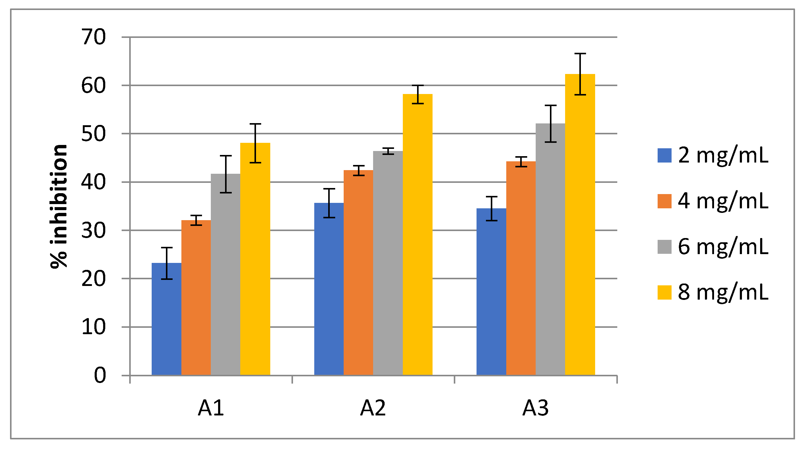
Figure 2.
Sensoric value (odour, taste and color) of juice of C. racemosa which is mixed with pineple 70:30 (A1) ,75:25 (A2) and 80% (A3).
Figure 2.
Sensoric value (odour, taste and color) of juice of C. racemosa which is mixed with pineple 70:30 (A1) ,75:25 (A2) and 80% (A3).
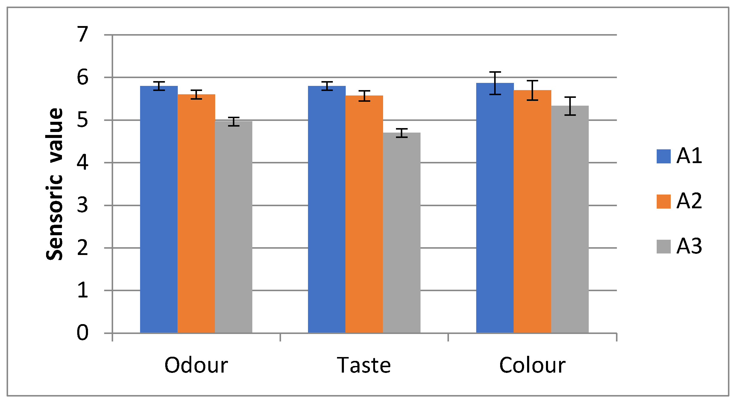
Figure 3.
GCMS Chromatogram of ethanol extract of C. racemosa.
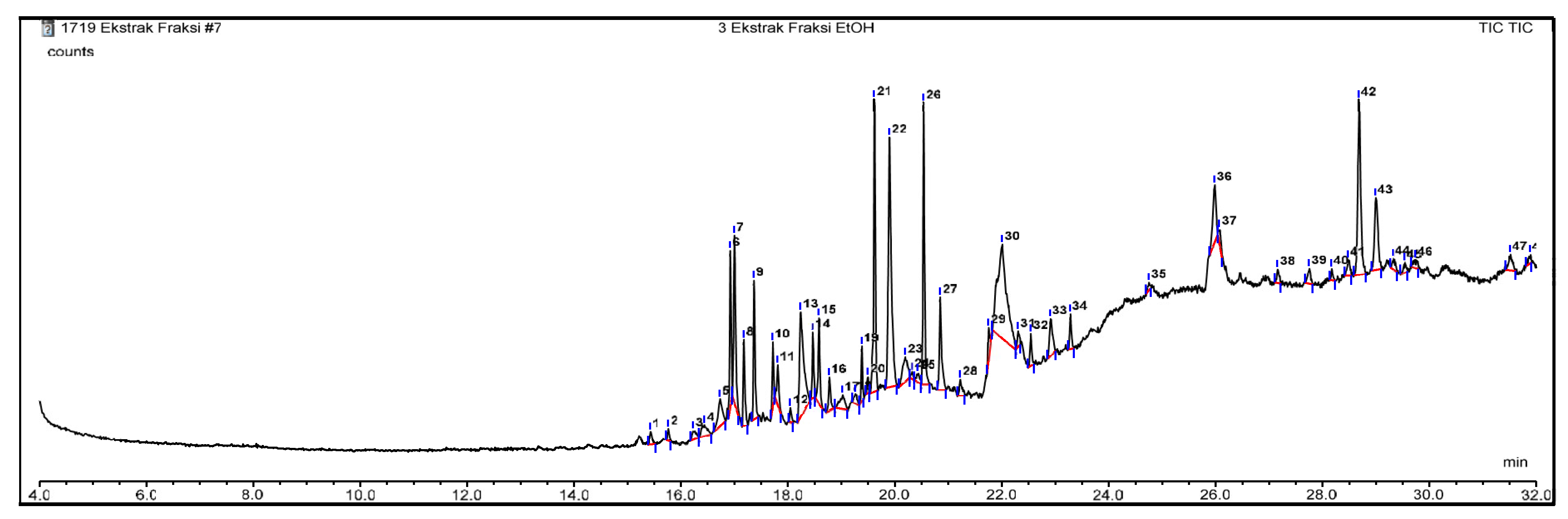
Figure 3.
Molecule structure of Fitochemical compunds of Caulerpa racemosa 1. 12-Octadecenal; 2. 13-Octadecenal; 3. 1H-Cyclopropa [3,4]benz [1,2-e]azulene-5,7b,9,9a-tetrol, 1a,1b; 4. 4a,5,7a,8,9-octahydro-1,1,6,8-tetramethyl-3-[(triphenylmethoxy)methyl]-; 5. 9,9a-triacetate 4. 1-Heptatriacotanol 5. 2-Methyl-1-hexadecanol; 6. 1H-Cyclopropa [3,4]benz [1,2-e]azulene-5,7b,9,9a-tetrol, 1a,1b,4,4a,5,7a,8,9-octahydro-1,1,6,8-tetramethyl-3-[(triphenylmethoxy)methyl]-, 5,9,9a-triacetate; 7. 2-[4-Methyl-6-(2,6,6-trimethylcyclohex-1-enyl)hexa-1,3,5-trienyl]cyclohex-1-en-1-carboxaldehyde; 8. 2-Methyl-E,E-3,13-octadecadien-1-ol; 9. 6,10,14-Trimethylpentadecan-2-one; 10. 3,7,11,15-Tetramethyl-2-hexadecen-1-ol; 11. 4H-Cyclopropa [5’,6’]benz [1’,2’7,8]azuleno [5,6-b]oxiren-4-one, 8,8a-bis(acetyloxy)-2a-[(acetyloxy)methyl]-1,1a,1b,1c,2a,3,3a,6a,6b,7,8,8a-dodecahydro-3,3a,6b-trihydroxy-1,1,5,7-tetramethyl- ;12. 5H-Cyclopropa [3,4]benz [1,2-e]azulen-5-one, 4,9,9a-tris(acetyloxy)-3-[(acetyloxy)methyl]-1,1a,1b,4,4a,7a,7b,8,9,9a-decahydro-4a,7b-dihydroxy-1,1,6,8-tetramethyl- ;13. 17-Chloro-7-heptadecene; 14. 7-Heptadecene, 1-chloro- 15. 7-Methyl-Z-tetradecen-1-ol acetate; 15. 9,10-Secocholesta-5,7,10(19)-triene-3beta,25-diol; 16.. 9,12,15-Octadecatrienoic acid, 2-phenyl-1,3-dioxan-5-yl ester; 17. 9-Desoxo-9-x-acetoxy-3,8,12-tri-O-acetylingol; 18. 4-Methylcholesta-8,24-dien-3-ol ;19. cis-10-Nonadecenoic acid 20. Cyclopropanebutanoic acid, 2-[[2-[[2-[(2-pentylcyclopropyl)methyl]cyclopropyl]methyl]cyclopropyl]methyl]-, methyl ester; 21. Cyclopropanedodecanoic acid, 2oktil metil ester; 22. Docosanoic acid, 1,2,3-propanetriyl ester; 23. (22E)-Ergosta-5,22-dien-3-ol acetate; 24. Ethanol, 2-(9-octadecenyloxy)-, (E)- 25. Ethyl iso-allocholate; 26. Furo(3’,4’:6,7)naphtho(2,3-d)-1,3-dioxol-6(5aH)-one, 27. 9-((4-fluorophenyl)amino)-5,8,8a,9-tetrahydro-5-(3,4,5-trimethoxyphenyl)-, (5R,5aR,8aS,9S)-; 28. 1,1-Bis(dodecyloxy)hexadecane; 29.. Hexadecanoic acid; 30.. Hexadecanoic acid, 31. 1-(hydroxymethyl)-1,2-ethanediyl ester; 32. i-Propyl 11,12-methylene-octadecanoate; 33. Neophytadiene; 34. Oxiraneundecanoic acid,3-pentyl-, methyl ester, cis- 35. Phytol 36. Phytol, acetate 36. Phytyl decanoate 37. Pregn-4-ene-3,20-dione, 21-(acetyloxy)-9-fluoro-11,17-dihydroxy-2-methyl-, (2beta,11beta)- 38. Rhodopin; 39. ß-Sitosterol; 39. Z-(13,14-Epoxy)tetradec-11-en-1-ol acetate; 40. Z-5-Methyl-6-heneicosen-11-one; 41. Z-7-Octadecen-1-ol acetate.
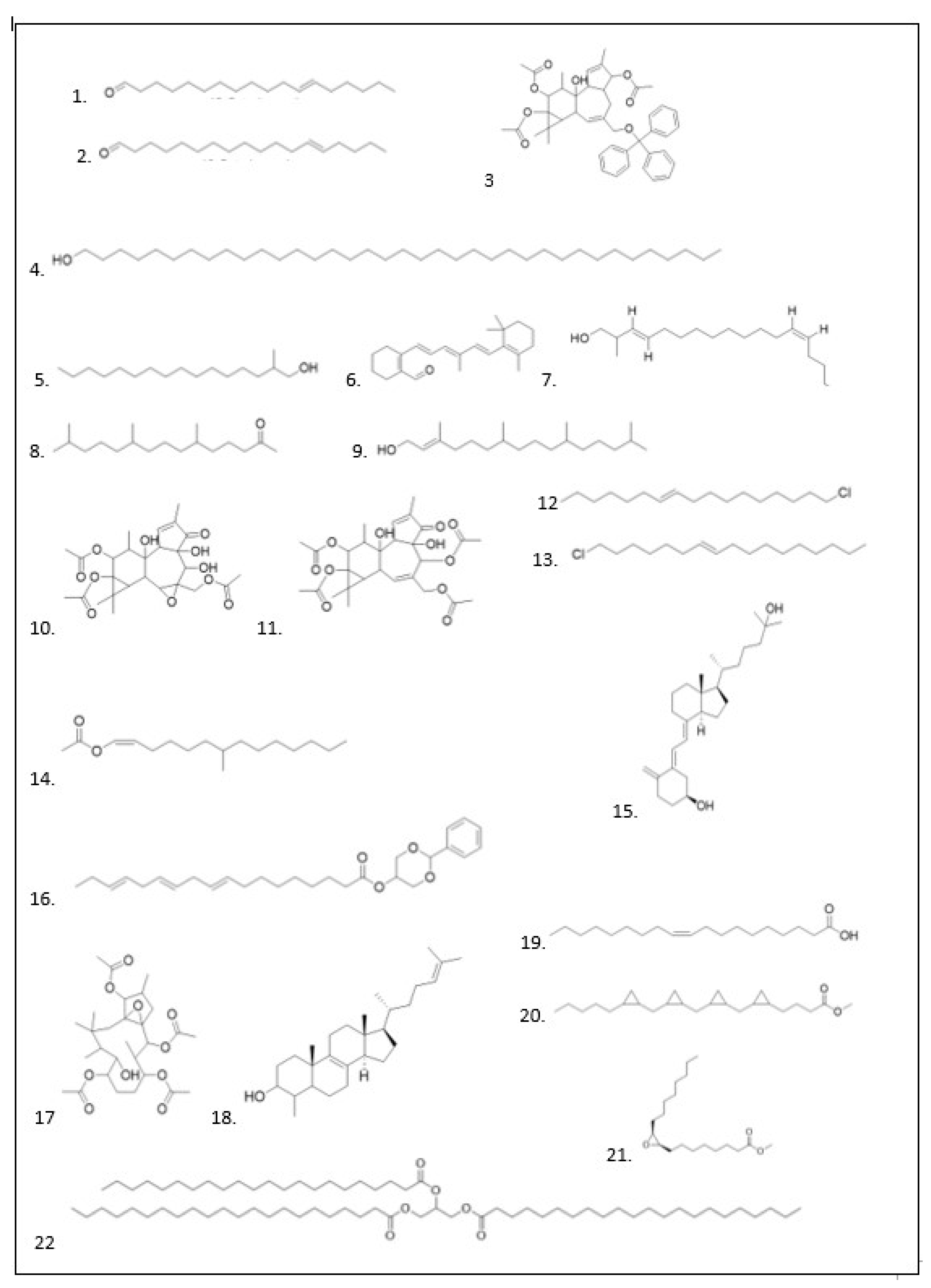
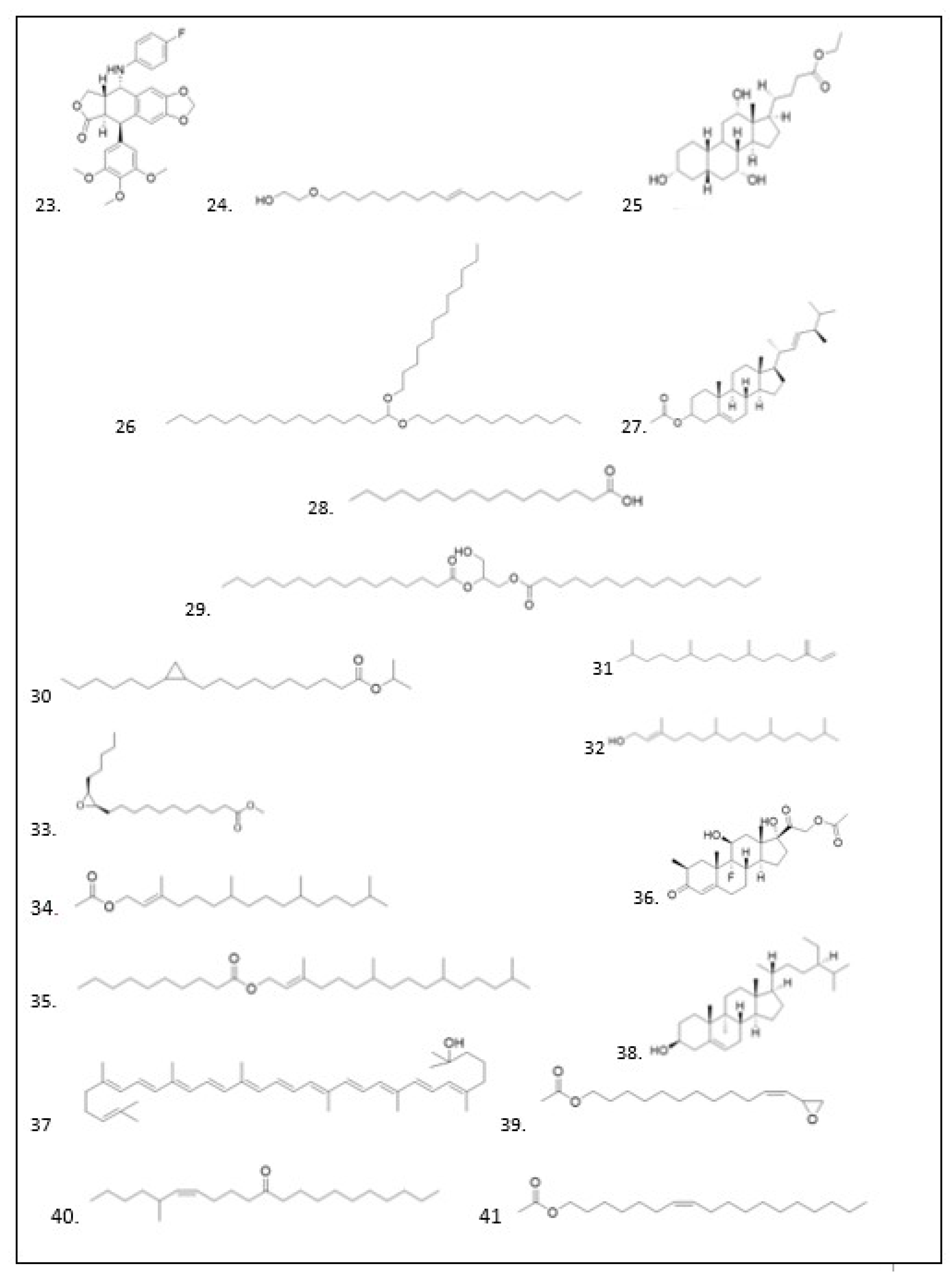
Figure 4.
Validation protokol Molecular docking of Cytochrome P450 (CP450) X S-Walfarin; Lipoxygenase (LO) X Protocatecuic acid; Myeloperoxidase (MP), X N-acethyl-D-glucosamine; NADPH Oxidase (NO) X Adenocine-5-diphosphat; Xanthine oxidase (XO) X Hypoxanthin.
Figure 4.
Validation protokol Molecular docking of Cytochrome P450 (CP450) X S-Walfarin; Lipoxygenase (LO) X Protocatecuic acid; Myeloperoxidase (MP), X N-acethyl-D-glucosamine; NADPH Oxidase (NO) X Adenocine-5-diphosphat; Xanthine oxidase (XO) X Hypoxanthin.
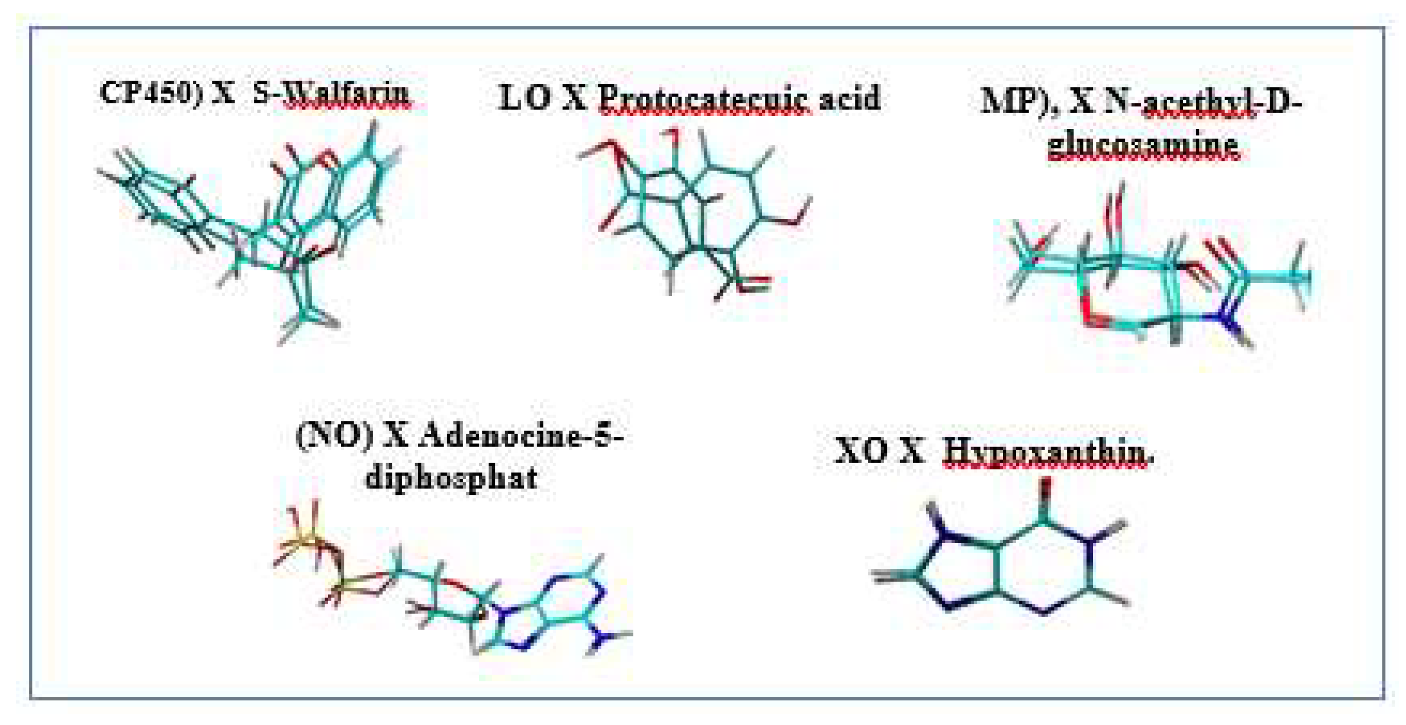
Figure 5.
Ligand control available as a commercially available drug: FLU, ZIL, MEL, DEX and FED.
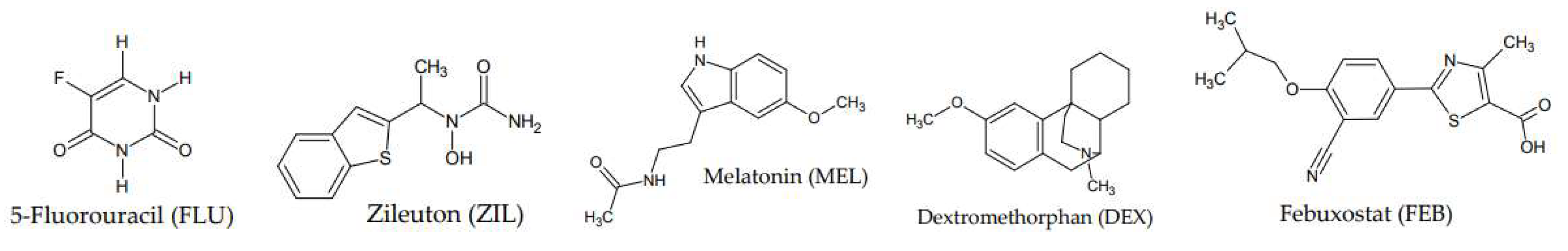
Figure 5.
Perbandingan Binding Affinity antara senyawa aktif Cyclopropanedodecanoic acid, 2-octyl-, methyl ester; Oxiraneundecanoic acid, 3-pentyl-, methyl ester, cis-; cis-10 Nonadecenoic acid dan ligand kontrol CP450, LO, MP, NO dan XO .
Figure 5.
Perbandingan Binding Affinity antara senyawa aktif Cyclopropanedodecanoic acid, 2-octyl-, methyl ester; Oxiraneundecanoic acid, 3-pentyl-, methyl ester, cis-; cis-10 Nonadecenoic acid dan ligand kontrol CP450, LO, MP, NO dan XO .
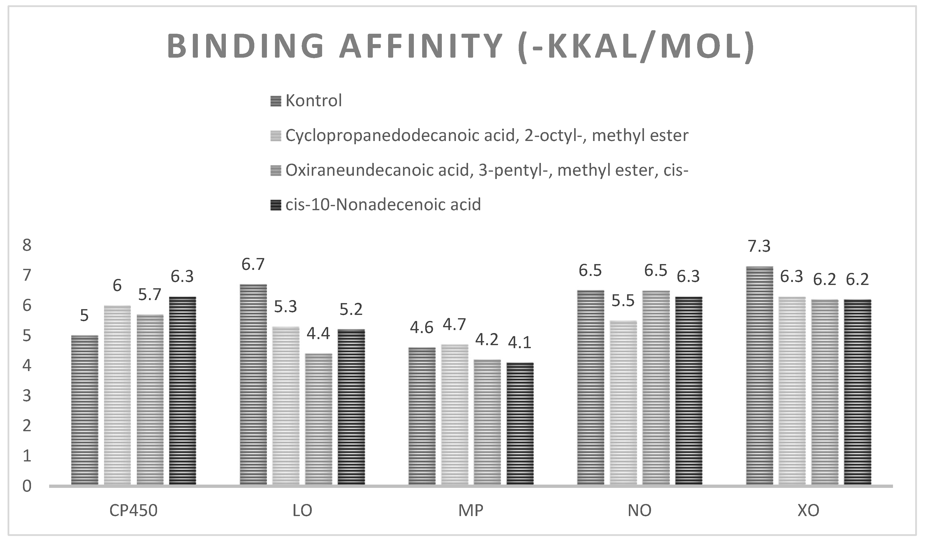
Figure 6.
Visualisasi Hasil Molekular Docking antara Ligan control Flu, Cyclopropanedodecanoic acid, 2-octyl-, methyl ester (A) ; Oxiraneundecanoic acid, 3-pentyl-, methyl ester (B), cis- dan cis-10-Nonadecenoic acid (C) secara terhadap CP450. .
Figure 6.
Visualisasi Hasil Molekular Docking antara Ligan control Flu, Cyclopropanedodecanoic acid, 2-octyl-, methyl ester (A) ; Oxiraneundecanoic acid, 3-pentyl-, methyl ester (B), cis- dan cis-10-Nonadecenoic acid (C) secara terhadap CP450. .
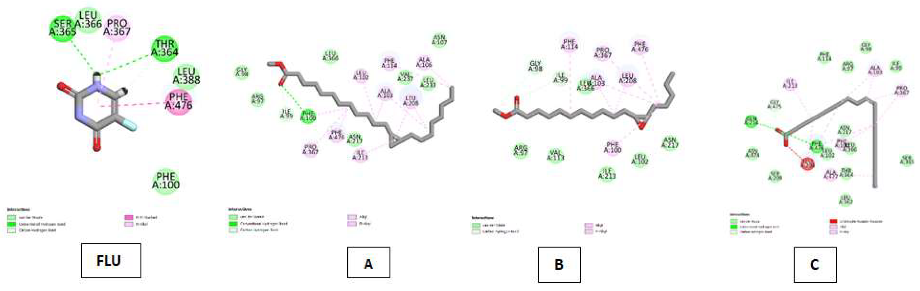
Figure 7.
Visualisasi Hasil Molekular Docking antara Ligan control ZIL , Cyclopropanedodecanoic acid, 2-octyl-, methyl ester (A) ; Oxiraneundecanoic acid, 3-pentyl-, methyl ester, cis- dan cis-10-Nonadecenoic acid (C) terhadap LO.
Figure 7.
Visualisasi Hasil Molekular Docking antara Ligan control ZIL , Cyclopropanedodecanoic acid, 2-octyl-, methyl ester (A) ; Oxiraneundecanoic acid, 3-pentyl-, methyl ester, cis- dan cis-10-Nonadecenoic acid (C) terhadap LO.
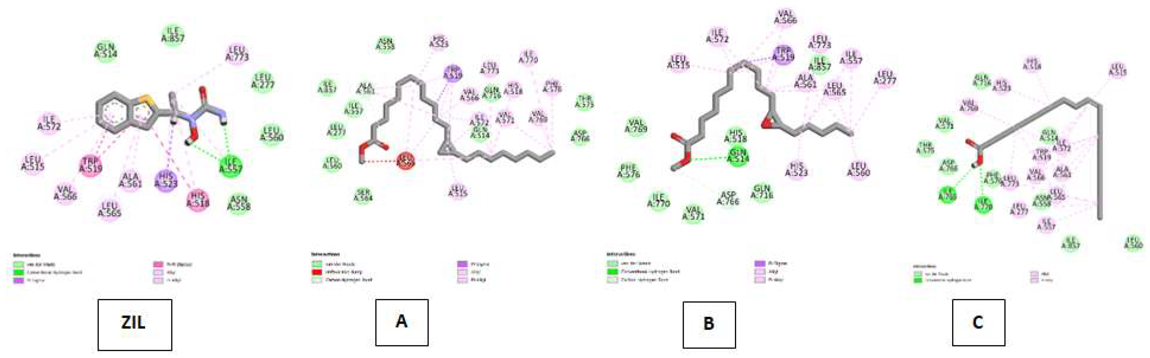
Figure 8.
Visualisasi Hasil Molekular Docking antara Ligan control MEL, Cyclopropanedodecanoic acid, 2-octyl-, methyl ester ; Oxiraneundecanoic acid, 3-pentyl-, methyl ester, cis- dan cis-10-Nonadecenoic acid (C) secara berturut-turut (kanan ke kiri) terhadap MP.
Figure 8.
Visualisasi Hasil Molekular Docking antara Ligan control MEL, Cyclopropanedodecanoic acid, 2-octyl-, methyl ester ; Oxiraneundecanoic acid, 3-pentyl-, methyl ester, cis- dan cis-10-Nonadecenoic acid (C) secara berturut-turut (kanan ke kiri) terhadap MP.
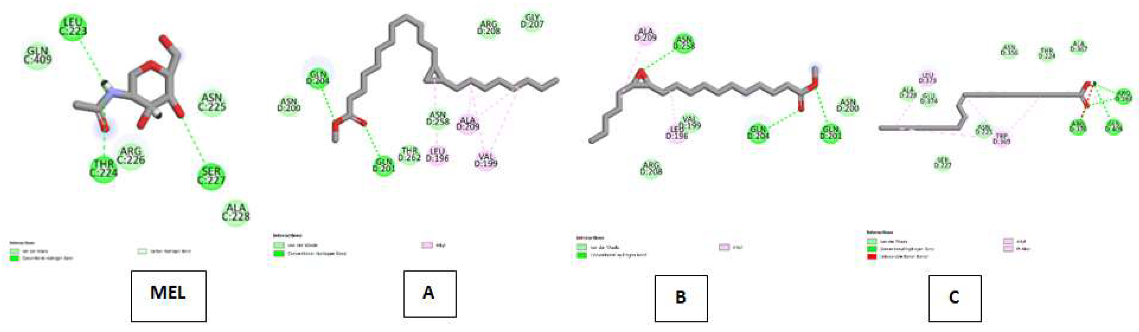
Figure 9.
Visualisasi Hasil Molekular Docking antara Ligan control FED , Cyclopropanedodecanoic acid, 2-octyl-, methyl ester ; Oxiraneundecanoic acid, 3-pentyl-, methyl ester, cis- dan cis-10-Nonadecenoic acid secara berturut-turut (kanan ke kiri) terhadap XO.
Figure 9.
Visualisasi Hasil Molekular Docking antara Ligan control FED , Cyclopropanedodecanoic acid, 2-octyl-, methyl ester ; Oxiraneundecanoic acid, 3-pentyl-, methyl ester, cis- dan cis-10-Nonadecenoic acid secara berturut-turut (kanan ke kiri) terhadap XO.
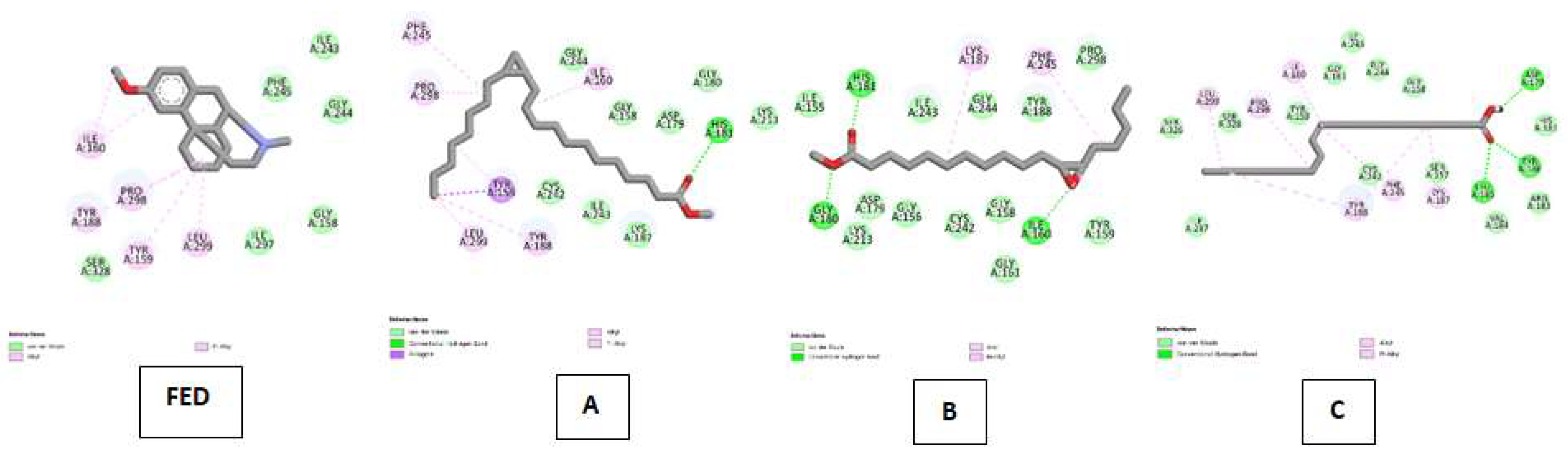
Figure 10.
Visualisasi Hasil Molekular Docking antara Ligan control DEX , Cyclopropanedodecanoic acid, 2-octyl-, methyl ester ; Oxiraneundecanoic acid, 3-pentyl-, methyl ester, cis- dan cis-10-Nonadecenoic acid terhadap NO.
Figure 10.
Visualisasi Hasil Molekular Docking antara Ligan control DEX , Cyclopropanedodecanoic acid, 2-octyl-, methyl ester ; Oxiraneundecanoic acid, 3-pentyl-, methyl ester, cis- dan cis-10-Nonadecenoic acid terhadap NO.
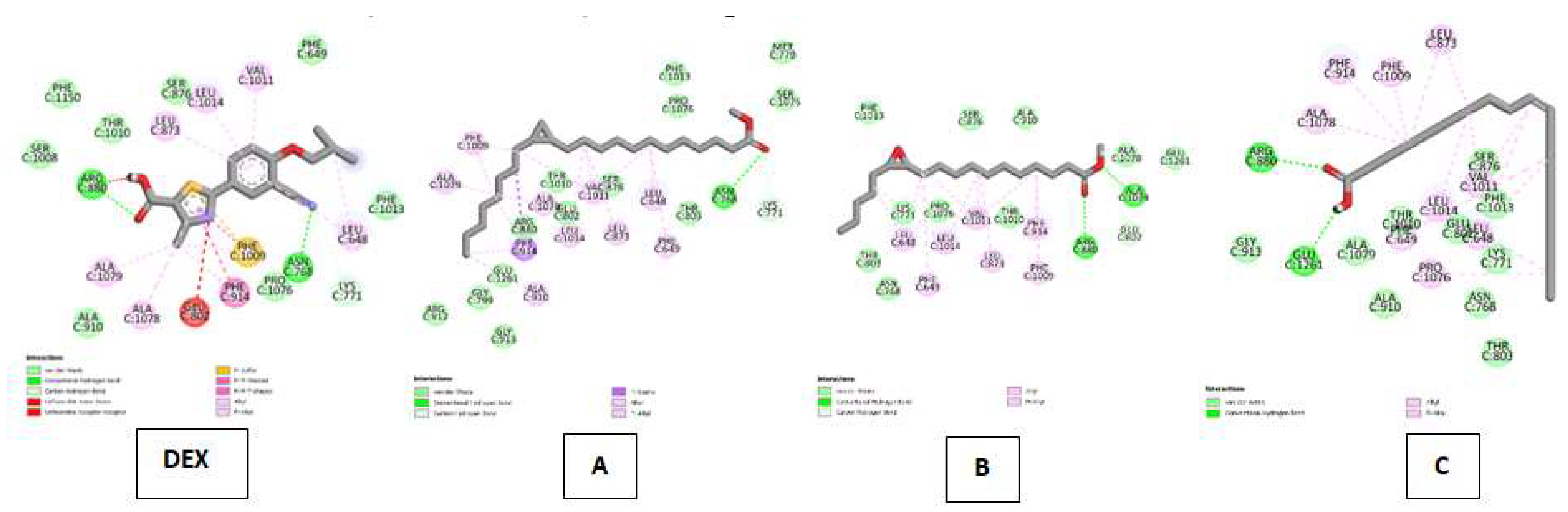
Table 1.
Total phenolic Content (TPC), fiber, Vitamin C, and pH value of juice of C. racemosa which is mixed with pineple 70:30 (A1) ,75:25 (A2), and 80:20 % (A3).
Table 1.
Total phenolic Content (TPC), fiber, Vitamin C, and pH value of juice of C. racemosa which is mixed with pineple 70:30 (A1) ,75:25 (A2), and 80:20 % (A3).
| Juice | TPC (µg GAE/g) |
Fiber (%) | Vitamine C (µg/mL) |
pH value |
|---|---|---|---|---|
| A1 | 32,1788±0,509 | 0,80±0,04 | 2,8923±0,076 | 4.47±0,379 |
| A2 | 31,7723±0,141 | 0,84±0,01 | 2,7953±0,049 | 4.57±0,115 |
| A3 | 31.8646±0,616 | 0.94±0,03 | 2.8215±0,048 | 4.5±0,172 |
Table 2.
The phytocompounds, molecule formula, retention time, % are PubChem (CID) from GCMS of ethanol extrat C. racemosa.
Table 2.
The phytocompounds, molecule formula, retention time, % are PubChem (CID) from GCMS of ethanol extrat C. racemosa.
| No | Ligan | Molecule formula | RT | Persen area |
PubChem (CID) | Compound Group |
|---|---|---|---|---|---|---|
| 1 | 12-Octadecenal | C 18H34O | 17.71 | 1.31 | 5367671 | Aldehide |
| 2 | 13-Octadecenal | C 18H34O | 17.71 | 1.31 | 5367670 | Aldehide |
| 3 | 1H-Cyclopropa [3,4]benz [1,2-e]azulene-5,7b,9,9a-tetrol, 1a,1b,4,4a,5,7a,8,9-octahydro-1,1,6,8-tetramethyl-3-[(triphenylmethoxy)methyl]-, 5,9,9a-triacetate | C28H38O9 | 19.02 | 1.09 | 596973 | Ester |
| 4 | 1-Heptatriacotanol | C37H76O | 21.22 | 0.63 | 537071 | Alcohol |
| 5 | 2-Methyl-1-hexadecanol | C17H36O | 15.42 | 0.49 | 17218 | Alcohol |
| 6 | 2-[4-Methyl-6-(2,6,6-trimethylcyclohex-1-enyl)hexa-1,3,5-trienyl]cyclohex-1-en-1-carboxaldehyde | C23H32O | 16.99 | 0.96 | 5363101 | Aldehide |
| 7 | 2-Methyl-E,E-3,13-octadecadien-1-ol | C19H36O | 16.72 | 1.65 | 5364413 | Alcohol |
| 8 | 6,10,14-Trimethylpentadecan-2-one | C18H36O | 16.99 | 4.48 | 10408 | Keton |
| 9 | 3,7,11,15-Tetramethyl-2-hexadecen-1-ol | C20H40O | 17.36 | 3.25 | 5366244 | Alcohol |
| 10 | 4H-Cyclopropa [5’,6’]benz [1’,2’7,8]azuleno [5,6-b]oxiren-4-one, 8,8a-bis(acetyloxy)-2a-[(acetyloxy)methyl]-1,1a,1b,1c,2a,3,3a,6a,6b,7,8,8a-dodecahydro-3,3a,6b-trihydroxy-1,1,5,7-tetramethyl- | C26H34O11 | 25,98 | 2.76 | 538186 | Keton |
| 11 | 5H-Cyclopropa [3,4]benz [1,2-e]azulen-5-one, 4,9,9a-tris(acetyloxy)-3-[(acetyloxy)methyl]-1,1a,1b,4,4a,7a,7b,8,9,9a-decahydro-4a,7b-dihydroxy-1,1,6,8-tetramethyl- | C28H36O11 | 25,98 | 2.76 | 538181 | Aldehide |
| 12 | 17-Chloro-7-heptadecene | C17H33Cl | 19.61 | 7.09 | 5364489 | Alkena |
| 13 | 7-Heptadecene, 1-chloro- | C17H33Cl | 19.61 | 7.09 | 5364485 | Alkena |
| 14 | 7-Methyl-Z-tetradecen-1-ol acetate | C17H32O2 | 16,72 | 1.65 | 5363222 | Ester |
| 15 | 9,10-Secocholesta-5,7,10(19)-triene-3beta,25-diol | C27H44O2 | 28.68 | 7.48 | 6506392 | Alcohol |
| 16 | 9,12,15-Octadecatrienoic acid, 2-phenyl-1,3-dioxan-5-yl ester | C28H40O4 | 16.43 | 0.96 | 5367498 | Ester |
| 17 | 9-Desoxo-9-x-acetoxy-3,8,12-tri-O-acetylingol | C28H40O10 | 24.74 | 0.27 | 537583 | Alcohol |
| 18 | 4-Methylcholesta-8,24-dien-3-ol | C28H46O | 22.90 | 1.69 | 44150703 | Alcohol |
| 19 | cis-10-Nonadecenoic acid | C19H36O2 | 18.46 | 1.39 | 5312513 | Carboxilic acid |
| 20 | Cyclopropanebutanoic acid, 2-[[2-[[2-[(2-pentylcyclopropyl)methyl]cyclopropyl]methyl]cyclopropyl]methyl]-, methyl ester | C25H42O2 | 17.81 | 1.18 | 554084 | Ester |
| 21 | Cyclopropanedodecanoic acid, 2oktil metil ester | C15H28O2 | 16.22 | 0.51 | 11447830 | Carboxilic acid |
| 22 | Docosanoic acid, 1,2,3-propanetriyl ester | C69H134O6 | 22.54 | 0.81 | 62726 | Ester |
| 23 | (22E)-Ergosta-5,22-dien-3-ol acetate | C30H48O2 | 28,99 | 3.62 | 5352877 | Ester |
| 24 | Ethanol, 2-(9-octadecenyloxy)-, (E)- | C20H40O2 | 17.17 | 1.99 | 5367702 | Ester |
| 25 | Ethyl iso-allocholate | C26H44O5 | 16.22 | 0.51 | 6452096 | Ester |
| 26 | Furo(3’,4’:6,7)naphtho(2,3-d)-1,3-dioxol-6(5aH)-one, 9-((4-fluorophenyl)amino)-5,8,8a,9-tetrahydro-5-(3,4,5-trimethoxyphenyl)-, (5R,5aR,8aS,9S)- | C28H26FNO7 | 19.25 | 0.44 | 468876 | Alkaloid |
| 27 | 1,1-Bis(dodecyloxy)hexadecane | C40H82O2 | 19.61 | 7.09 | 41920 | Alkena |
| 28 | Hexadecanoic acid | C16H32O2 | 18,23 | 6.35 | 985 | Carboxilic acid |
| 29 | Hexadecanoic acid, 1-(hydroxymethyl)-1,2-ethanediyl ester | C35H68O5 | 20.84 | 3.04 | 99931 | Ester |
| 30 | i-Propyl 11,12-methylene-octadecanoate | C22H42O2 | 18.77 | 1.02 | 91692516 | Ester |
| 31 | Neophytadiene | C20H38 | 16,91 | 3.19 | 10446 | Alkena |
| 32 | Oxiraneundecanoic acid,3-pentyl-, methyl ester, cis- | C13H24O3 | 17.81 | 1.18 | 86131368 | Carboxilic aid |
| 33 | Phytol | C20H40O | 19.89 | 10.81 | 5280435 | Alcohol |
| 34 | Phytol, acetate | C22H42O2 | 20.53 | 6.04 | 6428538 | Ester |
| 35 | Phytyl decanoate | C30H58O2 | 20.53 | 6.04 | 91711717 | Ester |
| 36 | Pregn-4-ene-3,20-dione, 21-(acetyloxy)-9-fluoro-11,17-dihydroxy-2-methyl-, (2beta,11beta)- | C24H33FO6 | 19.02 | 1.09 | 31719 | Steroid |
| 37 | Rhodopin | C40H58O | 29,32 | 0.45 | 5365880 | Terpenoid |
| 38 | ß-Sitosterol | C29H50O | 22.01 | 12,44 | 175268335 | Steroid |
| 39 | Z-(13,14-Epoxy)tetradec-11-en-1-ol acetate | C16H28O3 | 19.89 | 10.81 | 5363633 | Ester |
| 40 | Z-5-Methyl-6-heneicosen-11-one | C22H42O | 17.71 | 1.31 | 5363254 | Keton |
| 41 | Z-7-Octadecen-1-ol acetate | C20H38O2 | 16.99 | 4.48 | 5363553 | Ester |
Table 3.
Profil Farmakokinetik dari Ligan Target menggunakan parameter Lipinski’s Rule of Five.
| No | Ligan | Berat Molekul <500 | H-Donor <5 | H-Akseptor <10 | Log P <5 | Refraksi Molar 40-130 | Penyimpangan |
|---|---|---|---|---|---|---|---|
| 1 | 12-Octadecenal | 266.000000 | 0 | 1 | 6.222801 | 85.515968 | 1 |
| 2 | 13-Octadecenal | 266.000000 | 0 | 1 | 6.222801 | 85.515968 | 1 |
| 3 | 1H-Cyclopropa [3,4]benz [1,2-e]azulene-5,7b,9,9a-tetrol, 1a,1b,4,4a,5,7a,8,9-octahydro-1,1,6,8-tetramethyl-3-[(triphenylmethoxy)methyl]-, 5,9,9a-triacetate | 312.000000 | 5 | 6 | -0.053101 | 77.145782 | 1 |
| 4 | 1-Heptatriacotanol | 312.000000 | 5 | 6 | -0.053101 | 77.145782 | 1 |
| 5 | 2-Methyl-1-hexadecanol | 256.000000 | 1 | 1 | 5.706000 | 81.944763 | 1 |
| 6 | 2-[4-Methyl-6-(2,6,6-trimethylcyclohex-1-enyl)hexa-1,3,5-trienyl]cyclohex-1-en-1-carboxaldehyde | 81.944763 | 0 | 1 | 6.641202 | 103.926971 | 1 |
| 7 | 2-Methyl-E,E-3,13-octadecadien-1-ol | 280.000000 | 1 | 1 | 6.038201 | 90.990761 | 1 |
| 8 | 6,10,14-Trimethylpentadecan-2-one | 268.000000 | 0 | 1 | 6.014501 | 85.399971 | 1 |
| 9 | 3,7,11,15-Tetramethyl-2-hexadecen-1-ol | 296.000000 | 1 | 1 | 6.364101 | 95.561760 | 1 |
| 10 | 4H-Cyclopropa [5’,6’]benz [1’,2’7,8]azuleno [5,6-b]oxiren-4-one, 8,8a-bis(acetyloxy)-2a-[(acetyloxy)methyl]-1,1a,1b,1c,2a,3,3a,6a,6b,7,8,8a-dodecahydro-3,3a,6b-trihydroxy-1,1,5,7-tetramethyl- | 522.000000 | 0 | 11 | 4.684239 | 129.402466 | 2 |
| 11 | 5H-Cyclopropa [3,4]benz [1,2-e]azulen-5-one, 4,9,9a-tris(acetyloxy)-3-[(acetyloxy)methyl]-1,1a,1b,4,4a,7a,7b,8,9,9a-decahydro-4a,7b-dihydroxy-1,1,6,8-tetramethyl- | 548.000000 | 0 | 11 | 5.092820 | 138.002487 | 2 |
| 12 | 17-Chloro-7-heptadecene | 272.500000 | 0 | 0 | 6.872602 | 85.554970 | 1 |
| 13 | 7-Heptadecene, 1-chloro- | 272.500000 | 0 | 0 | 6.872602 | 85.554970 | 1 |
| 14 | 7-Methyl-Z-tetradecen-1-ol acetate | 268.000000 | 0 | 2 | 5.272600 | 82.163971 | 1 |
| 15 | 9,10-Secocholesta-5,7,10(19)-triene-3beta,25-diol | 400.000000 | 2 | 2 | 6.733902 | 122.648552 | 1 |
| 16 | 9,12,15-Octadecatrienoic acid, 2-phenyl-1,3-dioxan-5-yl ester | 440.000000 | 0 | 4 | 6.540953 | 135.386978 | 1 |
| 17 | 9-Desoxo-9-x-acetoxy-3,8,12-tri-O-acetylingol | 536.000000 | 0 | 10 | 5.651121 | 141.818985 | 2 |
| 18 | 4-Methylcholesta-8,24-dien-3-ol | 398.000000 | 1 | 1 | 7.698903 | 123.645737 | 1 |
| 19 | cis-10-Nonadecenoic acid | 296.000000 | 1 | 2 | 5.039920 | 102.327286 | 0 |
| 20 | Cyclopropanebutanoic acid, 2-[[2-[[2-[(2-pentylcyclopropyl)methyl]cyclopropyl]methyl]cyclopropyl]methyl]-, methyl ester | 374.000000 | 0 | 2 | 6.222682 | 127.046967 | 1 |
| 21 | Cyclopropanedodecanoic acid, 2oktil metil ester | 240.000000 | 1 | 2 | 4.772099 | 71.146782 | 0 |
| 22 | Docosanoic acid, 1,2,3-propanetriyl ester | 312.000000 | 5 | 6 | -0.053101 | 77.145782 | 0 |
| 23 | (22E)-Ergosta-5,22-dien-3-yl acetate | 440.000000 | 0 | 2 | 7.507115 | 150.931992 | 1 |
| 24 | Ethanol, 2-(9-octadecenyloxy)-, (E)- | 312.000000 | 1 | 2 | 5.749202 | 110.685280 | 1 |
| 25 | Ethyl iso-allocholate | 436.000000 | 3 | 5 | 3.927198 | 119.034340 | 0 |
| 26 | Furo(3’,4’:6,7)naphtho(2,3-d)-1,3-dioxol-6(5aH)-one, 9-((4-fluorophenyl)amino)-5,8,8a,9-tetrahydro-5-(3,4,5-trimethoxyphenyl)-, (5R,5aR,8aS,9S)- | 507.000000 | 1 | 8 | 4.435399 | 130.275681 | 1 |
| 27 | 1,1-Bis(dodecyloxy)hexadecane | 312.000000 | 5 | 6 | -0.053101 | 77.145782 | 1 |
| 28 | Hexadecanoic acid | 312.000000 | 5 | 6 | -0.053101 | 77.145782 | 1 |
| 29 | Hexadecanoic acid, 1-(hydroxymethyl)-1,2-ethanediyl ester | 256.000000 | 1 | 2 | 5.552299 | 77.947777 | 1 |
| 30 | i-Propyl 11,12-methylene-octadecanoate | 338.000000 | 0 | 2 | 6.129292 | 118.590973 | 1 |
| 31 | Neophytadiene | 278.000000 | 0 | 0 | 7.167703 | 94.055969 | 1 |
| 32 | Oxiraneundecanoic acid, 3-pentyl-, methyl ester, cis- | 228.000000 | 1 | 3 | 3.370800 | 63.545784 | 0 |
| 33 | Phytol | 296.000000 | 1 | 1 | 6.364101 | 95.561760 | 0 |
| 34 | Phytol, acetate | 338.000000 | 0 | 2 | 6.934901 | 105.108963 | 1 |
| 35 | Phytyl decanoate | 450.000000 | 0 | 2 | 8.799607 | 162.773972 | 1 |
| 36 | Pregn-4-ene-3,20-dione, 21-(acetyloxy)-9-fluoro-11,17-dihydroxy-2-methyl-, (2beta,11beta)- | 436.000000 | 2 | 6 | 2.690500 | 109.585564 | 0 |
| 37 | Rhodopin | 554.000000 | 1 | 1 | 9.998867 | 189.588913 | 2 |
| 38 | ß-Sitosterol | 368.000000 | 0 | 1 | 1.510640 | 93.379990 | 0 |
| 39 | Z-(13,14-Epoxy)tetradec-11-en-1-ol acetate | 565.000000 | 2 | 10 | 1.031598 | 152.134506 | 1 |
| 40 | Z-5-Methyl-6-heneicosen-11-one | 322.000000 | 0 | 1 | 6.447083 | 118.461472 | 1 |
| 41 | Z-7-Octadecen-1-ol acetate | 310.000000 | 0 | 2 | 5.705202 | 108.268982 | 1 |
Table 4.
Data obtained in the validation of the molecular docking protocols for the receptors cytochrome P450 (CP450), lypoxygenase (LO), myeloperoxidase (MP), NADPH oxidase (NO) and xanthine oxidase (XO).
Table 4.
Data obtained in the validation of the molecular docking protocols for the receptors cytochrome P450 (CP450), lypoxygenase (LO), myeloperoxidase (MP), NADPH oxidase (NO) and xanthine oxidase (XO).
| No | Molecular docking | Resolution | ∆G (Kkal/mol) |
RMSD |
|---|---|---|---|---|
| 1 | Cytochrome P450 (CP40) X S walfarin |
2.55 Å |
-10.4260 |
0.6311 Å |
| 2 | Lipoxygenase (LO) X Protocatecuic acid |
2.10 Å |
-6.6490 | 1.9756 Å |
| 3 | Myeloperoxidase (MP) X N-acethyl-D-glucosamine |
1.85 Å |
-4.2060 |
0.7344 Å |
| 4 | NADPH Oxidase (NO) X Adenocine-5-diphosphat |
1.80 Å |
:-8.8380 | 0.9495 Å |
| 5 | Xanthine oxidase (XO) X Hypoxanthin. |
1.80 Å |
- 5.9700 | 0.1940 Å |
Table 7.
ADME properties cyclopropanedodecanoic acid, 2-octyl-, methyl ester ; oxiraneundecanoic acid, 3-pentyl-, methyl ester, dan cis-10-Nonadecenoic acid.
Table 7.
ADME properties cyclopropanedodecanoic acid, 2-octyl-, methyl ester ; oxiraneundecanoic acid, 3-pentyl-, methyl ester, dan cis-10-Nonadecenoic acid.
| No. | Compound | HIA | BBB | Plasma protein Binding | CP450 | p-Gp | Log Kp | BiA Score |
|---|---|---|---|---|---|---|---|---|
| 1. | Cyclopropanedodecanoic acid, 2-octyl-, methyl ester | 100.000000 | 17.5431 | 100.000000 | inhibitor | Inhibitor | -1,25 | 0,55 |
| 2 | Oxiraneundecanoic acid, 3-pentyl-, methyl ester, cis- | 98.555395 | 6.62728 | 100.000000 | Inhibitor | No | -3,62 | 0,55 |
| 3 | cis-10-Nonadecenoic acid | 98.386939 | 9.69018 | 100.000000 | Inhibitor | Inhibitor | 0,525741 | 0,85 |
Note: HIA (human gastrointestinal absorption); blood-brain barrier (BBB) permeation; P-gp (permeability glycoprotein), cytochrome P450 (CYP), i: inhibition, Log Kp (skin permeability coefficient), BiA Score (Bioavailability Score).
Disclaimer/Publisher’s Note: The statements, opinions and data contained in all publications are solely those of the individual author(s) and contributor(s) and not of MDPI and/or the editor(s). MDPI and/or the editor(s) disclaim responsibility for any injury to people or property resulting from any ideas, methods, instructions or products referred to in the content. |
© 2025 by the authors. Licensee MDPI, Basel, Switzerland. This article is an open access article distributed under the terms and conditions of the Creative Commons Attribution (CC BY) license (http://creativecommons.org/licenses/by/4.0/).
Copyright: This open access article is published under a Creative Commons CC BY 4.0 license, which permit the free download, distribution, and reuse, provided that the author and preprint are cited in any reuse.


