Preprint
Article

This version is not peer-reviewed.

Phytochemical Profile, Antioxidant and Antiproliferative Activity of Randia spp Fruit Extracts Obtained by Ultrasound-Assisted Extraction

A peer-reviewed article of this preprint also exists.

Submitted:

01 September 2025

Posted:

09 September 2025

You are already at the latest version

Abstract

Randia spp. is a medicinal plant traditionally used in the treatment of various diseases. In this study, the phytochemical composition and the antioxidant, antiproliferative, and cytotoxic activities of hydroalcoholic extracts from fresh and dried Randia spp. fruits were evaluated. The phytochemical profile was determined through qualitative assays and high-performance liquid chromatography (HPLC). Antioxidant activity was assessed using the 2,2-diphenyl-1-picrylhydrazyl (DPPH) and 2,2-azinobis-(3-ethylbenzothiazoline-6-sulfonic acid) (ABTS) assays. The antiproliferative effect was tested against CaCo-2 cells (human colon adenocarcinoma), while cytotoxicity was evaluated using J774.2 murine macrophages, and the selectivity index (SI) was calculated. The fresh and dried fruit extracts contained 50.27 and 47.22 mg QE/g extract of total phenols (TPC), and 27.08 and 35.53 mg QE/g extract of total flavonoids (TFC), respectively. In fresh fruit extracts, four phenolic acids (caffeic, hydroxybenzoic, ferulic, and coumaric) and one flavonoid (kaempferol) were identified, while dried fruit extracts contained ferulic acid, vanillic acid, and kaempferol. Kaempferol was the predominant compound in both extracts (137.55 and 42.10 mg/g dry sample in fresh and dried fruits, respectively). Both extracts displayed antioxidant activity, with IC₅₀ values of 18.29 mg/mL (DPPH) and 8.70 mg/mL (ABTS). Among the tested samples, the dried fruit extract demonstrated the highest antiproliferative activity. Furthermore, the extract showed moderate antiproliferative effects against CaCo-2 cells (IC₅₀ 25.44 ± 0.16 µg/mL) and low cytotoxicity toward J774.2 cells (CC₅₀ > 100 µg/mL), resulting in an SI = 3.92. Overall, the antioxidant and antiproliferative activities can be attributed mainly to kaempferol, given its high abundance in both extracts. The favorable selectivity index suggests that hydroalcoholic extracts of Randia spp. are safe and effective, highlighting their potential as candidates for further preclinical and clinical evaluations.

Keywords: 
;  ;  ;  

1. Introduction

Under normal physiological conditions, the human body continuously generates free radicals, which play essential roles in regulating key cellular processes, including tumor cell apoptosis, immune cell activation, and cell differentiation [1]. The balance of these reactive species is tightly controlled by endogenous antioxidant enzymes that protect cells from excessive oxidative damage. However, when the production of free radicals exceeds the capacity of these defense systems, oxidative stress occurs, contributing to the onset and progression of numerous chronic diseases, such as inflammation, cardiovascular disorders, neurodegenerative conditions, and cancer [2].
Inflammation, while being one of the critical stages of the wound-healing process and a vital component of the innate immune response to tissue injury, can become harmful when dysregulated or chronic [3]. Similarly, despite advances in modern medicine, cancer remains one of the leading causes of mortality worldwide, highlighting the need for novel, safer, and more effective therapeutic strategies [4].
Since antiquity, medicinal plants have served as primary remedies for the treatment of diverse ailments. Even today, plant-derived compounds continue to represent an invaluable source of bioactive molecules due to their safety, accessibility, and therapeutic efficacy [5]. In particular, growing attention has been given to natural antioxidants capable of mitigating oxidative stress and modulating inflammatory responses. Compounds such as polyphenols, saponins, flavonoids, and vitamins have been extensively investigated for their pharmacological properties, including antioxidant, anti-inflammatory, and anticancer activities [6].
In this context, the exploration of medicinal plants and their secondary metabolites provides an important avenue for the discovery of novel therapeutic agents. By elucidating their phytochemical composition and biological activities, it becomes possible to identify potential candidates for the prevention or complementary treatment of oxidative stress-related diseases, including cancer and chronic inflammation.
Randia is a neotropical genus comprising approximately 60 to 70 species, distributed from the southern United States to South America. Mexico represents the center of greatest diversity, harboring 33 recognized species [7]. Commonly known as “crucetillo”, this genus is characterized by the presence of cross-shaped thorns along the stems of its trees [8]. The fruits are traditionally used in local medicine, often prepared as an alcoholic extract mixed with brandy, to counteract the effects of snake and insect envenomation (e.g., snakes, spiders, scorpions, toads, bees, wasps). Additionally, they are employed in the treatment of cancer, diabetes, inflammation, and pain [9].
Several species within the genus have demonstrated notable pharmacological properties. For example, Randia aculeata has shown partial inhibition of necrosis in skeletal and myocardial muscles following exposure to snake venom, thereby offering protection against venom-induced tissue damage. Furthermore, hydroalcoholic extracts of its fruits have been reported to exert analgesic activity at the visceral level [9,10]. These findings highlight the therapeutic potential of Randia species and support their relevance as a source of bioactive compounds for further pharmacological and biomedical research.
Despite its traditional importance, most species of the Randia genus remain poorly characterized from a phytochemical perspective. Existing qualitative studies are scarce and incomplete, with reports available for only eight species, in which flavonoids and tannins have been the most frequently detected compounds. Moreover, the isolation and identification of specific metabolites have been achieved in just six species [11]. Among them, Randia spp., distributed in the northern region of Oaxaca, Mexico, is particularly noteworthy due to its long-standing traditional use in the treatment of poisonous animal bites and in the management of chronic conditions such as diabetes and cancer. This ethnopharmacological relevance underscores the urgent need to advance research on its chemical composition and therapeutic potential.
The present study addresses this gap by conducting a detailed investigation of hydroalcoholic extracts obtained from fresh and dried fruits of Randia spp. Specifically, the phytochemical profile was characterized, and the antioxidant, antiproliferative, and cytotoxic activities were systematically evaluated. By integrating both chemical and biological assessments, this research contributes to expanding the current knowledge of Randia spp., while also highlighting its potential as a promising source of bioactive compounds with applications in the pharmaceutical and functional food industries.

2. Materials and Methods

2.1. Sample Collection and Preparation

A total of 100 fruits of Randia spp. (5.7 kg) were collected in October 2023 from the city of Tuxtepec, Oaxaca, Mexico (18°01′21″ N, 96°12′10″ W; 35 m a.s.l.). The fruits were thoroughly washed to remove soil residues, and any samples showing signs of fungal contamination or physical damage (e.g., bruising) were discarded. The selected fruits were ground using a knife mill (model HC-2000Y) until a homogeneous paste was obtained. A portion of this paste was subjected to drying in a batch refractive window dryer at a constant temperature of 55 °C for 2.5 h. The dried material was subsequently re-ground in the same knife mill to obtain a fine powder with a particle size of 0.420 mm (mesh No. 40).

2.2. Extraction Procedures and Traditional Wine Preparation Techniques.

Fresh fruit paste and dried fruit powder of Randia spp. were subjected to ultrasound-assisted extraction. The process was performed in an ultrasonic bath (Elmasonic P, D-78224, Singen/Htw., Germany) operating at a frequency of 80 kHz and 100% power for 30 min. A hydroalcoholic mixture of 70% ethanol in water was used as the extraction solvent, at a plant material-to-solvent ratio of 1:10 (g/mL). After extraction, the solvent was filtered through Whatman No. 1 filter paper, and the filtrate was concentrated under reduced pressure using a Rotavapor® R-3 (Büchi Labortechnik, Flawil, Switzerland) at 40 °C and 60 rpm. The resulting extracts were stored at 4 °C until further analysis [13].
The traditional beverage was formulated by combining 750 mL of sherry wine (La Lupe Mexican brand) with 250 mL of brandy, both acquired from the local market. Subsequently, eight ground fruits (40 g) were incorporated into the mixture to obtain the final preparation.

2.3. Phytochemical Profile

2.3.1. Qualitative Identification of Families

An aliquot of 50 mg of each extract was weighed and diluted in 2.5 mL of a 70% ethanol–water solution. The resulting mixture was clarified by filtration through a Pasteur pipette packed with diatomaceous earth and activated carbon, after which it was used for subsequent determinations. Phytochemical screening of the clarified extracts was carried out using standard qualitative tests: alkaloids were detected with Dragendorff’s reagent, saponins by their ability to produce stable foam, phenolic compounds with the Folin–Ciocalteu reagent, flavonoids by colorimetric reactions with NaOH and HCl, and sterols and triterpenes with the Liebermann–Burchard reagent [14].

2.3.2. Total Polyphenol Content

The total polyphenol content (TPC) was determined following the procedure described by Singleton and Rossi [15], using gallic acid as the reference standard. Results were expressed as milligrams of gallic acid equivalents per gram of extract (mg GAE/g). For the assay, 600 µL of deionized water, 10 µL of sample solution (2.5 mg extract/mL H₂O), and 50 µL of Folin–Ciocalteu reagent were added sequentially to Eppendorf tubes. After mixing, 150 µL of 20% (w/v) sodium carbonate solution was added, and the reaction mixtures were incubated at 27 °C for 2 h. The absorbance of each sample was then measured at 760 nm using a UV–Vis spectrophotometer.

2.3.2. Total Flavonoid Content

The total flavonoid content (TFC) was determined following the methodology described by Zhishen et al. [16]. Briefly, 1,250 µL of deionized water was mixed with the sample solution (2.5 mg extract/mL H₂O), followed by the addition of 75 µL of 5% NaNO₂ solution. After incubation at room temperature for 6 min, 150 µL of 10% AlCl₃ solution was added, and the mixture was allowed to stand for an additional 5 min. Subsequently, 500 µL of 1 M NaOH was added, and the final mixture was thoroughly mixed. Absorbance was recorded at 510 nm using a UV–Vis spectrophotometer. Quercetin was used as the reference standard, and results were expressed as milligrams of quercetin equivalents per gram of extract (mg QE/g).

2.4. Identification of Phenols by HPLC

Phenolic acids were identified using a Perkin-Elmer Flexar high-performance liquid chromatography (HPLC) system equipped with a quaternary pump and UV detector, operated with Chromera software (version 4.1.16396).
The procedure followed the method described by Méndez-Lagunas et al. [17]. Separation was achieved on a Zorbax Bonus-RP column (4.6 × 150 mm i.d.; Agilent Technologies) coupled to a UV detector (Perkin-Elmer Flexar). The mobile phase consisted of two solvents with different polarities: 0.085% orthophosphoric acid in water and acetonitrile. Identification of phenolic acids was carried out by comparison with authentic standards, including ferulic, p-coumaric, syringic, caffeic, chlorogenic, 4-hydroxybenzoic, and vanillic acids. Quantification was performed using calibration curves prepared from the corresponding standards at a detection wavelength of 280 nm, and peak areas were used for calculations. Results were expressed as milligrams per 100 g of dry solids.

2.5. Antioxidant Activity

The antioxidant activity of the extracts was evaluated based on their ability to inhibit DPPH and ABTS radicals. The percentage of DPPH inhibition was determined following the method of Brand-Williams et al. [18]. A stock solution of DPPH was prepared by dissolving 2.4 mg of the radical in methanol and adjusting the volume to 100 mL. For the assay, 975 μL of the DPPH solution was mixed with 25 μL of the sample at concentrations of 10,000, 5,000, 2,500, 1,000, and 500 ppm in Eppendorf tubes. The mixtures were incubated under dark conditions at room temperature for 30 min prior to absorbance measurement at 515 nm.
The ABTS radical inhibition assay was performed according to the procedure described by Re et al. [19]. ABTS was prepared at a concentration of 7 mM in water, and the ABTS radical cation was generated by reacting the stock solution with 2.45 mM potassium persulfate (final concentration). The mixture was kept in the dark at room temperature for 12–16 h before use. The working ABTS solution was then diluted with ethanol to an absorbance of 0.70 ± 0.05 at 732 nm. The ABTS radical solution was added to the samples at a 1:10 (v/v) ratio. Samples at concentrations of 10,000, 5,000, 2,500, 1,000, 500, 250, 50, and 25 ppm were evaluated, and the results were expressed as percentage inhibition.

2.6. Antiproliferative Activity and Cytotoxicity

The cytotoxicity assay was performed using mouse macrophage cells J774.2 (ATCC® TIB-67, Manassas, VA, USA). Cells were cultured in RPMI 1640 medium supplemented with 10% FBS, 100 U/mL penicillin, 100 µg/mL streptomycin, and 2 mM glutamine, and maintained at 37 °C in a humidified atmosphere containing 5% CO₂. A defined cell suspension (1 × 10⁵ cells/mL) was incubated with fresh and dried fruit extracts of Randia spp. at concentrations ranging from 0.78 to 200 µg/mL for 48 h under the same conditions. Wells containing cells treated with 0.2% DMSO served as negative controls, while cisplatin was used as a positive control. Cell metabolic activity was assessed by the MTT assay, and cell viability (%) was calculated. The 50% cytotoxic concentration (CC₅₀) was determined by probit analysis. All assays were performed in triplicate across three independent experiments [20].
The antiproliferative activity was evaluated using human colorectal adenocarcinoma Caco-2 cells. Cells were cultured in high-glucose DMEM supplemented with 20% FBS and 1% antibiotic–antimycotic solution, maintained at 37 °C in a humidified atmosphere with 5% CO₂. The culture medium was replaced every 2–3 days, depending on cell confluence. For the assay, 15,000 cells per well were seeded in 96-well plates and incubated for 24 h. Cells were then treated with fresh and dried fruit extracts of Randia spp. at concentrations ranging from 0.78 to 200 µg/mL, washed with PBS, and incubated with fresh, treatment-free medium for an additional 22 h. Cisplatin was used as a positive control.
Cell viability was assessed using the MTT assay. After treatments, culture supernatants were removed, wells were washed with PBS, and 100 µL of serum-free medium containing 0.5 mg/mL MTT was added. Plates were incubated at 37 °C for 2 h, after which the resulting formazan crystals were dissolved in isopropyl alcohol. Absorbance was measured at 540 nm using a microplate reader [21]. Experiments were performed in triplicate across three independent assays, and results are expressed as mean ± standard error. The half-maximal inhibitory concentration (IC₅₀) was determined by probit analysis.
The selectivity index (SI) of the fresh and dried fruit extracts was calculated as the ratio between CC₅₀ and IC₅₀ (SI = CC₅₀ / IC₅₀) [20].

2.7. Statistical Analysis

All results are presented as mean ± standard deviation (SD). Statistical analysis was performed using one-way analysis of variance (ANOVA), followed by Tukey’s test to assess significant differences between means at a 95% confidence level (p < 0.05). Analyses were conducted using Statistica software, version 10.0 (StatSoft Inc., 1984–2008, USA).

3. Results

3.1. Phytochemical Profile

3.1.1. Qualitative Family Identification

The colorimetric assays revealed the presence (+) or absence (–) of secondary metabolites, as summarized in Table 1. Extracts from the fresh and dried fruit, as well as from the traditional beverage, were positive for saponins, phenols, and flavonoids, compounds that are widely recognized for their potential contribution to antioxidant activity.
In the extracts of fresh and dried fruit, phenols and saponins were identified as the predominant metabolite families, whereas lower levels of flavonoids were detected compared to those found in the traditional drink. In contrast, alkaloids, steroids, and triterpenes were not detected in any of the evaluated extracts.

3.1.2. Total Polyphenol and Flavonoid Content

The total polyphenol content of the fruit extracts is presented in Table 1, with values of 47.22 ± 0.96 mg GAE/g for the fresh fruit extract and 50.27 ± 0.14 mg GAE/g for the dried fruit extract. No significant differences were observed between the fresh and dried samples, indicating that drying using a refractive window did not notably affect the polyphenol content.
Significant differences in total flavonoid content were observed among the extracts, with the dried fruit extract showing the highest value (35.53 ± 2.20 mg QE/g, Table 2). The increase is likely due to water removal during drying, which concentrates the compounds and disrupts cellular vacuoles, facilitating flavonoid extraction [22].

3.1.3. Identification of Phenols by HPLC

The phenolic profiles of Randia spp. fruit extracts, as determined by HPLC, are presented in Table 3. In the fresh fruit extract, four phenolic acids (caffeic, hydroxybenzoic, ferulic, and coumaric acids) and one flavonoid (kaempferol) were identified and quantified. In contrast, the dried fruit extract contained only two phenolic acids (ferulic and vanillic acids) along with kaempferol (Table 3), suggesting that several phenolic compounds were degraded during the drying process due to thermal effects [23].
The detection of vanillic acid in the dried fruit extract is noteworthy, as it was not observed in the fresh fruit. This may be attributed to its formation as an intermediate from ferulic acid (present in the fresh fruit) during vanillin biosynthesis, reflecting the structural diversity of phenolic compounds arising from oxidative coupling reactions [24]. No phenolic compounds were detected in the traditional beverage.

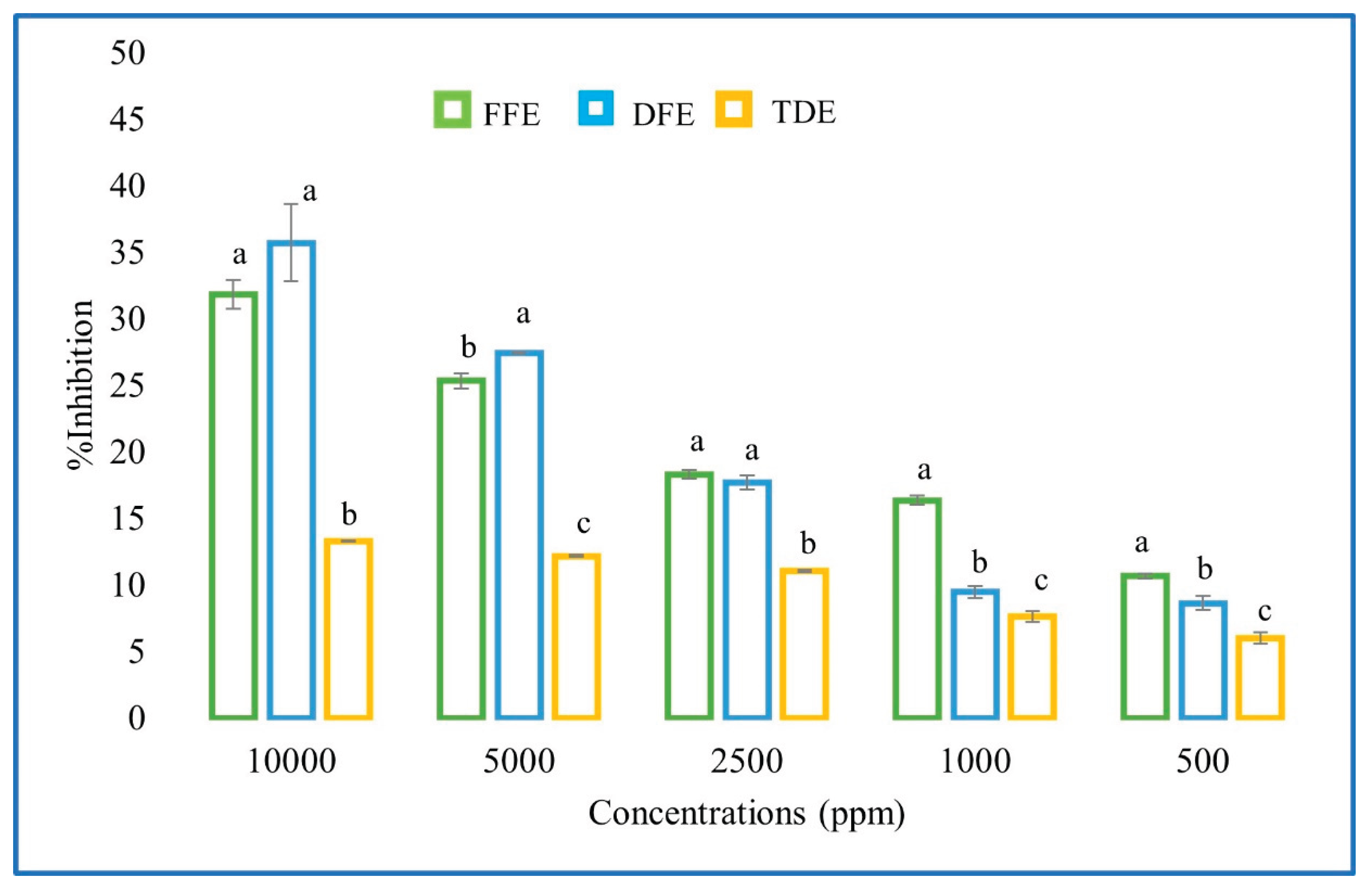
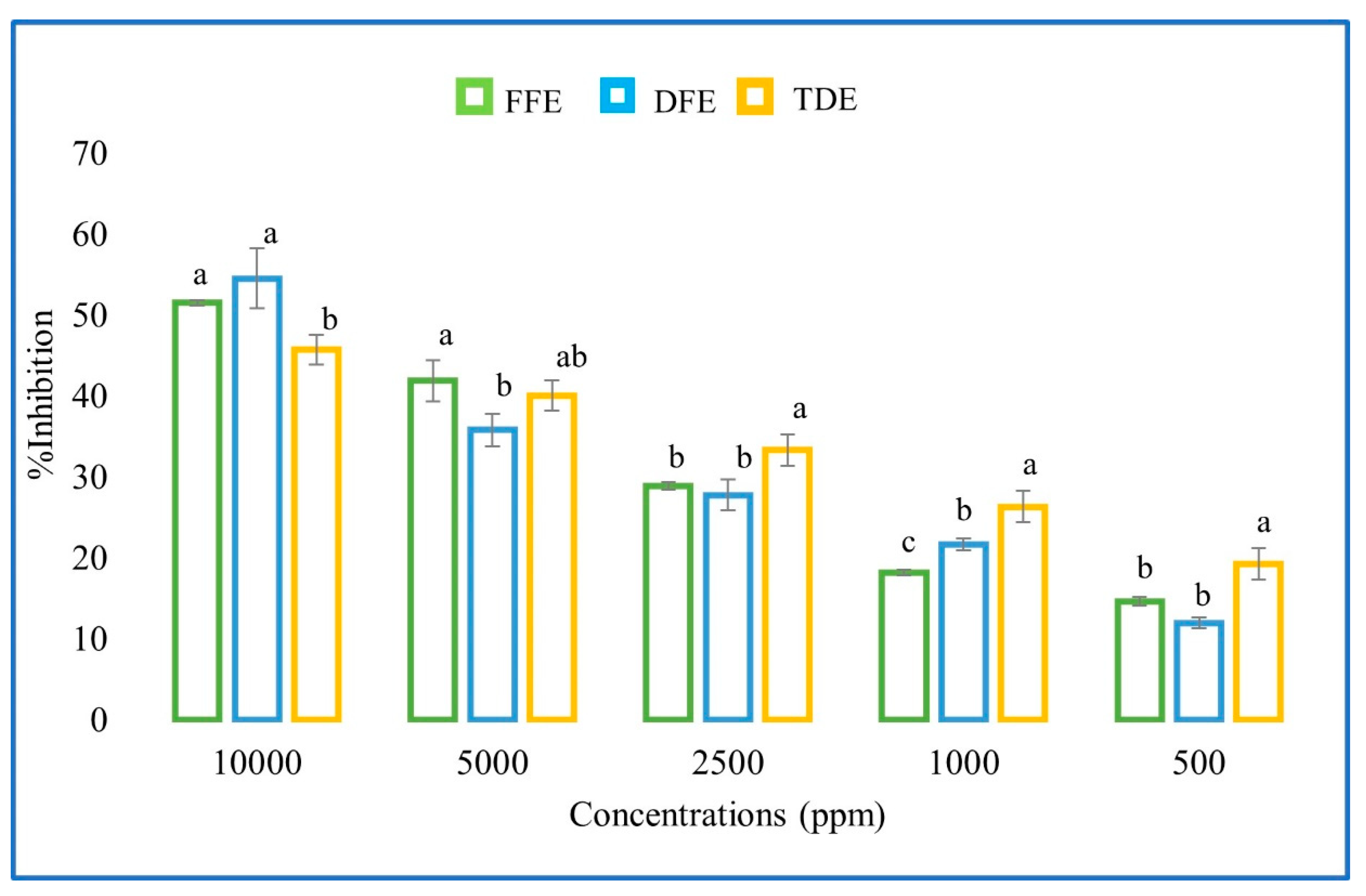
3.2. Antioxidant Activity

Several methods have been developed to assess the antioxidant activity of plant extracts, each providing a distinct perspective on their antioxidant potential. Consequently, the most comprehensive evaluation is achieved by combining two or more complementary methods [25]. In this study, the in vitro antioxidant activity of the extracts was evaluated using the DPPH radical scavenging assay and the ABTS method. Figure 1 and Figure 2 present the percentage inhibition of DPPH and ABTS radicals across concentrations ranging from 500 to 10,000 ppm. The highest inhibition was observed at the maximum concentration tested (10 mg/mL), indicating a concentration-dependent radical scavenging activity. No significant differences in antioxidant activity were observed between the fresh and dried fruit extracts with either method (Table 4), which correlates with the similar total polyphenol content observed in both extracts. This finding aligns with the observations of Martínez-Ceja et al. [26], who reported a strong relationship between phenolic compound concentration and antioxidant activity.
The mean inhibitory concentration (IC50) was calculated to quantify the antioxidant capacity of the extracts. For the DPPH assay, IC50 values were 18.29 ± 0.8 mg/mL for the fresh fruit extract and 14.22 ± 1.35 mg/mL for the dried fruit extract. For the ABTS assay, IC50 values were 8.70 ± 0.14 mg/mL and 8.68 ± 0.62 mg/mL for the fresh and dried fruit extracts, respectively (Table 4). No significant differences were detected between the IC50 values obtained for the two extracts using either method.

3.3. Antiproliferative Activity and Cytotoxicity

The assessment of cytotoxicity in medicinal plants has gained considerable importance, as it serves as an initial step in evaluating their safety and is also indicative of the biological activity of plant extracts or isolated compounds [27]. Table 5 presents the cytotoxic concentration (CC50) values of the extracts. Both the fresh and dried fruit extracts exhibited CC50 values greater than 200 µg/mL, indicating low cytotoxicity. In contrast, the reference compound cisplatin, a chemotherapeutic agent, displayed a CC50 value of 2.22 ± 0.21 µg/mL.
The antiproliferative activity of the extracts, evaluated using the Caco-2 cell line, is presented in Table 4. The dried fruit extract exhibited an activity of 25.44 ± 0.16 µg/mL, which was higher than that of the control (7.52 ± 0.41 µg/mL). Regarding the selectivity index, both the fresh and dried fruit extracts demonstrated greater selectivity compared to the traditional beverage and the control, with values of 1, whereas the control and beverage exhibited a selectivity index of 0.29.

4. Discussion

Most Randia species remain poorly characterized, with the earliest chemical studies dating back to the 1990s and focusing primarily on Randia echinocarpa [28]. To date, qualitative phytochemical studies are still limited and incomplete for several species, with flavonoids and tannins being the most commonly reported metabolites. Cano-Campos and Ojeda-Ayala et al. [7,11] identified phenols, flavonoids, and saponins in Randia echinocarpa, Randia nitida, and Randia laevigata; these same classes of compounds were also detected in both fresh and dried fruit extracts in the present study. Such similarities may be related not only to the species but also to methodological aspects, including the extraction procedure and solvent system employed [29].
With respect to total polyphenol content (TPC), previous reports on the dried pulp of Randia monantha Bent showed values of 413 ± 0.61, 125 ± 0.63, and 57 ± 0.70 mg GAE/g for aqueous, methanolic, and ethanolic extracts, respectively [10]. For the dried seeds of the same species, TPC values of 276 ± 0.36, 146 ± 1.76, and 268 ± 0.27 mg GAE/g were reported for aqueous, methanolic, and ethanolic extracts, respectively [10]. Martínez-Ceja et al. [26] reported a TPC of 30.65 ± 0.00 mg GAE/g in methanolic extracts of Randia aculeata leaves. In comparison, the values obtained in the present work were lower than those reported for the seeds and dried pulp of R. monantha, but higher than those reported for the methanolic extract of R. aculeata leaves.
It is important to note that, in contrast to most previous studies where pulp and seeds were analyzed separately, the present study evaluated the whole fruit. This methodological difference likely explains the variation in TPC values, as polyphenolic compounds are differentially distributed across plant tissues [30]. Such distributional differences highlight the importance of considering the plant part analyzed and the extraction strategy employed when comparing phytochemical profiles across Randia species.
The phytochemical composition observed in this study is consistent with the biological activities obtained. The comparable antioxidant activities of fresh and dried fruit extracts, measured by DPPH and ABTS assays, correlate with the similar polyphenol contents observed between the two treatments. Moreover, the presence of bioactive compounds such as phenolic acids and flavonoids may underlie the low cytotoxicity and selective antiproliferative effects detected in the Caco-2 cell line. These findings reinforce the idea that phenolic composition not only varies among Randia species and plant parts but also plays a critical role in defining their functional and therapeutic potential.
Flavonoids are a major subclass of polyphenolic compounds widely distributed in plants and known for their diverse biological activities. Qualitative phytochemical studies in Randia species have consistently reported the presence of flavonoids, with seeds being particularly rich in these metabolites [11]. In the present study, flavonoids were quantitatively assessed, and the dried fruit extract exhibited a higher flavonoid content compared to the fresh fruit extract. This increase may be attributed to the mechanical processes involved in powder production (grinding and sieving), which likely promoted the rupture of cellular vacuoles and cell walls, thereby enhancing the release and extraction efficiency of flavonoids [22]. Such findings highlight the influence of postharvest processing on the phytochemical yield of plant materials.
Despite the growing interest in Randia species, there is still a limited number of studies that have systematically identified and quantified individual phenolic compounds in Randia fruits. A notable contribution is the work of Juárez-Trujillo et al. [10], who conducted the first comprehensive profiling of phenolic compounds in pulp and seed extracts of Randia monantha using aqueous, methanolic, and ethanolic solvents. They identified 10 compounds in the pulp and 13 in the seeds, most of which were classified as phenolic acids. The seed extracts consistently exhibited a higher diversity and concentration of individual phenolics compared to the pulp.
In their study, chlorogenic acid was the predominant compound in the aqueous seed extract (81.11 ± 1.94 μg/g), followed by rutin (51.61 ± 3.12 μg/g), 4-coumaric acid (30.29 ± 0.06 μg/g), and caffeic acid (21.95 ± 0.25 μg/g). Other compounds detected in lower concentrations across all seed extracts included ferulic acid, kaempferol, vanillic acid, quercetin, (–)-epicatechin, 4-hydroxybenzoic acid, vanillin, 2,4-dimethoxy-6-methylbenzoic acid, and scopoletin. In contrast, the pulp extracts contained lower concentrations of phenolics overall. Chlorogenic acid was also the major compound in the pulp, with concentrations of 39.81, 20.74, and 13.01 μg/g in the ethanolic, aqueous, and methanolic extracts, respectively. Vanillic acid was consistently present in all pulp extracts, with the highest concentration in the aqueous extract (9.83 ± 0.28 μg/g), followed by the methanolic (4.87 ± 0.48 μg/g) and ethanolic (2.17 ± 0.22 μg/g) extracts. Caffeic acid was also detected at low levels in the ethanolic pulp extract (3.11 ± 0.09 μg/g).
Several of these phenolic acids—including chlorogenic acid, vanillic acid, and caffeic acid—were also detected and quantified in the fresh and dried fruit extracts of Randia spp. analyzed in the present work. The overlap in compound profiles between our results and previous studies supports the reproducibility of the phytochemical composition across different Randia species and extraction methods. However, differences in compound abundance are likely influenced by several factors, including the plant organ analyzed (whole fruit versus separated pulp or seed), the solvent polarity, and the extraction conditions employed.
These findings contribute to the limited but growing body of knowledge on the phytochemistry of Randia fruits. By combining qualitative and quantitative data, the present study underscores the relevance of both processing techniques and analytical methods in shaping the phytochemical profile, particularly with respect to flavonoid and phenolic acid content. Such insights are essential for understanding the bioactive potential of Randia species and for guiding future research aimed at their pharmacological and nutraceutical applications.
Comparison of the results obtained in this study with previous reports highlights important differences in the phenolic profiles of Randia spp. The main compound identified in both the fresh and dried fruit extracts was kaempferol, whereas in the literature this flavonoid has been reported at low concentrations in seed extracts of Randia monantha Benth and was not detected in the pulp. A key distinction is that the present study evaluated the whole fruit (pulp and seeds), which likely accounts for part of the observed variation.
The chemical composition of plant extracts is strongly influenced by the solubility of metabolites in the solvent system employed. In this study, 70% ethanol was selected, as it is well recognized for its efficiency in extracting flavonoids and phenolic acids, though its selectivity toward different classes of polyphenols can vary. This factor, together with the use of whole fruit, may explain the differences between the extracts analyzed here and those previously reported [30,31]. Beyond solvent choice, phytochemical composition can also be affected by other variables, including the extraction method, analytical protocols, environmental conditions, and geographical location of plant growth [12].
All compounds identified in the fresh and dried fruit extracts of Randia spp. contain conjugated double bonds in their chemical structures, which enable electronic delocalization. This structural feature is closely associated with the antioxidant and anti-inflammatory properties of polyphenols described in various plants [32] and may therefore contribute to the biological activities observed in the present study.
In contrast, no compounds were detected in the traditional beverage extract. This absence is likely attributable to the low concentration of bioactive compounds, which falls below the detection limits of the analytical method employed. To overcome this limitation, modifications in the chromatographic protocol—such as adjustments to the mobile phase composition, retention times, or inclusion of additional standards—are recommended to achieve a more comprehensive characterization of the beverage extract.
Vanillic acid, identified in the dried fruit extract of Randia spp., is a phenolic compound previously reported to possess anti-snakebite properties [33]. Phenolic compounds are also recognized for their anti-inflammatory potential in various pathologies, which may explain their traditional use in treating animal bites. Another key compound detected in the extracts was kaempferol, a flavonoid widely known for its antioxidant, anti-inflammatory, and neuroprotective activities. Kaempferol contributes to cellular protection by neutralizing reactive oxygen species and reducing oxidative stress [34]. The high content of this compound in both fresh and dried fruit extracts highlights Randia spp. as a rich source of bioactive secondary metabolites with potential applications in the pharmaceutical and food industries [30].
Furthermore, the strong correlation between total polyphenol content and antioxidant activity, consistently demonstrated in this and other studies [35,36], reinforces the role of phenolic compounds as major contributors to the biological activity of Randia spp. extracts.
Although studies have reported the antioxidant activity of pulp, seeds, and leaves of Randia species [18], no previous evaluations have been conducted specifically on the whole fruit. In the present study, antioxidant activity was assessed by determining the percentage of inhibition of DPPH and ABTS radicals, as well as the IC50 values for both assays. The activity observed in the fresh and dried fruit extracts can be largely attributed to kaempferol, which, as noted above, exhibits antioxidant, anti-inflammatory, and neuroprotective properties by effectively neutralizing reactive oxygen species and protecting cells against oxidative stress [34].
It is noteworthy that the ABTS assay exhibited higher inhibition percentages compared to the DPPH assay. This difference is consistent with the broader applicability of the ABTS method, which is capable of evaluating both hydrophilic and lipophilic antioxidant systems, whereas the DPPH assay is more restricted to hydrophobic environments [37].
The evaluation of cytotoxicity has gained increasing importance as an initial step in assessing the safety of medicinal plants, since it provides valuable insights into the biological activity of plant extracts and isolated compounds [27]. In this context, previous studies on Randia ferox leaf extracts demonstrated that peripheral blood mononuclear cells treated for 24 h maintained normal cell viability. Moreover, all tested concentrations reduced intracellular reactive oxygen species (ROS) levels without affecting nitric oxide (NO) production, and most did not alter double-stranded DNA (dsDNA) release [38].
In the present study, the cytotoxicity of hydroalcoholic extracts from fresh and dried fruit, as well as from the traditional beverage prepared from Randia spp., was evaluated in the J774.2 cell line. The extracts exhibited low cytotoxicity compared to cisplatin, suggesting a favorable safety profile. These findings highlight the potential of Randia spp. fruit as a safe source of bioactive compounds for therapeutic applications and support its traditional use in the treatment of various diseases and symptoms.
Species of the genus Randia have been attributed with diverse biological activities; however, only antioxidant, anti-inflammatory, and antimicrobial properties have been experimentally validated to date [11]. In the present study, hydroalcoholic extracts of fresh and dried fruit of Randia spp. exhibited an IC₅₀ > 200 µg/mL against the colon adenocarcinoma cell line (CaCo-2) using the MTT assay. These findings suggest a potential antiproliferative effect, which may be primarily attributed to kaempferol, a flavonoid identified in the extracts. Previous studies have demonstrated that flavonoids possess anticancer activity while generally exhibiting lower toxicity than conventional chemotherapeutic agents [39,40].
Furthermore, comparison of the selectivity index (SI) of the fresh and dried Randia spp. fruit extracts with that of cisplatin, a standard drug used in cancer therapy, revealed that the extracts displayed higher SI values. This is a desirable characteristic, as a higher SI indicates greater safety and efficacy. Ideally, a compound should demonstrate minimal cytotoxicity at high concentrations while maintaining biological activity at low concentrations, thus yielding a high SI value [41]. These results highlight the potential of Randia spp. fruit extracts as promising candidates for the development of safer, plant-derived anticancer agents.

5. Conclusions

In this study, the phytochemical composition, antioxidant and antiproliferative activities, and cytotoxicity of hydroalcoholic extracts from fresh and dried Randia spp. fruits were evaluated. Qualitative analysis confirmed the presence of saponins, phenols, and flavonoids. The dried fruit extract exhibited higher levels of total polyphenols and flavonoids, likely due to concentration effects during drying using a refractive window. HPLC analysis revealed that the fresh fruit extract contained a greater diversity of phenolic compounds, while kaempferol was the predominant compound in both extracts, contributing significantly to their antioxidant and antiproliferative activities. Both extracts demonstrated low cytotoxicity and high selectivity indices, supporting their potential safety and therapeutic value in the prevention or management of chronic degenerative diseases.

Author Contributions

Conceptualization, V.M.F-L.; methodology, V.M.F-L., L.L.M-L., A.M-R. and I.G-M..; software, E.H-L.; validation, A.M-R., L.L.M-L.; formal analysis, C.E.M-S., E.H-L. and I.G-M.; investigation, V.M.F-L., C.E.M-S. and I.G.M.; resources, C.E.M-S. and A.M-R.; data curation, methodology, writing—original draft preparation, V.M.F-L., I.G-M.; writing—review and editing, C.E.M-S, A.M-R, L.A-F, and I.G-M..; visualization, E.H-L-; supervision, L.L.M-L, L.A-F. All authors have read and agreed to the published version of the manuscript.

Funding

Secretariat of Science, Technology, Humanities and Innovation of Mexico (SECIHTI) through grant 832851 and the National Technological Institute of Mexico Campus Tuxtepec for infrastructure support.

Data Availability Statement

All relevant data are provided in this published article. Any further questions can be directed to the corresponding author.

Conflicts of Interest

The authors declare no conflicts of interest.

References

  1. Gómez, X.; Sanon, S.; Zambrano, K.; Asquel, S.; Bassantes, M.; Morales, J.E.; Otáñez, G.; Pomaquero, C.; Villarroel, S.; Zurita, A.; Calvache, C; Celi, K; Contreras, T; Corrales, D; Naciph, M.B.; Peña, P.; Caicedo; A Key points for the development of antioxidant cocktails to prevent cellular stress and damage caused by reactive oxygen species (ROS) during manned space missions. npj Microgravity 2021, 7, 35. [CrossRef]
  2. Sies, H.; Berndt, C.; Jones, D.P. Oxidative Stress. Annu. Rev. Biochem. 2017, 86, 715–748.
  3. Jiang, F.; Ding, Y.; Tian, Y.; Yang, R.; Quan, M.; Tong, Z.; Zhang, X.; Luo, D.; Chi, Z.; Liu, C. Hydrolyzed low-molecular-weight polysaccharide from Enteromorpha prolifera exhibits high anti-inflammatory activity and promotes wound healing. Biomater. Adv. 2022, 133, 112637. [CrossRef]
  4. Manikandan, R.; Anjali, R.; Beulaja, M.; Prabhu, N.M.; Koodalingam, A.; Saiprasad, G.; Chitra, P.; Arumugam, M. Synthesis, characterization, anti-proliferative and wound healing activities of silver nanoparticles synthesized from Caulerpa scalpelliformis. Process Biochem. 2019, 79, 135–141. [CrossRef]
  5. Said Nasser Al-Owamri, F.; Saleh Abdullah Al Sibay, L.; Hemadri Reddy, S.; Althaf Hussain, S.; Subba Reddy Gangireddygari, V. Phytochemical, Antioxidant, hair growth and wound healing property of Juniperus excelsa, Olea oleaster and Olea europaea. J. King Saud Univ. Sci 2023, 35, 102446. [CrossRef]
  6. Hang, C.; Sun, H.; Zhang, A.; Yan, G.; Lu, S.; Wang, X. Recent Developments in the Field of Antioxidant Activity on Natural Products. Aмурский Медицинский Журнaл 2015, 2, 44–48.
  7. Cano-Campos, M.C., Díaz-Camacho, S.P., Uribe-Beltran, M.J., López-Angulo, G., Montes-Avila, J., Paredes-López, O.; Delgado-Vargas, F. Bio-guided fractionation of the antimutagenic activity of methanolic extract from the fruit of Randia echinocarpa (Sessé et Mociño) against 1-nitropyrene. Food Res. Int. 2011, 44(9), 3087-3093. [CrossRef]
  8. Stevens, W. D., Ulloa, C., Pool, A., & Montiel, O. M. Flora de Nicaragua, 1st ed. St. Louis: Missouri Botanical Garden Press, 2001; pp.943.
  9. Gallardo-Casas, C.A.; Guevara-Balcázar, G.; Morales-Ramos, E.; Tadeo-Jiménez, Y.; Gutiérrez-Flores, O.; Jiménez-Sánchez, N.; Valadez-Omaña, M.T.; Valenzuela-Vargas, M.T.; Castillo-Hernández, M.C. Ethnobotanic study of Randia aculeata (Rubiaceae) in Jamapa, Veracruz, Mexico, and its anti-snake venom effects on mouse tissue. J. Venom. Anim. Toxins Incl. Trop. Dis. 2012, 18 (3), 287–294. [CrossRef]
  10. Juárez-Trujillo, N.; Monribot-Villanueva, J.L.; Alvarado-Olivarez, M.; Luna-Solano, G.; Guerrero-Analco, J.A.; Jiménez-Fernández, M. Phenolic profile and antioxidative properties of pulp and seeds of Randia monantha Benth. Ind. Crops Prod. 2018., 124, 53-58. [CrossRef]
  11. Ojeda-Ayala, M.; Gaxiola-Camacho, S. M.; Delgado-Vargas, F. Phytochemical composition and biological activities of the plants of the genus Randia. Bot. Sci. 2024, 100(4), 779-796. [CrossRef]
  12. Mërtiri, I.; Coman, G.; Cotârlet, M.; Turturică, M.; Balan, N.; Râpeanu, G.; Stanciuc, N.; Mihalcea, L. Phytochemical Profile Screening and Selected Bioactivity of Myrtus communis Berries Extracts Obtained from Ultrasound-Assisted and Supercritical Fluid Extraction. Separations 2025, 12(1), 8. [CrossRef]
  13. Capataz-Tafur, J.; Orozco-Sánchez, F.; Vergara-Ruiz, R.; Hoyos-Sánchez, R Efecto antialimentario de los extractos de suspensiones celulares de Azadirachta indica sobre Spodoptera frugiperda JE Smith en condiciones de laboratorio. Rev. Fac. Nac. de Agr. Medellín. 2007, 60(1), 3703-3715.
  14. Bulugahapitiya, V.P.; Rathnaweera, T.N.; Manawadu, H.C. Phytochemical composition and antioxidant properties of Dialium ovoideum thwaites (Gal Siyambala) leaves. Int. J Min. Fruits, Med. and Arom. Plants. 2020, 6 (1), 13-19.
  15. Singleton, V.L.; Rossi, J.A. Colorimetry of total phenolics with phosphomolybdicphosphotungstic acid reagents. Am. J. Enol. Vitic. 1965, 16(3), 144-158. [CrossRef]
  16. Zhishen, J.; Mengcheng, T.; Jianming, W. The determination of flavonoid contents in mulberry and their scavenging effects on superoxide radicals. Food Chem. 1999, 64(4), 555–559. [CrossRef]
  17. Méndez-Lagunas, L.L.; Cruz-Gracida, M.; Barriada-Bernal, L.G.; Rodríguez-Méndez, L.I. Profile of phenolic acids, antioxidant activity and total phenolic compounds during blue corn tortilla processing and its bioaccessibility. J. Food Sci.Tech. 2020, 57(12), 4688–4696. [CrossRef]
  18. Brand-Williams, W.; Cuvelier, M.E.; Berset, C. Use of a free radical method to evaluate antioxidant activity. LWT - Food Sci. Tech. 1995, 28(1), 25–30.
  19. Re, R.; Pellegrini, N.; Proteggente, A.; Pannala, A.; Yang, M.; Rice-Evans, C. Antioxidant activity applying an improved ABTS radical cation decolorization assay. Free Radic. Biol. Med. 1999, 26(9-10), 1231–1237. [CrossRef]
  20. González-Morales, L. D.; Moreno-Rodríguez, A.; Vázquez-Jiménez, L.K.; Delgado-Maldonado, T.; Juárez-Saldivar, A.; Ortiz-Pérez, E.; Rivera, G. Triose Phosphate Isomerase Structure-based virtual screening and in vitro biological activity of natural products as Leishmania Mexicana inhibitors. Pharmaceutics 2023, 15(8), 2046.
  21. Medina-Reyes, E.I.; Mancera-Rodríguez, M.A.; Delgado-Buenrostro, N.L.; Moreno-Rodríguez, A.; Bautista-Martínez, J.L.; Díaz-Velásquez, C.E.; Martínez-Alarcón, S.A.; Torrens, H.; Godínez-Rodríguez, M.A.; Terrazas-Valdés, L.I.; Chirino, Y.I.; Vaca Paniagua, F. Novel thiosemicarbazones induce high toxicity in estrogen receptor-positive breast cancer cells (MCF7) and exacerbate cisplatin effectiveness in triple-negative breast (MDA-MB231) and lung adenocarcinoma (A549) cells. Invest. New Drugs. 2019, 38, 558-573. [CrossRef]
  22. Chaparro-Hernández, I.; Méndez-Lagunas, L.; Rodríguez-Ramírez, J.; Sandoval-Torres, S.; Aquino-González, L.; Barriada-Bernal, G. Spray Drying of Stevia Rebaudiana Bertoni Aqueous Extract: Effect on Polyphenolic Compounds. Chem. Eng. Trans. 2023, 98, 33-38.
  23. Sepúlveda, C.T.; Zapata, J.E. Efecto de la Temperatura, el pH y el Contenido en Sólidos sobre los Compuestos Fenólicos y la Actividad Antioxidante del Extracto de Bixa orellana L. Inf. Tec. 2019, 30(5), 57-66.
  24. Lesage-Meessen, L.; Bou, M.; Sigoillot, J.C.; Faulds, C.B.; Lomascolo, A. Essential oils and distilled straws of lavender and lavandin: a review of current use and potential application in white biotechnology. Appl. Microbiol. Biotechnol. 2015, 99(8), 3375–3385. [CrossRef]
  25. Ozer, M.S.; Kirkanb, B.; Sarikurkcuc, C.; Cengizd, M.; Ceylane, O.; Atılgand, N.; Tepef, B. Onosma heterophyllum: phenolic composition, enzyme inhibitory and antioxidant activities. Ind. Crops Prod. 2018, 111, 179–184.
  26. Martínez-Ceja, A.; Romero-Estrada, A.; Columba-Palomares, M.C.; Hurtado-Díaz, I.; Alvarez, L.; Teta-Talixtacta, R.; Bernabé-Antonio, A. Anti-inflammatory, antibacterial and antioxidant activity of leaf and cell cultures extracts of Randia aculeata L. and its chemical components by GC-MS. S. Afr. J. Bot. 2022, 144, 206-218. [CrossRef]
  27. McGaw, L.J.; Elgorashi, E.E.; Eloff, J.N. Cytotoxicity of African medicinal plants against normal animal and human cells. In Toxicological survey of African medicinal plants,1st ed.; Kuete,V. Elsevier: Jamestown Road, London, 2014, pp. 181-233.
  28. Bye, R.; Linares, E.; Mata, R.; Albor C.; Casteñeda, P.C.; Delgado, G. Ethnobotanical and phytochemical investigation of Randia echinocarpa (Rubiaceae). An. Inst. Biol. Serie Bot. 1991, 62, 87-106.
  29. Charoensin, S. (2014). Antioxidant and anticancer activities of Moringa oleifera leaves. J. Med. Plants Res, 2014, 8(7), 318-325.
  30. Sahlabgi, A.; Lupuliasa, D.; Stoicescu, I.; Vlaia, L.L.; Licu, M.; Popescu, A.; Mititelu, M. Determination of the Phytochemical Profile and Antioxidant Activity of Some Alcoholic Extracts of Levisticum officinale with Pharmaceutical and Cosmetic Applications. Separations, 2025, 12(4), 79.
  31. Do, Q.D.; Angkawijaya, A.E.; Tran-Nguyen, P.L.; Huynh, L.H.; Soetaredjo, F.E.; Ismadji, S.; Ju, Y.H. Effect of extraction solvent on total phenol content, total flavonoid content, and antioxidant activity of Limnophila aromatica. J. Food Drug Anal. 2014, 22, 296–302. [CrossRef]
  32. Rice-Evans, C.A.; Miller, NJ.; Paganga, G. (1996). Structure-antioxidant activity relationships of flavonoids and phenolic acids. Free Radic Biol. Med. 1996, 20(7), 933-956.
  33. Budryn, G.; Zaczyńska, D.; Oracz, J. (2016). Effect of addition of green coffee extract and nanoencapsulated chlorogenic acids on aroma of different food products. Lwt, 2016, 73, 197-204.
  34. Zhuang, X., Shi, W., Shen, T., Cheng, X., Wan, Q., Fan, M., & Hu, D. Research Updates and Advances on Flavonoids Derived from Dandelion and Their Antioxidant Activities. Antioxidants, 2024, 13(12), 1449. [CrossRef]
  35. Krishnaiah, D.; Sarbatly, R.; Nithyanandam, R. A review of the antioxidant potential of medicinal plant species. Food Bioprod. Process. 2011, 89(3), 217-233. [CrossRef]
  36. Juárez-Trujillo, N.; Tapia-Hernández, F.E.; Alvarado-Olivarez, M.; Beristain-Guevara, C.I.; Pascual-Pineda, L.A.; Jiménez-Fernández, M. Antibacterial activity and acute toxicity study of standardized aqueous extract of Randia monantha Benth fruit. Biotecnia, 2022, 24(1), 38-45. [CrossRef]
  37. Floegel, A.; Kim, D.O.; Chung, S.J.; Koo, S.I.; Chun, O.K. (). Comparison of ABTS/DPPH assays to measure antioxidant capacity in popular antioxidant-rich US foods. J. Food Compos. Anal. 2011, 24(7), 1043-1048.
  38. Pappis, L.; Prates-Ramos, A.; Fontana, T.; Geraldo-Sangoi, G.; Castro-Dornelles, R.; Bolssoni-Dolwitsch, C.; Rorato-Sagrillo, M.; Cadoná, FC.; Kolinski-Machado, A.; De Freitas, Bauermann, L. Randia ferox (Cham & Schltdl) DC: phytochemical composition, in vitro cyto-and genotoxicity analyses. Nat. Prod. Res. 2021, 4, 1-7.
  39. Saklani, A.; Kutty S.K. Plant-derived compounds in clinical trials. Drug Discovery Today, 2008, 13(3-4), 161–171.
  40. Solowey, E.; Lichtenstein, M.; Sallon, S.; Paavilainen, H.; Solowey, E., & Lorberboum-Galski, H. (2014). Evaluating medicinal plants for anticancer activity. Sci. World J. 2014, 1, 721402.
  41. Subramani, C.; Sharma, G.; Chaira, T.; Barman, T.K. High content screening strategies for large-scale compound libraries with a focus on high-containment viruses. Antivir. Res. 2024, 221, 105764. [CrossRef]
Figure 1. Antioxidant activity (% inhibition) of the DPPH radical for fruit extracts and traditional fruit drink of Randia spp. at various concentrations. FFE = fresh fruit extract; DFE = dried fruit extract; TDE = traditional fruit drink extract. Different letters within the same concentration indicate significant differences (p < 0.05) according to Tukey’s test.
Preprints 174900 g001
Figure 2. Antioxidant activity (% inhibition) of the ABTS radical for fruit extracts and traditional fruit drink of Randia spp. at different concentrations. FFE = fresh fruit extract; DFE = dried fruit extract; TDE = traditional drink extract. Different letters within the same concentration indicate significant differences (p < 0.05) according to Tukey’s test.
Preprints 174900 g002
Table 1. Identification of chemical families of extracts from the fresh and dried fruit of Randia spp.
Family Fresh fruit Dried fruit Traditional drink
Alkaloids - - -
Saponins ++ ++ +
Sterols and triterpenes - - -
Phenols ++ ++ +
Flavonoids + + ++
Absent (-), little (+), moderate (++), abundant (+++). The values represent the mean of three determinations ± standard deviation. Different letters in the same column indicate significantly different values (p<5) according to Tukey's test.
Table 2. Total polyphenol and flavonoid content of fresh and dried fruit extracts of Randia spp.
Extract TPC
(mg GAE/g extract)
TFC
(mg QE/g extract)
Fresh 47.22 ± 0.96 A 27.08 ± 1.36 B
Dry 50.27 ± 0.14 A 35.53 ± 2.20 A
Traditional drink 38.583 ± 2.677B 18.660 ± 1.696C
The values represent the mean of three determinations ± standard deviation. Different letters in the same column indicate significantly different values (p≤<0.05) according to Tukey's test.
Table 3. Phenolic profile of extracts from the fresh and dried fruit of Randia spp.
Compound (mg/gdb) Fresh Dry Traditional drink
Caffeic acid 1.05 ± 0.01 nd nd
Hydroxybenzoic acid 5.34 ± 0.1 nd nd
Ferulic acid 2.30 ± 0.03 A 1.94 ± 0.05 B nd
Cumaric acid 1.54 ± 0.05 nd nd
Kaempferol 137.55 ± 0.16 A 42.10 ± 0.20 B nd
Vanillic acid nd 7.51 ± 0.33 nd
The values represent the mean of three determinations ± the standard deviation. Different letters in the same row indicate significantly different values (p<0.05). nd: not detected.
Table 4. Antioxidant activity of fruit extracts of Randia spp.
Extract DPPH (%) IC50 (mg/mL) ABTS (%) IC50 (mg/mL)
Fresh 31.8 ± 1.85 A 18.29 ± 0.8 A 51.54 ± 2.12 A 8.70 ± 0.14 A
Dry 35.7 ± 1.92 A 14.22 ± 1.35 A 54.54 ± 1.87 A 8.68 ± 0.62 A
Traditional drink 14.6 ± 1.92 B 61.933 ± 1.042 B 45.54 ± 1.92 B 61.933 ± 1.042 B
The values represent the mean of three determinations ± the standard deviation. Different letters in the same column indicate significantly different values (p<0.05).
Table 5. Cytotoxicity and antiproliferative activity of extracts from the fruit of Randia spp.
Extract Cytotoxicity a
CC50 (µg/mL)
Antiproliferative Activityb
IC50 (µg/mL)
SIc
Fresh fruit >100 >100 1
Dried fruit >100 25.44± 0.16 3.92
Traditional drink 29.99± 8.45 100.50 ± 6.54 0.29
Control (cisplatin) 2.22 ± 0.21 7.52 ± 0.41 0.29
a CC50 concentration of the extract that produces a 50% reduction in the J774.2 cell line. b IC50 concentration of the extract that produces a 50% reduction of the CaCo-2 cell line. c IS selectivity index (CC50/IC50).
Disclaimer/Publisher’s Note: The statements, opinions and data contained in all publications are solely those of the individual author(s) and contributor(s) and not of MDPI and/or the editor(s). MDPI and/or the editor(s) disclaim responsibility for any injury to people or property resulting from any ideas, methods, instructions or products referred to in the content.
Copyright: This open access article is published under a Creative Commons CC BY 4.0 license, which permit the free download, distribution, and reuse, provided that the author and preprint are cited in any reuse.
Prerpints.org logo

Preprints.org is a free preprint server supported by MDPI in Basel, Switzerland.

Subscribe

Disclaimer

Terms of Use

Privacy Policy

Privacy Settings

© 2025 MDPI (Basel, Switzerland) unless otherwise stated