
Submitted:
06 September 2025
Posted:
09 September 2025
You are already at the latest version
Abstract
The antigenic relationship between Eurasian avian-like H1N1 swine influenza viruses (EA H1N1) and human pandemic 2009 H1N1 viruses (2009/H1N1) remains a critical question for influenza surveillance and vaccine efficacy. This study systematically investigated the antigenic differences between strains A/swine/Tianjin/312/2016 (TJ312, EA H1N1) and A/Guangdong-Maonan/SWL1536/2019 (GD1536, 2009/H1N1). Cross-hemagglutination inhibition (HI) assays revealed a significant antigenic disparity, with a 16-fold reduction in heterologous versus homologous HI titers. Comparative sequence analysis identified 22 amino acid differences across the five major antigenic sites (Sa, Sb, Ca1, Ca2, Cb) of the HA1 subunit. Using reverse genetics, a panel of mutant viruses was generated. This study revealed that a single histidine (H)-to-asparagine (N) substitution at residue 128 (H3 numbering) in the Sa antigenic site acts as a primary determinant of antigenic variation, sufficient to cause a 4-fold change in HI titers and a measurable shift in antigenic distance. Structural modeling via AlphaFold3 and PyMOL software reveals that the H128N mutation alters the local conformation of the antigenic site, by which H at position 128 exerts electrostatic repulsion with adjacent amino acids, while N facilitates hydrogen bond formation with adjacent amino acids, ultimately leading to changes in the protein structure of the antigenic site. Our findings confirm that residue 128 is a critical molecular marker for the antigenic differentiation of EA H1N1 and 2009/H1N1 viruses. The study underscores the necessity of monitoring specific HA mutations that could reduce cross-reactivity and provides valuable insights for refining vaccine strain selection and pandemic preparedness strategies.
Keywords:
influenza A virus
; antigenic variation
; hemagglutinin
; amino acid substitution
1. Introduction
Influenza A viruses, as RNA viruses characterized by high mutation rates, pose a significant threat to global public health due to their continuous evolution and cross-species transmission capabilities. Since its initial isolation from European swine in 1979, the Eurasian avian-like H1N1 swine influenza virus (EA H1N1) has evolved over decades into a stable and geographically distinct lineage, now circulating widely in pig herds across Eurasia (9; 12; 16; 32). Recent surveillance data indicate an increasing prevalence of this virus in swine populations, establishing it as a major target for regional influenza control efforts. Notably, prevalent EA H1N1 strains exhibit high binding affinity for human-type α-2,6-linked sialic acid receptors on respiratory epithelial cells, providing a molecular basis for overcoming species barriers and enabling zoonotic transmission (23). Since 2011, sporadic human infections with EA H1N1 viruses have been reported in countries including China, the Netherlands, and Germany, with some cases documenting direct contact with pigs (3; 4; 6-8; 11; 15; 26; 29; 33; 34). These incidents confirm the virus’s capacity for human infection and transmission potential, marking it as a candidate virus with pandemic potential.
To date, the 2009/H1N1 pandemic virus, which emerged in 2009 via the reassortment of swine influenza viruses, has been incorporated into the seasonal human influenza cycle and remains a major focus of annual surveillance efforts. Genetic analyses indicate that the hemagglutinin (HA) gene of the 2009/H1N1 virus originated from classical swine influenza lineages, whereas the HA of EA H1N1 viruses is derived from an avian influenza source (14; 21). Although both are classified as H1N1 subtypes, they belong to distinct evolutionary lineages (1; 25). Current serological surveillance data suggest generally low pre-existing immunity in humans against EA H1N1 viruses (32). The majority of commonly used seasonal influenza vaccines include an antigen similar to A/California/07/2009 as their 2009/H1N1 component; that said, the extent to which these vaccines offer cross-protection against EA H1N1 viruses is still largely unclear.
Antigenicity is a critical determinant of influenza virus immunogenicity and vaccine matching (10; 18). Differences in antigenicity directly impact viral transmissibility, epidemic dynamics, and the effectiveness of control strategies. Serological assays, such as hemagglutination inhibition (HI) and microneutralization (MN), have demonstrated significant antigenic differences between EA H1N1 and 2009/H1N1 viruses. Some EA H1N1 strains show a greater than 4-fold reduction in neutralization titers by sera raised against 2009/H1N1 vaccines, meeting the criterion for significant antigenic variation (20). However, existing studies have been largely limited to in vitro serological comparisons of a small number of strains. The molecular mechanisms by which amino acid variations in key antigenic sites of HA alter epitope structure and neutralizing antibody binding remain poorly understood. The specific residues driving the antigenic divergence between these two lineages and their evolutionary patterns have not been elucidated. Significant antigenic differences would imply that current 2009/H1N1 vaccines may offer limited protection against EA H1N1 infection. Furthermore, in the context of low population immunity, continued antigenic drift in EA H1N1 viruses could facilitate immune escape, potentially leading to localized outbreaks or larger epidemics.
Therefore, we employed serological assays, reverse genetics, and three-dimensional structural modeling to systematically analyze the antigenic differences between EA H1N1 and human 2009/H1N1 influenza viruses. We aim to clarify the molecular mechanisms underlying their antigenic variation and immune escape characteristics. The findings are intended to inform the development of targeted surveillance strategies, facilitate the selection of candidate vaccine strains, optimize seasonal vaccine design, and mitigate the risk of a potential pandemic.
2. Materials and Methods
2.1. Cells, Viruses, Sera, and Reagents
Human embryonic kidney (HEK293T) cells and Madin-Darby canine kidney (MDCK) cells, maintained in our laboratory, were used for virus rescue and in vitro culture. Both cell lines were cultured in Dulbecco’s modified Eagle’s medium (DMEM) supplemented with 10% fetal bovine serum (FBS), 100 U/mL penicillin, and 100 μg/mL streptomycin at 37°C in a 5% CO₂ atmosphere. The human influenza 2009/H1N1 virus strain A/Guangdong-Maonan/SWL1536/2019 (abbreviated as GD1536) was used as the human reference virus. The EA SIV A/swine/Tianjin/312/2016 (TJ312) served as the representative EA H1N1 virus. Additional strains, A/swine/Guangxi/18/2011 (GX18) and A/swine/Guangdong/104/2013 (GD104), representing EA Genetic group 1 and EA Genetic group 2, respectively, were included for antigenic testing. Virus stocks were propagated in the allantoic cavities of 9- to 11-day-old SPF embryonated eggs incubated at 37°C for 72 hours. Allantoic fluid was harvested, clarified by centrifugation at 3000 × g for 10 minutes, and titrated for hemagglutination activity before storage at -70°C for subsequent serological characterization. Detailed background information and antigenic properties of these strains are described in previous studies (12; 32).
Ferret antisera raised against TJ312 and GD1536 were used for HI assays. Receptor-destroying enzyme (RDE; Denka Seiken, Japan) was used to remove non-specific inhibitors from serum samples. Cell transfections were carried out using Lipofectamine™ LTX and Plus Reagent (Thermo Fisher Scientific). A reverse genetics system based on the PBD plasmid constructed as described in reference (13) was employed for virus rescue.
2.2. Antigenic Analysis
Ferret antisera were treated with RDE, serum was mixed with RDE at a 1:3 (v/v) ratio, incubated at 37°C for 18 hours, and then inactivated at 56°C for 30 minutes. Treated sera were serially diluted twofold (starting dilution 1:20). Then, 25 μL of each serum dilution was mixed with 25 μL of virus suspension containing 4 hemagglutination units (4 HAU) and incubated at room temperature for 30 minutes. Next, 25 μL of a 1% chicken red blood cell suspension was added, followed by an additional 30-minute incubation at room temperature. HI was then assessed. The HI titer was expressed as the reciprocal of the highest serum dilution that completely inhibited hemagglutination. Each serum sample was tested against viruses from different antigenic groups. An HI titer difference of ≥4-fold between two viruses was considered indicative of a significant antigenic difference.
2.3. HA1 Protein Sequence Alignment and Analysis
The HA amino acid sequences of GD1536 and TJ312 were obtained from the Global Initiative on Sharing All Influenza Data (GISAID) database. Sequence alignment was performed using the MegAlign module within the Lasergene Suite software (v7.1.0). The amino acid sequences, deduced from the full-length HA genes of both strains, were aligned using the Clustal W method. The homology of the HA1 region was analyzed, with particular focus on amino acid differences within the five known antigenic sites (Sa, Sb, Ca1, Ca2, Cb).
2.4. Rescue and Antigenic Validation of Single-Site Mutant Viruses
Recombinant viruses GD1536 and TJ312 were generated using the PBD plasmid-based reverse genetics system. Briefly, a mixture of eight PBD plasmids (250 ng each) was co-transfected into HEK293T cells using Lipofectamine™ LTX and Plus Reagent. At 48 hours post-transfection, cell culture supernatants were harvested and inoculated into the allantoic cavities of SPF embryonated eggs. After incubation at 37°C for 72 hours for virus amplification, rescued viruses were subjected to full-genome sequencing to confirm identity with the parental wild-type virus sequences. Chimeric viruses were constructed: chimeric G-T (containing GD1536 HA1 and TJ312 HA2) and chimeric T-G (containing TJ312 HA1 and GD1536 HA2), to determine whether the antigenic differences mapped to the HA1 region. Using TJ312 as the backbone, 43 mutant viruses were generated via single or combined amino acid mutations (Table 1, 4). With the GD1536 backbone, reverse substitution was implemented for mutant viruses that were suspected of causing antigenic reversal. The antigenic properties of each mutant virus were determined through HI assays. The antigenic map was created using Antigenic Cartography Software (http://www.antigenic-cartography.org), a tool that automatically calculates the antigenic distance based on the coordinates of viruses and sera.
2.5. Structural and Mutational Simulation Analysis of the HA1 128 Amino Acid Position
The three-dimensional structures of the HA proteins of TJ312 and GD1536 were predicted using AlphaFold3 (https://alphafoldserver.com/) based on their full-length HA amino acid sequences (12). Default parameters were applied, incorporating evolutionary constraints generated from multiple sequence alignment (MSA). The neural network model was iteratively optimized, and high-confidence structural models were selected. Structure visualization was performed using PyMOL software (v2.5.2). The structural boundaries between HA1 (amino acids 1–330, H3 numbering) and HA2 were defined. All amino acid variation sites within the HA1 protein of both strains and the five key antigenic sites (Sa, Sb, Ca1, Ca2, Cb) were annotated. Mutant models focusing on the amino acid at position 128 (H3 numbering) were constructed to analyze conformational changes in the HA1 head domain before and after mutation.
3. Results
3.1. HI Assays Reveal Significant Antigenic Differences Between TJ312 and GD1536
Serological analysis results showed that the HI titer of TJ312 antiserum against the heterologous GD1536 virus was 1:40, while its homologous titer was 1:640, representing a 16-fold difference (Table 2 and Figure 1). Conversely, the HI titer of GD1536 antiserum against TJ312 was 1:80, compared to its homologous titer of 1:1280, a 16-fold difference (Table 2 and Figure 1). According to the standard criterion for cross-HI assays (≥4-fold titer difference), these results confirm a significant antigenic difference between TJ312 and GD1536.
3.2. HA1 Protein Sequence Alignment Reveals Multiple Amino Acid Differences in Antigenic Sites
The amino acid sequence homology of the HA1 protein between the two strains was 70.6%. The five antigenic sites (Sa, Sb, Ca1, Ca2, Cb) of H1N1 influenza virus HA1 are known to comprise 13, 12, 11, 8, and 5 amino acid residues, respectively (Figure 2 and 3A). Sequence alignment (Figure 2) and amino acid variation sites on the structure of HA1 protein (Figure 3B) revealed that the number of amino acid differences between GD1536 and TJ312 within these sites was 4 (Sa), 6 (Sb), 6 (Ca1), 4 (Ca2), and 2 (Cb), suggesting that multiple amino acid variations in the HA1 antigenic sites likely form the molecular basis for their antigenic divergence.
3.3. The Amino Acid at Position 128 in the HA1 Protein Is a Key Determinant of Antigenic Differences Between TJ312 and GD1536 Strains
With TJ312 serving as the backbone, 43 mutant viruses were generated via single or combined amino acid mutations, and 41 of these mutant viruses were successfully rescued (Table 1). The HI assay results of the two constructed chimeric viruses, chimeric G-T and chimeric T-G, indicated that the amino acids responsible for the antigenic differences between the two viruses are located on the HA1 protein (Table 3). HI testing of 41 mutant viruses generated on the TJ312 backbone identified the TJ312/Mut-16 mutant, which exhibited antigenic reversal. Specifically, its HI titer against TJ312 antiserum was 1:160, a 4-fold decrease compared to the homologous wild-type titer (1:640). Its HI titer against GD1536 antiserum was 1:320, a 4-fold increase compared to the homologous GD1536 titer (1:80) (Table 3). Sequence analysis confirmed that this mutant contained a single H128N amino acid substitution (H to N at position 128, H3 numbering) in the HA1 protein.
A reverse mutant, GD1536HA-N128H, was constructed on the GD1536 backbone. HI assays showed that this mutant’s HI titer against TJ312 antiserum increased from 1:40 (wild-type) to 1:320, while its HI titer against homologous GD1536 antiserum decreased from 1:1280 to 1:320 (Table 3). Antigenic cartography analysis indicated that the antigenic distance between GD1536HA-N128H and wild-type GD1536 decreased from ≥3 grid units (≥8-fold titer difference) to 1.5 units (approximately 3-fold difference). Similarly, the antigenic distance between TJ312HA-H128N and GD1536 decreased from ≥3 units to 1.2 units (approximately 2.3-fold difference) (Figure 4). These results confirm that the H128N substitution in the Sa antigenic site of HA1 causes antigenic shift between EA H1N1 and 2009/H1N1 influenza viruses.

Table 4.
Primers used to generate mutant viruses.
| Purpose | Primers (5’-3’) a | |
| Forward | Reverse | |
| TJ312/Mut-1 | TTAAAAGCTGATACCCTTTGTATAGGCTACCATGCCAAC | GGCATGGTAGCCTATACAAAGGGTATCAGCTTTTAATGT |
| TJ312/Mut-2 | GACACTGTCGACACAGTATTGGAGAAAAATGTGACTGT | AGTCACATTTTTCTCCAATACTGTGTCGACAGTGTCTGTG |
| TJ312/Mut-3 | ATTTACTGGAAGACAAGCATAATGGGAAACTCTGCAGCC | CAGAGTTTCCCATTATGCTTGTCTTCCAGTAAATTAACTG |
| TJ312/Mut-4 | GAAACTCTGCAAACTGAGAGGAGTGGCCCCCTTACAACTG | TAAGGGGGCCACTCCTCTCAGTTTGCAGAGTTTCCCATT |
| TJ312/Mut-5 | CTCTGCAAACTGAGAGGAGTGGCCCCCTTACAACTGGGAAACTG | CAGTTGTAAGGGGGCCACTCCTCTCAGTTTGCAGAGTTTCCCAT |
| TJ312/Mut-6 | AGATCCCCTTACAACTGGGAAAATGCAACGTAGCAGGATG | ATCCATCCTGCTACGTTGCATTTTCCCAGTTGTAAG |
| TJ312/Mut-7 | TACAACTGGGAAACTGCAACATAGCAGGATGGATCCTTG | TTGCCAAGGATCCATCCTGCTATGTTGCAGTTTCCCAGT |
| TJ312/Mut-8 | CTTGGCAACCCAGAATGTGAATCGCTGTCCACAGCGAGATCGTGGTCTT | GATCTCGCTGTGGACAGCGATTCACATTCTGGGTTGCCAAGGATCCATC |
| TJ312/Mut-9 | TTCGTGGTCTTACATAGTAGAGACTTCAAATTCAAAAAAT | TTTGAATTTGAAGTCTCTACTATGTAAGACCACGAATTCG |
| TJ312/Mut-10 | TAGAGACTTCAAATTCAGACAATGGAGCATGCTACCCCG | GGGTAGCATGCTCCATTGTCTGAATTTGAAGTCTCTAT |
| TJ312/Mut-11 | CAAATTCAAAAAATGGAACATGCTACCCCGGAGAATTTGC | AATTCTCCGGGGTAGCATGTTCCATTTTTTGAATTTGAAG |
| TJ312/Mut-12 | CTACCCCGGAGATTTTATTAATTATGAAGAGTTAAAGGAG | TTAACTCTTCATAATTAATAAAATCTCCGGGGTAGCATGC |
| TJ312/Mut-13 | CTGATTATGAAGAGTTAAGGGAGCAGCTGAGTACAGTTTC | ACTGTACTCAGCTGCTCCCTTAACTCTTCATAATCAGC |
| TJ312/Mut-14 | TAAAGGAGCAGCTGAGTTCAGTTTCTTCATTTGAAAGAT | CTTTCAAATGAAGAAACTGAACTCAGCTGCTCCTTTAACT |
| TJ312/Mut-15 | AAATTTTCCCAAAGACAAGTTCATGGCCACACCATGATAC | TGGTGTGGCCATGAACTTGTCTTTGGGAAAATTTCAAATC |
| TJ312/Mut-16 | AGGCAACTTCATGGCCAAACCATGATACCACCAGAGGTAC | CCTCTGGTGGTATCATGGTTTGGCCATGAAGTTGCCTTTG |
| TJ312/Mut-17 | GGCCACACCATGATTCCGACAAAGGTACCACGGTTTCATG | ACCGTGGTACCTTTGTCGGAATCATGGTGTGGCCATGAAG |
| TJ312/Mut-18 | CACCAGAGGTGTCACGGCTGCATGCCCCCACGCTGGAGCCAACAGCTTTTATCG | TGTTGGCTCCAGCGTGGGGGCATGCAGCCGTGACACCTCTGGTGGTATCATGGT |
| TJ312/Mut-19 | GCTCCCACTCTGGAGCCAAAAGCTTTTATCGGAATTTACT | AAATTCCGATAAAAGCTTTTGGCTCCAGAGTGGGAGCATG |
| TJ312/Mut-20 | GAGCCAACAGCTTTTATAAGAATTTACTATGGATAGT | ACTATCCATAGTAAATTCTTATAAAAGCTGTTGGCTCCAG |
| TJ312/Mut-21 | TTTATCGGAATTTAATATGGCTAGTAAAGAAAGGAAACTC | CCTTTCTTTACTAGCCATATTAAATTCCGATAAAAGCTGT |
| TJ312/Mut-22 | CTAAGCTCAACCAGACATACATAAACGATAAGGGAAAGGAAGTGCTTGT | TTTCCCTTATCGTTTATGTATGTCTGGTTGAGCTTAGGATAGGAGTTTCT |
| TJ312/Mut-23 | GAAAGGAAGTGCTTGTACTTTGGGGAGTGCACCACCCTCC | GGGTGGTGCACTCCCCAAAGTACAAGCACTTCCTTTCCCT |
| TJ312/Mut-24 | TGCTTGTAATTTGGGGAATTCACCACCCTCCAACTGATAG | TCAGTTGGAGGGTGGTGAATTCCCCAAATTACAAGCACTT |
| TJ312/Mut-25 | CACCCTCCAACTATTGCTGTCCAAGAAAGCCTCTACCAGAATAATCATAC | TTCTGGTAGAGGCTTTCTTGGACAGCAATAGTTGGAGGGTGGTGCACTC |
| TJ312/Mut-26 | CTACCAGAATGCTGATGCATATGTTTCAGTTGGATCATC | ACTGAAACATATGCATCAGCATTCTGGTAGAGGGTTTGTT |
| TJ312/Mut-27 | ATAATCATACATATGTTTTTGTTGGATCATCAAAATACT | TATTTTGATGATCCAACAAAAACATATGTATGATTATTCT |
| TJ312/Mut-28 | TTCAGTTGGAACATCAAGATACTCCAAAAAGTTCAAACCAGAAATAGTAGCAAGACC | TACTATTTCTGGTTTGAACTTTTTGGAGTATCTTGATGTTCCAACTGAAACATATGTAT |
| TJ312/Mut-29 | TCACACCAGAAATAGCAACAAGACCTAAAGTCAGAGAAC | CTGACTTTAGGTCTTGTTGCTATTTCTGGTGTGAACCTTT |
| TJ312/Mut-30 | CTAAAGTCAGAGATCAAGAAGGCAGAATGAATTATTACTG | TTCATTCTGCCTTCTTGATCTCTGACTTTAGGTCTTGCT |
| TJ312/Mut-31 | ACTGGACACTGGTAGAACCAGGGGACACCATAACTTTTG | ATGGTGTCCCCTGGTTCTACCAGTGTCCAGTAATAATTC |
| TJ312/Mut-32 | TGTTAGATCAAGGGGACAAAATAACTTTTGAAGCCACTGG | GTGGCTTCAAAAGTTATTTTGTCCCCTTGATCTAACAGTG |
| TJ312/Mut-33 | GCCACTGGAAATTTAGTAGTACCAAGGTATGCATTTGCATTGAAAAAAGG | TTTCAATGCAAATGCATACCTTGGTACTACTAAATTTCCAGTGGCTTC |
| TJ312/Mut-34 | GCATGCATTTACAATGGAAAGAGATGCTGGATCTGGAATTATGAGGTCGG | TAATTCCAGATCCAGCATCTCTTTCCATTGTAAATGCATGCCATGGTGCT |
| TJ312/Mut-35 | CTAGTTCTGGAATTATCATTTCGGATGCTCAGGTTCAC | ACCTGAGCATCCGAAATGATAATTCCAGAACTAGAACCTT |
| TJ312/Mut-36 | TTATGAGGTCGGATACTCCGGTTCACAATTGCACTAC | GTGCAATTGTGAACCGGAGTATCCGACCTCATAATTCCAG |
| TJ312/Mut-37 | ATGCTCAGGTTCACGATTGCAATACAACGTGCCAAACTCCCCATGGGGC | TGGGGAGTTTGGCACGTTGTATTGCAATCGTGAACCTGAGCATCCGACC |
| TJ312/Mut-38 | CAAAGTGCCAAACTCCCGAGGGGGCCTTGAAAGGCAACCT | TTGCCTTTCAAGGCCCCCTCGGGAGTTTGGCACTTTGTAG |
| TJ312/Mut-39 | CATGGGGCCATAAACACCAGCCTTCCCTTTCAGAATGTAC | AAGGGAAGGCTGGTGTTTATGGCCCCATGGGGAGTTTGGC |
| TJ312/Mut-40 | TTCAGAATGTACATCCCATCACTATTGGGAAATGCC | GGGCATTTCCCAATAGTGATGGGATGTACATTCTGAAAGG |
| TJ312/Mut-41 | AATATGTTAAAAGCACCAAACTGAGAATGGCAACAGGACT | CCTGTTGCCATTCTCAGTTTGGTGCTTTTAACATATTTGG |
| TJ312/Mut-42 | AAAGCACCCAACTGAGACTGGCAACAGGACTAAGAAATAT | TTTCTTAGTCCTGTTGCCAGTCTCAGTTGGGTGCTT |
| TJ312/Mut-43 | CAACAGGACTAAGAAATGTCCCCTCTATTCAATCCAGAGG | CTGGATTGAATAGAGGGGACATTTCTTAGTCCTGTTGCC |
| GD1536/N128H | TTCATGGCCTCATCATGACTCGGAC | TCCGAGTCATGATGAGGCCATGAAC |
aThe nucleotides highlighted with red color represent the mutated amino acids.
3.4. Structural Analysis Reveals the Impact of the 128 Amino Acid Substitution on HA1 Conformation
The HA1 domains of both TJ312 and GD1536 exhibited the typical globular head-stem fold, with the head domain consisting of β-sheets and random coils. The Sa antigenic site (residues 127 to 128) was located at the apex of the head domain and was a primary target for neutralizing antibodies (Figure 3 and 5). Structural prediction using AlphaFold3 and visualization with PyMOL software revealed distinct local conformations surrounding residue 128 in the wild-type HA1 proteins of TJ312 and GD1536, with H present in TJ312 and N in GD1536. Given the high conservation of proline (P) at position 127 and H at position 129, comparative analysis of wild-type and mutant structures, along with hydrogen bond network assessment, elucidated the molecular mechanism. In TJ312, the imidazole ring of 128H may carry a weak positive charge at physiological pH, potentially causing electrostatic repulsion with 129H. In GD1536, the polar side chain of 128N can form a hydrogen bond with 129H, stabilizing a more compact pocket conformation (Figure 5).
4. Discussion
This study, utilizing serological assays, reverse genetics, and three-dimensional structural modeling, identifies the H128N mutation in the HA1 protein as a key molecular determinant of the antigenic difference between the EA H1N1 virus TJ312 and the 2009/H1N1 virus GD1536. This residue is located within the core region of the Sa antigenic site, and its side chain directly contributes to the conformation of the antibody-binding pocket. It is hypothesized that, with adjacent amino acids remaining unchanged, the single amino acid substitution from N to H induces a structural remodeling of the pocket from a compact to a more open state due to the repulsion between positive charges, thereby establishing it as a critical site governing antigenic variation.
Influenza viruses need to evolve continuously via antigenic shift and antigenic drift if they are to keep circulating among humans, as this allows them to escape herd immunity derived from both natural infection and vaccination (28). The antigenicity of influenza virus HA protein is primarily determined by five antigenic sites (Sa, Sb, Ca1, Ca2, Cb) within the HA1 domain. Among these, the Sa site, due to its location at the apex of the HA head and its role as a major target for neutralizing antibodies, is a primary driver of antigenic drift (2; 5; 17; 22). The Sa antigenic site is a key antigenic site for cross-immune protection against the H1N1 influenza virus, and its conservation determines the cross-immune protection across pandemic viruses (31). Although this study identifies position 128 as a critical residue, the single amino acid change did not fully recapitulate the wild-type antigenic phenotype of the heterologous lineage. HA1 sequence alignment revealed a total of 22 amino acid differences between TJ312 and GD1536 across the five antigenic sites, suggesting that the antigenic divergence between these distinct lineages likely results from a mechanism dominated by key sites with synergistic augmentation of other sites. This finding underscores the diversity and complexity of the molecular mechanisms underlying antigenic change, which is consistent with the observation in previous studies that multiple mutations at antigenic sites promote antigenic drift through a cumulative effect (19). It highlights the necessity of considering both variations at core sites and the synergistic effects of surrounding residues when evaluating viral antigenicity, particularly for strains with high HA1 homology where peripheral sites may serve as crucial complementary factors.
5. Conclusions
The findings of this study provide a clear molecular marker for the control of EA H1N1 viruses and offer insights into relevant technical strategies. For viral surveillance, the H128N mutation in the HA protein can serve as a core marker to differentiate the antigenicity of EA H1N1 viruses from that of 2009/H1N1 viruses. Furthermore, this study not only elucidates one of the molecular mechanisms underlying the antigenic differences between EA H1N1 and 2009/H1N1 viruses but also clarifies the multi-site synergistic mechanism governing their antigenic distinction. One study has shown that ferrets vaccinated with a human seasonal influenza vaccine were protected against infection with the antigenically matched 2009/H1N1 virus, but not against infection with the EA H1N1 virus (24). Together with our previous research on the antigenic variation of H1N1 subtype influenza viruses (27; 30), these findings provide insights for the precise development of influenza vaccines and the monitoring of cross-species transmission, thereby holding significant theoretical and practical importance for mitigating the risk of influenza pandemics. As influenza viruses continue to evolve, such structure-based, site-specific analyses will remain critical for anticipating antigenic drift and sustaining an effective public health defense system against emerging H1N1 variants.
Author Contributions
Conceptualization, Fei Meng, Zhang Cheng and Huanliang Yang; Data curation, Fei Meng Zhang Cheng and Zijian Feng; Funding acquisition, Huanliang Yang; Software, Yijie Zhang, Yali Zhang, Yanwen Wang, Yujia Zhai, Peichun Kuang and Rui Qu; Supervision, Huanliang Yang; Validation, Zijian Feng, Yali Zhang and Yujia Zhai; Writing - original draft, Fei Meng and Huanliang Yang; Writing-review & editing, Yan Chen, Chuanling Qiao, Hualan Chen and Huanliang Yang.
Funding
This study was supported by the National Key Research and Development Program of China (grant number 2022YFC2303800).
Informed Consent Statement
Not applicable.
Data Availability Statement
The data are available upon request.
Conflicts of Interest
The authors declare no conflict of interest.
References
- Anderson TK, Macken CA, Lewis NS, Scheuermann RH, Van Reeth K, et al. 2016. A Phylogeny-Based Global Nomenclature System and Automated Annotation Tool for H1 Hemagglutinin Genes from Swine Influenza A Viruses. mSphere 1. [CrossRef]
- Caton AJ, Brownlee GG, Yewdell JW, Gerhard W. 1982. The antigenic structure of the influenza virus A/PR/8/34 hemagglutinin (H1 subtype). Cell 31:417-27. [CrossRef]
- Durrwald R, Wedde M, Biere B, Oh DY, Hessler-Klee M, et al. 2020. Zoonotic infection with swine A/H1(av)N1 influenza virus in a child, Germany, June 2020. Euro surveillance : bulletin Europeen sur les maladies transmissibles = European communicable disease bulletin 25.
- Eggink D, Kroneman A, Dingemans J, Goderski G, van den Brink S, et al. 2025. Human infections with Eurasian avian-like swine influenza virus detected by coincidence via routine respiratory surveillance systems, the Netherlands, 2020 to 2023. Euro surveillance : bulletin Europeen sur les maladies transmissibles = European communicable disease bulletin 30. [CrossRef]
- Gerhard W, Yewdell J, Frankel ME, Webster R. 1981. Antigenic structure of influenza virus haemagglutinin defined by hybridoma antibodies. Nature 290:713-7. [CrossRef]
- He F, Yu H, Liu L, Li X, Xing Y, et al. 2024. Antigenicity and genetic properties of an Eurasian avian-like H1N1 swine influenza virus in Jiangsu Province, China. Biosaf Health 6:319-26. [CrossRef]
- He Y, Song S, Wu J, Wu J, Zhang L, et al. 2024. Emergence of Eurasian Avian-Like Swine Influenza A (H1N1) virus in a child in Shandong Province, China. BMC infectious diseases 24:550. [CrossRef]
- He YJ, Song SX, Wu J, Wu JL, Zhang LF, et al. 2024. Emergence of Eurasian Avian-Like Swine Influenza A (H1N1) virus in a child in Shandong Province, China. Bmc Infectious Diseases 24. [CrossRef]
- Henritzi D, Petric PP, Lewis NS, Graaf A, Pessia A, et al. 2020. Surveillance of European Domestic Pig Populations Identifies an Emerging Reservoir of Potentially Zoonotic Swine Influenza A Viruses. Cell Host Microbe 28:614-27 e6. [CrossRef]
- Kilbourne ED, Smith C, Brett I, Pokorny BA, Johansson B, Cox N. 2002. The total influenza vaccine failure of 1947 revisited: Major intrasubtypic antigenic change can explain failure of vaccine in a post-World War II epidemic. Proceedings of the National Academy of Sciences of the United States of America 99:10748-52. [CrossRef]
- Li X, Guo L, Liu C, Cheng Y, Kong M, et al. 2019. Human infection with a novel reassortant Eurasian-avian lineage swine H1N1 virus in northern China. Emerg Microbes Infect 8:1535-45. [CrossRef]
- Meng F, Chen Y, Song ZC, Zhong Q, Zhang YJ, et al. 2023. Continued evolution of the Eurasian avian-like H1N1 swine influenza viruses in China. Science China-Life Sciences 66:269-82. [CrossRef]
- Meng F, Yang H, Qu Z, Chen Y, Zhang Y, et al. 2022. A Eurasian avian-like H1N1 swine influenza reassortant virus became pathogenic and highly transmissible due to mutations in its PA gene. Proceedings of the National Academy of Sciences of the United States of America 119:e2203919119. [CrossRef]
- Neumann G, Noda T, Kawaoka Y. 2009. Emergence and pandemic potential of swine-origin H1N1 influenza virus. Nature 459:931-9. [CrossRef]
- Parys A, Vandoorn E, King J, Graaf A, Pohlmann A, et al. 2021. Human Infection with Eurasian Avian-Like Swine Influenza A(H1N1) Virus, the Netherlands, September 2019. Emerging Infectious Diseases 27:939-43. [CrossRef]
- Pensaert M, Ottis K, Vandeputte J, Kaplan MM, Bachmann PA. 1981. Evidence for the natural transmission of influenza A virus from wild ducts to swine and its potential importance for man. Bull World Health Organ 59:75-8.
- Retamal M, Abed Y, Rheaume C, Baz M, Boivin G. 2017. In vitro and in vivo evidence of a potential A(H1N1)pdm09 antigenic drift mediated by escape mutations in the haemagglutinin Sa antigenic site. The Journal of general virology 98:1224-31. [CrossRef]
- Schotsaert M, García-Sastre A. 2016. A High-Resolution Look at Influenza Virus Antigenic Drift. Journal of Infectious Diseases 214:982-. [CrossRef]
- Shih ACC, Hsiao TC, Ho MS, Li WH. 2007. Simultaneous amino acid substitutions at antigenic sites drive influenza A hemagglutinin evolution. Proceedings of the National Academy of Sciences of the United States of America 104:6283-8. [CrossRef]
- Smith DJ, Forrest S, Ackley DH, Perelson AS. 1999. Variable efficacy of repeated annual influenza vaccination. Proceedings of the National Academy of Sciences of the United States of America 96:14001-6. [CrossRef]
- Smith GJ, Vijaykrishna D, Bahl J, Lycett SJ, Worobey M, et al. 2009. Origins and evolutionary genomics of the 2009 swine-origin H1N1 influenza A epidemic. Nature 459:1122-5. [CrossRef]
- Stray SJ, Pittman LB. 2012. Subtype- and antigenic site-specific differences in biophysical influences on evolution of influenza virus hemagglutinin. Virol J 9:91. [CrossRef]
- Sun H, Xiao Y, Liu J, Wang D, Li F, et al. 2020. Prevalent Eurasian avian-like H1N1 swine influenza virus with 2009 pandemic viral genes facilitating human infection. Proceedings of the National Academy of Sciences of the United States of America 117:17204-10. [CrossRef]
- van Diemen PM, Byrne AMP, Ramsay AM, Watson S, Nunez A, et al. 2023. Interspecies Transmission of Swine Influenza A Viruses and Human Seasonal Vaccine-Mediated Protection Investigated in Ferret Model. Emerg Infect Dis 29:1798-807. [CrossRef]
- Vijaykrishna D, Smith GJ, Pybus OG, Zhu H, Bhatt S, et al. 2011. Long-term evolution and transmission dynamics of swine influenza A virus. Nature 473:519-22. [CrossRef]
- Wang DY, Qi SX, Li XY, Guo JF, Tan MJ, et al. 2013. Human infection with Eurasian avian-like influenza A(H1N1) virus, China. Emerg Infect Dis 19:1709-11. [CrossRef]
- Wang Z, Chen Y, Chen H, Meng F, Tao S, et al. 2022. A single amino acid at position 158 in haemagglutinin affects the antigenic property of Eurasian avian-like H1N1 swine influenza viruses. Transboundary and emerging diseases 69:e236-e43. [CrossRef]
- Wu NC, Wilson IA. 2020. Influenza Hemagglutinin Structures and Antibody Recognition. Cold Spring Harb Perspect Med 10. [CrossRef]
- Xie JF, Zhang YH, Zhao L, Xiu WQ, Chen HB, et al. 2018. Emergence of Eurasian Avian-Like Swine Influenza A (H1N1) Virus from an Adult Case in Fujian Province, China. Virol Sin 33:282-6. [CrossRef]
- Xu C, Zhang N, Yang Y, Liang W, Zhang Y, et al. 2022. Immune Escape Adaptive Mutations in Hemagglutinin Are Responsible for the Antigenic Drift of Eurasian Avian-Like H1N1 Swine Influenza Viruses. Journal of virology 96:e0097122. [CrossRef]
- Xu R, Ekiert DC, Krause JC, Hai R, Crowe JE, Jr., Wilson IA. 2010. Structural basis of preexisting immunity to the 2009 H1N1 pandemic influenza virus. Science 328:357-60. [CrossRef]
- Yang H, Chen Y, Qiao C, He X, Zhou H, et al. 2016. Prevalence, genetics, and transmissibility in ferrets of Eurasian avian-like H1N1 swine influenza viruses. Proceedings of the National Academy of Sciences of the United States of America 113:392-7. [CrossRef]
- Zhu W, Zhang H, Xiang X, Zhong L, Yang L, et al. 2016. Reassortant Eurasian Avian-Like Influenza A(H1N1) Virus from a Severely Ill Child, Hunan Province, China, 2015. Emerg Infect Dis 22:1930-6. [CrossRef]
- Zhu WF, Feng ZM, Chen YK, Yang L, Liu J, et al. 2019. Mammalian-adaptive mutation NP-Q357K in Eurasian H1N1 Swine Influenza viruses determines the virulence phenotype in mice. Emerg Microbes Infec 8:989-99. [CrossRef]
Figure 1.
The antigenic map of the H1N1 viruses in this study. The antigenic map was created based on the HI experimental data. Squares represent the positive sera used for analysis, and circles with different colors represent the viruses. Each square cell in the grid represents a 2-fold difference in antigenicity. Each of the three coordinate axes represents the antigen distance. The viruses within the circles belong to the same antigen group. Detailed information of the HI detection data can be found in the previously published article (Meng et al., 2023). The antigenic map was created using the antigen software (http://www.antigenic-cartography.org/).
Figure 2.
Amino acid sequence alignment of the HA1 proteins of H1N1 influenza virus strains (TJ312 and GD1536). The sequence positions are numbered according to the H3 HA numbering. Sa, Sb, Ca1, Ca2, and Cb, which denote antigenic sites within the HA1 domain, are indicated in green, purple, blue, orange, and light green, respectively.
Figure 3.
The HA1 structure of TJ312. Alphafold3 was used to predict the HA1 structure of the influenza virus TJ312, and the prediction results were visualized using PyMOL software (v2.5.2). Then, the antigenic regions of HA1 were highlighted with different colors as indicated (A), and the different amino acids between TJ312 and GD1536 in the 3D structure were marked in pink (B).
Figure 4.
The effect of the 128 N or H in HA on the antigenicity of the H1N1 strain. The antigenic map was created using the same method as in Figure 1, based on the HI experimental data. In this diagram, the three yellow circles represent three antigen groups, and the mutated viruses are marked in red and purple, respectively.
Figure 5.
Structural analysis of amino acid site 128 (H3 numbering) of the HA1 head region. The HA1 structures of TJ312 and GD1536 were predicted using Alphafold3, and visualized with the PyMOL software (v2.5.2). The simulated mutations of histidine and aspartic acid were demonstrated in the 3D structure, including TJ312-HA1/H128N (A) and GX1536-HA1/N128H (B). H: Histidine; N: aspartic acid.
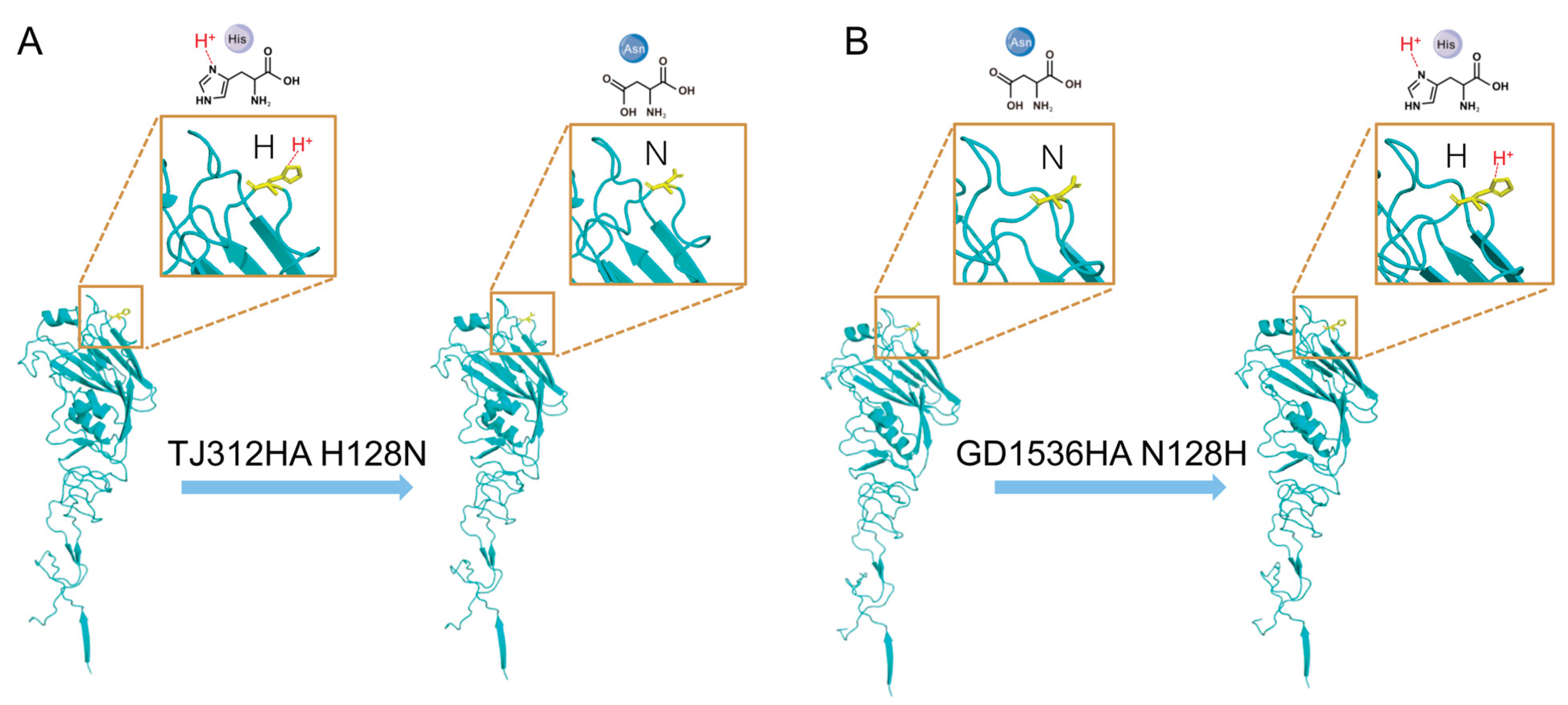
Table 1.
Viruses with mutations generated in the TJ312 background.
| Viruses | Virus rescue | |
| Mutation of HA | Successfully rescued | |
| TJ312/Mut-1 | I6L, V8I | Yes |
| TJ312/Mut-2 | I22V | Yes |
| TJ312/Mut-3 | N38D, S39K | Yes |
| TJ312/Mut-4 | S46K, N48R, K50V, I51A | Yes |
| TJ312/Mut-5 | Q54H | Yes |
| TJ312/Mut-6 | N57K | Yes |
| TJ312/Mut-7 | V60I | Yes |
| TJ312/Mut-8 | K69E, D71E, L72S, L74S, N77R | Yes |
| TJ312/Mut-9 | I83V | Yes |
| TJ312/Mut-10 | K89D | Yes |
| TJ312/Mut-11 | A92T | Yes |
| TJ312/Mut-12 | E97D, A99I, D100N | Yes |
| TJ312/Mut-13 | K105R | Yes |
| TJ312/Mut-14 | T110S | Yes |
| TJ312/Mut-15 | A123T, T124S | Yes |
| TJ312/Mut-16 | H128N | Yes |
| TJ312/Mut-17 | T131S, T132D, R133K | Yes |
| TJ312/Mut-18 | T135V, V137A, S138A, S140P, S142A | Noa |
| TJ312/Mut-19 | N145K | Yes |
| TJ312/Mut-20 | R149K | Yes |
| TJ312/Mut-21 | L152I, I154L | Yes |
| TJ312/Mut-22 | S165N, K166Q, S167T, T169I, N171D | No |
| TJ312/Mut-23 | I179L | Yes |
| TJ312/Mut-24 | V182I | Yes |
| TJ312/Mut-25 | D188I, S189A, D190V, Q192E, T193S | Yes |
| TJ312/Mut-26 | N198A, H199D, T200A | Yes |
| TJ312/Mut-27 | S203F | Yes |
| TJ312/Mut-28 | S206T, K208R, Y210S, R212K, T214K | Yes |
| TJ312/Mut-29 | V218A, A219T | Yes |
| TJ312/Mut-30 | E225D, A227E | Yes |
| TJ312/Mut-31 | L237V, D237E, Q239P | Yes |
| TJ312/Mut-32 | T242K | Yes |
| TJ312/Mut-33 | I252V, A253V, W255R, H256Y | Yes |
| TJ312/Mut-34 | A259T, L260M, K261E, K262R, G263D, S264A S265G | Yes |
| TJ312/Mut-35 | M269I, R270I | Yes |
| TJ312/Mut-36 | A273T, Q274P | Yes |
| TJ312/Mut-37 | N277D, T279N, K281T | Yes |
| TJ312/Mut-38 | H286E | Yes |
| TJ312/Mut-39 | L289I, K290N, G291T, N292S | Yes |
| TJ312/Mut-40 | V301I | Yes |
| TJ312/Mut-41 | Q314K | Yes |
| TJ312/Mut-42 | M317L | Yes |
| TJ312/Mut-43 | I324V | Yes |
Notes. aThe reverse genetics system was used for three times to ensure that the virus cannot be rescued.
Table 2.
The HI titers of the H1N1 viruses with antisera of different lineage viruses.
| Virus | Genetic group | Ferret antisera | |||
| TJ312 | GD1536 | GD104 | GX18 | ||
| TJ312 | EA1 | 640a | 80 | 20 | 640 |
| GD1536 | 2009/H1N1 | 40 | 1280 | 20 | 320 |
| GD104 | EA2 | <20 b | <20 | 1280 | 20 |
| GX18 | EA1 | 160 | 160 | 20 | 640 |
Notes. aHomologous tiers were marked in bold; b The lowest detectable level of the serum is 20.
Table 3.
The HI titers of the chimeric and mutant viruses reacting with the serum of TJ312 and GD1536.
Table 3.
The HI titers of the chimeric and mutant viruses reacting with the serum of TJ312 and GD1536.
| Virus | Ferret antisera | Virus | Ferret antisera | |||
| TJ312 | GD1536 | TJ312 | GD1536 | |||
| TJ312 | 640a | 80 | TJ312/Mut-21 | 640 | 80 | |
| GD1536 | 40 | 1280 | TJ312/Mut-22 | - | - | |
| chimeric G-T | 40 | 1280 | TJ312/Mut-23 | 640 | 40 | |
| chimeric T-G | 640 | 80 | TJ312/Mut-24 | 640 | 80 | |
| TJ312/Mut-1 | 640 | 80 | TJ312/Mut-25 | 160 | 10 | |
| TJ312/Mut-2 | 640 | 40 | TJ312/Mut-26 | 640 | 80 | |
| TJ312/Mut-3 | 640 | 80 | TJ312/Mut-27 | 640 | 160 | |
| TJ312/Mut-4 | 640 | 80 | TJ312/Mut-28 | 640 | 80 | |
| TJ312/Mut-5 | 640 | 80 | TJ312/Mut-29 | 640 | 40 | |
| TJ312/Mut-6 | 640 | 80 | TJ312/Mut-30 | 320 | 80 | |
| TJ312/Mut-7 | 640 | 80 | TJ312/Mut-31 | 640 | 80 | |
| TJ312/Mut-8 | 640 | 80 | TJ312/Mut-32 | 640 | 80 | |
| TJ312/Mut-9 | 640 | 80 | TJ312/Mut-33 | 640 | 80 | |
| TJ312/Mut-10 | 640 | 80 | TJ312/Mut-34 | 640 | 40 | |
| TJ312/Mut-11 | 640 | 80 | TJ312/Mut-35 | 640 | 80 | |
| TJ312/Mut-12 | 640 | 80 | TJ312/Mut-36 | 640 | 80 | |
| TJ312/Mut-13 | 640 | 80 | TJ312/Mut-37 | 640 | 40 | |
| TJ312/Mut-14 | 640 | 80 | TJ312/Mut-38 | 640 | 80 | |
| TJ312/Mut-15 | 640 | 80 | TJ312/Mut-39 | 640 | 80 | |
| TJ312/Mut-16 | 160 | 320 | TJ312/Mut-40 | 640 | 80 | |
| TJ312/Mut-17 | 320 | 80 | TJ312/Mut-41 | 640 | 80 | |
| TJ312/Mut-18 | -b | - | TJ312/Mut-42 | 640 | 80 | |
| TJ312/Mut-19 | 640 | 80 | TJ312/Mut-43 | 640 | 80 | |
| TJ312/Mut-20 | 640 | 80 | GD1536/ N128H | 320 | 320 | |
Notes. aHomologous tiers were marked in bold; bThe virus was not successfully rescued; G-T is a chimeric virus composed of the HA1 of GD1536 and the HA2 of TJ312, and T-G is a chimeric virus composed of the HA1 of TJ312 and the HA2 of GD1536.
Disclaimer/Publisher’s Note: The statements, opinions and data contained in all publications are solely those of the individual author(s) and contributor(s) and not of MDPI and/or the editor(s). MDPI and/or the editor(s) disclaim responsibility for any injury to people or property resulting from any ideas, methods, instructions or products referred to in the content. |
© 2025 by the authors. Licensee MDPI, Basel, Switzerland. This article is an open access article distributed under the terms and conditions of the Creative Commons Attribution (CC BY) license (http://creativecommons.org/licenses/by/4.0/).
Copyright: This open access article is published under a Creative Commons CC BY 4.0 license, which permit the free download, distribution, and reuse, provided that the author and preprint are cited in any reuse.


