Preprint
Article

This version is not peer-reviewed.

Convergent Gut Microbiome Adaptation and Pervasive Antibiotic Resistome in Qinghai‒Tibet Plateau Passerines

Submitted:

02 September 2025

Posted:

04 September 2025

You are already at the latest version

Abstract

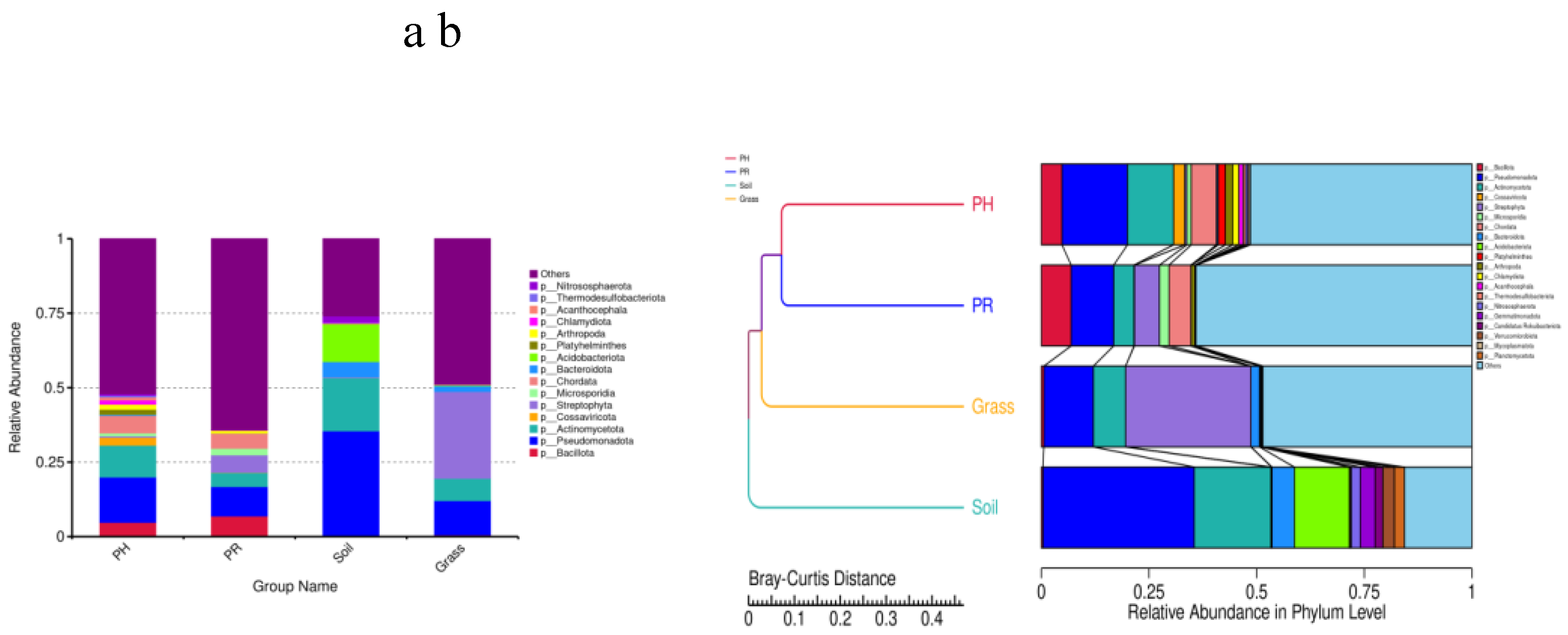
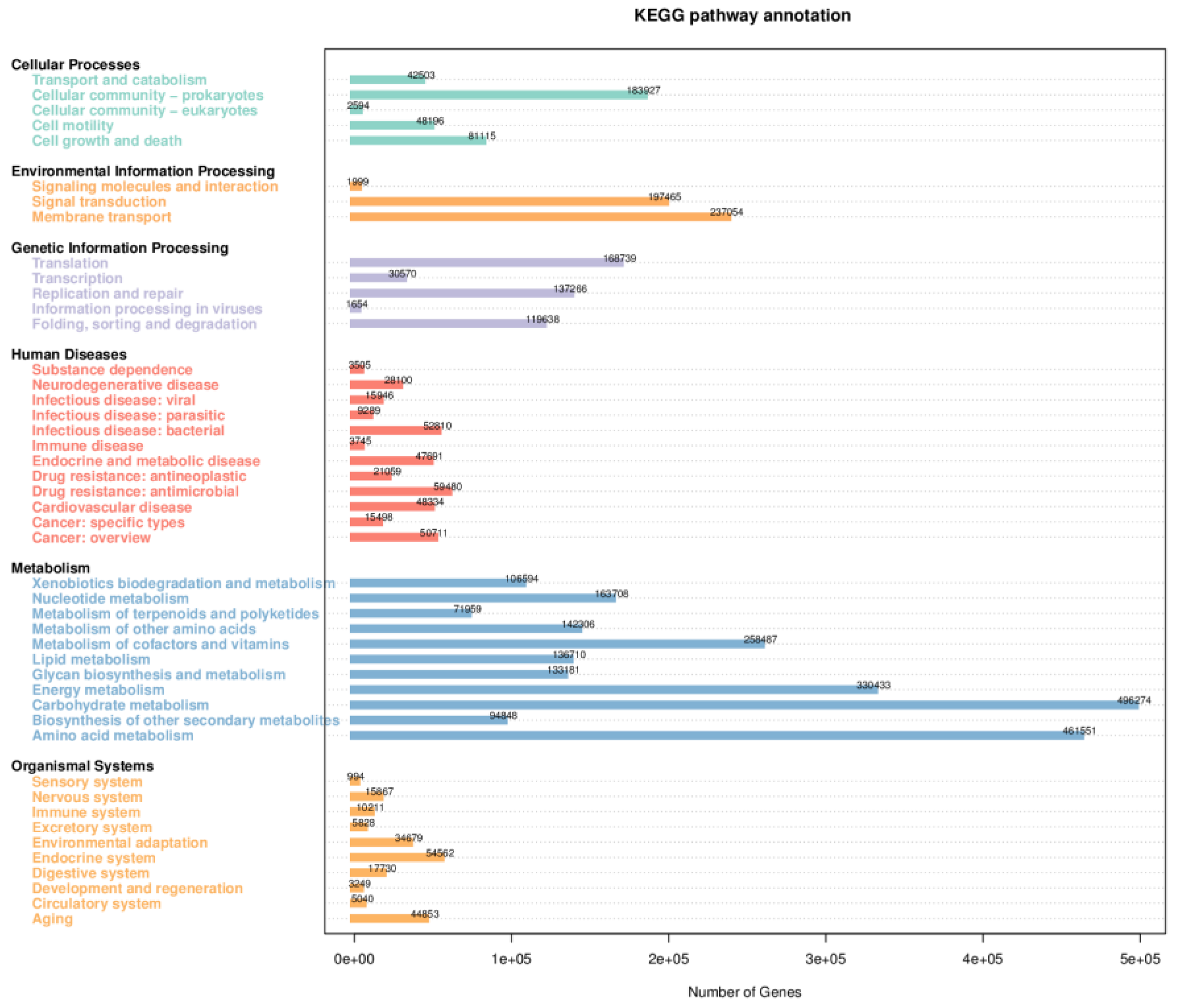
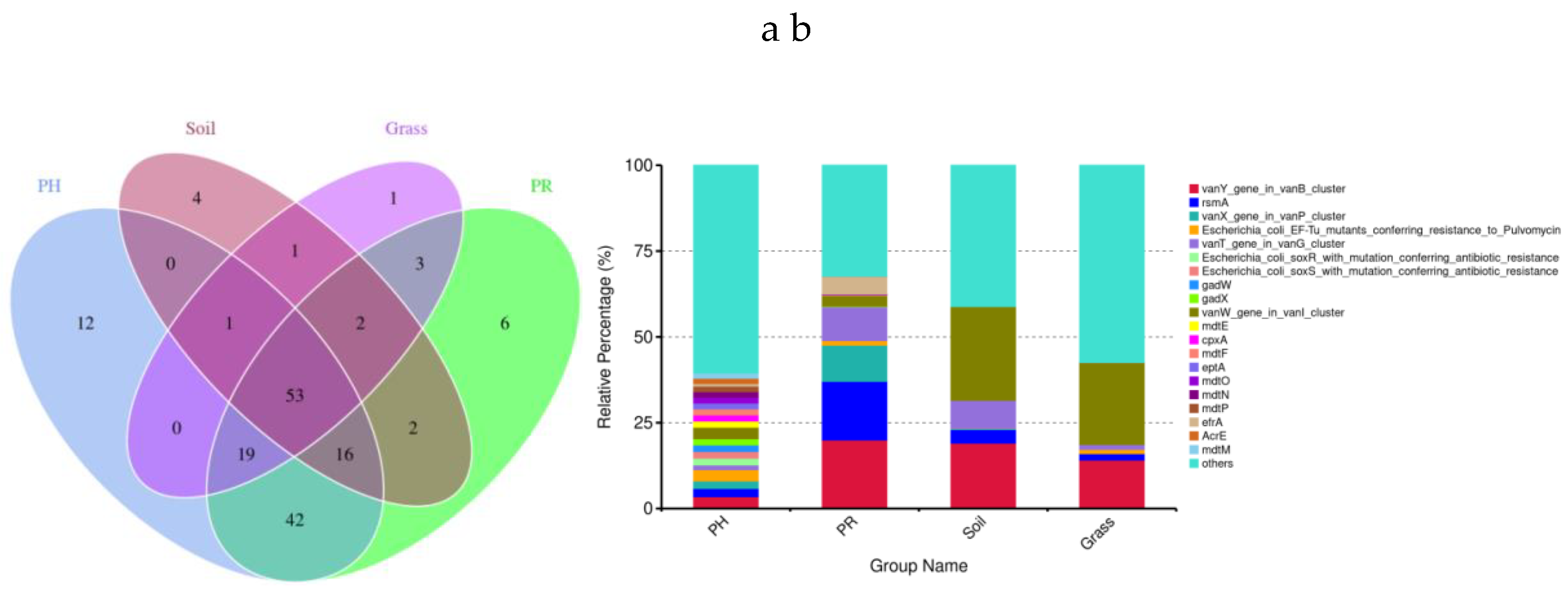
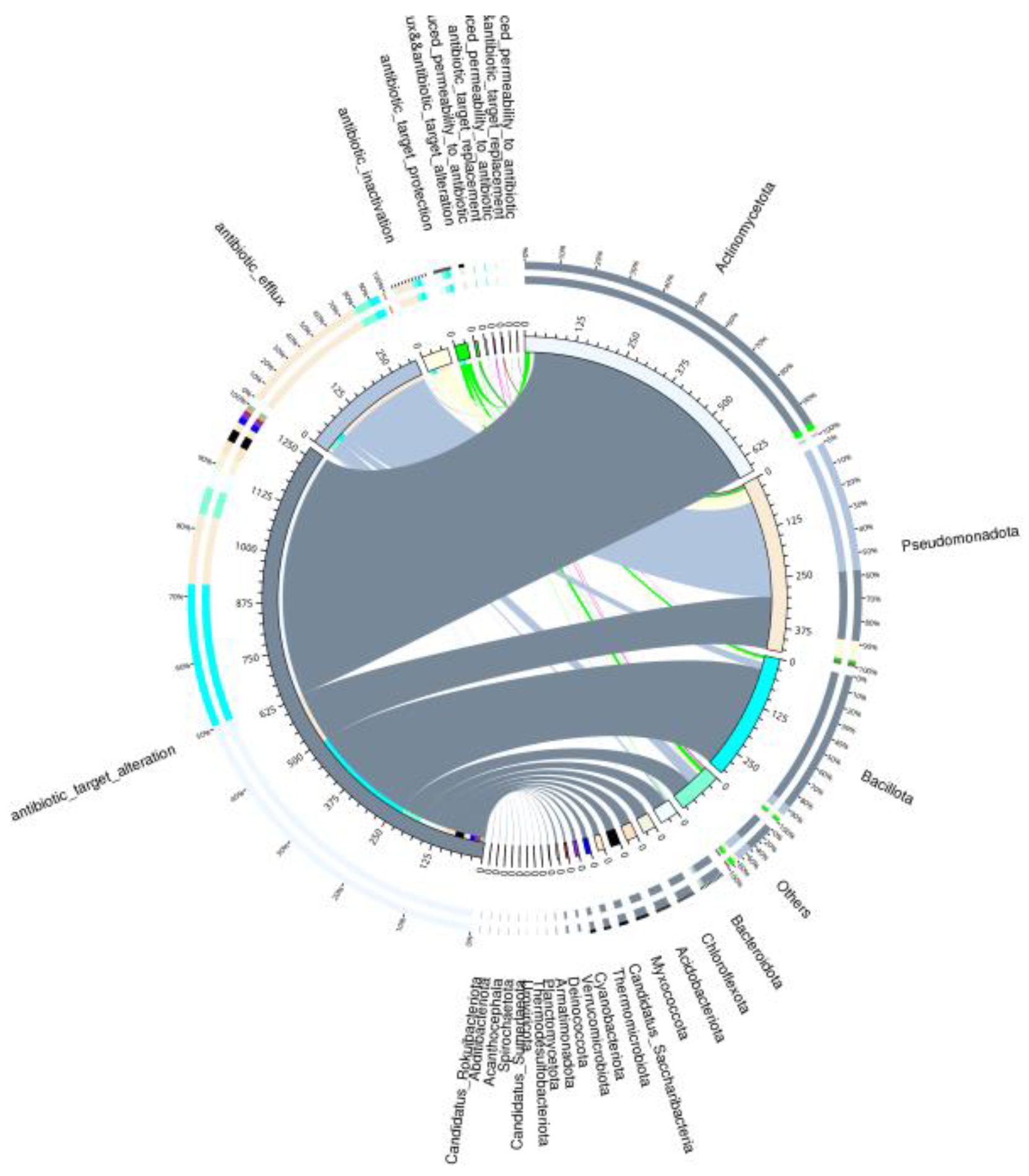
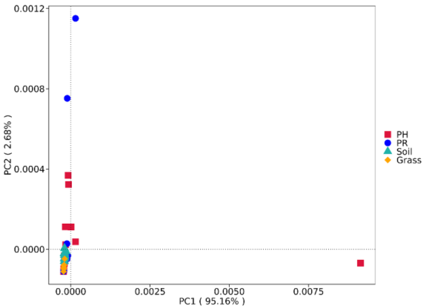
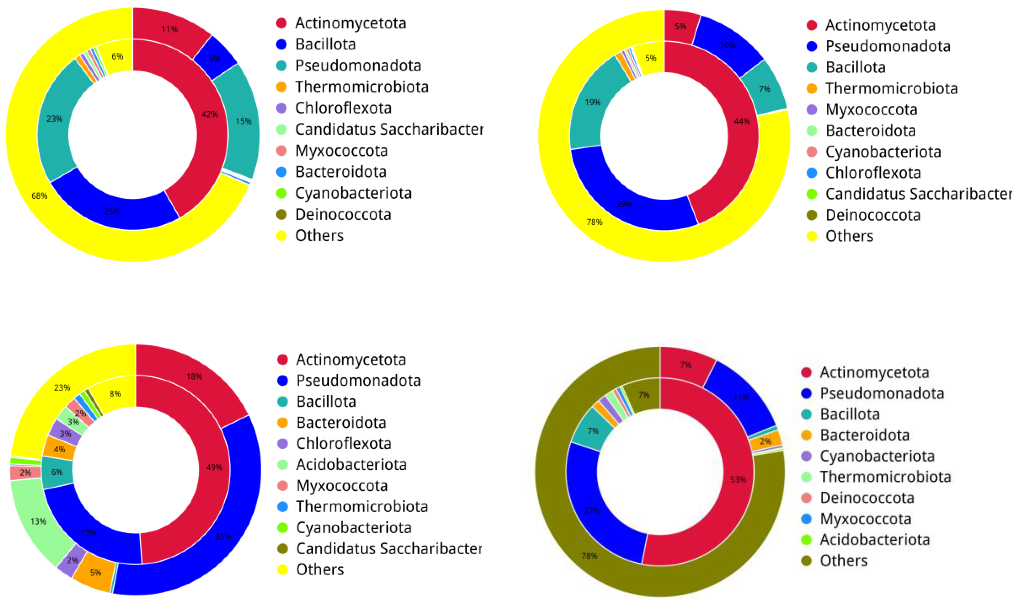
Metagenomic analysis of nonmigratory passerines (Pseudopodoces humilis and Pyrgilauda ruficollis) and their habitats on the Qinghai‒Tibet Plateau revealed convergent adaptations in gut microbial composition and function dominated by Bacillota and Pseudomonadota. Functional enrichment in carbohydrate metabolism and genetic information processing underpins host energy optimization in extreme high-altitude environments. Critically, these birds constitute a major reservoir of antibiotic resistance genes (ARGs), harbouring 162 antibiotic resistance ontologies (AROs) with nearly universal resistance to clinical antibiotic classes. The core resistome—comprising glycopeptide (van clusters), fluoroquinolone, and tetracycline resistance genes—reflects anthropogenic contamination amplified by environmental persistence. Environmental transmission pathways were unequivocally demonstrated via 53 AROs shared between avian hosts and proximal matrices (soil/grass), coupled with livestock-derived antibiotic influx through excreta, establishing the plateau as a hotspot for resistance gene flux. Strikingly, "low-abundance-high-resistance" taxa (Pseudomonadota, Actinomycetota, and Bacillota; ≤20% abundance but >90% ARG contribution) drive resistome plasticity, potentially facilitated by horizontal gene transfer. Our findings redefine resident passerines as sentinels of ecosystem health and bridges for cross-boundary antimicrobial resistance (AMR) spread. Mitigating global AMR thus necessitates interdisciplinary strategies targeting environmental reservoirs (e.g., regulating livestock antibiotic use) and monitoring avian-mediated gene flow.

Keywords: 
;  ;  ;  

1. Introduction

The Qinghai‒Tibet Plateau, the highest-altitude ecosystem on Earth, is characterized by a unique combination of hypobaric hypoxia, low temperatures, and intense ultraviolet radiation, which collectively drive coevolution between native species and their symbiotic microbial communities [1,2]. As a critical symbiotic interface, the gut microbiota facilitates host adaptation to extreme environments through two regulatory mechanisms: metabolic optimization and immune homeostasis maintenance [3]. The assembly of microbial communities results from the tripartite interplay of host genetics, dietary substrates, and environmental selection pressures [4,5,6]. Emerging evidence indicates that indigenous plateau mammals (e.g., plateau pikas and Tibetan antelope) exhibit seasonally stratified remodelling of the gut microbiota, enabling ecological niche-specific energy harvesting strategies (cellulose degradation in winter vs. protein catabolism in summer) [7,8]. These findings elucidate the microbe-mediated mechanisms underlying host extremophilic adaptation.
Meanwhile, tens of thousands of tons of antibiotics are consumed annually in healthcare, livestock, and agricultural settings worldwide [9]. This anthropogenic pressure has accelerated the global dissemination of antibiotic-resistant bacteria (ARB) and antibiotic resistance genes (ARGs), constituting a planetary-scale threat to ecological integrity and One Health security [10]. Critically, the increasing prevalence of multidrug-resistant (MDR) pathogens has established antimicrobial resistance (AMR)-associated infections as a leading cause of mortality worldwide, including AIDS-related deaths [11]. Environmental compartments—including soil matrices, aquatic systems, and animal waste reservoirs—serve as ARG hotspots, enabling cross-ecosystem transmission through horizontal gene transfer (HGT), hydrological connectivity, and mobile vectors (e.g., migratory avifauna) [12,13]. Mounting experimental evidence has demonstrated that gut microbiomes function as critical reservoirs for persistent ARB populations and transmissible ARG repertoires [10,14]. Notably, wild birds represent underappreciated mobile vectors facilitating the intercontinental spread of evolving ARB lineages and novel ARG combinations through latitudinal migration networks [13].
Compared with those of mammals, avian gut microbiomes exhibit lower stability and greater plasticity [15], traits strongly influenced by selective pressures from their complex life-history strategies, including dietary flexibility, flight-associated physiological adaptations, and long-distance migrations [16]. Metagenomic analyses by Lin et al [17] revealed that migratory birds harbour mobile genetic elements (MGEs) and demonstrate high coselection potential for antibiotic resistance genes (ARGs), increasing the risk of environmental contamination. Notably, endemic Tibetan Plateau species such as Pseudopodoces humilis and Pyrgilauda ruficollis have evolved hypoxia-responsive mechanisms to thrive in extremely high-altitude environments (3200–4550 m) [18]. Despite these adaptations, current avian microbiome research remains disproportionately focused on low-altitude and migratory waterfowl. Field sampling challenges have hindered investigations of plateau-dwelling resident birds, which serve dual roles as AMR reservoirs and bioindicators of ecosystem health in low-disturbance regions. The distribution patterns of ARGs and host‒microbe coadaptation mechanisms in these birds’ unique habitats remain poorly characterized, underscoring the urgent need for targeted studies to address the increasing global threat of antibiotic resistance.
Previous studies have characterized the gut microbiota of avian species such as Haradrius alexandrinus, Charadrius alexandrinus, and Passer montanus using 16S rRNA amplicon sequencing and culture-dependent methods [19,20]. However, these approaches offer limited resolution of microbial community dynamics and fail to comprehensively resolve antibiotic resistance gene (ARG) profiles in wild birds. Recent advances in high-throughput metagenomics have enabled systematic annotation of functional pathways and ARG identification across host-associated and environmental microbiomes [21,22]. To address these gaps, we selected two plateau-endemic resident birds, the Ground Tit (Pseudopodoces humilis) and the Rufous-necked Snowfinch (Pyrgilauda ruficollis), as model systems. Leveraging metagenomic sequencing, we aimed to (1) elucidate functional regulatory networks governing host‒microbe interactions under extreme environmental stress, (2) characterize community-wide ARG distribution patterns and horizontal transmission potential, and (3) evaluate the role of resident birds as ARG reservoirs and the implications for ecosystem health. Our findings advance the understanding of microbial evolutionary adaptation in extremely high-altitude ecosystems while providing actionable insights into the wildlife-mediated global dissemination of antimicrobial resistance, thereby informing strategies to mitigate this pressing public health challenge.

2. Materials and Methods

2.1. Sampling

Sampling efforts were carried out at three sites in Tianjun County, Menyuan County and Xinghai County, Qinghai Province, from July to September 2024 (Figure S1 and Table S1). During the feeding of cattle and sheep in pastoral areas, Tibetan people often observe birds competing for food. We trapped Pseudopodoces humilis (in text abbreviation: PH group) and Pyrgilauda ruficollis (in text abbreviation: PR group) using live trapping and locked them in cages previously sterilized with 75% alcohol. Fresh faeces were collected in 2-mL tubes (Kejin, China) and immediately frozen in liquid nitrogen before being sent for analysis to the Qinghai Provincial Institute for Endemic Disease Prevention and Control, Xining, China. The faecal samples were collected at a minimum distance interval of five metres to ensure that all the fresh droppings were expelled from different individuals, and the captured individuals were marked with nontoxic avian leg bands prior to release. In total, we collected 16 PH and 16 PR faecal samples, 15 soil samples and 6 grass samples.

2.2. DNA Extraction and Sequencing

Metagenomic DNA was isolated from approximately 0.25 g samples using a TIANamp Soil DNA Kit (Tiangen Biotech; Beijing; China) following the manufacturer’s instructions. The DNA concentration and quality were assessed with an Agilent 5400 instrument. The high-quality DNA was randomly fragmented into segments of approximately 350 bp using a Covaris ultrasonic disruptor to construct the library. Library preparation was completed through steps including end repair, addition of A-tails, ligation of sequencing adapters, purification, and PCR amplification. All libraries were then subjected to 2 × 150 bp paired-end sequencing on the BGISEQ DNBSEQ-T7 platform (Novogene, Beijing, China).

2.3. Sequence Analyses and Metagenome Assembly

The raw BGISEQ paired-end sequencing data were preprocessed using Fastp (v0.23.1) with the following criteria. Reads were discarded if either paired read contained (a) adapter sequences (detected by default overlap analysis); (b) >10% ambiguous bases (N); or (c) >50% low-quality bases (Phred score <5). Considering the possibility of host contamination in samples, clean data were subjected to BLAST analysis against the host database to filter out reads of host origin. Bowtie2 software (v2.5.4) was used with the following default parameter settings: --end-to-end, --sensitive, -I 200, and -X 400 [23,24,25]. MEGAHIT software (v1.2.9) was used for assembly analysis of the clean data, with the following assembly parameter settings: --presets meta-large (--end-to-end, --sensitive, -I 200, -X 400) [24,26], and scaftigs without N were obtained by breaking the resulting scaffolds from the N junction [27,28].

2.4. Gene Prediction and Construction of the Nonredundant Gene Set

With the default parameters, MetaGeneMark (v2.1) was used to perform ORF prediction for scaftigs (≥ 500 bp) of each sample [23,29,30,31,32], and sequences with a length of less than 100 nt in the prediction results were filtered out [26,27,33,34,35]. For the ORF prediction results, CD-HIT software (v4.5.8) was used to eliminate redundancy [36,37] and obtain the nonredundant initial gene catalogue (the nucleic acid sequences encoded by successive nonredundant genes are called genes) [34], with parameter settings: -c 0.95, -G 0, -aS 0.9, -g 1, -d 0 [30,32]. Clean data from each sample were aligned to the initial gene catalogue by using Bowtie2 to calculate the number of reads of the genes in each sample alignment, with the following parameter settings: --end-to-end, --sensitive, -I 200, -x 400 [27,30]. Genes with <= 2 reads in each sample were filtered out to finally determine the gene catalogue (unigenes) for subsequent analysis [34]. On the basis of the abundance of each gene in the gene catalogue in each sample, the abundance information of each unigene in each sample was statistically analysed.

2.5. Gene Taxonomic Prediction

DIAMOND software (v2.1.9) [38] was used for alignment of unigene sequences with the Micro_NR database, which included sequences from bacteria, fungi, archaea, and viruses extracted from the NCBI NR database (https://www.ncbi.nlm.nih.gov/). The alignment was performed using the BLASTP algorithm with a parameter setting of 1e-5 [24]. From the alignment results of each sequence, the one with evalue <= min. evalue *10 was selected. Since each sequence may have multiple alignment results, the LCA algorithm (applied to the systematic taxonomy of MEGAN software (https://en.wikipedia.org/wiki/Lowest_common_ancestor)) was adopted to determine the species annotation information of the sequence [39]. In addition to the results of the LCA annotation and gene abundance table, the abundance of each sample at each taxonomic level and the corresponding gene abundance tables were acquired. The abundance of a species in a sample. Microbiome analyses were conducted using a standardized bioinformatics pipeline. Taxonomic abundance profiles across hierarchical levels were visualized through relative abundance profiles and Bray‒Curtis distance-based clustering trees implemented in Perl SVG (v5.18.2). Alpha diversity metrics, including the Chao1 richness and Shannon entropy indices, were computed using the R vegan package (v2.15.3). Beta diversity patterns were examined via principal coordinate analysis (PCoA) employing the R packages extrafont, ggplot2, and grid (v2.15.3 for core dependencies), with dimensionality reduction performed on normalized abundance matrices. Intersectional analysis of taxonomic features across sample groups was visualized using the R VennDiagram package (v3.0.3).

2.6. Functional Gene Annotation

Functional annotation of the unigenes was performed using DIAMOND (v2.0.15) with BLASTP alignment against the KEGG (v2023.1) [40,41] and PHI (v4.12) databases, applying an e-value cut-off of 1e−5 [30,42]. From the alignment results of each sequence, the best BLAST hit results were selected for subsequent analysis [30,32,43,44]. The relative abundance at different functional levels was calculated according to the alignment results (the relative abundance at each functional level was equal to the sum of the relative abundance of genes annotated at that functional level) [23,30]. For antibiotic resistance profiling, the unigenes were aligned to the CARD database (https://card.mcmaster.ca/) [45] using Resistance Gene Identifier (RGI) software (v6.0.2) [46] provided by the CARD database (RGI built-in BLASTP, default evalue < 1e-30) [47]. The relative abundance of each ARO was calculated according to the RGI alignment result and unigene abundance information. To reveal the relationship between microbial composition and the resistome, Circos (v0.64)-generated visualization of abundance distribution circle map.resistance genes (unigenes annotated as ARO) and species attribution analysis of the resistance mechanism were carried out (some AROs with long names are abbreviated as the first three words plus underlines).

3. Results

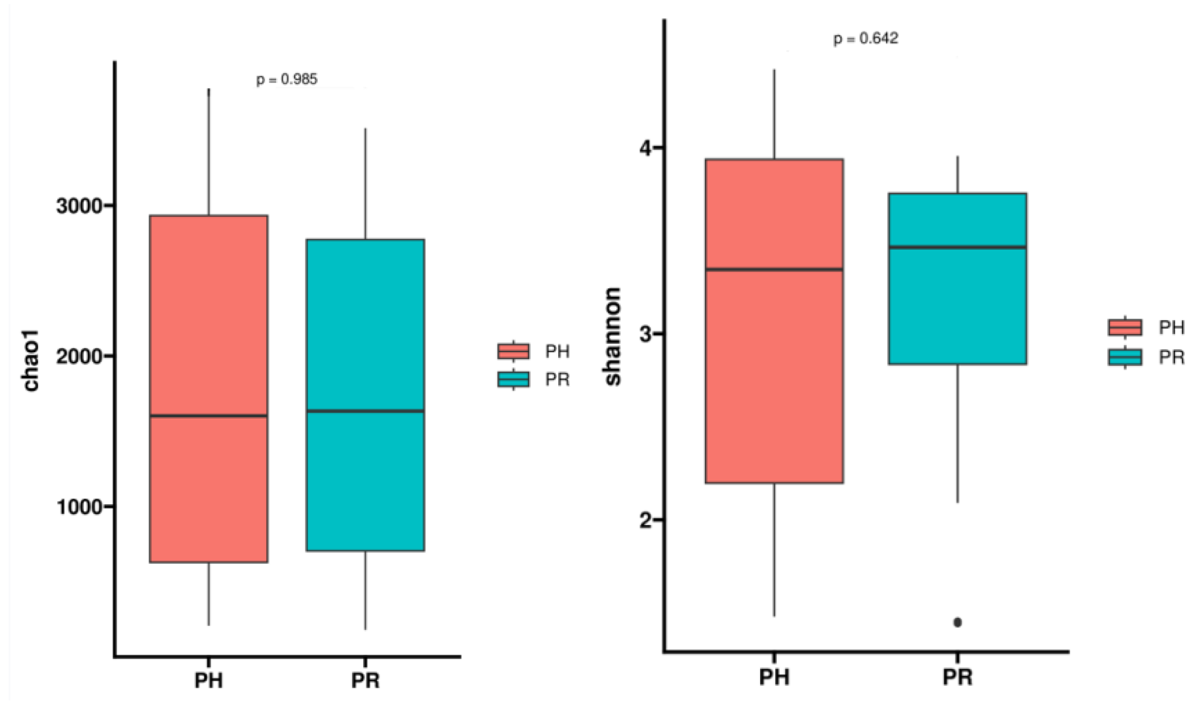
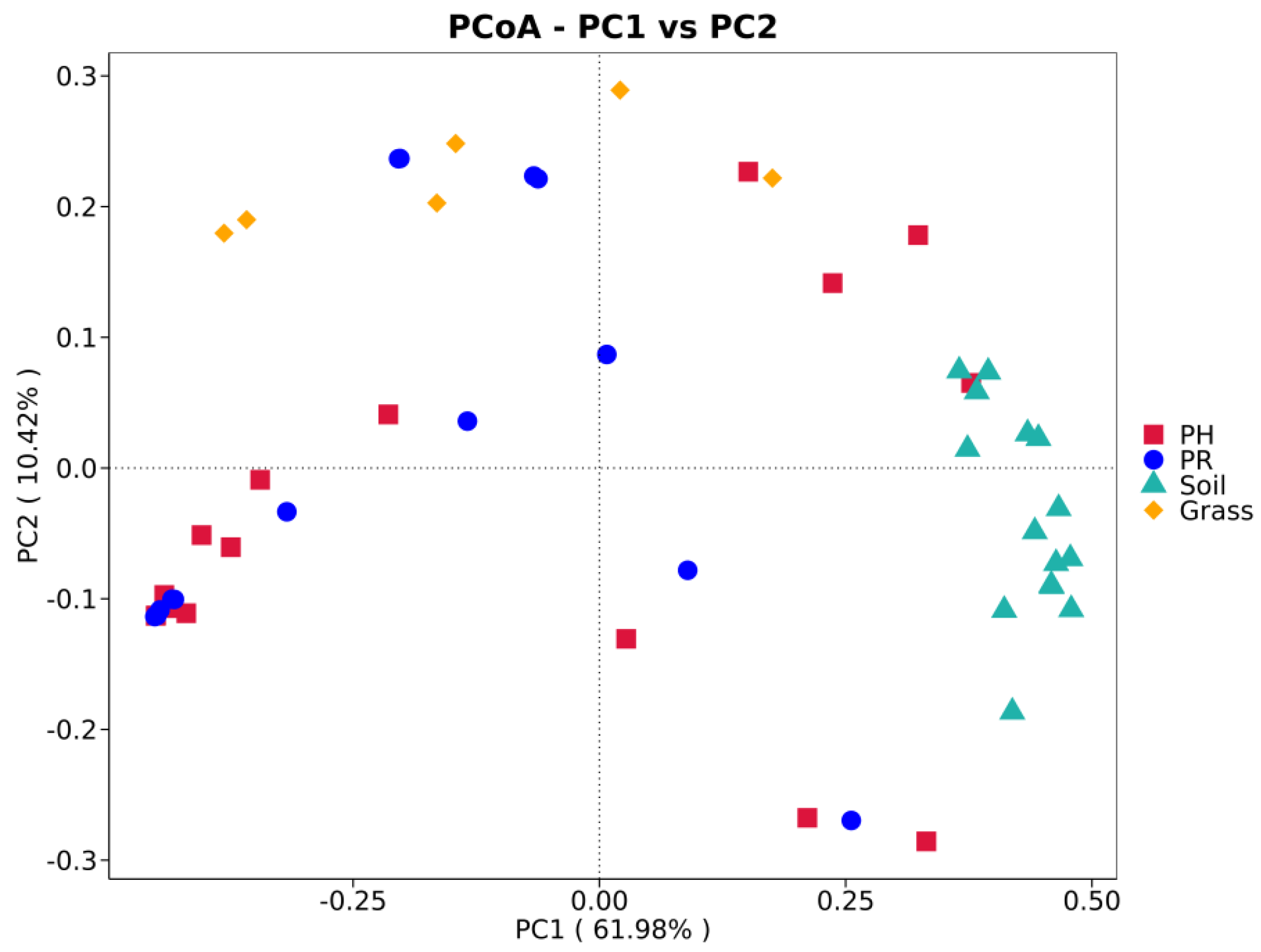
3.1. Bacterial Community Profile

A total of 53 metagenomes were sequenced using the BGISEQ DNBSEQ-T7 platform (2 × 150 bp). High-throughput sequencing of the microbial DNA samples generated 391.4 Gb of high-quality clean data from the 53 samples. An average of 7.38 Gb were obtained per sample (Supplementary File 1). Furthermore, after de novo assembly, 1306.62 Mb of scaftigs (with total lengths ranging from 0.94 to 52.13 Mb) were generated, with an N50 of 76426 bp (Supplementary File 2).
Taxonomic annotation of the metagenomic sequencing data against the NR database revealed that Bacteria dominated the microbial community and presented significantly greater phylogenetic diversity than did Archaea, Eukaryota, and viral sequences (Supplementary File 3). The top four phyla in the PH group were Pseudomonadota (15.21%), Actinomycetota (10.70%), Chordata (5.86%), and Bacillota (4.79%) (Figure 1a and (Supplementary File 4). In the PR group, Pseudomonadota also predominated, with an average relative abundance of 9.88%, followed by Bacillota (6.91%), Streptophyta (5.82%), and Chordata (5.10%). Unclassified sequences and low-abundance sequences were categorized as "Others," accounting for 52.04% and 64.18% of the total community in each sample, respectively, indicating the presence of a significant number of unknown bacteria in the guts of these two bird species. A considerable proportion of Pseudomonadota was detected in the soil (35.13%) and grass (11.49%) samples, while Streptophyta (29.14%) was the most dominant phylum in the grass samples. At the genus level, Enterococcus represented the most abundant bacterial taxon in both avian species, with relative abundances of 2.06% (PH) and 4.26% (PR), while host-specific secondary genera emerged—Rhodococcus (1.84%) and Pseudomonas (1.75%) in PH versus Turicibacter (1.97%) and Hordeivirga (1.65%) in PR (Figure S1 and (Supplementary File 5). Hierarchical clustering analysis revealed that bacterial communities in the faeces of Tibetan PH and PR clustered closely between species and were obviously distinct from other environmental communities (Figure 1b). Moreover, the grass and soil samples were grouped together.
Venn diagrams revealed a conserved gut microbial architecture between PH and PR (Figure S2), despite divergent abundance patterns in specific taxa. Concordantly, α diversity metrics demonstrated comparable community complexity between species (Figure 2). To distinguish the differences in microbial communities across various ecological environments, we compared the metagenomic sequencing data from wild bird faeces with those from environmental samples collected from their surrounding areas. The PCoA results indicated that, at the phylum level, the intestinal microbiota of most passerine hosts (PH) exhibited compositional convergence with the predominant microbial profile of proximal environmental reservoirs (PR) (Figure 3). Significant divergence emerged between specific PH species and PR cohorts, suggesting host phylogenetic constraints on microbial assembly. Subsequent genus-level PCoA corroborated these macroecological patterns while resolving finer taxonomic stratification among niches (Figure S3).
A total of 228 opportunistically pathogenic species were identified via comparison against a previously published pathogen list [48] Salmonella enterica, Pseudomonas aeruginosa, and E. coli were the most prevalent bacteria. Furthermore, several clinically critical pathogens, such as Staphylococcus aureus, Yersinia pestis, and Vibrio cholerae, were consistently observed.

3.2. Functional Profiling of the Gut Metagenome

The metabolic and functional pathways of the nonredundant (NR) gene catalogue derived from the microbiome were annotated using the KEGG database. Approximately 50% of the NR genes (17,408 KEGG Orthology (KO) functions) were assigned to 44 level II orthology groups (Figure 4). Among the annotated functions, metabolic pathways dominated (40.47% of total KO assignments), with the following subcategories: carbohydrate metabolism (10.73%), amino acid metabolism (10.05%), energy metabolism (9.57%), metabolism of cofactors and vitamins (5.83%), nucleotide metabolism (3.75%), and lipid metabolism (3.00%). The remaining functional categories primarily included genetic information processing (13.86%) and environmental information processing (10.88%). Furthermore, Bray‒Curtis distance analysis based on the microbial functional module composition revealed similar clustering patterns among the samples (Figure S4).

3.3. Antibiotic Resistance Profiles

To profile antibiotic resistance genes (ARGs) in the gut microbiota of avian and environmental samples, metagenomic unigenes were screened against the Comprehensive Antibiotic Resistance Database (CARD v3.2.5). We identified 162 distinct antibiotic resistance ontology (ARO) categories across the 4 metagenome samples (Supplementary File6). Avian-associated samples dominated the resistome, containing 143 AROs (88.27% of the total), of which 53 were shared with the environmental samples(Figure 5a). Notably, 130 AROs (90.91% of avian-associated AROs) were common to both the PH and the PR, indicating conserved resistance gene pools between these sympatric passerines.
The 20 most abundant AROs across samples revealed that the faecal samples from PR presented relatively high ARG abundances, followed by the soil samples from the environment (Figure 5b). Although PH shared a large portion of AROs with PR (n=130), the abundance of these AROs in PH was much lower than that in PR. Most of these genes represented antibiotic target alterations, encompassing four classical glycopeptide resistance gene clusters, namely, the vanI cluster, vanB cluster, vanG cluster, and vanP cluster, which confer resistance to glycopeptide and tetracycline antibiotics. The second most common function was antibiotic efflux, represented by three types of classic multidrug efflux pumps: the resistance-nodulation-cell division (RND)-type, ATP-binding cassette (ABC)-type and major facilitator superfamily (MFS)-type, which confer resistance to fluoroquinolones, macrolides, diaminopyrimidine, phenicol, lincosamides, nucleosides, and rifamycin, as well as disinfecting agents and antiseptics.
To systematically elucidate the associations between bacterial communities and antibiotic resistance mechanisms within the samples, we generated a double-circle plot depicting the relationships between metagenomic bacteria and resistance mechanisms (Figure 6). Antibiotic target alteration has emerged as the most prevalent resistance mechanism, exhibiting extensive associations with multiple bacterial phyla and demonstrating significant enrichment within Pseudomonadota. Furthermore, antibiotic efflux was also highly prevalent in both Pseudomonadota and Bacillota, indicating that efflux pumps represent a common resistance strategy in these taxa. Additionally, certain phyla, including Bacteroidota, Chloroflexota, and Verrucomicrobiota, exhibited moderate associations with multiple resistance mechanisms. This pattern reflects the diverse adaptation strategies employed by bacterial populations in response to antibiotic pressure in the sampled environment. We next assessed the similarity of ARO composition among each sample group via PCoA. Our findings revealed significant overlap between avian and environmental (soil/grass) resistomes (Figure 7). Notably, avian samples presented greater β diversity than did environmental samples. Antibiotic target alterations were more enriched in the PH resistome, whereas antibiotic efflux mechanisms were more prevalent in the PR resistome.
These AROs were further analysed for their microbial origin. Taxonomic analysis revealed that the AROs were predominantly associated with Pseudomonadota, Actinomycetota, and Bacillota across all the samples (Figure 8a–d). Approximately 90% of AROs in PH, 92% in PR, and 87% in grass samples originated from these phyla, despite their relatively low microbial abundances: Pseudomonadota constituted 15%, 10%, and 11% of the total microbiota in PH, PR, and grass, respectively; Actinomycetota accounted for 11% (PH), 5% (PR), and 7% (grass); and Bacillota represented 5% (PH), 7% (PR), and 0.6% (grass). In contrast, the soil samples exhibited divergent patterns, with 49% of the AROs linked to Actinomycetota (10% microbial abundance) and 23% to Pseudomonadota (35% abundance). These findings underscore a systemic disproportionality between microbial prevalence and resistance gene contribution, where phylogenetically restricted taxa drive ARG reservoirs despite their low ecological representation.

4. Discussion

This study provides the first systematic characterization of gut microbiome adaptation and ARG dissemination in two plateau-endemic passerines—Pseudopodoces humilis (PH) and Pyrgilauda ruficollis (PR)—under extreme environmental conditions on the Qinghai‒Tibet Plateau. These species were selected not only for their ecological representativeness as endemic taxa but also for their dual importance in elucidating extremophilic adaptation and ARG transmission dynamics. Taxonomic analysis revealed no significant divergence in the gut microbial community structure at the phylum level between species, with Bacillota and Pseudomonadota dominating both assemblages, which is consistent with the typical passerine microbiota [49,50,51]. This similarity likely arises from their shared ecological niche as cave-dwelling species and partial dietary overlap, underscoring the roles of host ecology, diet, and environmental selection in shaping microbial communities, in accordance with the host‒microbe coevolution theory [52,53]. Members of the phylum Bacillota (e.g., Butyrivibrio spp.) mediate the degradation of insoluble fibres (e.g., cellulose and resistant starch) and produce butyrate, conferring anti-inflammatory benefits and enhancing intestinal barrier integrity. These metabolic functions enable hosts to extract energy and nutrients from carbohydrates, polysaccharides, sugars, and fatty acids [54,55,56]. Notably, sequences assigned to Chordata were detected at substantial abundances in both species (Pseudopodoces humilis (PH): 5.86%; Pyrgilauda ruficollis (PR): 5.10%), potentially reflecting increased meat consumption during summer sampling (July–September) or indicating the presence of intestinal parasites (e.g., nematodes). At the genus level, Enterococcus was predominant in both birds. This genus frequently exhibits multidrug resistance (particularly to β-lactams and quinolones), highlighting its ARG dissemination potential [57]. Key distinctions included elevated abundances of Rhodococcus (1.84%) and Pseudomonas (1.75%) in Pseudopodoces humilis (PH), which aligned with their relatively high proportions of Pseudomonadota (15.21%) and Actinomycetota (10.70%). Pseudomonas species possess metabolic capabilities for denitrification, sulfur oxidation, and urea degradation, potentially serving as biocontrol agents against pathogens such as Vibrio species [58]. Conversely, Pyrgilauda ruficollis (PR) presented relatively high abundances of plant/soil-associated genera (e.g., Triticum: 1.97%; Hordeum: 1.65%) and increased Streptophyta abundance in grass samples (snowfinch: 5.82%; grass: 29.14%). This suggests greater grass seed consumption in Pyrgilauda ruficollis (PR), with their microbiota more substantially influenced by environmental bacteria (e.g., soil- and plant-derived microbes) [59]. In support of this, the soil samples presented high abundance of Acidobacteriota (12.78%), a diverse and ubiquitous soil phylum linked to local geochemical properties [60,61]. The microbial structure of birds is different from that of soil, and the phylogenetic composition of the soil microbiota is less heterogeneous than that of the bird microbiota and grass microbiota, potentially because of the diverse and variable conditions in the environmental samples. The diversity and composition of soil bacterial communities, on a large spatial scale, can be predicted largely by a single variable: soil pH [62].
We further elucidated the microbial risks associated with Pseudopodoces humilis (PH) and Pyrgilauda ruficollis (PR), in which 228 opportunistic pathogens were detected. The identification of such a high abundance of opportunistic pathogens underscores the complexity of their microbial communities' pathogenic potential and suggests that wild birds may serve as potential reservoirs of infection, highlighting the need for continuous surveillance of these pathogens. Systematic screening of pathogen profiles using metagenomic technology provides novel strategies for disease prevention, control, and early diagnosis while also establishing a more robust data foundation for related pathological studies.
The 21st-century public health challenge posed by the global spread of antibiotic resistance (AR) involves complex environmental drivers in avian species inhabiting the Qinghai‒Tibet Plateau. Environmental compartments in China demonstrate pervasive antibiotic contamination, with detection rates reaching 100% in soils, 98.0% in surface waters, and 96.4% in coastal waters [63], establishing reservoirs for ARG dissemination. As critical environmental vectors, birds facilitate the transmission of ARB through faecal deposition and contaminated water sources [64,65,66]. This study employed metagenomic sequencing to conduct unbiased resistome profiling of Pseudopodoces humilis (PH) and Pyrgilauda ruficollis (PR), circumventing the limitations inherent in targeted detection approaches [67]. We comprehensively identified 162 ARO categories. While both species shared >90% of the ARGs, Pyrgilauda ruficollis (PR) had a significantly greater total ARG abundance. The gut microbiomes of these species have demonstrated resistance to nearly all major classes of antibiotics relevant to clinical and agricultural practice, with glycopeptide-, fluoroquinolone-, and tetracycline-resistance genes constituting the core resistome. Glycopeptide antibiotics, notably vancomycin and teicoplanin, have served as last-resort therapeutics against bacterial infections for more than half a century and remain essential for treating methicillin-resistant Staphylococcus aureus (MRSA) and penicillin-resistant Streptococcus pneumoniae. The historical use of avoparcin as a livestock growth promoter [68] likely contributed to the evolution of vancomycin-resistant enterococci (VREs) [69,70]. Critically, homologous resistance genes (vanA, B, C, D, E, and G) have been detected in >10,000-year-old permafrost samples [71,72] and are highly abundant in contemporary soil, marine, and human faecal matrices [73], confirming that environmental reservoirs are intrinsic sources of ARGs. The persistence of fluoroquinolone resistance is linked to environmental stability and low biodegradability [74], whereas tetracycline resistance genes are widely distributed among plateau nonmigratory birds (e.g., corvids and Gyps himalayensis) [75,76], collectively indicating a regional ecological network of resistance. Given the absence of direct antibiotic exposure in both bird species, their ARGs likely originate from environmental pathways (e.g., antibiotic residues from free-grazing livestock enter ecosystems via excreta [77] ), with 53 AROs shared between birds and environmental samples (soil, grass), highlighting the critical role of environmental transmission. Substantial quantities of antibiotics discharged from hospitals and livestock operations contaminate rivers, sediments, and soils [78], enabling the transmission of antibiotic-resistant bacteria (ARB) from contaminated matrices to avian species [79]. ARGs may be disseminated to hosts via contaminated water, food chains, or aerosols, posing elevated risks in regions with limited public health infrastructure [80]. The resistome composition was significantly associated with microbial community structure. Although Pseudomonadota, Actinomycetota, and Bacillota represented only 20% of the total relative microbial abundance, they harboured >90% of the identified ARGs. This "low-abundance-high-resistance" phenomenon suggests that these taxa serve as key vectors for ARG dissemination, potentially facilitated by horizontal gene transfer (HGT) [81]. Consequently, structural shifts in microbial communities may directly modulate resistance transmission.
The present study has several limitations that should be acknowledged. First, the relatively small sample size (*n* = 53) and cross-sectional design constrained our ability to investigate gut microbiota dynamics and their impacts across the host life cycle. Future studies should incorporate broader taxonomic representations, encompassing both migratory and resident avian populations across diverse ecosystems, to systematically elucidate the ecological and evolutionary drivers shaping microbial communities and their role in antibiotic resistance transmission. Longitudinal monitoring is particularly essential to resolve the effects of seasonal fluctuations, environmental stressors (e.g., extreme climatic events, variations in food resources), and migratory behaviour on microbial community structure and resistome dynamics. Additionally, annotations relying on the NR and CARD databases are inherently subject to taxonomic bias towards well-characterized taxa, potentially overlooking plateau-specific antibiotic resistance genes (ARGs) or novel resistance mechanisms. Therefore, integrating culturomics with single-cell sequencing technologies represents a critical approach for characterizing the biological traits and resistance potential of uncultivated/unclassified microorganisms, thereby advancing the current understanding. Furthermore, the absence of quantitative data on environmental factors (e.g., antibiotic residue concentrations, pH, and heavy metal pollution) impedes the precise delineation of the environmental drivers underpinning the resistome. Future investigations should employ multivariate statistical modelling frameworks (e.g., structural equation modelling and machine learning algorithms) to quantify causal relationships between multiple environmental stressors and the evolution of microbial antibiotic resistance.

5. Conclusions

On the basis of metagenomic analysis of nonmigratory passerines and their habitats on the QingHai Tibetan Plateau, this study revealed significant convergence in gut microbial composition and function across species. These birds represent critical reservoirs of antibiotic resistance genes (ARGs), which serve as major sources of resistance determinants for local environmental bacteria. Transmission pathways were clearly demonstrated by the sharing of 53 resistance determinants between avian hosts and proximal environmental matrices (soil/grass), combined with livestock-derived antibiotic influx via excreta. These mechanisms establish the Tibetan Plateau as a hotspot for resistance gene dissemination. Collectively, these findings reposition resident passerines as sentinel species for ecosystem health and bridge the need for cross-boundary antimicrobial resistance (AMR) transmission. Mitigating global AMR thus necessitates interdisciplinary strategies targeting environmental reservoirs (e.g., regulating antibiotic use in livestock operations) and monitoring avian-mediated gene flow.

Supplementary Materials

The following supporting information can be downloaded at the website of this paper posted on Preprints.org. Fig.S1 Taxonomic profiles of the microbial communities at the genus level in each sample.Fig.S2 Venn diagrams showing the unique and shared microbial genera between Group PH and Group PR.Fig.S3 Principal Co-ordinates Analysis (PCoA) based on abundance of genus lever (Bray-Curtis algorithm).Fig.S4 KEGG clustering tree based on Bray-Curtis distance. File 1: Statistical Results of Raw Sequencing Data Preprocessing.File 2:Summary of Assembled Scaftigs (≥ 500 bp) Across Samples.File 3:Relative Ab:ndance of Taxonomic Annotation at Kingdom Level Across Samples.File 4:Relative Ab:ndance of Taxonomic Annotation at Phylum Level Across Samples.File 5:Relative Ab:ndance of Taxonomic Annotation at Genus Level Across Samples.File 6:Antibiotic Resistance Ontology(ARO)annotation results Across Samples.

Author Contributions

Conceptualization, HM.X. and SA.S.; Methodology, JC.Q.; Software, SA.S.; Validation, WL.W. and K.J.; Formal Analysis, XD.S and P.C.; Investigation, L.M and W.L.; Resources, HM.X. and YS.W ; Data Curation, SB.X. and JC.Q; Writing – Original Draft Preparation, SA.S.; Writing – Review & Editing, HM.X. and YS.W.; Visualization, JC.Q and ZG.L.; Supervision,HM.X. and YS.W.; Project Administration, HM.X.; Funding Acquisition, HM.X and W.L.

Funding

This work was supported by Qinghai Provincial Science and Technology Information Research Institute Co., Ltd.2024-ZJ-747.

Institutional Review Board Statement

This study and all related procedures were reviewed, managed, and supervised by the Laboratory Animal Management and Ethics Committee of Qinghai Institute for Endemic Disease Prevention and Control. The protocol was approved by the same committee (approval number. SYXK (Qing) 2021-0003, approved on 17 March 2023).

Informed Consent Statement

Not applicable.

Data Availability Statement

All data and materials are available upon request.

Acknowledgments

We gratefully acknowledge funding support from the Qinghai Provincial Science and Technology Information Research Institute Co., Ltd. (Grant No. 2024-ZJ-747). Special thanks to field teams from the Qinghai Provincial Institute for Endemic Disease Control, Tianjun County CDC, and China CDC's National Institute for Communicable Disease Control for sample collection; local herders in Menyuan, Xinghai, and Tianjun for ecological insights; Novogene Co., Ltd. for sequencing services; and anonymous reviewers for constructive feedback.

Conflicts of Interest statement

The authors declare that they have no competing interests.

References

  1. Li JT, Gao YD, Xie L, et al. Comparative genomic investigation of high-elevation adaptation in ectothermic snakes. Proc Natl Acad Sci U S A. 2018;115(33):8406-8411. [CrossRef]
  2. Liu Y, Priscu JC, Yao T, et al. Bacterial responses to environmental change on the Tibetan Plateau over the past half century. Environ Microbiol. 2016;18(6):1930-1941. [CrossRef]
  3. Tang B, Wang Y, Dong Y, et al. The Catalog of Microbial Genes and Metagenome-Assembled Genomes from the Gut Microbiomes of Five Typical Crow Species on the Qinghai-Tibetan Plateau. Microorganisms. 2024;12(10):2033. Published 2024 Oct 8. [CrossRef]
  4. Fragiadakis GK, Smits SA, Sonnenburg ED et al. (2019).Links between environment, diet, and the huntergatherer microbiome. Gut Microbes 10, 216–27.
  5. Orkin JD, Campos FA, Myers MS et al. (2019). Seasonality of the gut microbiota of free-ranging white-faced capuchins in a tropical dry forest. The ISME Journal 13, 183–96.
  6. Kešnerová L, Emery O, Troilo M et al. (2020). Gut microbiota structure differs between honeybees in winterand summer. The ISME Journal 14, 801–14.
  7. Fan C, Zhang L, Jia S, et al. Seasonal variations in the composition and functional profiles of gut microbiota reflect dietary changes in plateau pikas. Integr Zool. 2022;17(3):379-395. [CrossRef]
  8. Ma Y, Ma S, Chang L, et al. Gut microbiota adaptation to high altitude in indigenous animals. Biochem Biophys Res Commun. 2019;516(1):120-126. [CrossRef]
  9. Van Boeckel TP, Brower C, Gilbert M, et al. Global trends in antimicrobial use in food animals. Proc Natl Acad Sci U S A. 2015;112(18):5649-5654. [CrossRef]
  10. Knapp CW, Dolfing J, Ehlert PA, Graham DW. Evidence of increasing antibiotic resistance gene abundances in archived soils since 1940. Environ Sci Technol. 2010;44(2):580-587. [CrossRef]
  11. Sommer MOA, Dantas G, Church GM. Functional characterization of the antibiotic resistance reservoir in the human microflora. Science. 2009;325(5944):1128-1131. [CrossRef]
  12. Smillie CS, Smith MB, Friedman J, Cordero OX, David LA, Alm EJ. Ecology drives a global network of gene exchange connecting the human microbiome. Nature. 2011;480:241–4.
  13. Allen HK, Donato J, Wang HH, Cloud-Hansen KA, Davies J, Handelsman J. Call of the wild: antibiotic resistance genes in natural environments. Nat Rev Microbiol. 2010;8(4):251-259. [CrossRef]
  14. Hu Y, Yang X, Qin J, et al. Metagenome-wide analysis of antibiotic resistance genes in a large cohort of human gut microbiota. Nat Commun. 2013;4:2151. [CrossRef]
  15. Hird SM. Evolutionary Biology Needs Wild Microbiomes [J]. Front Microbiol. 2017;8:725. [CrossRef]
  16. García-Amado MA, Shin H, Sanz V, et al. Comparison of gizzard and intestinal microbiota of wild neotropical birds [J/OL]. PLoS One. 2018;13(3):e0194857. [CrossRef]
  17. Lin Y, Dong X, Sun R, et al. Migratory birds-one major source of environmental antibiotic resistance around Qinghai Lake, China. Sci Total Environ. 2020;739:139758. [CrossRef]
  18. Yang SJ, Yin ZH, Ma XM, Lei FM. Phylogeography of ground tit (Pseudopodoces humilis) based on mtDNA: evidence of past fragmentation on the Tibetan Plateau. Mol Phylogenet Evol. 2006;41(2):257-265. [CrossRef]
  19. Sun M, Halimubieke N, Fang B, et al. Gut microbiome in two high-altitude bird populations showed heterogeneity in sex and life stage. FEMS Microbes. 2024;5:xtae020. Published 2024 Jul 4. [CrossRef]
  20. Sun Y, Hao Y, Zhang Q, et al. Coping with extremes: Alternations in diet, gut microbiota, and hepatic metabolic functions in a highland passerine. Sci Total Environ. 2023;905:167079. [CrossRef]
  21. Kohl, K.D. Diversity and function of the avian gut microbiota. J Comp Physiol B 182, 591–602 (2012). [CrossRef]
  22. Turaev D, Rattei T. High definition for systems biology of microbial communities: metagenomics gets genome-centric and strain-resolved. Curr Opin Biotechnol. 2016;39:174-181. [CrossRef]
  23. Karlsson FH, Fåk F, Nookaew I, et al. Symptomatic atherosclerosis is associated with an altered gut metagenome. Nat Commun. 2012;3:1245. [CrossRef]
  24. Karlsson FH, Tremaroli V, Nookaew I, et al. Gut metagenome in European women with normal, impaired and diabetic glucose control. Nature. 2013;498(7452):99-103. [CrossRef]
  25. Scher JU, Sczesnak A, Longman RS, et al. Expansion of intestinal Prevotella copri correlates with enhanced susceptibility to arthritis. Elife. 2013;2:e01202. Published 2013 Nov 5. [CrossRef]
  26. Nielsen HB, Almeida M, Juncker AS, et al. Identification and assembly of genomes and genetic elements in complex metagenomic samples without using reference genomes. Nat Biotechnol. 2014;32(8):822-828. [CrossRef]
  27. Qin J, Li R, Raes J, et al. A human gut microbial gene catalogue established by metagenomic sequencing. Nature. 2010;464(7285):59-65. [CrossRef]
  28. Li D, Liu CM, Luo R, Sadakane K, Lam TW. MEGAHIT: an ultra-fast single-node solution for large and complex metagenomics assembly via succinct de Bruijn graph. Bioinformatics. 2015;31(10):1674-1676. [CrossRef]
  29. Mende DR, Waller AS, Sunagawa S, et al. Assessment of metagenomic assembly using simulated next generation sequencing data [published correction appears in PLoS One. 2014;9(11):e114063]. PLoS One. 2012;7(2):e31386. [CrossRef]
  30. Li J, Jia H, Cai X, et al. An integrated catalog of reference genes in the human gut microbiome. Nat Biotechnol. 2014;32(8):834-841. [CrossRef]
  31. Oh J, Byrd AL, Deming C, et al. Biogeography and individuality shape function in the human skin metagenome. Nature. 2014;514(7520):59-64. [CrossRef]
  32. Qin N, Yang F, Li A, et al. Alterations of the human gut microbiome in liver cirrhosis. Nature. 2014;513(7516):59-64. [CrossRef]
  33. Zhu W, Lomsadze A, Borodovsky M. Ab initio gene identification in metagenomic sequences. Nucleic Acids Res. 2010;38(12):e132. [CrossRef]
  34. Zeller G, Tap J, Voigt AY, et al. Potential of fecal microbiota for early-stage detection of colorectal cancer. Mol Syst Biol. 2014;10(11):766. Published 2014 Nov 28. [CrossRef]
  35. Sunagawa S, Coelho LP, Chaffron S, et al. Ocean plankton. Structure and function of the global ocean microbiome. Science. 2015;348(6237):1261359. [CrossRef]
  36. Li W, Godzik A. Cd-hit: a fast program for clustering and comparing large sets of protein or nucleotide sequences. Bioinformatics. 2006;22(13):1658-1659. [CrossRef]
  37. Fu L, Niu B, Zhu Z, Wu S, Li W. CD-HIT: accelerated for clustering the next-generation sequencing data. Bioinformatics. 2012;28(23):3150-3152. [CrossRef]
  38. Buchfink B, Xie C, Huson DH. Fast and sensitive protein alignment using DIAMOND. Nat Methods. 2015;12(1):59-60. [CrossRef]
  39. Huson DH, Mitra S, Ruscheweyh HJ, Weber N, Schuster SC. Integrative analysis of environmental sequences using MEGAN4. Genome Res. 2011;21(9):1552-1560. [CrossRef]
  40. Kanehisa M, Goto S, Hattori M, et al. From genomics to chemical genomics: new developments in KEGG. Nucleic Acids Res. 2006;34(Database issue):D354-D357. [CrossRef]
  41. Kanehisa M, Furumichi M, Tanabe M, Sato Y, Morishima K. KEGG: new perspectives on genomes, pathways, diseases and drugs. Nucleic Acids Res. 2017;45(D1):D353-D361. [CrossRef]
  42. Feng Q, Liang S, Jia H, et al. Gut microbiome development along the colorectal adenoma-carcinoma sequence. Nat Commun. 2015;6:6528. Published 2015 Mar 11. [CrossRef]
  43. Qin J, Li Y, Cai Z, et al. A metagenome-wide association study of gut microbiota in type 2 diabetes. Nature. 2012;490(7418):55-60. [CrossRef]
  44. Bäckhed F, Roswall J, Peng Y, et al. Dynamics and Stabilization of the Human Gut Microbiome during the First Year of Life. Cell Host Microbe. 2015;17(6):852. [CrossRef]
  45. Martínez JL, Coque TM, Baquero F. What is a resistance gene? Ranking risk in resistomes. Nat Rev Microbiol. 2015;13(2):116-123. [CrossRef]
  46. Jia B, Raphenya AR, Alcock B, et al. CARD 2017: expansion and model-centric curation of the comprehensive antibiotic resistance database. Nucleic Acids Res. 2017;45(D1):D566-D573. [CrossRef]
  47. McArthur AG, Waglechner N, Nizam F, et al. The comprehensive antibiotic resistance database. Antimicrob Agents Chemother. 2013;57(7):3348-3357. [CrossRef]
  48. Li B, Ju F, Cai L, Zhang T. Profile and fate of bacterial pathogens in sewage treatment plants revealed by high-throughput metagenomic approach.Environ Sci Technol. 2015;49:10492–502. [CrossRef]
  49. Kropáčková L, Pechmanová H, Vinkler M, Svobodová J, Velová H, Těšičký M, et al. Variation between the oral and faecal microbiota in a free-living passerine bird, the great tit (Parus major) PLoS ONE. 2017. [CrossRef]
  50. Lewis WB, Moore FR, Wang S. Characterization of the gut microbiota of migratory passerines during stopover along the northern coast of the Gulf of Mexico. J Avian Biol. 2016;47:659–668. [CrossRef]
  51. Joakim RL, Irham M, Haryoko T, et al. Geography and elevation as drivers of cloacal microbiome assemblages of a passerine bird distributed across Sulawesi, Indonesia. Anim Microbiome. 2023;5(1):4. Published 2023 Jan 16. [CrossRef]
  52. Maritan E, Quagliariello A, Frago E, Patarnello T, Martino ME. The role of animal hosts in shaping gut microbiome variation. Philos Trans R Soc Lond B Biol Sci. 2024;379(1901):20230071. [CrossRef]
  53. Moeller AH, Caro-Quintero A, Mjungu D, et al. Cospeciation of gut microbiota with hominids. Science. 2016;353(6297):380-382. [CrossRef]
  54. Berry D. The emerging view of Firmicutes as key fibre degraders in the human gut [published correction appears in Environ Microbiol. 2016 Dec;18(12):5305. https://doi.org/10.1111/1462-2920.13590.]. Environ Microbiol. 2016;18(7):2081-2083. [CrossRef]
  55. Tap J, Mondot S, Levenez F, et al. Towards the human intestinal microbiota phylogenetic core. Environ Microbiol. 2009;11(10):2574-2584. [CrossRef]
  56. Flint HJ, Bayer EA, Rincon MT, Lamed R, White BA. Polysaccharide utilization by gut bacteria: potential for new insights from genomic analysis. Nat Rev Microbiol. 2008;6:121–31.
  57. Fujii A, Kawada-Matsuo M, Nguyen-Tra Le M, et al. Antibiotic susceptibility and genome analysis of Enterococcus species isolated from inpatients in one hospital with no apparent outbreak of vancomycin-resistant Enterococcus in Japan. Microbiol Immunol. 2024;68(8):254-266. [CrossRef]
  58. Isnansetyo A, Kamei Y. Bioactive substances produced by marine isolates of Pseudomonas. J Ind Microbiol Biotechnol. 2009;36(10):1239-1248. [CrossRef]
  59. Mikaelyan A, Dietrich C, Köhler T, Poulsen M, Sillam-Dussès D, Brune A. Diet is the primary determinant of bacterial community structure in the guts of higher termites. Mol Ecol. 2015;24(20):5284-5295. [CrossRef]
  60. Barns, S. M., Takala, S. L., and Kuske, C. R. (1999) Wide distri bution and diversity of members of the bacterial kingdom Acidobacteria in the environment. Appl. Environ. Microb. 65, 1731–1737.
  61. Cole, J. R., Chai, B., Farris, R. J., Wang, Q., Kulam, S. A., McGarrell, D. M., Garrity, G. M., and Tiedje, J. M. (2005) The Ribosomal Database Project (RDP-II): sequences and tools for high-throughput rRNA analysis. Nucleic Acids Res. 33, D294–D296.
  62. N. Fierer, & R.B. Jackson, The diversity and biogeography of soil bacterial communities, Proc. Natl. Acad. Sci. U.S.A. 103 (3) 626-631. (2006).). [CrossRef]
  63. Lyu J, Yang L, Zhang L, Ye B, Wang L. Antibiotics in soil and water in China-a systematic review and source analysis. Environ Pollut. 2020;266(Pt 1):115147. [CrossRef]
  64. Phan TG, Vo NP, Boros Á, et al. The viruses of wild pigeon droppings. PLoS One. 2013;8(9):e72787. Published 2013 Sep 4. [CrossRef]
  65. Wille M, Shi M, Hurt AC, Klaassen M, Holmes EC. RNA virome abundance and diversity is associated with host age in a bird species. Virology. 2021;561:98–106. [CrossRef]
  66. Ramirez-Martinez LA, Loza-Rubio E, Mosqueda J, Gonzalez-Garay ML, Garcia-Espinosa G. Fecal virome composition of migratory wild duck species. PLoS ONE. 2018;13(11):e0206970. [CrossRef]
  67. Li L.G., Huang Q., Yin X., Zhang T. Source tracking of antibiotic resistance genes in the environment—Challenges, progress, and prospects. Water. Res. 2020;185:116127. [CrossRef]
  68. Phillips, I. Withdrawal of growth-promoting antibiotics in Europe and its effects in relation to human health. Int. J. Antimicrob. Agents 30, 101–107 (2007).
  69. Manson JM, Keis S, Smith JM, Cook GM. A clonal lineage of VanA-type Enterococcus faecalis predominates in vancomycin-resistant Enterococci isolated in New Zealand. Antimicrob Agents Chemother. 2003;47(1):204-210. [CrossRef]
  70. Phillips, I. et al. Does the use of antibiotics in food animals pose a risk to human health? A critical review of published data. J. Antimicrob. Chemother. 53, 28–52 (2004).
  71. D'Costa, V. M. et al. Antibiotic resistance is ancient. Nature 477, 457–461 (2011).
  72. Wright, G. D. & Poinar, H. Antibiotic resistance is ancient: implications for drug discovery. Trends Microbiol. 20, 157–159 (2012).
  73. Nesme, J. et al. Large-Scale Metagenomic-Based Study of Antibiotic Resistance in the Environment. Curr. Biol. 24, 1096–1100 (2014).
  74. Bhatt S, Chatterjee S. Fluoroquinolone antibiotics: Occurrence, mode of action, resistance, environmental detection, and remediation - A comprehensive review. Environ Pollut. 2022;315:120440. [CrossRef]
  75. Wang Y, Cui Q, Hou Y, et al. Metagenomic Insights into the Diverse Antibiotic Resistome of Non-Migratory Corvidae Species on the Qinghai-Tibetan Plateau. Vet Sci. 2025;12(4):297. Published 2025 Mar 23. [CrossRef]
  76. Zhai J, Wang Y, Tang B, et al. A comparison of antibiotic resistance genes and mobile genetic elements in wild and captive Himalayan vultures. PeerJ. 2024;12:e17710. Published 2024 Jul 9. [CrossRef]
  77. Zubair et al. (2023).Zubair M, Li Z, Zhu R, Wang J, Liu X, Liu X. The antibiotics degradation and its mechanisms during the livestock manure anaerobic digestion. Molecules. 2023;28:4090. [CrossRef]
  78. Zhang QQ, Ying GG, Pan CG, Liu YS, Zhao JL. A comprehensive evaluation of antibiotics emission and fate in the river basins of China: source analysis, multimedia modelling, and linkage to bacterial resistance. Environ Sci Technol. 2015;49:6772–6782. [CrossRef]
  79. Bonnedahl J, Jarhult JD. Antibiotic resistance in wild birds. Upsala J Med Sci. 2014;119:113–116. [CrossRef]
  80. Munk P., Knudsen B.E., Lukjancenko O., Duarte A.S.R., Van Gompel L., Luiken R.E.C., Smit L.A.M., Schmitt H., Garcia A.D., Hansen R.B., et al. Abundance and diversity of the faecal resistome in slaughter pigs and broilers in nine European countries. Nat. Microbiol. 2018;3:898–908. [CrossRef]
  81. Larsson DGJ, Flach CF. Antibiotic resistance in the environment. Nat Rev Microbiol. 2022;20(5):257-269. [CrossRef]
Figure 1. Taxonomic profiles and beta diversity of bacterial communities at the phylum level in finch species and environmental samples. (a) Relative abundance of bacterial taxa at the phylum level across different sample groups, including PH, PR, grass, and soil samples. Each bar represents one group and is colour-coded by phylum. (b) The dendrogram indicates similarity in microbial communities on the basis of Bray–Curtis distance, with corresponding stacked bar plots showing phylum-level composition.
Preprints 174938 g001
Figure 2. Alpha diversity at the genus level (Chao1 estimator and Shannon estimator).
Preprints 174938 g002
Figure 3. Principal coordinate analysis (PCoA) based on abundance at the phylum level (Bray‒Curtis algorithm).
Preprints 174938 g003
Figure 4. The KEGG functional annotation of the predicted nonredundant gene catalogue based on the functional database.
Preprints 174938 g004
Figure 5. Distribution and composition of antibiotic resistance genes (ARGs) in the finch gut and environmental microbiota. (a) Venn diagram showing the shared and unique antibiotic resistance ontologies (AROs) among the PH, PR, grass, and soil samples. (b) Relative abundance of the top 20 antibiotic resistance ontology terms in each sample group.
Preprints 174938 g005
Figure 6. Circos plot illustrating the associations between bacterial taxa and antibiotic resistance mechanisms in the metagenomic dataset.
Preprints 174938 g006
Figure 7. PCoA (Bray‒Curtis) of the annotated CARD between birds and their associated environmental microbiota.
Preprints 174938 g007
Figure 8. a-d Circos plots representing the alignment of the proportions of different antibiotic resistance ontologies and microbial phyla in the PH (a), PR (b), soil (c) and grass (d) groups. The inner ring shows the distribution of different antibiotic resistance ontologies in the corresponding microbial phyla. The outer ring shows the relative abundance of different phyla in each group.
Preprints 174938 g008
Disclaimer/Publisher’s Note: The statements, opinions and data contained in all publications are solely those of the individual author(s) and contributor(s) and not of MDPI and/or the editor(s). MDPI and/or the editor(s) disclaim responsibility for any injury to people or property resulting from any ideas, methods, instructions or products referred to in the content.
Copyright: This open access article is published under a Creative Commons CC BY 4.0 license, which permit the free download, distribution, and reuse, provided that the author and preprint are cited in any reuse.
Prerpints.org logo

Preprints.org is a free preprint server supported by MDPI in Basel, Switzerland.

Subscribe

Disclaimer

Terms of Use

Privacy Policy

Privacy Settings

© 2025 MDPI (Basel, Switzerland) unless otherwise stated