
Submitted:
12 August 2025
Posted:
13 August 2025
You are already at the latest version
Abstract
The skin barrier is essential for maintaining the body's internal homeostasis and protecting against harmful external substances; its impairment may cause different dermatological diseases. Algae compounds are used for skin care with the aim of preventing skin aging, improving hydration, and protecting against environmental aggressors. In this context, it can be assumed that these compounds may serve to strengthen the skin barrier, and therefore, the purpose of this review is to test this hypothesis. This review surveys the literature on the potential of algae-derived compounds in skin care, focusing on skin barrier repair, hydration, and emollience. From the review of published studies, it can be concluded that polysaccharides, phenols, carotenoids, and extracts from macro- and microalgae can indeed be effective in skin barrier maintenance and recovery after injuries.
Keywords:
algae compounds
; skin physiology/structure
; skin care
; skin barrier
; skin hydration
; cosmeceuticals
1. Introduction
Skin is the largest organs of the human body,
organized in layers of different origin: epidermis, dermis and subcutaneous
tissue, containing also cutaneous appendix as sebaceous glands, sweat glands,
and hair follicles, blood and lymphatic vessels, nerves and muscles (arrector
pili muscle). The skin exerts important functions as protection against
chemical and physical injuries, body temperature regulation, and also
participates in immune function [1].
The skin barrier is essential for maintaining the
body’s internal homeostasis, protecting against harmful external substances,
and regulating water and electrolyte balance. The skin barrier impairment is
linked to several chronic skin diseases as atopic dermatitis, contact
dermatitis, and skin allergies [2,3,4]. Damage
from environmental insults or genetic or inflammatory causes, can impair the
skin barrier, increasing transepidermal water loss (TEWL), accompanied by
scaling skin and, frequently, itching [5].
The “epithelial barrier theory” hypothesizes that
these diseases are aggravated by an ongoing periepithelial inflammation
triggered by exposure to a wide range of epithelial barrier-damaging insults
that lead to “epithelitis” and the release of alarmins (endogenous
immunomodulatory molecules, that recruit and activate the immune system).
Furthermore, a permeable epithelial barrier allows the translocation of the
microbiome from the periphery to the deeper interepithelial and subepithelial
areas together with allergens, toxins and pollutants. This can lead to a
microbial dysbiosis, characterized by the colonization of opportunistic
pathogenic bacteria and the loss of the skin microbiota biodiversity [6].
The external agents involved in skin barrier damage are mainly environment aggressors (pollution, particulate matter, ozone, sun radiation, etc.), including cigarette smoking, other pollutants derived from human activity such as microplastics or food additives, components of cosmetics or cleaning products, and bacteria, viruses, mites, fungi, and allergens in general [7]. Together they are called the external exposome, understood as the set of environmental and external exposures of a person throughout his or her life [8]. Changes in skin microbiota should also be taken into account, as have been shown that in some skin diseases, as atopic dermatitis or rosacea, the microbial diversity disbalance is present, and several studies confirmed an important role of skin microbiota in the development of cutaneous tolerance and maintaining the skin barrier against allergens [1,9].
Figure 1 summarizes the external exposome factors that can damage the skin barrier.
The barrier function of the skin depends primarily on the structural and functional integrity of the stratum corneum (SC) and stratum granulosum (SG). The outermost structure of the SC is made up of keratinized cells filled primarily with keratin and keratin intermediate filament-associated proteins. The intercellular spaces of the corneocytes are filled with structural lipids (lamellar lipids) and proteins. These structures mentioned above are called the “brick and concrete structure” and compose the outer barrier of human skin. SC cells are filled with keratin and filaggrin, which provide them with some barrier function. In addition, tight junctions (TJs) seal the intercellular spaces of cells, thereby restricting the movement of water and hydrophilic molecules through intercellular pathways. During the keratinization process, the natural moisturizing factor is formed from the degradation of filaggrin. Skin hydration therefore depends on the integrity of this barrier and on the presence of sufficient natural moisturizing factor (NMF) and barrier lipids, but also on the integrity of the TJs of the SC [10,11].
In this scenario, it is essential to repair the skin barrier and improve hydration, which is one of the first steps to reducing TEWL and skin scaling. Moisturizers and emollients are widely used in cosmetics, mainly in the treatment of dry skin, but also as adjuvants to reduce discomfort in skin affected by various skin conditions, such as psoriasis and certain dermatitis. Strugar et al., considering that dysfunction of the skin barrier can lead to atopic march, allergies, and contact dermatitis, proposed to use emollients to improve the barrier function of the SC by providing water and lipids, which can also enhance ceramide synthesis, and restore the skin microbiota balance through skin care products containing prebiotics and thermal spring water [9]. For its part, Elias et al. postulated, in addition to emollients to repair the skin barrier, including moisturizers and ingredients that reduce the skin’s pH, which is increased in some disorders such as atopic dermatitis [12]. Rajkumar et al. suggested that a consistent and methodic moisturization may strengthen the immunologic skin barrier by reducing permeability and subsequent allergen penetration and sensitization [13]. Similarly, Madnani et al. claimed that a moisturizer based on biomimetic technology (i.e., containing skin-like lipids, such as ceramide-3 and hydrogenated lecithin, involved in bi-layer lipid-forming, together with squalene, glycerin, and triglycerides, resulting in mimicking the natural structure of the skin) is beneficial for the treatment of impaired skin barrier function [14].
So that, it can be summarized that the three pillars of skin barrier repair are the combination of emollients, moisturizers/humectants and physiological lipids as ceramides and other lipids that can mimic those of the skin lamellar lipids (Figure 2).
Macro- and microalgae contain active ingredients of great interest in skin care, as they are easy to obtain and can be included in natural cosmetic products. Both produce primary and secondary metabolites that are used as active ingredients in cosmetics, such as proteins, polysaccharides, polyunsaturated fatty acids, polyphenols, pigments, vitamins, sterols, and other bioactive compounds, with a wide range of biological activities of interest in the cosmetic field, such as moisturizing, antioxidant, skin whitening, protection against UV radiation, or repairing the skin barrier [15]. Although macroalgae have been more studied, microalgae and cyanobacteria also offer great potential for exploitation for cosmetic uses due to their ease of cultivation.
Due to the increase in consumer demand for products that are more respectful of the skin and the environment, derivatives of macro and microalgae can provide solutions within the “natural”, “green,” and “eco-friendly” trends [16].
This review aims to discuss the role of bioactive compounds of macro and microalgae in skin barrier repair, highlighting their potential use in cosmetics, especially organic cosmetics, which benefit from marine resources to obtain high-quality active ingredients and excipients suitable for the most sensitive skin and even those with dermatological problems.
2. Materials and Methods
For the non-systematic literature review, PubMed, and Web of Science databases, and Google Scholar searching system were consulted, and relevant search terms as “macroalgae”, “microalgae”, and “seaweed” were analyzed in association with other terms such as “skin care”, “cosmetics”, “cosmeceuticals”, “moisturizer”, “emollient”, “skin hydration”, and “skin barrier repair”.
To ensure that relevant information was obtained to answer the research question, inclusion and exclusion criteria were established. The inclusion criteria were articles focusing on the problem under study, articles describing the bioactive compounds of macro and microalgae in skin care or skin barrier dysfunction, and articles describing the different specific activities of the metabolites of algae reporting the dermo-therapeutic or cosmetic use. As an exclusion criterion, articles not related to the problem under study and in which these bioactive were related to pharmacological use or drug development are considered.
The search data was performed until June 2025.
Algae, and specifically seaweed, produce primary metabolites, which are directly involved in normal conditions of growth, development, or reproduction to perform physiological functions, and secondary metabolites, which are carried out under different stress conditions, such as exposure to UV radiation, salinity, temperature changes or environmental poisons. Compounds from algae include polysaccharides, proteins, fatty acids, amino acids, pigments, phenolic compounds, sterols, vitamins and other bioactive agents [17] which may be of interest for cosmetic use.
Microalgae, depending on the genera and species, are rich in pigments (chlorophylls, carotenoids, and phycobiliproteins), polyphenols, polysaccharides, lipids (PUFAs), glycolipids, steroids, and short peptides and proteins [18,19].
To the best our knowledge, there are no specific reviews related to skin barrier and algae. We have found articles related to the actions of certain algae compounds on the skin, but not specifically on the skin barrier. For example, Conde et al. [20] refer to algae lipids, but the review focuses on antioxidant and anti-inflammatory capacity, which is not exactly the objective of this review. We also include reviews and articles related to photoprotective capacity of algae compounds as complementary information.
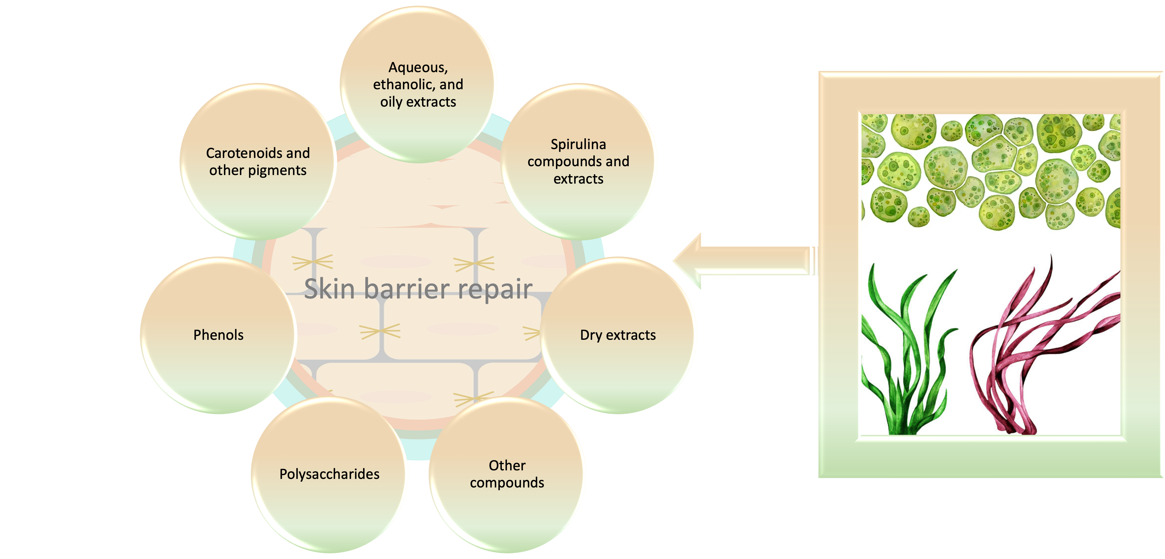
Table I summarizes the bioactive compounds of macro and microalgae that have shown to be effective in repairing skin barrier both related to skin hydration, and emollience. Among the bioactive compounds, polysaccharides, carotenoids, and phenols are the ones that have proven to be most effective in repairing the skin barrier, but also aqueous and oily extracts. In this review, compounds from Spirulina (Arthrospira sp., also known as Limnospira sp.) are also considered. Despite not been a microalga, as has been classified as cyanobacteria, carry out photosynthesis, and it is one of the most cultivated genera to obtain biomass for food, and nutraceuticals, but also for its richness in bioactive compounds, such as phycocyanin, and proteins that are of interest for skin care. In the following subsections, selected research is discussed.
As mentioned above, the current trend towards consuming cosmetic products that are more skin-friendly and also sustainable an[d non-polluting, has led to an increase in research into natural [58], and eco-sustainable cosmetics [59], frequently referred to as “green cosmetics” [60]. This tendency also fits with the trend of circular economy, to maximize the exploitation of resources with minimum waste and energy costs, water use, etc. [61].
In the last two decades, a large amount of scientific literature has been generated regarding the bioactive compounds of algae and their use in cosmetics. Several authors highlight the great potential of algae in this field, especially providing bioactive antioxidant, anti-inflammatory, and UV light protective molecules, but also other classic uses such as stabilizers and thickeners [62,63,64,65,66,67,68,69]. Joshi et al. revised the macroalgal species used in cosmetics, highlighting Chondrus crispus, Ulva lactuca, Fucus vesiculosus, Porphyra umbilicalis, and Ascophyllum nodosum, that are already used in the cosmetics industry [70], but since 2018 the market increased exponentially and other species have joined, such as Ecklonia cava, Undaria pinnatifida, Sargassum horneri, Palmaria palmata, and Neopyropia yezoensis (formerly Porphyra yezoensis), among others. And among microalgae, cyanobacteria and diatom, the following stand out: Porphyridium cruentum, and P. purpureum, Phaeodactylum tricornutum, Haematococcus pluvialis (currently Haematococcus lacustris), Scenedesmus sp., Odontella aurita, Chlorella sp., Nannochloropsis sp., Tetraselmis sp., and Arthrospira sp. [69].
Several seaweeds’ molecules already demonstrated a high potential as a cosmetic active ingredient (such as, fucoidan, phenolic compounds, pigments, and mycosporine-like amino acids) or to provide consistency (agar, alginate, carrageenan) [71,72]. For example, in marine brown algae, compounds like fucoxanthin, polysaccharides, mycosporine-like amino acids (MAAs), and phlorotannins have a variety of functions to combat ultraviolet radiation and protect human skin [73]. Fabrowska et al. revised the cosmetics properties of algal bioactive compounds, finding that polysaccharides, alginates, carrageenans, and agar are mainly used as thickening agents; ulvans exert moisturizing and antioxidant properties; fucoidans and laminarans have antioxidant and anti-inflammatory activity; amino acids (histidine, taurine, glutamic acid, serine, alanine, and MAAs) may be used as moisturizers, antioxidants, and natural sunscreen; peptides as carnosine are potential radical scavengers; and lectins and cyclic peptides may exert antibacterial, antiviral, and antifungal activities. Lipids from algae are also used as emollients, while pigments (carotenoids, chlorophylls, and phycobiliproteins) may exert a wide range of activities, as antioxidant, anti-inflammatory, anti-photoaging, or antiallergic. Phenolic compounds (phlorotannins, bromophenols, and terpenoids) are also well-known for its antioxidant properties, among others [74].
Even though algal extracts are the most studied in the pharmacological and cosmeceutical field, recently, there has been an increasing interest in studying the health properties of various specific marine algae compounds, so polysaccharides, phlorotannins, carotenoids, and terpenes emerged as prominent compounds, collectively representing 42.4% of the investigated compounds [75].
Kalasariya et al. revised the beneficial effect of marine macroalgae in moisturizing the skin (and photoprotection) finding that the most investigated algae were terpenoids, polysaccharides-fucoidan, carrageenan, and alginates [76]. These compounds have an additional advantage for the cosmetic industry, because it can be cheap and at the same time satisfy consumer demands for “natural” and “healthier” products [77].
Ersoydan et al. revised the anti-inflammatory activity of various brown algae species, finding that fucoidan can ameliorate not only inflammation in atopic dermatitis but also hydration and itching. Phlorotannins also exert anti-inflammatory effects and can contribute to skin health by inhibiting enzymes such as collagenase and elastase, which are involved in the degradation of the skin structure and function, and impaired wound healing [78]. Photoprotective effect is also of interest to maintain the skin barrier, so the algae bioactive compounds as phlorotannins [79], and fucoidan [80] should be taken into consideration when formulating skin care cosmetics, as both have shown to protect skin against UV damage. Fucoxanthin from different macro and microalgae also showed anti-inflammatory activities [81].
Rye et al. compiled the applications of microalgae-derived active ingredients as cosmeceuticals, resulting that the genera and species most frequently used in commercialized cosmetics are Spirulina sp., Arthrospira maxima, Chlorella vulgaris, Anacystis nidulans, Halymenia durvillei, Dunaliella salina, and Porphyridium sp. [82]. Later on, Castro et al. revised the cosmetic applications of microalgae and cyanobacteria, finding that the main genera are Chlorella, Arthrospira, Tetraselmis, and Scenedesmus, being the crude extracts or bio-mass, pigments, and polysaccharides the based products more reported [83].
However, the most studied activity of the bioactive compounds of algae is the antioxidant capacity, although for the case at hand, the repair of the skin barrier, the photoprotective capacity is of interest [84,85,86,87], which will also be discussed throughout this review.
Polysaccharides
Carbohydrates are the major and abundant constituent of marine algae and are used in cosmetic formulations as moisturizing and thickening agents [88,89], and also for developing transdermal drug delivery systems [90]. In marine algae, polysaccharides are biomacromolecules made up of repeating monosaccharide units joined by glycosidic bonds. The taxonomic distribution of polysaccharides varies according to the type of algae: in brown algae (Ochrophyta, Phaeophyceae), fucoidans, alginates and laminarins predominate; in red algae (Rhodophyta), porphyran, carrageenan and floridean starch are the main ones; and in green algae (Chlorophyta), rhamnan sulphate and ulvan stand out [91]. It can be said that one of the most studied compounds from algae are polysaccharides, which mainly focus on the antioxidant and photoprotection properties [92].
Thus, Patel et al. reviewed the applicability of algae for skin moisturizing finding that polysaccharides from certain algal species like S. japonica, Chondrus crispus, and Codium tomentosum help in the absorption of water or moisture, providing soothing effect, and may keep the skin moisturized in extremely hot and dry environments [93]. And, poly- and oligosaccharide Ulva sp. fractions were able to modulate the extracellular matrix of human dermal fibroblast, showing that may be also used to develop protective skin care cosmetics [94]. Furthermore, Ulva sp., possesses a representative content of rhamnose, a water-soluble sulfated polysaccharide with an important moisturizing action [95].
Several research showed that macroalgae polysaccharides have a great moisture retention ability, in many cases higher than hyaluronic acid (HA). Some examples are described in Table II. It can be summarized that sulfate and low molecular weight (LMW) polysaccharides have higher moisture retention ability than HA, glycerol or urea, and, in general, in brown algae it is higher than in red algae.
One brown algae Saccharina japonica, one red alga Porphyra haitanensis and three green algae Codium fragile, Enteromorpha linza, and Bryopsis plumose were investigated to evaluate their moist retention ability. The moisture-absorption of polysaccharides extracted from these five algae was examined and compared with that of HA, showing that with a decrease in molecular weight, the moisture-absorption/retention ability increased, being higher in red algae compared to green algae, and demonstrating that all of them possess higher moist retention capacity than HA [96].
The moisture-preserving property of the algae polysaccharide from Ulva lactuca was examined gravimetrically and compared with that of glycerol, resulting being higher for algae polysaccharide (43% vs 34%) [97]. Li et al. investigated the moisture retentions of different polysaccharides obtained from Enteromorpha prolifera; results showed that all polysaccharides, both low molecular weight and sulfated types, achieved high moisture retention rates, and were close to that of HA. Furthermore, the higher the sulphate content, the higher the water retention [98].
A study conducted by Cai et al. investigated the moist retention and absorption of powder and ooze of two macroalgae: Porphyra yezoensis and Sargassum horneri. The results showed that seaweed powder generally has a higher absorption capacity than ooze, and that S. horneri powder obtained the maximum moisture retention after 30 min (95%), while P. yezoensis powder had the highest moisture absorptions, up to 93% of that of glycerol. In addition, the polysaccharide extracted from S. horneri was superior to that of P. yezoensis in moisture retention [99]. Jesumani et al. studied the moisture absorption and retention abilities of a polysaccharide-rich extract from Sargassum vachellianum, showing that moisture retention is higher (65.84%) than of that of glycerol (51.35%) [100]. Other investigation found that polysaccharide from Caulerpa microphysa could be also useful for skin moisturizing. In terms of water-absorption capacity, C. microphysa extract was better than collagen, similar to HA, and poorer than urea. And the moisture-retention capacity over 24 h was better than that of collagen and HA, and similar to that of urea [101]. Finally, microalgae polysaccharides were also studied. In vitro moisture-absorption and -retention properties of Nostoc polysaccharide were compared to chitosan and urea; moisture retention resulted in higher (78.5%, 75.2%, and 62.7% respectively) and, additionally, was able to improve water retention in mouse stratum corneum under dry conditions [29].
In addition to the interesting and useful studies related to moisture retention cited, various in vitro and in vivo studies support the use of algae polysaccharides in cosmetics for skin hydration and barrier repair. Fucoidan is the most studied polysaccharide in skin disorders, such as atopic dermatitis, mainly due to its anti-inflammatory activity [102]. Low molecular fucoidan fractions showed to exert a significant recovery of skin barrier proteins and molecular mediators, indicating its potential for attenuating skin barrier dysfunction. In vitro study (HaCaT cells) demonstrated that a low molecular fucoidan fraction from the brown algae Sargassum confusum was able to suppress impairment of stratum corneum hydration in UVB irradiated cells, and causing a significant recovery of skin barrier proteins and molecular mediators. Authors postulated that the activation of MAPK and NF-κB mediators significantly declined following fucoidan low molecular fraction treatment and this could explain its protective functionality [21].
It has been observed that fucoidan from Undaria pinnatifida is able to promote the recovery of epidermal barrier disruption under low calcium condition by up-regulating the expression of calcium-sensing receptor (CaSR), which could then lead to the activation of the Catenin/PLC pathway, and also increase the expression of CaSR through activating the ERK and p38 pathway [22]. The research is based on a situation of low calcium levels, after having caused a disruption of the skin barrier in mice by tape stripping on their shaved back. Further clinical studies are needed to test this assumption in cases of eczema, psoriasis and other dermatological disorders that alter the skin barrier. Considering that several studies have suggested that intracellular Ca2+ stores, such as the endoplasmic reticulum, are the main components of the epidermal calcium gradient, and endoplasmic reticulum calcium homeostasis is crucial for regulating keratinocyte differentiation, intercellular junction expression, and permeability barrier homeostasis [103], these types of studies represent another way to repair the skin barrier and confirm that fucoidan can be very useful in the treatment of dermatoses associated to skin barrier impairment.
Other studies focus on the interest of fucoidan in protecting the skin against environmental particles (fine particle matter, PM). This aspect must be considered in skin protection, especially in areas with high pollution. Two studies investigated the activity of low molecular fucoidan fractions in fine-dust stimulated HaCaT keratinocytes. Fine dust (FD) is considered the most adverse among ambient PM, which can easily penetrate the skin; FD exposure of keratinocytes results in ROS-dependent production of inflammatory cytokines, transmutes the differentiation and cornification of keratinocytes and influences the downregulation of skin barrier proteins in the stratum corneum thus causing defects in skin barrier function [104]. Low molecular fucoidan fraction from Sargassum horneri, in vitro study (FD-induced HaCaT keratinocytes) ameliorated key tight junction proteins and skin hydration factors, outlining the effects of fucoidan in reducing FD-induced inflammation and skin barrier deterioration [23]. Similarly, low molecular fucoidan fraction from Sargassum confusum was able to increasing the cell viability of FD-induced HaCaT keratinocytes and to protect from inflammation [24]. Furthermore, fucoidan from Sargassum fusiforme, in vitro study (HaCaT cells and HDF cells) exerted protective effect against PM pollution [25]. These in vitro and in vivo studies show that polysaccharides, especially low molecular weight fucoidan fractions, can exert important functions not only in protecting the skin against environmental pollutants, but also in repairing the skin barrier which, as indicated, is damaged in certain dermatological conditions such as dermatitis and eczema.
Other studies evaluated cosmetic formulations. For example, a cream with 1% fucoidan obtained for Sargassum horneri, clinical study, showed to improving skin barrier function and reduced TEWL [26].
Likewise, a mixture of sulfated polysaccharides and glucuronic acid from Sargassum fusiforme, in vivo study, decreased the skin moisture loss [27]; and sacran gel from the red algae Aphanothece sacrum showed to increase skin hydration and decrease TEWL as well as promoting normal epidermal differentiation and improvement of the maturation of corneocytes [28].
Finally, it should take into consideration that polysaccharides from microalgae could be also of interest; as has been mentioned before, polysaccharides form Nostoc have higher water retention than urea, in vitro study in mouse stratum corneum [29].
Oligosaccharides such as agaro-oligosaccharides and carrageenan-oligosaccharides (obtained from agar and carrageenan, respectively, by enzymatic and chemical hydrolysis) have shown in vitro studies their water retention capacity, suggesting their potential use as skin moisturizers, and it has been suggested that they can also be used as prebiotics [105].
Additionally, exopolysaccharides (EPS) from cyanobacteria and microalgae could be a good raw material for cosmetic use, as has been shown that for example sacran or spirulan from cyanobacteria Aphanothece sacrum or Arthrospira (Spirulina) sp. is an acidic extracellular EPS that has a high-water absorption property [106,107], and Parachlorella sp. is also able to produce a high number of EPS [108]. Therefore, although these studies do not show specific effects of EPS on skin barrier repair, the moisturizing effect is of great interest, since it is the first step, along with emollience, to prevent dehydration.
Carotenoids and Other Pigments
Algae are rich in pigments. Brown algae possess chlorophyll a, c, carotenoids, fucoxanthin, and other pigments, whereas red algae contain chlorophyll, phycobilin, carotenoids, carotene, lutein, phycocyanin, and phycoerythrin, and Chlorophyta possess chlorophyll-a, -b, and -c and carotenoids [59].
Algae carotenoids, despite not been directly involved in skin barrier recovery, are of great interest in the field of cosmeceuticals as can exert antioxidant and anti-inflammatory activities; for example, astaxanthin is one of the strongest antioxidant, as well as β-carotene; Furthermore, fucoxanthin is able to counteract oxidative stress caused by UV radiation, which is why it is currently used in cosmeceuticals; and the carotenoid lutein protects skin structures against UV-induced oxidative damage, especially in combination with other antioxidant systems and immunoprotective substances [109].
Other examples are the following. Undaria pinnatifida fucoxanthin showed the ability to regulate filaggrin genes expression, and to restore the skin barrier by filaggrin stimulation, in vitro and in vivo studies [30]; and a clinical study demonstrated that zeaxanthin-based oral supplementation plus topical gel serum was able to improve skin hydration [31].
On the other hand, given the high abundance of carotenoids in microalgae, they can be considered potential producers of carotenoids in biotechnology [110] and, therefore, a good source of active ingredients for cosmetics, easy to produce and cheap.
Phenols
Algae polyphenols are very well-known for its antioxidant activity [111]; specifically, phlorotannins [112]. In addition to this protective function, it has been observed that other phenols can contribute to repairing the skin barrier. One of the most studied is fucosterol. For example, Hannan et al. revised the pharmacological properties and health benefits of marine algae phytosterol, finding that, particularly fucosterol, possesses substantial health benefits, including anti-obesity, anti-Alzheimer’s, anti-diabetes, anticancer, antiaging, and hepatoprotection, among many others, which are attributed to their antioxidant, anti-inflammatory, immunomodulatory and cholesterol-lowering properties, indicating their potentiality as therapeutic leads. These sterols interact with enzymes and other proteins that actively participate in different cellular pathways, including the antioxidant defense system, apoptosis and cell survival, metabolism, and homeostasis [113]. And Hwang et al. assessed the effects of fucosterol from the brown algae Hizikia fusiformis on photodamage, finding that fucosterol significantly decreased the UVB-induced expression of several matrix metalloproteinases (MMPs) and interleukins, as MMP-1 and IL-6 [114].
Fucosterol from Sargassum fusiforme, in vitro study, showed to modulate MAPK in irradiated HaCaT cells [32]; and Sargassum binderi fucosterol also demonstrated cytoprotective effects against xenobiotics [33] which can be also involved in certain dermatoses. Bromophenol (3-bromo-4,5 – dihydroxybenzaldehyde) from Polysiphonia morrowii was able to increasing the production of skin hydration proteins and tight junction proteins, in vitro study [34].
Other Compounds
Other research that combines different algae compounds deserve to be cited. A mixture of compounds including Fucus vesiculosus extract, Ulva lactuca extract, and Ectoine, in split-face clinical study, showed to increasing skin hydration and maintain the skin barrier function [35].
On the other hand, Ferreira et al. revised the ingredients of algae that have shown efficacy in the care of sensitive skin, highlighting that carotenoids, polysaccharides, and lipids are the chemical classes, but also Ascophyllum nodosum, and Asparagopsis armata extracts [115]. Further clinical research is needed to confirm this assumption.
Microalgae produce various bioactive compounds that exhibit potential pharmacological effects, including antidiabetic, anticancer, anti-inflammatory, and antioxidant activities. These bioactive compounds include fatty acids, phycobiliproteins, chlorophylls, carotenoids, and vitamins. Given their tremendous structural diversity and biological availability, microalgae-derived bioactive compounds have been shown to be useful in the treatment of inflammatory skin disorders [116], and, thus, could be an excellent source of active ingredients for skin care cosmetics. The activity of microalgae and cyanobacteria extracts would be discussed later on.
Besides, other algae compounds of interest for organic beauty products are chitin, terpenoids and vitamins [117]. For example, microalgae are rich in vitamins; Dunaliella tertiolecta synthesizes vitamin B12, B2, E, and beta-carotene, and Tetraselmis suecica produces vitamin C [118]. Microalgal species such as Thalassiosira sp., rich in amino acids, and Monodus subterraneus, with a high content of PUFA, could be able to reduce transepidermal water loss (TEWL); and also the genus Nannochloropsis sp. rich in linoleic acid, are good candidates as active ingredients for skin hydration [119].
Aquaous, Ethanolics and Oily Extracts
In addition to the bioactive compounds mentioned above, the cosmetic industry makes extensive use of macro and microalgae extracts, both aqueous, ethanolic and oily, as bioactive ingredients. For example, Sargassum spp. extracts and derivative compounds have excellent potential for skincare, as they exhibit skin health-promoting properties, including antioxidants, anti-inflammation, whitening, skin barrier repair, and moisturizing [120]. Other recent review deals with the skin protective effects, among others activities, of Antarctic algae, such as Micractinium sp., Chlamydomonas sp., Iridaea cordata, Curdiea racovitzae, and Phaeodactylum tricornutum finding that most of them can produce a quite amount of MMAs (porphyra-334, shinorine, and mycosporine-glycine) with interesting photo-protective activities in the UVR spectral range [121]. Cyanobacterial metabolites were also investigated finding that proteins and peptides (dry powder), methanolic extracts of exopolysaccharides, and other compounds such as vitamins A, C, B1, B2, B12 may improve skin hydration [122].
Macroalgae aqueous / oily extracts are widely used in the cosmetics industry to protect skin and increase hydration, prevent aging, or skin barrier recovery. Doria et al. argues that “it is necessary to consider that overall action of an algal extract is most probably due to the joint action of different substances” [123]. Considering that this is one of the principles of skin care cosmetics, the synergy between bioactive compounds rather than a high concentration of a specific compound that could lead to irritation or alterations in skin homeostasis, algae extracts can be important allies in the protection and recovery of the skin barrier.
Choi et al. screened Laminaria japonica extracts for skin moisturizing activity, showing that skin hydration increased by 14.44% compared with a placebo; and TEWL (using a test cream with 10% L. japonica extract), decreased to 4.01 g/cm2 (8 h after applying the cream), which was approximately 20% of that seen with the control [36]. In vitro study (Human Primary Epidermal Keratinocytes, HPEK) demonstrated that Sargassum glaucescens extracts induced the expressions of skin barrier-related genes TGM1 (Transglutaminase 1), KRT10 (Keratin 10) and KRT14 in keratinocytes. Furthermore, these algae extract induced the gene expression of filaggrin, which promoted the production of NMF for maintaining the moisture and barrier functions of skin [37]. Recent studies performed by Jang et al. demonstrated that guanosine and uridine nucleosides-rich extracts from Codium fragile exerted anti-inflammatory and skin protective effects on macrophages and human keratinocytes in atopic dermatitis. Specifically, enhanced the expression of factors related to skin barrier function, filaggrin, involucrin, and loricrin [38].
Two studies of Sargassum horneri ethanolic extracts showed to be also useful in skin barrier improvement. Dias et al., in vitro study in FD-induced HaCaT keratinocytes, found that S. horneri ethanolic extract ameliorated filaggrin, involucrin, lymphoepithelial Kazal-type-related inhibitor (LEKTI), signifying its beneficial effects on deteriorated skin hydration caused by FD-induced inflammation. Additionally, the extract exhibited skin protective effects regulating the tight junction proteins; occludin, zonula occludens (ZO)-1, claudin-1, claudin-4, claudin-7, and claudin-23, while increasing the production of HA minimizing skin damage [39]. And Mihindukulasooriya et al. showed that S. horneri ethanolic extract directly inhibited the expression of keratinocyte-produced TSLP (thymic stromal lymphopoietin), which is known to exacerbate skin barrier impairment. Especially, the decrease of filaggrin observed in DNCB-induced Atopic Dermatitis mice was significantly improved when treated with S. horneri ethanolic extract [40]. Further clinical studies are needed to confirm the potential of these extracts.
Extracts from microalgae have strongly approached the cosmetics market due to their ease of cultivation. Coccoid and filamentous algae extracts (type 1 and type 2) obtained from Blue Lagoon and also from silica mud were tested in vitro and in vivo (clinical study). Results showed that stimulation of keratinocytes with silica mud extracts increased mRNA steady-state levels for involucrin, filaggrin and transglutaminase-1 in a time- and dose-dependent manner. Similarly, expression of keratinocyte differentiation markers was also increased upon stimulation of cells with extracts from algae type 1, although to a lesser extent from algae type 2. Additionally, topical application of a galenic formulation containing all three extracts studied, once daily for 4 weeks, significantly increased mRNA expression for involucrin, filaggrin and transglutaminase-1. Upregulation of these keratinocyte differentiation markers were associated with a significant reduction in transepidermal water loss of treated skin areas [41].
Buono et al. investigated the biological activities of dermatologic interest of the water extract from the microalga Botryococcus braunii, in vitro study, finding that 1% extract induced gene expression of proteins involved in the maintenance of skin cells water balance such as aquaporin-3 (AQP3), filaggrin, and involucrin, in addition to antioxidant activities [42].
Aqueous extracts from microalgae Neochloris oleoabundans was investigated to evaluate its antioxidant properties and cutaneous compatibility. For that, a gel with 1.0% N. oleoabundans aqueous extract was formulated and in vivo study were performed. Results showed that, in addition to its anti-inflammatory properties, in vivo essay concluded that TEWL did not increase, so the formulation did not negatively affect the skin barrier function, and stratum corneum hydration was maintained in all the participants [43].
Microalgae ethanolic extract were also investigated as ingredient in cosmetics. Six different polar microalgae Micractinium sp. (KSF0015 and KSF0041), Chlamydomonas sp. (KNM0029C, KSF0037, and KSF0134), Chlorococcum sp. (KSF0003) were collected from the Antarctic or Arctic regions, and aqueous extract were prepared and analyzed. The biological activity of polar microalgae extracts in protecting against damage induced by oxidative stress and UVB in human HaCaT keratinocyte cells was evaluated. Additionally, to study the anti-inflammatory activity, imiquimod-induced murine model of psoriatic dermatitis was used. Results showed that polar microalgae extracts reduced oxidative stress generated in HaCaT cells, and KNM0029C and KSF0041 extracts prevented the reduced viability of HaCaT cells caused by UVB radiation, suggesting that these polar microalgae contain cytoprotective substances for skin epithelial cells. Furthermore, topical application of KSF0041 extract almost completely alleviated the clinical features of psoriasis (C57BL/6 mice), such as scaling, redness, and loss of epithelial water and weight polar, concluding that KSF0041 extract were able to reduce the barrier integrity damage. Authors suggested that the presence of abundant and diverse fatty acids in polar microalgae extracts, which dominant compounds were docosahexaenoic acid methyl ester, linolenic acid methyl ester, 13-Docosenamide (Z)-, and methyl 4,7,10,13-Hexadecatetraenoate, may play a role in epithelial protection, considering fatty acids promote epithelial preservation and barrier integrity [44].
Ethanolic extracts from Nannochloropsis sp., which main compounds were fatty acids 58.2%, carotenoids 1.6%, phenolics 7.7%, and flavonoids 2.0% (crude extract), in vitro study, enhanced the expression of HAS-2 (moisturizing-related gene) in a dose-dependent manner [45].
As has been mentioned before, emollients can improve the barrier function, and lipids from algae could be good allies. Macroalgae are rich in polyunsaturated ω-6 and ω -3 fatty acids (PUFAs), and other lipidic compounds; Rhodophyta and Phaeophyta have a high percentage of ω-3 fatty acids (linolenic, DHA, and EPA) and ω-6 fatty acids (linoleic acid, γ-linolenic acid, and arachidonic acid), and red and brown algae have high levels of ω-3 fatty acids (EPA) and the ω-6 fatty acids (arachidonic acid and linoleic acid) [15]. So that, algae oils are particularly enriched with PUFAs, including DHA, which finds application in skin protection formulations. Microalgae can also produce various types of lipids such as triacylglycerols, phospholipids, glycolipids or phytosterols, mainly in oleaginous microalgae such as Chlorella sp., Nannochloropsis sp., Scenedesmus sp., and Dunaliella sp. [124]; improving cultivation conditions, harvesting, and extraction methods may lead to microalgae being a sustainable and green source of lipids for the cosmetics industry. For example, during the last five year there is being an increase extraction yields of fatty acids from Nannochloropsis sp. microalgae; depending on the extraction efficacy of the different technologies, various types of lipids and/or fatty acids are obtained, namely PUFA, including EPA [125]. Other example are extracts from the green algae Cladophora glomerata biomass containing unsaturated fatty acids, especially ω-3, ω-6, and ω-9 are considered a valuable material for the production of cosmetic products, as these compounds may act as emollients, and prevent excessive TEWL [84].
Research into the role of algae-derived lipids in the epidermal barrier is scarce, although some studies on macroalgae can be cited, such as that of Kok et al. who studied the effects of Macrocystis pyrifera lipid extracts in three-dimensional cultures of HaCaT cells; results showed that M. pyrifera lipid pre-treatment reduced trans-epidermal leakage in cytokine-stimulated 3D epidermal constructs [46]. Additionally, a cream prepared with 0,5% carotenoid and phenolic-rich compounds of Cladophora glomerata oily extract showed, in a randomized clinical study, to be able to improve skin moisturizing[ [47]. The biological potential of the various bioactive compounds in the polar marine algae extracts were also studied. Oily extracts from macroalgae Himantothallus grandifolius, Plocamium cartilagineum, Phaeurus antarcticus, and Kallymenia antarctica (currently Trematocarpus antarcticus), in addition to increasing cell viability, showed to protect cells against inflammatory stimulation and increase the barrier integrity of cells damaged by lipopolysaccharide or ultraviolet radiation, both in intestine and skin [48].
Diatom Phaeodactylum tricornutum is very well-known for its high content on fucoxanthin, but is also rich in fatty acids; P. tricornutum extracts have been shown to exert anti-inflammatory properties [126], but the studies related to skin barrier improvement are scarce. An oily extract from P. tricornutum (fatty acids content up to 99% of and less than 1% of xanthophyll’s) showed the ability to stimulate 20S proteasome peptidases activities both in vitro and within human keratinocytes and to reduce the level of oxidized proteins [49], and is therefore a potential candidate for protection and the skin barrier against environmental aggressions. Furthermore, encapsulated liposomal lipid extracts obtained from P. tricornutum, in combination with thermal spring water, seemed to contribute to repairing the altered skin barrier, increasing the skin’s resistance threshold in sensitive skin, calming the capsaicin-induced stinging and burning sensation (randomized, clinical split-face study) [50].
Lipid extract from Nannochloropsis oceanica, rich in phosphatidylcholine, and phosphatidylethanolamine, were investigated in human immortalized keratinocytes CDD 1102 KERTr exposed to UV radiation. After irradiation, the cells were treated with a lipid extract from N. oceanica algae in 0.1% DMSO (dimethyl sulfoxide) at concentrations ranging from 1 μg/ml to 1 mg/ml for 24 h. Results showed the ability of N. oceanica lipid extract to modulate the sphingomyelin-ceramide (SM-CER) pathway, accompanied by a significant upregulation of both classes of ceramides, non-hydroxy fatty acid/dihydrosphingosine base ceramide CER[NDS], and non-hydroxy fatty acid/sphingosine base ceramide CER[NS] [51].
Other algae lipids are used by the cosmetic brands to improve skin barrier. For example, a bio-based algae oil (INCI name Triolein), in vivo single-blind study, showed to improve skin moisturizing, and also was able to reduce TEWL, which is an indicator of the skin’s barrier function [52].
Spirulina Compounds and Aqueous Extracts
In this review studies related to Spirulina (Arthrospira sp.) were selected considering its extensive exploitation for obtaining bioactive molecules, mainly for the food industry. Jang and Kim investigated the photoprotective effects of spirulina-derived C-phycocyanin against UVB radiation using keratinocytes (HaCaT cells). Results showed that 80 μg/mL C-phycocyanin increased involucrin, filaggrin, and loricrin expression by >25% [53].
Aqueous extract of Spirulina sp. (Arthrospira sp.) composed by 50 and 70% proteins (dry weight), 8 to 14% of polysaccharides, and about 6% of lipids was used to prepared a formulation containing 0.1% dry extract (w/w). A clinical study was performed to evaluate several skin parameters (skin hydration, TEWL, sebum content, skin microrelief, dermis thickness, and structural and morphological properties of the epidermis), showing that after 28 days of application of the formulation, a significant increase of the SC water content was observed in both groups (young and mature skin). This effect was more pronounced on the mature skin group. In addition, a reduction of the TEWL was observed in both groups; however, these results were significant only in the older group, when compared to the group that received the vehicle formulation. Furthermore, the skin microrelief was improved observing a reduction of the surface roughness, and keratinocytes were more uniformly distributed and homogeneous [54]. Subsequently, similar studies were carried out with this same gel formulation based on spirulina extract, in Tertiary care Hospital, Bangladesh, observing a replication of the results [55].
Dry Extracts
Dry extracts of microalgae and cyanobacteria are also used as cosmetics ingredient. A double-blind, randomized, placebo-controlled clinical trial was performed applying a formulation containing olive oil and spirulina extract. After 12 weeks of application of the formulations, clinical evaluation using instrumental measurements showed an increase in skin hydration, an improvement in the skin barrier and in the morphological characteristics of the epidermis. A significant increase in the brightness of the stratum corneum was also observed, suggesting a film-forming effect [56], another aspect to take into consideration in the preservation of the skin barrier, since excessive TEWL is prevented. And Ma’or et al. evaluated the skincare effect of a cream composed by Dead Sea minerals (mineral-botanical complex) and Dunaliella salina dry extract, resulting 42,3% improvement in skin roughness, and a slight improvement of skin hydration compared to the control (the same cream without D. salina extract) [57]. In this last study, the influence of Dead Sea salts in improving skin hydration must also be considered.
Potential of Other Algae Compounds in Skin Barrier Recovery
Amino acids, peptides, and proteins from algae are also of great interest in skin care, as seaweed proteins are effective in their use as cosmeceuticals, as they are excellent moisturizers for hair and body [63,127]. For example, Chondrus crispus contain several amino acids, including alanine, arginine, aspartic acid, citrulline, glutamic acid, glycine, histidine, isoleucine, leucine, serine, lysine, methionine, threonine, ornithine, tyrosine, phenylalanine, proline, taurine, valine, and also peptides [128]. And Chaetomorpha crassa was investigated, finding that is rich in aspartic acid, glutamic acid, hydroxyproline, glycine, and alanine [59], therefore, it should also be considered as a potential source of cosmetic ingredients for skin hydration.
Thus, recently, the focus has been on research to obtain algal peptides for dermocosmetic use, arguing that cost-effective extraction and purification of bioactive peptides, specifically from red algae, will be promising for the wider application of algal oligopeptides. Enzymatic hydrolysis, in combination with other extraction methods, can provide peptides with reasonably high yield and good bioactivity [129]. Water-soluble hydrolysates, protein and peptides rich, from Chlorella vulgaris have also been studied for their antioxidant properties [130]; considering that proteins can improve skin moisturizing, these hydrolysates could also be of interest in cosmetics for skin barrier recovery.
Ectoine, (S)-2-methyl-1,4,5,6-tetrahydropyrimidine-4-carboxylic acid, is a cyclic amino acid naturally produced by extremophile microorganisms living under conditions of extreme salinity, drought, irradiation, pH, and temperature; the species which produce higher amount of Ectoine are Halomonas elongata and Halomonas salina [131]. Ectoine provides multiple cosmetic benefits such as immune protection, cell protection, UV protection, and membrane protection, as well as decreases skin inflammation in atopic dermatitis [15]. For example, Kauth and Trusova revised the effects of Ectoine topical application in inflammatory diseases associated with an impaired skin barrier, suggesting that topical formulations containing Ectoine could be a beneficial alternative as basic therapy or to increase the efficacy of the pharmacological treatment regimen for patients with inflammatory skin diseases, including infants and children [132]. Thus, Ectoine may be a good ally for treating impaired skin barrier diseases.
Algae and microalgae are also rich in minerals and trace elements, which can help to rebalance skin hydration and metabolism. Palmaria palmata, Fucus vesiculosus, Laminaria sp., and Ulva sp., among others, contain minerals such as Na, K, Ca, Mg, and also trace elements (Fe, Zn, Mn, Cu) [133]. For example, the consumption of 10 g of Ulva lactuca can provide 70% of the body’s daily magnesium requirements and over half of its iron requirements [134]. Among the microalgae species, Tetraselmis chuii exhibited the highest concentrations of calcium (Ca) and manganese (Mn), while Chaetoceros muelleri showed prominence in magnesium (Mg), sodium (Na), and iron (Fe). Thalassiosira weissflogii stood out for its potassium (K) content, and Tisochrysis lutea contained notable amounts of copper (Cu), and zinc (Zn) [135].
Other field of interest is the study of aeroterrestrial and extreme environment microalgae which need to adapt to extreme conditions and is commonly accepted that these adaptations include series of protective natural compounds, such us phenols, scytonemin, (a dimer of indolic and phenolic subunits), MAAs, bioactive peptides, glucans, etc. [136].
Other investigations related to improving wound healing with the aid of algae and microalgae are also of interest in skincare. The red seaweed Gelidium corneum aqueous extracts showed wound healing properties, among other activities [137]; polysaccharides fractions from Gracilaria lemaneiformis promoted cell proliferation and migration through activation of PI3 K/aPKC signaling during human keratinocytes wound healing [138]. Lectin isolated from the red algae Bryothamnion seaforthii showed to improve wound healing effects [139]. A skin cream including 1.125% S. platensis (currently Arthrospira platensis) crude extract enhanced wound healing effect on HS2 keratinocyte cell line [140]. And Choi et al. investigated a microalgae-based biohybrid microrobot for accelerated diabetic wound healing [141], which constitutes a novel approach to the treatment of wounds and ulcers.
Finally, it deserves to mentioning other research of great interest for enhancing cosmetic bioactive compounds through bio-vectors and nanoparticles of algae, specifically microalgae. Some examples are microspheres from purified Sargassum horneri alginate from [142], and nanoliposomal peptides derived from Spirulina platensis (A. platensis) protein which were able to accelerating full-thickness wound healing [143]. In vitro research about Chitin-Hyaluronan nanoparticles entrapping different ingredients showed that may lead to the development of more effective cosmetics that can be used in the cosmeceutical field but also in aesthetic medicine [144]. Other example is a spray drying microencapsulation of phytochemicals from berry pomaces with Spirulina protein which was incorporated into a cosmeceutical topical formulation to mitigate pollution skin damage [145].
Cosmetic Patents Based on the Use of Algae and Spiruline for Skin Barrier Improvement
Several patents and commercial products from algae which claim to be effective in skin barrier strengthening or improving skin hydration can be found. Table III shows some examples.
3. Future Challenges and Conclusions
Cosmetic consumer behavior has changed during this century, and more and more are searching for natural ingredients, clean labeling, and non-synthetic chemicals, mainly due to increased awareness of toxicity and chemical cocktails in cosmetic products [146]. The need to repair the skin barrier, whose deterioration is associated with various dermatological disorders, also promotes the search for new natural bioactive compounds, in which algae derivatives play a prominent role.
Marine resources, specifically marine algae, exist in vast numbers and show enormous diversity. As a result, there are likely many possible applications for algae marine molecules of interest in the cosmetic industry, whether as excipients or additives, but especially as active substances [147]. Microalgae and cyanobacteria are also a good source of compounds for the cosmetic industry [83], so that investigation aimed at optimizing the production of bio-compounds and/or algae extracts constitutes a route to the production of cosmetics that are respectful of skin physiology and kinder to the environment in the line of green cosmetics and blue research, in addition to search the extensive use of renewable resources to produce more sustainable ingredients. In this scenario, it is important to decide which extraction method will be used and biorefinery methods should be developed. The greener technologies improve yields, minimize solvent waste, save time and energy, and facilitate automation. Some examples include supercritical CO2 extraction, pressurized liquid extraction, subcritical water extraction, and microwave-assisted extraction [148]. Hence, there is a need to develop new efficient and innovative extraction procedures that surpass conventional technologies to obtain high quality biomolecules with higher yield [149].
Other important aspects that require study are related to the more ecological extraction of bioactive compounds, their chemical and biological characterization, as well as their stabilization and delivery in new products [150]. Additionally, it has been proposed to optimize the minor compounds that might be discarded or lost during seaweed processing. The algae processing usually targets major components, such as agar, carrageenan, and alginate, and the rest of the material, as minor components, are refused or considerer impurities. But nowadays some minor components have been recognized as potential cosmetic ingredients, such as phycocyanin, phycoerythrin, and MAAs. Therefore, integrated biorefinery concepts have been proposed in algae process lines to obtain polysaccharides but also active compounds as targeted products [151,152]. On the other hand, biopolymers resulting from the use of waste from the industry linked to marine resources, including algae, create sustainable and value-added goods, it may be worthwhile to value these abundant and accessible biowastes [153]. Additionally, it has been proposed that different culturing conditions, including incubation with predators, influence microalgal bioactivities (the so-called OSMAC approach: one strain many compounds) triggering the activation of specific metabolic pathways [18]. So, new strategies to increase the profitability of the extraction process are also needed to improve the cosmetic industry interest, and in this context, biotechnology may present advantages by reducing the environmental impact from the exploitation of these resources. Additionally, it is essential to evaluate the presence of heavy metal like arsenic, mercury, lead and cadmium, pesticides, such as organochlorine, allergens, toxins, and other chemical contaminations in the algae samples, but also phototoxicity due to the presence of phototoxins or photoallergens [154].
Finally, it is important to note that it is necessary to evaluate the efficacy and safety of bioactive compounds through standardized in vitro assays [79] as is required from different cosmetics regulations. In a recent review performed by Bouafir et al., authors highlight that marine algae bioactive substances are safe but there is a need of standardization, and also deeper understanding of their mechanisms of action and efficacy [155].
This review showed the potential of macro and microalgae compounds in skin barrier repair, as well as to improving skin hydration, and acting as emollients. In addition to the aqueous, ethanolic and oily extracts, which can act through the synergy of their bioactive compounds, the polysaccharides stand out, especially fucoidan, among the carotenoids, fucoxanthin, and, among the phenols, fucosterol. The advancement in the discovery of new natural and versatile active ingredients for the cosmetics industry is linked to the research and improvement of extraction techniques, benefiting all actors linked to cosmetics and skin care, companies, scientists, dermatologists and skin care specialists, thus promoting cosmetic science.
Funding
This research received no external funding.
Conflicts of Interest
The authors declare no conflict of interest.
References
- Harris-Tryon TA, Grice EA. Microbiota and maintenance of skin barrier function. Science. 2022;376(6596):940-945. [CrossRef]
- Egawa G, Kabashima K. Barrier dysfunction in the skin allergy. Allergology International. 2018;67(1):3-11. [CrossRef]
- Yang J, Guo J, Tang P, et al. Insights from Traditional Chinese Medicine for Restoring Skin Barrier Functions. Pharmaceuticals. 2024;17(9):1176. [CrossRef]
- Schild J, Kalvodová A, Zbytovská J, Farwick M, Pyko C. The role of ceramides in skin barrier function and the importance of their correct formulation for skincare applications. Intern J of Cosmetic Sci. 2024;46(4):526-543. [CrossRef]
- Yosipovitch G, Misery L, Proksch E, Metz M, Ständer S, Schmelz M. Skin Barrier Damage and Itch: Review of Mechanisms, Topical Management and Future Directions. Acta Derm Venereol. 2019;99(13):1201-1209. [CrossRef]
- Kucuksezer UC, Ozdemir C, Yazici D, et al. The epithelial barrier theory: development and exacerbation of allergic and other chronic inflammatory diseases. Asia Pacific Allergy. Published online March 31, 2023. [CrossRef]
- Orioli D, Dellambra E. Epigenetic Regulation of Skin Cells in Natural Aging and Premature Aging Diseases. Cells. 2018;7(12):268. [CrossRef]
- Celebi Sozener Z, Ozdel Ozturk B, Cerci P, et al. Epithelial barrier hypothesis: Effect of the external exposome on the microbiome and epithelial barriers in allergic disease. Allergy. 2022;77(5):1418-1449. [CrossRef]
- Strugar TL, Bs AK, Seité S, Lin M, Lio P. Connecting the Dots: From Skin Barrier Dysfunction to Allergic Sensitization, and the Role of Moisturizers in Repairing the Skin Barrier. 2019;18(6).
- Wickett RR, Visscher MO. Structure and function of the epidermal barrier. American Journal of Infection Control. 2006;34(10):S98-S110. [CrossRef]
- Baroni A, Buommino E, De Gregorio V, Ruocco E, Ruocco V, Wolf R. Structure and function of the epidermis related to barrier properties. Clinics in Dermatology. 2012;30(3):257-262. [CrossRef]
- Elias PM, Wakefield JS, Man MQ. Moisturizers versus Current and Next-Generation Barrier Repair Therapy for the Management of Atopic Dermatitis. Skin Pharmacol Physiol. 2019;32(1):1-7. [CrossRef]
- Rajkumar J, Chandan N, Lio P, Shi V. The Skin Barrier and Moisturization: Function, Disruption, and Mechanisms of Repair. Skin Pharmacol Physiol. 2023;36(4):174-185. [CrossRef]
- Madnani N, Deo J, Dalal K, et al. Revitalizing the skin: Exploring the role of barrier repair moisturizers. J of Cosmetic Dermatology. 2024;23(5):1533-1540. [CrossRef]
- Mourelle ML, Gómez CP, Legido JL. Role of Algal Derived Compounds in Pharmaceuticals and Cosmetics. In: Rajauria G, Yuan YV, eds. Recent Advances in Micro and Macroalgal Processing. 1st ed. Wiley; 2021:537-603. [CrossRef]
- Fonseca S, Amaral MN, Reis CP, Custódio L. Marine Natural Products as Innovative Cosmetic Ingredients. Marine Drugs. 2023;21(3):170. [CrossRef]
- Pereira, L. Pereira L. Seaweeds as Source of Bioactive Substances and Skin Care Therapy—Cosmeceuticals, Algotheraphy, and Thalassotherapy. Cosmetics. 2018;5(4):68. [CrossRef]
- Saide A, Martínez KA, Ianora A, Lauritano C. Unlocking the Health Potential of Microalgae as Sustainable Sources of Bioactive Compounds. IJMS. 2021;22(9):4383. [CrossRef]
- Cagney MH, O’Neill EC. Strategies for producing high value small molecules in microalgae. Plant Physiology and Biochemistry. 2024;214:108942. [CrossRef]
- Conde T, Lopes D, Łuczaj W, et al. Algal Lipids as Modulators of Skin Disease: A Critical Review. Metabolites. 2022;12(2):96. [CrossRef]
- Fernando IPS, Dias MKHM, Madusanka DMD, et al. Fucoidan refined by Sargassum confusum indicate protective effects suppressing photo-oxidative stress and skin barrier perturbation in UVB-induced human keratinocytes. International Journal of Biological Macromolecules. 2020;164:149-161. [CrossRef]
- Chen Y, Li X, Gan X, et al. Fucoidan from Undaria pinnatifida Ameliorates Epidermal Barrier Disruption via Keratinocyte Differentiation and CaSR Level Regulation. Marine Drugs. 2019;17(12):660. [CrossRef]
- Fernando IPS, Dias MKHM, Madusanka DMD, et al. Low molecular weight fucoidan fraction ameliorates inflammation and deterioration of skin barrier in fine-dust stimulated keratinocytes. International Journal of Biological Macromolecules. 2021;168:620-630. [CrossRef]
- Kirindage KGIS, Jayasinghe AMK, Cho N, et al. Fine-Dust-Induced Skin Inflammation: Low-Molecular-Weight Fucoidan Protects Keratinocytes and Underlying Fibroblasts in an Integrated Culture Model. Marine Drugs. 2022;21(1):12. [CrossRef]
- Wang X, Huang C, Yang F, et al. Fucoidan isolated from the edible seaweed Sargassum fusiforme suppresses skin damage stimulated by airborne particulate matter. Algal Research. 2024;77:103339. [CrossRef]
- Kang J, Hyun SH, Kim H, et al. The effects of fucoidan-rich polysaccharides extracted from Sargassum horneri on enhancing collagen-related skin barrier function as a potential cosmetic product. J of Cosmetic Dermatology. 2024;23(4):1365-1373. [CrossRef]
- Ye Y, Ji D, You L, Zhou L, Zhao Z, Brennan C. Structural properties and protective effect of Sargassum fusiforme polysaccharides against ultraviolet B radiation in hairless Kun Ming mice. Journal of Functional Foods. 2018;43:8-16. [CrossRef]
- Masaki H, Doi M. Function of Sacran as an Artificial Skin Barrier and the Development of Skincare Products. YAKUGAKU ZASSHI. 2019;139(3):371-379. [CrossRef]
- Li H, Xu J, Liu Y, et al. Antioxidant and moisture-retention activities of the polysaccharide from Nostoc commune. Carbohydrate Polymers. 2011;83(4):1821-1827. [CrossRef]
- Matsui M, Tanaka K, Higashiguchi N, et al. Protective and therapeutic effects of fucoxanthin against sunburn caused by UV irradiation. Journal of Pharmacological Sciences. 2016;132(1):55-64. [CrossRef]
- Schwartz S, Frank E, Gierhart D, Simpson P, Frumento R. Zeaxanthin-based dietary supplement and topical serum improve hydration and reduce wrinkle count in female subjects. J of Cosmetic Dermatology. 2016;15(4). [CrossRef]
- Kim M, Oh G, Kim M, Hwang J. Fucosterol Inhibits Matrix Metalloproteinase Expression and Promotes Type-1 Procollagen Production in UVB -induced HaCaT Cells. Photochem & Photobiology. 2013;89(4):911-918. [CrossRef]
- Fernando IPS, Jayawardena TU, Kim HS, et al. A keratinocyte and integrated fibroblast culture model for studying particulate matter-induced skin lesions and therapeutic intervention of fucosterol. Life Sciences. 2019;233:116714. [CrossRef]
- Jayasinghe AMK, Han EJ, Kirindage KGIS, et al. 3-Bromo-4,5-dihydroxybenzaldehyde Isolated from Polysiphonia morrowii Suppresses TNF-α/IFN-γ-Stimulated Inflammation and Deterioration of Skin Barrier in HaCaT Keratinocytes. Marine Drugs. 2022;20(9):563. [CrossRef]
- Janssens-Böcker C, Wiesweg K, Doberenz C. The Tolerability and Effectiveness of Marine-Based Ingredients in Cosmetics: A Split-Face Clinical Study of a Serum Spray Containing Fucus vesiculosus Extract, Ulva lactuca Extract, and Ectoin. Cosmetics. 2023;10(3):93. [CrossRef]
- Choi JS, Moon WS, Choi JN, et al. Effects of seaweed Laminaria japonica extracts on skin moisturizing activity in vivo. JOURNAL OF COSMETIC SCIENCE. 2013;64:193-205.
- Li Z yi, Yu CH, Lin YT, et al. The Potential Application of Spring Sargassum glaucescens Extracts in the Moisture-Retention of Keratinocytes and Dermal Fibroblast Regeneration after UVA-Irradiation. Cosmetics. 2019;6(1):17. [CrossRef]
- Jang A yeong, Choi J, Rod-in W, Choi KY, Lee DH, Park WJ. In Vitro Anti-Inflammatory and Skin Protective Effects of Codium fragile Extract on Macrophages and Human Keratinocytes in Atopic Dermatitis. J Microbiol Biotechnol. 2024;34(4):940-948. [CrossRef]
- Dias MKHM, Madusanka DMD, Han EJ, et al. Sargassum horneri (Turner) C. Agardh ethanol extract attenuates fine dust-induced inflammatory responses and impaired skin barrier functions in HaCaT keratinocytes. Journal of Ethnopharmacology. 2021;273:114003. [CrossRef]
- Mihindukulasooriya SP, Dinh DTT, Herath KHINM, et al. Sargassum horneri extract containing polyphenol alleviates DNCB-induced atopic dermatitis in NC/Nga mice through restoring skin barrier function. Histol Histopathol. 2022;37(09):839-852. [CrossRef]
- Grether-Beck S, Marini A, Jaenicke T, et al. Blue Lagoon Algae Improve Uneven Skin Pigmentation: Results from in vitro Studies and from a Monocentric, Randomized, Double-Blind, Vehicle-Controlled, Split-Face Study. Skin Pharmacol Physiol. 2022;35(2):77-86. [CrossRef]
- Buono S, Langellotti AL, Martello A, et al. Biological activities of dermatological interest by the water extract of the microalga Botryococcus braunii. Arch Dermatol Res. 2012;304(9):755-764. [CrossRef]
- Morocho-Jácome AL, Santos BBD, Carvalho JCMD, et al. Microalgae as a Sustainable, Natural-Oriented and Vegan Dermocosmetic Bioactive Ingredient: The Case of Neochloris oleoabundans. Cosmetics. 2022;9(1):9. [CrossRef]
- Lim Y, Park SH, Kim EJ, et al. Polar microalgae extracts protect human HaCaT keratinocytes from damaging stimuli and ameliorate psoriatic skin inflammation in mice. Biol Res. 2023;56(1):40. [CrossRef]
- Kim SY, Kwon YM, Kim KW, Kim JYH. Exploring the Potential of Nannochloropsis sp. Extract for Cosmeceutical Applications. Marine Drugs. 2021;19(12):690. [CrossRef]
- Kok JML, Dowd GC, Cabral JD, Wise LM. Macrocystis pyrifera Lipids Reduce Cytokine-Induced Pro-Inflammatory Signalling and Barrier Dysfunction in Human Keratinocyte Models. IJMS. 2023;24(22):16383. [CrossRef]
- Fabrowska J, KAPUåCI A, Feliksik-Skrobich K, Nowak I. IN VIVO STUDIES AND STABILITY STUDY OF CLADOPHORA GLOMERATA EXTRACT AS A COSMETIC ACTIVE INGREDIENT. Acta Poloniae Pharmaceutica. 2017;74:633-641.
- Ko SH, Lim Y, Kim EJ, et al. Antarctic Marine Algae Extracts as a Potential Natural Resource to Protect Epithelial Barrier Integrity. Marine Drugs. 2022;20(9):562. [CrossRef]
- Bulteau AL, Moreau M, Saunois A, Nizard C, Friguet B. Algae Extract-Mediated Stimulation and Protection of Proteasome Activity Within Human Keratinocytes Exposed to UVA and UVB Irradiation. Antioxidants & Redox Signaling. 2006;8(1-2):136-143. [CrossRef]
- Vitale M, Truchuelo MT, Nobile V, Gómez-Sánchez MJ. Clinical Tolerability and Efficacy Establishment of a New Cosmetic Treatment Regimen Intended for Sensitive Skin. Applied Sciences. 2024;14(14):6252. [CrossRef]
- Łuczaj W, Gęgotek A, Conde T, Domingues MR, Domingues P, Skrzydlewska E. Lipidomic assessment of the impact of Nannochloropsis oceanica microalga lipid extract on human skin keratinocytes exposed to chronic UVB radiation. Sci Rep. 2023;13(1):22302. [CrossRef]
- Birjandi Nejad H, Blasco L, Moran B, et al. Bio-based Algae Oil: an oxidation and structural analysis. Intern J of Cosmetic Sci. 2020;42(3):237-247. [CrossRef]
- Jang YA, Kim BA. Protective Effect of Spirulina-Derived C-Phycocyanin against Ultraviolet B-Induced Damage in HaCaT Cells. Medicina. 2021;57(3):273. [CrossRef]
- Delsin DS, Mercurio, S.G., Fossa MM, Maia Campos, P.M.B.G M. Clinical Efficacy of Dermocosmetic Formulations Containing Spirulina Extract on Young and Mature Skin: Effects on the Skin Hydrolipidic Barrier and Structural Properties. Clin Pharmacol Biopharm. 2015;04(04). [CrossRef]
- Mondal KA, Ahmed R. Clinical Profile of Spirulina on Skin Diseases-A Study in Tertiary care Hospital, Bangladesh. Global Academic Journal of Medical Sciences. 2021;3(3):54-62.
- D’Angelo Costa GM, Maia Campos PMBG. Development of Cosmetic Formulations Containing Olive Extract and Spirulina sp.: Stability and Clinical Efficacy Studies. Cosmetics. 2024;11(3):68. [CrossRef]
- Ma’Or Z, Meshulam-Simon, G, Yehuda, S, Gavrieli, J. A. Anti wrinkle and skin moisturizing effect of a mineral-algal-botanical complex. JOURNAL OF COSMETIC SCIENCE. 2000;51(1):27-36.
- Guillerme JB, Couteau C, Coiffard L. Applications for Marine Resources in Cosmetics. Cosmetics. 2017;4(3):35. [CrossRef]
- Kalasariya HS, Yadav VK, Yadav KK, et al. Seaweed-Based Molecules and Their Potential Biological Activities: An Eco-Sustainable Cosmetics. Molecules. 2021;26(17):5313. [CrossRef]
- Leong HJY, Teoh ML, Beardall J, Convey P. Green beauty unveiled: Exploring the potential of microalgae for skin whitening, photoprotection and anti-aging applications in cosmetics. J Appl Phycol. 2024;36(6):3315-3328. [CrossRef]
- Lourenço-Lopes C, Fraga-Corral M, Jimenez-Lopez C, et al. Metabolites from Macroalgae and Its Applications in the Cosmetic Industry: A Circular Economy Approach. Resources. 2020;9(9):101. [CrossRef]
- Bedoux G, Hardouin K, Burlot AS, Bourgougnon N. Bioactive Components from Seaweeds. In: Advances in Botanical Research. Vol 71. Elsevier; 2014:345-378. [CrossRef]
- Ariede MB, Candido TM, Jacome ALM, Velasco MVR, De Carvalho JCM, Baby AR. Cosmetic attributes of algae - A review. Algal Research. 2017;25:483-487. [CrossRef]
- Salehi B, Sharifi-Rad J, Seca AML, et al. Current Trends on Seaweeds: Looking at Chemical Composition, Phytopharmacology, and Cosmetic Applications. Molecules. 2019;24(22):4182. [CrossRef]
- Cikoš AM, Jerković I, Molnar M, Šubarić D, Jokić S. New trends for macroalgal natural products applications. Natural Product Research. 2021;35(7):1180-1191. [CrossRef]
- Dussably J, Mshvildadze V, Pichette A, Ripoll L. Microalgae and Diatom - Potential Pharmaceutical and Cosmetic Resources – Review. J Biomed Res Environ Sci. 2022;3(9):1082-1092. [CrossRef]
- Kalasariya HS, Pereira L, Patel NB. Pioneering Role of Marine Macroalgae in Cosmeceuticals. Phycology. 2022;2(1):172-203. [CrossRef]
- Zhuang D, He N, Khoo KS, Ng EP, Chew KW, Ling TC. Application progress of bioactive compounds in microalgae on pharmaceutical and cosmetics. Chemosphere. 2022;291:132932. [CrossRef]
- Yadav A, Kumar S, Deepak B, Bhateria R, Mona S. Prospects of algal bioactive compounds in the cosmetic industry. In: Algae Based Bioelectrochemical Systems for Carbon Sequestration, Carbon Storage, Bioremediation and Bioproduct Generation. Elsevier; 2024:69-76. [CrossRef]
- Joshi S, Kumari R, Upasani VN. Applications of Algae in Cosmetics: An Overview. 2018;7(2).
- Morais T, Cotas J, Pacheco D, Pereira L. Seaweeds Compounds: An Ecosustainable Source of Cosmetic Ingredients? Cosmetics. 2021;8(1):8. [CrossRef]
- Mendes M, Cotas J, Pacheco D, et al. Red Seaweed (Rhodophyta) Phycocolloids: A Road from the Species to the Industry Application. Marine Drugs. 2024;22(10):432. [CrossRef]
- Parikh HS, Singh PK, Tiwari A. Algal biorefinery: focus on cosmeceuticals. Syst Microbiol and Biomanuf. 2024;4(4):1239-1261. [CrossRef]
- Fabrowska J, Łęska B, Schroeder G, Messyasz B, Pikosz M. Biomass and Extracts of Algae as Material for Cosmetics. In: Kim S, Chojnacka K, eds. Marine Algae Extracts. 1st ed. Wiley; 2015:681-706. [CrossRef]
- Silva M, Avni D, Varela J, Barreira L. The Ocean’s Pharmacy: Health Discoveries in Marine Algae. Molecules. 2024;29(8):1900. [CrossRef]
- Kalasariya HS, Dave DrMP, Yadav DrVK, Patel DrNB. BENEFICIAL EFFECTS OF MARINE ALGAE IN SKIN MOISTURIZATION AND PHOTOPROTECTION. IJPHC. Published online 2020. [CrossRef]
- Pimentel F, Alves R, Rodrigues F, P. P. Oliveira M. Macroalgae-Derived Ingredients for Cosmetic Industry—An Update. Cosmetics. 2017;5(1):2. [CrossRef]
- Ersoydan S, Rustemeyer T. Investigating the Anti-Inflammatory Activity of Various Brown Algae Species. Marine Drugs. 2024;22(10):457. [CrossRef]
- Freitas R, Martins A, Silva J, et al. Highlighting the Biological Potential of the Brown Seaweed Fucus spiralis for Skin Applications. Antioxidants. 2020;9(7):611. [CrossRef]
- Jing R, Guo K, Zhong Y, et al. Protective effects of fucoidan purified from Undaria pinnatifida against UV-irradiated skin photoaging. Ann Transl Med. 2021;9(14):1185-1185. [CrossRef]
- Mayer A, Mayer V, Swanson-Mungerson M, et al. Marine Pharmacology in 2019–2021: Marine Compounds with Antibacterial, Antidiabetic, Antifungal, Anti-Inflammatory, Antiprotozoal, Antituberculosis and Antiviral Activities; Affecting the Immune and Nervous Systems, and Other Miscellaneous Mechanisms of Action. Marine Drugs. 2024;22(7):309. [CrossRef]
- Ryu B, Himaya SWA, Kim SK. Applications of Microalgae-Derived Active Ingredients as Cosmeceuticals. In: Handbook of Marine Microalgae. Elsevier; 2015:309-316. [CrossRef]
- Castro V, Oliveira R, Dias ACP. Microalgae and cyanobacteria as sources of bioactive compounds for cosmetic applications: A systematic review. Algal Research. 2023;76:103287. [CrossRef]
- Messyasz B, Michalak I, Łęska B, et al. Valuable natural products from marine and freshwater macroalgae obtained from supercritical fluid extracts. J Appl Phycol. 2018;30(1):591-603. [CrossRef]
- Gürlek C, Yarkent Ç, Köse A, Oral İ, Öncel SŞ, Elibol M. Evaluation of Several Microalgal Extracts as Bioactive Metabolites as Potential Pharmaceutical Compounds. In: Badnjevic A, Škrbić R, Gurbeta Pokvić L, eds. CMBEBIH 2019. Vol 73. IFMBE Proceedings. Springer International Publishing; 2020:267-272. [CrossRef]
- Yang F, Hyun J, Nagahawatta DP, Kim YM, Heo MS, Jeon YJ. Cosmeceutical Effects of Ishige okamurae Celluclast Extract. Antioxidants. 2022;11(12):2442. [CrossRef]
- Yuan M, Wang J, Geng L, Wu N, Yang Y, Zhang Q. A review: Structure, bioactivity and potential application of algal polysaccharides in skin aging care and therapy. International Journal of Biological Macromolecules. 2024;272:132846. [CrossRef]
- Ruocco N, Costantini S, Guariniello S, Costantini M. Polysaccharides from the Marine Environment with Pharmacological, Cosmeceutical and Nutraceutical Potential. Molecules. 2016;21(5):551. [CrossRef]
- Kim JH, Lee JE, Kim KH, Kang NJ. Beneficial Effects of Marine Algae-Derived Carbohydrates for Skin Health. Marine Drugs. 2018;16(11):459. [CrossRef]
- Lomartire S, Gonçalves AMM. Algal Phycocolloids: Bioactivities and Pharmaceutical Applications. Marine Drugs. 2023;21(7):384. [CrossRef]
- Valado A, Cunha M, Pereira L. Biomarkers and Seaweed-Based Nutritional Interventions in Metabolic Syndrome: A Comprehensive Review. Marine Drugs. 2024;22(12):550. [CrossRef]
- Wang Z, Man MQ, Li T, Elias PM, Mauro TM. Aging-associated alterations in epidermal function and their clinical significance. Aging. 2020;12(6):5551-5565. [CrossRef]
- Patel NB, Tailor V, Rabadi M, Jain DA, Kalasariya H. Role of marine macroalgae in Skin hydration and photoprotection benefits: A review. International Journal of Botany Studies.
- Fournière M, Bedoux G, Lebonvallet N, et al. Poly- and Oligosaccharide Ulva sp. Fractions from Enzyme-Assisted Extraction Modulate the Metabolism of Extracellular Matrix in Human Skin Fibroblasts: Potential in Anti-Aging Dermo-Cosmetic Applications. Marine Drugs. 2021;19(3):156. [CrossRef]
- Alves A, Sousa RA, Reis RL. In Vitro Cytotoxicity Assessment of Ulvan, a Polysaccharide Extracted from Green Algae. Phytotherapy Research. 2013;27(8):1143-1148. [CrossRef]
- Wang J, Jin W, Hou Y, Niu X, Zhang H, Zhang Q. Chemical composition and moisture-absorption/retention ability of polysaccharides extracted from five algae. International Journal of Biological Macromolecules. 2013;57:26-29. [CrossRef]
- Shao P, Shao J, Han L, Lv R, Sun P. Separation, preliminary characterization, and moisture-preserving activity of polysaccharides from Ulva fasciata. International Journal of Biological Macromolecules. 2015;72:924-930. [CrossRef]
- Li J, Chi Z, Yu L, Jiang F, Liu C. Sulfated modification, characterization, and antioxidant and moisture absorption/retention activities of a soluble neutral polysaccharide from Enteromorpha prolifera. International Journal of Biological Macromolecules. 2017;105:1544-1553. [CrossRef]
- Cai CE, Yang YY, Cao RD, Jia R, He PM. Derivatives from Two Algae: Moisture Absorption-Retention Ability, Antioxidative and Uvioresistant Activity. j biobased mat bioenergy. 2018;12(3):277-282. [CrossRef]
- Jesumani V, Du H, Pei P, Aslam M, Huang N. Comparative study on skin protection activity of polyphenol-rich extract and polysaccharide-rich extract from Sargassum vachellianum. Achal V, ed. PLoS ONE. 2020;15(1):e0227308. [CrossRef]
- Lee MC, Yeh HY, Shih WL. Extraction Procedure, Characteristics, and Feasibility of Caulerpa microphysa (Chlorophyta) Polysaccharide Extract as a Cosmetic Ingredient. Marine Drugs. 2021;19(9):524. [CrossRef]
- Tian T, Chang H, He K, et al. Fucoidan from seaweed Fucus vesiculosus inhibits 2,4-dinitrochlorobenzene-induced atopic dermatitis. International Immunopharmacology. 2019;75:105823. [CrossRef]
- Lee SE, Lee SH. Skin Barrier and Calcium. Ann Dermatol. 2018;30(3):265. [CrossRef]
- Hänel K, Cornelissen C, Lüscher B, Baron J. Cytokines and the Skin Barrier. IJMS. 2013;14(4):6720-6745. [CrossRef]
- Cheong KL, Qiu HM, Du H, Liu Y, Khan BM. Oligosaccharides Derived from Red Seaweed: Production, Properties, and Potential Health and Cosmetic Applications. Molecules. 2018;23(10):2451. [CrossRef]
- Choi J, Kim S, Kim S. Spirulan from Blue-Green Algae Inhibits Fibrin and Blood Clots: Its Potent Antithrombotic Effects. J Biochem & Molecular Tox. 2015;29(5):240-248. [CrossRef]
- Ngatu NR, Okajima MK, Yokogawa M, et al. Anti-inflammatory effects of sacran, a novel polysaccharide from Aphanothece sacrum, on 2,4,6-trinitrochlorobenzene-induced allergic dermatitis in vivo. Annals of Allergy, Asthma and Immunology. 2012;108(2):117-122.e2. [CrossRef]
- Sasaki M, Takagi A, Ota S, Kawano S, Sasaki D, Asayama M. Coproduction of lipids and extracellular polysaccharides from the novel green alga Parachlorella sp. BX1.5 depending on cultivation conditions. Biotechnology Reports. 2020;25:e00392. [CrossRef]
- Sathasivam R, Ki JS. A Review of the Biological Activities of Microalgal Carotenoids and Their Potential Use in Healthcare and Cosmetic Industries. Marine Drugs. 2018;16(1):26. [CrossRef]
- Chekanov, K. Chekanov K. Diversity and Distribution of Carotenogenic Algae in Europe: A Review. Marine Drugs. 2023;21(2):108. [CrossRef]
- Jayawardhana HHACK, Jayawardena TU, Sanjeewa KKA, et al. Marine Algal Polyphenols as Skin Protective Agents: Current Status and Future Prospectives. Marine Drugs. 2023;21(5):285. [CrossRef]
- Aging Biomarker Consortium, Bao H, Cao J, et al. Biomarkers of aging. Sci China Life Sci. 2023;66(5):893-1066. [CrossRef]
- Hannan MdA, Sohag AAM, Dash R, et al. Phytosterols of marine algae: Insights into the potential health benefits and molecular pharmacology. Phytomedicine. 2020;69:153201. [CrossRef]
- Hwang E, Park SY, Sun Z wang, Shin HS, Lee DG, Yi TH. The Protective Effects of Fucosterol Against Skin Damage in UVB-Irradiated Human Dermal Fibroblasts. Mar Biotechnol. 2014;16(3):361-370. [CrossRef]
- Ferreira MS, Resende DISP, Lobo JMS, Sousa E, Almeida IF. Marine Ingredients for Sensitive Skin: Market Overview. Marine Drugs. 2021;19(8):464. [CrossRef]
- Choo WT, Teoh ML, Phang SM, et al. Microalgae as Potential Anti-Inflammatory Natural Product Against Human Inflammatory Skin Diseases. Front Pharmacol. 2020;11:1086. [CrossRef]
- Mago Y, Sharma Y, Thakran Y, Mishra A, Tewari S, Kataria N. Next-Generation Organic Beauty Products Obtained from Algal Secondary Metabolites: A Sustainable Development in Cosmeceutical Industries. Mol Biotechnol. Published online August 21, 2023. [CrossRef]
- Martínez-Ruiz M, Martínez-González CA, Kim DH, et al. Microalgae Bioactive Compounds to Topical Applications Products—A Review. Molecules. 2022;27(11):3512. [CrossRef]
- Couteau C, Coiffard L. Microalgal Application in Cosmetics. In: Microalgae in Health and Disease Prevention. Elsevier; 2018:317-323. [CrossRef]
- Lee MK, Ryu H, Lee JY, et al. Potential Beneficial Effects of Sargassum spp. in Skin Aging. Marine Drugs. 2022;20(8):540. [CrossRef]
- Lim MCX, Loo CT, Wong CY, et al. Prospecting bioactivity in Antarctic algae: A review of extracts, isolated compounds and their effects. Fitoterapia. 2024;176:106025. [CrossRef]
- Nowruzi B, Sarvari G, Blanco S. The cosmetic application of cyanobacterial secondary metabolites. Algal Research. 2020;49:101959. [CrossRef]
- Doria E, Temporiti M, Verri M, Dossena M, Buonocore D. Algae as Alternative Source of Bioactive Compounds for Cosmetics. Published online 2020.
- De Luca M, Pappalardo I, Limongi AR, et al. Lipids from Microalgae for Cosmetic Applications. Cosmetics. 2021;8(2):52. [CrossRef]
- Sousa SC, Freitas AC, Gomes AM, Carvalho AP. Extraction of Nannochloropsis Fatty Acids Using Different Green Technologies: The Current Path. Marine Drugs. 2023;21(6):365. [CrossRef]
- Mosxou D, Letsiou S. Exploring the Protective Effects of Phaeodactylum tricornutum Extract on LPS-Treated Fibroblasts. Cosmetics. 2021;8(3):76. [CrossRef]
- Kalasariya HS, Maya-Ramírez CE, Cotas J, Pereira L. Cosmeceutical Significance of Seaweed: A Focus on Carbohydrates and Peptides in Skin Applications. Phycology. 2024;4(2):276-313. [CrossRef]
- Park C, Kim JH, Choi W, et al. Natural peloids originating from subsea depths of 200 m in the hupo basin, South Korea: physicochemical properties for potential pelotherapy applications. Environ Geochem Health. 2024;46(7):240. [CrossRef]
- Wani HMUD, Chen CW, Huang CY, et al. Development of Bioactive Peptides Derived from Red Algae for Dermal Care Applications: Recent Advances. Sustainability. 2023;15(11):8506. [CrossRef]
- Cunha SA, Coscueta ER, Nova P, Silva JL, Pintado MM. Bioactive Hydrolysates from Chlorella vulgaris: Optimal Process and Bioactive Properties. Molecules. 2022;27(8):2505. [CrossRef]
- Liu M, Liu H, Shi M, Jiang M, Li L, Zheng Y. Microbial production of ectoine and hydroxyectoine as high-value chemicals. Microb Cell Fact. 2021;20(1):76. [CrossRef]
- Kauth M, Trusova OV. Topical Ectoine Application in Children and Adults to Treat Inflammatory Diseases Associated with an Impaired Skin Barrier: A Systematic Review. Dermatol Ther (Heidelb). 2022;12(2):295-313. [CrossRef]
- Leandro A, Pereira L, Gonçalves AMM. Diverse Applications of Marine Macroalgae. Marine Drugs. 2019;18(1):17. [CrossRef]
- Khan N, Sudhakar K, Mamat R. Eco-friendly nutrient from ocean: Exploring Ulva seaweed potential as a sustainable food source. Journal of Agriculture and Food Research. 2024;17:101239. [CrossRef]
- Čmiková N, Kowalczewski PŁ, Kmiecik D, et al. Characterization of Selected Microalgae Species as Potential Sources of Nutrients and Antioxidants. Foods. 2024;13(13):2160. [CrossRef]
- Stoyneva-Gärtner M, Uzunov B, Gärtner G. Enigmatic Microalgae from Aeroterrestrial and Extreme Habitats in Cosmetics: The Potential of the Untapped Natural Sources. Cosmetics. 2020;7(2):27. [CrossRef]
- Matias M, Martins A, Alves C, et al. New Insights into the Dermocosmetic Potential of the Red Seaweed Gelidium corneum. Antioxidants. 2023;12(9):1684. [CrossRef]
- Veeraperumal S, Qiu HM, Zeng SS, et al. Polysaccharides from Gracilaria lemaneiformis promote the HaCaT keratinocytes wound healing by polarised and directional cell migration. Carbohydrate Polymers. 2020;241:116310. [CrossRef]
- Gonzaga Do Nascimento-Neto L, Carneiro RF, Da Silva SR, et al. Characterization of Isoforms of the Lectin Isolated from the Red Algae Bryothamnion seaforthii and Its Pro-Healing Effect. Marine Drugs. 2012;10(9):1936-1954. [CrossRef]
- Gunes S, Tamburaci S, Dalay MC, Deliloglu Gurhan I. In vitro evaluation of Spirulina platensis extract incorporated skin cream with its wound healing and antioxidant activities. Pharmaceutical Biology. 2017;55(1):1824-1832. [CrossRef]
- Choi H, Kim B, Jeong SH, et al. Microalgae-Based Biohybrid Microrobot for Accelerated Diabetic Wound Healing. Small. 2023;19(1):2204617. [CrossRef]
- Fernando IPS, Kirindage KGIS, Jeon HN, Han EJ, Jayasinghe AMK, Ahn G. Preparation of microspheres by alginate purified from Sargassum horneri and study of pH-responsive behavior and drug release. International Journal of Biological Macromolecules. 2022;202:681-690. [CrossRef]
- Mirzaei H, Sedighi S, Kouchaki E, et al. Probiotics and the Treatment of Parkinson’s Disease: An Update. Cell Mol Neurobiol. 2022;42(8):2449-2457. [CrossRef]
- Morganti P, Palombo M, Fabrizi G, et al. New Insights on Anti-Aging Activity of Chitin Nanofibril-Hyaluronan Block Copolymers Entrapping Active Ingredients: In Vitro and In Vivo Study.
- Hoskin RT, Grace MH, Guiotto A, Pecorelli A, Valacchi G, Lila MA. Development of Spray Dried Spirulina Protein-Berry Pomace Polyphenol Particles to Attenuate Pollution-Induced Skin Damage: A Convergent Food-Beauty Approach. Antioxidants. 2023;12(7):1431. [CrossRef]
- Tang S, Chen Y, Song G, et al. A Cocktail of Industrial Chemicals in Lipstick and Nail Polish: Profiles and Health Implications. Environ Sci Technol Lett. 2021;8(9):760-765. [CrossRef]
- Couteau C, Coiffard L. Phycocosmetics and Other Marine Cosmetics, Specific Cosmetics Formulated Using Marine Resources. Marine Drugs. 2020;18(6):322. [CrossRef]
- Hempel MDSS, Colepicolo P, Zambotti-Villela L. Macroalgae Biorefinery for the Cosmetic Industry: Basic Concept, Green Technology, and Safety Guidelines. Phycology. 2023;3(1):211-241. [CrossRef]
- Hentati F, Tounsi L, Djomdi D, et al. Bioactive Polysaccharides from Seaweeds. Molecules. 2020;25(14):3152. [CrossRef]
- López-Hortas L, Flórez-Fernández N, Torres MD, et al. Applying Seaweed Compounds in Cosmetics, Cosmeceuticals and Nutricosmetics. Marine Drugs. 2021;19(10):552. [CrossRef]
- Sadhukhan J, Gadkari S, Martinez-Hernandez E, et al. Novel macroalgae (seaweed) biorefinery systems for integrated chemical, protein, salt, nutrient and mineral extractions and environmental protection by green synthesis and life cycle sustainability assessments. Green Chem. 2019;21(10):2635-2655. [CrossRef]
- Siahaan EA, Agusman, Pangestuti R, Shin KH, Kim SK. Potential Cosmetic Active Ingredients Derived from Marine By-Products. Marine Drugs. 2022;20(12):734. [CrossRef]
- Bose I, Nousheen, Roy S, et al. Unveiling the Potential of Marine Biopolymers: Sources, Classification, and Diverse Food Applications. Materials. 2023;16(13):4840. [CrossRef]
- Thiyagarasaiyar K, Goh BH, Jeon YJ, Yow YY. Algae Metabolites in Cosmeceutical: An Overview of Current Applications and Challenges. Marine Drugs. 2020;18(6):323. [CrossRef]
- Bouafir Y, Bouhenna MM, Nebbak A, et al. Algal bioactive compounds: A review on their characteristics and medicinal properties. Fitoterapia. 2025;183:106591. [CrossRef]
Figure 1.
External exposome and skin epithelial barrier dysfunction. Environmental substances (detergents, microplastics, nanoparticles, etc.), environmental aggressors and lifestyle factors (sun radiation, pollution, tobacco smoke, etc.), opportunistic pathogens (bacteria, virus), allergens, etc., are involved in the leaky epidermal barrier.
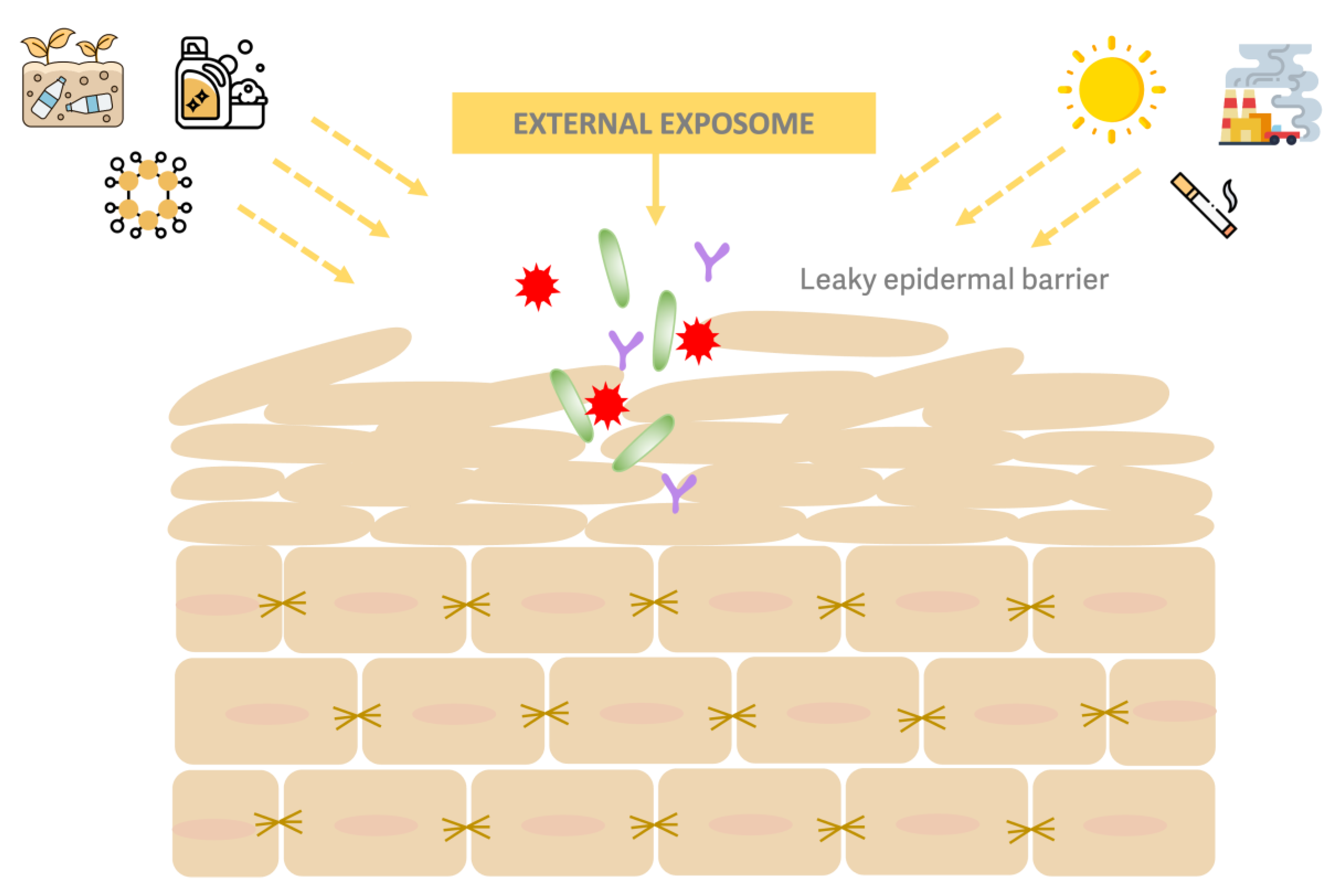
Figure 2.
The three pillars of skin barrier repair. Ceramides-like repair skin lamellar lipids; emollients form a water-repellent film that decreases TEWL; moisturizers and humectants retain TEWL.
Figure 2.
The three pillars of skin barrier repair. Ceramides-like repair skin lamellar lipids; emollients form a water-repellent film that decreases TEWL; moisturizers and humectants retain TEWL.
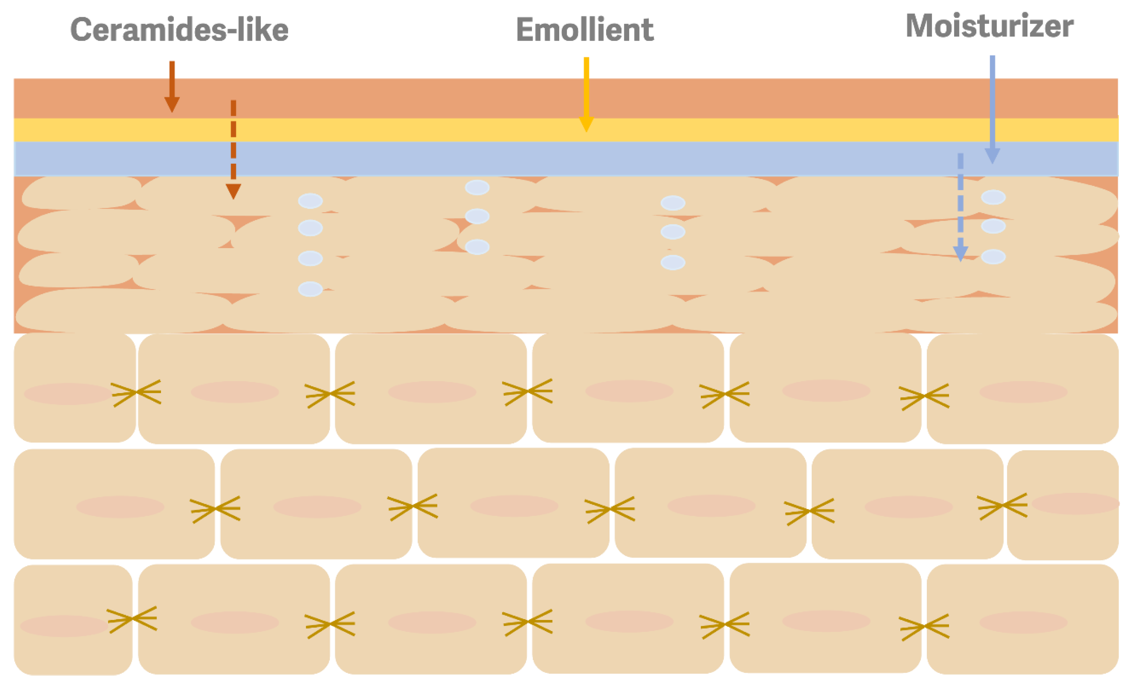

Table 1.
Bioactive compounds/extracts from algae with potential application in skin barrier repair.
| Bioactive Compound Group / Extract | Specific Bioactive Compound | Macro /Microalgae | Type of Study | Results | Ref. |
|---|---|---|---|---|---|
| Polysaccharides | Low molecular fucoidan fraction | Sargassum confusum | In vitro (HaCaT cells) |
Moisture-preserving repair structural proteins in UVB-damaged HaCaT cells | [21] |
| Fucoidan | Undaria pinnatifida | In vivo (ICR mice) |
Promotion of the recovery of epidermal barrier disruption | [22] | |
| Low molecular fucoidan fraction | Sargassum horneri | In vitro (FD-induced HaCaT keratinocytes) |
Amelioration of key tight junction proteins and skin hydration factors Reduction of FD-induced inflammation and skin barrier deterioration |
[23] | |
| Low molecular fucoidan fraction | Sargassumconfusum | In vitro (FD-induced HaCaT keratinocytes) |
Increasing the cell viability in FD-stimulated HaCaT keratinocytes |
[24] | |
| Fucoidan | Sargassum fusiforme | In vitro (HaCaT cells and HDF cells) |
Protective effect against particular matter pollution | [25] | |
| Fucoidan (cream with 1% fucoidan) |
Sargassum horneri | Clinical trial |
Improvement skin barrier function Reduction of TEWL |
[26] | |
| Sulfated polysaccharides + Glucuronic acid | Sargassum fusiforme | In vivo (Hairless Kun Ming mice) |
Decreasing skin moisture loss | [27] | |
| Sacran gel | Aphanothece sacrum | Clinical study | Skin hydration increase TEWL decrease Promotion of normal epidermal differentiation and improvement of the maturation of corneocytes |
[28] | |
| Polysaccharide (specific type NR) | Nostoc commune | In vitro (Mouse stratum corneum) |
Higher water retention than urea | [29] | |
| Carotenoids and other pigments | Fucoxanthin | Undaria pinnatifida | In vitro In vivo |
Regulation of filaggrin genes expression Restoration of the skin barrier by filaggrin stimulation |
[30] |
| Zeaxanthin-based oral supplementation + topical gel serum | Laminaria and Porphyra extracts | Clinical study | Skin hydration improvement | [31] | |
| Phenols | Fucosterol | Sargassum fusiforme | In vitro (HaCaT cells) |
Modulation of MAPK in irradiated HaCaT cells | [32] |
| Fucosterol | Sargassum binderi | In vitro (HaCaT keratinocytes) |
Cytoprotective effects against xenobiotics | [33] | |
| Bromophenol (3-bromo-4,5 – dihydroxybenzaldehyde) |
Polysiphonia morrowii |
In vitro (HaCaT keratinocytes) |
Increasing the production of skin hydration proteins and tight junction proteins | [34] | |
|
Mixture of compounds (Fucus vesiculosus extract, Ulva lactuca extract, and Ectoine) |
Fucoidan, ulvans, and ectoine |
Fucus vesiculosus Ulva lactuca |
Split-Face Clinical Study | Increasing skin hydration Maintenance of the skin barrier function |
[35] |
| Aqueous extracts | NR |
Laminaria japonica (currently Saccharina japonica) |
In vivo | Hydration increase TEWL decrease |
[36] |
| Aqueous extracts | NR |
Sargassum glaucescens |
In vitro (Human Primary Epidermal Keratinocytes, HPEK) |
Induction of the expressions of skin barrier-related genes in HPEK Increasing expression levels of TGM1, KRT10 and FLG Promotion of NMF production |
[37] |
| Aqueous extracts | Guanosine and uridine nucleosides | Codium fragile | In vitro (TNF-α/IFN-γ stimulated HaCaT keratinocytes) |
Expression of factors related to skin barrier function, FLG, IVL, and LOR enhancement | [38] |
| Ethanolic extract | NR | Sargassum horneri | In vitro (FD-induced HaCaT keratinocytes) |
Amelioration of filaggrin, involucrin, and lymphoepithelial Kazal-type-related inhibitor (LEKTI) Regulation of tight junction proteins |
[39] |
| Ethanolic extract | NR | Sargassum horneri | In vivo (DNCB-induced AD) |
Improvement of skin barrier function | [40] |
| Aqueous extracts | NR | Coccoid and filamentous algae | In vivo | Increasing mRNA expression for involucrin, filaggrin and transglutaminase-1 |
[41] |
| Aqueous extract | NR | Botryococcus braunii | In vitro | Induction of gene expression of aquaporin-3, filaggrin and involucrin | [42] |
| Aqueous extract | NR | Neochloris oleoabundans | In vivo | Not skin barrier perturbation Anti-inflammatory activity |
[43] |
| Ethanolic extract | Dominant compounds: Docosahexaenoic acid methyl ester, linolenic acid methyl ester, 13-Docosenamide (Z)-, and methyl 4,7,10,13-hexadecatetraenoate |
Micractinium sp. (KSF0015 and KSF0041) Chlamydomonas sp. (KNM0029C, KSF0037, and KSF0134) Chlorococcum sp. (KSF0003) |
In vivo (C57BL/6 mice) |
Reduction of barrier integrity damage | [44] |
| Ethanolic extract | Main compounds: fatty acids 58.2%, carotenoids 1.6%, phenolics 7.7%, flavonoids 2.0% (crude extract) | Nannochloropsis sp. | In vitro NHDF cells |
Enhance the expression of HAS-2 in a dose-dependent manner | [45] |
| Lipid extract | Myristic acid, palmitoleic acid, and α-linolenic acid | Macrocystis pyrifera | In vitro Three-dimensional cultures of HaCaT cells |
Barrier protective effect | [46] |
| Oily extract | Rich in carotenoids and phenolic compounds (cream with 0,5% extract) |
Cladophora glomerata | Randomized clinical study | Moisturizing improvement | [47] |
| Lipid extract | Palmitic, oleic, myristic, stearic, and linoleic acids | Himantothallus grandifolius, Plocamium cartilagineum, Phaeurus antarcticus, and Kallymenia antarctica | In vitro (HaCaT cells) |
Protection of skin barrier function | [48] |
| Oily extract | Up to 99% of fatty acids + less than 1% of xanthophyll’s | Phaeodactylum tricornutum | In vitro Human keratinocytes |
Stimulation and protection of proteasome peptidase activities | [49] |
| Encapsulated (liposomal) lipid exact | ω-3 fatty acids, and standardized to fucoxanthin levels |
Phaeodactylum tricornutum | Randomized, clinical split-face study |
Soothing effect Barrier function improvement |
[50] |
| Lipid extract | Rich in phosphatidylcholine, and phosphatidylethanolamine | Nannochloropsis oceanica | In vitro | Down-regulation of sphingomyelin Upregulation of ceramides CER[NDS] and CER[NS] |
[51] |
|
Oily extract (Bio-Based Algae Oil) |
Triglyceride (three monounsaturated oleic acid chains with very low polyunsaturated fatty acid content) |
Not described (INCI name Triolein) |
In vivo Single-blind study |
Skin hydration increase TEWL decrease |
[52] |
| Phycocyanin | Spirulina-derived C-phycocyanin |
Spirulina sp. (Arthrospira sp.) |
In vitro (HaCaT cells) |
Protection and maintaining the expression of filaggrin, involucrin, and loricrin after UV radiation |
[53] |
| Aqueous extract | 50 and 70% proteins (dry weight) 8-14% polysaccharides |
Spirulina sp. (Arthrospira sp.) |
Clinical study (Gel-cream formulation with 0,1% w/w spirulina extract) |
Skin hydration increase TEWL decrease Skin microrelief improved Reduction of the surface roughness |
[54] |
| Aqueous extract | 50 and 70% proteins (dry weight) |
Spirulina sp. (Arthrospira sp.) |
Clinical study (Gel-cream formulation with 0,1% w/w spirulina extract) |
Skin hydration increase TEWL decrease Skin microrelief improved Reduction of the surface roughness |
[55] |
| Dry extract | NR |
Spirulina sp. (Arthrospira sp.) |
Double-blind, randomized, placebo-controlled clinical trial (Formulation containing olive oil and spirulina extract) | Skin hydration increase TEWL decrease Skin barrier improvement |
[56] |
| Dry extract | NR (Cream with 5% complex of Dead Sea Mineral salts + algae extract + “desert plants”) |
Dunaliella salina | Clinical trial | Skin roughness decrease Skin moisturizing improvement |
[57] |
*FD, fine dust. NR, not reported. DNCB, 2,4-dinitrobenzene. TGM1, transglutaminase-1. KRT10, Keratin-10. FLG, filaggrin. IVL, involucrin. Loricrin, LOR. NMF, Natural Moisturizing Factor. NHDF, Normal Human Dermal Fibroblasts. MAPK, mitogen-activated protein kinases. HAS2, Hyaluronan Synthase 2. TEWL, Transepidermal Water Loss.
Table 2.
Polysaccharides moisture retention capacity.
| Polysaccharide Type | Moist Retention | Ref. |
|---|---|---|
| LMW polysaccharides (brown algae) | The lower the molecular weight, the greater the moist retention capacity Higher than HA |
[96] |
| Sulfated polysaccharide (Ulva lactuca) | Higher than glycerol (43% vs 34%) |
[97] |
| Sulfated polysaccharides (Enteromorpha prolifera) | The higher the sulphate content, the higher the water retention Close to HA |
[98] |
| Brown and red algae polysaccharides | Higher in Sargassum horneri than in Porphyra yezoensis | [99] |
| Sulfated polysaccharide (fucoidan-rich extract) (Sargassum vachellianum) | Higher (65.84%) than glycerol (51.35%) | [100] |
| Polysaccharides-rich extract (Caulerpa microphysa) | Better than collagen and HA, and similar to urea | [101] |
| Polysaccharides-rich extract (Nostoc) | Higher (78.5%) than chitosan (75.2%), and urea (62.7%) | [29] |
*LMW, Low molecular weight. HA, hyaluronic acid.
Table 3.
Examples of patents related to the use of algae and Spirulina for skin barrier improvement.
Table 3.
Examples of patents related to the use of algae and Spirulina for skin barrier improvement.
| Compound | Specie / Strain | Claim | Patent Number |
|---|---|---|---|
| Protein and peptides (dry powder) | Porphyra sp., Wakame sp., Spirulina sp., and Chlorella sp. | Glossing and moisturizing skin (cream) | EP1433463B1 |
| Extracts | Chondrus crispus and Codium tomentosum | Skin moisturizing | CN110339102A |
| Extracts of coccoid filamentous cyanobacteria | Cyanobacterium sp. | Enhancement of skin barrier | US8795679B2 |
| Lysate | Chlamydocapsa sp. | Enhancement of skin barrier | US8206721B2 |
| Cell algae or extracts | Prototheca, Auxenochlorella, Chlorella or Parachlorella genus | Improvement of skin hydration | US20150352034A1 |
| Extracts | Senedesmus sp. | Protection against UV damage and moisturizing | KR101825683B1 |
| Liposomes or algaesomes | Dunaliella salina | Moisturizing agent | KR102008870B1 |
| Extract | Leptolyngbya tenuis | Promote the production of Aquaporine-3 | JP2022029111A |
| Extract | Chlorella sorokiniana | Strengthening the skin barrier | FR3064481A1 |
| Peptide extract | Spirulina | Restructuring the cutaneous barrier | FR2857978, 27 |
| Extract |
Haematococcus pluvialis (H. lacustris) |
Moisturizing mask | CN106963670B |
Disclaimer/Publisher’s Note: The statements, opinions and data contained in all publications are solely those of the individual author(s) and contributor(s) and not of MDPI and/or the editor(s). MDPI and/or the editor(s) disclaim responsibility for any injury to people or property resulting from any ideas, methods, instructions or products referred to in the content. |
© 2025 by the authors. Licensee MDPI, Basel, Switzerland. This article is an open access article distributed under the terms and conditions of the Creative Commons Attribution (CC BY) license (http://creativecommons.org/licenses/by/4.0/).
Copyright: This open access article is published under a Creative Commons CC BY 4.0 license, which permit the free download, distribution, and reuse, provided that the author and preprint are cited in any reuse.


