
Submitted:
01 August 2025
Posted:
04 August 2025
You are already at the latest version
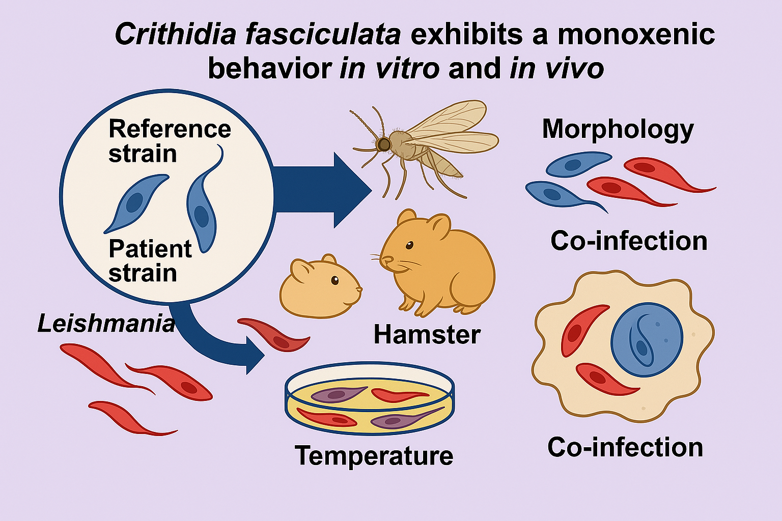
Abstract
There is increasing evidence on the occurrence of Crithidia spp. in patients presenting either cutaneous or visceral leishmaniasis, solely or associated with Leishmania. We analyzed the influence of temperature in the growth rate and morphology of two Crithidia fasciculata strains (a reference strain and one isolated from a patient), and the effect of the co-cultivation of Leishmania and Crithidia in parasite isolation, in the infection of macrophages in vitro, and also in infections of hamsters, BALB/c mice and sandflies. In culture, both Crithidia strains could undergo 32oC for 96 h, although major morphological alterations and a decrease in mitochondrial membrane potential were observed. At 34°C, there was an 80% reduction on the number of cells from the patient strain. Mixed cultivation of Crithidia-Leishmania led to the recovery of only Crithidia. In macrophages, C. fasciculata alone was virtually eliminated, and in the co-infection only Leishmania was recovered. The same was observed in vivo. Curiously, C. fasciculata is more resistant to Amphotericin B. Our results indicate that both C. fasciculata strains are unable to reproduce the pathogenic effect in vitro and in vivo models.
Keywords:
Leishmania
; co‐infection
; macrophage
; sandfly
; Amphotericin B
1. Introduction
Neglected tropical diseases (NTDs) such as those caused by trypanosomatid protozoa represent a significant global health challenge, particularly in tropical and subtropical regions [1]. These parasites are transmitted by various arthropod vectors, causing a broad range of diseases responsible for substantial morbidity or even mortality [2]. The diverse trypanosomatid species are traditionally divided into heteroxenous, characterized by completing their life cycle alternating between invertebrate and vertebrate or plant hosts; and monoxenous trypanosomatids, presumably restricted to invertebrate hosts, mainly insects. Although the heteroxenic counterpart are the most extensively studied due to their considerable medical, veterinary, and economic impacts [3], monoxenous trypanosomatids have been attracting attention due to the increasing reports on the occurrence of these presumably nonpathogenic parasites in mammalian hosts, including humans [4,5,6,7,8,9,10,11,12,13,14,15,16].
Most of the recent reports about monoxenic infections have emerged from the atypical nature of some cases, inspiring further investigations into the pathogenic species involved. Most monoxenic infections reported in humans were observed either in co-infection with HIV or other trypanosomatids, such as Leishmania [7]. So far, the impact of monoxenous trypanosomatid on the pathogenesis of leishmaniasis is still unclear, raising critical questions about how mixed infections affect disease severity, diagnosis, and treatment.
Our research group received an isolate from a cutaneous lesion of an immunocompetent man in Cusco, Peru [4]. The parasite initially was identified as Leishmania. braziliensis based on clinical presentation and geographic distribution, but further studies revealed inconsistent characteristics, such as non-fastidious cultivation. Nevertheless, through a more accurate genomic analysis employing the molecular markers gGAPDH and V7V8 ribosomal RNA region, the parasite was identified as Crithidia fasciculata, and was deposited in our Protozoa Collection in Oswaldo Cruz Foundation (COLPROT-Fiocruz) [14]. In the present study, we addressed the issue of Leishmania–Crithidia co-infection from the point of view of the monoxenous partner. A comprehensive analysis of the growth dynamics and infectivity of C. fasciculata isolated from a human patient were performed. The parasite's behavior in different culture media was assessed, as well as its in vitro and in vivo infectivity against mouse and hamster models. Finally, the potential effect of the co-infection of C. fasciculata and Leishmania braziliensis was investigated, and the ability of such co-infection to colonize the sandfly vector Lutzomyia longipalpis, contributing to our understanding of its potential as a vector-borne pathogen. This study shed some light on the ecological and pathological roles of monoxenous trypanosomatids in human infections, providing valuable information into co-infection mechanisms and the pathogenic potential of C. fasciculata.
2. Materials and Methods
2.1. Parasite Cultivation
The following strains from our Protozoan Collection (COLPROT- Fiocruz) were used: i) the type strain of C. fasciculata (COLPROT048), isolated from Anopheles quadrimaculatus in 1926; ii) C. fasciculata (COLPROT606), isolated from a human patient in 1994; and iii) L. braziliensis (ThorMCAN/BR/97/P142) obtained from LBTq/IOC and maintained in BALB/c mice. All strains were cultivated in NNN’/LIT medium with 10% inactivated FBS. C. fasciculata strains were cultured in LIT medium with 10% inactivated FBS (Gibco) at 27°C, while L. braziliensis was maintained in Schneider´s medium (Sigma-Aldrich) with 20% inactivated FBS and 2% sterile human urine at 27°C. Passages were done twice a week up to the fifth passage.
2.2. Growth Curve
Growth curves were established by inoculating cultures (C. fasciculata COLPROT606 and COLPROT048, and L. braziliensis MCAN/BR/97/P142) starting with 1.0 × 10⁶ parasites/mL in LIT or Schneider´s medium supplemented with 10% and 20% inactivated FBS, respectively. Parasites were maintained in triplicate and monitored every 24 hours during five days. Parasite viability was assessed using a Neubauer chamber and Trypan blue (Sigma-Aldrich) exclusion. The effect of temperature on COLPROT606 was evaluated at 27°C, 32°C and 34°C. Replicates were passed during the logarithmic growth phase.
Alternatively, to evaluate mixed cultures, L. braziliensis and COLPROT606 were co-cultured in Schneider medium with 20% inactivated FBS and 2% sterile male human urine at 27°C in the following ratios: L. braziliensis (control), COLPROT606 (control), and three mixed ratios (1:1, 2:1, 1:2). For mixed cultures, 1 × 10⁶ or 2 × 10⁶ parasites were added to 5 mL of culture medium. Every 3 days, cultures were checked for contamination and growth, and 300 µL were passed into fresh medium. After five passages, the cultures were washed three times with sterile PBS, centrifuged, and stored in TRIzol® (Invitrogen) at -80°C for RNA extraction and qPCR analysis [17].
2.3. Morphological Analysis
For cell morphology analysis, C. fasciculata COLPROT606 and COLPROT048 were stained with Giemsa (Sigma-Aldrich) and observed under a Zeiss AxioObserver M1 optical microscope with a 100 × objective. Smears were prepared from log phase samples (5.0 × 10⁶ parasites/mL), fixed in 100% methanol for 10 minutes, air-dried, treated with 5N HCl for 10 minutes, washed, and stained with Giemsa for 1 hour.
2.4. Mitochondrial Membrane Potential (ΔΨm) Assay
To evaluate the impact of the temperature on C. fasciculata strains, the mitochondrial membrane potential (ΔΨm) of parasites incubated at 32°C was evaluated by flow cytometry. The parasites were incubated with 50 nM tetramethylrhodamine (TMRE) (Molecular Probes) for 15 min at 28°C, using 50 μM carbonyl cyanide m-chlorophenyl hydrazone (CCCP) (Sigma) as a control for ΔΨm dissipation. Variations in TMRE fluorescence were quantified using an index of variation (IV), calculated by the equation of the median of fluorescence for parasites in experimental condition of 32°C (ME) less the median of fluorescence of the control parasites (MC), and divided by MC (ME-MC/MC). The fluorescence of parasites incubated with CCCP were reduced from the fluorescence of both ME and MC groups. Negative IV values correspond to depolarization of the mitochondrial membrane [19]. In parallel, the parasites were labeled with 0.1 µM TO-PRO3 iodide (Invitrogen) for 30 min to evaluate the plasma membrane integrity. Parasites incubated in 0.01% Triton X-100 (Sigma) were used as positive control. The parasites (10,000) were kept on ice until the acquisition on Beckman Coulter's CyAn Flow Cytometer, and finally analyzed in the CytExpert software.
2.5. Murine Peritoneal Macrophages In Vitro Infection
Peritoneal macrophages were extracted from BALB/c mice and adhered in coverslips (3 × 10⁵ cells) with RPMI 1640 medium (Gibco) in a 4% CO2 atmosphere at 37ºC for 24 h, and were then washed thrice with PBS. Cells (1.0 × 10⁶ cells/well) were infected with L. braziliensis (MCAN/BR/97/P142 strain) and both C. fasciculata strains (COLPROT048 and COLPROT606) at a 5 parasites per macrophage ratio, both individually and in co-infection. Cells were cultured in RPMI medium supplemented with 10% FBS, 1% glutamine, and 1% pyruvate for 24, 48, 72, and 96 hours at 35°C with 5% CO2. After incubation, slides were stained with Panotic Fast (Laborclin), and the infection index was determined by counting under an optical microscope using the formula: % infected macrophages × amastigotes count/total macrophages [20].
2.6. Amphotericin B Sensitivity Assays
For Amphotericin B assays, 4×10⁶ parasites/mL were used in 96-well plates. The drug was evaluated in a range of diluted concentrations starting from 1 μM. After 72 hours of incubation at 26°C, cell viability was assessed by fluorometry using 50 µM Alamar Blue®resazurin. Readings were taken with a SpectraMax GEMINI XPS (Molecular Devices), with an excitation of 560 nm to a detection emission of 590 nm. All assays were performed in triplicate. The results were expressed as the 50% inhibitory concentration (IC50/72h) for parasite growth, using a nonlinear regression analysis on a semi-logarithmic scale obtained from GraphPad Prism 5.0 software [21].
2.7. In Vivo Experimental Infection Models
In vivo experiments were conducted in two models. Female BALB/c mice (4-8 weeks old, 5 per group) from ICTB Fiocruz, housed at the Oswaldo Cruz Institute (approved by the Animal Ethics Committee: L002/2022), were infected in the right ear with 2.0 × 10⁶ parasites in the following groups: Group I - L. braziliensis (control), Group II –C. fasciculata COLPROT048, Group III - C. fasciculata COLPROT606, Group IV - L. braziliensis + COLPROT 048 (1:1), and Group V - L. braziliensis + COLPROT606 (1:1). Lesion size was monitored twice weekly for 35 days with a dial caliper (Mitutoyo) and expressed as the difference between the thickness of the infected and uninfected paws, and parasitic load was evaluated by limiting dilution and RT-qPCR. The second in vivo model was Golden hamsters (6-8 weeks old) from the UERJ animal facility (Animal Ethics Committee: 023/2022), which were used in similar experiments, i.e., infected in the right paw with 1.0 × 10⁶ parasites and monitored for lesion size. After euthanasia, the infected paw and popliteal lymph nodes were removed for analysis [21].
For the immunosuppression assays, BALB/c mice (10 per group) were treated with cyclophosphamide (150 mg/kg) intraperitoneally, and subsequently infected with 1x 10⁷ C. fasciculata (COLPROT048 or COLPROT606). Lesions were monitored weekly, and parasitic load was assessed by limiting dilution.
2.8. RT-qPCR Quantification
Parasitic load was quantified using reverse transcriptase quantitative PCR (RT-qPCR). Primers and probes (IDT Inc.) were designed based on conserved regions of the SSU rRNA gene sequences of L. braziliensis and C. fasciculata using MUSCLE and Primer Express software. RNA was extracted with TRIzol and RNeasy Mini Kit (Qiagen), treated with DNase (Sigma), and converted to cDNA using the SuperScript III kit (Invitrogen) [17]. qPCR was performed on an ABI Prism 7500 Fast system, with the following primers and probes: SSU_Cfa_FwCCGTGCCCTCAAGAACAT, SSU_Cfa_RvGGGATGTTCACACCGTACAA and SSU_Cfa_probeFAM-TGCACAAGAAGAAGCAGGAGCAGA-3IABkFQ for C. fasciculata amplification; ii) SSU_Lbr_FwTGACGAACCCACACAACAA, SSU_Lbr_RvGGTCGCGAATTATCTCCCAATA and SSU_Cfa_probeHEX-ACCGAACGAAAGCTGAACCACACT-3IABkFQ for L. braziliensis detection; and iii) Mm.PT.39a_Fw GGGTGGAACTGTGTTACGTAG, Mm.PT.39a_Rv TGGTCTTTCTGGTGCTTGTC and Mm.PT.39a_pb Cy5-CCGGAGAATGGGAAGCCGAACATAc-3IAbRQSp for mouse microtubulin beta (Mm.PT.39a.22214835, IDT Inc.) amplification. Standard curves were generated from serial dilutions of cDNA (10⁷ to 100 parasites) to quantify parasitemia and assess primer efficiency. All the reactions were performed in triplicate.
2.9. Sandflies Experimental Infection
Infection and colonization of sandflies were performed using 5-8-day-old female Lutzomyia longipalpis by artificially feeding on mice blood containing L. braziliensis (MCAN/BR/97/P142) or C. fasciculata (COLPROT606) at a concentration of 5.0 × 10⁶ parasites/mL, or a 1:1 mixture of L. braziliensis-C. fasciculata. Pools of 10 sandflies from 3- and 7-days post-infection had their RNA extracted by the protocol described previously. The parasite load was evaluated by qPCR using SYBR Green (Invitrogen) and employing the same SSU rRNA primers for the trypanosomatids’ detection and Lu. longipalpis ribosomal protein (RP49) as a normalization control [22]. The parasite load was represented by the number of quantified parasites (L. braziliensis or C. fasciculata) × 104 per the number of sandflies in the pools (trypanosomatid × 104/sandfly pool).
2.10. Statistical Analysis
The results were analyzed using the t-test for non-parametric variables and Two-way ANOVA/Bonferroni for parametric variables. The significance level was set at P < 0.05. Analyses were performed using GraphPad Prism 5.0 Software (San Diego, CA, USA).
3. Results
3.1. Growth Kinetics of C. fasciculata COLPROT606 and ΔΨm Analysis
The presence of monoxenous co-infections with Leishmania should imply several adaptations to the environment of the human body, such as temperature. Pathogenic Leishmania species exhibit intracellular amastigotes in mammalian cells and extracellular promastigotes in the vector, while Crithidia species typically display the coanomastigote form, but intermediate forms may appear in cultures. To investigate the growth kinetic of the isolated C. fasciculata COLPROT606, parasites were cultured in LIT medium with 10% FBS and Schneider’s medium with 20% FBS and 2% sterile human urine at 27°C (Figure 1A). Growth analysis revealed faster growth of the reference strain (COLPROT048) in Schneider’s medium compared to LIT, and a slight increase in cell number for C. fasciculata COLPROT606 in Schneider’s medium. L. braziliensis failed to grow in LIT medium, leading to the use of Schneider’s medium for subsequent experiments. Additionally, C. fasciculata COLPROT606 survived and replicated at 32°C, although in fewer numbers than at 27°C (Figure 1B). However, the parasites were not capable of surviving at 34°C, resulting in an 80% cell death within 24 hours (Figure 1C). Similar results were observed using C. fasciculata COLPROT048 (data not shown). The ability of C. fasciculata to withstand 32°C could explain the isolation of the parasite from human cutaneous lesions, where temperatures are lower than in internal organs. Additionally, 32°C is the temperature used to induce Leishmania amastigogenesis, and so it may mimic the temperature within mammalian host cells.
Morphological parameters at different temperatures of the growth kinetics were assessed using Giemsa staining prepared every 24 hours (Figure 2). Subtle morphological differences were observed at 32°C, as the parasites displayed a more rounded shape, whereas at 27°C they maintained a less altered morphology with rosette formation, which indicates high cellular growth and adaptation.
To analyze the effect of the elevated temperature on the parasite’s viability, ΔΨm was assessed by the use of TMRE probe (Table 1). It´s important to mention that CCCP was employed to exclude the unspecific labeling. At 48h, C. fasciculata COLPROT 048 slightly decreased the ΔΨm between parasites cultivated at 27°C and 32°C, but the number of TMRE+ cells and the IV were not significant. Conversely, the strains isolated from a patient reduced significantly the IV, as the pathogenic L. braziliensis parasites. After 96 hours the TMRE fluorescence of both C. fasciculata strains decreased to 60% and 64%, respectively. L. braziliensis mitochondria was even more affected by the 32°C temperature, reaching a reduction of 90% in ΔΨm (Table 1).
3.2. Cell Growth Evaluation in Leishmania-Crithidia Co-Cultures
A common approach to isolate parasites from clinical lesions requires the inoculation of the biopsy material in culture for taxonomic identification following cell growth. However, this methodology raises the concern of whether one parasite might overcome the other in the culture. To explore this question, we simulated an in vitro mixture under the following conditions: L. braziliensis alone (control), C. fasciculata COLPROT606 alone (control), a 1:1 mixture of L. braziliensis and C. fasciculata, and a 2:1 mixture of the two parasites. The cultures were subcultured every 3 days, and after 5 passages the cells had their RNA extracted for quantitative RT-qPCR analysis.
To quantify the number of each trypanosomatid in the co-infection’s assays, a sensitive and reproducible qPCR was developed. We standardized parasite load quantification by RT-qPCR employing the small subunit of the ribossomal RNA (SSU rRNA) primers and probes, which provides a higher specificity and sensibility to quantify each trypanosomatid (Fig. S1). In the mixed cultures, the results demonstrated that C. fasciculata successfully outgrew L. braziliensis in all mixed infection conditions (Figure 3). The absolute quantification didn’t reveal the presence of L. braziliensis in any co-infection condition.
3.3. Susceptibility to Amphotericin B
Due to the increasing presence of non-pathogenic species in vertebrate hosts, which may exacerbate symptoms or promote resistance to leishmaniasis treatment [5,6], we evaluated C. fasciculata susceptibility to amphotericin B, a drug commonly used for leishmaniasis treatment. L. braziliensis showed an IC50/72 h of 0.1 ± 0.02 μM, while the values were 0.4 ± 0.02 and μM for and 0.5 ± 0.01 μM for COLPROT048 and COLPROT606, respectively (Figure 4).
3.4. In Vitro Coinfection of C. fasciculata and L. braziliensis
Macrophages are key immune cells controlling Leishmania infections, and the failure to control parasite replication within these cells is critical for disease progression. Here, we analyzed the ability of C. fasciculata parasites to infect macrophages, and maintain the infection for 96 hours, by optical microscopy. As expected, L. braziliensis was internalized by macrophages and maintained infection for 4 days (Figure 5). In contrast, C. fasciculata strains (COLPROT048 and COLPROT606) showed a significantly lower number of parasites inside macrophages at 24 hours post infection. This number persisted low at 48 and 72 hours, decreasing even more at 96 hours (Figure 5A). No statistical difference was observed between C. fasciculata COLPROT048 and COLPROT606 strains.
Considering that L. braziliensis might be able to modulate the immune response to support the survival of C. fasciculata, in vitro coinfection of murine macrophages was also evaluated by RT-qPCR. As observed previously, L. braziliensis alone was successfully internalized and maintained the macrophage for 96 hours, but C. fasciculata COLPROT606 failed to persist alone in macrophages over the same period. In coinfected murine macrophages, L. braziliensis RNA was detected in high number at the 96 hours of infection, but C. fasciculata was only detected at 24h pos-infection (Figure 5B). These findings suggest that L. braziliensis does not enhance the survival of C. fasciculata within macrophages or exacerbate infection, but rather eliminated C. fasciculata from the vertebrate host cells.
3.5. In Vivo Experimental Infections
The infectivity of C. fasciculata was evaluated in both BALB/c mice and golden hamsters, two established experimental models for Leishmania spp. infection. The golden hamsters are the most susceptible in vivo experimental model for L. braziliensis infection, capable of developing large and self-resolving lesions [23], Therefore, female hamsters were infected in the dorsum of their right hind paw with 1.0 × 10⁶ parasites according to the following protocol: Group I - L. braziliensis alone, Group II - C. fasciculata COLPROT048, Group III - C. fasciculata COLPROT606, Group IV - L. braziliensis co-infected with C. fasciculata COLPROT048, and Group V - L. braziliensis co-infected with C. fasciculata COLPROT606. The animals had the lesion size measured for 78 days post-infection, and were euthanized at the end of the experiment. The infected paw and popliteal lymph nodes were removed and macerated for analysis by limiting dilution (LDA) and qPCR absolute quantification (Figure 6).
Our results reported apparent lesions in control Group I, and in the co-infections of L. braziliensis with C. fasciculata COLPROT048 and COLPROT606, Groups IV and V, including the typical ulceration of this lesion in this experimental model (Figure 6A,C). However, no apparent lesions were observed on the paws of the hamsters infected only with C. fasciculata, Groups II and III. Moreover, the LDA parasite burden analysis didn’t report any parasite growth of in C. fasciculata strains infected alone, Groups II and III (Figure 6B). In the systems in which it was possible to observe a LDA positive parasite growth, Groups I, IV and V, the paw samples had a higher burden compared to popliteal lymph node (data not shown).
In order to improve our co-infection analysis, we performed a qPCR quantification of the removed paws to detect L. braziliensis and C. fasciculata RNA, which could indicate the presence of live parasites at the end of the experiment. No C. fasciculata RNA was detected by RT-qPCR in the co-infection’s groups after 78 days of infection, or Crithidia alone (Figure 6D). However, L. braziliensis was detected in all the three groups where this parasite was used. A mean of 1,145 parasites/mg skin equivalents were observed in Group I, 1,625 parasites/mg in the co-infected Group IV, and 1,141 parasites/mg in the co-infected Group V (Figure 6D). The changes in the qPCR parasite load were not statistically significant, suggesting that the co-infected were not capable of improving the hamster infection. The absence of C. fasciculata RNA in Groups II and III leads us to assume that this parasite is not able to maintain itself in hamsters.
In the BALB/c mouse model, infection with L. braziliensis and C. fasciculata COLPROT048 and COLPROT606 induced significant inflammation, as measured by increased ear thickness (Figure S2A). However, in hamsters C. fasciculata COLPROT048 and COLPROT606 alone failed to induce any visible signs of infection or growth, indicating that C. fasciculata are also not capable of surviving or establishing an infection in BALB/c mice (Figure S2B). In addition, no C. fasciculata growth or RNA were observed. Notwithstanding, we decided to investigate the persistence of C. fasciculata in immunosuppressed BALB/c mice treated weekly with cyclophosphamide (3 mg/animal, intraperitoneally). The animals were infected in the left hind paw with C. fasciculata COLPROT048 and with the clinical isolate C. fasciculata COLPROT606. A control group received the same treatment without infection. Immunosuppression efficacy was confirmed by a decrease in leukocyte count from 13.9 to 1.9 over 5 weeks (Figure 7A). The infection was monitored by measuring paw thickness, and after 14 weeks, both the paws and draining popliteal lymph nodes were collected for LDA parasite burden analysis. An initial increase in paw thickness was observed, followed by a stabilization in the size of the lesion (Figure 7B), but no parasite growth was detected in cultures from the paws or lymph nodes of cyclophosphamide-treated mice (data not shown). These results suggest that C. fasciculata is unable to persist in BALB/c mice, even under immunosuppression.
3.6. Co-Infection of Sandflies with L. braziliensis and C. fasciculata COLPROT606
To assess the transmissibility of C. fasciculata in an invertebrate host, we tested its ability to colonize the Lu. longipalpis sandfly, a known permissive vector of Leishmania spp. [24]. Female sandflies (100 per group) were fed with blood containing: (i) L. braziliensis, (ii) C. fasciculata (COLPROT606), or (iii) both parasites (1:1). A pool of 10 insects was collected on day 3 and 5 post-infection and analyzed by RT-qPCR. Results showed that C. fasciculata was present at all time points, both alone and in coinfection, while L. braziliensis colonized the sandfly as expected (Figure 8). These findings suggest that C. fasciculata can colonize Lu. longipalpis, offering insights into the transmission dynamics of these trypanosomatids in vertebrate hosts. Moreover, the co-infection significantly increased the number of both parasites on the third day, rising from 2.4 × 10⁴ to 9.5× 10⁴ C. fasciculata/sandfly and 2.5 × 10⁴ to 9.5 x 10⁴ L. braziliensis/sandfly. On day 10 the number of both parasites decreased and no statical difference was observed both alone and in the mixed infection (Figure 8).
4. Discussion
Recent reports of human infections by monoxenous trypanosomatids, previously assumed to be insect-exclusive, are raising concerns about their pathogenic potential in vertebrates, including humans. Leptomonas seymouri has been frequently identified in co-infections with Leishmania donovani in the Indian subcontinent, often associated with more severe clinical outcomes and possible drug resistance [15,16]. Similarly, Crithidia spp. have been reported in both humans and animals, with cases documented in Brazil and Iran [4,14]. In humans, monoxenous infections are primarily described in immunocompromised individuals co-infected with HIV and L. major or L. infantum, but have also been detected in immunocompetent hosts as the sole pathogen [4,5,6,7,8,9,10,11,12,13,14]. Additionally, species of Herpetomonas and Blechomonas (formerly classified as Leptomonas) have been sporadically detected in humans [7]. These emerging reports suggest an expanding host range and increased adaptability of monoxenous trypanosomatids, reinforcing the need for intensified surveillance and continued investigation into their pathogenic mechanisms. Therefore, here we further investigated the biological and pathogenic characteristics of a C. fasciculata isolate obtained from a skin lesion of a patient from Cusco, Peru [4,17], to better understand its behavior under experimental conditions and its potential role in human infections.
Our findings demonstrate that C. fasciculata exhibits remarkable adaptability, being capable of surviving and proliferating in two media specifically designed for monoxenous trypanosomatids, i.e., LIT and Schneider's medium; the last typically used for Leishmania species. Furthermore, the parasite's limited growth at 32°C, a temperature usually employed for Leishmania axenic amastigogenesis [25], indicates a certain degree of thermo tolerance, although the reduction of C. fasciculata ΔΨm at 32°C after 96 h suggests the loss of parasites´ viability in this adverse condition. The capability to withstand a higher temperature supports previous observations of the parasite’s ability to grow in environments with low nutrient availability and inconstant temperatures, such as the conditions in the regions where C. fasciculata has been reported causing mammals infection [4]. Interestingly, in co-culture experiments, C. fasciculata outgrew L. braziliensis, highlighting its faster growth rate under laboratory conditions. However, in contrast to prior studies with Crithidia coinfection, we observed that C. fasciculata COLPROT606 failed to survive at higher temperatures, such as 34°C, suggesting that environmental temperature may limit some isolates to thrive in warmer conditions [14,26].
Following these findings, it is important to consider the "environment-biased selection hypothesis", which suggests that in co-infection scenarios, laboratory culture conditions may favor the faster-growing monoxenous trypanosomatids, such as C. fasciculata, potentially overshadowing the growth of Leishmania species [27]. In our co-culture experiments, C. fasciculata consistently outgrew L. braziliensis, making it unfeasible to detect Leishmania parasites after extended incubation periods. This result supports the hypothesis mentioned, indicating that C. fasciculata outgrew the pathogenic species in laboratory conditions due to the monoxenous faster growth, which could lead to potential diagnostic misinterpretations in co-infections. On the other hand, in the vertebrate model, Leishmania outcompetes the monoxenous trypanosomatids, which may be eliminated by host immune responses, or even by the lack of other mechanisms to establishes infection [27].
Unlike pathogenic Leishmania species, C. fasciculata did not survive or proliferate within murine peritoneal macrophages despite an initial infection, indicating the absence of immune evasion mechanisms commonly associated with Leishmania infections [28,29]. In contrast, Crithidia sp. CLA-KP1, isolated from the biting midge Culicoides peregrinus, was cleared from murine peritoneal exudate macrophages (PEMs) by 48 hours [26]; whereas the Brazilian clinical isolate Crithidia sp. LVH60 infected THP-1 cells for up to 72 h [11]. Another C. fasciculata isolated in Iran infected both J774 and THP-1 cells, yet the persistence was not specified [6]. The results presented here are aligned with findings on Le. seymouri, which also failed to persist in mammalian macrophages during co-infection with L. donovani [15]. Together, these findings suggest that C. fasciculata isolated from Peru could lack the virulence factors required to cause disease in vertebrate hosts. Furthermore, its inability to establish infection in murine or golden hamster models, even under immunosuppressed conditions, further supports the conclusion that C. fasciculata could be non-pathogenic under the experimental conditions assayed here.
Another key aspect of our study was assessing C. fasciculata resistance to Amphotericin B, a drug commonly used for the treatment of leishmaniasis [30]. Our results showed that C. fasciculata parasites isolated from a human patient exhibited greater resistance to Amphotericin B compared to both L. braziliensis and the reference C. fasciculata strain. This result suggests that C. fasciculata may harbor intrinsic mechanisms of drug resistance, as supported by a previous study comparing growth inhibition between C. fasciculata and Leptomonas, where C. fasciculata required 3 to 6 times higher concentrations of phenanthridines and diamidines to achieve 50% inhibition compared to Leptomonas [31]. Nevertheless, further studies with additional drug treatments are necessary to better characterize the specific mechanisms of resistance and assess their clinical relevance, particularly in regions where C. fasciculata may be co-existing with Leishmania species.
In the sandfly insect host, C. fasciculata was detected in all samples after artificial blood infection, indicating its ability to establish in the vector. A meta-analysis has shown that insects exhibit a higher prevalence of monoxenous trypanosomatids, likely due to a trade-off favoring dissemination over complex host adaptation [32]. This increased prevalence could favor contact with vertebrate hosts, presenting a challenge which should be overcome by the monoxenous parasite. In the presence of a heteroxenous parasite the survival of the monoxenous counterpart could be favored by a synergistic interaction between both trypanosomatids. In this context, studies reporting sandflies naturally infected with monoxenous trypanosomatids suggest that human infection with C. fasciculata could share similar transmission dynamics with pathogenic species [33,34,35,36,37]. Notwithstanding, the passage of the parasite through the vector could select for more virulent populations [38], and factors such as immunomodulatory molecules presented in sandfly saliva may also influence the success of infection [39]. Additional research should aim to evaluate these natural transmission conditions more closely to better understand how the vector influences the pathogenic potential of C. fasciculata, and to clarify the ecological and biological dynamics of trypanosomatids in their natural environments.
5. Conclusions
Over the last few years, an increased number of reports of monoxenous trypanosomatids in vertebrate hosts have emerged. However, the mechanisms involved in the pathogenesis and transmission of such parasites in co-infection with pathogenic trypanosomatids remain poorly understood. Our findings suggest that the Crithidia fasciculata isolated from an atypical human infection does not exhibit mechanisms that would promote its persistence or pathogenicity in the mammalian experimental models used in this study. These results highlight the need for further investigation about the presence of monoxenous trypanosomatids in pathological conditions, as well as the routine use of molecular methods such as DNA sequencing for a better diagnostic, and to clarify the role of these organisms in vertebrate infections or even avoid misidentification in clinical settings.
Supplementary Materials
The following supporting information can be downloaded at the website of this paper posted on Preprints.org. Figure S1: Standardization of qPCR assays for parasite load quantitation from culture mixtures, in vitro peritoneal macrophage infection and in vivo experiments with L. braziliensis and C. fasciculata COLPROT 606; Figure S2: Experimental infection in the right ear of BALB/c mice.
Author Contributions
Conceptualization, CMD and VE-V.; methodology, JFBdS, CBM, VVA-N and TLS; formal analysis, RFSM-B, ECT-S and VE-V.; resources, YMT-S, SAGdS, RFSM-B, ECT-S and CMD; writing—original draft preparation, JFBdS, CBM and VE-V; writing—review and editing, YMT-S, SAGdS, RFSM-B, ECT-S,CMD and VE-V. All authors have read and agreed to the published version of the manuscript.
Funding
This research was funded by grants from Fundação Carlos Chagas Filho de Amparo à Pesquisa do Estado do Rio de Janeiro (FAPERJ), Conselho Nacional de Desenvolvimento Científico e Tecnológico (CNPq), Coordenação de Aperfeiçoamento de Pessoal de Nível Superior (CAPES, Financial code 001). The APC was funded by Instituto Oswaldo Cruz (IOC), Fundação Oswaldo Cruz (FIOCRUZ).
Institutional Review Board Statement
This study performed all animal care and experimental procedures in compliance with Guide for the Care and Use of Laboratory Animals of the Brazilian National Council of Animal Experimentation (COBEA). The procedures have the approval of the Animal Ethics Committee of Oswaldo Cruz Foundation (license number L002/2022) and Committee on of the Instituto de Biologia Roberto Alcantara Gomes of the Universidade do Estado do Rio de Janeiro-UERJ (license number 023/2022).
Informed Consent Statement
Not applicable.
Data Availability Statement
The original contributions presented in this study are included in the article. Further inquiries can be directed to the corresponding author.
Acknowledgments
We are grateful to Dra. Ana Cristina Souza Bombaça and Msc. Sheila Medeiros dos Santos Pereira their technical assistance, and the Protozoa Collection (COLPROT) from Fundação Oswaldo Cruz (FIOCRUZ) for providing Crithidia fasciculata parasites. We also thank the multi-user facilities from Instituto Oswaldo Cruz - FIOCRUZ: flow cytometry, real time PCR and DNA sequencing.
Conflicts of Interest
The authors declare no conflicts of interest.
References
- Hamida, A.; Ilyas, D.; Zahra, M.; Shehnaz, G. In Leishmaniasis: A Neglected Tropical Disease. In Global Immunological & Infectious Diseases Review; APA, 2019; Volume IV, Chapter 1, pp. 17–23. [CrossRef]
- Karmakar, S.; Volpedo, G.; Zhang, W.W.; Lypaczewski, P.; Ismail, N.; Oliveira, F.; et al. Centrin-deficient Leishmania mexicana confers protection against Old World visceral leishmaniasis. NPJ Vaccines 2022, 7(1), 157. [CrossRef]
- Maslov, D.A.; Votýpka, J.; Yurchenko, V.; Lukeš, J. Diversity and phylogeny of insect trypanosomatids: all that is hidden shall be revealed. Trends Parasitol. 2013, 29, 43–52. [CrossRef]
- Toledo, P.D. Comportamiento in vitro de um Kinetoplastido Trypanosomatidae Aislado de uma Lesion Cutanea. Licenciate Thesis, Universidad Nacional Siglo XX, Llallagua, Bolivia, 2007.
- Maruyama, S.R.; Santana, A.K.M.; Takamiya, N.T.; Takahashi, T.Y.; Rogerio, L.A.; Oliveira, C.A.B.; et al. Non-Leishmania parasite in fatal visceral leishmaniasis-like disease, Brazil. Emerg. Infect. Dis. 2019, 25, 2088–2092. [CrossRef]
- Ghobakhloo, N.; Motazedian, M.H.; Naderi, S.; Ebrahimi, S. Isolation of Crithidia spp. from lesions of immunocompetent patients with suspected cutaneous leishmaniasis in Iran. Trop. Med. Int. Health 2019, 24, 116–126. [CrossRef]
- Boucinha, C.; Andrade-Neto, V.V.; Ennes-Vidal, V.; Branquinha, M.H.; Santos, A.L.S.; Torres-Santos, E.C. A stroll through the history of monoxenous trypanosomatids infection in vertebrate hosts. Front. Cell. Infect. Microbiol. 2022, 12, 804707. [CrossRef]
- Fakhar, M.; Derakhshani-nia, M.; Gohardehi, S.; Karamian, M.; Hezarjaribi, H.Z.; Mohebali, M.; et al. Domestic dogs carriers of Leishmania infantum, Leishmania tropica, and Crithidia fasciculata as potential reservoirs for human visceral leishmaniasis in northeastern Iran. Vet. Med. Sci. 2022, 8, 2329–2336. [CrossRef]
- Rodrigues, E.S.; Santos, G.Q.; Silva, M.V.d.; Barros, J.H.S.; Bernardo, A.R.; Diniz, R.L.; et al. Chagas immunochromatographic rapid test in the serological diagnosis of Trypanosoma cruzi infection in wild and domestic canids. Front. Cell. Infect. Microbiol. 2022, 12, 835383. [CrossRef]
- Rogerio, L.A.; Takahashi, T.Y.; Cardoso, L.; Takamiya, N.T.; de Melo, E.V.; Ribeiro de Jesus, A.; et al. Co-infection of Leishmania infantum and a Crithidia-related species in a case of refractory relapsed visceral leishmaniasis with non-ulcerated cutaneous manifestation in Brazil. Int. J. Infect. Dis. 2023, 133, 85–88. [CrossRef]
- Takamiya, N.T.; Rogerio, L.A.; Torres, C.; Leonel, J.A.F.; Vioti, G.; Oliveira, T.M.F.S.; et al. Parasite detection in visceral leishmaniasis samples by dye-based qPCR using new gene targets of Leishmania infantum and Crithidia. Trop. Med. Infect. Dis. 2023, 8(8), 405. [CrossRef]
- Fadhil, S.A.; Ali, M.J. Molecular identification of Crithidia sp. from naturally infected dogs. Iraqi J. Vet. Sci. 2024, 38(4), 831–837. [CrossRef]
- Khademi, A.; Mohammadi, Z.; Tohidi, F. Investigating Crithidia spp. in ulcer smear of patients suspected of leishmaniasis in Aq-Qala, Golestan province, Northern Iran, 2019–2020. Jorjani Biomed. J. 2024, 12(3), 15–17. [CrossRef]
- Boucinha, C.; Caetano, A.R.; Santos, H.L.; Helaers, R.; Vikkula, M.; Branquinha, M.H.; et al. Analysing ambiguities in trypanosomatids taxonomy by barcoding. Mem. Inst. Oswaldo Cruz 2020, 115, e200504. [CrossRef]
- Kraeva, N.; Butenko, A.; Hlaváčová, J.; Kostygov, A.; Myškova, J.; Grybchuk, D.; et al. Leptomonas seymouri: adaptations to the dixenous life cycle analyzed by genome sequencing, transcriptome profiling and co-infection with Leishmania donovani. PLoS Pathog. 2015, 11, e1005127. [CrossRef]
- Sukla, S.; Nath, H.; Kamran, M.; Ejazi, S.A.; Ali, N.; Das, P.; et al. Detection of Leptomonas seymouri in an RNA-like virus in serum samples of visceral leishmaniasis patients and its possible role in disease pathogenesis. Sci. Rep. 2022, 12, 14436. [CrossRef]
- Ennes-Vidal, V.; Vitório, B.S.; Menna-Barreto, R.F.S.; Pitaluga, A.N.; Gonçalves-da-Silva, S.A.; Branquinha, M.H.; et al. Calpains of Leishmania braziliensis: genome analysis, differential expression, and functional analysis. Mem. Inst. Oswaldo Cruz 2019, 114, e190147. [CrossRef]
- Bombaça, A.C.S.; Gandara, A.C.P.; Ennes-Vidal, V.; Bottino-Rojas, V.; Dias, F.A.; Farnesi, L.C.; et al. Aedes aegypti infection with trypanosomatid Strigomonas culicis alters midgut redox metabolism and reduces mosquito reproductive fitness. Front. Cell. Infect. Microbiol. 2021, 11, 732925. [CrossRef]
- Santa-Rita, R.M.; Henriques-Pons, A.; Barbosa, H.S.; De Castro, S.L. Effect of the lysophospholipid analogues edelfosine, ilmofosine and miltefosine against Leishmania amazonensis. J. Antimicrob. Chemother. 2004, 54, 704–710. [CrossRef]
- Andrade-Neto, V.V.; Cunha-Júnior, E.F.; Canto-Cavalheiro, M.M.; Atella, G.C.; Fernandes, T.A.; Costa, P.R.; et al. Antileishmanial activity of Ezetimibe: inhibition of sterol biosynthesis, in vitro synergy with azoles, and efficacy in experimental cutaneous leishmaniasis. Antimicrob. Agents Chemother. 2016, 60, 6844–6852. [CrossRef]
- Inacio, J.D.F.; Gervazoni, L.; Canto-Cavalheiro, M.M.; Almeida-Amaral, E.E. The effect of (-)-epigallocatechin 3-O in vitro and in vivo in Leishmania braziliensis: involvement of reactive oxygen species as a mechanism of action. PLoS Negl. Trop. Dis. 2014, 8(8), e3093. [CrossRef]
- Meireles, A.C.A.; Amoretty, P.R.; Souza, N.A.; Kyriacou, C.P.; Peixoto, A.A. Rhythmic expression of the cycle gene in a hematophagous insect vector. BMC Mol. Biol. 2006, 7, 38. [CrossRef]
- Gomes-Silva, A.; Valverde, J.G.; Ribeiro-Romão, R.P.; Placido-Pereira, R.M.; Da-Cruz, A.M. Golden hamster (Mesocricetus auratus) as an experimental model for Leishmania (Viannia) braziliensis infection. Parasitology 2013, 140(6), 771–779. [CrossRef]
- Volf, P.; Myskova, J. Sand flies and Leishmania: specific versus permissive vectors. Trends Parasitol. 2007, 23, 91–92. [CrossRef]
- de Melo, L.V.; Vasconcelos dos Santos, T.; Ramos, P.K.; Lima, L.V.; Campos, M.B.; Silveira, F.T. Antigenic reactivity of Leishmania (Viannia) lainsoni axenic amastigote proved to be a suitable alternative for optimizing Montenegro skin test. Parasites Vectors 2024, 17, 402. [CrossRef]
- Kaewmee, S.; Mano, C.; Phanitchakun, T.; Ampol, R.; Yasanga, T.; Pattanawong, U.; et al. Natural infection with Leishmania (Mundinia) martiniquensis supports Culicoides peregrinus (Diptera: Ceratopogonidae) as a potential vector of leishmaniasis and characterization of a Crithidia sp. isolated from the midges. Front. Microbiol. 2023, 14, 1235254. [CrossRef]
- Domagalska, M.A.; Dujardin, J. Non-Leishmania parasite in fatal visceral leishmaniasis-like disease, Brazil. Emerg. Infect. Dis. 2020, 26, 388. [CrossRef]
- Barral-Netto, M.; Badaró, R.; Barrai, A.; Carvalho, E.M. Imunologia da Leishmaniose Tegumentar. Rev. Soc. Bras. Med. Trop. 1986, 19, 173–191. [CrossRef]
- Kaye, P.; Scott, P. Leishmaniasis: complexity at the host-pathogen interface. Nat. Rev. Microbiol. 2011, 9, 604–615. [CrossRef]
- Alves, L.L.; Freire, M.L.; Troian, I.L.; Morais-Teixeira, E.; Cota, G. Local amphotericin B therapy for cutaneous leishmaniasis: a systematic review. PLoS Negl. Trop. Dis. 2024, 18(4), e0012127. [CrossRef]
- Bacchi, C.J.; Lambros, C.; Goldberg, B.; Hutner, S.H.; De Carvalho, G.D.F. Susceptibility of an insect Leptomonas and Crithidia fasciculata to several established anti-trypanosomatid agents. Antimicrob. Agents Chemother. 1974, 6, 785. [CrossRef]
- Al-Ghafli, H.; Barribeau, S.M. Double trouble: trypanosomatids with two hosts have lower infection prevalence than single host trypanosomatids. Evol. Med. Public Health 2023, 11(1), 202–218. [CrossRef]
- Ferreira, T.S.; Minuzzi-Souza, T.T.; Andrade, A.J.; Coelho, T.O.; Rocha, D.A.; Obara, M.T.; et al. Molecular detection of Trypanosoma sp. and Blastocrithidia sp. (Trypanosomatidae) in phlebotomine sand flies (Psychodidae) in the Federal District of Brazil. Rev. Soc. Bras. Med. Trop. 2015, 48, 776–779. [CrossRef]
- Kalantari, M.; Motazedian, M.H.; Asgari, Q.; Soltani, Z.; Soltani, A.; Azizi, K. Bionomics of phlebotomine sand flies species (Diptera: Psychodidae) and their natural infection with Leishmania and Crithidia in Fars Province, Southern Iran. J. Parasit. Dis. 2018, 42, 511–518. [CrossRef]
- Tanure, A.; Rêgo, F.D.; Tonelli, G.B.; Campos, A.M.; Shimabukuro, P.H.F.; Gontijo, C.M.F.; et al. Diversity of phlebotomine sand flies and molecular detection of trypanosomatids in Brumadinho, Minas Gerais, Brazil. PLoS One 2020, 15, e0234445. [CrossRef]
- Songumpai, N.; Promrangsee, C.; Noopetch, P.; Siriyasatien, P.; Preativatanyou, K. First evidence of co-circulation of emerging Leishmania martiniquensis, Leishmania orientalis, and Crithidia sp. in Culicoides biting midges (Diptera: Ceratopogonidae), the putative vectors for autochthonous transmission in Southern Thailand. Trop. Med. Infect. Dis. 2022, 7, 379. [CrossRef]
- Ennes-Vidal, V.; Pitaluga, A.N.; Britto, C.F.P.C.; Branquinha, M.H.; dos Santos, A.L.S.; Menna-Barreto, R.F.S.; et al. Expression and cellular localisation of Trypanosoma cruzi calpains. Mem. Inst. Oswaldo Cruz 2020, 115, e200142. [CrossRef]
- Fayaz, S.; Bahrami, F.; Parvizi, P.; Fard-Esfahani, P.; Ajdary, S. An overview of the sand fly salivary proteins in vaccine development against leishmaniases. Iran J. Microbiol. 2022, 14(6), 792–801. [CrossRef]
Figure 1.
Cell growth kinetics of L. braziliensis and C. fasciculata COLPROT048 and COLPROT606. (A) The L. braziliensis and C. fasciculata growth pattern at 27°C in LIT and Schneider`s medium supplemented with 10% FBS and 20% FBS, respectively; (B) C. fasciculata COLPROT606 growth at 27°C and 32°C in Schneider medium supplemented with 20% FBS. (C) C. fasciculata COLPROT606 kinetics of growth at 34 ºC in Schneider medium supplemented with 20% SFB. An initial inoculum of 1.0 × 10⁶ parasites/mL in the logarithmic phase was used to start the growth curves. Cells were quantified using a Neubauer chamber every 24 hours over five days.
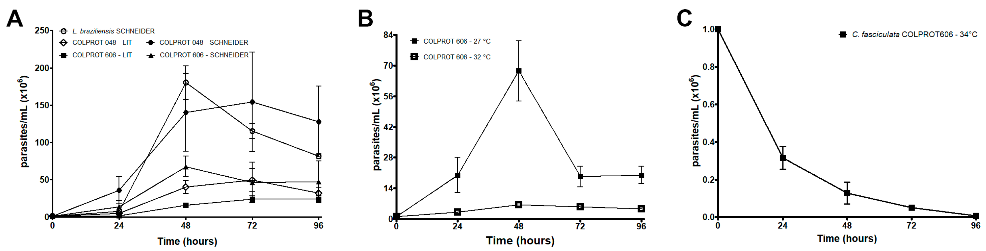
Figure 2.
Morphology of C. fasciculata isolated from a patient (COLPROT606). Illustrative panel of both growth kinetics’ morphology (27°C and 32°C) of the COLPROT606 strain stained with Giemsa. Parasites were cultured for 5 days with an initial concentration of 1 × 10⁶ parasites/mL in Schneider's medium supplemented with 20% FBS and 2% sterile human male urine. Bar: 10 µm.
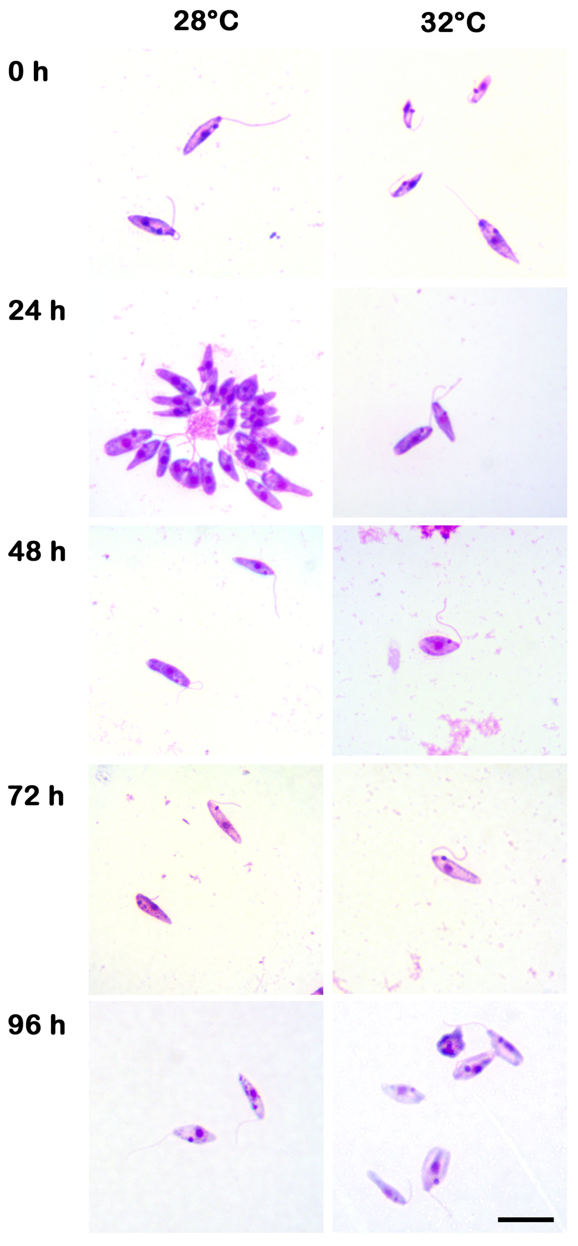
Figure 3.
RT-qPCR analysis of cell growth in different ratio mixed cultures of L. braziliensis-C. fasciculata COLPROT606. L. braziliensis (circles) and C. fasciculata COLPROT606 (squares) at 1 × 10⁶ or 2 × 10⁶ parasites were co-cultured in 5 mL of Schneider medium with 20% inactivated FBS at 27°C in the following ratios: L. braziliensis alone (Lbr), C. fasciculata COLPROT606 alone (Cfa), 1:1 Leishmania-Crithidia and 2:1 mixture of the two parasites. Cultures were passed into fresh medium until the fifth passage for RNA extraction and RT-qPCR analysis with SSU rRNA primers and probes.
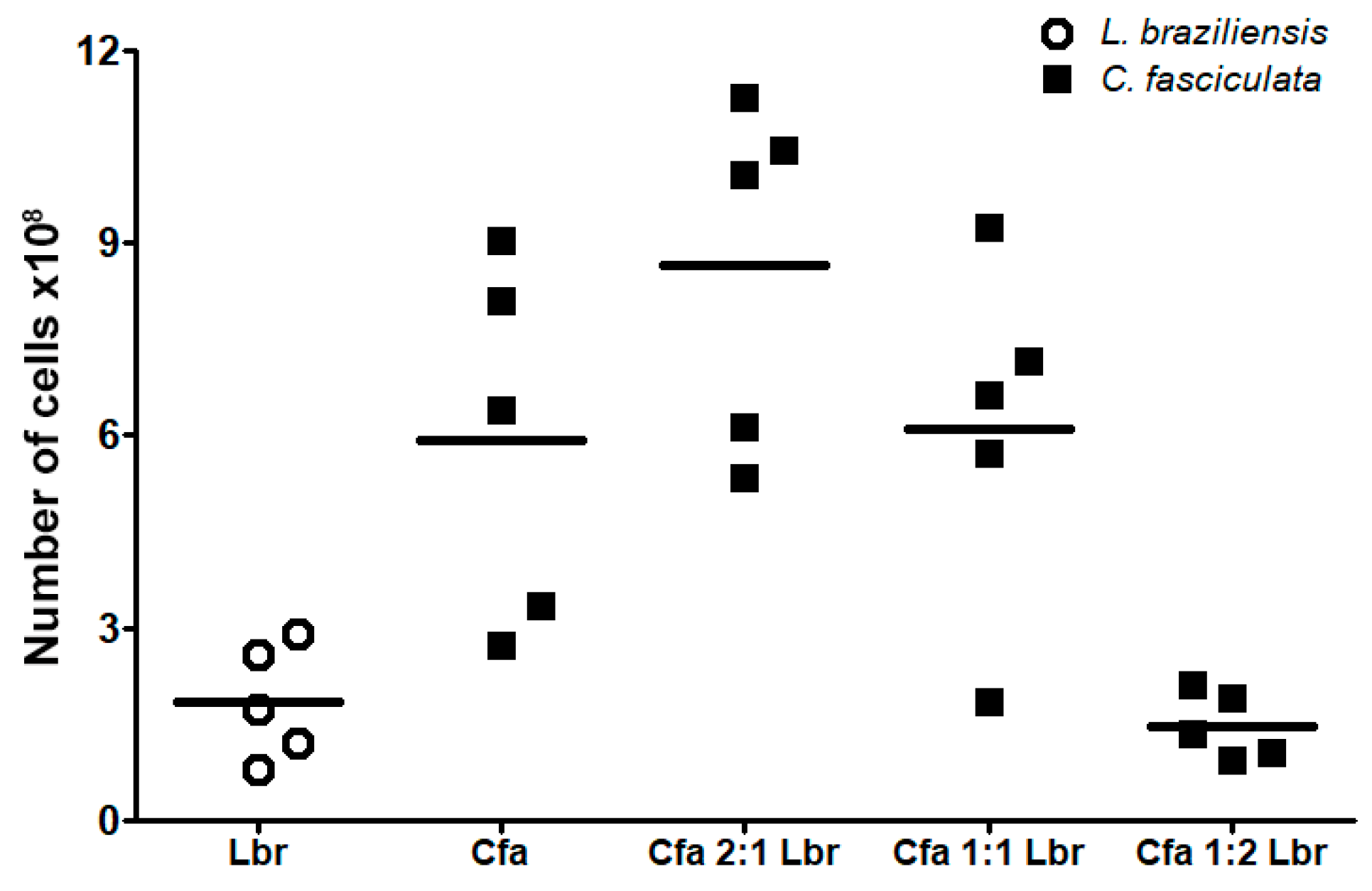
Figure 4.
Dose-response curve of L. braziliensis, C. fasciculata COLPROT048 and COLPROT606 to Amphotericin B. 4 ×10⁶ parasites/mL were treated with a serial dilution of Amphotericin B starting from 1 µM for 72 h. After the incubation period, cell viability was assessed by fluorometry by adding 50 µM of resazurin (Alamar Blue®) per well. The reads were performed using a Spectra Max GEMINI XPS (Molecular Devices, Silicon Valley, USA) at an excitation of 560 nm and emission at 590 nm. The IC50/72h value was determined using GraphPad Prism software. Statistical test: One-way ANOVA. P < 0.05.
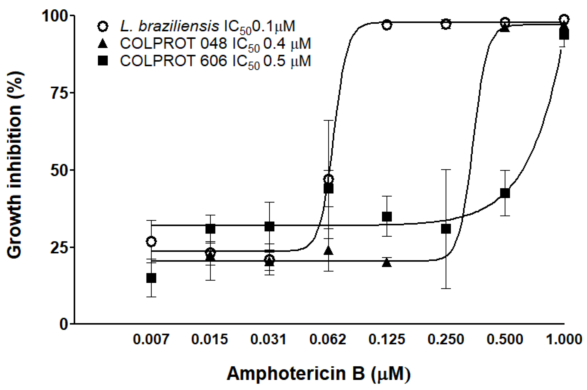
Figure 5.
Infection of BALB/c peritoneal macrophages with L. braziliensis, C. fasciculata COLPROT606, and coinfection (1:1) with both parasites (macrophages/parasites 1:5). (A) The values represent the number of parasites per infected macrophage at 24-, 48-, 72-, and 96-hours post-infection. The infection rate was determined by light microscope counting and calculated using the formula: % number of infected macrophages × number of amastigotes/total number of macrophages. (B) Absolute quantification by RT-qPCR of the macrophage coinfection by L. braziliensis (circles) and C. fasciculata COLPROT606 (squares) employing the SSU rRNA primers and probes for each parasite, and tubulin beta (Mm.PT.39a.22214835, IDT Inc.) for normalization with mice cDNA. Statistical test performed: 2-way ANOVA. (*) P<0.001.
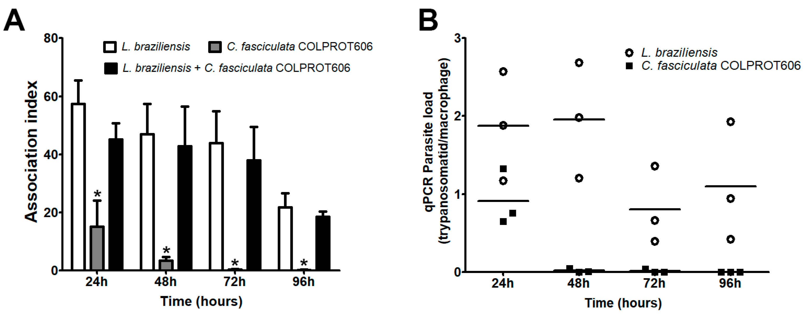
Figure 6.
Experimental infection in the right hind paw of golden hamsters. (A) Course of golden hamsters’ infection with L. braziliensis and C. fasciculata parasites. The right hind paw was infected with 2.0 × 10⁶ L. braziliensis alone (Group I, empty circle), C. fasciculata COLPROT048 alone (Group II, empty diamond), C. fasciculata COLPROT606alone (Group III, closed square), L. braziliensis in co-infection with COLPROT048 (Group IV, closed circle) and with COLPROT606 (Group V, closed triangle). (B) The size of the infected paws was monitored weekly using a dial caliper for 78 days. (C) At the end of the experiment the animals were euthanized and had the infected paw removed and macerated for parasite burden analysis by limiting dilution (LDA) in a microplate. No culture growth was observed in the wells from hamsters infected only with C. fasciculata strains COLPROT048 and COLPROT606 (D) The infected paws were also submitted to absolute quantification analysis by RT-qPCR employing specific SSU rRNA primers and probes for each parasite, and tubulin beta (Mm.PT.39a.22214835, IDT Inc.) for normalization with the hamsters’ cDNA. Statistical test performed: 1-way ANOVA *P<0.05. Statistical test performed: 2-way ANOVA *P<0.05 **P<0.01 ***P<0.001.
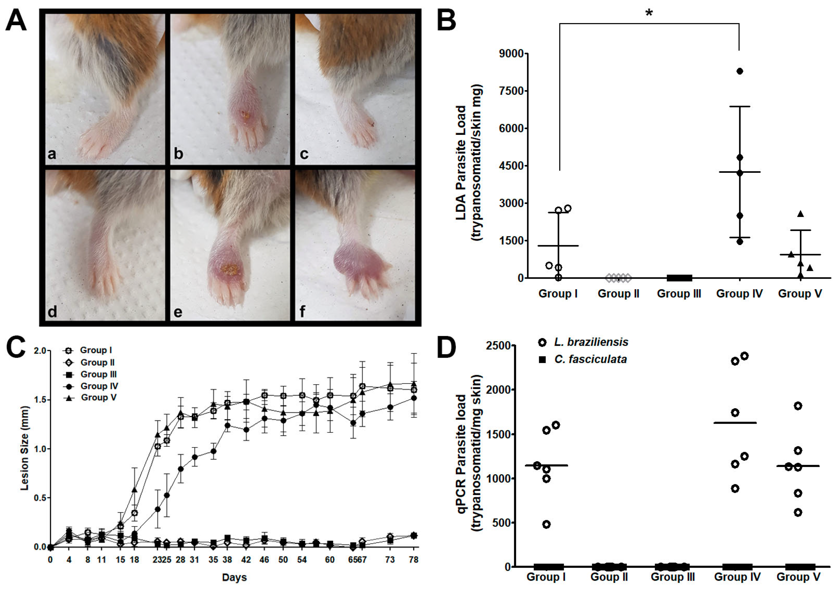
Figure 7.
Experimental infection of immunosuppressed BALB/c cyclophosphamide treated mice. (A) Uninfected BALB/c mice were treated intraperitoneally with cyclophosphamide at a dose of 150 mg/kg (3 mg/animal) and had their leukocytes counted weekly. (B) Cyclophosphamide treated BALB/c females were infected on the left posterior paw with 1 × 10⁷C. fasciculata COLPROT048 and C. fasciculata COLPROT606. The size of the infected paws was monitored weekly using a dial caliper. Statistical test performed: 1-way ANOVA *P<0.05.
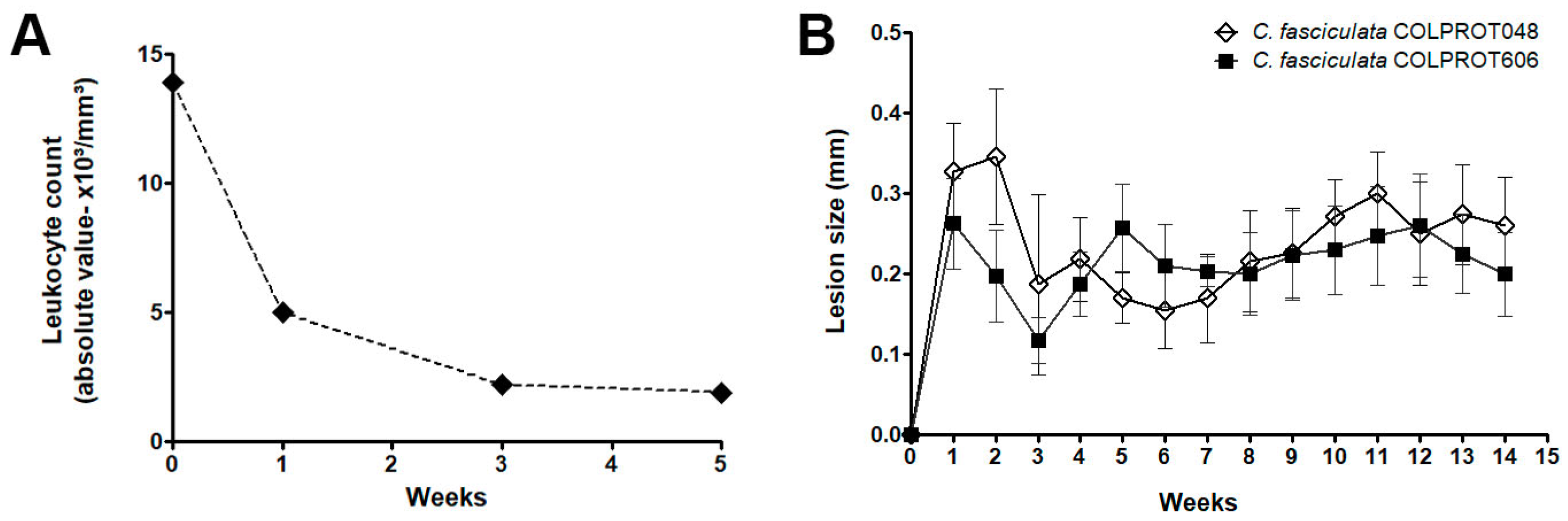
Figure 8.
Experimental infection and co-infection of Lu. longipalpis. The sandflies were infected with 5.0 × 10⁶ parasites/mL of L. braziliensis (circles), C. fasciculata COLPROT606 (squares) and a 1:1 mixture of both parasites. The RNA from infected sandflies were extracted on days 3- and 7-post-infection to quantify the number of L. braziliensis and C. fasciculata COLPROT606 per sandfly by RT-qPCR analysis. The horizontal straight lines indicate the median value of L. braziliensis, and the dotted lines indicates C. fasciculata median. Statistical test performed: one-way ANOVA, *P<0.05.
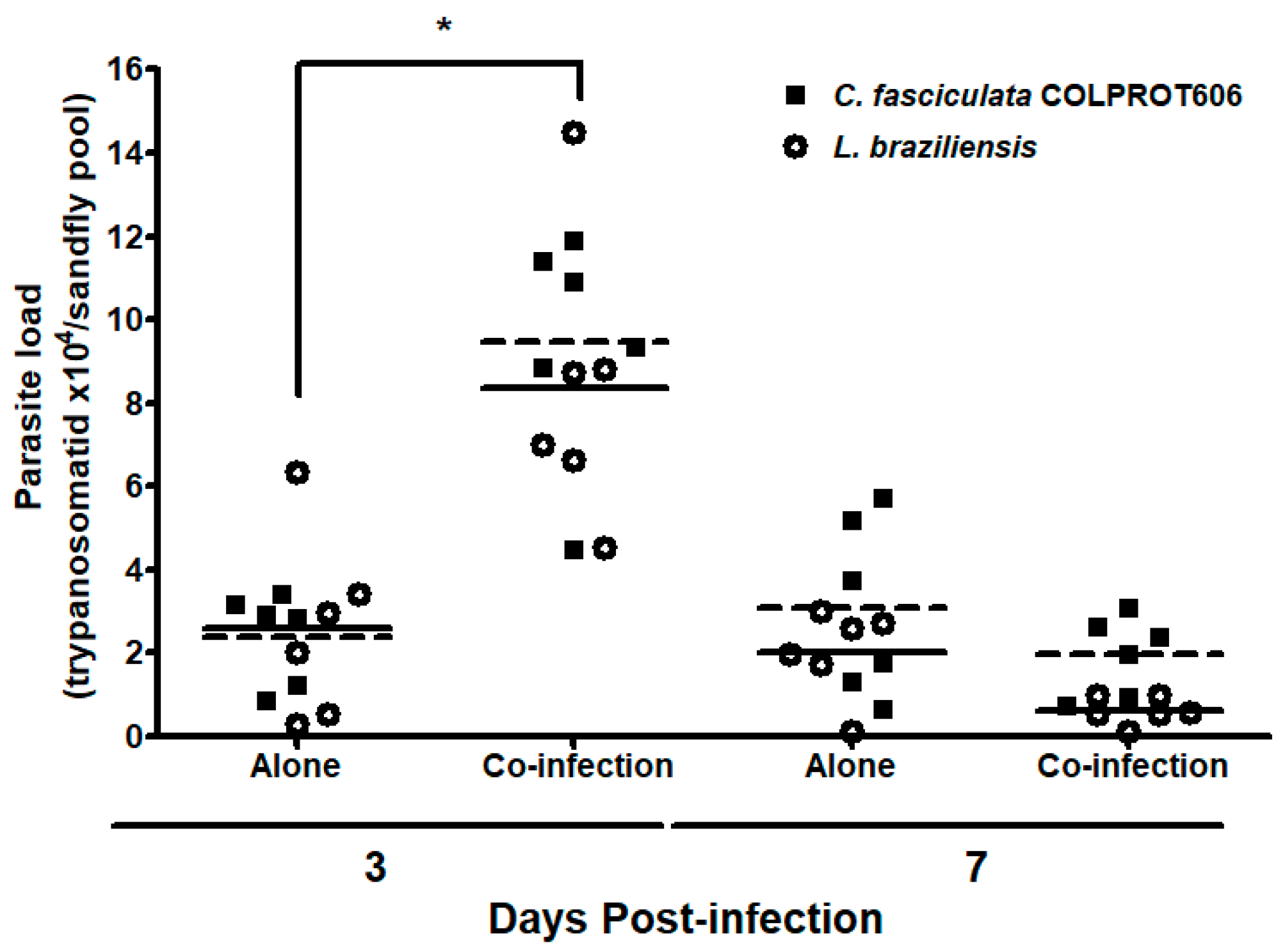

Table 1.
Δψm analysis of C. fasciculata COLPROT048, C. fasciculata COLPROT606 and L. braziliensis at 27°C and 32°C.
Table 1.
Δψm analysis of C. fasciculata COLPROT048, C. fasciculata COLPROT606 and L. braziliensis at 27°C and 32°C.
| Trypanosomatid | 48 hours | 96 hours | ||||
| TMRE+ cells (%) | IVa | TMRE+ cells (%) | IVa | |||
| C. fasciculataCOLPROT048 | 27°C | 91.0±1.1b | 0.00 | 84.1±1.7 | 0.00 | |
| 27°C +CCCP10 µM |
20.1±1.8* | -0.58* | 14.9±1.0* | -0.95* | ||
| 32°C | 82.8±2.0 | 0.07 | 79.6±0.5 | -0.60* | ||
| C. fasciculataCOLPROT606 | 27°C | 87.5±1.7 | 0.00 | 74.4±0.8 | 0.00 | |
| 27°C +CCCP10 µM |
29.9±2.0* | -0.63* | 26.8±1.9* | -0.81* | ||
| 32°C | 66.1±6.6 | -0.62* | 52.1±1.3* | -0.64* | ||
| L. braziliensis | 27°C | 86.0±0.7 | 0.00 | 73.8±2.6 | 0.00 | |
| 27°C +CCCP10 µM |
3.3 ±4.1* | -0.57* | 10.0±2.1* | -0.96* | ||
| 32°C | 51.8±4.1 | -0.30* | 13.2±0.9* | -0.90* | ||
aIV = (ME – MC)/MC, were ME corresponds to the median of fluorescence for parasites in experimental conditions of 32°C, and MC corresponds to control parasites at 27°C. The fluorescence of CCCP was reduced from ME and MC of each trypanosomatid. bMean ± standard deviation of 3 independent experiments. Asterisks indicate significant differences to the control group at 27°C (p ≤ 0.05).
Disclaimer/Publisher’s Note: The statements, opinions and data contained in all publications are solely those of the individual author(s) and contributor(s) and not of MDPI and/or the editor(s). MDPI and/or the editor(s) disclaim responsibility for any injury to people or property resulting from any ideas, methods, instructions or products referred to in the content. |
© 2025 by the authors. Licensee MDPI, Basel, Switzerland. This article is an open access article distributed under the terms and conditions of the Creative Commons Attribution (CC BY) license (http://creativecommons.org/licenses/by/4.0/).
Copyright: This open access article is published under a Creative Commons CC BY 4.0 license, which permit the free download, distribution, and reuse, provided that the author and preprint are cited in any reuse.


