Submitted:
02 August 2025
Posted:
04 August 2025
You are already at the latest version
Abstract
Oligofructans are fructose-based molecules composed of oligosaccharides and fructans linked by -glycosidic bonds. With the growing market demand for functional foods and bioactive ingredients, oligofructans have attracted considerable interest in their health-promoting properties. This study enhanced the production of oligofructans powder based on Bacillus subtilis TISTR 001 fermentation and analyzed the physicochemical properties of viscosity at concentrations of 10 %, 30 %, and 50 % (w/v), as well as stability at different pH levels (3, 5, 7, 9, and 11) and temperatures (25, 75, 100, and 125 °C) over time. Structural characterization was investigated using advanced analytical techniques (FTIR, NMR, and MALDI-TOF-MS). Based on the results, oligofructans production was successful, achieving a maximum concentration of 75.86 g/L under the optimized conditions at pH 6.5, 32.5 °C, and 250 rpm for 48 h. The oligofructans solution had low viscosity (10 cP at 50 % (w/v)) with Newtonian flow behavior and maintained excellent stability under neutral pH and moderate temperature conditions. The structural analysis confirmed the presence of characteristic fructose-based glycosidic linkages. Based on the results of the functional property assessment, there were dual bioactivity functions, with antioxidant activity having an IC₅₀ value of 7.11 mg/mL and the cytotoxicity studies indicating significant inhibitory effects on HepG2 liver cancer cells at concentrations ≥100 mg/mL. These findings supported the potential of oligofructans as multifunctional ingredients for application in the food, nutraceutical, and pharmaceutical industries. Further research will focus on process optimization for industrial-scale production and stability testing in various food matrices, along with investigating the bioactive and therapeutic potential, particularly for liver health and cancer prevention.
Keywords:
Bacillus subtilis
; bioactivity
; cancer cell
; functional food
; oligofructans
1. Introduction
Oligofructans comprise a combination of fructose chains, encompassing both short-chain fructose molecules, known as fructo-oligosaccharides (FOSs), and long-chain fructose molecules, referred to as fructans. The repeating fructose molecules are linked together by β-2,1 or β-2,6 glycosidic linkages and bonded with glucose molecules by α-glycosidic linkage at the end of oligofructans structures in the form GFn [1,2]. FOSs are mainly composed of repeating fructose units with less than 10 molecules, such as kestose (GF2), nystose (GF3), and 1-fructofuranosyl-D-nystose (GF4), while fructans are composed of more than 10 long-chain fructose units, such as inulin and levan.
Oligofructans can be enzymatically synthesized using two main processes: fructosyl-linkage hydrolysis and transfructosylation. Both processes are catalyzed by enzymes belonging to the glycoside hydrolase families, such as GH32 or GH68 [3]. First, the substrate (typically sucrose) is hydrolytically cleaved into its constituent monosaccharides of glucose and fructose. Subsequently, the liberated fructose molecules, or fructosyl units, are transferred and linked to form short-chain FOSs. These oligosaccharides may then undergo polymerization to form larger fructose polymers, potentially containing up to 100,000 fructose units [4,5]. Levansucrase (EC 2.4.1.10) is a bacterial enzyme classified within the GH 68 enzymes family and has potentially dual enzymatic functions for fructose polymer production. Oligofructans have been reported to be synthesized through microbial fermentation involving extracellular levansucrase, with the participation of microorganisms such as Pseudomonas spp., Zymomonas spp., Bacillus spp., and Corynebacterium spp. [6]. Among these, Bacillus subtilis is a promising levansucrase-producing bacterium capable of synthesizing a mixture of FOSs and fructans and is classified as generally recognized as safe (GRAS) [5]. In nature, oligofructans can be found in various plant foods such as onions, garlic, bananas, and asparagus [7].
Oligofructans are widely applied in many industries, particularly in the food industry, where they function as emulsifiers, encapsulating agents, flavor enhancers, and fat substitutes [8]. In addition to their applications in food, oligofructans are utilized in the cosmetic and pharmaceutical industries [9]. Beyond their industrial uses, oligofructans have notable health-promoting properties and have been associated with beneficial effects on liver health and cancer prevention [7]. Specifically, they act as prebiotics, stimulating the growth of beneficial gut microbiota, thereby promoting a balanced gut microbiome linked to reduced inflammation and improved metabolic health [10]. Their antioxidant and anti-inflammatory activities can help mitigate oxidative stress and inflammation, both of which are critical factors in the progression of liver diseases [11]. In addition, oligofructans have prebiotic properties that help balance the gut microbiome by promoting the growth of beneficial bacteria. This activity reduces dysbiosis, strengthens the intestinal barrier, and limits the translocation of microbial products into the liver [12]. Consequently, oligofructans influence the gut-liver axis by enhancing gut barrier function and indirectly supporting liver health [13]. Furthermore, the consumption of oligofructans has been correlated with improved metabolic parameters, including better blood glucose regulation and lipid metabolism, which potentially reduce the risk of non-alcoholic fatty liver disease (NAFLD), a known precursor to liver cancer [14]. Further health benefits include the promotion of bowel regularity, enhanced calcium absorption contributing to reduced osteoporosis risk, and potential prevention of obesity [15].
Liver cancer, or hepatocellular carcinoma (HCC), has emerged as a major global public health concern due to its steadily increasing incidence; currently, HCC ranks as the fifth most common cancer and the fourth leading cause of cancer-related mortality worldwide [16]. The primary contributing factors to this growing prevalence include chronic viral hepatitis infections, particularly hepatitis B and C, along with NAFLD and exposure to aflatoxins. Additionally, lifestyle factors, such as smoking and poor dietary habits, further exacerbate the risk. In Thailand, liver cancer is particularly important, ranking as the most common cancer among men and the third most common among women [17]. According to the National Cancer Institute [18], the major risk factors include hepatitis B virus infection, liver cirrhosis, exposure to harmful chemicals (such as insecticides), prolonged use of certain medications, fungal toxins, chemicals present in fermented foods, alcohol consumption, weakened immune function, and genetic predisposition. Several studies have highlighted the considerable health benefits of oligofructans, which are classified as natural prebiotics with diverse biological activities [19,20,21,22]. Beyond promoting the growth of beneficial gut microflora, oligofructans have demonstrated potential in inhibiting the progression of several cancer types, including colon, breast, and liver cancers, where these anticancer effects have been attributed primarily to their ability to induce apoptosis and promote oxidative stress, specifically within cancer cells [11]. For example, Pool-Zobel et al [23] reported that inulin reduced colon cancer risk in experimental animals by decreasing cancer cell proliferation at the molecular level. Similarly, Dahech et al [24] investigated levan produced by Bacillus licheniformis through the action of the enzyme levansucrase that primarily consists of fructose and has been shown to inhibit HepG2 liver cancer cells in vitro. Their experimental results demonstrated that levan had anticancer activity, specifically at high concentrations, without damaging normal cells.
Noidee and Ninchan [5] investigated the optimal conditions for oligofructans production by B. subtilis TISTR 001 using sucrose as the fermentation substrate to achieve higher production efficiency compared to other sucrose-based substrates, including raw sugar, cane juice, and molasses The optimal conditions, determined using response surface methodology, were identified as 37 °Brix sucrose concentration, pH 6.5, a temperature of 32.5 °C, a shaking speed of 250 rpm, and a fermentation period of 48 h. Building on these findings, the present study aimed to further investigate the production of an oligofructans powder based on analyzing its physicochemical properties, characterizing the structure of the produced oligofructans, and evaluating their functional properties. In Addition, this research explored the potential inhibitory effects of oligofructans on liver cancer cells. The ultimate objective was to assess the feasibility of using oligofructans as potential therapeutic agents or supplementary materials for the prevention or treatment of liver cancer, or both.
2. Materials and Methods
The bacterial strain, B. subtilis TISTR 001, was obtained from the Thailand Institute of Scientific and Technological Research (TISTR, Thailand). All chemicals used were analytical grade and the culture media were purchased from HiMedia Laboratories Pvt. Ltd. (India). Sucrose (pure refined sugar, Mitr Phol Sugar Corp., Ltd., Thailand) as the substrate fermentation was purchased from a supermarket. Additionally, maltodextrin was provided by Matsutani Chemical Industry Co., Ltd. (Japan).
2.1. Production of Oligofructans Powder
2.1.2. Inoculum Preparation
The B. subtilis TISTR 001 inoculum was prepared. First, a single colony of B. subtilis was cultured in sterilized nutrient broth (NB). Next, the B. subtilis culture was transferred to fresh NB and incubated at 37 °C at 200 rpm in a shaking incubator (VS-8480SFN; Vision Scientific Co. Ltd.; South Korea) for 24 h. Subsequently, 10 % (v/v) inoculum culture was transferred again into fresh NB supplemented with 0.1 % (w/v) sucrose and incubated under the same conditions for 18 h.
2.1.3. Oligofructans Fermentation
A 10 % (v/v) inoculum of B. subtilis was transferred into a 35 °Brix sucrose solution as substrate for fermentation, supplemented with yeast extract (0.2 % w/v), meat extract (0.3 % w/v), peptone (0.5 % w/v), (NH4)2SO4 (0.2 % w/v), and MgSO4·7H2O (0.06 % w/v). The oligofructans fermentation was carried out at pH 6.5, 32.5 °C, and 250 rpm for 48 h. The kinetic changes during oligofructans fermentation were determined in terms of the sugar contents (sucrose, fructose, and glucose), short-chain FOSs (ketose and nystose), and total oligofructans.
2.1.4. Oligofructans Powder
The B. subtilis cells were separated from the fermentation medium prior to the oligofructans precipitation. The cell-free oligofructans were precipitated twice using cold absolute ethanol and then kept at 4 °C for 24 h to completely polymerize the precipitate. Next, the precipitated polymer was collected using centrifugation and subsequently dissolved into water at a precipitated polymer-to-water ratio of 1:1 (w/w). Then, maltodextrin was added to the polymer solution prior to drying. The concentrations of maltodextrin at 1 % (w/v) and 2 % (w/v) were compared with no addition of maltodextrin. The oligofructans powders were produced using a mini spray dryer (B-290; Büchi Labortechnik, Switzerland), according to Amrutha et al [25]. The spray-drying conditions were set at an inlet temperature of 140 °C, an outlet temperature of 85 °C, and a feed flow rate in the range 1.4- 6.2 mL/min.
2.2. Analysis of Fermentation Kinetics and Properties of Oligofructans Powder
2.2.1. Sugar Contents and Short-Chain of Fructo-Oligosaccharide Using High Performance Liquid Chromatography
The analytical methods followed Ninchan and Noidee [2], Showa Denko K.K. [26], and Vertical Chromatography Co., Ltd. [27]. Cell-free fermented samples were diluted to appropriate levels of concentration and filtered through a 0.45 μm cellulose acetate membrane. Then, the sugar content was analyzed using high performance liquid chromatography (HPLC) and refractive index detection (RID-10A; Shimadzu Corporation; Japan), with a VertiSep™ Sugar CMP column (7.8 × 300 mm; 8 μm diameter; Vertical Chromatography Co., Ltd.; Thailand) at 80 °C, using deionized water as the mobile phase at a flow rate of 0.4 mL/min. Standard sugars (sucrose, glucose, and fructose) were used for calibration to monitor changes in the sugar concentrations during fermentation. The quantitative analysis of short-chain fructo-oligosaccharides used a Shodex Asahipak NH2P-50-4E column (4.6 mm internal diameter × 250 mm; Showa Denko America, Inc.; Japan) and was performed at 40 °C, with the mobile phase consisting of an acetonitrile-to-deionized water ratio of 70:30 and at a flow rate of 1 mL/min. Two standard short-chains of fructo-oligosaccharides (kestose and nystose) were analyzed for identification and quantification.
2.2.2. Total Oligofructans in Form of Total Free Fructose
The total oligofructans were analyzed, according to the method described by Noidee and Ninchan [5]. Briefly, 1 mL of cell-free samples were mixed with 2 mL of absolute ethanol to precipitate the fructo-oligosaccharides and fructose polymers. Then, the mixture was centrifuged at 10,000 rpm for 10 min using a Hettich Rotina 35R refrigerated tabletop centrifuge (UK). After centrifugation, the supernatant was discarded and the resulting precipitate was hydrolyzed with 2 mL of 0.1 N hydrochloric acid. The hydrolysis process was carried out by boiling for 30 min to cleave the oligosaccharide and fructose polymer bonds into monosaccharides. The concentration of hydrolyzed monosaccharides was expressed as reducing sugars based on the Somogyi-Nelson method [28], with fructose as the standard, to calculate the total amount of free fructose units involved in the total oligofructans content.
2.3. Determination of Physicochemical Properties of Oligofructans Powder
2.3.1. Viscosity
Oligofructans powder samples, with and without maltodextrin addition, were dissolved in distilled water and prepared at 10 %, 30 %, and 50 % (w/v). All solutions were allowed to stand for 10 min to ensure complete dissolution. The viscosity of the solutions was measured using a viscometer (NDJ-8S; Nanjing T-Bota SciTech Instruments & Equipment Co., Ltd.; China) operated at 25 ± 0.5 °C with spindle No. 1 at a rate of 60 rpm. The viscosity readings were recorded in terms of centipoise (cP) at time intervals of 5, 10, and 15 min to study the physical behavior of oligofructans. Each sample was tested in triplicate.
2.3.2. pH and Thermal Stability
A 10 % (w/v) oligofructans solution was prepared and tested for pH and temperature stability. The pH stability of oligofructans powder was determined following a modified method of Glibbowski and Bukowska [29]. The solution was adjusted to various pH values (3, 5, 7, 9, and 11) using phosphate buffer. Samples were collected at specific intervals (0, 5, 15, 30, and 60 min). Each reaction was stopped using neutralization, followed by boiling for 10 min and rapid cooling in an ice bath. Subsequently, the concentration of reducing sugar formed and the remaining total oligofructans were determined using the Somogyi-Nelson method [28]. Thermal stability was tested based on heating 10 % (w/v) of oligofructans solution at various temperatures (25, 75, 100, and 125 °C). Samples were taken at specific intervals (0, 5, 15, 30, and 60 min at each temperature) to compare the stability based on measuring the contents of reducing sugar and total oligofructans using the same analytical method.
2.4. Structural Analysis of B. subtilis TISTR 001 Oligofructans Powder
2.4.1. Fourier Transform-Infrared Spectroscopy
The functional groups of oligofructans were analyzed using Fourier transform-infrared (FTIR) spectroscopy. The FTIR spectra were recorded in transmittance mode in the wavenumber range 4000–400 cm-1 using a Shimadzu IRSpirit FTIR spectrometer equipped with a QATR-S AT, accessory (Japan). Infrared measurement was carried out in transmission mode, in which the infrared beam passed directly through the sample and was then recorded in the transmittance unit. The infrared spectra were presented using the Star Software version 16.30a.
2.4.2. 1H and 13C Nuclear Magnetic Resonance Spectroscopy
The oligofructans powder was structurally characterized using nuclear magnetic resonance (NMR) spectroscopy (AVANCE UltraShield 400 MHz spectrometer; Bruker; Germany). A 0.5 mg of oligofructans sample was dissolved in D2O for the 1H NMR and 13C NMR analyses. In addition, heteronuclear single-quantum correlation analysis was performed using D2O solvent. All spectra were recorded at 70 °C using the Bruker UltraShield 400 MHz spectrometer with a 54 mm bore.
2.4.3. Matrix-Assisted Laser Desorption/Ionization Mass Spectrometry
An oligofructans sample was prepared as a solution using water for matrix-assisted laser desorption/ionization mass spectrometry (MALDI-TOF-MS). Next, the 2,5-dihydroxybenzoic acid (DHB) matrix was prepared by dissolving 10–20 mg of DHB in 1 mL of water. After that, equal volumes of the sample solution and the DHB matrix solution were mixed and then spotted onto a clean MALDI target plate. The spots were dried at room temperature. The target plate was analyzed using a JEOL SpiralTOF™ MALDI-TOF/TOF Mass Spectrometer (JMS-S3000; Japan). The laser settings were optimized and the mass spectra were acquired to identify and quantify peaks based on the mass-to-charge ratio (m/z).
2.5. Evaluation of Bioactivity Properties of Oligofructans Powders
The bioactivity levels of the oligofructans powder samples produced using B. subtilis TISTR 001 were evaluated based on assessing the antioxidant activity and cytotoxicity effects on HepG2 liver cancer cells. The methodologies are detailed below.
2.5.1. Antioxidant Activity
The antioxidant activity of oligofructans was evaluated using the 2,2-diphenyl-1-picrylhydrazyl hydrate (DPPH) assay, based on a modified procedure from Srikanth et al [30]. Each preparation of DPPH solution was dissolved in 95 % ethanol to obtain a 0.1 mM concentration. The oligofructans powders were dissolved in the different concentrations of oligofructans in water (0–50 mg/mL). Then, 600 µL of DPPH were mixed with 2 mL of the oligofructans solution. The mixtures were thoroughly mixed and incubated in the dark at room temperature for 30 min after incubation, the absorbance of each mixture was measured at 517 nm using the spectrophotometer. An oligofructans solution in ethanol was used as a blank and a mixture of DPPH and ethanol was used as the control. The antioxidant activity was calculated based on comparing the absorbance of the sample with that of the control using the equation:
where A0 is the absorbance of the DPPH solution without any sample, A1 is the absorbance of the DPPH solution with the sample, and B is the absorbance of the sample solution without DPPH.
2.5.2. Cytotoxicity Test
Each oligofructans power sample was dissolved using Dulbecco’s modified eagle medium supplemented with 10 % fetal bovine serum (FBS). Subsequently, the solution was sterilized by passing through a 0.45 µm syringe filter and then prepared for the cytotoxicity testing. The cytotoxic effect of the oligofructans was evaluated against human hepatocarcinoma (HepG2) cells (human liver cancer cells) using the 3-(4,5-dimethylthiazol-2-yl)-2,5-diphenyltetrazolium bromide (MTT) assay. The HepG-2 Cells were seeded at a density of 1 × 10⁵ cells/mL into a 96-well plate, with a volume of 100 µL per well, and incubated at 37 °C in a humidified atmosphere containing 5 % CO₂ for 24 h. After incubation, the culture medium was aspirated and 100 µL oligofructans solutions at concentrations of 1, 10, 20, 100, 200, 300, and 500 mg/mL were added to the respective wells. Then, the plates were incubated for an additional 24 h under the same conditions. Following treatment, 10 µL of MTT solution (5 mg/mL) was added to each well, after which the plates were incubated for 4 h at 37 °C with 5 % CO₂. After incubation, the MTT solution was carefully removed and 100 µL of Formazan solubilizing solution (a mixture of 100 % dimethyl sulfoxide (DMSO) and 10 % sodium dodecyl sulfate (SDS) in a 9:1 ratio) was added to each well. Next, the plates were shaken for 5 min before the measurement of optical density (OD) at 570 nm using a microplate reader. Cytotoxicity was calculated using the equation:
where A is the absorbance of the sample and B is the absorbance of the blank (a solution of 100 % DMSO to 10 % SDS) that must be subtracted from the absorbance of each sample (A).
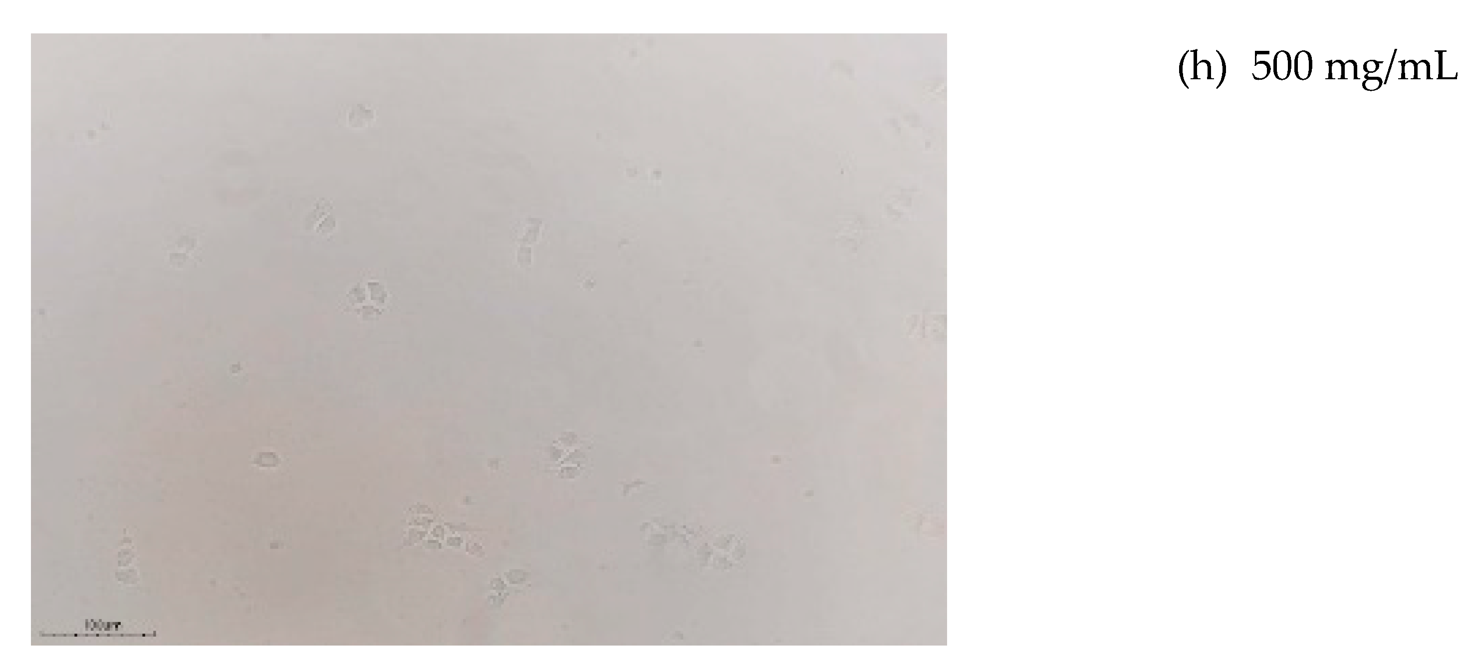
Additionally, the morphology and cell density of the HepG2 cells following the oligofructans treatments at various concentrations were observed using an inverted microscope (ECLIPSE; TS100; Nikon; Japan at 100X magnification. The imaging system consisted of a digital camera (Model: MC4KW-G1; Lanoptik, China) mounted on the microscope to evaluate cellular responses.
2.6. Statical Analysis
Data were analyzed and reported as mean ± standard deviation values. All reported values were based on the mean of three replicates. Analysis of variance (ANOVA) was used for data analysis in the IBM SPSS Statistics Version: 19.0.0 software (IBM Corp., Armonk, USA). Significance was tested at the p < 0.05 level in the ANOVA.
3. Results and Discussion
3.1. Production of Oligofructans Powder by B. subtilis TISTR001
The kinetic changes in oligofructans production by B. subtilis TISTR 001 fermentation during fermentation for 48 h under the optimized conditions established by Noidee and Ninchan [5] are shown in Figure 1. The optimized fermentation conditions were 35 °Brix of sucrose concentration as substrate fermentation at pH 6.5 and 32.5 °C, with shaking at 250 rpm for 48 h, resulting in a maximum oligofructans concentration of 75.86 g/L. During fermentation, sucrose was enzymatically hydrolyzed, leading to an increase in the concentrations of monosaccharides as glucose and fructose (Figure 1a). Theoretically, the molar ratio of glucose-to-fructose after sucrose hydrolysis should be 1:1. However, the observed fructose content was lower than that of glucose due to fructose being further utilized as a substrate for the synthesis of oligofructans. These products consisted of a mixture of short-chain FOSs (such as kestose and nystose) and higher molecular weight fructose polymers, whose concentrations increased progressively throughout the fermentation period, along with the total oligofructans content (Figure 1b).
After fermentation, the oligofructans were precipitated and subsequently processed through a spray dryer to produce oligofructans powder. The efficiency of maltodextrin addition at concentrations of 1 % (w/v) and 2 % (w/v) on the yield of oligofructans powder content after spray-drying were compared to no addition (control), as shown in Figure 2. Based on the results, the addition of maltodextrin significantly increased the powder yield compared to the control (Figure 2). However, there was no significant difference in yield between the 1 % and 2 % (w/v) maltodextrin concentrations. Notably, increasing the maltodextrin concentration to 2 % (w/v) led to a slight decrease in yield which could be attributed to the excessive polymer encapsulation caused by the higher maltodextrin content, which can negatively impact product recovery [31]. As a stabilizer, maltodextrin at high concentrations may increase the matrix viscosity or alter the particle formation characteristics, ultimately interfering with oligosaccharide recovery during spray-drying [32]. Therefore, optimizing the maltodextrin concentration is a critical factor in the spray-drying process to maximize product yield. Based on these findings, the addition of 1 % (w/v) maltodextrin was identified as the most suitable concentration for producing oligofructans powder using spray-drying.
3.2. Physical Properties of Oligofructans Powder
3.2.1. Viscosity of Oligofructans Solution
Viscosity plays a crucial role in influencing the physical behavior of material solutions. Theoretically, an increase in solution concentration is expected to positively correlate with higher viscosity, which was consistent with the experimental results observed in the oligofructans solutions at concentrations of 10 %, 30 %, and 50 % (w/v), both with and without the maltodextrin addition, as shown in Figure 3. However, no significant differences in viscosity were observed between the oligofructans solutions with and without maltodextrin under the same conditions due to the interaction strength of oligofructans and maltodextrin is considered weak. At the highest concentration used of 50 % (w/v), the oligofructans solution had a viscosity of approximately 10 cP. Additionally, when measured over time (5, 10, and 15 min) at a constant concentration, the viscosity of the oligofructans solutions remained relatively stable over time. This behavior of oligofructans acting as a stable polymer network was also reported by Aprodu et al. [33]. Maltodextrin, a polysaccharide produced through starch hydrolysis, is widely used as a carrier or stabilizer in food and pharmaceutical formulations owing to its high solubility and low viscosity even at elevated concentrations. The addition of maltodextrin can enhance product stability, protect bioactive compounds during processing, and improve the physical properties of powders in spray-drying or freeze-drying applications [34,35]. Then, there was a consistent viscosity over time with the addition of maltodextrin and the viscosity of the solution was slightly enhanced without causing any major alterations to the structural dynamics of the oligofructans (Figure 3).
3.2.2. pH and Thermal Stability of Oligofructans
The stability of oligofructans was determined under various pH levels (3, 5, 7, 9, and 11) and temperatures (25 °C, 75 °C, 100 °C, and 125 °C) by monitoring the increase in the reducing sugar content over time, as shown in Figure 4. An increase in the reducing sugar content implied the degradation of oligofructans as the polymer chains were hydrolyzed into smaller sugar units resulting in decreased stability of the oligofrcutans. As shown in Figure 4a, the oligofructans were clearly degraded under strongly acidic (pH 3) and alkaline (pH 11) conditions, reflected by a substantial increase in the reducing sugar content under these severe conditions, consistent with the findings of Glibowski and Biadun [36]. Notably, the oligofructans were more stable under alkaline than under acidic conditons (Figure 4a). Additionally, a prolonged incubation time further contributed to oligofructans degradation. In contrast, within the pH range 5–9, the oligofructans maintained excellent stability, with minimal increases in the reducing sugar content. These results aligned with those of Glibowski and Bukowska [29], who reported that neutral pH conditions effectively minimized the hydrolytic activity on fructans chains.
The thermal stability of the oligofrcutans is shown in Figure 4b. Increases in both temperature and time had a substantial negative effect on the stability of the oligofrcutans, as evidenced by the increase in the reducing sugar content. At 25 °C, the oligofructans remained stable, with only a slight increase in the reducing sugar concentration over 60 min. However, at elevated temperatures (75 °C and higher), the degradation rate increased markedly, particularly at 100 °C and 125 °C, where rapid breakdown of the glycosidic bonds in the fructose polymer led to the release of monosaccharides and other breakdown products, resulting in a substantial increase in reducing sugars [37]. These findings suggested that pH and temperature control were the critical factors when applying oligofructans in food processing applications, because elevated heat and severe pH condition may compromise structural integrity and functional benefits [38].
3.3. Structure of B. subtilis TISTR 001 Oligofructans Powder
3.3.1. FTIR Spectra
The oligofructans powder produced by B. subtilis TISTR 001 was analyzed structurally using FTIR and compared with the structure of standards: short-chain FOSs (kestose and nystose) and fructose polymer (levan), as shown in Figure 5. The FTIR spectra revealed frequency regions of infrared absorptions on the glycosidic linkages and the functional groups of structural samples and standards. The spectra of the oligofructans powder samples had absorption patterns very similar to those of kestose and nystose, particularly the absorption bands at 3700–3584 cm-1 (O-H stretching) and 1685–1666 cm-1 (C=O stretching), as shown in Figure 5. Notably, the short-chain FOSs and oligofructans had a band at 2349 cm-1, indicating O=C=O stretching vibration. Specific spectral regions detected in the oligofructans, short-chain FOSs, and levan indicated the presence of similar structural characteristics. The broad band at 3004–2867 cm⁻¹ indicated C-H stretching, signifying the presence of aliphatic groups within the sugar structures, with the strong absorption at 1200–1000 cm⁻¹ referring to C-O-C and C-OH strong stretching, corresponding to the glycosidic bonds (β-2,1 and β-2,6 linkages) between the fructose units, while those as 1000–800 cm-1 indicated the fingerprint region of a pyran ring [39,40]. Thus, the results of FTIR spectral comparisons confirmed that the B. subtilis TISTR 001 oligofructans had a mixed structure of short-chain FOSs (kestose and nystose) and a complex fructose polymer (levan), supporting their classification and affirming their potential application in functional food products or in health supplements, aligning with similar applications of the FOS family.
3.3.2. NMR Spectra
The NMR spectra were systematically analyzed based on both ¹H and ¹³C NMR data to elucidate the molecular structure and confirm the presence of specific functional groups within the compounds. The ¹H NMR spectrum provides detailed information about the hydrogen atoms (protons) in a molecule to reveal the number, type, their environment, and how they are connected, with this information being critical in deducing molecular frameworks and functional groups. In addition, the ¹³C NMR data reveals the number and types of distinct carbon environments in a molecule, their degree of substitution, and the electron environment, with this information being essential for mapping out the carbon framework. The combined analysis of the ¹H and ¹³C NMR spectra strongly suggested that the investigated compounds had structures rich in hydroxyl functionalities, likely corresponding to a carbohydrate derivative structure.
The ¹H and ¹³C NMR spectra of the oligofructans produced by B. subtilis TISTR 001 were analyzed using NMR, as shown in Figure 6 and Figure 7, respectively. The ¹H spectra were recorded on the chemical shift region from 4.20 to 3.20 ppm that is dominated by the polyhydroxylated sugar structures and is associated with the extensive presence of hydroxylated methine (–CH–OH) and methylene (–CH₂–OH) groups. The ¹H spectra contained features at 4.20–4.10 ppm (H1: (H-1, the anomeric protons or H at terminal glucose or fructose), at 4.00–3.80 ppm (fructosyl H-3 and H-4 protons), and at 3.70–3.40 ppm (H-5 and H-6 protons of fructose units) [39,41,42,43]. The integration values displayed at the base of the spectrum (notably at 21.29, 18.42, and 8.03 ppm) suggested a high degree of polymerization or the presence of repeating fructosyl units, as the protons (H-3 to H-6) were repeated multiple times within the oligomer [44]. The ¹³C NMR spectrum of the oligofructans produced by B. subtilis TISTR 001 had strong signals at 105–60 ppm. which were characterized as sugar carbon atoms, particularly the C1–C6 carbons of fructose residues, as shown in Figure 7. The 13C spectra indicated the presence of the fructose-based oligosaccharides typically involved in β-glycosidic linkages [45] at 103.2 ppm (C2: anomeric carbon of β-D-fructofuranose residues), 81.3 ppm (C5: –CH–OH), 77.5 ppm (C3: –CH–OH), 74.8 ppm (C4: –CH–OH), 62.3 ppm (C6: –CH₂–OH ), and 61.2 ppm (C1: anomeric carbon atoms of glucose units or terminal fructose residues), corresponding to the signals reported in the results of Aramsangtienchai et al [39] and Chaves et al [46].
3.3.3. MALDI-TOF-Mass Spectrum
In the MALDI-TOF mass spectrum, each peak represents an ion detected at specific mass-to-charge (m/z) ratio. For the oligofructans and fructose polymers, these peaks correspond to molecules with varying degrees of polymerization (DP), which refer to the number of fructose monomer units linked together in the polymer chain. The molecular weight of each molecule, directly related to its DP, determines the position of its peak in the spectrum. During MALDI ionization, molecules typically gain a proton, so the m/z value of each peak reflects the molecular weight (MW) of the oligomer plus the mass of a proton. A single fructose unit (monosaccharide) has a molecular weight of about 162 Da. As more units are linked, the molecular weight—and thus the m/z value—increases accordingly. Figure 8 shows the MALDI-TOF mass spectrum peaks for the oligofructans powder in the range ~2128 to ~6196 m/z, representing oligomers with DP values in the range ~10 to ~34. Major peaks appeared at m/z values indicative of oligomers with DPs ranging from 10 to 12 that were similar to those reported in FOSs production using microbial fermentation by Evans et al. [47]. Notably, higher DPs (DP > 12) were detected in B. subtilis TISTR 001 oligofructans powder (Figure 8).
The detection of the higher DP oligomers was particularly notable, as larger oligofructans are often associated with enhanced functional properties by having greater resistance to hydrolysis in the gastrointestinal tract, leading to prolonged fermentation and improved prebiotic effects [48]. Oligofructans with higher DP values have been reported to have strong anticancer properties, particularly in inhibiting the proliferation of liver cancer cells [49]. The present research studied the potential cytotoxicity of B. subtilis TISTR 001 oligofructans with mixed DP of oligomers on liver cancer cells, which is discussed ion the next section.
3.4. Bioactivity Properties of Oligofructans Powder
3.4.1. Antioxidant Activity
The oligofructans powder was evaluated using DPPH assay at 0–50 mg/mL oligofructans concentrations; subsequently, the radical scavenging activity or the percentage of inhibition (% inhibition) was calculated (Figure 9) as an indicator of antioxidation activity. The inhibition (%) increased with increasing concentration of oligofructans from 0 to 20 mg/mL, achieving a high inhibition of 83.20 %, followed by a slight increase before remaining constant at higher concentrations, as shown in Figure 9. The highest inhibition was 88.40 % at 50 mg/mL of oligofructans concentration. The antioxidant property of the B. subtilis TISTR 001 oligofructans powder was indicated by the half maximal inhibitory concentration (IC50) which was 7.11 mg/mL. The oligofructans produced by B. subtilis TISTR 001 had a high IC50 value compared to the IC50 values of standard antioxidants such as ascorbic acid (approximately 20–50 µg/mL) and tocopherols (approximately 30–80 µg/mL) [50], that indicated greater activity for effective free radical scavenging. However, the effective potential in antioxidant activity in terms of radical scavenging properties depends on the source and degree of polymerization of oligofructans [51,52,53]. Microbial oligofructans had a reported lower radical scavenging activity than the oligomer extracts from plant [51] due to effect of phenolic functional groups in plant components that are likely a key factor in their ability to donate protons or electrons to neutralize free radicals [54]. Although the direct antioxidant activity of oligofructans was relatively low, oligofructans are well recognized for their indirect biological effects. For example, oligofructans can stimulate the growth of probiotic microorganisms in the gut thereby enhancing the host’s endogenous antioxidant defenses [55]. Additionally, Roberfroid [11] reported that oligofructans could reduce oxidative stress through modulation of the gut microbiota. Thus, despite limited radical-scavenging capacity, oligofructans act as health-promoting agents, particularly through their prebiotic properties. Further research will investigate the optimization of their bioactive properties and practical applications of the oligofructans produced in functional food or nutraceutical formulations and study the bioactivity in food applications and their beneficial health effects.
3.4.2. Cytotoxicity
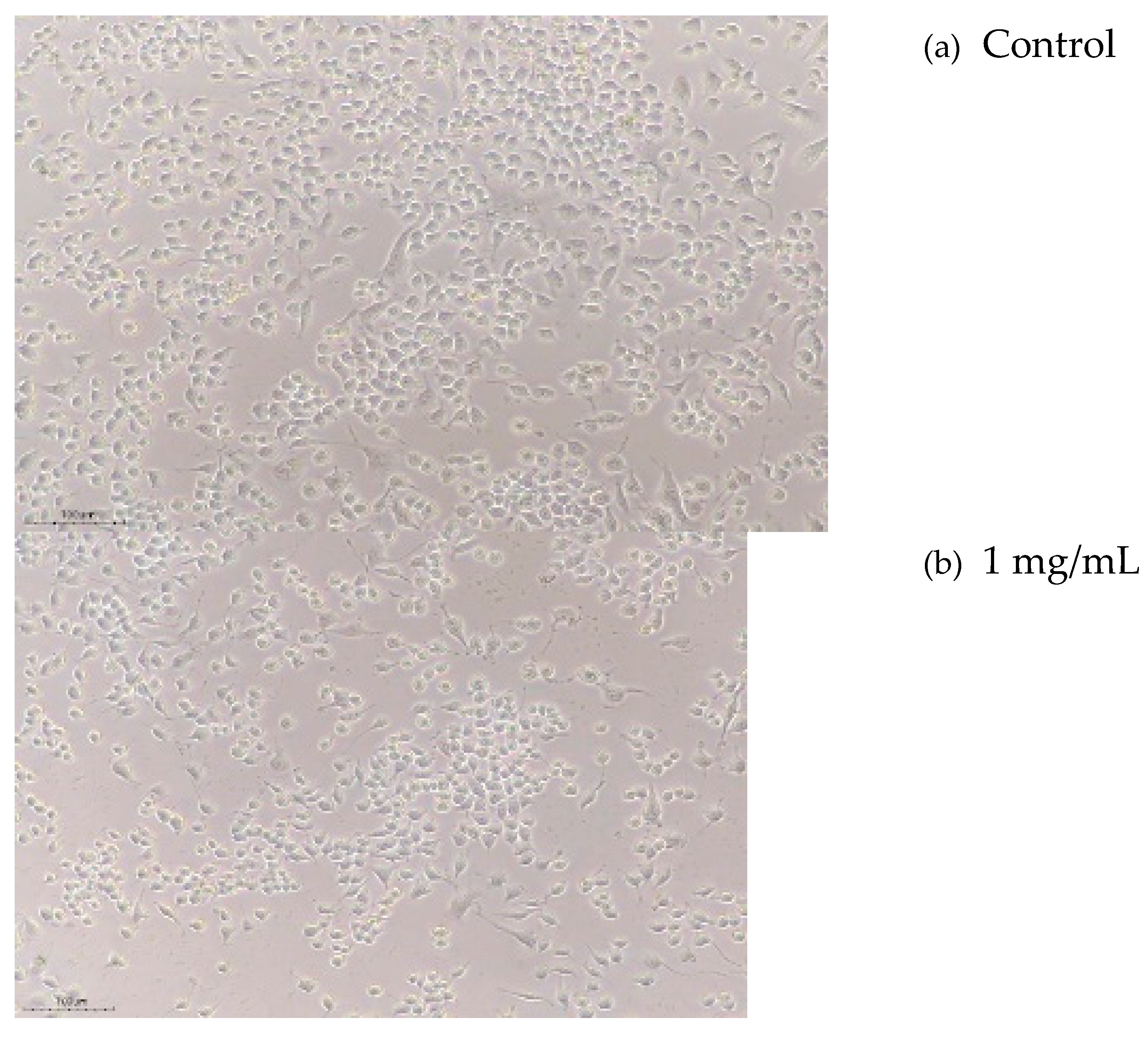
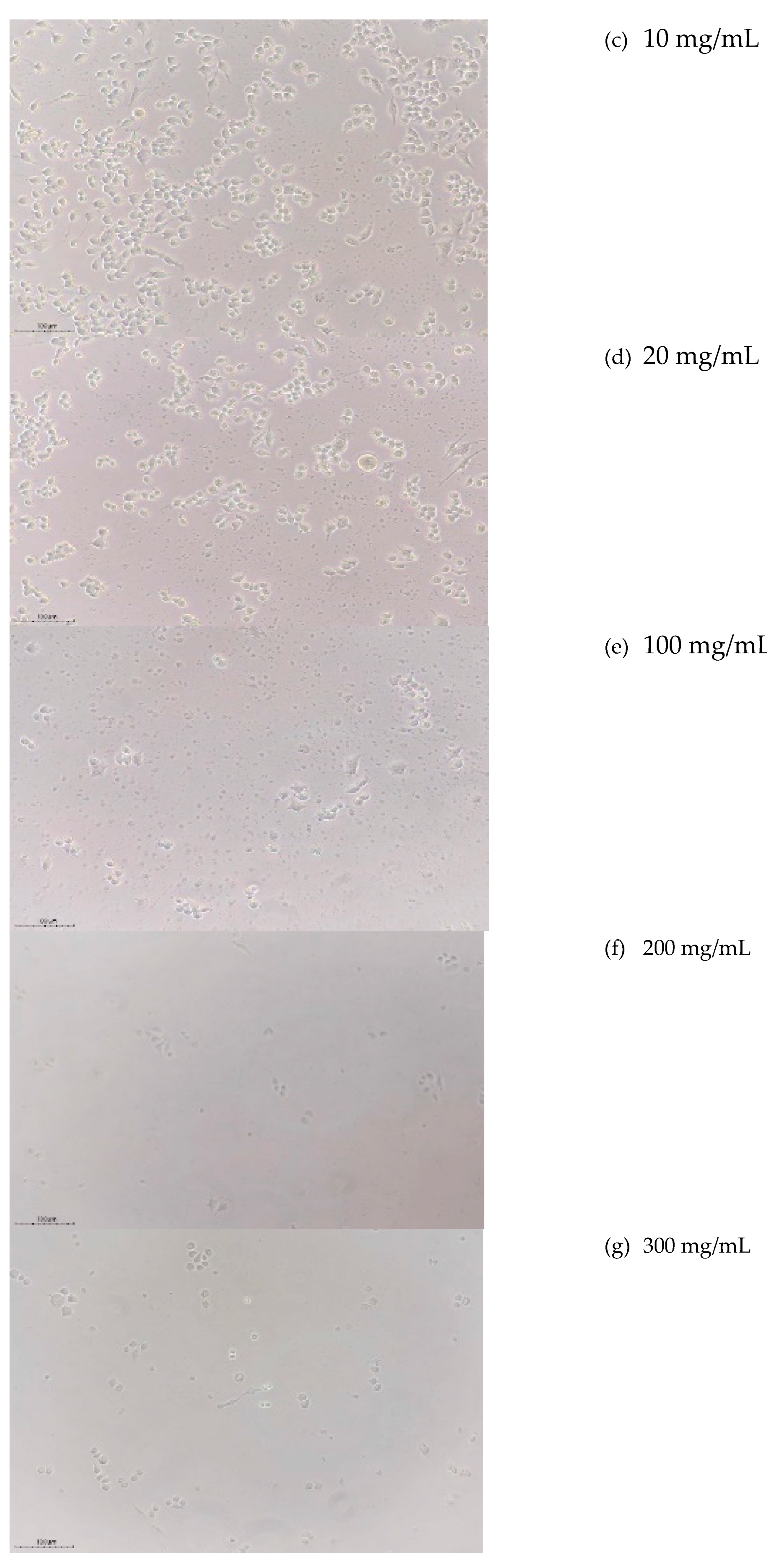
The bioactivity on the cytotoxicity of the oligofructans powder produced by B. subtilis TISTR 001 was evaluated on HepG2 liver cancer cells. The results are presented in Figure 10 and demonstrate the dose-dependent cytotoxicity of oligofructans on HepG2 liver cancer cells at concentrations in the range 0–500 mg/mL, in addition, Figure 11 shows the morphology and density of the HepG2 liver cancer cells after treatment with oligofructans powder. Based on these results, increasing concentrations of oligofructans positively influenced the cytotoxicity properties, resulting in reduced viability of HepG2 liver cancer cells, which was supported by the results reported by Shi et al. [56] and Li et al. [57]. Notably, concentrations of oligofructans of more than 100 mg/mL produced strong bioactivity, with high cytotoxicity levels reaching approximately 70–80 % (Figure 10), resulting in a significant reduction in HepG2 cell growth and cell density (Figure 11).
Notably, oligofructans has antioxidant properties (Figure 9), which support the mechanism of prebiotic properties to boost the immune system [58]. Zhang et al. [40] reported that the extract of FOSs had an anti-tumor effect and could boost immune cell activity, suggesting its potential as a natural immune-enhancing agent. Similarly, Moharib [59] confirmed the anti-cancer potential of FOSs, particularly through their ability to prevent colon cancer, improve gut health, and decrease oxidative stress, which may contribute to a reduced cancer risk. These results highlight their potential properties in mitigating oxidative stress and contributing to overall health enhancement. Additionally, the present study demonstrated that the oligofructans produced B. subtilis TISTR 001 had considerable potential to inhibit liver cancer cells proliferation, indicating their promise as a functional bioactive compound or therapeutic agent. Notably, one of the most important outcomes of the present study was the dose-dependent cytotoxicity observed against HepG2 liver cancer cells. The cytotoxicity assays revealed that higher concentrations of oligofructans effectively reduced cancer cell viability, suggesting their potential as a natural anticancer agent. The dual functionality of oligofructans—as both antioxidants and anticancer agents—underscores their value in the food, nutraceutical, and pharmaceutical industries, where the increasing demand for functional, health-promoting ingredients continues to grow for overall consumer well-being. Nonetheless, further investigations are necessary to elucidate the precise mechanisms underlying the anticancer effects of oligofructans. Future research should prioritize in vivo studies, clinical trials, and the exploration of potential synergistic effects when combined with other therapeutic agents.
4. Conclusion
This study successfully demonstrated the production of oligofructans powder using B. subtilis TISTR 001, followed by comprehensive characterization to analyze the structure and physicochemical properties. Based on the results, the oligofructans solution had low viscosity and showed Newtonian flow behavior while remaining very stable under neutral pH and moderate temperature conditions, with minimal degradation, thereby indicating potential suitability for diverse industrial and nutritional applications. Advanced analytical techniques (FTIR, NMR, and MALDI-TOF-MS) were applied to determine molecular structures and confirmed the presence of characteristic glycosidic linkages, supporting the powder classification as a fructose polymer. The analysis of the results indicated that the oligofructans had notable bioactivity properties, with both antioxidant activity (based on the DPPH radical scavenging assays) and cytotoxicity effects (based on HepG2 liver cancer cells). Despite these promising findings, this study has several limitations. Bioactivity was assessed only in-vitro study, with no in-vivo or clinical evaluations to confirm the biological effects and safety in complex biological systems. However, further research will investigate optimizing production to enhance the yield and purity on a pilot scale or an industrial scale, along with evaluating the stability of oligofructans in various food formulations to further expand their practical applications. Overall, the present study of bioactive activity has contributed valuable insights into the health benefits of prebiotic compounds and highlighted the promising potential of oligofructans as natural supplements or therapeutic agents, particularly for liver health and cancer prevention.
Funding
This research was financially supported by The Capacity Building of KU Students on Internationalization Program (KUCSI), Kasetsart University, Bangkok, Thailand.
Authors Contributions
Conceptualization, B.N.; methodology, B.N. and C.N.; investigation, data curation, C.N.; writing − original draft preparation, C.N.; writing – review & editing, B.N. and F.B.; supervision, B.N.; co-supervision, F.B.; funding acquisition, B.N and C.N.
Institutional Review Board Statement
Not applicable.
Informed Consent Statement
Not applicable.
Data Availability Statement
The raw data supporting the conclusions of this article will be made available by the authors upon request.
Acknowledgments
This research was financially supported by The Capacity Building of KU Students on Internationalization Program (KUCSI), Kasetsart University, Bangkok, Thailand. The Sugars and Derivatives Analytical Laboratory (SuDAL), the Department of Biotechnology, Faculty of Agro-Industry, Bangkok, Thailand, and the Department of Food Science and Technology, Faculty of Agriculture, Hasanuddin University, Indonesia provided laboratory space for the project, support and advice during the research. .
Conflicts of Interest
The authors declare that there are no conflicts of interest.
References
- Bersaneti, G. T.; Pan, N. C.; Baldo, C.; Celligoi, M. A. P. C. Co-production of fructo-oligosaccharides and levan by levansucrase from Bacillus subtilis natto with potential application in the food industry. Applied Biochemistry and Biotechnolog. 2018, 184, 838–851. [Google Scholar] [CrossRef] [PubMed]
- Ninchan, B.; C. Noidee. Optimization of oligofructans production from sugarcane juice fermentation using Bacillus subtilis TISTR001. Agriculture and Natural Resources 2021, 55, 1005–1014. [Google Scholar] [CrossRef]
- Okuyama, M.; Serizawa, R.; Tanuma, M.; Kikuchi, A.; Sadahiro, J.; Tagami, T.; Lang, W.; Kimura, A. Molecular insight into regioselectivity of transfructosylation catalyzed by GH68 levansucrase and β-fructofuranosidase. Journal of Biological Chemistry 2021, 296, 100398. [Google Scholar] [CrossRef] [PubMed]
- Goldman, D.; Lavid, N.; Schwartz, A.; Shoham, G.; Danino, D.; Shoham, Y. Two active forms of Zymomonas mobilis levansucrase: An ordered microfibril structure of the enzyme promotes levan polymerization. Journal of Biological Chemistry. 2008, 283, 32209–32217. [Google Scholar] [CrossRef]
- Noidee, C.; Ninchan, B.; Ninchan, B. Investigation of optimized microbial oligofructans production by Bacillus subtilis TISTR 001 using response surface methodology. Sugar Tech. 2024, 26, 585–594. [Google Scholar] [CrossRef]
- Belghith, K. S.; Dahech, I.; Belghith, H.; Mejdoub, H. Microbial production of levansucrase for synthesis of fructooligosaccharides and levan. International Journal of Biological Macromolecules. 2012, 50, 451–458. [Google Scholar] [CrossRef]
- Guo, X.; Li, X.; Xu, Q.; Zhang, Y. Dietary fibers and their emerging benefits for liver health: The role of oligofructans. Nutrition Reviews. 2020, 78, 282–298. [Google Scholar] [CrossRef]
- Benkeblia, N. . Fructooligosaccharides and fructans analysis in plants and food crops. Journal of Chromatography A, 2013, 1313, 54–61. [Google Scholar] [CrossRef]
- Kherade, M.; Solanke, S.; Tawar, M.; Wankhede, S. Fructooligosaccharides: A comprehensive review. Journal of Ayurvedic and Herbal Medicine. 2021, 7, 193–200. [Google Scholar] [CrossRef]
- Gibson, G. R.; Hutkins, R.; Sanders, M. E.; Prescott, S. L.; Reimer, R. A.; Salminen, S. J.; Scott, T.; Stanton, C.; Swanson, K.S.; Cani, P.D.; Vevbeke, V.; Reid, G. Expert consensus document: The International Scientific Association for Probiotics and Prebiotics (ISAPP) consensus statement on the definition and scope of prebiotics. Nature Reviews Gastroenterology & Hepatology 2017, 14, 491–502. [Google Scholar] [CrossRef]
- Roberfroid, M. Prebiotics: The concept revisited. The Journal of Nutrition 2007, 137, 830S–837S. [Google Scholar] [CrossRef]
- Wang, Y.; Yan, H.; Zheng, Q.; Sun, X. The crucial function of gut microbiota on gut-liver repair. hLife 2025. [Google Scholar] [CrossRef]
- Tripathi, A. , Debelius, J., Brenner, D. A., Karin, M., Loomba, R., Schnabl, B., & Knight, R. The gut–liver axis and the intersection with the microbiome. Nature Reviews Gastroenterology & Hepatology 2018, 15, 397–411. [Google Scholar] [CrossRef]
- Cani, P. D.; Neyrinck, A. M.; Fava, F.; Knauf, C.; Burcelin, R. G.; Tuohy, K. M.; Gibson, G.R.; Delzenne, N. M. Selective increases of bifidobacteria in gut microflora improve high-fat-diet-induced diabetes in mice through a mechanism associated with endotoxaemia. Diabetologia 2007, 50, 2374–2383. [Google Scholar] [CrossRef] [PubMed]
- Gibson, G. R.; Roberfroid, M. B. Dietary modulation of the human colonic microbiota: Introducing the concept of prebiotics. The Journal of Nutrition. 1995, 125, 1401–1412. [Google Scholar] [CrossRef]
- Al Hrout, A. A.; Cervantes-Gracia, K.; Chahwan, R.; Amin, A. Modelling liver cancer microenvironment using a novel 3D culture system. Scientific Reports 2022, 12, 8003. [Google Scholar] [CrossRef] [PubMed]
- Cao, P.; Rozek, L. S.; Pongnikorn, D.; Sriplung, H.; Meza, R. Comparison of cholangiocarcinoma and hepatocellular carcinoma incidence trends from 1993 to 2012 in Lampang, Thailand. International Journal of Environmental Research and Public Health 2022, 19, 9551. [Google Scholar] [CrossRef]
- National Cancer Institute. 2024. Hospital-based cancer registry 2022. Department of Medical Services, Ministry of Public Health, Thailand. Retrieved from http://www.nci.go.th.
- Bosscher, D.; Van Loo, J.; Franck, A. Inulin and oligofructose as prebiotics in the prevention of intestinal infections and diseases. Nutrition Research Reviews 2006, 19, 216–226. [Google Scholar] [CrossRef]
- Femia, A. P.; Luceri, C.; Dolara, P.; Giannini, A.; Biggeri, A.; Salvadori, M.; Clune, Y.; Collins, K. J.; Paglierani, M.; Caderni, G. Antitumorigenic activity of the prebiotic inulin enriched with oligofructose in combination with the probiotics Lactobacillus rhamnosus and Bifidobacterium lactis on azoxymethane-induced colon carcinogenesis in rats. Carcinogenesis 2002, 23, 1953–1960. [Google Scholar] [CrossRef]
- Kolida, S.; Tuohy, K.; Gibson, G. R. Prebiotic effects of inulin and oligofructose. British Journal of Nutrition. 2002, 87, S193–S197. [Google Scholar] [CrossRef]
- Nobre, C.; Simões, L. S.; Gonçalves, D. A.; Berni, P.; Teixeira, J. A. Fructooligosaccharides production and the health benefits of prebiotics. In Current developments in biotechnology and bioengineering. 2022, 109–138. [Google Scholar] [CrossRef]
- Pool-Zobel, B. L. Inulin-type fructans and reduction in colon cancer risk: review of experimental and human data. British Journal of Nutrition. 2005, 93(S1), S73–S90. [Google Scholar] [CrossRef] [PubMed]
- Dahech, I.; Belghith, K. S.; Belghith, H.; Mejdoub, H. Partial purification of a Bacillus licheniformis levansucrase producing levan with antitumor activity. International Journal of Biological Macromolecules. 2012, 51, 329–335. [Google Scholar] [CrossRef] [PubMed]
- Amrutha, N.; Hebbar, H. U.; Prapulla, S. G.; Raghavarao, K. S. M. S. Effect of additives on quality of spray-dried fructooligosaccharide powder. Drying Technology. 2014, 32, 1112–1118. [Google Scholar] [CrossRef]
- Showa Denko, K.K. 2021. Fructo-oligosaccharide Syrup (NH2P-50 4E). Showa Denko K.K. Tokyo, Japan. https://www.shodex.com/en/dc/03/03/17.html, 11 July 2021.
- Vertical Chromatography, Co. Ltd. 2021. VertiSepTM sugar HPLC columns. Vertical Chromatography Co. Ltd. Nonthaburi, Thailand. http://www.vertichrom.com/pdf/hplc_sugar.pdf, 11 July 2021.
- Nelson, N. A photometric adaptation of the Somogyi method for the determination of glucose. Journal of Biological Chemistry. 1944, 153, 375–380. [Google Scholar] [CrossRef]
- Glibowski, P.; Bukowska, A. The effect of pH, temperature and heating time on inulin chemical stability. Acta Scientiarum Polonorum Technologia Alimentaria. 2011, 10, 189–196. [Google Scholar]
- Srikanth, R.; Siddartha, G.; Reddy, C. H. S.; Harish, B. S.; Ramaiah, M. J.; Uppuluri, K. B. Antioxidant and anti-inflammatory levan produced from Acetobacter xylinum NCIM2526 and its statistical optimization. Carbohydrate Polymers. 2015, 123, 8–16. [Google Scholar] [CrossRef]
- Yarlina, V. P.; Rizky, A.; Diva, A.; Zaida, Z.; Djali, M.; Andoyo, R.; Lani, M. N. Maltodextrin concentration on the encapsulation efficiency of tempeh protein concentrated from Jack Bean (Canavalia ensiformis): Physical, chemical, and structural properties. International Journal of Food Properties. 2024, 27, 1120–1132. [Google Scholar] [CrossRef]
- Sobulska, M.; Zbicinski, I. Advances in spray drying of sugar-rich products. Drying Technology. 2021, 39, 1774–1799. [Google Scholar] [CrossRef]
- Aprodu, I.; Banu, I.; Banu, I. . Effect of starch and dairy proteins on the gluten free bread formulation based on quinoa. Journal of Food Measurement and Characterization. 2021, 15, 2264–2274. [Google Scholar] [CrossRef]
- Siccama, J. W.; Pegiou, E.; Zhang, L.; Mumm, R.; Hall, R. D.; Boom, R. M.; Schutyser, M. A. I. , Maltodextrin improves physical properties and volatile compound retention of spray-dried asparagus concentrate. LWT - Food Science and Technology 2021, 142, 111058. [Google Scholar] [CrossRef]
- Chaudhary, V.; Thakur, N.; Kajla, P.; Thakur, S.; Punia, S. Application of encapsulation technology in edible films. Frontiers in Sustainable Food Systems 2021, 5, 734921. [Google Scholar] [CrossRef]
- Glibowski, P.; Biaduń, P. Chemical stability of inulin in acidic environment as an effect of a long-term storage. Polish Journal of Natural Sciences 2021, 35, 323–329. [Google Scholar]
- Matusek, A.; Merész, P.; Le, T. K. D.; Örsi, F. Effect of temperature and pH on the degradation of fructo-oligosaccharides. European Food Research and Technology 2009, 228, 355–365. [Google Scholar] [CrossRef]
- Mensink, M. A.; Frijlink, H. W.; van der Voort Maarschalk, K.; Hinrichs, W. L. Inulin, a flexible oligosaccharide. II: Review of its pharmaceutical applications. Carbohydrate Polymers 2015, 134, 418–428. [Google Scholar] [CrossRef]
- Aramsangtienchai, P.; Kongmon, T.; Pechroj, S.; Srisook, K. Enhanced production and immunomodulatory activity of levan from the acetic acid bacterium, Tanticharoenia sakaeratensis. International Journal of Biological Macromolecules 2020, 163, 574–581. [Google Scholar] [CrossRef]
- Zhang, J.; Yue, X.; Zeng, Y.; Hua, E.; Wang, M.; Sun, Y. Bacillus amyloliquefaciens levan and its silver nanoparticles with antimicrobial properties. Biotechnology & Biotechnological Equipment. 2018, 32, 1583–1589. [Google Scholar] [CrossRef]
- De Oliveira, A. J. B.; Gonçalves, R. A. C.; Chierrito, T. P. C.; Dos Santos, M. M.; de Souza, L. M.; Gorin, P. A. J.; Sassaki, G.L.; Iacomini, M. Structure and degree of polymerisation of fructooligosaccharides present in roots and leaves of Stevia rebaudiana (Bert.) Bertoni. Food Chemistry. 2011, 129, 305–311. [Google Scholar] [CrossRef] [PubMed]
- Han, J.; Xu, X.; Gao, C.; Liu, Z.; Wu, Z. Levan-producing Leuconostoc citreum strain BD1707 and its growth in tomato juice supplemented with sucrose. Applied and Environmental Microbiolog. 2016, 82, 1383–1390. [Google Scholar] [CrossRef] [PubMed]
- Madia, V. N.; De Vita, D.; Messore, A.; Toniolo, C.; Tudino, V.; De Leo, A.; Pindinello, I.; Lalongo, D.; Saccoliti, F.; D’Ursa, A.M.; Grimaldi, M.; Ceccobelli, P.; Scipione, L.; Santo, R.D.; Ialongo, D. Analytical characterization of an inulin-type fructooligosaccharide from root-tubers of Asphodelus ramosus L. Pharmaceuticals. Pharmaceuticals. 2021, 14, 278. [Google Scholar] [CrossRef] [PubMed]
- Cérantola, S.; Kervarec, N.; Pichon, R.; Magné, C.; Bessieres, M. A.; Deslandes, E. NMR characterisation of inulin-type fructooligosaccharides as the major water-soluble carbohydrates from Matricaria maritima (L.). Carbohydrate Research. 2004, 339, 2445–2449. [Google Scholar] [CrossRef]
- Park, Y. K.; Park, Y. H.; Shin, B. A.; Choi, E. S.; Park, Y. R.; Akaike, T.; Cho, C.S. Synthesis and characterization of fructo-oligosaccharides produced by β-fructofuranosidase from Aspergillus niger ATCC 20611. Carbohydrate Research. 2000, 328, 593–603. [Google Scholar] [CrossRef]
- Chaves, P. F. P.; Iacomini, M.; Cordeiro, L. M. Chemical characterization of fructooligosaccharides, inulin and structurally diverse polysaccharides from chamomile tea. Carbohydrate Polymers. 2019, 214, 269–275. [Google Scholar] [CrossRef]
- Evans, M.; Gallagher, J. A.; Ratcliffe, I.; Williams, P. A. Determination of the degree of polymerisation of fructans from ryegrass and chicory using MALDI-TOF mass spectrometry and gel permeation chromatography coupled to multiangle laser light scattering. Food Hydrocolloids. 2016, 53, 155–162. [Google Scholar] [CrossRef]
- Kang, S.; You, H. J.; Lee, Y. G.; Jeong, Y.; Johnston, T. V.; Baek, N. I.; Ku, S.; Ji, G. E. Production, structural characterization, and in vitro assessment of the prebiotic potential of butyl-fructooligosaccharides. International Journal of Molecular Sciences. 2020, 21, 445. [Google Scholar] [CrossRef] [PubMed]
- Xu, J.; Chen, D.; Liu, C.; Wu, X. Z.; Dong, C. X.; Zhou, J. Structural characterization and anti-tumor effects of an inulin-type fructan from Atractylodes chinensis. International Journal of Biological Macromolecules. 2016, 82, 765–771. [Google Scholar] [CrossRef] [PubMed]
- Sasikumar, J. M.; Erba, O.; Egigu, M. C. In vitro antioxidant activity and polyphenolic content of commonly used spices from Ethiopia. Heliyon 2020, 6. [Google Scholar] [CrossRef]
- Liu, H.; Wang, Q.; Liu, Y.; Chen, G.; Cui, J. Antimicrobial and antioxidant activities of Cichorium intybus root extract using orthogonal matrix design. Journal of Food Science. 2013, 78, M258–M263. [Google Scholar] [CrossRef]
- Shang, H. M.; Zhou, H. Z.; Yang, J. Y.; Li, R.; Song, H.; Wu, H. X. X. In vitro and in vivo antioxidant activities of inulin. PloS One. 2018, 13, e0192273. [Google Scholar] [CrossRef]
- Roupar, D.; Coelho, M. C.; Gonçalves, D. A.; Silva, S. P.; Coelho, E.; Silva, S.; Coimbra, M.A.; Pintada, M.; Teixeira, J.A.; Nobre, C. Evaluation of microbial-fructo-oligosaccharides metabolism by human gut microbiota fermentation as compared to commercial inulin-derived oligosaccharides. Foods 2022, 11, 954. [Google Scholar] [CrossRef]
- Rice-Evans, C.; Miller, N.; Paganga, G. Antioxidant properties of phenolic compounds. Trends in Plant Science 1997, 2, 152–159. [Google Scholar] [CrossRef]
- Tungland, B. C.; Meyer, D. Nondigestible oligo-and polysaccharides (Dietary Fiber): their physiology and role in human health and food. Comprehensive Reviews in Food Science and Food Safety 2002, 1, 90–109. [Google Scholar] [CrossRef]
- Shi, L.; Li, Y.; Zhang, S.; Gong, X.; Xu, J.; Guo, Y. Construction of inulin-based selenium nanoparticles to improve the antitumor activity of an inulin-type fructan from chicory. International Journal of Biological Macromolecules 2022, 210, 261–270. [Google Scholar] [CrossRef]
- Li, Y.; Elmén, L.; Segota, I.; Xian, Y.; Tinoco, R.; Feng, Y.; Fujita, Y.; Munoz, R.R.S.; Schmaltx, R.; Bradley, L.M.; Ramer-Tait, A.; Zarecki, R.; Long, T.; Peterson, S.N.; Ze’ev, A. R. Prebiotic-induced anti-tumor immunity attenuates tumor growth. Cell Reports 2020, 30, 1753–1766. [Google Scholar] [CrossRef]
- Roberfroid, M. B. Introducing inulin-type fructans. British Journal of Nutrition 2005, 93, S13–S25. [Google Scholar] [CrossRef]
- Moharib, S. A. Anticancer and antioxidant effects of fructooligosaccharide (FOS) on chemically-induced colon cancer in rats. Electronic Journal of Polish Agricultural Universities 2016, 19, 1–21. [Google Scholar]
Figure 1.
Kinetic changes of oligofructans fermentation using B. subtilis TISTR001 during 48 h under optimized condition (pH 6.5 and 32.5 °C, shaking at 250 rpm): (a) sugar contents, (b) short-chain FOSs (kestose and nystose) and oligofructans content.
Figure 1.
Kinetic changes of oligofructans fermentation using B. subtilis TISTR001 during 48 h under optimized condition (pH 6.5 and 32.5 °C, shaking at 250 rpm): (a) sugar contents, (b) short-chain FOSs (kestose and nystose) and oligofructans content.
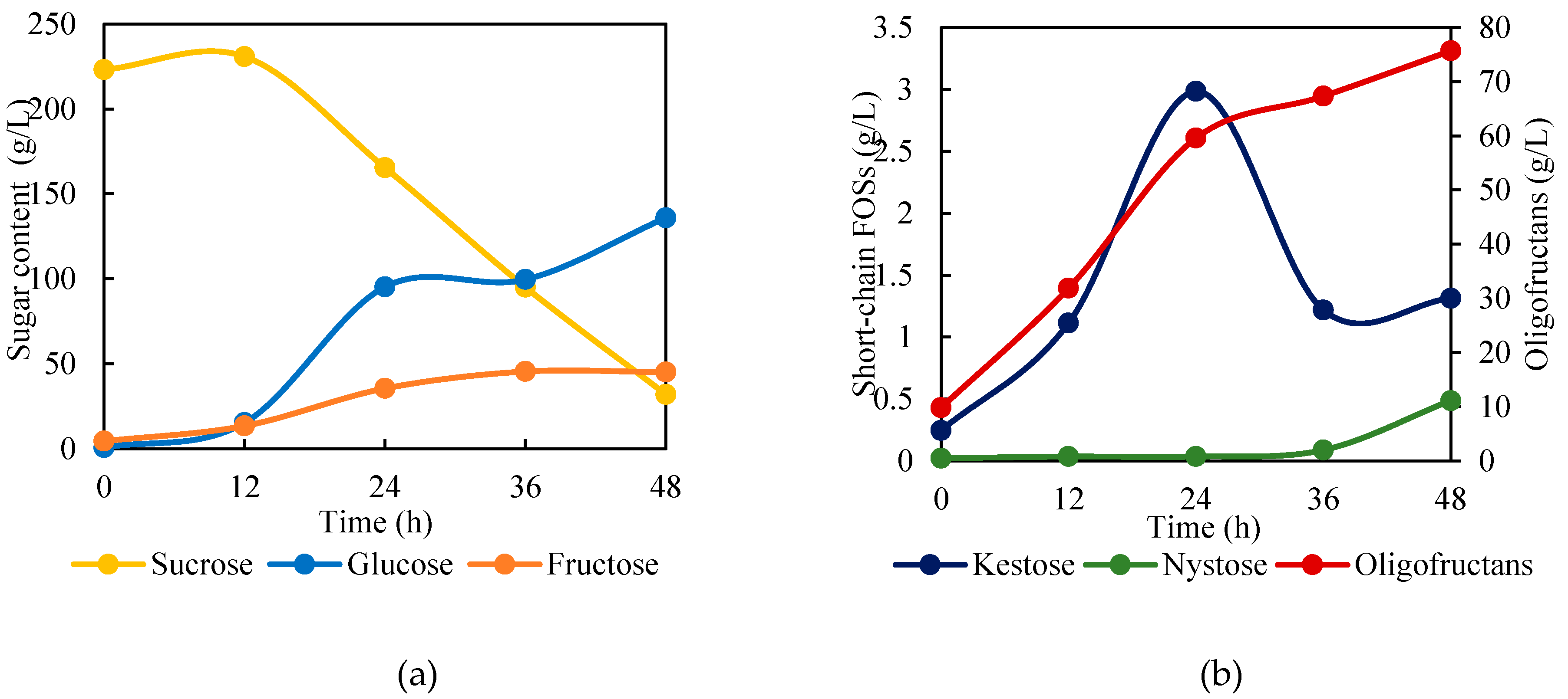
Figure 2.
Oligofructans powder content for addition and no addition of maltodextrin in spray-drying process, where different lowercase letters above columns indicate significant (p < 0.05) differences.
Figure 2.
Oligofructans powder content for addition and no addition of maltodextrin in spray-drying process, where different lowercase letters above columns indicate significant (p < 0.05) differences.
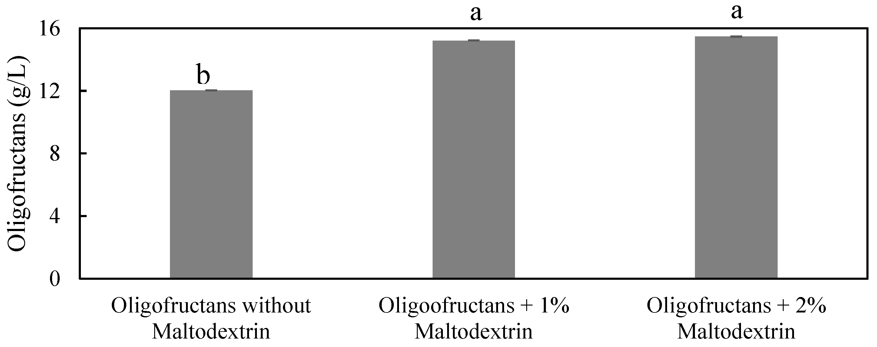
Figure 3.
Viscosity of oligofructans solutions at different concentrations (10 %, 30 %, and 50 % (w/v)) over time (5, 10, and 15 min) measured at 25 + 0.5 °C at a rate of 60 rpm.
Figure 3.
Viscosity of oligofructans solutions at different concentrations (10 %, 30 %, and 50 % (w/v)) over time (5, 10, and 15 min) measured at 25 + 0.5 °C at a rate of 60 rpm.
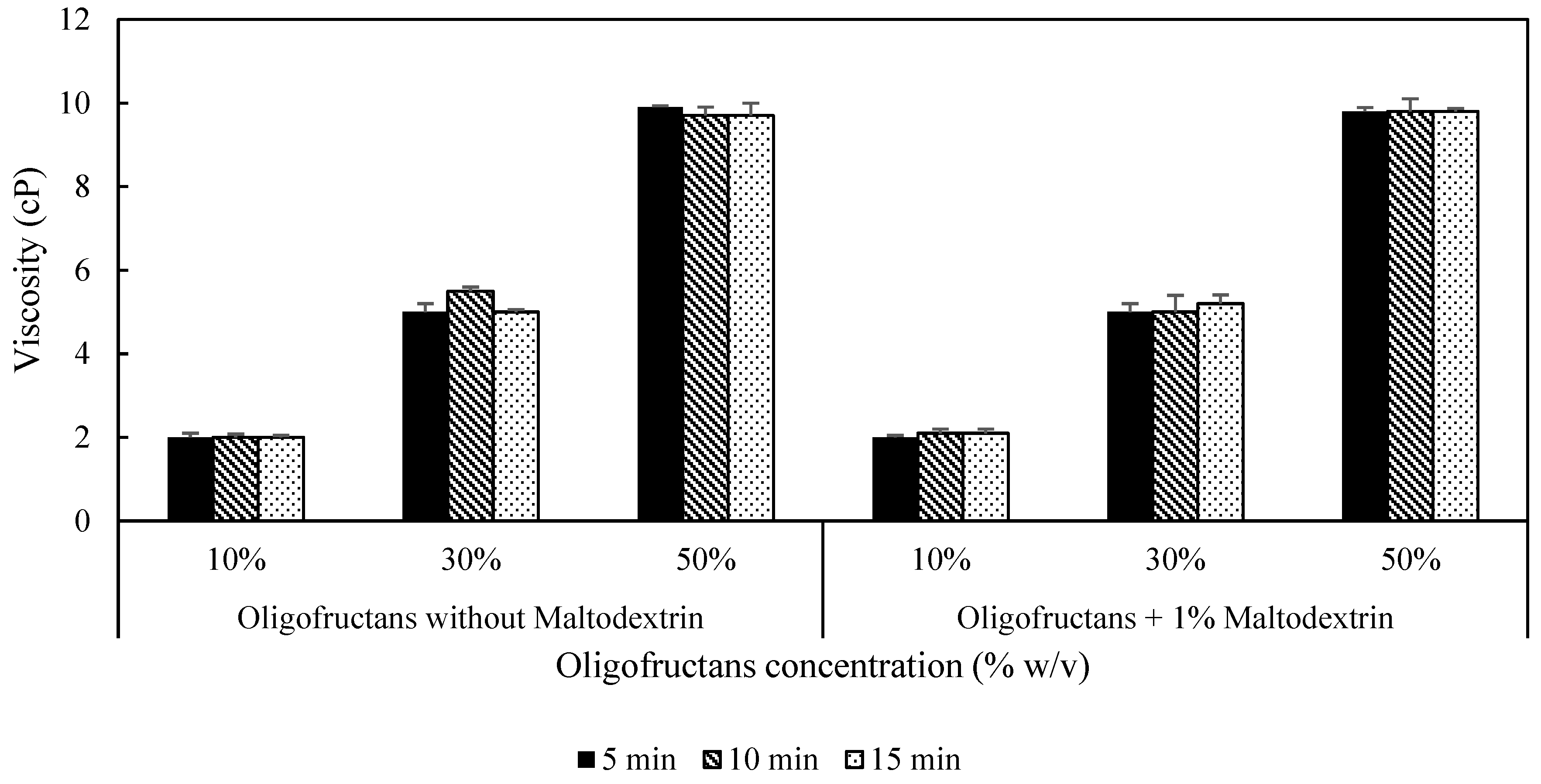
Figure 4.
Effect on reducing sugar content in oligofructans of: (a) pH and (b) Temperature.
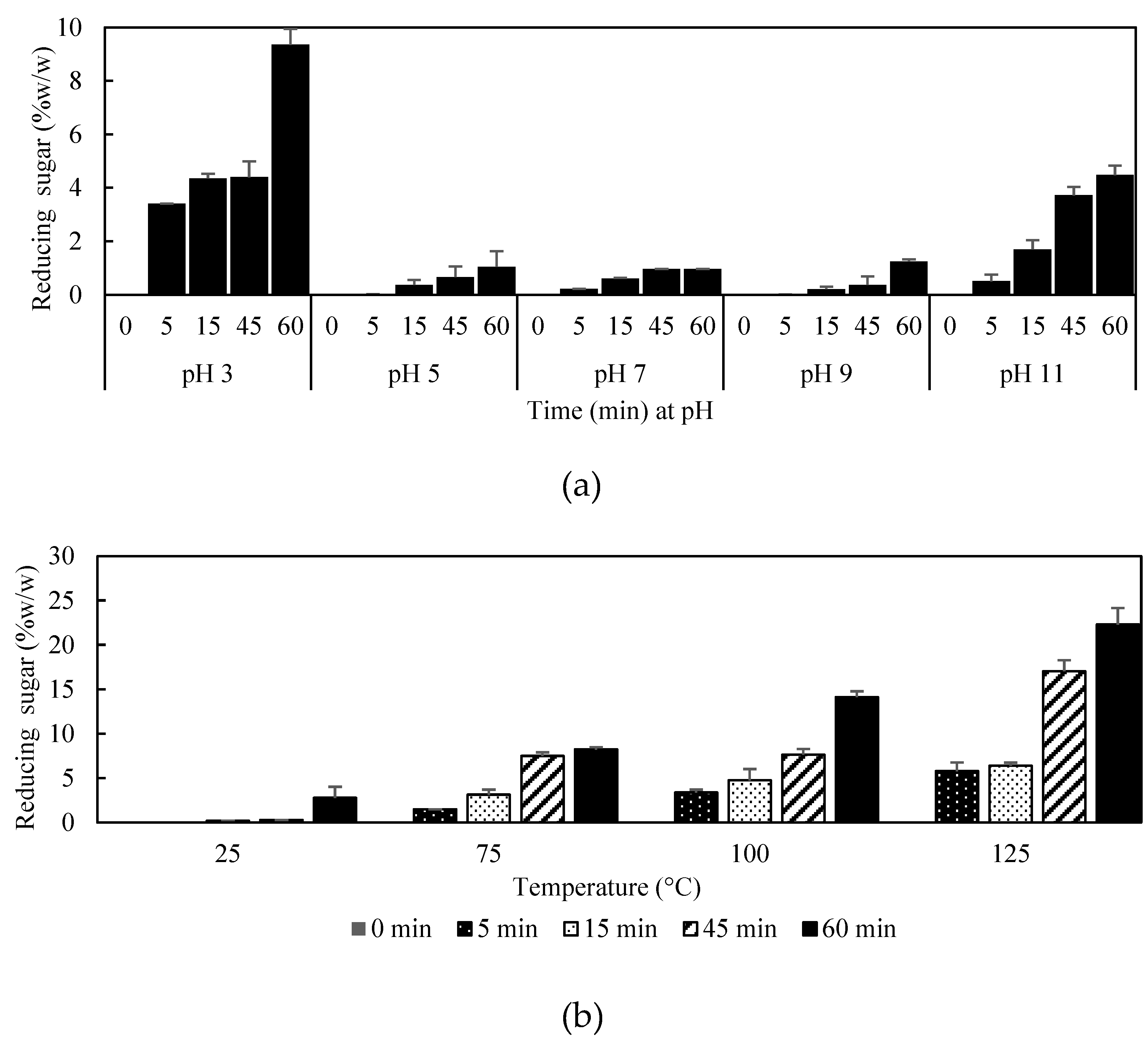
Figure 5.
FTIR spectra of oligofructans, short-chain FOSs (kestose and nystose) and fructose polymer (levan).
Figure 5.
FTIR spectra of oligofructans, short-chain FOSs (kestose and nystose) and fructose polymer (levan).
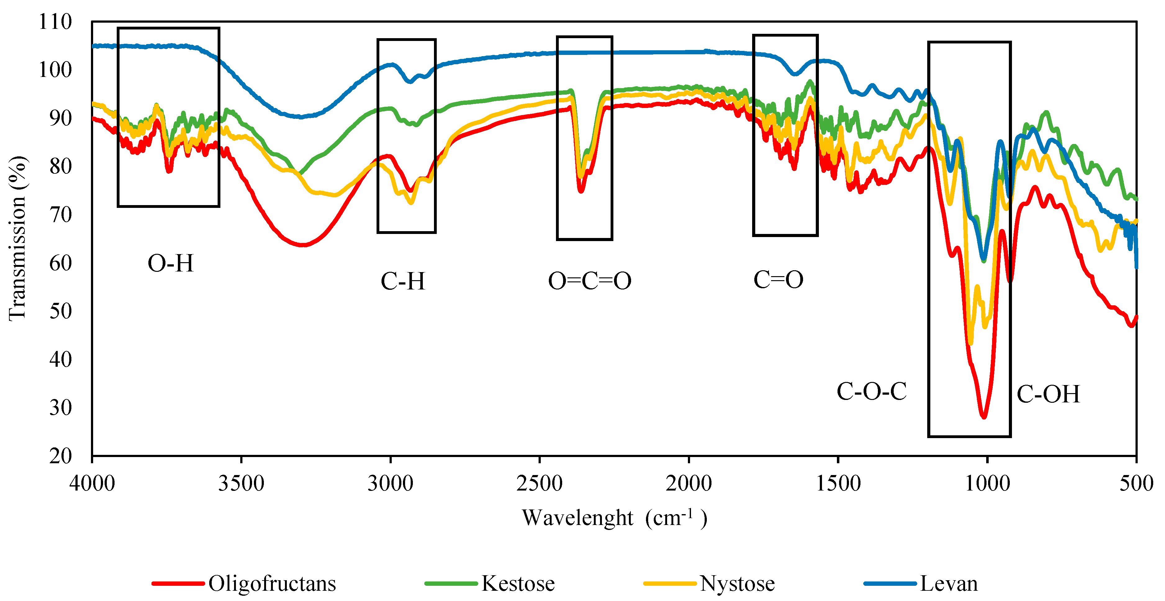
Figure 6.
¹H NMR spectra of oligofructans powder produced by B. subtilis TISTR 001.
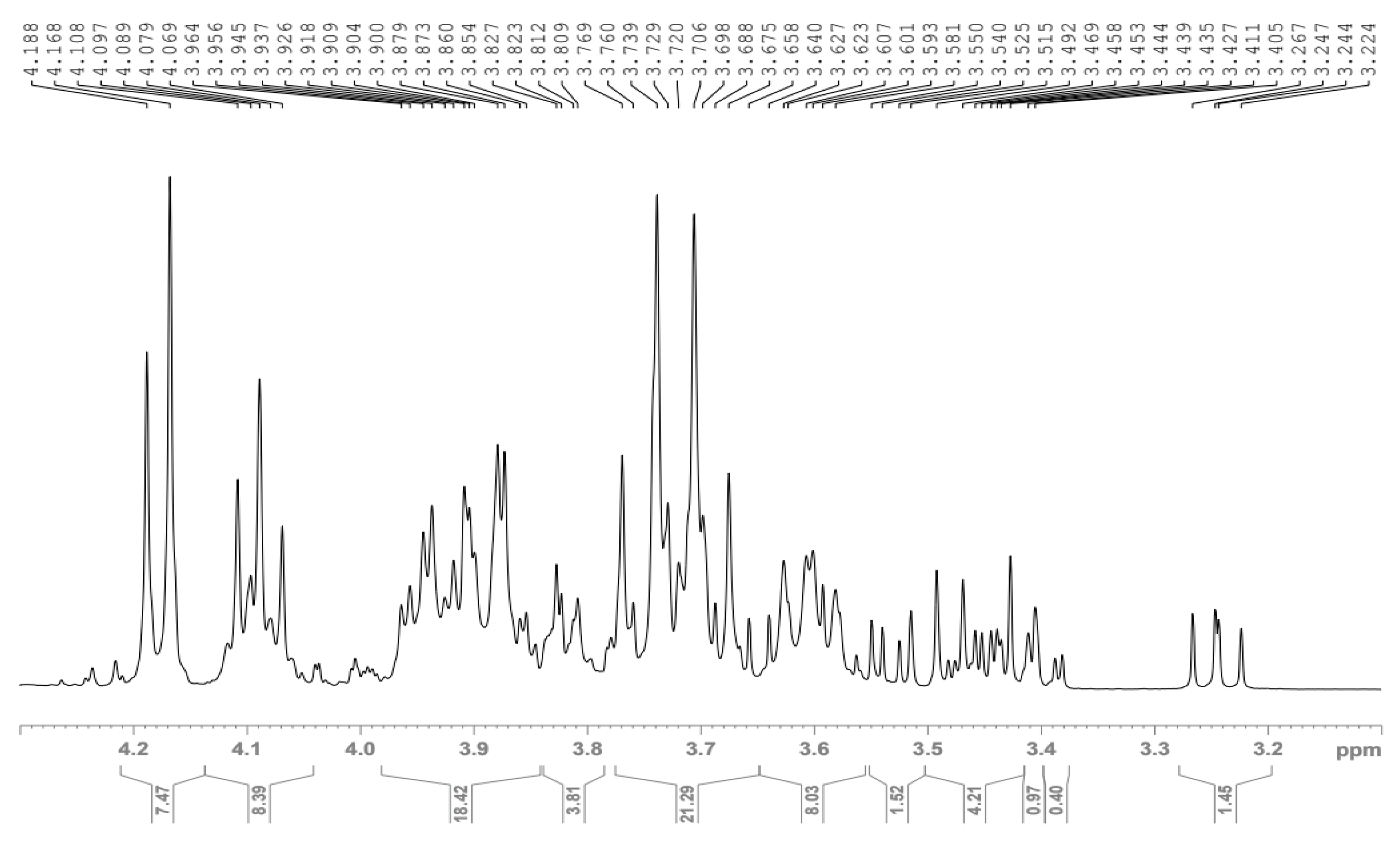
Figure 7.
¹³C NMR spectra of oligofructans powder produced by B. subtilis TISTR 001.
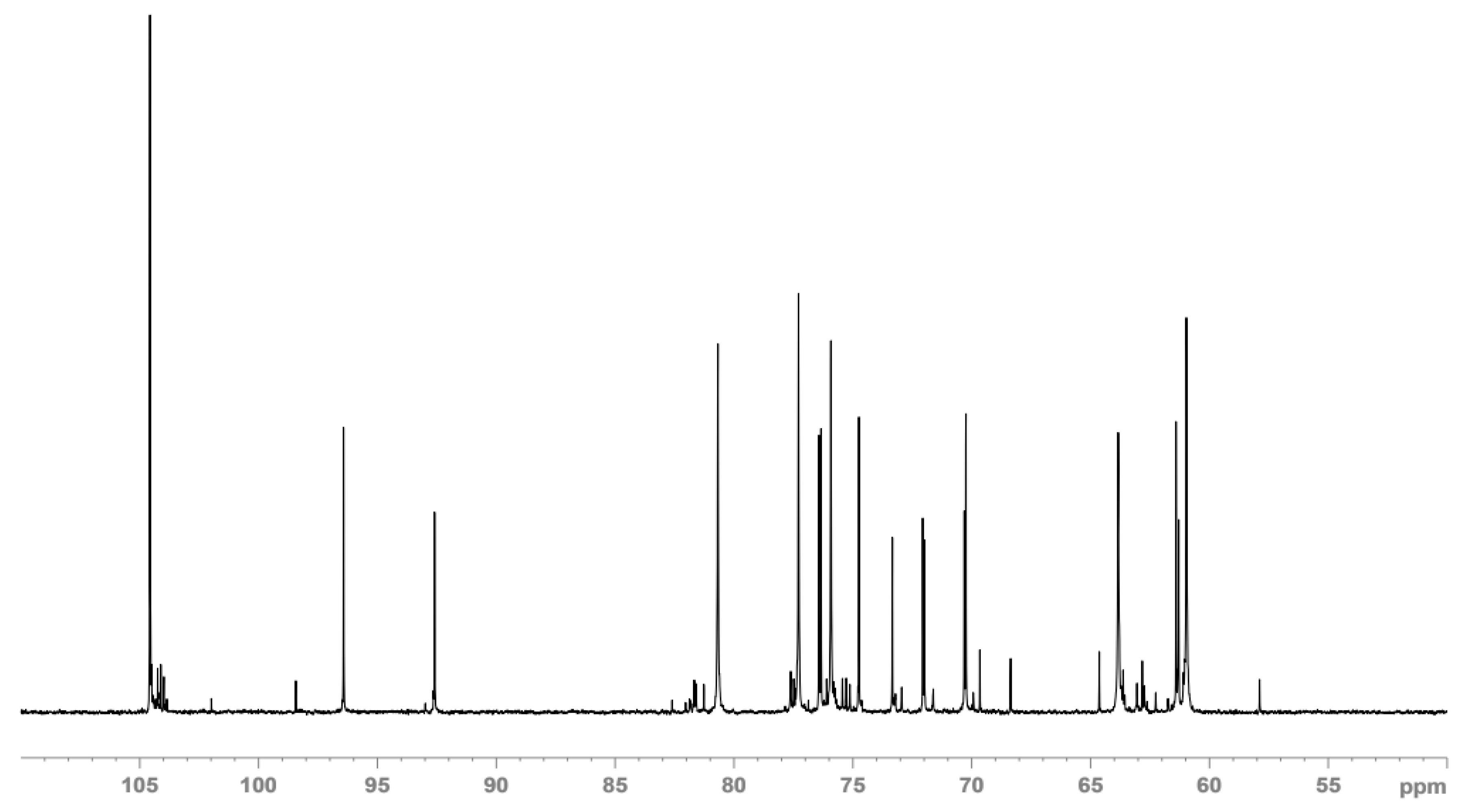
Figure 8.
MALDI-TOF_mass spectrum of oligofructans powder produced by B. subtilis TISTR 001.
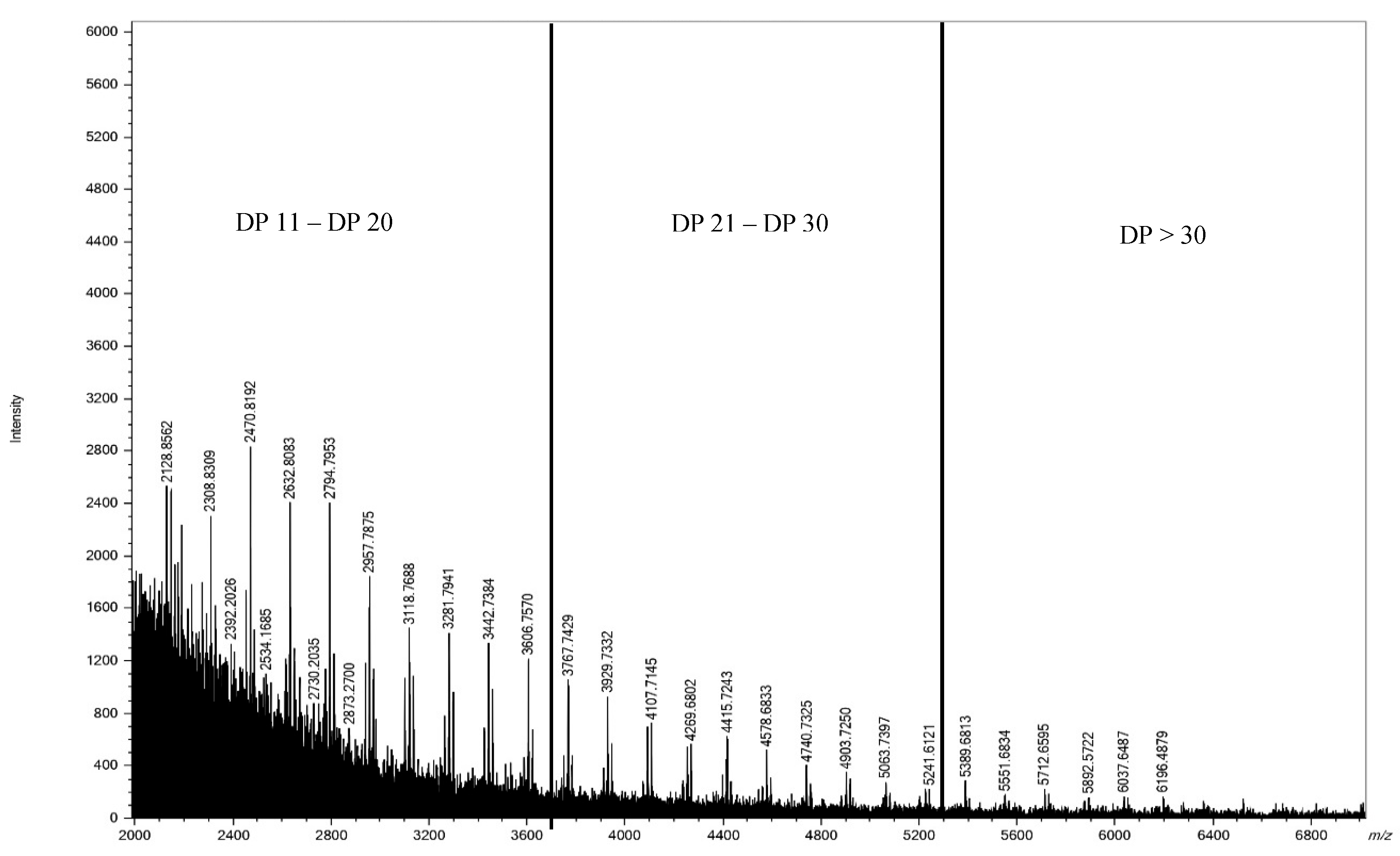
Figure 9.
Radical scavenging activity of oligofructans powder produced by B. subtilis TISTR 001.
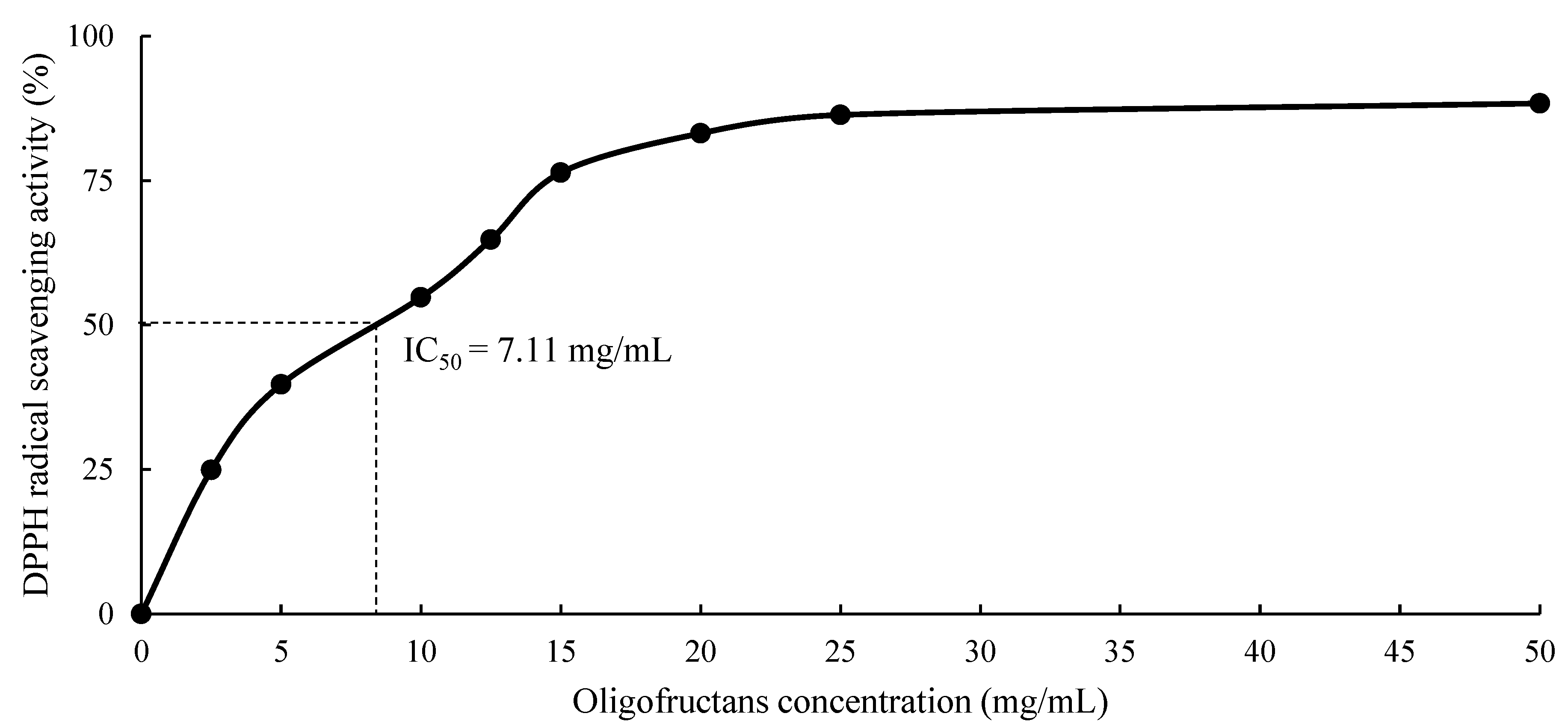
Figure 10.
Cytotoxicity assay of oligofructans powder produced by B. subtilis TISTR 001 on HepG2 liver cancer cells, where different lowercase letters above columns indicate significant (p < 0.05) differences.
Figure 10.
Cytotoxicity assay of oligofructans powder produced by B. subtilis TISTR 001 on HepG2 liver cancer cells, where different lowercase letters above columns indicate significant (p < 0.05) differences.
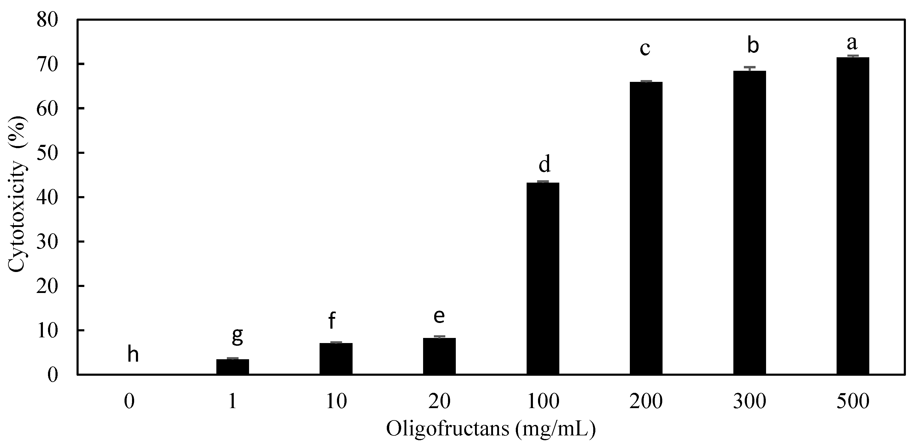
Figure 11.
The morphology and cell density of HepG2 liver cancer cells under oligofructans treatment.
Figure 11.
The morphology and cell density of HepG2 liver cancer cells under oligofructans treatment.
Disclaimer/Publisher’s Note: The statements, opinions and data contained in all publications are solely those of the individual author(s) and contributor(s) and not of MDPI and/or the editor(s). MDPI and/or the editor(s) disclaim responsibility for any injury to people or property resulting from any ideas, methods, instructions or products referred to in the content. |
© 2025 by the authors. Licensee MDPI, Basel, Switzerland. This article is an open access article distributed under the terms and conditions of the Creative Commons Attribution (CC BY) license (http://creativecommons.org/licenses/by/4.0/).
Copyright: This open access article is published under a Creative Commons CC BY 4.0 license, which permit the free download, distribution, and reuse, provided that the author and preprint are cited in any reuse.


