
Submitted:
24 June 2025
Posted:
25 June 2025
You are already at the latest version
Abstract
Background: Fluorescence-guided surgery (FGS) has emerged as a groundbreaking surgical technique in recent decades. As a real-time, non-invasive, and high-resolution diagnostic technology, FGS provides invaluable assistance to surgeons by offering real-time intraoperative navigation. Research on FGS has evolved substantially over time. Despite extensive research on FGS and its potential clinical applications, there is a notable absence of bibliometric analyses in existing studies. This study aims to fill this gap by mapping the knowledge structure and assessing the evolving trends related to FGS.
Materials and methods: This study involved searches conducted on the Web of Science Core Collection database, spanning from 2003 to the end of 2023. To explore emerging trends, we integrated the keyword “NIR-II imaging”. The collected data underwent a comprehensive analysis and visualization based on publications, country of origin, authorship, and keywords. We employed bibliometric software such as VOSviewer, CiteSpace, the R bibliometrix package, and Carrot2 for analysis and visualization purposes.
Results: The initial analysis of 1693 articles revealed that the most contributing countries were the USA and China. The most relevant topics encompassed clinical applications such as tumor resection and sentinel lymph node mapping. The most frequently occurring keywords included fluorescent dyes, notably 5-aminolevulinic acid and indocyanine green. Furthermore, statistical analyses of institutions and journals revealed that Leiden University was the leading contributor institution, whereas the Journal of Biomedical Optics was the most published journal. Subsequently, 102 publications specifically addressing NIR-II imaging in FGS were identified and subjected to further analyses.
Conclusion: This study offers a comprehensive depiction of FGS. The insights obtained from this analysis hold the potential to inform and guide future research in this field.
Keywords:
fluorescence-guided surgery
; bibliometric analysis
; indocyanine green
; NIR-II
1. Introduction
In recent decades, fluorescence-guided surgery (FGS) has emerged as a groundbreaking technique with a multitude of clinical applications. This innovative approach aims to enhance the precision of routine surgeries by illuminating the target tissue using an external light source, which then excites fluorescent agents, thereby providing surgeons with an augmented view of the surgical field[1]. As a real-time, non-invasive, and high-resolution diagnostic technology, FGS provides invaluable assistance to surgeons by offering real-time intraoperative navigation. In essence, FGS serves as a pair of “clairvoyant eyes” for surgeons during surgical procedures. Specifically, FGS enables surgeons to precisely locate and excise diseased tissues while minimizing collateral damage to surrounding healthy tissue[2]. It facilitates the identification of tumor boundaries, detection of minor lesions, guidance for lymph node dissection, and identification of critical structures such as nerves and blood vessels[3]. Overall, FGS contributes to increased surgical success rates and improves patient prognosis.
The development of FGS has undergone a long process. In the early 20th century, scientists began to realize that certain compounds fluoresced when excited by light of a specific wavelength. These early studies laid the foundation for the development of FGS[4]. As the mid-20th century approached, clinical studies began to explore the potential applications of FGS in various fields, including neurosurgery and tumor resection. A milestone moment occurred in 1948 when Moore et al. reported the pioneering use of fluorescein, a yellow-green fluorescent dye with an excitation wavelength of around 400 nm, to guide the resection of intracranial malignancies[5,6]. This breakthrough marked a substantial advancement in the field of FGS. Further progress was made in 1957 with the discovery that indocyanine green (ICG) fluoresced near-infrared (NIR) light and was a safe fluorescent probe. This discovery further propelled progress in the field of FGS[7,8]. With the onset of the 21st century, the introduction of advanced fluorescence imaging devices, such as fluorescence microscopy and cameras, have significantly broadened the clinical application of FGS. A range of fluorescent surgical navigation systems have been approved for clinical use to meet various surgical needs[9]. These navigation systems encompassed portable handheld systems, open surgery systems, and laparoscopic surgery systems, and have found widespread application in neurosurgery, general surgery, urology, gynecology, and other specialized fields[10,11,12,13].
In recent years, the evolution of FGS has led to the enhancement of targeted fluorescent molecular probes and the advancement of more precise and portable fluorescent systems. This strategic shift in response to the escalating demand for precision and minimally invasive surgical techniques. Among the latest developments, Near-Infrared II (NIR-II) fluorescent surgical navigation has garnered significant attention within the field[14]. NIR-II fluorescence offers several distinct advantages over traditional methods, including deeper tissue penetration depth, lower background signal, higher signal-to-noise ratio, and superior spatial resolution[15]. These attributes make NIR-II fluorescence an attractive option for FGS with greater accuracy and efficiency[16]. In addition, novel types of fluorescent probes have made significant progress in pre-clinical research. These probes include organic probes, inorganic quantum dots, and rare-earth nanoprobes [17,18,19]. These innovations hold promise for further improving the capabilities of FGS and expanding its applications in clinical practice.
Bibliometric analysis is a quantitative method used to assess the quantity, quality, impact, and interrelatedness of scientific literatures[20]. It involves statistical and metric processing of literature, including lexical, citations, authors, and topic analysis. Through bibliometric analysis, researchers gain insights into research trends, hotspots, disciplinary development, author collaboration, and journal impact within a specific field. This information provides clues for future research directions and decision-making. In the era of information technology and the burgeoning of interdisciplinary studies, bibliometric analysis has been applied across various fields including medicine, education, environmental science, and economics. Typically conducted using computer software like VOSviewer, Bibliometrix, CiteSpace, etc., bibliometric analysis enables efficient processing of large-scale literature data and visualization analysis[21].
In this study, we conducted a bibliometric analysis to gather literature related to FGS from the Web of Science Core Collection (WOSCC) database. Visualization software such as VOSviewer, Bibliometrix, and CiteSpace were utilized to analyze the number of publications and authors involved in FGS. Additionally, the analysis included publishing institutions, countries, journals, keywords, citation frequency, and overall contribution of the literature. Through this comprehensive analysis, our study aimed to review the developmental trajectory of FGS, identify research hotspots, and exhibit potential future trends in the field, with a particular emphasis on NIR-II surgical navigation. We anticipate that the insights obtained from this study will provide valuable guidance for future researchers focusing on FGS.
2. Materials and Methods
2.1. Data Collection
The literature search was conducted using the Web of Science Core Collection (WoSCC) database with the search queries ‘TS =((Fluorescen* OR Autofluorescen* ) AND (“Surg* navigation” OR”guided surgery” OR”Surg* guidance”))’ for the period 2003.1-2023.12 (inclusive). Full data, including country, institution, source, year of publication, authorship, title, abstract, keywords, citation, and reference, were collected.
2.2. Data Analysis
To perform bibliometric analysis, we used CiteSpace (version 6.2. R7), R bibliometrix package (version 4.1.3), VOSviewer (version 1.6.20), SCImago Graphica software (version 1.0.38), and the Carrot2 workbench (version 3.10.2). The top related institutions, keywords, and clusters were visualized by CiteSpace. The map of global collaboration, the dynamic of top related journals and authors, and three-field plot were produced by R bibliometrix package. International collaborative networks of authors and journals were drawn using VOSviewer. The maps of global collaboration were produced by SCImago Graphica software with converting data by VOSviewer in advance. The keyword tree was generated using Carrot2. GraphPad Prism (Version 9.0), Origin (Version 2021), and Adobe Illustrator (Version 2023) were used to draw diagrams, such as graphing of productions and citations, trend topics, and dynamic alterations of top-related journals and authors.
3. Results
3.1. Global Publication Trends and Inter-Country Collaboration
A detailed flowchart depicting the data collection and extraction process was presented in Figure 1. Based on the specified search terms, a total of 2346 publications were collected from the WoSCC database. After excluding review articles, meeting abstracts, editorial materials, and other studies that did not meet the criteria for our research, 1693 publications remained for further analysis.
The annual publications and citations of the FGS were illustrated in Figure 2A. It was observed that the development of FGS commenced around the early 21st century and has since demonstrated consistent growth trends over two decades, highlighting significant interest and ongoing research efforts in this field. To identify the primary countries contributing to this field, we conducted a statistical analysis of the publication trends across different nations. Table 1 presented the top 20 most relevant countries, and the stacked area diagram illustrated the annual number of publications in the top 10 countries (Figure 2B). The USA has emerged as the most prolific contributor, with 586 publications recorded at the end of 2023, maintaining a stable publication rate. Despite its relatively late start, China has exhibited a robust growth trajectory.
The cooperation between countries in the field of FGS is reflected in the Multiple Country Publications (MCP) ratio presented in Table 1, which indicates the proportion of international collaboration. Notably, China's MCP ratio is approximately 16.6%, lower than that of other European and American countries, which means that China should strengthen international academic exchange and cooperation in the FGS research field. Then, we provided a clearer depiction of the cooperative relationships between countries. It showed that the USA, China, Japan, Canada, Germany, the Netherlands, and other nations have established relatively strong academic cooperation ties (Figure 2C). Furthermore, to gain insight into the evolution of global collaboration, we divided the development timeline and presented visual networks of FGS publications for the periods 2003-2013 and 2014-2023, respectively (Figures 2D, 2E). It is evident that research in this field has grown rapidly over the past decade, with closer academic links between countries. Moreover, more countries and regions, such as Egypt, South Africa, Brazil, and Australia, have engaged in varying degrees of cooperation and exchange. This observation underscored the increasing frequency of academic exchanges in the field of fluorescent surgical navigation research facilitated by the ongoing process of globalization.
3.2. Most Related Institutions of FGS Research
The institutions that have published literatures related to FGS from 2003 to 2023 were identified using the CiteSpace software, revealing a total of 97 institutions with more than 10 publications. The co-occurrence network map was presented (Figure 3A). For instance, Leiden University was closely linked to the Netherlands Cancer Institute. The node representing Leiden University is notably large, indicating a high number of publications, and the concentric circles surrounding it transition from red to yellow, suggesting a longstanding presence and contribution to the field of FGS. In contrast, the concentric circles surrounding nodes representing the Chinese Academy of Sciences (CAS) are yellow, indicating that its development in this field is at an early stage.
Simultaneously, a statistical analysis of the top 20 institutions and their respective countries was conducted (Figure 3B). The figure revealed that the top five institutions in terms of the number of articles published were Leiden University (139 publications), University of California (137 publications), University of California San Diego (101 publications), Chinese Academy of Sciences (86 publications), and Harvard University (81 publications). Notably, Leiden University had the largest number of publications, accounting for 8.21% of all publications. Moreover, Figure 3B illustrated that among the top 20 institutions with the most literature on surgical fluorescence navigation, half are universities in the USA. Two institutions from China, namely the CAS and the Institute of Automation affiliated with the CAS, are also represented. The remaining institutions belong to European countries, such as the Netherlands, Germany, and France.
A three-field plot of the top 10 institutions is presented to elucidate the relationships among countries, institutions, and authors (Figure 3C). It is noteworthy that CAS accounted for the majority of studies in China. Specifically, Jie Tian and colleagues from CAS contributed 34 articles on fluorescent surgical navigation (Table 3). Moreover, the Netherlands has made significant contributions to research in the field of FGS. In detail, Leiden University has gained considerable recognition, particularly for its expertise in utilizing fluorescence imaging technology for surgical navigation and tumor diagnosis. In recent years, Leiden University has spearheaded advancements in NIR fluorescence imaging technology, employing fluorescent agents, such as ICG, to enhance detection and navigation in various complex surgical procedures[22].
3.3. Most Contributing Authors and Collaborative Networks
In the dynamic realm of FGS, the past two decades have witnessed the dedicated efforts of 8988 authors. In bibliometric circles, the assessment of an author's research impact traditionally depends on key metrics such as publication frequency, citation counts, journal impact factor (IF), and h-index (a composite measure considering both the quantity of publications and their respective citation frequencies)[23]. By leveraging these established metrics, we scrutinized the most influential authors in the FGS field and listed the top 20 contributors in Table 2. As presented in the table, the top 5 prolific authors are Robert M. Hoffman (n=83), Michael Bouvet (n=79), Alexander L. Vahrmeijer (n=68), F.W.B. van Leeuwen (n=68), and John V. Frangioni (n=40).

Table 2.
Top 20 most relevant authors.
| Rank | Authors | Country | Articles | AF1 | TC2 | H-index |
|---|---|---|---|---|---|---|
| 1 | Robert M. Hoffman | USA | 83 | 9.97 | 2377 | 30 |
| 2 | Michael Bouvet | USA | 79 | 8.42 | 2310 | 30 |
| 3 | Alexander L Vahrmeijer | Netherlands | 68 | 6.30 | 2578 | 31 |
| 4 | F.W.B van Leeuwen | Netherlands | 68 | 8.17 | 2326 | 29 |
| 5 | John V. Frangioni | USA | 40 | 5.46 | 2843 | 30 |
| 6 | Zhang, Yong | Singapore | 37 | 4.15 | 970 | 19 |
| 7 | Michele Diana | France | 36 | 4.03 | 623 | 14 |
| 8 | Tessa Buckle | Netherlands | 35 | 4.31 | 1267 | 17 |
| 9 | Tian, Jie | China | 34 | 4.16 | 1169 | 16 |
| 10 | Nynke S. van den Berg | Netherlands | 34 | 3.27 | 1402 | 20 |
| 11 | C.J.H. van de Velde | Netherlands | 32 | 3.18 | 1948 | 26 |
| 12 | Jaxques Marescaux | France | 31 | 3.64 | 563 | 13 |
| 13 | Keith. D. Paulsen | USA | 30 | 3.58 | 972 | 18 |
| 14 | Eben L. Rosenthal | USA | 30 | 2.95 | 1218 | 16 |
| 15 | Yukihiro Hiroshima | Japan | 29 | 2.78 | 1067 | 21 |
| 16 | Henk G. van der Poel | Netherlands | 29 | 3.38 | 1321 | 18 |
| 17 | Georg Widhalm | Austria | 26 | 2.05 | 263 | 8 |
| 18 | David W. Roberts | USA | 25 | 3.37 | 952 | 18 |
| 19 | M.N. van Oosterom | Netherlands | 25 | 2.82 | 344 | 13 |
| 20 | Brain W. Pogue | USA | 24 | 3.17 | 552 | 15 |
1AF=Articles Fractionalized; 2TC=total citations.
Table 3.
Top 20 most relevant Journals.
| Rank | Sources | Articles | TC1 | AAC2 |
|---|---|---|---|---|
| 1 | Journal of Biomedical Optics | 64 | 1303 | 20.36 |
| 2 | Molecular Imaging and Biology | 46 | 808 | 17.57 |
| 3 | Theranostics | 46 | 1556 | 33.83 |
| 4 | Journal of Nuclear Medicine | 38 | 1343 | 35.34 |
| 5 | Surgical Endoscopy and Other Interventional Techniques | 34 | 747 | 21.97 |
| 6 | Frontiers In Oncology | 32 | 197 | 6.16 |
| 7 | World Neurosurgery | 31 | 566 | 18.26 |
| 8 | Acta Neurochirurgica | 28 | 820 | 29.29 |
| 9 | Biomedical Optics Express | 28 | 411 | 14.68 |
| 10 | Journal of Neurosurgery | 28 | 831 | 29.68 |
| 11 | PLOS One | 28 | 882 | 31.50 |
| 12 | Scientific Reports | 28 | 667 | 23.82 |
| 13 | Cancers | 27 | 177 | 6.56 |
| 14 | Journal of Neuro-Oncology | 25 | 423 | 16.92 |
| 15 | European Journal of Nuclear Medicine and Molecular Imaging | 24 | 506 | 21.08 |
| 16 | Oncotarget | 23 | 725 | 31.52 |
| 17 | Analytical Chemistry | 22 | 623 | 28.32 |
| 18 | Photodiagnosis and Photodynamic Therapy | 22 | 310 | 14.09 |
| 19 | Journal of Surgical Oncology | 20 | 826 | 41.30 |
| 20 | Molecular Pharmaceutics | 20 | 407 | 20.35 |
1TC=total citations; 2AAC=average article citations.
Cluster analysis of co-authorship with authors was conducted using VOSviewer software, resulting in the formation of 10 colorful clusters based on the authors' collaborations (Figure 4A). Each cluster was represented by the corresponding authors who played significant roles in their respective clusters. For instance, Cluster 1, led by Alexander L. Vahrmeijer and John V. Frangioni, is deeply involved in clinical surgery with a focus on precise tumor surgeries. They were committed to developing multimodal intraoperative imaging devices for precise tumor resection. Meanwhile, other clusters, like Cluster 5 and Cluster 9, have distinct expertise represented by Jie Tian and Zhen Cheng, respectively. Jie Tian is an expert in optical molecular imaging, having made significant contributions to fields such as biological spontaneous light and excitation fluorescence tomography imaging. His work has been particularly innovative and impactful in advancing optical molecular imaging techniques and applications. Zhen Cheng's research interests lie in medicinal chemistry and molecular imaging technology. Over the years, he has been instrumental in driving advancements in molecular imaging, multimodal molecular probes, radiopharmaceuticals, nanomedicine, and related fields. Notably, his efforts have led to successful clinical translation research in multiple molecular probes and imaging technologies. These clusters illustrated the intricate collaborative networks within fluorescence-guided surgery (FGS) research, underscoring the diverse regions, institutions, and collaborative endeavors of researchers across various domains.
To observe the publications of authors over time, an analysis of authors' production over time was conducted using the R bibliometrix package. The top 20 authors with the highest overall publications were selected for this analysis, and the data was visualized (Figure 4B). This analysis provides insights into the temporal trends of publication output for each of the top authors, allowing for a comprehensive understanding of their research productivity and evolution over time[24]. The trends observed in research on FGS vary among different authors. For instance, the top two authors, Robert M. Hoffman and Michael Bouvet, exhibited a period of prolific publication and high citation rates between 2014 and 2016. However, this trend has shown a downward trajectory in recent years, suggesting a potential shift in research focus. On the other hand, Jie Tian, a scientist from China, began their research in FGS in 2013. Over the years, there has been a noticeable increase in the number of published articles, indicating a growing contribution to the field.
3.4. Analysis of the Most Relevant Journals and Papers in the FGS Research Field
We further analyzed the most influential journals publishing articles in the field of FGS research. Over 400 journals have contributed articles on the topic of FGS, with the top 20 most productive journals summarized in Table 3 and Figure 5A. The most relevant journal is “Journal of Biomedical Optics” (64 publications and 1303 citations), followed by “Molecular Imaging and Biology” (46 publications and 808 citations), “Theranostics” (46 publications and 1556 citations), “Journal of Nuclear Medicine” (38 publications and 1343 citations), “Surgical Endoscopy and Other Interventional Techniques” (34 publications and 747 citations), and so forth. In addition, we identified the 20 most-cited journals, as shown in Figure 5B. Notably, “Theranostics”, “Journal of Biomedical Optics” and “Journal of Nuclear Medicine” maintained their positions in the top five most-cited journals, reaffirming their prominent influence and impact in this field. As for average article citations (AAC), the ACC value of the “Journal of Nuclear Medicine” was the highest among all the journals.
A graphical analysis of journal production over time in the FGS research field illustrated a significant developmental trend over the last decade (Figure 5C). In 2003, the “Journal of Neurosurgery” emerged as a pioneer with the publication of “Fluorescence-guided resection of glioblastoma multiforme, by using high-dose fluorescein sodium” (DOI:10.3171/jns.2003.99.3.0597), marking a milestone moment in the field[25]. Subsequently, the “Journal of Biomedical Optics” followed this forefront in 2009, and a surge in publications from various other journals appeared in 2010. Additionally, a timeline-based co-occurrence network of journals was constructed. Journals with purple nodes, such as “PLOS One” and “Neurosurgery,” tended to be active at an earlier period, while journals with yellowish nodes, such as “Advanced Science” and “Nature Communications,” was more active recently (Figure 5D).
The academic influence of a particular researcher is inseparable from their remarkable works. In this regard, we presented the top 10 most-cited documents (Table 4), highlighting the significant impact of their scholarly work. Leading the list is an article titled “Fluorescence-guided surgery with 5-aminolevulinic acid for resection of malignant glioma: a randomized controlled multicenter phase III trial,” published in 2006 by Walter Stummer et al.[26]. Professor Stummer, a distinguished surgeon affiliated with the Department of Neurosurgery at the University of Münster, spearheaded the study. This study assessed the efficacy of fluorescence-guided resection using 5-aminolevulinic acid in enhancing surgical radicality, progression-free survival, overall survival, and morbidity rates[26]. This study laid a solid foundation for subsequent investigations in the field of FGS. Notably, in 2020, Jie Tian et al. made a groundbreaking contribution with their publication in Nature Biomedical Engineering titled “First-in-human liver-tumor surgery guided by multispectral fluorescence imaging in the visible and near-infrared-I/II windows.” This study was the first introduce the use of NIR-II fluorescence imaging in clinical research for the detection and resection of liver cancer[27]. Even though this study published in 2020, which was the last among the ten studies, it has become the third most cited paper in this field. Their pioneering use of multispectral fluorescence imaging in the visible and NIR-I/II windows has opened new avenues for research and clinical applications. This study not only expanded the horizons of medical imaging but also ushered in a new era of precision surgery for liver tumors[27]. In summary, the impactful contributions of researchers such as Stummer and Hu Zhenhua continue to drive innovation and enhance patient recovery through pioneering work in medical imaging technologies and FGS.
3.5. Research Hotspots Analyzing Through Keywords and Bursts Analyses from 2003 to 2023
To gain insight into current research directions and hotspots, we utilized the CiteSpace software to collect and analyze keywords presented in relevant documents. The frequency and publication timespan of the keywords extracted from these publications were calculated and visualized (Figure 6A). Notably, keywords such as “fluorescence guided surgery,” “near-infrared fluorescence,” “indocyanine green,” “image guided surgery,” and “5-aminolevulinic acid” exhibited highest frequencies. These keywords were further categorized into 18 clusters using the literature and referencing log-likelihood (LLR) methods in the CiteSpace software (Figure 6B). As illustrated, “fluorescence-guided surgery” emerged as a prominent cluster. Additionally, several clusters focused on fluorescent dyes, including “5-aminolevulinic acid,” “sodium fluorescein,” and “indocyanine green.” Moreover, numerous clusters encompassed keywords related to various cancers such as “brain tumor,” “breast cancer,” “colorectal cancer,” and “prostate cancer”, signifying the growing importance and research focus of FGS in these clinical areas.
Keyword burst analysis refers to the phenomenon wherein a specific keyword or topic experiences a sudden surge in frequency within academic literature over a defined period[28]. This concept was first introduced by Kleinberg in 2002[29]. Identifying keyword bursts during a literature analysis is instrumental in uncovering novel research directions, pivotal issues, and potential future developments. In our study, we used this approach to identify the top 20 keywords that exhibited the strongest citation bursts (Figure 6C). Notably, keywords such as “surgical guidance” and “glioblastoma multiforme” demonstrated bursts between 2003 and 2012. Conversely, during subsequent years, a surge in interest was observed for keywords including “hepatocellular carcinoma,” “NIR-II fluorescence imaging,” “aggregation-induced emission,” and “confocal endomicroscopy,” starting from 2020 onward. This suggested a continuous evolution of fluorescence techniques and corresponding clinical developments.
Subsequently, we utilized the CiteSpace software to generate a timeline illustrating the cluster analysis of keyword bursts (Figure 6D). Keyword timelines of the six clusters of interest were presented. Clusters 3 and 6 represent the typical dyes used in FGS. Clusters 9, 12, and 16 signify clinical research directions relevant to the field of FGS. Taking cluster 9 as an example, the developmental timeline extends from 2003 to 2023. Prior to 2010s, the predominant emphasis was on surgical guidance and multimodality imaging. However, in later years, the focus shifted towards hyperspectral imaging and artificial intelligence. Additionally, in clusters 6 and 12, recent research has increasingly concentrated on NIR-I/II imaging. These observations underscore a trend towards a heightened exploration of intelligent and precise applications of FGS.
3.6. In-Depth Exploration of NIR-II Imaging in FGS Research Field
To decipher the forefront areas of Fluorescence-Guided Surgery (FGS), we conducted a thorough analysis using hotspot foam tree analysis via the web-based tool Carrot2 (https://search.carrot2.org/#/workbench). (Figure 7A). Our findings revealed several influential hotspots, including “fluorescence imaging,” “fluorescence guided surgery,” “cancer surgery,” “Near-infrared fluorescence,” and “indocyanine green.” NIR-II fluorescence imaging is a remarkable surgical navigation technique. As shown in Table 4, the first report of clinical translation of NIR-II fluorescence rapidly drew great attention from surgeons and engineers once it was published. Of particular interest, our analysis underscored “NIR-II imaging” as a significant field with a noteworthy frequency (n=67), as visually highlighted by the dotted red lines in Figure 7A. To delve deeper into the development and application of NIR-II imaging in the FGS domain, we compiled publications related to NIR-II from previous literature using the search term TS= ((Fluorescen* OR Autofluorescen*) AND (“Surg* navigation” OR “guided surgery” OR “Surg* guidance”) AND (“NIR-II” OR “Near-Infrared-II” OR “second near-infrared”)). A total of 101 relevant publications were retrieved. Figure 7B illustrated the top 5 most-cited articles, their authors, and publication years identified through our search.
Time-dependent trend topic analysis of NIR imaging in the FGS field was then performed (Figure 7C). As depicted in the figure, topics such as the “near-infrared window” and “nanoparticles” experienced bursts between 2020 and 2022. Terms such as “NIR-II fluorescence,” “lymph node,” “fluorescence-guided surgery,” and “liver cancer” have emerged as bursts from 2022 to 2023, indicating a novel trend in scientific research concerning NIR-II fluorescence-guided surgery. Based on this analysis, we proceeded with an interaction network analysis of the countries contributing to the literature on FGS in NIR-II (Figure 7D). China, the USA, France, and Singapore have emerged as the top four contributing countries. The analysis revealed robust cooperation networks between European and American countries, whereas China demonstrated collaboration links with European and Asian nations. Additionally, a co-author network analysis was performed using VOSviewer, highlighting significant engagement from Chinese authors (Figure 7E). Five distinct clusters were categorized: Cluster #1, represented by Benzhong Tang, Shoujun Zhu and Xiaoyuan Chen; Cluster #2, led by Jie Tian and Zhenhua Hu; Cluster #3, centered around Zhen Cheng; Cluster #4, associated with Xuechuan Hong; and Cluster #5, represented by Fan Zhang.
4. Discussion
FGS, often referred to as “intelligent navigation” in surgery, represents a significant advancement in precision surgery[30]. Over the past two decades, remarkable progress has been made in the development of FGS. Consequently, there is a pressing need for comprehensive bibliometric analysis to explore its development, hotspots, and trends. In our study, we utilized visualization software to analyze the FGS-related literatures from 2003-2023. Global cooperation has also significantly strengthened in recent years. Notable scholars such as Robert M. Hoffman, Michael Bouvet, and Alexander L. Vahrmeijer held leadership positions in this field, while institutions such as Leiden University and the University of California emerged as prolific contributors. We also vividly visualized researcher collaboration networks, keyword co-occurrence networks, the most publicized and cited journals, in-depth explorations related to NIR-II fluorescence, and frontier perspectives on the FGS research field. Our study aimed to summarize the developmental trajectory of FGS, identify research hotspots, and forecast potential future trends in the field. We anticipate that the insights obtained from this comprehensive analysis provide valuable guidance for future research endeavors focused on advancing FGS.
Our analysis highlighted the significant contributions of several authors, with particular emphasis on the pivotal role of Robert M. Hoffman in research related to FGS. Dr. Hoffman's impact on advancing cancer visualization research is undeniable. He is recognized for his groundbreaking work in developing green fluorescent protein (GFP) as a transformative tool for optically labeling tumor cells[31]. This innovation has enabled non-invasive, real-time, and in vivo monitoring of tumor dynamics, thereby revolutionizing the landscape of cancer diagnosis and treatment[32]. Additionally, Alexander L. Vahrmeijer emerged as a standout author with the highest H-index (H-index=31), underscoring his unparalleled achievements within the field. As a surgeon, Vahrmeijer brings extensive research experience and notable accomplishments in optical imaging, fluorescent surgical navigation, and tumor labeling[18,33]. For instance, Vahrmeijer utilized the zwitterionic NIR fluorophore ZW800-1, which exhibited rapid delivery into the urine following intravenous injection, enabling visualization of ureteral structure and function for several hours. This novel technology held promise for laparoscopic abdominopelvic surgery, potentially reducing the risk of iatrogenic urethral injury[34]. Vahrmeijer's efforts have played a pivotal role in advancing the field of FGS, contributing to its prosperity, and expanding its applications in clinical practice.
The frequency analysis of keywords revealed the extensive utilization of various fluorescent dyes over different periods, including ICG, 5-ALA, sodium fluorescein, and IRDye800CW, among others. Notably, fluorescein has emerged as a pioneering dye, both discovered and applied clinically [5]. A pivotal breakthrough occurred in 1957 when scientists discovered the ability of ICG to fluoresce under NIR stimulation[35]. Subsequently, in 1959, ICG obtained FDA approval for assessing hepatic function and was further employed in retinal angiography. ICG readily binds to albumin in the bloodstream, forming a complex sized between 5 to 10 nm, which then accumulates in tumor tissue via the enhanced permeability and retention effect (EPR)[8]. Currently, ICG is widely utilized in various tumor surgeries, including breast cancer, colorectal cancer, liver cell carcinoma, and others, which lies in the real-time visualization of tumor margins, aiding in the detection of minute lesions that may go unnoticed in preoperative imaging examinations[36,37,38]. Moreover, ICG fluorescence imaging has also found extensive utility in visualizing lymphatic vessels, identifying sentinel lymph nodes, and assessing blood flow in cardiovascular and neurosurgical procedures[39,40,41]. However, nonspecific targeting is the major flaw of current fluorescent probes, such as ICG, fluorescein and methylene blue. The notion of specific targeting towards tumor cells has been proposed, leading to the conjugation of fluorophores with entities possessing specific targeting affinity, such as antibodies, peptides, or small molecules[1]. For instance, Phase III clinical trials of anti-CEA-labeled agents in primary CRC and panitumumab-IRDye800CW in head and neck cancer were conducted in 2020 and 2021, signifying notable advancements in the field of precise targeting imaging[42,43].
NIR imaging technology stands at the forefront of FGS, showcasing significant advancements in recent years, as revealed in our study. Our comprehensive exploration of NIR imaging in field of FGS has also highlighted the dominance of countries China and the USA in research within this domain. As imaging technology continues its forward march, the NIR optical window has shifted from the NIR-I region (700-900 nm) to the NIR-II region (900-1880 nm)[44]. This shift leverages reduced light scattering and tissue absorption within the NIR-II region, leading to enhanced imaging resolution and deeper tissue penetration capabilities. NIR-II imaging has emerged as a promising frontier in biomedical imaging applications. Researchers have persistently innovated new NIR-II fluorescent probes to enhance the imaging sensitivity and specificity. These probes typically have high fluorescence quantum yields, excellent biocompatibility, and targeted properties, enabling precise imaging of specific biomarkers or biological processes[45,46]. Simultaneously, ongoing enhancements in imaging equipment and techniques contribute to refining the resolution, sensitivity, and imaging speed within the NIR-II region imaging technology. This evolution enables finer and faster imaging of biological tissues[47,48].
It is important to acknowledge the limitations of this study. Firstly, our study exclusively gathered articles in English from the WoSCC database, thus potentially overlooking valuable studies. However, given the extensive coverage by WoSCC, we believe that a few omissions are unlikely to significantly impact the overall trend. In future research, we aim to refine our data sources and screening methods to enhance the quality of our analyses and predictions. Secondly, our analysis was confined to abstracts and keywords collected by algorithms, thus limiting our ability to delve deeply into certain aspects. Traditional reading and summarization methods remain essential for gaining comprehensive insight into these areas. Finally, recent publications, despite their high quality, may not have garnered proportional attention owing to lower citation rates. Nevertheless, our study contributes academically by helping researchers understand the developing trends, hotspots, and frontiers in the field of fluorescence-guided surgery.
5. Conclusion
Our study presented a thorough bibliometric analysis of scientific literature pertaining to FGS from 2003 to 2023, complemented by a historical review within a specific dimension. With growing international collaboration and expanding research scope, investigations into FGS have primarily focused on expanding their clinical applications and exploring advancements in imaging equipment and technology. Of notable interest is the emergence of NIR-II imaging in FGS research, which has garnered significant attention and is poised to remain a focal point in the field. We sincerely hope that our study can serve as a valuable reference for future researchers, providing insights to propel further advancements in understanding and addressing FGS.
Author Contributions
XLL, JXC and SXJ contributed equally to the study. HL, XXF, XLL and SXJ conceived the project and wrote the manuscript. JXC and MTJ collected the data and performed analyses. JY helped to visualize the data. XXF and HL provided important suggestions for revisions. XXF and HL supervised the study and provided the financial support. All authors have discussed the results and comments on the manuscript.
Funding
This work was supported by the National Natural Science Foundation of China (U23A20487 and 82102105), Natural Science Foundation of Zhejiang Province (LQ22H160017 and LQ21H030035), Zhejiang Province Science and Technology Plan Project (2022C03134), and Science and Technology Innovation 2030 Plan Project (2022ZD0160703).
Data Availability Statement
The data that support the findings of this study are available within the article and the supplementary materials are publicly available. Further inquiries can be directed to the corresponding authors.
Ethics approval and Consent to participate
Not applicable.
Consent for publication
Not applicable.
Declaration of Competing Interest
The authors declare no competing interests.
References
- Mieog, J.S.D.; Achterberg, F.B.; Zlitni, A.; Hutteman, M.; Burggraaf, J.; Swijnenburg, R.J.; Gioux, S.; Vahrmeijer, A.L. Fundamentals and developments in fluorescence-guided cancer surgery. Nat Rev Clin Oncol 2022, 19, 9–22. [Google Scholar] [CrossRef] [PubMed]
- Vahrmeijer, A.L.; Hutteman, M.; van der Vorst, J.R.; van de Velde, C.J.; Frangioni, J.V. Image-guided cancer surgery using near-infrared fluorescence. Nat Rev Clin Oncol 2013, 10, 507–518. [Google Scholar] [CrossRef]
- Liu, J.T.C.; Sanai, N. Trends and Challenges for the Clinical Adoption of Fluorescence-Guided Surgery. J Nucl Med 2019, 60, 756–757. [Google Scholar] [CrossRef]
- Stewart, H.L.; Birch, D.J.S. Fluorescence Guided Surgery. Methods Appl Fluoresc 2021, 9. [Google Scholar] [CrossRef]
- Moore, G.E. Fluorescein as an Agent in the Differentiation of Normal and Malignant Tissues. Science 1947, 106, 130–131. [Google Scholar] [CrossRef] [PubMed]
- Moore, G.E.; Peyton, W.T.; et al. The clinical use of fluorescein in neurosurgery; the localization of brain tumors. J Neurosurg 1948, 5, 392–398. [Google Scholar] [CrossRef]
- Egloff-Juras, C.; Bezdetnaya, L.; Dolivet, G.; Lassalle, H.P. NIR fluorescence-guided tumor surgery: new strategies for the use of indocyanine green. Int J Nanomedicine 2019, 14, 7823–7838. [Google Scholar] [CrossRef]
- Porcu, E.P.; Salis, A.; Gavini, E.; Rassu, G.; Maestri, M.; Giunchedi, P. Indocyanine green delivery systems for tumour detection and treatments. Biotechnol Adv 2016, 34, 768–789. [Google Scholar] [CrossRef] [PubMed]
- Chi, C.; Du, Y.; Ye, J.; Kou, D.; Qiu, J.; Wang, J.; Tian, J.; Chen, X. Intraoperative imaging-guided cancer surgery: from current fluorescence molecular imaging methods to future multi-modality imaging technology. Theranostics 2014, 4, 1072–1084. [Google Scholar] [CrossRef]
- Solidoro, R.; Centonze, A.; Miciaccia, M.; Baldelli, O.M.; Armenise, D.; Ferorelli, S.; Perrone, M.G.; Scilimati, A. Fluorescent imaging probes for in vivo ovarian cancer targeted detection and surgery. Med Res Rev 2024. [Google Scholar] [CrossRef]
- Wang, X.; Teh, C.S.C.; Ishizawa, T.; Aoki, T.; Cavallucci, D.; Lee, S.Y.; Panganiban, K.M.; Perini, M.V.; Shah, S.R.; Wang, H.; et al. Consensus Guidelines for the Use of Fluorescence Imaging in Hepatobiliary Surgery. Ann Surg 2021, 274, 97–106. [Google Scholar] [CrossRef] [PubMed]
- Acerbi, F.; Broggi, M.; Schebesch, K.M.; Hohne, J.; Cavallo, C.; De Laurentis, C.; Eoli, M.; Anghileri, E.; Servida, M.; Boffano, C.; et al. Fluorescein-Guided Surgery for Resection of High-Grade Gliomas: A Multicentric Prospective Phase II Study (FLUOGLIO). Clin Cancer Res 2018, 24, 52–61. [Google Scholar] [CrossRef] [PubMed]
- Pu, T.; Liu, Y.; Pei, Y.; Peng, J.; Wang, Z.; Du, M.; Liu, Q.; Zhong, F.; Zhang, M.; Li, F.; et al. NIR-II Fluorescence Imaging for the Detection and Resection of Cancerous Foci and Lymph Nodes in Early-Stage Orthotopic and Advanced-Stage Metastatic Ovarian Cancer Models. ACS Appl Mater Interfaces 2023, 15, 32226–32239. [Google Scholar] [CrossRef]
- Yang, R.Q.; Lou, K.L.; Wang, P.Y.; Gao, Y.Y.; Zhang, Y.Q.; Chen, M.; Huang, W.H.; Zhang, G.J. Surgical Navigation for Malignancies Guided by Near-Infrared-II Fluorescence Imaging. Small Methods 2021, 5, e2001066. [Google Scholar] [CrossRef]
- Feng, Z.; Tang, T.; Wu, T.; Yu, X.; Zhang, Y.; Wang, M.; Zheng, J.; Ying, Y.; Chen, S.; Zhou, J.; et al. Perfecting and extending the near-infrared imaging window. Light Sci Appl 2021, 10, 197. [Google Scholar] [CrossRef]
- Li, S.; Cheng, D.; He, L.; Yuan, L. Recent Progresses in NIR-I/II Fluorescence Imaging for Surgical Navigation. Front Bioeng Biotechnol 2021, 9, 768698. [Google Scholar] [CrossRef] [PubMed]
- He, W.; Zhang, Z.; Luo, Y.; Kwok, R.T.K.; Zhao, Z.; Tang, B.Z. Recent advances of aggregation-induced emission materials for fluorescence image-guided surgery. Biomaterials 2022, 288, 121709. [Google Scholar] [CrossRef]
- Hernot, S.; van Manen, L.; Debie, P.; Mieog, J.S.D.; Vahrmeijer, A.L. Latest developments in molecular tracers for fluorescence image-guided cancer surgery. Lancet Oncol 2019, 20, e354–e367. [Google Scholar] [CrossRef]
- Qu, Z.; Shen, J.; Li, Q.; Xu, F.; Wang, F.; Zhang, X.; Fan, C. Near-IR emissive rare-earth nanoparticles for guided surgery. Theranostics 2020, 10, 2631–2644. [Google Scholar] [CrossRef]
- Jiang, S.; Liu, Y.; Zheng, H.; Zhang, L.; Zhao, H.; Sang, X.; Xu, Y.; Lu, X. Evolutionary patterns and research frontiers in neoadjuvant immunotherapy: a bibliometric analysis. Int J Surg 2023, 109, 2774–2783. [Google Scholar] [CrossRef]
- Chen, C. Searching for intellectual turning points: progressive knowledge domain visualization. Proc Natl Acad Sci U S A 2004, 101 Suppl 1, 5303–5310. [Google Scholar] [CrossRef]
- van der Vorst, J.R.; Schaafsma, B.E.; Hutteman, M.; Verbeek, F.P.; Liefers, G.J.; Hartgrink, H.H.; Smit, V.T.; Lowik, C.W.; van de Velde, C.J.; Frangioni, J.V.; et al. Near-infrared fluorescence-guided resection of colorectal liver metastases. Cancer 2013, 119, 3411–3418. [Google Scholar] [CrossRef] [PubMed]
- Ruscio, J. Taking Advantage of Citation Measures of Scholarly Impact: Hip Hip h Index! Perspect Psychol Sci 2016, 11, 905–908. [Google Scholar] [CrossRef]
- Yeung, A.W.K.; Tosevska, A.; Klager, E.; Eibensteiner, F.; Laxar, D.; Stoyanov, J.; Glisic, M.; Zeiner, S.; Kulnik, S.T.; Crutzen, R.; et al. Virtual and Augmented Reality Applications in Medicine: Analysis of the Scientific Literature. J Med Internet Res 2021, 23, e25499. [Google Scholar] [CrossRef]
- Shinoda, J.; Yano, H.; Yoshimura, S.; Okumura, A.; Kaku, Y.; Iwama, T.; Sakai, N. Fluorescence-guided resection of glioblastoma multiforme by using high-dose fluorescein sodium. Technical note. J Neurosurg 2003, 99, 597–603. [Google Scholar] [CrossRef] [PubMed]
- Stummer, W.; Pichlmeier, U.; Meinel, T.; Wiestler, O.D.; Zanella, F.; Reulen, H.J.; Group, A.L.-G.S. Fluorescence-guided surgery with 5-aminolevulinic acid for resection of malignant glioma: a randomised controlled multicentre phase III trial. Lancet Oncol 2006, 7, 392–401. [Google Scholar] [CrossRef] [PubMed]
- Hu, Z.; Fang, C.; Li, B.; Zhang, Z.; Cao, C.; Cai, M.; Su, S.; Sun, X.; Shi, X.; Li, C.; et al. First-in-human liver-tumour surgery guided by multispectral fluorescence imaging in the visible and near-infrared-I/II windows. Nat Biomed Eng 2020, 4, 259–271. [Google Scholar] [CrossRef]
- Tan, L.; Wang, X.; Yuan, K.; Yin, T.; Du, R.; Shen, L.; Zhu, Z.; Yu, S.; Zhang, H.; Wang, G. Structural and temporal dynamics analysis on drug-eluting stents: History, research hotspots and emerging trends. Bioact Mater 2023, 23, 170–186. [Google Scholar] [CrossRef]
- Kleinberg, J. Bursty and hierarchical structure in streams. Data Min Knowl Disc 2003, 7, 373–397. [Google Scholar] [CrossRef]
- Nezhat, C.H.; Odunsi, T. Intelligent light and florescence-guided surgery augmenting the surgeon's visual perception. Fertil Steril 2020, 114, 980. [Google Scholar] [CrossRef]
- Hoffman, R.M. Application of GFP imaging in cancer. Lab Invest 2015, 95, 432–452. [Google Scholar] [CrossRef] [PubMed]
- Yang, M.; Li, L.; Jiang, P.; Moossa, A.R.; Penman, S.; Hoffman, R.M. Dual-color fluorescence imaging distinguishes tumor cells from induced host angiogenic vessels and stromal cells. Proc Natl Acad Sci U S A 2003, 100, 14259–14262. [Google Scholar] [CrossRef] [PubMed]
- Sier, C.F.M.; Vahrmeijer, A.L. NIR Fluorescence Imaging of Colon Cancer With cRGD-ZW800-1-Response. Clin Cancer Res 2021, 27, 4938. [Google Scholar] [CrossRef]
- de Valk, K.S.; Handgraaf, H.J.; Deken, M.M.; Sibinga Mulder, B.G.; Valentijn, A.R.; Terwisscha van Scheltinga, A.G.; Kuil, J.; van Esdonk, M.J.; Vuijk, J.; Bevers, R.F.; et al. A zwitterionic near-infrared fluorophore for real-time ureter identification during laparoscopic abdominopelvic surgery. Nat Commun 2019, 10, 3118. [Google Scholar] [CrossRef]
- Fox, I.J.; Wood, E.H. Indocyanine green: physical and physiologic properties. Proc Staff Meet Mayo Clin 1960, 35, 732–744. [Google Scholar]
- Liberale, G.; Bohlok, A.; Bormans, A.; Bouazza, F.; Galdon, M.G.; El Nakadi, I.; Bourgeois, P.; Donckier, V. Indocyanine green fluorescence imaging for sentinel lymph node detection in colorectal cancer: A systematic review. Eur J Surg Oncol 2018, 44, 1301–1306. [Google Scholar] [CrossRef] [PubMed]
- Leiloglou, M.; Kedrzycki, M.S.; Chalau, V.; Chiarini, N.; Thiruchelvam, P.T.R.; Hadjiminas, D.J.; Hogben, K.R.; Rashid, F.; Ramakrishnan, R.; Darzi, A.W.; et al. Indocyanine green fluorescence image processing techniques for breast cancer macroscopic demarcation. Sci Rep 2022, 12, 8607. [Google Scholar] [CrossRef]
- Alfano, M.S.; Molfino, S.; Benedicenti, S.; Molteni, B.; Porsio, P.; Arici, E.; Gheza, F.; Botticini, M.; Portolani, N.; Baiocchi, G.L. Intraoperative ICG-based imaging of liver neoplasms: a simple yet powerful tool. Preliminary results. Surg Endosc 2019, 33, 126–134. [Google Scholar] [CrossRef]
- Desmettre, T.; Devoisselle, J.M.; Mordon, S. Fluorescence properties and metabolic features of indocyanine green (ICG) as related to angiography. Surv Ophthalmol 2000, 45, 15–27. [Google Scholar] [CrossRef]
- Okubo, K.; Uenosono, Y.; Arigami, T.; Matsushita, D.; Yanagita, S.; Kijima, T.; Amatatsu, M.; Ishigami, S.; Maemura, K.; Natsugoe, S. Quantitative assessment of fluorescence intensity of ICG in sentinel nodes in early gastric cancer. Gastric Cancer 2018, 21, 776–781. [Google Scholar] [CrossRef]
- Giordano, L.; Familiari, M.; Galli, A.; Howardson, B.; Bussi, M. Trimming of Facial Artery Myomucosal Flap (FAMM) using Indocyanine Green Fluorescence Video-Angiography: Operative Nuances. Ann Surg Oncol 2022, 29, 8361. [Google Scholar] [CrossRef] [PubMed]
- de Valk, K.S.; Deken, M.M.; Schaap, D.P.; Meijer, R.P.; Boogerd, L.S.; Hoogstins, C.E.; van der Valk, M.J.; Kamerling, I.M.; Bhairosingh, S.S.; Framery, B.; et al. Dose-Finding Study of a CEA-Targeting Agent, SGM-101, for Intraoperative Fluorescence Imaging of Colorectal Cancer. Ann Surg Oncol 2021, 28, 1832–1844. [Google Scholar] [CrossRef]
- Nishio, N.; van den Berg, N.S.; van Keulen, S.; Martin, B.A.; Fakurnejad, S.; Zhou, Q.; Lu, G.; Chirita, S.U.; Kaplan, M.J.; Divi, V.; et al. Optimal Dosing Strategy for Fluorescence-Guided Surgery with Panitumumab-IRDye800CW in Head and Neck Cancer. Mol Imaging Biol 2020, 22, 156–164. [Google Scholar] [CrossRef] [PubMed]
- Tian, R.; Ma, H.; Zhu, S.; Lau, J.; Ma, R.; Liu, Y.; Lin, L.; Chandra, S.; Wang, S.; Zhu, X.; et al. Multiplexed NIR-II Probes for Lymph Node-Invaded Cancer Detection and Imaging-Guided Surgery. Adv Mater 2020, 32, e1907365. [Google Scholar] [CrossRef]
- Sar, D.; Ostadhossein, F.; Moitra, P.; Alafeef, M.; Pan, D. Small Molecule NIR-II Dyes for Switchable Photoluminescence via Host -Guest Complexation and Supramolecular Assembly with Carbon Dots. Adv Sci (Weinh) 2022, 9, e2202414. [Google Scholar] [CrossRef] [PubMed]
- Chen, L.L.; Zhao, L.; Wang, Z.G.; Liu, S.L.; Pang, D.W. Near-Infrared-II Quantum Dots for In Vivo Imaging and Cancer Therapy. Small 2022, 18, e2104567. [Google Scholar] [CrossRef]
- Guo, B.; Feng, Z.; Hu, D.; Xu, S.; Middha, E.; Pan, Y.; Liu, C.; Zheng, H.; Qian, J.; Sheng, Z.; et al. Precise Deciphering of Brain Vasculatures and Microscopic Tumors with Dual NIR-II Fluorescence and Photoacoustic Imaging. Adv Mater 2019, 31, e1902504. [Google Scholar] [CrossRef]
- Wang, S.; Liu, J.; Goh, C.C.; Ng, L.G.; Liu, B. NIR-II-Excited Intravital Two-Photon Microscopy Distinguishes Deep Cerebral and Tumor Vasculatures with an Ultrabright NIR-I AIE Luminogen. Adv Mater 2019, 31, e1904447. [Google Scholar] [CrossRef]
Figure 1.
the flowchart of this study.
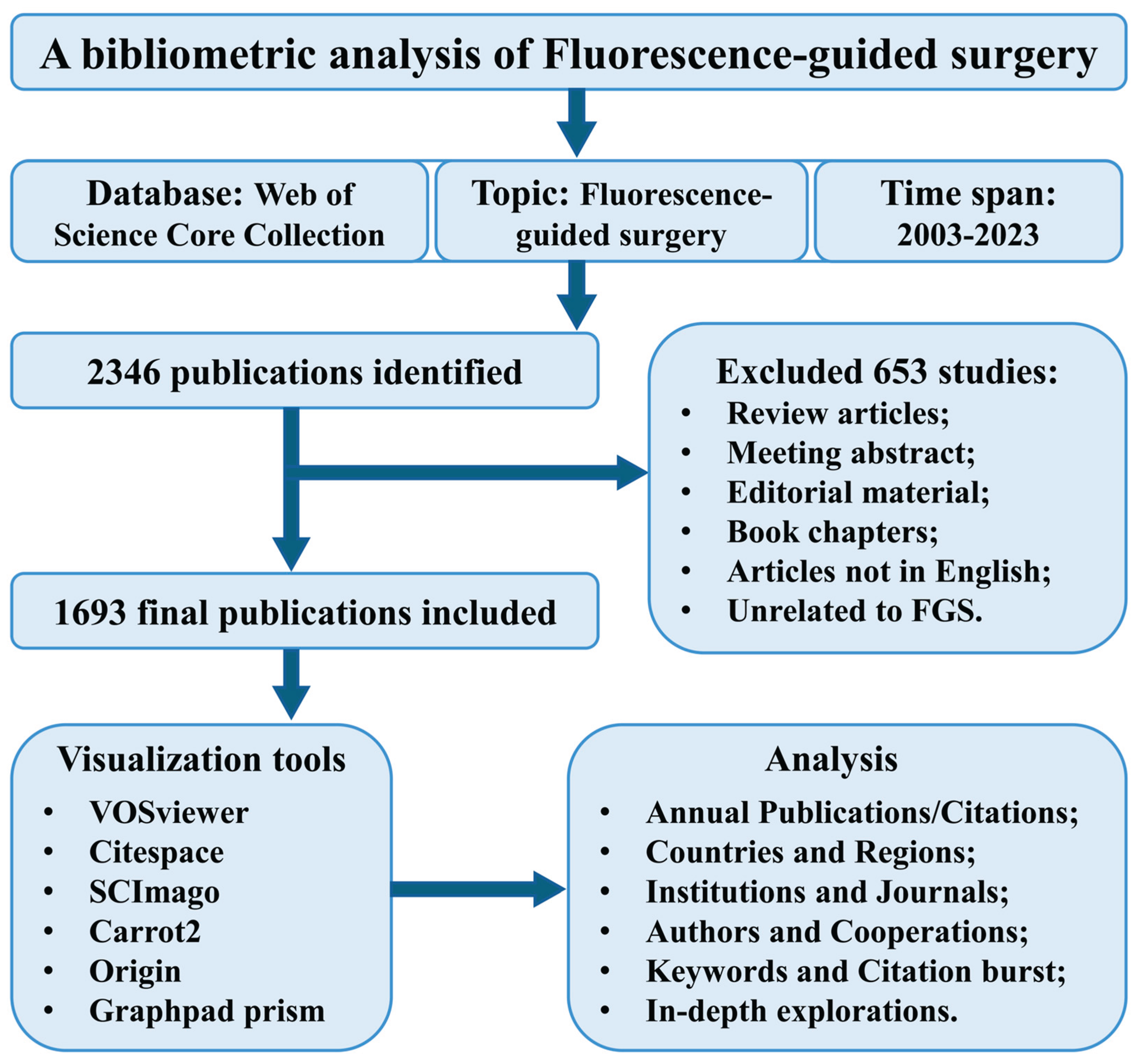
Figure 2.
Global publication trends and inter-country collaboration in the field of fluorescence-guided surgery. (A) Statistical chart of annual publications and citations about FGS research. (B) Area stacking diagram of annual publications in the top 10 countries. (C) Global collaboration in FGS research among countries. Different colors represent different clusters. (D, E) Global collaboration in FGS research among countries from 2003 to 2013 and 2014-2023, respectively. The thickness of the lines between nodes indicates the strength of cooperation between respective countries.
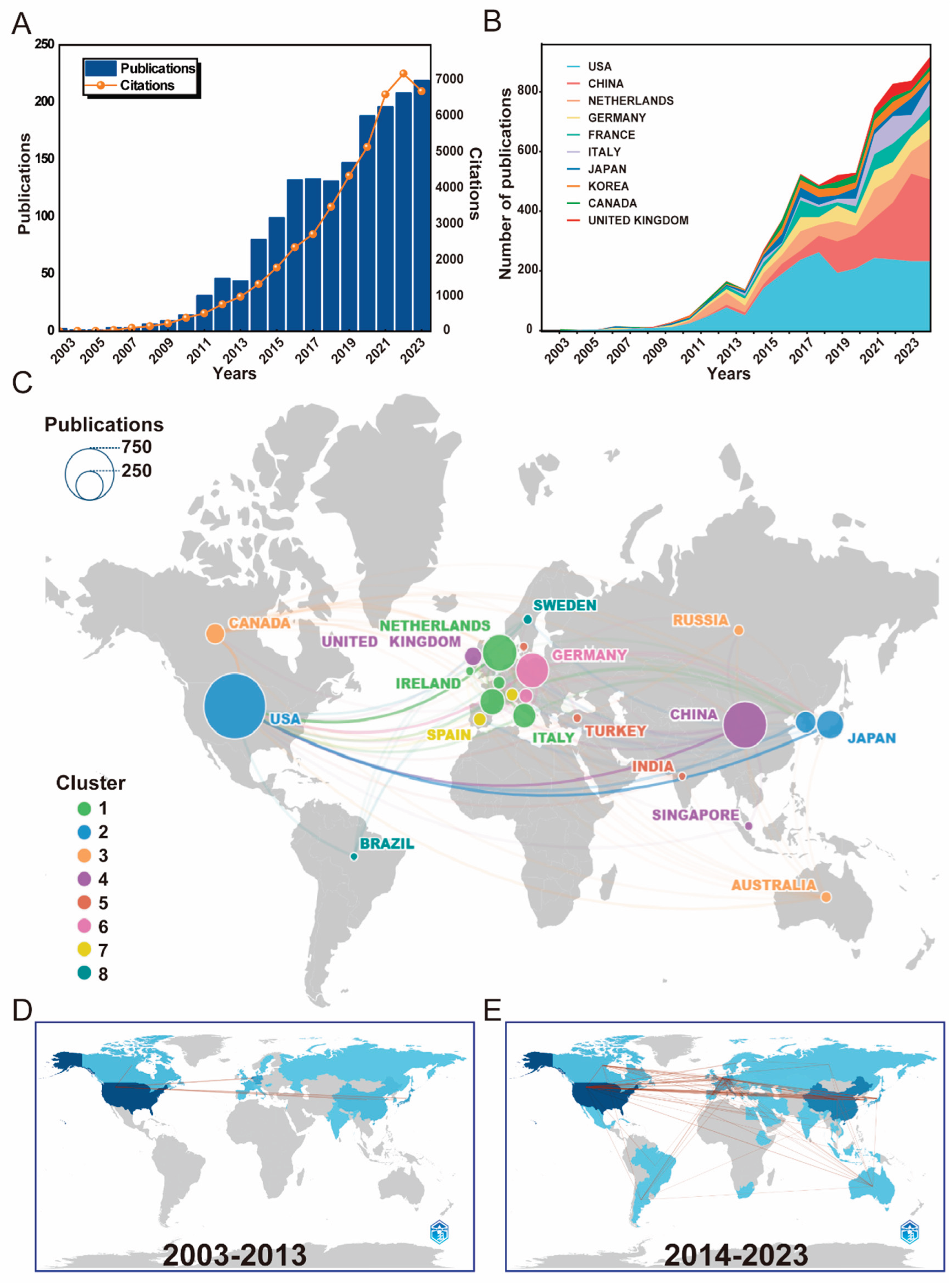
Figure 3.
Most Related Institutions of FGS research. (A)The co-occurrence network map of institutions from 2003-2023 in the FGS field. The size of each circular node corresponds to the number of papers published by the respective institution. The connections between nodes represent cooperative links among different institutions. Additionally, the concentric circles around the nodes indicate the trend of publication over time, with more scattered yellow appearances indicating recently published articles. (B) Statistical chart of the top 20 institutions in terms of publication volume. (C) The three-field plot of the top 10 institutions with countries and authors.
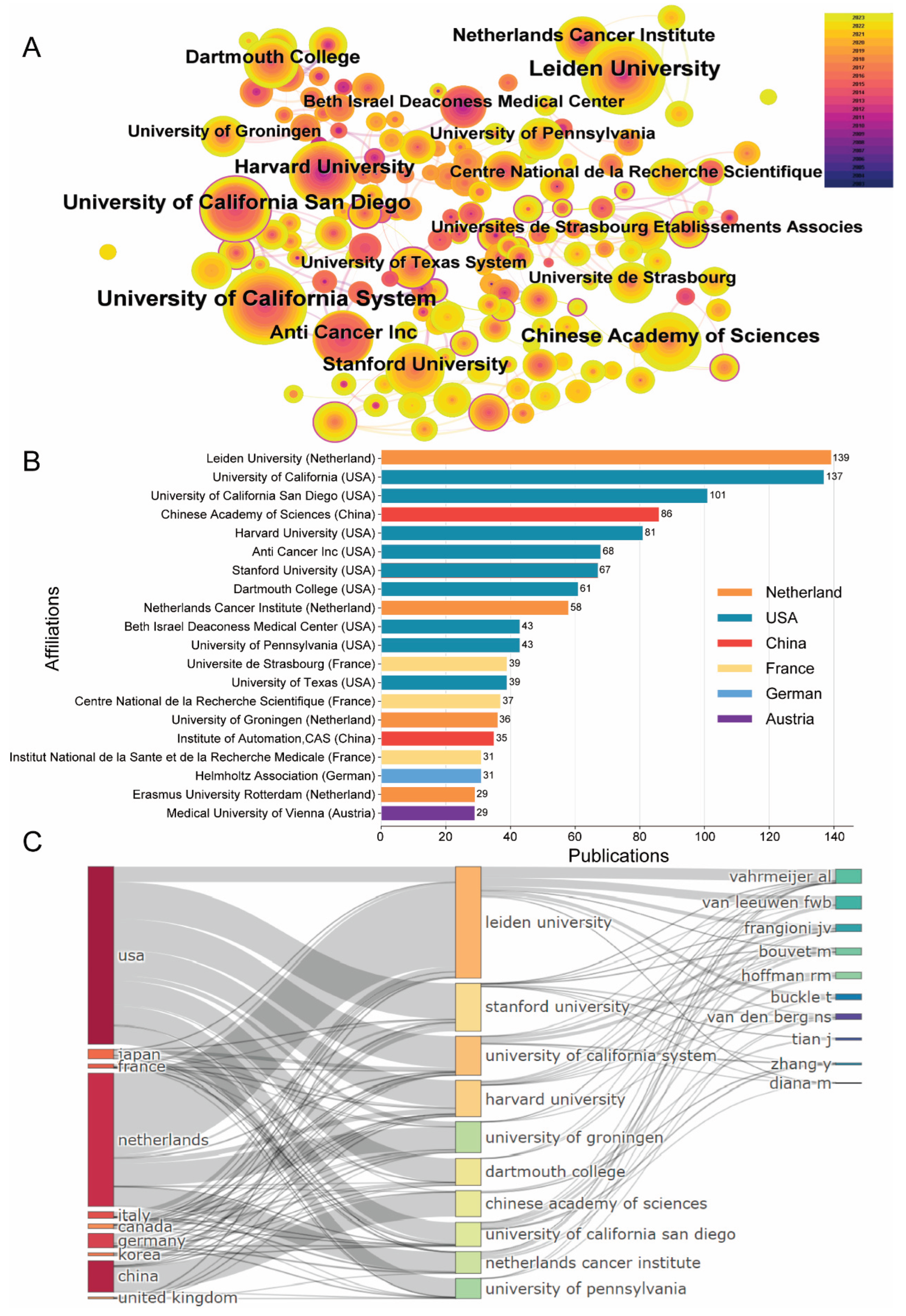
Figure 4.
Most Contributing authors and collaborative networks. (A) Cluster analysis of co-authorship was conducted using the VOSviewer software. Each color represents a cluster. The node size indicates the contribution of the author within the research field. (B) The graphic of the top 20 authors' production over time from 2008 to 2023.
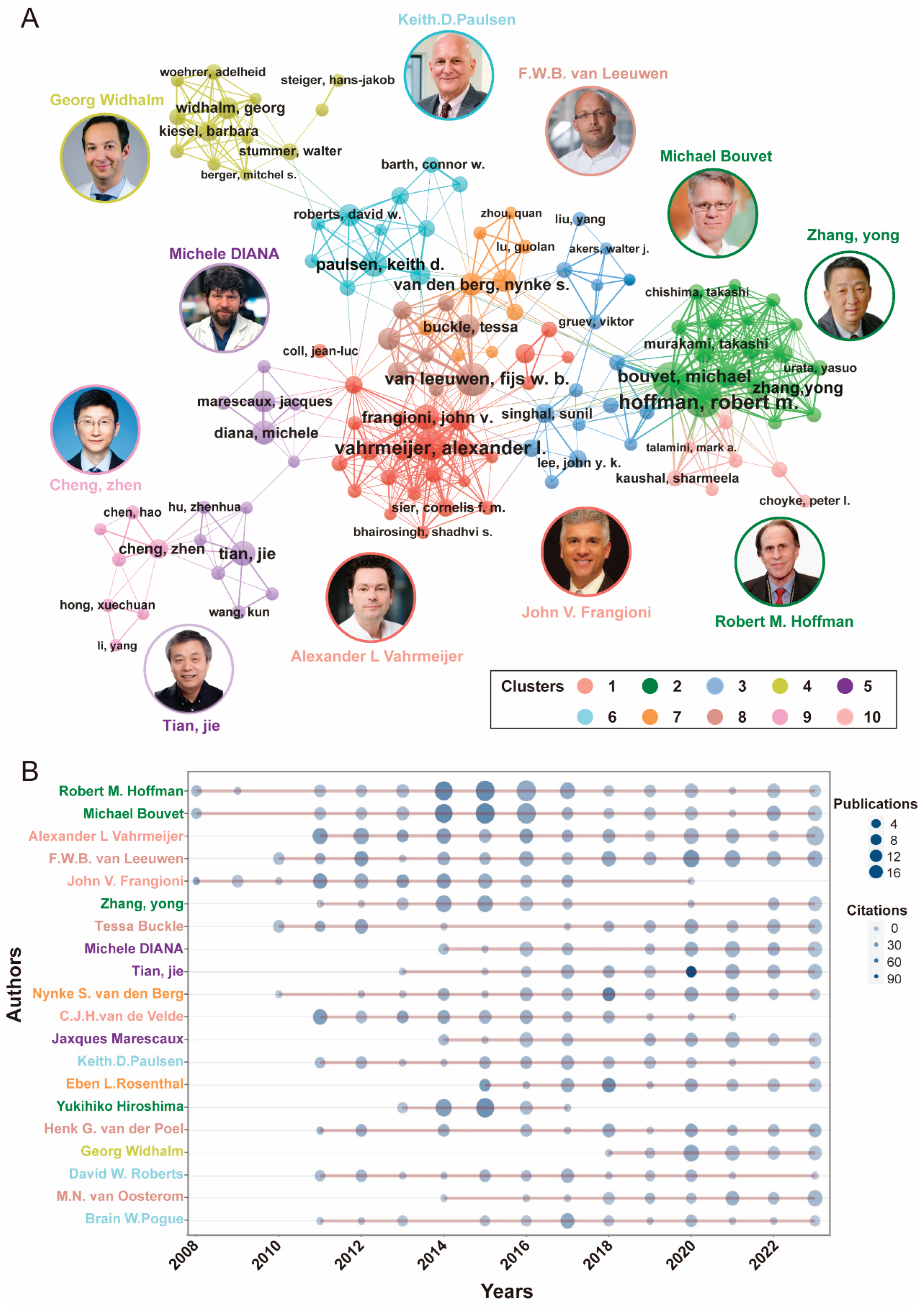
Figure 5.
Most relevant Journals in the FGS research field. (A)The top 20 most productive journals in FGS research. (B) The top 20 most cited journals in FGS research. (C) The graphical analysis of Top 10 Journal production over time in the FGS research field. (D) The timeline-based co-occurrence network of journals.
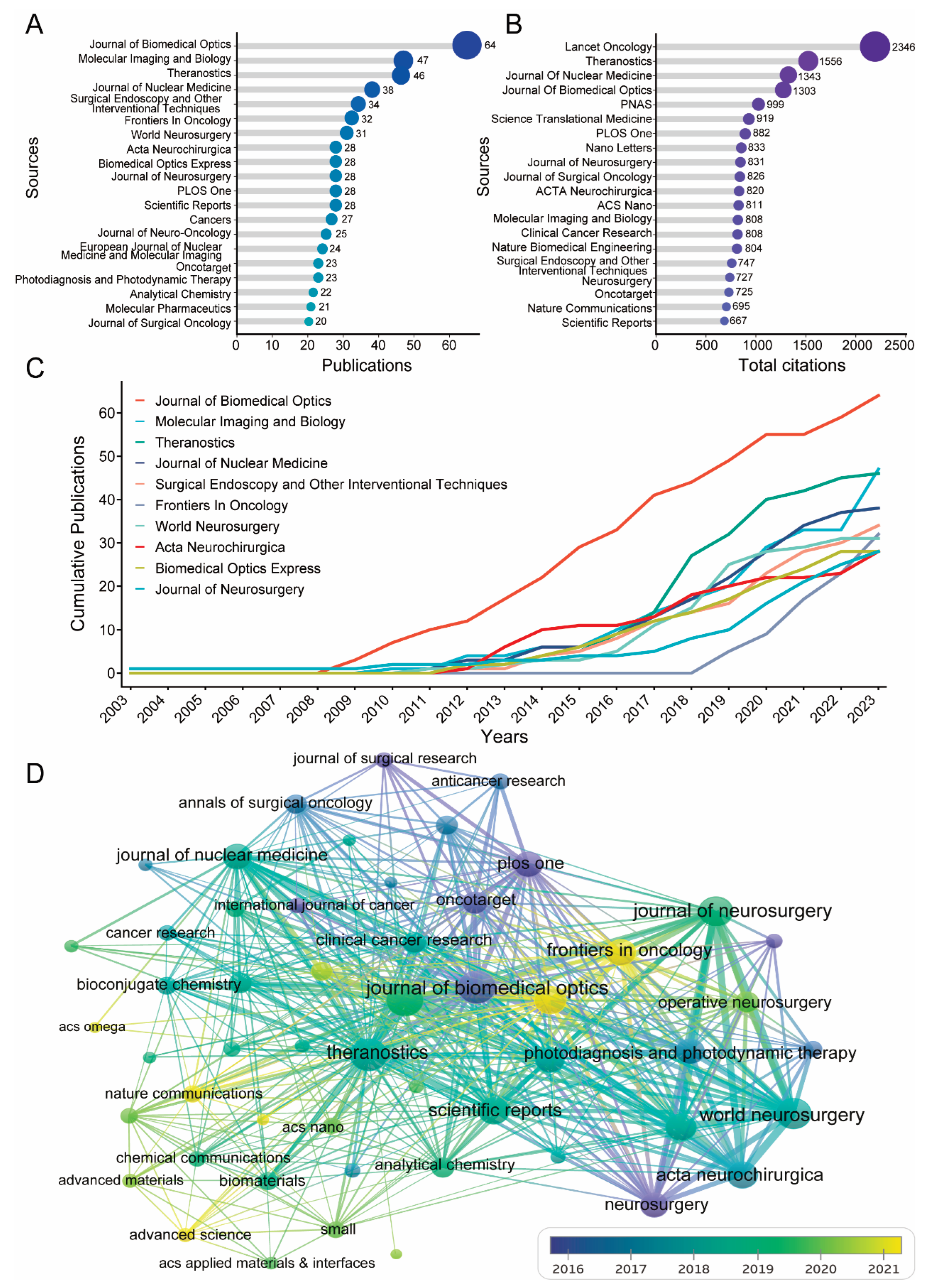
Figure 6.
Research hotspots analyzing through keywords and bursts analyses from 2003 to 2023. (A) Network visualization map of keywords started from 2003 to 2023. (B) 18 keyword clusters were categorized by CiteSpace software. (C) The graphic of the Top 20 keywords with the strongest citation bursts. (D) The timeline map of 6 keyword clusters.
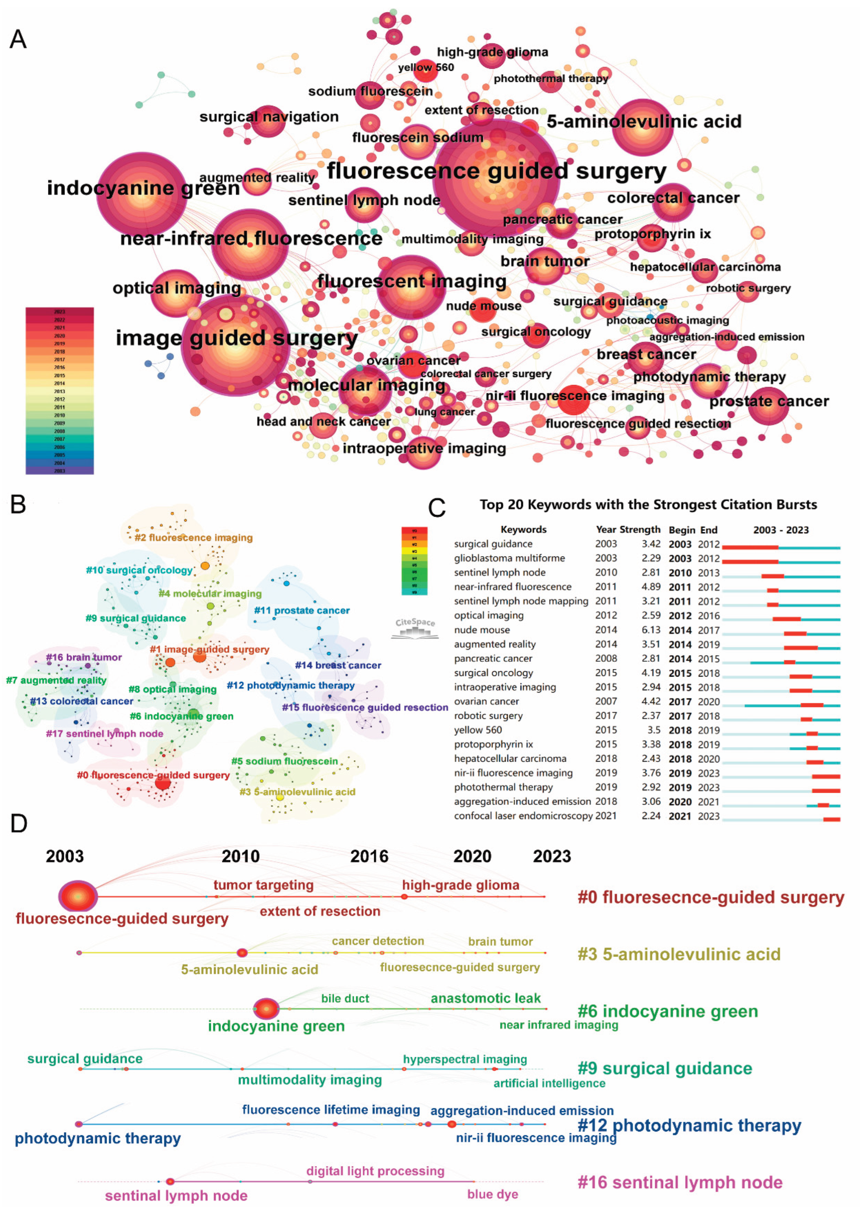
Figure 7.
In-depth Exploration of NIR-II Imaging in the FGS Research Field. (A) Hotspot Foam-Tree Analysis of FGS Field from Carrot2. The dotted red lines represent NIR-II imaging. Each foam depicted a certain field, with foam size reflecting the number of publications. (B) Top 5 most cited articles and their authors and publication years of NIR-II topic in FGS field. (C) Time-dependent trend topic analysis of NIR-II topic in FGS field. (D) Interaction network analysis of countries contributing to literature in the field of FGS in NIR-II. (E) The graphic of co-author network analysis.
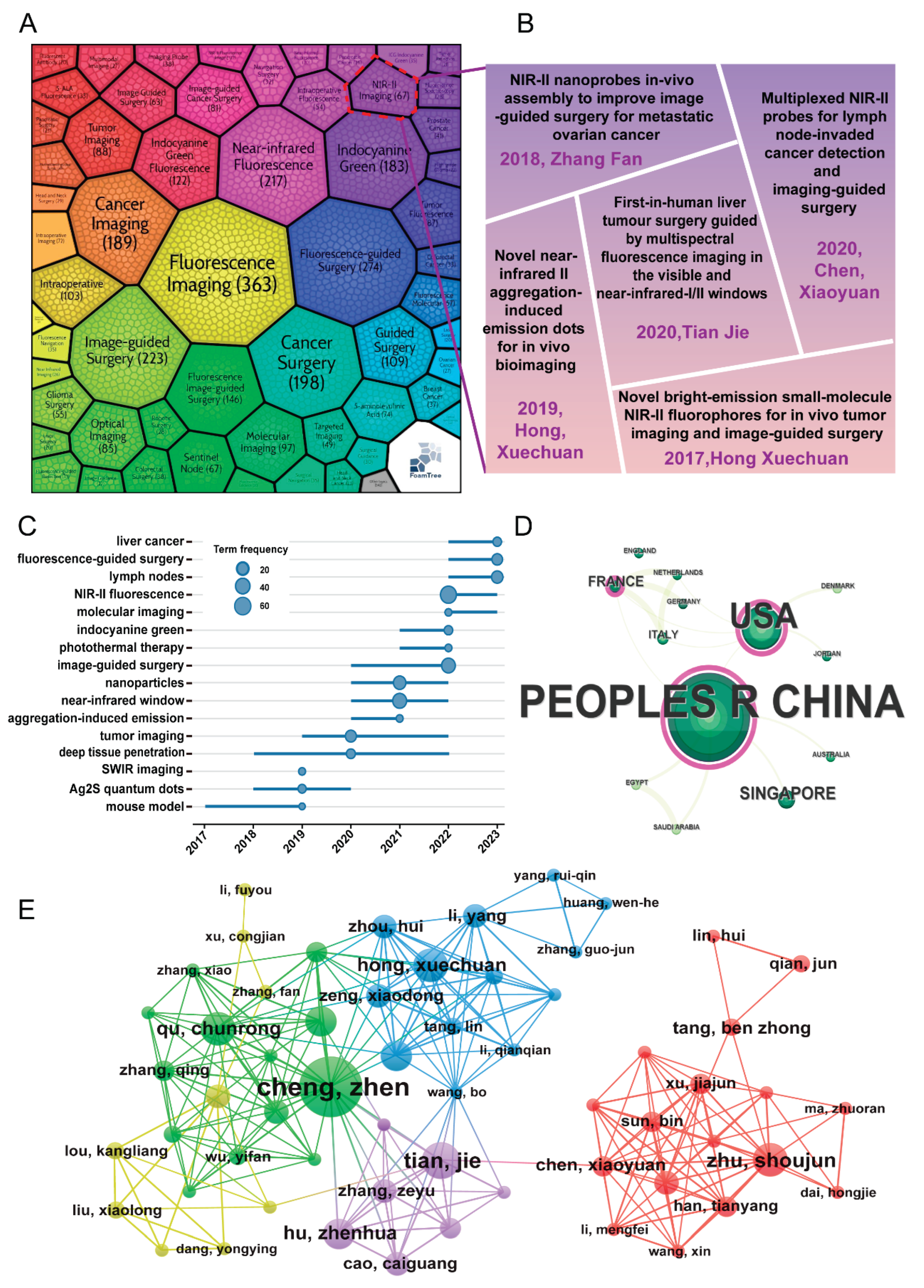
Table 1.
Top 20 most relevant Countries.
| Rank | Country | Articles | Freq | MCP Ratio1 | Total Citations |
Average article citations |
|---|---|---|---|---|---|---|
| 1 | USA | 586 | 0.34389671 | 0.34300341 | 17986 | 30.60 |
| 2 | China | 313 | 0.18368545 | 0.16613419 | 7793 | 24.80 |
| 3 | Netherlands | 176 | 0.10328638 | 0.55113636 | 5855 | 33.30 |
| 4 | Germany | 122 | 0.07159624 | 0.2704918 | 4719 | 41.40 |
| 5 | France | 79 | 0.0463615 | 0.36708861 | 1354 | 18.30 |
| 6 | Italy | 66 | 0.03873239 | 0.27272727 | 1286 | 19.20 |
| 7 | Japan | 58 | 0.03403756 | 0.12068966 | 1511 | 26.10 |
| 8 | Korea | 48 | 0.02816901 | 0.27083333 | 788 | 16.40 |
| 9 | United Kingdom | 35 | 0.02053991 | 0.2 | 483 | 13.80 |
| 10 | Canada | 34 | 0.01995305 | 0.38235294 | 778 | 22.90 |
| 11 | Austria | 26 | 0.01525822 | 0.61538462 | 215 | 8.30 |
| 12 | Spain | 19 | 0.01115023 | 0.15789474 | 393 | 26.20 |
| 13 | Belgium | 14 | 0.00821596 | 0.42857143 | 274 | 19.60 |
| 14 | Denmark | 13 | 0.00762911 | 0.07692308 | 225 | 17.30 |
| 15 | Ireland | 13 | 0.00762911 | 0.23076923 | 136 | 10.50 |
| 16 | Switzerland | 12 | 0.00704225 | 0.33333333 | 252 | 21.00 |
| 17 | RUSSIA | 10 | 0.00586854 | 0.2 | 133 | 13.30 |
| 18 | Sweden | 8 | 0.00469484 | 0 | 100 | 12.50 |
| 19 | Turkey | 7 | 0.00410798 | 0.28571429 | 101 | 14.40 |
| 20 | Australia | 6 | 0.00352113 | 0.66666667 | 31 | 5.20 |
1
Table 4.
Top 10 most cited articles.
| Rank | Year | Source | First Author | Paper Title | TC1 | TC per Year | Normalized TC |
|---|---|---|---|---|---|---|---|
| 1 | 2006 | Lancet Oncol | Walter Stumme | Fluorescence-guided surgery with 5-aminolevulinic acid for resection of malignant glioma: a randomised controlled multicentre phase III trial | 2344 | 123.37 | 2.13 |
| 2 | 2006 | Nano Lett | Weibo Cai | Peptide-Labeled Near-Infrared Quantum Dots for Imaging Tumor Vasculature in Living Subjects | 807 | 42.47 | 0.73 |
| 3 | 2020 | Nat Biomed Eng | Zhenhua Hu | First-in-human liver-tumour surgery guided by multispectral fluorescence imaging in the visible and near-infrared-I/II windows | 527 | 105.40 | 21.46 |
| 4 | 2014 | Sci Transl Med | Evan Phillips | Clinical translation of an ultrasmall inorganic optical-PET imaging nanoparticle probe | 527 | 47.91 | 10.70 |
| 5 | 2010 | PNAS | Emilia S Olson | Activatable cell penetrating peptides linked to nanoparticles as dual probes for in vivo fluorescence and MR imaging of proteases | 462 | 30.80 | 5.44 |
| 6 | 2018 | Nat Commun | Peiyuan Wang | NIR-II nanoprobes in-vivo assembly to improve image-guided surgery for metastatic ovarian cancer | 319 | 45.57 | 9.74 |
| 7 | 2015 | Clin Cancer Res | Eben L Rosenthal | Safety and Tumor Specificity of Cetuximab-IRDye800 for Surgical Navigation in Head and Neck Cancer | 317 | 31.70 | 8.14 |
| 8 | 2011 | Eur Urol | Henk G van der Poel | Intraoperative laparoscopic fluorescence guidance to the sentinel lymph node in prostate cancer patients: clinical proof of concept of an integrated functional imaging approach using a multimodal tracer | 257 | 18.36 | 4.23 |
| 9 | 2009 | J Surg Oncol | Kunihito Gotoh | A novel image-guided surgery of hepatocellular carcinoma by indocyanine green fluorescence imaging navigation | 241 | 15.06 | 3.30 |
| 10 | 2013 | Cancer | Joost R van der Vorst | Near-infrared fluorescence-guided resection of colorectal liver metastases | 224 | 18.67 | 4.75 |
1TC=total citations.
Disclaimer/Publisher’s Note: The statements, opinions and data contained in all publications are solely those of the individual author(s) and contributor(s) and not of MDPI and/or the editor(s). MDPI and/or the editor(s) disclaim responsibility for any injury to people or property resulting from any ideas, methods, instructions or products referred to in the content. |
© 2025 by the authors. Licensee MDPI, Basel, Switzerland. This article is an open access article distributed under the terms and conditions of the Creative Commons Attribution (CC BY) license (http://creativecommons.org/licenses/by/4.0/).
Copyright: This open access article is published under a Creative Commons CC BY 4.0 license, which permit the free download, distribution, and reuse, provided that the author and preprint are cited in any reuse.


