Preprint
Article

This version is not peer-reviewed.

The Assessment of Sentinel Lymph Node Mapping Methods in Endometrial Cancer

A peer-reviewed article of this preprint also exists.

Submitted:

20 December 2024

Posted:

23 December 2024

You are already at the latest version

Abstract

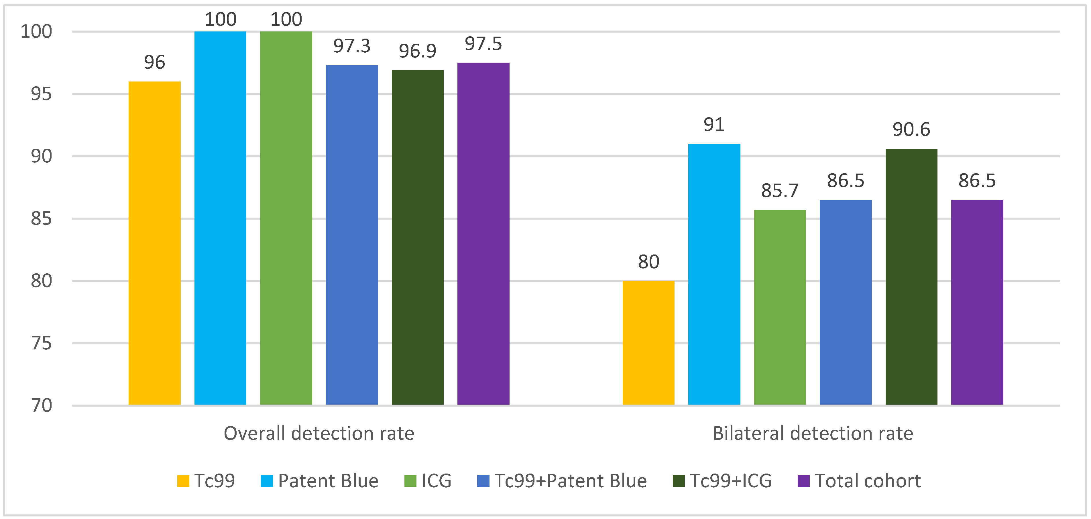
Background/Objectives: Methods: This retrospective cohort study included 119 patients with early-stage EC treated at the Maria Skłodowska-Curie National Research Institute of Oncology between 2016 and 2021. Patients underwent SLNB using technetium-99m (Tc99m), indocyanine green (ICG), Patent Blue, or combinations of these tracers. Detection rates for unilateral and bilateral SLNs and the accuracy of metastasis identification were analyzed. Results: The overall SLN detection rate was 97.5%. Detection rates for individual tracers were 100% for ICG, 100% for Patent Blue, and 96% for Tc99m. Combining tracers achieved detection rates of 96.9% (Tc99m + ICG) and 97.3% (Tc99m + Patent Blue). Bilateral detection was highest with Tc99m + ICG (90.6%) and Patent Blue alone (91%). Metastases were identified in 12% of cases, with combined methods improving metastatic detection. No "empty nodes" were observed with Tc99m, compared to 1.7% with Patent Blue and 0.8% with ICG. Conclusions: While combining Tc99m with dyes did not significantly improve overall detection rates, it enhanced metastasis identification and reduced false-negative results. The findings suggest that combined tracer methods can optimize SLNB accuracy in endometrial cancer. Prospective studies are warranted to validate these results.

Keywords: 
;  ;  ;  ;  

1. Introduction

Endometrial cancer (EC) is one of the three most common malignant tumors of the reproductive system in women in developed countries [1]. In recent years, the incidence of EC has increased, making it the most common gynecological cancer. This rise can be attributed to risk factors such as obesity, diabetes, prolonged exposure to unopposed estrogen, and the aging population.
The standard treatment for early-stage EC remains total hysterectomy with bilateral salpingo-oophorectomy and pelvic or para-aortic lymphadenectomy [2]. Lymph node status plays a crucial role in determining prognosis and guiding decisions regarding adjuvant therapy. However, routine lymphadenectomy is controversial due to the associated risks of complications such as vascular and nerve injury, lymphedema, and lymphocyst formation [3]. These risks are particularly significant in obese patients and those with comorbidities, where extended surgical procedures may lead to increased perioperative morbidity and prolonged recovery time.
Sentinel lymph node biopsy (SLNB) is a minimally invasive alternative to traditional lymphadenectomy, enabling the detection of lymph node metastases while reducing surgical morbidity [4,5]. SLNB involves identifying and removing the first lymph node (sentinel node) to which lymph from the primary tumor drains, thus enabling precise staging [6]. The effectiveness of this procedure relies on accurate SLN identification, achieving bilateral detection, and minimizing false-negative rates.
Comparative studies have demonstrated that SLNB is an effective and safe method for determining lymph node status in EC, with results comparable to full lymphadenectomy [7,8]. For SLNB to replace traditional lymphadenectomy as the standard of care, it is essential to achieve high bilateral detection rates and ensure that removed nodes contain true lymphatic tissue rather than fatty tissue alone.
Currently, three main techniques are used to identify SLNs: radioisotopic tracers (e.g., Tc99m), dyes (Patent Blue), and fluorescent optical tracers (e.g., indocyanine green, ICG). Each method has its strengths and limitations. Tc99m offers deep tissue penetration and allows preoperative localization via SPECT/CT imaging. Patent Blue is a simple and cost-effective technique that does not require specialized equipment. In contrast, ICG provides excellent optical resolution with near-infrared fluorescence, making it particularly effective in obese patients and those with altered anatomy. However, each technique has inherent limitations: Tc99m requires access to nuclear medicine facilities, Patent Blue can cause allergic reactions, and ICG has a short retention time in lymphatic channels, necessitating precise timing of the procedure. Combining these methods has been proposed to enhance detection rates and overcome individual limitations, but the optimal combination remains a subject of ongoing investigation.
Despite extensive research, the optimal method for SLN detection in EC is still the focus of active inquiry. Technical challenges, such as “empty nodes,” dye diffusion, and lymphatic channel obstruction due to tumor emboli, underscore the need for continued procedural refinements [9,10,11,12]. Combining techniques, such as Tc99m with ICG or Tc99m with Patent Blue, may enhance both SLN detection rates and the accuracy of metastasis identification.
The introduction of pathological ultrastaging has further improved the ability to detect micrometastases and isolated tumor cells (ITCs) in SLNs, which may have significant prognostic implications [13]. Ultrastaging involves detailed histopathological evaluation, including serial sectioning and immunohistochemistry, increasing the likelihood of detecting low-volume metastases.
The aim of this study was to compare the effectiveness of different SLN detection methods (Tc99m, ICG, Patent Blue) and to evaluate the impact of combining these tracers on overall detection rates, sample quality, and metastasis identification in patients with early-stage EC.

2. Materials and Methods

2.1. Study Design and Setting

A retrospective cohort study was conducted at the Maria Skłodowska-Curie National Research Institute of Oncology (NIO-PIB), Kraków Branch. The study included patients with endometrial cancer (EC) who underwent surgical treatment along with the sentinel lymph node biopsy (SLNB) procedure between 2016 and 2021.

2.2. Inclusion and Exclusion Criteria

Patients meeting the following criteria were included in the study:
  • Clinically early-stage disease (FIGO stage I-II, 2009 classification) [14,15].
  • No prior neoadjuvant therapy (chemotherapy or radiotherapy).
  • Availability of complete clinical and histopathological data.
Exclusion criteria included:
  • Advanced-stage disease (FIGO stage III-IV).
  • Allergic reactions to tracers (ICG, Patent Blue).
  • Age below 18 years or above 85 years.
  • Comorbidities preventing surgical intervention.

2.3. Sentinel Lymph Node Identification Procedure

Each patient underwent surgical staging of endometrial cancer, including total hysterectomy with bilateral salpingo-oophorectomy and sentinel lymph node identification using the following methods:

2.3.1. Radioactive Tracer Administration (Tc99m):

  • Tracer: Technetium-99m-labeled human albumin colloid (NanoColl, GE Healthcare).
  • Technique: A dose of 1 mCi (60 MBq) Tc99m was injected into the cervix at the 3 and 9 o’clock positions. Half the dose was administered superficially (2–3 mm) and half deeply (10–15 mm) using thin-walled needles (21G).
  • Preoperative Imaging: Static scintigraphy and SPECT-CT imaging were performed at 5 minutes, 60 minutes, and 18 hours after tracer administration, using AnyScan Mediso equipment.

2.3.2. Dye Administration:

  • Indocyanine Green (ICG): 0.5 ml (1.250 mg ICG) diluted in 5 ml of water was injected into the cervix at the same locations.
  • Patent Blue: 2 ml of dye (1 ml per injection site) was administered.
  • Timing: Dyes were injected 15–30 minutes before the start of surgery.

2.3.3. Sentinel Lymph Node Identification:

  • Surgical Technique: Sentinel lymph node identification was performed laparoscopically using:
  • A gamma probe Gamma Finder 2 Word of medicine to localize Tc99m.
  • The VS3 Iridium laparoscopic system (Visionsense 3DHD & IR Fluorescence V) for ICG fluorescence visualization and Patent Blue color channels.
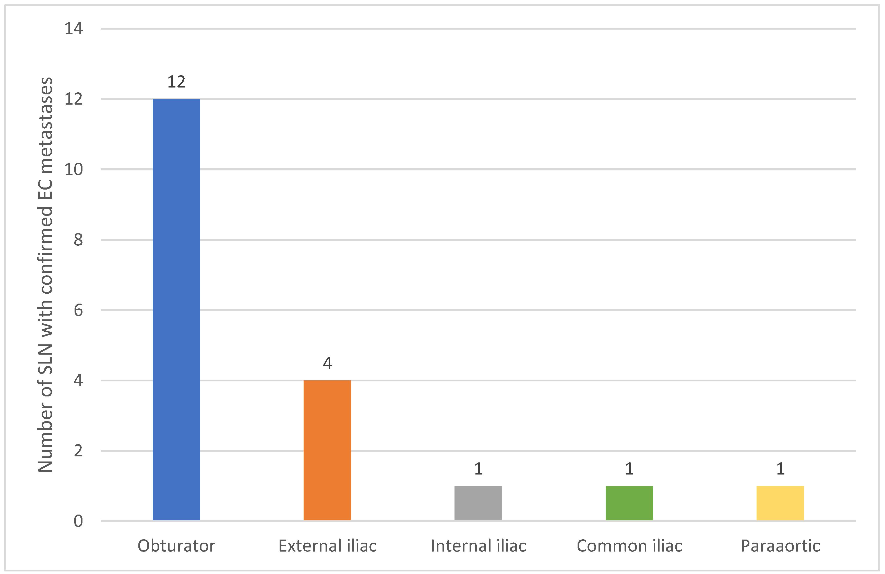
  • Anatomical Classification: Retrieved sentinel lymph nodes were anatomically classified as:
  • Obturator, external iliac, internal iliac, common iliac, and para-aortic.
  • Verification: Each excised sentinel lymph node was double-checked ex vivo using a gamma probe to confirm the presence of Tc99m.

2.4. Histopathological Examination

All excised lymph nodes were fixed in 10% formalin solution and subjected to detailed histopathological analysis:
  • Lymph Node Sections: Sections were cut at 2 mm thickness.
  • Staining: Hematoxylin and eosin (H&E) staining and immunohistochemistry (CK – cytokeratins) were performed.
  • Definitions:
    -
    Macrometastases: Lesions >2 mm.
    -
    Micrometastases: Lesions 0.2–2 mm in diameter.
    -
    Isolated Tumor Cells (ITC): Lesions ≤0.2 mm.

2.5. Statistical Analysis

Data were analyzed using IBM SPSS Statistics v25.0 software. Detection rates were compared using the chi-square test for qualitative variables and the Student’s t-test for continuous variables. A p-value <0.05 was considered statistically significant.

2.6. SLN Evaluation Parameters

  • Detection Rate (DR): The percentage of patients in whom at least one sentinel lymph node (SLN) was identified.
  • Bilateral Detection Rate (BDR): The percentage of patients with SLN detected on both sides.
  • Sensitivity: The ratio of true positive results to the number of patients with metastases.

3. Results

3.1. Patient Characteristics

The study cohort included 119 patients with early-stage endometrial cancer. The median age was 60.8 years (range: 38–85). Clinical and pathological characteristics are summarized in Table 1. Most patients had endometrioid histology (95%), with less common subtypes including serous (2.5%) and clear cell carcinoma (2.5%). Lymphovascular space invasion (LVSI) was identified in 12 patients (10%), while myometrial invasion was noted in 36% of cases with >50% involvement.

3.2. Sentinel Lymph Node Detection

The overall SLN detection rate was 97.5% across the entire cohort. Bilateral detection was achieved in 86.5% of cases. Detailed detection rates for each method are presented in Table 2 and Figure 1.

3.3. Metastases and Sample Quality

Metastases to SLNs were identified in 14 patients (12%), including 8 cases of unilateral metastases (57%), 5 cases of bilateral metastases (36%), and 1 case of isolated metastasis to a para-aortic node (7%) (Table 3 and Figure 2).
Among the metastases:
  • 9 were macrometastases,
  • 2 were micrometastases,
  • 3 were isolated tumor cells.
Detection of metastases using a single identification method occurred in 6 patients: 5 cases with Tc99m and 1 with Patent Blue. In 8 additional cases, combined methods were used. The average number of SLNs excised per patient was 2. SLN samples identified with Tc99m showed no “empty nodes” (0%), compared to 1.7% with Patent Blue and 0.8% with ICG.
In the conducted study, the overall detection rate of sentinel lymph nodes (SLN) was 97.5% across the entire patient cohort. Analysing the individual methods, SLN detection rates were as follows: 100% for ICG, 100% for Patent Blue, 96% for Tc99m, 97.3% for the combination of Patent Blue + Tc99m, and 96.9% for the combination of ICG + Tc99m. The total bilateral SLN detection rate was 86.5%. The individual bilateral detection rates were 85.7% for ICG, 91% for Patent Blue, 80% for Tc99m, 90.6% for the combination of ICG + Tc99m, and 86.5% for the combination of Patent Blue + Tc99m. These results align with data available in the literature regarding both bilateral and unilateral detection of sentinel lymph nodes.
In the final histopathological examination, metastases to sentinel lymph nodes were found in 14 patients, representing 12% of the entire study group. Among these patients, 8 (57%) had unilateral metastases, 5 (36%) had bilateral metastases, and 1 patient (7%) had an isolated metastasis to a para-aortic node.
Among the metastases, 9 were macrometastases, 2 were micrometastases, and in 3 cases, isolated tumor cells were detected. Detection of metastases in sentinel lymph nodes using a single identification method was achieved in 6 patients: 5 cases of metastases were identified using Tc99m, and 1 case using Patent Blue. For the remaining 8 patients with metastases, combined identification methods were used. In this group, 6 patients received Tc99m and Patent Blue, while 2 patients received Tc99m and ICG. In two cases of the Patent Blue + Tc99m combination, SLNs were identified exclusively using Tc99m (Patent Blue did not stain the node in either of these cases).
In both cohorts, the average number of SLNs excised per patient was similar, with a mean of 2 nodes per patient. The quality assessment of SLN samples, particularly regarding the presence of lymphatic tissue, showed differences depending on the identification method. No “empty” nodes were noted in samples identified with Tc99m (0%). In samples detected using Patent Blue and ICG, the rate of nodes without lymphatic tissue was 1.7% (2 cases) and 0.8% (1 case), respectively.

4. Discussion

Our study compared the effectiveness of different sentinel lymph node (SLN) identification methods in early-stage endometrial cancer (EC). The results demonstrate that combining Tc99m with dyes such as indocyanine green (ICG) or Patent Blue does not significantly improve SLN detection rates compared to using dyes alone. However, this combination positively influenced the detection of metastases in SLNs while minimizing the issue of “empty nodes,” thereby enhancing the accuracy of the SLNB procedure.

4.1. SLN Identification Techniques

The cervical injection technique remains the most commonly described method in the literature due to its reproducibility and accessibility of the cervix, which rarely undergoes distortion in patients with EC [16,17]. Optimizing SLN detection methods involves not only identifying SLNs but also ensuring that they are the true first lymph nodes in the lymphatic drainage pathway and contain lymphatic tissue. The goal is to detect at least one SLN on each side and two SLNs in cases of bilateral mapping [18].

4.2. Detection Failures and the Issue of “Empty Nodes”

Failures in SLN detection using ICG remain a challenge. Sozzi et al. reported that bilateral SLN detection with ICG failed in 23.7% of cases, with risk factors including lymphovascular space invasion (LVSI), non-endometrioid histology, and enlarged lymph nodes [9]. In our study, LVSI did not influence the detection rate, which may reflect differences in technique or population characteristics.
ICG, despite its advantages, is associated with the issue of “empty nodes,” where nodes devoid of lymphatic tissue are mistakenly identified as SLNs. This phenomenon results from the albumin-binding properties of ICG, which increase oncotic pressure in lymphatic vessels, causing them to swell and be misinterpreted as lymph nodes. Studies estimate that 40% of SLN detection failures with ICG are due to the removal of empty nodes, with initial experiences showing rates as high as 20% during the first 25 procedures, decreasing to 7% with increasing surgical expertise [19]. Furthermore, ICG rapidly diffuses through the lymphatic system into second-tier nodes, complicating true SLN localization. A second injection improves bilateral detection rates to 96% [20]. I Khoury-Collado et al. emphasized the importance of the learning curve, reporting an increase in SLN detection rates from 77% to 94% after performing 30 procedures [21].

4.3. Strengths and Limitations of Individual Techniques

Patent Blue is a simple and cost-effective technique that does not require specialized equipment. The dye binds to serum proteins and migrates to SLNs within 5–10 minutes, staining them blue [22]. However, the SLN must be identified early in the procedure before the dye migrates further along the lymphatic vessels [21]. Additionally, severe allergic reactions, including anaphylaxis, have been reported in 0.7–1.9% of cases [23].
ICG provides deep tissue penetration and low autofluorescence, making it particularly effective in obese patients. The dye is generally safe, but it is contraindicated in patients with iodine allergies [22].
Tc99m, as a radioisotopic tracer, offers deep tissue penetration and longer retention time in SLNs [24]. In our study, the use of Tc99m eliminated the issue of empty nodes, which occurred in 1.7% of cases with Patent Blue and 0.8% with ICG. Another advantage of Tc99m is its compatibility with preoperative SPECT/CT imaging, which provides higher spatial resolution compared to planar imaging, enhancing SLN localization accuracy [25,26]. Papadia et al. reported that combining Tc99m with ICG achieved a detection rate of 96.9%, while combining Tc99m with Patent Blue reached 97.3%. However, ICG was superior in bilateral detection (84.1% vs 73.5%; p = 0.007) [27].

4.4. Histopathological Evaluation and Ultrastaging

The introduction of ultrastaging in SLN evaluation represents a significant advancement in the SLNB procedure. Ultrastaging involves detailed pathological assessment, including serial sectioning and immunohistochemistry, allowing for the detection of micrometastases and isolated tumor cells (ITCs). These findings have important prognostic implications and can influence therapeutic decisions [13]. In our study, combined SLN identification methods improved the detection of low-volume metastases, emphasizing the value of advanced histopathological analysis.

4.5. Study Limitations

Our study has several limitations. Its retrospective nature and relatively small sample size limit the statistical power of the findings. Additionally, variability in tracer injection techniques and operator experience may have influenced detection rates and overall SLN identification success.

5. Conclusions

The results of our study confirm that combining Tc99m with dyes such as ICG or Patent Blue does not significantly increase SLN detection rates compared to dyes alone. However, this combination improves metastasis detection and reduces the incidence of empty nodes. The use of a “long Tc99m protocol,” along with advanced histopathological techniques such as ultrastaging, can significantly enhance the accuracy of SLNB in EC patients. Further randomized prospective studies are needed to define the optimal SLN detection protocols in endometrial cancer.

Author Contributions

“Conceptualization, W.Sz., K.P., P.B. and A.Sz.; methodology, W.Sz., A.Sz., J.J., K.K., T.B.; software, W.Sz., K.P., K.M., M.J. and E.I.; validation, W.Sz, K.M., J.R. and T.B.; formal analysis, W.Sz., K.P., P.B., M.J., E.I., K.M., A.Sz., J.J., K.K., T.B. and J.R.; investigation, W.Sz., K.P., P.B., M.J., E.I., K.M., A.Sz., J.J., K.K., T.B. and J.R.; resources, W.Sz., P.B., K.K. and J.J; data curation, W.Sz., K.P..; writing—original draft preparation, W.Sz, P.B. and J.R.; writing—review and editing, W.Sz., K.P., P.B. and T.B.; supervision, P.B., J.R. and T.B.; project administration, W.Sz and K.P. All authors have read and agreed to the published version of the manuscript.”

Funding

“This research received no external funding”

Institutional Review Board Statement

The study was conducted in accordance with the Declaration of Helsinki, and approved by the Institutional Review Board of Maria Skłodowska-Curie National Research Institute of Oncology (NIO-PIB), Krakow Branch, Poland.

Informed Consent Statement

At the start of the treatment, written consent was obtained from all subjects involved in the study for the purpose of retrospective analysis of their medical data.

Conflicts of Interest

The authors declare no conflicts of interest the representation or interpretation of reported research results.

References

  1. Ferlay, J.; Colombet, M. Cancer incidence and mortality patterns in Europe: Estimates for 40 countries and 25 major cancers in 2018. Eur J Cancer. 2018 Nov;103:356-387. [CrossRef]
  2. Concin, N.; Matias-Guiu, X. ESGO/ESTRO/ESP guidelines for the management of patients with endometrial carcinoma. Int J Gynecol Cancer. 2021 Jan;31(1):12-39. [CrossRef]
  3. Volpi, L.; Sozzi, G. Long term complications following pelvic and para-aortic lymphadenectomy for endometrial cancer, incidence and potential risk factors: a single institution experience. Int J Gynecol Cancer. 2019 Feb;29(2):312-319. [CrossRef]
  4. Ballester, M.; Dubernard, G. Use of the sentinel node procedure to stage endometrial cancer. Ann Surg Oncol. 2008 May;15(5):1523-9. [CrossRef]
  5. Delpech, Y.; Cortez, A. The sentinel node concept in endometrial cancer: histopathologic validation by serial section and immunohistochemistry. Ann Oncol. 2007 Nov;18(11):1799-803. [CrossRef]
  6. Keshtgar, M.R.; Ell, P.J. Sentinel lymph node detection and imaging. Eur J Nucl Med. 1999 Jan;26(1):57-67. [CrossRef]
  7. Bodurtha Smith, A.J.; Fader, A.N. Sentinel lymph node assessment in endometrial cancer: a systematic review and meta-analysis. Am J Obstet Gynecol. 2017 May;216(5):459-476.e10. [CrossRef]
  8. Daraï, E.; Dubernard, Gnc. E. Sentinel node biopsy for the management of early stage endometrial cancer: long-term results of the SENTI-ENDO study. Gynecol Oncol. 2015 Jan;136(1):54-9. [CrossRef]
  9. Sozzi, G.; Fanfani, F. Laparoscopic sentinel node mapping with intracervical indocyanine green injection for endometrial cancer: the SENTIFAIL study - a multicentric analysis of predictors of failed mapping. Int J Gynecol Cancer. 2020 Nov;30(11):1713-1718. [CrossRef]
  10. Tortorella, L.; Casarin, J. Sentinel lymph node biopsy with cervical injection of indocyanine green in apparent early-stage endometrial cancer: predictors of unsuccessful mapping. Gynecol Oncol. 2019 Oct;155(1):34-38. [CrossRef]
  11. Taşkın, S.; Sarı, M.E. Risk factors for failure of sentinel lymph node mapping using indocyanine green/near-infrared fluorescent imaging in endometrial cancer. Arch Gynecol Obstet. 2019 Jun;299(6):1667-1672. [CrossRef]
  12. Tanner, E.J.; Sinno, A.K. Factors associated with successful bilateral sentinel lymph node mapping in endometrial cancer. Gynecol Oncol. 2015 Sep;138(3):542-7. [CrossRef]
  13. Altgassen, C.; Müller, N. Immunohistochemical workup of sentinel nodes in endometrial cancer improves diagnostic accuracy. Gynecol Oncol. 2009 Aug;114(2):284-7. [CrossRef]
  14. Amant, F.; Mirza, M.R. Cancer of the corpus uteri. Int J Gynaecol Obstet. 2018 Oct;143 Suppl 2:37-50. [CrossRef]
  15. Female Genital Tumours, WHO Classification of Tumours, 5th Edition, Volume 4. World Health Organization. 2020.
  16. Rocha, A.; Domínguez, A.M. Indocyanine green and infrared fluorescence in detection of sentinel lymph nodes in endometrial and cervical cancer staging - a systematic review. Eur J Obstet Gynecol Reprod Biol. 2016 Nov;206:213-219. [CrossRef]
  17. Abu-Rustum, N.R. Update on sentinel node mapping in uterine cancer: 10-year experience at Memorial Sloan-Kettering Cancer Center. J Obstet Gynaecol Res. 2014 Feb;40(2):327-34. [CrossRef]
  18. Kang, S.; Yoo, H.J. Sentinel lymph node biopsy in endometrial cancer: meta-analysis of 26 studies. Gynecol Oncol. 2011 Dec;123(3):522-7. [CrossRef]
  19. Body, N.; Grégoire, J. Tips and tricks to improve sentinel lymph node mapping with Indocyanin green in endometrial cancer. Gynecol Oncol. 2018 Aug;150(2):267-273. [CrossRef]
  20. Geppert, B.; Lönnerfors, C. A study on uterine lymphatic anatomy for standardization of pelvic sentinel lymph node detection in endometrial cancer. Gynecol Oncol. 2017 May;145(2):256-261. [CrossRef]
  21. Khoury-Collado, F.; Glaser, G.E. Improving sentinel lymph node detection rates in endometrial cancer: how many cases are needed? Gynecol Oncol. 2009 Dec;115(3):453-5. [CrossRef]
  22. Holloway, R.W.; Abu-Rustum, N.R. Sentinel lymph node mapping and staging in endometrial cancer: A Society of Gynecologic Oncology literature review with consensus recommendations. Gynecol Oncol. 2017 Aug;146(2):405-415. [CrossRef]
  23. Holub, Z.; Jabor, A. Laparoscopic detection of sentinel lymph nodes using blue dye in women with cervical and endometrial cancer. Med Sci Monit. 2004 Oct;10(10):CR587-91.
  24. Smith, B.; Backes, F. The role of sentinel lymph nodes in endometrial and cervical cancer. J Surg Oncol. 2015 Dec;112(7):753-60. [CrossRef]
  25. Vermeeren, L.; van der Ploeg, I.M. SPECT/CT for preoperative sentinel node localization. J Surg Oncol. 2010 Feb 1;101(2):184-90. [CrossRef]
  26. Keidar, Z.; Israel, O. SPECT/CT in tumor imaging: technical aspects and clinical applications. Semin Nucl Med. 2003 Jul;33(3):205-18. [CrossRef]
  27. Papadia, A.; Zapardiel, I. Sentinel lymph node mapping in patients with stage I endometrial carcinoma: a focus on bilateral mapping identification by comparing radiotracer Tc99m with blue dye versus indocyanine green fluorescent dye. J Cancer Res Clin Oncol. 2017 Mar;143(3):475-480. [CrossRef]
  28. Ballester, M.; Dubernard, G. Detection rate and diagnostic accuracy of sentinel-node biopsy in early stage endometrial cancer: a prospective multicentre study (SENTI-ENDO). Lancet Oncol. 2011 May;12(5):469-76. [CrossRef]
  29. Rossi, E.C., Kowalski, L.D. A comparison of sentinel lymph node biopsy to lymphadenectomy for endometrial cancer staging (FIRES trial): a multicentre, prospective, cohort study. Lancet Oncol. 2017 Mar;18(3):384-392. [CrossRef]
  30. Kessous, R.; How, J. Triple tracer (blue dye, indocyanine green, and Tc99) compared to double tracer (indocyanine green and Tc99) for sentinel lymph node detection in endometrial cancer: a prospective study with random assignment. Int J Gynecol Cancer. 2019 Sep;29(7):1121-1125. [CrossRef]
  31. Cabrera, S.; Bebia, V. Technetium-99m-indocyanine green versus technetium-99m-methylene blue for sentinel lymph node biopsy in early-stage endometrial cancer. Int J Gynecol Cancer. 2020 Mar;30(3):311-317. [CrossRef]
  32. How, J.; Gotlieb, W.H. Comparing indocyanine green, technetium, and blue dye for sentinel lymph node mapping in endometrial cancer. Gynecol Oncol. 2015 Jun;137(3):436-42. [CrossRef]
Figure 1. SLN Detection Rates by Method.
Preprints 143587 g001
Figure 2. Distribution of SLN Metastases.
Preprints 143587 g002
Table 1. Patient and Tumor Characteristics1.
Feature Characteristic N %
Histologic Type Endometrioid 113 95.0%
Serous 3 2.5%
Clear Cell 3 2.5%
Lymphovascular Space Invasion (LVSI) Present 12 10.0%
Absent 107 90.0%
Myometrial Invasion 0% 10 8.4%
<50% 66 55.5%
>50% 43 36.1%
Lymphadenectomy Bilateral Pelvic 20 16.8%
Paraaortic 7 5.9%
FIGO Stage IA 11 9.2%
IB 56 47.1%
II 31 26.1%
IIIA 3 2.5%
IIIB 2 1.7%
IIIC1 12 10.1%
IIIC2 4 3.3%
FIGO Grade G1 59 49.6%
G2 52 43.7%
G3 8 6.7%
Total 119 100.0%
1 Percentages may not sum to 100% due to rounding.
Table 2. Effectiveness of Sentinel Lymph Node (SLN) Detection1.
Parameter Tc99m (N=25) Blue Dye (N=11) ICG2 (N=14) Tc99m + Blue Dye (N=37) Tc99m + ICG2 (N=32) Total Cohort (N=119) p-value
Overall
Detection Rate
24 (96.0%) 11 (100.0%) 14 (100.0%) 36 (97.3%) 31 (96.9%) 116 (97.5%) 0.921
Bilateral
Detection Rate
20 (80.0%) 10 (91.0%) 12 (85.7%) 32 (86.5%) 29 (90.6%) 103 (86.5%) 0.815
Confirmed
Metastases
5 (25.0%) 1 (9.1%) 0 (0.0%) 6 (16.2%) 2 (6.3%) 14 (11.8%) 0.266
1 p-value < 0.05 was considered statistically significant. 2 ICG = indocyanine green.
Table 3. Location of Metastases in Sentinel Lymph Nodes1.
Lymph Node Location Unilateral Metastases (N=8) Bilateral Metastases (N=5) Isolated Metastasis (N=1)
Obturator 5 7 0
External Iliac 2 2 0
Internal Iliac 1 0 0
Common Iliac 0 1 0
Paraaortic 0 0 1
1 Some patients had metastases in more than one location. N represents the number of patients.
Disclaimer/Publisher’s Note: The statements, opinions and data contained in all publications are solely those of the individual author(s) and contributor(s) and not of MDPI and/or the editor(s). MDPI and/or the editor(s) disclaim responsibility for any injury to people or property resulting from any ideas, methods, instructions or products referred to in the content.
Copyright: This open access article is published under a Creative Commons CC BY 4.0 license, which permit the free download, distribution, and reuse, provided that the author and preprint are cited in any reuse.
Prerpints.org logo

Preprints.org is a free preprint server supported by MDPI in Basel, Switzerland.

Subscribe

Disclaimer

Terms of Use

Privacy Policy

Privacy Settings

© 2025 MDPI (Basel, Switzerland) unless otherwise stated