
Submitted:
13 June 2025
Posted:
16 June 2025
You are already at the latest version
Abstract
Affecting millions of individuals each year, chronic wounds place a substantial strain on both the healthcare system and healthcare providers, becoming a global health issue that requires a rapid and efficient solution. Unlike acute wounds that heal naturally without any external intervention, chronic wounds necessitate a proper medical treatment in order to promote the wound healing process and avoid any arising complications. However, the traditional therapeutic strategies are often limited when it comes to treating chronic wounds, reason why new approaches that facilitate the timely and effective healing of skin have been explored. Due to their unique properties, collagen-based hydrogels have been widely investigated as potential candidates for the management of chronic wound, owing to their good biocompatibility, high water retention capacity, which provides a moist microenvironment, and capacity to promote cell adhesion, proliferation, migration and differentiation for an optimal wound repair. In this context, the current paper discusses the recent advancements in collagen-based hydrogels as wound dressings, thus highlighting their potential as a future therapeutic approach for skin chronic wound care.
Keywords:
collagen‐based hydrogels
; wound healing
; skin
; wound dressings
; drug delivery systems
1. Introduction
As the largest organ of the human body, the skin serves as the primary physical barrier against various external invading pathogens, whilst also performing a plethora of vital functions, such as internal homeostasis regulation, excretion, external stimuli perception and immune regulation [1,2]. Positioned as the body’s outermost layer, the skin is more susceptible to a wide range of environmental factors, i.e. physical, biological and chemical, that can compromise its integrity. Thus, when damaged, a decline in the individual’s overall health may occur, highlighting the critical importance of maintaining the skin’s structural and functional integrity [3]. In response to injury, the outermost layer of the skin, namely epidermis, can initiate self-healing processes and regenerate normal skin structures, primarily due to the presence of resident stem cells. [4]. However, once the extent of the lesion surpasses the endogenous regenerative capacity of the skin, as seen in extensive full-thickness wounds or in individuals with a compromised innate regenerative capacity, the healing process becomes adequate and, without proper treatment, such injuries end up closing with great difficulty and may even aggravate, mainly due to infection and other associated complications [4,5].
With a high and constant increasing incidence, chronic wounds patients are estimated to account for approximatively 2-6% of the global population, with an average annual treatment cost that exceeds 20 billion dollars only in the United States alone [6], turning these cutaneous wounds into a major clinical problem that require an urgent solving.
The use of cutaneous dressings is of essence in wound healing, since they act as a temporary skin replacement, offering the injury protection against the external environment and bacterial infection whilst stopping the bleeding and absorbing the wound exudate [7,8]. However, the conventional dressings lack the essential anti-inflammatory and antibacterial properties required for an optimal wound healing. Additionally, they may adhere to the injury site and even impede the regenerative process [7]. In this light, it is imperative that novel multifunctional wound dressings are designed to accommodate all of the requirements for skin wound healing. In recent years, several important characteristics for an ideal wound dressing have been recognized: favourable biocompatibility with low immunogenicity and non-toxicity; good mechanical and physical durability and structural integrity; maintenance of a humid environment by long-lasting moisture preservation; excellent antibacterial properties; non-adherence to the wound site; ability to promote cell adhesion, proliferation and differentiation for an optimal cutaneous regeneration [9,10]. Encouraged by the “wet wound healing” theory, compared to other dressings, hydrogels (Figure 1) possess a significant advantage in the biomedical field, mainly due to their high water content which provides a moist three-dimensional microenvironment suitable not only for the cellular proliferation and migration, but also for the fast transfer and exchange of metabolites and nutrients, necessary for a rapid wound repair [11,12]. Moreover, in addition to their advantageous swelling capacity, hydrogels exhibit a favourable biocompatibility, adjustable physicochemical features and can be moulded into a varied selection of shapes and sizes as required. [13]. Likewise, by changing their composition and structure and by loading various active molecules, hydrogel dressings can be endowed with antioxidant, antibacterial and anti-inflammatory activities [8,14].
In recent years, a variety of natural and synthetic polymers were explored in the design and manufacturing process of advanced wound dressings [15,16,17,18]. Among these, collagen has gain significant attention as a particularly promising material for cutaneous regeneration. As a major constituent of the extracellular matrix (ECM), it is essential for preserving the structural integrity of the skin and regulating the various stages of wound healing, either in its fibrillar or soluble form [19]. Moreover, as a biomaterial, collagen exhibits excellent properties such as a good biocompatibility and biodegradability, reduced antigenicity, low toxicity, good in vivo adsorption and a superior synergism with other bioactive substances, reason why the incorporation of collagen into wound dressings has the potential to enhance the critical wound repairing process [19]. Furthermore, in the past few decades a wide array of collagen-based wound dressings, including hydrogels, with different compositions, have been clinically approved and commercialised. One example is the Integra® Dermal Regeneration Template, a membrane based on a layer of type I collagen crosslinked with chondroitin-6-sulfate and covered with a semipermeable silicone sheet used in the treatment of venous ulcers and combat-related wounds [20]. Other collagen-based dressings include FIBRACOL and CollaSorb with a composition of 90% collagen-10% alginate, and 90% pure collagen mixed with 10% calcium alginate, respectively, Wun’dres gel, a mixture of collagen-phenol-allantoin, Promogran with 55% collagen and 45% oxidized regenerated cellulose and Medifil – a 100% nonhydrolzed bovine type I-derived collagen in the natural triple-helical molecular form [21].
However, in spite of its outstanding properties and its already proven clinical efficiency as wound dressings, collagen exhibits a series of disadvantages such as thermal instability, enzymatic degradation and mechanical strength, which can be improved by using various methods, i.e. crosslinking, blending, grafting polymerization and covalent conjugating [22].
In light of this, the present review aims at summarising the recent advancements in the field of wound dressings with regards to the collagen-based hydrogels for cutaneous regeneration. It is important to note that in this review, we will confine our discussion only to studies in the field that have been published in the last 5 years, with an emphasis on data reported within the last two years in order not to overlap with previously published reviews that cover the same matter. Thus, the first part focuses on the natural wound healing process, the role of collagen in wound healing and the various sources of collagen, either natural or synthetic, while the second part is comprised of an overview of the collagen-based hydrogels, from methods of fabrication to in vitro and in vivo experiments based on collagen hydrogels for skin regeneration and wound healing. Lastly, the challenges, limitations and future research directions are also discussed.
2. Skin and Wound Healing Process
2.1. The Skin Structure
Skin is a sophisticated organ comprised of three distinct layers, namely the epidermis, dermis, and hypodermis. The outermost layer, the epidermis, is a non-vascular epithelial tissue measuring between 75 and 150 μm in thickness and while keratinocytes make up the majority of its cellular composition, it also contains melanocytes, Langerhans cells, and Merkel cells, each contributing to its diverse functions [23]. Accounting for approximatively 90% of the cells in the human epidermis, the keratinocytes play a crucial role in reducing water evaporation and protecting the skin against external traumas, while the melanocytes provide protection against the ultraviolet radiation through the production of melanin which determines skin tone [23]. In addition, the Langerhan cells combat bacteria, while the Merkel cells function as tactile and endocrine cells [23]. The next layer is represented by the dermis, a highly vascularised thick connective tissue (2 mm to 4 mm) located between the epidermis and the subcutaneous tissue, in which more complex cell types can be found [24]. The structural integrity of the cutaneous tissue is heavily impacted by its components, i.e. fibroblasts and the ECM components such as elastin, collagen, hyaluronic acid and glycosaminoglycans, which all together contribute to the skin’s tonicity and elasticity [24]. Moreover, it also contains blood and lymphatic vessels, receptors, macrophages, and various appendages such as hair follicles, sebaceous and sweat glands, characteristics which allow the involvement of dermis in the vascular, sensorial, and immune systems and connective tissue [1]. Finally, the subcutaneous tissue, or hypodermis, is a well-vascularised layer composed of adipocytes and collagen, with a primary role in thermoregulation, mechanical protection and dermal-skeletal attachment [25].
2.2. The Wound Healing Process and Type of Skin Wounds
Due to the constant and excessive exposure of skin to various internal and external factors such as physical, mechanical, chemical and other environmental risk factors, the anatomical and functional integrity of the cutaneous tissue can be physiologically disrupted, resulting in wounds [26], which based on their healing time are commonly categorized into acute and chronic wounds [15,27,28]. Resulting from traumas that damage the integrity of skin, acute lesions are considered to be a type of wound in which the tissue’s structure and function is recovered completely through a natural self-healing process, without the intervention of any external factors [29]. Normally, small burns, minor surgery wounds and minor cuts heal within 8-12 weeks, but the healing rate of acute wounds depends on two wound characteristics, namely the size, depth and location of the wound and most importantly on the individual’s overall condition, i.e. age or underlying disease [30].
The physiological wound repair process is a highly regulated and intricate sequence of events that can be divided into four overlapping stages, i.e. haemostasis, inflammation, proliferation, and remodelling, in which each phase involves intricate, dynamic interactions that contribute to tissue repair [27]. Following injury, the wound will enter the initial stage of the healing process, namely haemostasis, in which the blood flow is restricted through both vasoconstriction and cloth formation [28]. Once the blood cloth is formed, inflammatory cells (e.g. neutrophils and monocytes), under the action of cellular factors and locally secreted growth factors, will migrate to the site of injury and start clearing out any foreign bodies, bacteria, damaged endogenous tissue and reactive oxygen species (ROS) [31]. The purpose of the inflammatory stage is to prevent bacterial infection and to establish a microenvironment beneficial to wound healing process. To achieve this, the migrating monocytes will differentiate into macrophages, which, along with the resident macrophages, will secrete a range of inflammatory mediators, which help create a balanced inflammatory environment that supports tissue regeneration. Initially, in order to stimulate inflammation, the macrophages will differentiate into a classically activated M1 phenotype, secreting pro-inflammatory mediators (cytokines, chemokines), but as the process progresses, and the bacteria and dead tissue is removed, the macrophages will change their polarization state towards an anti-inflammatory phenotype (M2). The alternatively activated M2 macrophages will secrete anti-inflammatory cytokines and growth factors that help direct the process towards the next stage of wound healing, namely proliferation [27,32]. During this phase, epithelial cells, fibroblasts and keratinocytes migrate to the injury site and start filling it, resulting in the formation of a pale pink granulation tissue [28]. Additionally, new blood vessel also forms (angiogenesis) and fibroblasts start secreting collagen and other ECM components to promote re-epithelization. In the final stage, the myfibroblasts, which differentiated from the proliferating fibroblasts, will cover the site of injury, leading to scar formation [33]. During wound contraction, the granulation tissue will transform into the ECM, which, under the action of the matrix metalloproteinases (MMPs) will undergo a series of modifications that will lead to tissue restructuring and scar formation [33].
In healthy individuals, wound healing occurs properly and without interruption, but various pre-existing health conditions/pathologies may result in a disturbance in one of more of the aforementioned phases, leading to a slow and impaired healing process and chronic wound formation [34]. Chronic wounds are defined as wounds that fail to undergo through the normal healing process or restore the structural and functional integrity within a time frame of 3 months since initial injury [34]. The formation of chronic wounds involves a highly intricate process driven by various contributing factors, starting from an impaired blood flow, to peripheral vascular conditions, systemic illnesses, and infections. These wounds often lead to a significant loss of tissue that in more advanced cases, it may compromise deeper structures including nerves, joints and bones [34]. Compared to acute wounds, chronic wounds display a different phenotype as presented in Table 1.
2.3. The Role of Collgen in Skin Wound Healing
As an important component of the skin matrix, collagen plays a role in each of the wound healing stages. Thus, while not produced anew in the haemostatic and inflammatory stages, the exposed collagen fibres from damaged blood vessels trigger the clotting cascade, resulting in fibrin clot formation and prevention of further blood loss [8,36]. Type I collagen and type IV collagen fragments can serve as effective chemoattractants, helping to recruit immune cells (e.g. neutrophils) to the injury site by enhancing the phagocytosis and immunological responses and influencing gene expression [37]. Furthermore, data reported in literature showed that collagen can promote the polarization switch of macrophages towards an anti-inflammatory and pro-angiogenic phenotype via the activation of the microRNA signalling pathway [37]. In the proliferative phase, migrating fibroblasts begin synthesizing type III collagen, which acts as a temporary scaffold for the formation and organization of new cutaneous tissue. Moreover, research also indicates that the C-propeptide fragment of type I collagen can exert chemotactic effects on endothelial cells [37], thus promoting new blood vessel formation and tissue vascularization. As the process progresses, the collagen matrix substitutes the pre-existing fibronectin-rich matrix, and during the adult wound healing, type I collagen replaces type III collagen, leading in the end to the formation of the scarring tissue [26].
3. Collagen Structure, Sources and Extraction Methods
3.1. Collagen Structure
Accounting for almost one-third of the total protein mass in the human body, collagen is one of the most important ECM component, being responsible for the matrix’s durability and elasticity [38]. There are over 20 types of collagen in nature, and all of them display a hierarchical structure, that starts from the primary amino acid sequence and ends to its organization into complex fibres [38]. The unique structure of collagen consists of three polypeptide molecules, known as “α chains” positioned parallel to one another and coiled in a left-handed polyproline II-type helix (procollagen), that intertwine to create a right-handed triple helix structure referred to as tropocollagen [7,38,39]. The primary structure of collagen is represented by the specific sequence of amino acids in the polypeptide chains, and is characterised by the presence of a repeating glycine (Gly)-Xaa-Yaa residue triplet, in which Xaa and Yaa are usually proline (Pro) and hydroxyproline (Pro-HO) residues, respectively [40]. The repetitive presence of Gly and high content of Pro, combined with the hydrogen bonds and electrostatic interactions formed by Pro-HO between the aspartic acid and lysine residues, helps securing the compact organization of the three α chains into the tropocollagen configuration [41]. This unique conformation of the collagen triple helix coupled with the strong bonding between the amino acids gives collagen fibres their high flexibility and resistance to stretching [38]. The 29 different types of collagen are comprised of 25 different chains that are assembled in various combinations, and while the three α chains in a collagen molecule can be identical (homotrimeric), heterotimeric triple helices, where one or more of the α chains differ, are more common than homotrimeric ones [39].
3.2. Collagen Sources and Extraction Methods
Currently, collagen can be extracted from a varied selection of sources, including natural (e.g. animal and plant) sources or it can be obtained through the use of recombinant protein production systems based on bacteria, yeast, insects, artificial fibrils, plants or mammalian cells [21,42]. The collagen extraction techniques can be roughly divided into three categories: chemical methods, physical methods and biological enzyme methods, as detailed in Table 2. Among the aforementioned sources, animal-derived collagen remains the primary source for collagen products, with the majority obtained from raw materials such as those of bovine, porcine, and marine origins [43]. Bovine collagen, the primary source of commercially available collagen, is typically extracted from cow tendons, skin and bones, while porcine collagen derives from the animal’s skin, connective tissue and bones [38]. They are both Gly-Pro-Pro-HO rich collagens, and while porcine collagen exhibits a low hypersensitive immune reaction due to its structural resemblance to human collagen [21,43], some individuals may have sensitivities or dietary restrictions to bovine related products. For example, Keffe et al. (1992) reported unwanted reactions in a limited percentage of individuals undergoing treatment with bovine collagen implants [44], while a clinical trial conducted on 705 individuals reported a limited proportion of patients that showed both cellular and humoral immune responses while undergoing a treatment with a bovine collagen implant [45]. In the biomedical field, porcine-derived collagen is mostly used in wound dressings, surgical sutures and skin grafts, due to its biocompatibility and healing promoting abilities [42], while bovine collagen is more suitable for tissue scaffolding, joint health supplements and bone grafts [46].
Considering that the use of animal-derived collagen can be limited by various factors such as religious restrictions, immunological sensitivities or the high prevalence of mad cow disease [47], marine sources have been considered as a potential substitute by the collagen industry. By utilizing marine-derived collagens the probability of disease transmission becomes negligible, the collagen production can be raised significantly with low costs and the quantity of the wasted collagen-containing raw materials is reduced considerably [48]. The skin, scales, fins and bones of fish like salmon, pollock and cod are abundant in collagen, which in contrast to the animal-derived collagen, presents a greater bioavailability (due to the smaller peptize size) and a more effective body adsorption [38]. However, despite its biocompatibility, the mechanical properties of marine-derived collagen are comparatively inferior to those of its mammalian counterparts, in particular, certain collagens extracted from marine products denature at temperatures below physiological levels, a characteristic that makes some collagen-based biomaterials hard to use in human biomedical applications [49].
In contrast to the industrial application, the most common source of type I collagen utilized by researchers is the rat-tail tendon, mainly due to the comprehensive body of literature that focuses on its isolation and characterization. However, despite its extensive use in research, rat-tail tendon collagen lacks its medical grade, thus making it impossible to be used in any clinical products [42].
Although the extraction of collagen from natural origins represents one of the easiest and frequently used approach to obtain collagen, it is not without its limitations. For example, most of the time the collagen structure can be affected during the preparation process or some of the rarest collagen types cannot be efficiently isolated. Moreover, the isolated collagen may elicit pathogenic and immune adverse reactions in some individuals, while the high prevalence of diseases and health related conditions in animals can limit the use of animal raw products [8]. All this together, led to a new direction in collagen production, namely recombinant collagen. The recombinant collagen is obtained by splicing gene fragments of collagen into suitable vectors using tool enzymes, and their subsequent transfer into host cells to induce expression. By using this method, the structural sequence, quality, processability, water-solubility and immunogenicity of the obtained collagen can be controlled and customized as seen fitting [47]. Up to now, a wide range of recombinant expression systems were employed with the purpose of obtaining recombinant human collagen (HRC), amongst them bacteria (e.g. Escherichia coli) [50], animal cells, transgenic plants and animals, yeasts [51], etc. However, the bioengineered recombinant collagen cannot substitute nimal extracted collagen, mainly due to its limited quantity, ethical constraints and the disregard of researchers for the impact of high-level structure on the recombinant collagen performance as opposed to smaller collagen peptides fragments [7,47].

Table 2.
The extraction methods for collagen.
| Technique | Classification | Solvent | Ref |
|---|---|---|---|
| physical | dioxide acidified water high pressure carbon |
acidified water carbon dioxide |
[52] |
| extrusion-hydro-extraction | distilled water double distilled water |
[53] | |
| chemical | alkaline/acid solubilisation extraction | sodium hydroxide acetic/citric/lactic acid |
[54,55,56] |
| salt solubilisation extraction | sodium chloride | [57] | |
| enzymatic | porcine pepsin coupled with acetic acid | [58] | |
| others | Ultra-sonication assisted extraction |
ultrasonication coupled with acetic acid ultrasonication coupled with pepsin |
[59,60] |
4. Fabrication Methods of Collagen-Based Hydrogels
Since the self-assembly behaviour of collagen is fundamental to collagen-based hydrogel processing, currently the main strategy through which hydrogels are fabricated is the crosslinking process, which based on the type of bonds formed between the polymeric chains during crosslinking, can be divided into physical, chemical and enzymatic techniques (Figure 2) [61,62].
4.1. The Physical Crosslinking Process
During the physical crosslinking process, the crosslinking of collagen occurs under the effect of various physical treatments such as ultraviolet (UV) light, heating, freeze-drying cycles and γ-ray exposure, which leads to the formation of a reversible three-dimensional (3D) network structure, in the form of a viscoelastic gel system [8]. Compared to the chemical crosslinking method, the resulting hydrogels lack the potential cytotoxic effect of the chemical crosslinking agents [62] and exhibit self-healing properties in the sense that under specific conditions such as heat, they can change states from solid to liquid and re-crosslink when the external factors are withdrawn []. In addition, they also exhibit a high water sensitivity and thermal reversibility, with a short lifespan (from a few days to a month at maximum) in physiological conditions. Thus, in this form, the physically cross-linked hydrogels may serve as drug delivery platforms where the short-term release of the active substance is required [64].
Below, the two most common physical methods for collagen crosslinking, namely UV radiation and temperature, will be discussed in detail.
4.1.1. UV Radiation
The formation of cross-linked collagen through UV radiation is based on the formation of free radicals on aromatic amino acid residues such as tyrosine and phenylalanine [65], that interact with each other and form chemical bonds between the collagen molecules [66]. Being a physical method, the UV- induced crosslinking reaction does not entail the use of any additional and potentially cytotoxic reagents, but due to the fact that collagen is sensitive to UV light, a prolonged exposure and high temperatures may accelerate the photo-degradation of collagen [66]. Therefore, during UV irradiation, the crosslinking and denaturation processes oppose one another, reason why the final balance between these two may influence the overall mechanical and structural properties and degradation rate of the collagen-based hydrogels [67]. Moreover, the inability of UV irradiation alone to achieve a high degree of cross-linked fibres has prompted researchers to explore more efficient methods, which often involve the use of a photosensitizer in combination with UV light to generate both intra- and intermolecular bonds within the collagen fibres [8]. For example, the UV-riboflavin or UV- semi-synthetic gelatin methacrylate (Gel-MA)-induced crosslinking reaction of collagen is often used in treating skin wounds [68,69], while UV-A-riboflavin crosslinking reaction is commonly used to treat ocular injuries [70]. However, for all its advantages, this alternative method does have its limitations. One of the major drawbacks is the reaction threshold that arises when the rate of the crosslinking process is accelerated, which can affect the efficiency and control over the final structure [71].
It is worth mentioning that the crosslinking of collagen via UV irradiation is highly dependent on the ability of the UV light to penetrate the designed scaffold, meaning that only thin and translucent materials can be fabricated using this method [66].
4.1.2. Temperature
Similar to the UV-induced crosslinking method, the dehydrothermal (DHT) technique is a chemical reagent free process, that involves the exposure of collagen to elevated temperatures (i.e. > 90 °C) under vacuum conditions, resulting in the removal of water molecules and the formation of intramolecular amide bonds between the collagen molecules [72]. When applied properly, the DHT method preserves the inherent structure of collagen, enhancing its stability and mechanical characteristics [73]. However, it should be noted that DHT crosslinking may cause collagen deformation due to the rearrangement of its tertiary structure into a less ordered form when exposed to elevated temperatures and prolonged treatment times. Additionally, because the crosslinking process can take several days, DHT-cross-linked collagen supports have limited biomedical applications. [66].
4.2. The Chemical Crosslinking Process
Opposed to the physical crosslinking strategy, the chemical-induced crosslinking process requires the action of a synthetic reagent that under conditions of heat, light or irradiation, introduces exogenous crosslinks between and within the collagen fibres []. Through the presence of the covalent bonds, the hydrogel network is reinforced, becoming more resistant to environmental changes like fluctuations in pH and temperature. As a result, hydrogels cross-liked via the chemical method tend to display an enhanced long-term stability and mechanical strength [64]. Moreover, as opposed to the physical crosslinking, the chemically cross-linked hydrogel is easier to control mainly due to the fact that their fabrication is not dependent of the environmental pH [64]. However, even though this method is one of the most prevalent strategy that allows the rapid formation of collagen cross-linked fibres, the utilized crosslinking agent can still remain after washing [8] and the properties of the resulting cross-linked collagen-based support are heavily influenced by the used cross-linker [75]. Moreover, the efficiency of the process is heavily influenced by temperature and the reagent’s concentration, factors that determine the stability of the cross-linker, the number of the free amino acids residues that can react with the cross-linker and the bond energy that is associated with each cross-link [62].
The main cross-linkers, such as glutaraldehyde, genipin, dialdehyde starch, carbodiimides will be discussed in detail below.
4.2.1. Glutaraldehyde
Due to its cost efficiency and high reactivity, glutaraldehyde was the first crosslinking agent utilised in collagen-based hydrogel preparation trough chemical crosslinking. However, despite its extensive use, the exact mechanism behind the glutaraldehyde-collagen interactions is not fully elucidated, but what is known is the fact that the glutaraldehyde-protein crosslinks are formed through the reaction between the aldehyde groups of the crosslinking agent with the ε-amine groups of lysine or hydroxylysine residues within the collagen molecule [8]. This primary reaction leads to the formation of an intermediary Schiff base, which in turn will cause several subsequent reactions, that in the end will result in the formation of the cross-linked collagen fibres [62]. It is important to be mentioned, that depending on the concentration of collagen that participates in the reaction, the mechanical properties of the resulting hydrogel will differ. Thus, if the collagen contraction is high an inhomogenous reaction will occur, leading to a hydrogel with undesirable properties [76]. However, despite its advantages and wide application, glutaraldehyde is cytotoxic due to the impossibility of the by-products and unreacted chemicals to be completely removed at the end of the crosslinking reaction [77].
4.2.2. Dialdehyde Starch
Dialdehyde starch, a derivative of starch, is a macromolecule aldehyde that can be obtained through the oxidation process of the natural starch with various oxidants such as sodium priodate or periodic acid [78]. Due to the presence of many active aldehyde groups, it exhibits excellent physico-chemical and biological properties, such as strong adhesion and alkaline solubility, and it can be used as a crosslinking agent, catalysing the reaction between the amio and imino groups of collagen [79]. As a crosslinking reagent it is used to modify the mechanical properties of the collagen-based hydrogel, leading to an increase in their swelling degree and porosity [79]. Moreover, dialdehyde starch is non-toxic, biodegradable and exhibits an antiviral activity, making it a sought after candidate in the preparation of collagen-based hydrogels [80].
4.2.3. Carbodiimides
Less toxic to cells than glutaraldehyde, 1-ethyl-3-(3-dimethyl aminopropyl) carbodiimide hydrochloride and cyanamide are good examples of commonly used carbodiimides that belong to the unique class of zero-length cross-linkers – special chemical reagents that enables the covalent bonding of two polymeric chains without any additional atoms or linkers in between them, effectively creating a direct bond [8]. These crosslinking agents facilitate the intermolecular binding between the amino and the carboxyl groups of the glutamic or aspartic acid residues, with a highest level of efficiency in moderately acidic conditions, i.e. pH of approximately 4.5. However, the crosslinking reaction can also occur in physiological conditions, i.e. 37°C, pH 7.4, turning this crosslinking approach into an attractive option for various biomedical applications [62]. Moreover, during the crosslinking process, the carbodiimides are converted into non-toxic water-soluble urea derivatives that can be washed away with ease at the end of the crosslinking process [81] minimizing the risk of the by-product release into the collagen matrix. Therefore, carbodiimides exhibit a reduced cytotoxic potential, while significantly enhancing the physicochemical properties of the collagen-based hydrogel [82].
4.2.4. Genipin
Alongside synthetic crosslinking reagents, natural derived ones, such as genipin extracted from the Gardenia jasminoides fruit [83] have gather world-wide attention in the field of tissue engineering, mainly due to their low toxic potential and wide variety of active groups, such as ester bonds and hydroxyl groups that can react directly with the amino acid residues or proteins, maintaining the basic structure of the collagen support while also improving it biological stability [8]. Thus, genipin reacts non-specifically with primary amino groups, forming a secondary activated genipin whose ester groups then interact with proteins by creating secondary amide bonds [62]. From data reported in literature, it was revealed that the use of genipin as a crosslinking agent provides control over the degradation behaviour and mechanical properties of the hydrogels, thus turning the collagen-based hydrogels prepared through this method into ideal drug delivery platforms [84].
4.2.5. Other Reagents
In recent years, there has been a growing interest in employing phenolic compounds as chemical cross-linkers in the preparation of collagen-based hydrogels, largely due to the presence of various functional groups within the phenols’ structure that can interact with the functional groups in the polymeric collagen chains, primarily through the formation of hydrogen bonds [85]. As a representative compound of the hydroxycinnamic acids, caffeic acid (3, 4-dihydroxycinnamic acid), a secondary metabolite commonly found in fruits and vegetables, is another natural cross-linker used in the preparation of collagen-based hydrogels, due to its phenolic structure, which facilitates the formation of crosslinks between the collagen chains by reacting with the free amino groups of peptides under oxidative conditions [86]. In addition, data reported in literature revealed that caffeic acids exhibits a wide array of beneficial biological effects, such as an antioxidant, antibacterial, anti-inflammatory and immunostimulatory activity [87]. Thus, collagen-based hydrogels cross-linked with caffeic acid may be endowed with these features, alongside improved physicochemical characteristics, i.e. high degree of swelling, increased mechanical strength, higher stability, etc. [79]. Moreover, as a naturally occurring substance its use represents a sustainable and eco-friendly strategy for the crosslinking process. Tannic acid, a naturally occurring polyphenol, forms non-covalent interactions within the three-dimensional collagen framework, thereby allowing the creation of supramolecular materials with an enhanced functional performance. However, the use of tannic acid is limited due to its non-specific binding to hydrogen binding acceptors, phenomenon which leads to an amorphous structure with weak mechanical characteristics, as opposed to an ordered, natural structure [85].
4.3. The Enzymatic Crosslinking Process
In contrast to the physical and chemical crosslinking processes, the enzymatic-induced crosslinking method has attracted an intense interest in recent years due to its excellent specificity, mild reaction conditions, absence of secondary products, high yield and an enhanced catalytic efficiency [8,74]. Based on the type of catalytic reaction, the enzymatic cross-linkers can be categorized into transferases (glutamine transaminase), hydrolases (lysyl oxidase) and oxidoreductases (horseradish peroxidase) [66], which can modify the amino groups and produce protofibril bonds [88]. In a physiological environment, collagen undergoes several enzymatic post-translational modifications that are essential for its proper function and structural integrity, enabling the protein to maintain its structural integrity, elasticity and biological activity [8]. One of the enzymes responsible for these modifications is glutamine transaminase, a transferase that catalyses the acyl transfer reaction between a γ-carboxyamide group of glutamine residue in protein and a primary amine, leading to the formation of inter- or intramolecular ε-(γ-glutamyl) lysine bonds and the covalent crosslinking of collagen. Another enzyme used as a collagen crosslinker is lysyl oxidase, a hydrolase involved in the modification of the ε-amino groups of lysine and hydrolysine into aldehyde groups. The resulting aldehyde groups interact with the adjacent unmodified ε-amino groups, leading to the formation of a cross-linked collagen [66]. Lastly, horseradish peroxidase is a plant-derived enzyme capable of catalyzing the phenol-rich polymers via H2O2 consumption as an oxidant [89]. Since collagen is rich in tyrosine residues, the horseradish peroxidase-H2O2 system can oxidise them in order to generate active free radicals and crosslink the collagen fibres [90].
However, despite the lack of drawbacks that the physical and chemical crosslinking methods seem to suffer, the enzymatic crosslinking strategy is the most expensive technique, reason why its use is heavily limited.
The advantages and disadvantages of these three crosslinking methods for collagen-based hydrogels are summarised in Table 3.
5. Properties of the Collagen-Based Hydrogels
5.1. Hydrohpilicity and Moisturization
The hydrophilic nature and high water retention capacity of collagen-based hydrogels are two notable characteristics that result from the high proportion of hydrophilic functional groups found on the surface of the collagen, making them not only hydrophilic and moisturising, but also water-absorbent [91]. This exceptional hydrophilicity, coupled with their excellent biocompatibility, make collagen-based hydrogels ideal candidates for a variety of clinical uses, i.e. skin wound dressings. Furthermore, depending on the intended biomedical application, the absorptive capacity of the hydrogel can be tailored to suit the desired practical use, leading to improvements in its performance and stability [91].
5.2. Mechanical Properties
As previously mentioned, the mechanical characteristics of collagen-based hydrogels are highly sensitive to an array of factors, including the collagen’s origin, type and concentration, the used crosslinking technique, and the incorporation of additional materials (e.g. other polymers) [92]. For example, higher concentrations of collagen typically result in a much denser and more stable 3D framework, while the employment of the chemical crosslinking method can further improve the structural integrity of the hydrogel [91]. Furthermore, the mechanical performance of the hydrogel can also be further optimised through the inclusion of other polymers or nanofillers, which contribute to the formation of a more robust multi-network structure [92]. Therefore, by employing these strategies, not only the durability and integrity of hydrogels are significantly improved, but their mechanical properties can also be tailored to meet the specific demands of the application.
5.3. Degradability
The biodegradable nature of the collagen-based hydrogels represents one of their most important feature, which makes them highly attractive in the field of regenerative medicine. As a natural protein found in the human body, collagen is degraded by specific enzymes called collagenases, capable of cleaving the collagen fibres in specific points (peptide bonds), leading to the breakdown of the protein into smaller molecules or amino acids [93]. The resulting smaller peptides can be safely taken up and recycled by the circulatory system or be removed from the body via metabolic pathways [91]. In tissues like skin or cartilage, collagenases are naturally present, ensuring that the collagen-based hydrogel will degrade over time in a biologically controlled manner [92]. The degradation process is heavily impacted by a series of factors that include the collagen’s type, use of crosslinking agents, and surrounding environment [94,95].
The degradation process of collagen-based hydrogels is a vital feature which dictates their use in clinical applications, as it directly impacts their effectiveness as (i) drug delivery platforms and (ii) optimal scaffolds for wound repair and tissue regeneration [94]. For example, in wound healing and tissue engineering, the collagen hydrogel’s degradation rate should match the degree of new tissue formation. Thus, if the degradation process happens too fast, the scaffold may not provide enough support for the growing tissue, while if the degradation rate is too slow, it could affect the tissue remodelling process [96]. Moreover, the collagen-based hydrogels have also demonstrated their remarkable potential as drug delivery systems, especially in applications where the timed and targeted release of the active substance is required [94]. Thus, a controlled degradation rate can be used to release drugs over time.
5.4. Swelling Properties
The swelling rate of collagen hydrogels relates to its volume expansion upon water absorption and is impacted by a wide range of parameters, including the environmental pH and temperature, the collagen’s concentration and crosslinking method [97]. Amongst them, the cross-linking method is a key parameter that needs to be taken into account, and data reported in literature demonstrated a direct relationship between the crosslinking degree and the swelling capacity of the hydrogel. Thus, an extended crosslinking results in a more compact network structure, which in turn leads to a low swelling rate due to a restricted polymeric chain mobility [98]. In this light, the swelling capacity is a direct indicator of the polymer network hydrophilicity and crosslinking density, and it can be used as a criterion for batch-to-batch variations and consistency in hydrogel fabrication [99]. Consequently, by exercising precise control over these factors, the hydrogel’s degree of swelling can be tightly regulated.
5.5. Self-Healing
Due to the high number of reversible and dynamic chemical bonds (i.e. imine, borate, hydrazine, disulfide) present in their network structure, the collagen-based hydrogels exhibit a self-repair ability, which enables the hydrogel to recover its original structure and function after damage, via the breaking and re-formation of the chemical bonds [100,101]. Gu et al. [102] prepared a collagen-chitosan hydrogel cross-linked with oxidation modified konjac glucomannan (OKGM) in which silver nanoparticles were dispersed and the principle of Schiff based reactions was utilized to create a self-healing, typical injectable hydrogel. Thus, dynamic Schiff base bonds formed between the aldehyde groups of OKGM and the amino groups of both collagen and chitosan, creating a reversible and flexible hydrogel network. The in vitro and in vivo results demonstrated the potential of the newly developed construct as a dressing material for larger wounds with irregular shapes. Figure 3 denotes the properties of the collagen-based hydrogels dedicated to skin wound healing purpose.
6. Collagen Hydrogel for Skin Wound Healing
Due to their high water content of up to 70%-80%, collagen-based hydrogels are exceptionally well suited in the management/treatment of various skin wounds. Their semi-permeable nature permits the controlled exchange of both gases and liquids, which enables the autolytic debridement of the existing necrotic tissue by creating a moist wound environment, important for the enzymatic activity involved in the autolysis process [7]. Moreover, their soft and adjustable texture allows easy trimming for an exact fit to the wound site, and their naturally transparency simplifies the healthcare providers’ work by allowing them to observe the condition of the wound without having to constantly remove the dressing [103]. Furthermore, their non-adherent performance ensures that upon removal no secondary injury occurs and no residue is left behind, while their soothing cooling effect can significantly lower the post-surgical pain and inflammation [104].
In line with recent research, the collagen-based hydrogels for skin wounds can categorised as (Figure 4), namely (i) pure collagen; (ii) collagen blends with bioactive molecules (i.e. herbal extracts, metal nanoparticles (NPs), drugs, cell-penetrating peptide, growth factors, cytokines, etc.) for drug delivery; (iii) collagen blends with other natural and/or synthetic biopolymers (i.e. chitosan, hyaluronic acid, cellulose, etc.) [8,21].
The following section will review recent research into collagen-based hydrogels as a therapeutic strategy for chronic wounds, such as diabetic foot ulcers (DFUs), pressure ulcers (PrUs), and venous leg ulcers (VLUs). Diabetic foot ulcerations represent a prevalent complication in elderly diabetic individuals, and are characterised by epidermal lesions, ECM destruction, and ultimately loss of tissue integrity and amputation of the affected limb [15]. Similarly, VLUs are chronic injuries with a high prevalence among older people, characterized by open wounds in areas affected by hypertension, such as the lower tibia [105]. Individuals with VLU exhibit symptoms such as pain, heaviness of the affected limbs, varicose veins, gravitational dermatitis, subcutaneous fibrosis, cutaneous hyperpigmentation and dermal weeping [106]. With high prevalence in individuals with a reduced activity and mobility, pressure ulcers are defined as localised skin or subcutaneous tissue lesions that occur in the area of bone protuberance (i.e. heels, foot, hips, ankles, shoulders, elbow, coccyx, ear flaps) caused by continuous pressure coupled with shear and/or frictional forces [107]. Individuals with severe PrUs are susceptible to secondary infections that can lead to sepsis, skin cancer and ultimately death [15].
In light of the multifaceted nature of chronic wounds, collagen-based hydrogels emerge as a promising avenue for innovative therapeutic intervention.
As mentioned before, collagen-based hydrogels can be blended with various bioactive molecules or polymers in order to improve their biological performance, or in some instances, they can also be used in their pure form, without the addition of any bioactive agents or polymers [8]. Data reported in literature revealed that collagen in its purest form is safe and that simple collagen-based hydrogels can be used as efficient dressings for varying cutaneous wounds. For instance, Ge. et al [108] prepared a novel marine extracted collagen-based hydrogel containing 10 mg/ml of pepsine-soluble collagen (PSC) with the purpose of promoting burn wound healing and the in vitro and in vivo investigations demonstrated its feasibility as a potential candidate for skin wound treatment. Thus, the in vitro examination revealed a good biocompatibility and no cytotoxic effects, while the animal studies showed that after 14, 21 and 28 days, the healing process in the collagen-based hydrogel group improved significantly in comparison to both black control and the used commercially available product. Similarly, Li et al. [109] designed and fabricated a self-healing injectable codfish-derived collagen-peptide-based hydrogel, comprised of collagen-adipic acid dihydrazide functionalised collagen peptide (Col-ADH), oxidezed dextran (ODex), polyvinyl alcohol (PVA) and borax for wound healing and their reported data indicated an effective antibacterial activity, coupled with a satisfactory biocompatibility and biodegradability. Moreover, the Hematoxylin and Eosin and Masson’'s Trichrome staining revealed that the group treated with the hydrogel exhibited an enhanced collagen deposition, combined with the presence of a more ordered fibre structure at the site of the wound, in comparison to the control group (medical gauze) and the commercially available dressing used in the study. Cai et al [110] designed and developed a novel dual-dynamic-bond cross-linked collagen-based hydrogel extracted from marine fishery resources and the in vivo assessment indicated that the hydrogel facilitated faster wound closure by promoting the reepithelialisation process. In another study, a multifunctional dressing based on a collagen-derived hydrogel collagen and oxidized sodium (OSA) was fabricated and its wound healing ability was investigated. The obtained results indicated that the novel hydrogels exhibited a good in vitro biocompatibility and an accelerated in vivo wound healing performance [111], observations which suggested that the multifunctional-OSA cross-linked collagen-based hydrogel holds a great potential as a future candidate for wound dressings. Aguayo-Morales et al. [112] developed semi-permeable collagen-polyurethane-dextran-based hydrogels and the obtained results highlighted their excellent biocompatibility and anti-inflammatory activity, in fibroblasts and macrophages, while the animal experiments revealed their wound healing potential. Thus, the hydrogels were capable of promoting collagen fibrillogenesis and new blood vessel formation, demonstrating their potential as effective wound dressings for diabetic foot ulcers.
A step ahead of simple collagen-based hydrogels involves the incorporation of various bioactive molecules into the collagen matrices to enhance their therapeutic efficacy. This approach not only boosts the mechanical properties of the hydrogels but also imparts a range of beneficial biological activities. Metal NPs, plants extracts, pharmaceutical agents and growth factors are perfect examples of bioactive active molecules that once blended into the collagen-based hydrogels can significantly contribute to the wound healing process by mitigating the risk of microbial infection, reducing the pro-inflammatory responses and promoting tissue regeneration. The following section will explore studies that focus on the integration of these bioactive agents into collagen-based hydrogel systems.
Metal NPs are three dimensional crystalline materials with a high surface area-to-volume ratio, that can be synthesised in a wide variety of shapes and sizes, each exhibiting different physicochemical and biological activities [113]. Based on their strong stability, high loading capacity, specific targeting and trigger release abilities, metal nanoparticles have been used extendedly as both drug delivery vehicles and carries [113]. In wound healing, the use of metal nanoparticles is based on their ability to trigger neutrophil apoptosis, reduce the mitochondrial membrane potential and consequently reduce the cytokine production [114]. In addition, metal nanoparticles promote wound repair by promoting the proliferation and migration of epidermal cells and fibroblasts, primarily through photothermal effects [115]. Furthermore, they inhibit the growth of various microorganisms, exhibiting a superior antibacterial activity, feature which makes them particularly valuable in preventing wound infections [116]. In this context, collagen-based hydrogels blended with metal nanoparticles represent an outstanding progress in the field of wound management and skin regeneration. In comparison to pure collagen matrices, these innovative blended hydrogels present improved physical and mechanical characteristics due to the distribution of the metal nanoparticles, and exhibit an array of biological activities, e.g. a broad-spectrum antibacterial effect, low immunogenicity and pro-regenerative abilities [8,113]. Hu, et al. [117] developed a recombinant human collagen type III (rhCol III)-based hydrogel blended with silver-loaded polydopamine nanoparticles (NPs) (PDA@AgNPs) with the purpose of a controlled release of the NPs, and the reported results showed that the hydrogel provided a strong antibacterial effect and was capable of reducing the inflammatory response due to the gradual release of the silver NPs. Furthermore, the subsequent release of silver NPs promoted cell migration and proliferation. Moreover, the in vivo study showed that after 14 days the hydrogel group achieved a 98% wound healing rate in type II diabetic rat models. In another study, Zhang et al. [118] prepared a collagen/chitosan-based hydrogel incorporated with silver ions (COCAg) and the reported in vitro results suggested that the newly obtained COCAgNP hydrogel exhibited a favourable biocompatibility and excellent antibacterial activity. In addition, animal studies showed that the hydrogel significantly speed up the healing process of the infected full-thickness skin wounds by stimulating collagen formation, suppressing the inflammatory response, and supporting the processes of re-epithelialization and new blood vessel growth. Fu et al. [119] fabricated an injectable antibacterial collagen-derived hydrogel loaded with silver NPs (Col I/ AgNPs) as a possible therapeutical approach for full-thickness diabetic wounds. Noteworthy, the in vitro observations highlighted the antibacterial and pro-regenerative effects of the hydrogels, while the in vivo study revealed their ability to reduce the wound closing time, to promote the re-epithelization process and granulation tissue development, and to encourage collagen deposition. Alongside silver NPs, zinc oxide (ZnO) and copper oxide (CuO) NPs have also been integrated with collagen matrices, one example being that of the study conducted by Birca et al. [120], where ZnNPs and CuONPs were blended into collagen-derived hydrogels. The reported results highlighted the potential of the NPs-enriched collagen hydrogels as an effective therapeutic approach for wounds healing. Thus, the in vitro assessment showed a strong antimicrobial effect coupled with a good biocompatibility and lack of toxic effects.
Apart from metal nanoparticles, another example of bioactive agents that have been extensively incorporated into collagen-based matrices are natural compounds found in various herbal extracts. It is a well-known fact that a proper inflammatory response is required for an optimal healing process and that an excessive inflammatory activity can result in high levels of ROS, increase oxidative stress, and ultimately cellular death [121]. From data found in literature, it became apparent that a wide selection of herbal extracts exhibit antioxidant properties that can protect the skin tissue from oxidative damage [122]. Therefore, in order to maintain non-toxic ROS levels and a proper redox balance, different bioactive compounds extracted from a wide array of medicinal plants haven been employed with the purpose of endowing hydrogels with antioxidant properties that can encourage wound repair [121]. An example of a collagen-based hydrogel blended with an antioxidant compound is represented by the impregnated curcumin collagen-derived hydrogel developed by Shen et al. [123] for full-thickness burn wound healing. Curcumin is a natural bioactive compound extracted from the rhizome of the Curcuma longa plant, with antibacterial, anti-inflammatory and antioxidant properties, used with prevalence in wound healing studies [124]. Due to its extremely low water solubility, hydrogel matrices represent suitable carriers for the sustainable delivery of the substance at therapeutic levels to enhance its bioavailability [121]. Therefore, through the use of this newly designed hydrogel, the restoration of the skin’s structure and function was significantly enhanced, with a wound closure period of only 9 days as opposed to the control group that was still in the re-epithelization stage. Xiang et al. [125] investigated the effect of an amniotic membrane-derived collagen-based hydrogel loaded with quercetin on wound healing in diabetic rats and their findings indicated that, relative to the control group, the animals treated with the newly developed wound dressings presented an enhanced wound contraction rate, an increased in the epidermis and dermis volumes, new blood vessels, an increase in the collagen deposition and transforming growth factor (TGF)-1β and vascular endothelial growth factor (VEGF) concentration levels. The study’s observations are not surprising at all, considering that quercetin, a flavonoid substance abundant in vegetables and fruits, exhibits an antioxidant activity, and that data reported in literature demonstrated its inhibitory role in both acute and chronic phases of inflammation [126]. Other study employs the use of Continus coggygria extract in the preparation of a atelocollagen-based hydrogel for chronic wound management and the reported data reveal that the hydrogel was capable of generating a favourable microenvironment for an optimal reepithelization and granulation tissue development during the wound healing process [127].
Furthermore, collagen can also be combined with hydrophobic drugs for a targeted drug delivery. For example, in a study by Olivetti et al. [128] collagen hydrogels were grafted with dodecenylsuccinic anhydride (DDSA) to deliver simvastatin – a hydrophobic drug that exhibits anti-inflammatory properties. It was revealed that the concentrations of pro-inflammatory cytokines decreased when simvastatin was incorporated, but the addition of DDSA lowered the cellular attachment and proliferation. However, despite the slight reduction, the developed construct exhibited a favourable biocompatibility and no-cytotoxic effects. Feng et al. [129] incorporated bacteriocin and polymyxin sulfate B into collagen-based hydrogels, and the mixed constructs were shown to exhibit an efficient antibacterial activity and promote a slight acceleration in the wound closure rate after 7 and 14 days of treatment. Moreover, Jia et al. [130] demonstrated the wound healing potential of hyaluronic acid (HA)/collagen-based hydrogels blended with metformin microspheres. The in vitro investigations, showed that the hydrogels suppressed the proliferation of macrophages but not that of fibroblasts, reduced the inflammatory activity via the switch in the polarization phenotype of macrophages (from M1 to M2), stimulated the migration of fibroblasts and promoted the wound healing process. In addition, cytokines or growth factors can also be used to enhance the wound healing process. For instance, Zhang et al. [131] investigated the potential of an injectable collagen/polyethylene glycol-based hydrogel loaded with the stem cell factor (SCF) from the umbilical cord and the results indicated a pronounced new blood vessel formation, an increased collagen deposition and a macrophage transition towards the anti-inflammatory M2 phenotype which translates into a reduced inflammatory response. Thus, taken together the newly developed hydrogel can represent an ideal scaffold material that can be used as a dressing for diabetic foot ulcers. In another study, Suliman et al. [132] used stromal derived factor (SDF)-1α microsphere, which they incorporated into a collagen-based hydrogel derived from the amniotic membrane. Overall, the findings demonstrated that by treating the diabetic wounds with the loaded collagen-based hydrogel, an accelerated wound healing process was achieved, phenomenon attributed to the hydrogel’s ability to establish a chemoattractant microenvironment that facilitates cell recruitment and their active participation in tissue regeneration at the wound site.
As mentioned in section 5.1. the wound healing capacity of the collagen-based hydrogels can be drastically improved when other polymeric substances are added. For instance, the addition of chitosan into the collagen matrix can improve the therapeutic effect of the dressing by creating a hydrogel with enhanced mechanical properties and antibacterial, anti-inflammatory and pro-regenerative activities [133]. Moreover, compared to pure collagen, the hybrid hydrogel generates a more compatible microenvironment for cells, thus leading to an improved wound healing process [134]. Cao et al. [135] investigated the wound healing potential of double cross-linked human-like collagen (HCL)-carboxymethylated chitosan (CCS)-based hydrogel and the results showed that, in comparison to the control gel comprised of gelatine, the novel collagen-chitosan hydrogel met the standards of biological materials by exhibiting excellent mechanical properties and an excellent biocompatibility. Moreover, full-thickness skin injury experiments demonstrated that rats treated with the HCL-CCS hydrogel achieved complete restoration of skin structure and function in a shorter period compared to those in the control and gelatin hydrogel-treated groups. In addition, the immunohistochemical staining demonstrated the ability of the HCL-CSS hydrogels to stimulate the expression of various immune mediators such as CD31 and VEGF, involved in the wound repair. Similar, Zhang et al. [136] synthesized a ternary hydrogel system based on PVA, chitosan and collagen with the purpose of increasing the antibacterial properties and bioactivity of the collagen hydrogel and the animal experiments suggested that the wound healing rate of the hydrogel reached 98.2% after 14 days due to its excellent mechanical properties, efficient antibacterial activity and optimal biocompatibility. In another study by Demeter et al. [137] multi-component collagen based-hydrogels composed of collagen, chitosan, and carboxymethylcellulose (CMC) as natural moieties, and poly(vinylpyrrolidone) PVP and polyethyleneglycole (PEG)/polyethylene oxide (PEO) as synthetic backbone, were designed and prepared by e-beam crosslinking. The newly developed hydrogels were investigated in terms of their physico-chemical, mechanical, structural morphological and biological properties and the reported results indicated that these hydrogels were stable, non-adherent when handled and most importantly for skin wound applications, they exhibited an elastic structure. Moreover, their homogeneous pore distribution indicates their potential as targeted drug delivery platforms for various bioactive molecules. In terms of biological properties, the in vitro testing showed that they were well tolerated by the healthy cells (VERO cell line), therefore further highlighting their promising potential as drug delivery systems. Furthermore, the nanocomposite hydrogels can also be loaded with one or more bioactive substances to promote rapid wound healing. One example is the collagen/chitosan hydrogel developed by Li et al. [138], which was blended with phycocyanin nanoparticles loaded with a small molecule inhibitor of MMP-9 – ND-36, for a rapid wound healing of chronic foot ulcers. The in vitro experiments demonstrated that the newly designed hydrogel was biocompatible and capable of stimulating in situ cell migration, while the animal experiments indicated that the group treated with the collagen/chitosan matrix exhibited an improved collagen deposition and angiogenesis by effectively inhibiting the expression of MMP-9. Another hybrid collagen/chitosan dressing functionalized with propolis-ZnO NPs for a rapid wound healing was designed and fabricated by Zayed et al [139], and their findings indicated that after treating the full-thickness injuries for 14 days, the ratio of the wound closure area increased to 93.31%. In addition, the histological examination revealed a newly formed tissue characterized by a rich network of newly formed blood vessels, absence of inflammatory cells, and a substantial increase in collagen deposition. Furthermore, the biochemical assessment of the endogenous antioxidant enzymes (superoxide dismutase (SOD)) and lipid peroxidation levels (malondialdehyde (MDA) content) highlighted an increase in SOD, which could trigger the wound healing process and a suppression of MDA, a possible cause of wound healing delay.
In order for the structure and functionality of the skin to be maintain, ECM relies heavily on the presence of a wide range of components such as type I collagen, HA, fibrin, etc. [140], thus by designing hybrid hydrogels that incorporate natural components of the ECM, the cutaneous wound healing process can be significantly improved and accelerated. Hyaluronic acid is a natural polysaccharide with an excellent water retaining capacity (due to its anionic structure and high affinity of cations) that can interact with a wide spread variety of cell-surface receptors to support cell interaction and regulate cell behaviour [141]. Compared to pure collagen, matrices comprised of collagen and HA exhibit improved mechanical features (e.g. increased stiffness and postponed strain hardening) [142] and low immunogenicity. Liang et al. [143] reported the fabrication of a 3D printed collagen/HA hydrogel, and the obtained results revealed that the composite hydrogels exhibited good mechanical properties (high swelling capacity, optimal stiffness and toughness), high porosity and an excellent bacteriostatic effect against Streptococcus aureus and Pseudomonas aeruginosa. Moreover, the in vitro study highlighted the high efficiency of the hydrogels on the adhesion, migration and proliferation of the NIH3T3 cells. Additionally, in an in vivo microenvironment, the newly designed hydrogel was capable of regulating the inflammatory response, of increasing collagen fibrillogenesis and of sustaining tissue regeneration. In another study, Bindi et al. [140] fabricated a biomimetic hydrogel based on collagen, HA and fibrin and the reported results showed that the formulated dressing exhibited viscoelastic properties similar to those of native skin, an enhanced porous structure and an excellent biocompatibility. In their study, Yang et al. [144] developed an injectable, self-healing hydrogel composed of collagen and hyaluronic acid (HA) with inherent antioxidant properties, which demonstrated potential in effectively neutralizing excessive reactive oxygen species (ROS), enhancing vascular cell proliferation, and improving the inflammatory microenvironment, thereby promoting an accelerated wound healing process. Due to the presence of boronic ester-based covalent bonds, the developed hybrid hydrogel presented a great flexibility, an excellent self-repairing ability and an increase adhesion. Moreover, the use of natural polymers endowed the hydrogel with superior biocompatibility, biodegradability, antioxidant properties and ability to promote cell proliferation and migration. Additionally, the animal experiments showed that the novel hydrogel accelerated wound repair through the promotion of the angiogenic process, collagen deposition and the reduction of the inflammatory response.
7. Conclusions and Future Directions
The ineffective management of acute wounds often leads to chronic wounds, which presents a significant challenge in dermatology. The development of robust, durable and efficient wound dressings represents an essential requirement for the optimal treatment of patients suffering from various chronic skin wounds such as DFUs, PrUs or VLSs. In this context, various biomaterials have been employed in the design and development of wound-healing dressings, amongst them, collagen – a component of the natural ECM- has attracted the attention of researchers in the past few years, mainly due to its biocompatibility, biodegradability, low immunogenicity and ability to sustain the optimal wound healing process. Moreover, given its wide range of sources, lost costs of extraction, and therapeutic potential, collagen-based dressings represent promising candidates in the field of chronic wound management. However, despite collagen’s advantageous properties, its exclusive use in the crosslinking process leads to hydrogels limited by their weak mechanical properties, inadequate stability, inefficient antibacterial activity and low biological activity. In order to overcome these challenges, various strategies have been explored, including the blending of collagen with either natural and/or synthetic polymers or bioactive agents with a wide range of antibacterial and biological activities. However, the achievement of a hydrogel system that encompasses good mechanical properties, an efficient antimicrobial effect and ability to promote the healing process is challenging and requires future research. In addition, the animal models used in the in vivo studies were represented by either mice or rats, meaning that deviations in the experimental results could have occurred due to the fact that the wound healing process of these animals differs from human skin. Thus, while most rodent skin can heal the wound by contracting the injury site, the wound healing in humans is achieved through the formation of the granulation tissue and re-epithelization during injury. With this in mind, more appropriate animal models need to be explored, alongside with new approaches that are representative for the human wound healing while using animal models. In the last few years, due to the introduction of animal substitution tests and the extensive research and application of organoids, in vitro constructed skin organoids have been utilized to mimic the human skin environment for a more effective research and treatment of skin inflictions.
While a remarkable advancement has been made in the development of collagen-based wound dressings, there is still considerable room for innovation. Future research should adopt an interdisciplinary approach, combining advances in biomaterials science, nanotechnology, and modern healthcare systems to create personalized and high-performance wound care solutions. By overcoming current limitations and integrating cutting-edge technologies, collagen-based dressings harness the potential to become a key component of next-generation wound care, leading to better patient-reported outcomes and enhanced quality of life.
However, it is also important to recognize that, despite extensive research efforts, only a limited number of collagen-based hydrogel wound care products have received regulatory approval. Bridging the gap between laboratory research and clinical application remains a critical challenge, underscoring the urgent need for translational studies that facilitate the commercialization of promising experimental materials.
Author Contributions
Conceptualization, A.M.N and A.C.; validation, A.M.N. and A.C.; writing—original draft preparation, A.M.N..; writing—review and editing, A.C.; visualization, A.M.N.; supervision, A.C. All authors have read and agreed to the published version of the manuscript
Funding
This research received no external funding.
Acknowledgments
The authors gratefully acknowledge the project that supported this paper: CNFIS-FDI-2025-F-0364: UB Research Nexus: Transforming Research through Innovation, Digitalization and Exploration at the University of Bucharest.
Institutional Review Board Statement
Not applicable.
Informed Consent Statement
Not applicable.
Conflicts of Interest
The authors declare no conflicts of interest.
Abbreviations
The following abbreviations are used in this manuscript:
| HRC | Human Recombinant Collagen |
| ECM | Extracellular matrix |
| ROS | Reactive oxygen species |
| MMP | Matrix metalloproteinases |
| Gly | Glycine |
| Pro | Proline |
| Pro-HO | Hydroxyproline |
| UV | Ultraviolet |
| DHT | Dehydrothermal method |
| OKGM | Oxidation modified konjac glucomannan |
| NPs | Nanoparticles |
| DFUs | Diabetic foot ulcers |
| PrUs | Pressure ulcers |
| VLUs | Venous leg ulcers |
| PSC | Pepsine-soluble collagen |
| Col-ADH | Collagen peptide |
| ODex | Oxidezed dextran |
| PVA | Polyvinyl alcohol |
| OSA | oxidized sodium |
| ZnO NPs | Zinc oxide nanoparticles |
| CuO NPs | Copper oxide nanoparticles |
| TGF-1β | Transforming growth factor 1β |
| VEGF | Vascular endothelial growth factor |
| DDSA | Dodecenylsuccinic anhydride |
| HA | Hyaluronic acid |
| SDF-1α | Stromal derived factor 1α |
| CMC | Carboxymethylcellulose |
| PEG | Polyethyleneglycole |
| PEO | Polyethylene oxide |
| SOD | Superoxide dismutase |
| MDA | Malondialdehyde |
References
- Liu, H.; Xing, F.; Yu, P.; Zhe, M.; Duan, X.; Liu, M.; Xiang, Z.; Ritz, U. A review of biomacromolecue-based 3D bioprinting strategies for structure-function integrated repair of skin tissues. Int. J. Biol. Macromol. 2024, 268, 131623. [Google Scholar] [CrossRef]
- Cioce, A.; Cavani, A.; Cattani, C.; Scopelliti, F. Role of the Skin Immune System in Wound Healing. Cells 2024, 13, 624; Antibacterial Conductive Collagen-Based Hydrogels for Accelerated Full-Thickness Wound Healing. ACS Appl. Mater. Interfaces 2023, 15, 22817–22829. [Google Scholar]
- Olteanu, G.; Neacsu, S.M.; Joita, F.A.; Musuc, A.M.; Lupu, E.C.; Ionita-Mindrican, C.B.; Lupuliasa, D.; Mititelu, M. Advancements in Regenerative Hydrogels in Skin Wound Treatment: A Comprehensive Review. Int. J. Mol. Sci. 2024, 25, 3849. [Google Scholar] [CrossRef] [PubMed]
- Vig, K.; Chaudhari, A.; Tripathi, S.; Dixit, S.; Sahu, R.; Pillai, S. Dennis, V.A.; Singh, S.R. Advances in Skin Regeneration Using Tissue Engineering. Int. J. Mol. Sci. 2017, 18, 789. [Google Scholar] [CrossRef] [PubMed]
- Wang, M.; Hong, Y.; Fu, X.; Sun, X. Advances and applications of biomimetic biomaterials for endogenous skin regeneration. Bioactive Materials 2024, 39, 492–520. [Google Scholar] [CrossRef] [PubMed]
- Eriksson, E.; Liu, P.Y.; Schultz, S.G.; Martins-Green, M.M.; Tanaka, R.; Weir, D.; Gould, L.J.; Armostrong, D.G.; Gibbons, G.W.; Wolcott, R.; Olutoye, O.O.; Kirsner, R.S.; Gurtner, G.C. Chronic wounds: Treatment consensus. Wound Repair Regener. 2022, 30, 156. [Google Scholar] [CrossRef]
- Alberts, A.; Bratu, A.G.; Nicukescu, A.G.; Grumezescu, A.M. Collagen-Based Wound Dressings: Innovations, Mechanisms, and Clinical Applications. Gels 2025, 11, 271. [Google Scholar] [CrossRef]
- Zhang, Y.; Wang, Y.; Li, Y.; Yang, Y.; Jin, M.; Lin, X.; Zhuang, Z.; Guo, K.; Zhang, T.; Tan, W. Application of Collagen-Based Hydrogel in Skin Wound Healing. Gels 2023, 9, 185. [Google Scholar] [CrossRef]
- Peng, W.; Li, D.; Dai, K.; Wang, Y.; Song, P.; Li, H.; Tang, P.; Zhang, Z.; Li, Z.; Zhou, Y.; et al. Recent progress of collagen, chitosan, alginate and other hydrogels in skin repair and wound dressing applications. Int. J. Biol. Macromol. 2022, 208, 400–408. [Google Scholar] [CrossRef]
- Brown, T.E.; Anseth, K.S. Spatiotemporal hydrogel biomaterials for regenerative medicine. Chem. Soc. Rev. 2017, 46, 6532–6552. [Google Scholar] [CrossRef]
- Lei, H.; Zhu, C.; Fan. D. Optimization of Human-like Collagen Composite Polysaccharide Hydrogel Dressing Preparation Using Response Surface for Burn Repair. Carbohydr. Polym. 2020, 293, 116249.
- Lin. X.; Yang, X.; Li, P.; Xu, Z.; Zhao, L.; Mu, C.; Li, D.; Ge, L. Antibacterial Conductive Collagen-Based Hydrogels for Accelerated Full-Thickness Wound Healing. ACS Appl. Mater. Interfaces 2023, 15, 22817–22829. [CrossRef] [PubMed]
- Yang, Y.; Xu, L.; Wang, J.; Meng, Q.; Zhong, S.; Gao, Y.; Cui, X. Recent advances in polysaccharide-based self-healing hydrogels for biomedical applications. Carbohydr. Polym. 2022, 283. [Google Scholar] [CrossRef]
- Jose, G.; Shalumon, K.T.; Chen, J.-P. Natural Polymers Based Hydrogels for Cell Culture Applications. Curr. Med. Chem. 2020, 27, 2734–2776. [Google Scholar] [CrossRef] [PubMed]
- Shi, S.; Wang, L.; Song, C.; Yao, L.; Xiao, J. Recent progresses of collagen dressings for chronic skin wound healing. Collagen and Leather 2023, 5, 31. [Google Scholar] [CrossRef]
- Tran, H.Q.; Shahriar, S.M.S.; Yan, Z.; Xie, J. Recent Advances in functional Wound Dressings. Adv. Wound Care 2023, 12, 399–427. [Google Scholar] [CrossRef] [PubMed]
- Sheokand, B.; Vats, M.; Kumar, A.; Srivasta, C.M.; Bahadur, I.; Pathak, S. Natural polymers used in the dressing materials for wound healing: Past, present and future. Journal of Polymer Science 2023, 61. [Google Scholar] [CrossRef]
- Zhang, H.; Lin, X.; Cao, X.; Wang, Y.; Wang, J.; Zhao, Y. Developing natural polymers for skin wound healing. Bioactive Materials 2024, 33, 355-376; Aberbigbe, B.A. Hybrid-Based Wound Dressings: A Combination of Synthetic and Biopolymers. Polymers 2022, 14, 3806. [Google Scholar]
- Sorushanova, A.; Delgado, L.M.; Wu, Z.; Shologu, N.; Kshirsagar, A.; Raghunath, R.; Mullen, A.M.; Bayon, Y.; Pandit, A.; Raghunath, M.; et al. The Collagen Suprafamily: From Biosynthesis to Advanced Biomaterial Development. Adv. Mater. 2019, 31, e1801651. [Google Scholar] [CrossRef]
- Taupin, P.; Gandhi, A.; Saini, S. Integra® Dermal Regeneration Template: From Design to Clinical Use. Cureus 2023, 15, 1–6. [Google Scholar] [CrossRef]
- Abedi, M.; Shafiee, M.; Afshari, F.; Mohammadi, H.; Ghasemi, Y. Collagen-Based Medical Devices for Regenerative Medicine and Tissue Engineering. Appl. Biochem. Biotechnol. 2024, 169, 5563–5603. [Google Scholar] [CrossRef] [PubMed]
- Geanaliu-Nicole, R.E.; Andronescu, E. Blended Natural Support Materials-Collagen Based Hydrogels Used in Biomedicine. Materials 2020, 13, 5641. [Google Scholar] [CrossRef]
- Kumar, M.S., The skin. In Techniques in Small Animal Wound Management, 1st edition; Editor Buote, N.J., Ed. John Wiley and Sons, Inc.: Publisher: Hoboken, New Jersey, United States, 2024, pp. 1–36.
- Balavigneswaran, C.K.; Selvaraj, S.; Vasudha, T.K.; Iniyan, S.; Muthuvijayan, V. Tissue engineered skin substitues: A comprehensive review of basic design, fabrication using 3D printing, recent advances and challenges. Biomater. Adv. 2023, 153, 213570. [Google Scholar] [CrossRef] [PubMed]
- Zhun, R.; Huang, Z.; Zhang, J.; Shi, G.; Cai, X.; Dou, R.; Tang, J.; Zhang, C.; Zhao, Y.; Chen, J. The potential of collagen-based materials for wound management. Mater. Today Chem. 2024, 41, 102295. [Google Scholar]
- Wilkinson, H.N.; Hardman, M.J. Wound Healing: Cellular Mechanisms and Pathological Outcomes. Adv. Surg. Med. Spec. 2023, 10, 341–370. [Google Scholar] [CrossRef]
- La Monica, F.; Campora, S.; Ghersi, G. Collagen-Based Scaffolds for Chronic Skin Wound Treatment. Gels 2024, 10, 137. [Google Scholar] [CrossRef]
- Zhu, R.; Huang, Z.; Zhang, J.; Shi, G.; Cai, X.; Dou, R.; Tang, J.; Zhang, C.; Zhao, Y.; Chen, J. The potential of collagen-based materials for wound management. Mater. Today Chem. 2024, 41, 102295. [Google Scholar] [CrossRef]
- Li, J.; Chen, J.; Kirsner, R. ; Pathophysiology of acute wound healing. Clin. Dermatol. 2007, 25, 9–18. [Google Scholar] [CrossRef] [PubMed]
- Leonida, M.D.; Kumar, I. Wound Healing and Skin Regeneration. In Bionanomaterials for Skin Regeneration; Springer: Berlin/Heidelberg, Germany, 2016; pp. 17–25. [Google Scholar]
- Han, G.; Ceilley, R. Chronic wound healing: a review of current management and treatments. Adv. Ther. 2017, 34, 599–610. [Google Scholar] [CrossRef]
- Wang, Z.; Qi, F.; Luo, H.; Xu, G.; Wang, D. Inflammatory Microenvironment of Skin Wound. Front. Immunol. 2022, 13. [Google Scholar] [CrossRef]
- Wang, P.H.; Huang, B.S.; Horng, H.C.; Yeh, C.C.; Chen, Y.J. Wound healing. J. Chin. Med. Assoc. 2018, 81, 94–101. [Google Scholar] [CrossRef]
- Martin, P.; Nunan, R. Cellular and Molecular Mechanisms of Repair in Acute and Chronic Wound Healing. Br. J. Dermatol. 2015, 173, 370–378. [Google Scholar] [CrossRef]
- Morton, L.M.; Phillips, T.J. Wound healing and treating wounds: Differential diagnosis and evaluation of chronic wounds. Am. J. Clin. Dermatol. 2016, 74, 589–605. [Google Scholar]
- Manon-Jensen, T.; Kjeld, N.G.; Karsdal, M.A. Collagen-mediated hemostasis. J. Thromb. Haemost. 2016, 14, 438–448. [Google Scholar] [CrossRef] [PubMed]
- Kisling, A.; Lust, R.M.; Katwa, L.C. What is the role of peptide fragments of collagen I and IV in health and disease? Life Sci. 2019, 228, 30–34. [Google Scholar] [CrossRef] [PubMed]
- Wosicka-Frackowiak, H.; Poniedzialek, K.; Wozny, S.; Kuprianowicz, M.; Nyga, M.; Jadach, B.; Milanowski, B. Collagen and Its Derivatives Serving Biomedical Purposes: A Review. Polymers 2024, 16, 2668. [Google Scholar] [CrossRef]
- Chattopadhyay, S.; Raines, R.T. Collagen-Based Biomaterials for Wound Healing. Biopolymers 2014, 101, 821–833. [Google Scholar] [CrossRef] [PubMed]
- Mouw, J.K.; Ou, G.; Weaver, V.M. Extracellular matrix assembly: A multiscale deconstruction. Nat. Rev. Mol. Cell Biol. 2014, 15, 771–785. [Google Scholar] [CrossRef]
- Gordon, M.K.; Hahn, R.A. Collagens. Cell Tissue Res. 2010, 339, 247–257. [Google Scholar] [CrossRef]
- Davison-Kotler, E.; Marshall, W. S.; Garcia-Gareta, E. Sources of collagen for biomaterial in skin wound healing. Bioengineering (Basel) 2019, 6, 56. [Google Scholar] [CrossRef]
- Silvipriya, K.S.; Kumar, K.; Bhat, A.; Kumar, B.D.; John, A.; Lakshmanan, P. Collagen: Animal Sources and Biomedical Application. J. Appl. Pharm. Sci. 2015, 5, 123–127. [Google Scholar] [CrossRef]
- Keefe, J.; Wauk, L.; Chu, S.; & DeLustro, F. Clinical use of injectable bovine collagen: A decade of experience. Clinical Materials 1992, 9, 155–162.
- Charriere, G; Bejot, M.; Schnitzler, L.; Ville, G.; Hartmann, D. J. Reactions to a bovine collagen implant. Clinical and immunologic study in 705 patients. Journal of the American Academy of Dermatology 1989, 21, 1203–1208.
- Wang, H. A Review of the Effects of Collagen Treatment in Clinical Studies. Polymers 2021, 13, 3868. [Google Scholar] [CrossRef]
- Zheng, M.; Wang, X.; Chen, Y.; Yue, O.; Bai, Z.; Cui, B.; Jiang, H.; Liu, X. A Review of Recent Progress on Collagen-Based Biomaterials. Adv. Healthcare Mater. 2023, 12, 2202042. [Google Scholar] [CrossRef]
- Silva, T. H.; Moreira-Silva, J.; Marques, A. L.; Domingues, A.; Bayon, Y.; Reis, R. L. Marine origin collagens and its potential applications. Marine Drugs 2014, 12, 5881–5901. [Google Scholar] [CrossRef] [PubMed]
- Felician, F.F.; Xia, C.; Qi,W.; Xu, H. Collagen from Marine Biological Sources and Medical Applications. Chem. Biodivers. 2018, 15, e1700557.
- Wang, M.; Li, J.; Li, W.; Du, Z.; Qin, S. Preparation and characterization of novel poly (vinyl alcohol)/collagen double-network hydrogels. Int. J. Biol. Macromol. 2018, 118, 41–48. [Google Scholar] [CrossRef]
- Xi, C.; Liu, N.; Liang, F.; Zhao, X.; Long, J.; Yuan, F.; Yun, S.; Sun, Y.; Xi, Y. Molecular assembly of recombinant chicken type II collagen in the yeast Pichia pastoris. Sci. China Life Sci. 2018, 61, 815–825. [Google Scholar] [CrossRef]
- Barros, A.A.; Aroso, I.M.; Silva, T.H.; Mano, J.F.; Duarte, A.R.C.; Reis, R.L. Water and carbon dioxide, green solvents for the extraction of collagen/gelatin from marine sponges. ACS Sustain. Chem. Eng. 2015, 3, 254–260. [Google Scholar] [CrossRef]
- Bi, C.; Li, X.; Xin, Q.; Han, W.; Shi, C.; Guo, R.; Shi, W.; Qiao, R.; Wang, X.; Zhong, J. Effect of extraction methods on the preparation of electrospun/electrosprayed microstructures of tilapia skin collagen. J. Biosci. Bioeng. 2019, 128. [Google Scholar] [CrossRef] [PubMed]
- Zhang Xiaoliang, Z.Y. Characterization of rabbit skin collagen and modeling of critical gelation conditions. Food Sci. 2002, 41. [Google Scholar]
- Kurniasih, R.A.; Swastawati, F.; Riyadi, P.H.; Rianingsih, L. The influence of extraction period on the characteristics of acid soluble collagen from sea catfish (Arius thalassinus) swim bladder. IOP Conf. Ser. Earth Environ. Sci. 2021, 890. [Google Scholar] [CrossRef]
- Huang, C.Y.; Kuo, J.M.; Wu, S.J.; Tsai, H.T. Isolation and characterization of fish scale collagen from tilapia (Oreochromis sp.) by a novel extrusion-hydroextraction process. Food Chem. 2016, 190, 997–1006. [Google Scholar] [CrossRef] [PubMed]
- Liang, Q.; Wang, L.; Sun, W.; Wang, Z.; Xu, J.; Ma, H. Isolation and characterization of collagen from the cartilage of Amur sturgeon (Acipenser schrenckii). Process Biochem. 2014, 49, 318–323. [Google Scholar] [CrossRef]
- Mahboob, S. Isolation and characterization of collagen from fish waste material skin, scales and fins of Catla catla and Cirrhinus mrigala. J. Food Sci. Technol. 2015, 52, 4296–4305. [Google Scholar] [CrossRef]
- Kim, H.K.; Kim, Y.K.; Park, H.J.; Lee, N.H. Application of ultrasonic treatment to extraction of collagen from the skins of sea bass Lateolabrax japonicas. Fish. Sci. 2013, 79, 849–856. [Google Scholar] [CrossRef]
- Li, D.; Mu, C.; Cai, S.; Lin, W. Ultrasonic irradiation in the enzymatic extraction of collagen. Ultrason. Sonochem. 2009, 16, 605–609. [Google Scholar] [CrossRef]
- Tang, Y.; Xu, H.; Wang, X.; Dong, S.; Guo, L.; Zhang, S.; Yang, X.; Liu, C.; Jiang, X. Kan, M.; Wu, S.; Zhang, J.; Xu, C. Advances in preparation and application of antibacterial hydrogels. J. Nanobiotechnol. 2023, 21, 300. [Google Scholar] [CrossRef]
- Rana, D.; Desai, N.; Salave, S.; Karunakaran. B.; Giri, J.; Benival, D.; Gorantla, S.; Kommieni, N. Collagen-Based Hydrogels for the Eye: A Comprehensive Review. Gels 2023, 9, 643.
- Khattak, S.; Ullah, I.; Xie, H.; Tao, X.D.; Xu, H.T.; Shen, J. Self-healing hydrogels as injectable implants: advances in translational wound healing. Coord. Chem. Rev. 2024, 509, 215790. [Google Scholar] [CrossRef]
- Priya, A.S.; Premanand, R.; Ragupathi, I.; Bhaviripudi, V.R.; Aepuru, R.; Kannan, K.; Shanmugaraj, K. Comprenhensive Review of Hydrol Sythesis, Characterization and Emerging Applications. J. Comps. Sci. 2024, 8, 457. [Google Scholar] [CrossRef]
- Bax, D.V.; Davidenko, N.; Hamaia, S.W.; Farndale, R.W.; Best, S.M.; Cameron, R.E. Impact of UV- and carbodiimide-based crosslinking on the integrin-binding properties of collagen-based materials. Acta Biomater. 2019, 100, 280–291. [Google Scholar] [CrossRef] [PubMed]
- Adamiak, K.; Sionkowska, A. Current methods of collagen cross-linking: Review. Int. J. Biol. Macromol. 2020, 161, 550–560. [Google Scholar] [CrossRef]
- Sionkowska, A.; Lewandowska, K.; Adamiak, K. The Influence of UV Light on Rheological Properties of Collagen Extracted from Silver Carp Skin. Materials 2020, 13, 4453. [Google Scholar] [CrossRef]
- Xu, L.; Liu, Y.; Tang, L.; Xiao, H.; Yang, Z.; Wang, S. Preparation of Recombinant Human Collagen III Protein Hydrogels with Sustained Release of Extracellular Vesicles for Skin Wound Healing. Int. J. Mol. Sci. 2022, 23, 6289. [Google Scholar] [CrossRef]
- Yang, Y.; Xu, R.;Wang, C.; Guo, Y.; Sun,W.; Ouyang, L. Recombinant Human Collagen-Based Bioinks for the 3D Bioprinting of Full-thickness Human Skin Equivalent. Int. J. Bioprint. 2022, 8, 611.
- Kazobolis, V.; Labiris, G.; Gkika, M.; Sideroudi, H.; Kalaghianni, E.; Papadopoulou, D.; Taufexis. UV-A collagen cross-linking treatment of bullous keratopathy combined with corneal ulcer. Cornea 2010, 29, 235–238. [CrossRef]
- Richoz, O.; Hammer, A.; Tabibian, D.; Gatzioufas, Z.; Hafezi, F. The biochemical effect of corneal collagen crosslinking (CXL) with riboflavin and UV-A is oxygen dependent. Transl. Vis. Sci. Techn. 2013, 2, 6. [Google Scholar] [CrossRef]
- Chen, X.; Zhou, L.; Xu, H.; Yamamoto, M.; Shinoda, M.; Kishimoto, M.; Tanaka, T.; Yamane, H. Effect of the Application of a Dehydrothermal Treatment on the Structure and the Mechanical Properties of Collagen Film. Materials 2020, 13, 377. [Google Scholar] [CrossRef]
- Kuzma, J.; Suchy, T.; Horny, L.; Supova, M.; Sucharda, Z. Comparative Study of the Dehydrothermal Crosslinking of Electrospun Collagen Nanaofibers: The Effects of Vacuum Conditions and Subsequent Chemical Crosslinking. Polymers 2024, 16, 2453. [Google Scholar] [CrossRef] [PubMed]
- Gu, L.; Shan, T.; Ma, Y.; Tay, F.R.; Niu, L. Novel Biomedical Applications of Crosslinked Collagen. Trend in Biotechnol. 2019, 37, 464–491. [Google Scholar] [CrossRef] [PubMed]
- Huang, G.P.; Shanmugasundaram, S.; Masih, P.; Pandya, D.; Amara, S.; Collins, G.; Arinzeh, T.L. An investigation of common crosslinking agents on the stability of Electrospun collagen scaffolds. J. Biomed. Mater. Res. Part A 2014, 103, 726–771. [Google Scholar] [CrossRef] [PubMed]
- Tian, Z.; Wu, K.; Liu, W.; Shen, L.; Li, G. Two-dimensional infrared spectroscopic study on the thermally induced structural changes of glutaraldehyde-crosslinked collagen. Spectrochim.Acta A 2015, 140, 356–363. [Google Scholar] [CrossRef]
- Casali, D.M.; Yost, M.J.; Matthews, M.A. Eliminating glutaraldehyde from crosslinked collagen films using critical CO2. J. Biomed. Mater. Res. Part A 2018, 106, 86–94. [Google Scholar] [CrossRef]
- Zuo, Y.; Liu, W.; Xiao, J.; Zhao, X.; Zhu, Y.; Wu, Y. Preparation and Characterization of Dialdehyde Starch by One-Step Acid Hydrolysis and Oxidation. Int. J. Biol. Macromol. 2017, 103, 1257–1264. [Google Scholar] [CrossRef]
- Grabskaielinska, S. Cross-linking Agents in Three-component Materials Dedicated to Biomedical Applications: A Review. Polymers 2024, 6, 2679. [Google Scholar] [CrossRef]
- Xu, Z.; Yuan, L.; Liu, Q.; Li, D.; Mu, C.; Zhao, L.; Li, X.; Ge, L. Crosslinking effect of dialdehyde cholesterol modified starch nanoparticles on collagen hydrogel. Carbohydr. Polym. 2022, 285, 119237. [Google Scholar] [CrossRef]
- Hapach, L.A.; Vanderburgh, J.A.; Miller, J.P.; Reinhart-King, C.A. Manipulation of in Vitro Collagen Matrix Architecture for Scaffolds of Improved Physiological Relevance. Phys. Biol. 2015, 12, 61002. [Google Scholar] [CrossRef]
- Feng, X.; Zhang, X.; Li, S.; Zheng, Y.; Shi, X.; Li, F.; Guo, S.; Yang, J. Preparation of aminated fish scale collagen and oxidized sodium alginate hybrid hydrogel for enhanced full-thickness wound healing. Int. J. Biol. Macromol. 2020, 164, 626–637. [Google Scholar] [CrossRef]
- Lewandowska-Łańcucka, J.; Gilarska, A.; Buła, A.; Horak, W.; Łatkiewicz, A.; Nowakowska, M. Genipin crosslinked bioactive collagen/chitosan/hyaluronic acid injectable hydrogels structurally amended via covalent attachment of surface-modified silica particles. Int. J. Biol. Macromol. 2019, 136, 1196–1208. [Google Scholar] [CrossRef] [PubMed]
- MacAya, D.; Ng, K.K.; Spector, M. Injectable Collagen-Genipin Gel for the Treatment of Spinal Cord Injury: In Vitro Studies. Adv. Funct. Mater. 2011, 21, 4788–4797. [Google Scholar] [CrossRef]
- Kacmarek-Szczepanska, B.; Wekwejt, M.; Palubicka, A.; Michno, A.; Zasada, L.; Alsharabasy, A.M. Cold plasma treatment of tannic acid as a green technology for the fabrication of advanced cross-linkers for bioactive collagen/gelatin hydrogels. Macromolecules 2024, 258, 128870. [Google Scholar] [CrossRef] [PubMed]
- Ji, H.; Zhang, H.; Wang, Y.; Qiu, Z.; Wu, J.; Cao, J.; Xu, K.; Zhang, Y.; Jiang, Y.; Wang. M. Feasibility of caffeic acid as crosslinking agent in modifying accelular extracellular matrices. Res. Comm. 2023, 677, 82–189.
- Alam, M.; Ahmed, S.; Elasbali, A.M.; Adnan, M.; Alam, S.; Hassan, M.I.; Pasupuleti, V.R. Therapeutic Implications of Caffeic Acid in Cancer and Neurological Diseases. Front. Oncol. 2022, 12, 860508. [Google Scholar] [CrossRef]
- Li, P.; Zhong, Y.; Wang, X.; Hao, J. Enzyme-regulated healable polymeric hydrogels. ACS Cent. Sci. 2020, 6, 1507–1522. [Google Scholar] [CrossRef] [PubMed]
- Frayssinet, A.; Petta, D.; Illoul, C.; Haye, B.; Markitantova, A.; Eglin, D.; Mosser, G.; D’Este, M.; Hélary, C. Extracellular matrix-mimetic composite hydrogels of cross-linked hyaluronan and fibrillar collagen with tunable properties and ultrastructure. Carbohydr. Polym. 2020, 236, 116042. [Google Scholar] [CrossRef]
- Cao, J.; Wang, P.; Liu, Y.; Zhu, C.; Fan, D. Double crosslinked HLC-CCS hydrogel tissue engineering scaffold for skin wound healing. Int. J. Biol. Macromol. 2020, 155, 625–635. [Google Scholar] [CrossRef]
- She, J.; Liu, J.; Mu, Y.; Lv, S.; Tong, J.; Liu, L.; He, T.; Wang, J.; Wei, D. Recent advances in collagen-based hydrogels: Materials, preparation and applications. Reactive and Functional Polymers 2025, 207, 106136. [Google Scholar] [CrossRef]
- Sarrigiaddidis, S.O.; Rey, J.M.; Dobre, O.; Gonzales-Gracia, C.; Dlby, M.J.; Salmeron-Sanchez, M. A tough act to follow: collgen hydrogel modifications to improve mechanical and growth factor loading capabilities. Mater. Today Bio. 2021, 10, 100098. [Google Scholar]
- Liu, Q.; Dai, W.; Gao, Y.; Dong, L.; Jia, H.; Li, S.; Guo, L.; Fan, Y.; Zhang, X. The synergistic regulation of chondrogenesis by collagen-based hydrogels and cell co-culture. Acta Biomater. 2022, 154, 194–211. [Google Scholar] [CrossRef] [PubMed]
- Lu, P.; Ruan, D.; Huang, M.; Tian, M.; Zhu, K.; Gan, Z.; Xiao, Z. Harnessing the potential oh hydrogels for advanced therapeutic applications: current achievements and future directions. Signal Transduction and Targeted Therapy 2024, 9, 166. [Google Scholar] [CrossRef]
- Ho, T.C.; Chang, C.C.; Chan, H.P.; Chung, T.W.; Shu, C.W.; Chuang, K.P.; Duh, T.H.; Yang, M.H.; Tyan, Y.C. Hydrogels: Properties and Applications in Biomedicine. Molecules 2022, 27, 2902. [Google Scholar] [CrossRef]
- Molina, M.I.E.; Mollari, K.G.; Komvopoulos, K. Design Challenges in Polymeric Scaffolds for Tissue Engineering. Front. Bioeng. Biotechnol. 2021, 9, 617141. [Google Scholar]
- Rahvar, P.T.; Abdekhodaie, M.J.; Jooybar, E.; Gantenbein, B. An enzymatically crosslinked collagen type II/hyaluronic acid hybrid hydrogel: a biomimetic cell delivery system for cartilage tissue engineering. Int. J. Biol. Macromol. 2024, 279, 134614. [Google Scholar]
- Dinescu, S.; Albu Kaya, M.; Chiroiu, L.; Ignat, S.; Kaya, D.A.; Costache, M. Collagen-Based Hydrogels and Their Applications for Tissue Engineering and Regenerative Medicine. In: Mondal, M (eds) Cellulose-Based Superabsorbent Hydrogels. Polymers and Polymeric Compoistes: A Reference Series, Springer, Cham., 2019.
- Caliari, S.R.; Burdick, J.A. A practical guide to hydrogels for cell culture. Nature Methods 2016, 13, 405–414. [Google Scholar] [CrossRef]
- Yang, L; Sun, L.; Huang, H.; Zhu, W.; Wang, Y.; Wu, Z.; Neisiany, R.E.; Gu, S.; You, Z. Mechanically robust and room temperature self-healing Ionogel based on ionic liquid inhibited reversible reaction of disulfide bonds. Adv. Sci. 2023, 10, 2207527. [CrossRef] [PubMed]
- Cho, S.; Hwang, S.Y.; Oh, D.X.; Park, J. Recent progress in self-healing polymers and hydrogels based on reversible dynamic B-O bonds: boronic/boronate esters, borax, and benzoxaborole. J. Mater. Chem. A 2021, 9, 14630. [Google Scholar] [CrossRef]
- Gu, H.; Li, H.; Lu, J.; Wei, Q. Collagen-based injectable and-self healing hydrogel with multifunction for regenerative repairmen of infected wounds. Regen. Biomater. 2023, 10. [Google Scholar] [CrossRef]
- Rahmani, S.; Mooney, DJ. Tissue-engineered wound dressings for diabetic foot ulcers. Springer 2018, 247–56. [Google Scholar]
- Khampieng, T.; Wongkittithavorn, S.; Chaiarwut, S.; Ekabutr, P.; Pavasant, P.; Supaphol, P. Silver nanoparticles-based hydrogel: characterization of material parameters for pressure ulcer dressing applications. J. Drug Deliv. Sci. Technol. 2018, 44, 91–100. [Google Scholar] [CrossRef]
- Aleksandrowicz, H.; Owczarczyk-Saczonek, A.; Placek, W. Venous leg ulcers: advanced therapies and new technologies. Biomedicines. 2021, 9, 1569. [Google Scholar] [CrossRef] [PubMed]
- Krizanova, O.; Penesova, A.; Hokynkova, A.; Pokorna, A.; Samadian, A.; Babula, P. Chronic venous insufficiency and venous leg ulcers: Aetiology, on the pathophysiology-based treatment. Int. Wound J. 2024, 21, e14405. [Google Scholar] [CrossRef] [PubMed]
- Boyko, T.V.; Longaker, M.T.; Yang, G.P. Review of the current management of pressure ulcers. Adv Wound Care. 201, 7, 57–67.
- Ge, B.; Wang, H.; Li, J.; Liu, H.; Yin, Y.; Zhang, N.; Qin, S. Comprehensive Assessment of Nile Tilapia Skin (Oreochromis niloticus) Collagen Hydrogels for Wound Dressings. Mar. Drugs 2020, 18, 108. [Google Scholar] [CrossRef] [PubMed]
- Li, J.; Zhai, Y.N.; Xu, J.P.; Zhu, X.Y.; Yang, H.R.; Che, H.J.; Liu, C.K.; Qu, J.B. An injectable collagen peptide-based hydrogel with desirable antibacterial, self-healing and wound healing properties based on multiple-dynamic crosslinking. Int. J. Biol. Macromol. 2024, 259, 129006. [Google Scholar] [CrossRef]
- Cai, P.F.; Zheng, B.D.; Xu, Y.L.; Li, B.Z.; Liu, Z.Y.; Huang, Y.Y.; Ye, J.; Xiao, M.T. Multifunctional fish-skin collagen-based hydrogel sealant with dual-dynamic-bond cross-linked for rapid hemostasis and accelerated wound healing. Int. J. Biol. Macromol. 2024, 266, 131179. [Google Scholar] [CrossRef]
- Ju, F.; Hu, X.; Shi, X.; Li, T.; Yang, H. Exploration on the multifunctional hydrogel dressings based on collagen and oxidized sodium: A novel approach for dynamic wound treatment and monitoring. J. Mol. Liquids 2024, 409, 125478. [Google Scholar] [CrossRef]
- Aguayo-Morales, H.; Cobos-Puc, L.E.; Lopez-Badillo, C.M.; Oyervides-Munoz, E.; Ramirez-Garcia., G.; Claudio-Rizo, J.A. Collagen-polyurethane-dextran hydrogels enhace wound healing by inhibiting inflammation and promoting collagen fibrillogenesis. J. Biomed. Mater. Res. 2024, 112, 1760–1777. [CrossRef]
- Wang, Y.; Zhang, M.; Yan, Z.; Ji, S.; Xiao, S.; Gao, J. Metal nanoparticles hybrid hydrogels: the state-of-the-art of combining hard and soft materials to promote wound healing. Theranostics 2024, 14, 1534–1560. [Google Scholar] [CrossRef]
- Zeng, Q.; Qi, X.; Shi, G.; Zhang, M.; Haick, H. Wound Dressing: From Nanomaterials to Diagnostic Dressings and Healing Evaluations. ACS Nano. 2022, 16, 1708–1733. [Google Scholar] [CrossRef]
- Dzulkharnien, N.S.F.; Rohani, R. A Review on Current Designation of Metallic Nanocomposite Hydrogel in Biomedical Applications. Nanomaterials 2022, 12, 1629. [Google Scholar] [CrossRef] [PubMed]
- Kalantari, K.; Mostafavi, E.; Afifi, A.M. Wound dressings functionalized with silver nanoparticles: promises and pitfalls. Nanoscale 2020, 12, 2268–2291. [Google Scholar] [CrossRef]
- Hu, C.; Liu, W.; Long, L.;Wang, Z.; Yuan, Y.; Zhang,W.; He, S.;Wang, J.; Yang, L.; Lu, L.; et al. Microenvironment-responsive multifunctional hydrogels with spatiotemporal sequential release of tailored recombinant human collagen type III for the rapid repair of infected chronic diabetic wounds. J. Mater. Chem. B 2021, 9, 9684–9699.
- Zhao, F.; Liu, Y.; Song, T.; Zhang, B.; Li, D.; Xiao, Y.; Zhang, X. A chitosan-based multifunctional hydrogel containing in situ rapidly bioreduced silver nanoparticles for accelerating infected wound healing. J. Mater. Chem. B 2022, 10, 2135–2147. [Google Scholar] [CrossRef] [PubMed]
- Fu, C.; Fan, Y.; Liu, G.; Li, W.; Ma, J.; Xiao, J. One-step fabrication of an injectable antibacterial collagen hydrogel with in situ synthesized silver nanoparticles for accelerated diabetic wound healing. Chem. Eng. J. 2024, 480, 148288. [Google Scholar] [CrossRef]
- Birca, A.C.; Minculescu, M.A.; Niculescu, A.G.; Hudita, A.; Holban, A.M.; Alberts, A.; Grumezescu, A.M. Nanoparticles-Enhanced Collagen Hydrogels for Chronic Wound Manegement. J. Funct. Biomater. 2025, 16, 91. [Google Scholar] [CrossRef] [PubMed]
- Comino-Sanz, I.M.; Lopez-Franco, M.D.; Castro, B.; Pancorbo-Hidalgo, P.L. The Role of Antioxidants on Wound Healing: A Review of the Current Evidence. J. Clin. Med. 2021, 10, 3558. [Google Scholar] [CrossRef]
- Yazarlu, O.; Iranshahi, M.; Kashani, H.R.K.; Reshadat, S.; Habtemariam, S.; Iranshahy, M.; Hasanpour, M. Perspective on the application of medicinal plants and natural products in wound healing: A mechanistic review. Pharmacol. Res. 2021, 174, 105841. [Google Scholar] [CrossRef]
- Shen, S.; Fan, D.; Yuan, Y.; Ma, X.; Zhao, J.; Yang, J. An ultrasmall infinite coordination polymer nanomedicine-composited biomimetic hydrogel for programmed dressing-chemo-low level laser combination therapy of burn wounds. Chem. Eng. J. 2021, 426, 1306106. [Google Scholar] [CrossRef]
- Alven, S.; Nqoro, X.; Aderibigbe, B.A. Polymer-Based Materials Loaded with Curcumin for Wound Healing Applications. Polymers 2020, 12, 2286. [Google Scholar] [CrossRef]
- Xiang, X.; Peng, W.; Lu, Q.; Ma, S.; Wang, J.; Ma, J.; Wei, X.; Li, M.; Wang, H. Collagen-based hydrogel derived from amniotic membrane loaded with quercetin accelerates wound healing by improving sterelogical parameters and reducing inflammation in a diabetic rat model. Tissue and Cell 2025, 93, 102743. [Google Scholar] [CrossRef] [PubMed]
- Li, Y.; Yao, J.; Han, C.; Yang, J.; Chaudhry, M.T.; Wang, S.; Liu, H.; Yin, Y. Quercetin, inflammation and immunity. Nutrients 2016, 8, 167; Aceituno-Medina, M.; Mendoza, S.; Rodrıguez, B.A.; Lagaron, J.M.; López-Rubio, A. Improved antioxidant capacity of quercetin and ferulic acid during in-vitro digestion through encapsulation within food-grade electrospun fibers. J. Funct. Foods 2015, 12, 332–341. [Google Scholar]
- Ozdogan, C.Y.; Kenar, H.; Uzuner, H.; Karadenizli, A. Atelocollagen-based hydrogel loaded with Crotinus coggygria extract for treatment of type 2 diabetic wounds. Biomed. Mater. 2025, 20, 025009. [Google Scholar]
- Olivetti, C.E.; Alvarez Echazú, M.I.; Perna, O.; Perez, C.J.; Mitarotonda, R.; De Marzi, M.; Desimone, M.F.; Alvarez, G.S. Dodecenylsuccinic anhydride modified collagen hydrogels loaded with simvastatin as skin wound dressings. J. Biomed. Mater. Res. A 2019, 107, 1999–2012. [Google Scholar] [CrossRef] [PubMed]
- Feng, X.; Zhang, X.; Li, S.; Zheng, Y.; Shi, X.; Li, F.; Guo, S.; Yang, J. Preparation of aminated fish scale collagen and oxidized sodium alginate hybrid hydrogel for enhanced full-thickness wound healing. Int. J. Biol. Macromol. 2020, 164, 626–637. [Google Scholar] [CrossRef]
- Jia, Y.; Zhang, X.; Yang, W.; Lin, C.; Tao, B.; Deng, Z.; Gao, P.; Yang, Y.; Cai, K. A pH-responsive hyaluronic acid hydrogel for regulating the inflammation and remodeling of the ECM in diabetic wounds. J. Mater. Chem. B 2022, 10, 2875–2888. [Google Scholar] [CrossRef]
- Zhang, L.; Zhou, Y.; Su, D.; Wu, S.; Zhou, J.; Chen, J. Injectable, self-healing and pH responsive stem cell factor loaded collagen hydrogel as a dynamic bioadhesive dressing for diabetic wound repair. J. Mater. Chem. B 2021, 9, 5887–5897. [Google Scholar] [CrossRef]
- Suliman, M.; Alissa, M.; Alghamadi, A. Collagen-based hydrogel encapsulated with SDF-1α microspheres accelerate diabetic wound healing in rats. Tissue and Cell 2025, 95, 102877. [Google Scholar] [CrossRef]
- Zhang, L.; Tai, Y.F.; Liu, X.S.; Liu, Y.F.; Dong, Y.S.; Liu, Y.J.; Yang, C.; Kong, D.L.; Qi, C.X.; Wang, S.F.; Midgley, A.C. Natural polymeric and peptide-loaded composite wound dressings for scar prevention. Appl. Mater Today 2021, 25. [Google Scholar] [CrossRef]
- Zhang, M.X.; Zhao, W.Y.; Fang, Q.Q.; Wang, X.F.; Chen, C.Y.; Shi, B.H.; Zheng, B.; Wang, S.J.; Tan, W.Q.; Wu, L.H. Effects of chitosan-collagen dressings on wound healing in vitro and in vivo assays. J. Appl. Biomater. Funct. Mater. 2021, 19. [Google Scholar] [CrossRef]
- Cao, J.; Wang, P.; Liu, Y.; Zhu, C.; Fan, D. Double crosslinked HCL-CCS hydrogel tissue engineering scaffold for skin wound healing. Int. J. Biol. Macromol. 2020, 155, 625–635. [Google Scholar] [CrossRef] [PubMed]
- Zhang, X.H.; Yin, Z.; Guo, Y.; Huang, H.; Zhou, J.Y.; Wang, L.; Bai, J.Y.; Fan, Z. Facile and large-scale synthesis of PVA/Chitosan/Collagen hydrogel for wound healing. New J. Chem. 2020. [Google Scholar] [CrossRef]
- Demeter, M.; Negrescu, A.M.; Ion, C.; Scariosreanu, A.; Albu Kaya, M.; Micutz, M.; Dumitru, M.; Cimpean, A. Synthesis, Physicochemical Characteristics, and Biocompatibility of Multi-Component Collagen-Based Hydrogels Developed by E-Beam Irradiation. J. Funct. Biomater. 2023, 14, 454. [Google Scholar] [CrossRef]
- Li, Z.; Qian, C.; Zheng, X.; Qi, X.; Bi, J.; Wang, H.; Cao, J. Collagen/chitosan/genipin hydrogel loaded with phycocyanin nanoparticles and ND-336 for diabetic wound healing. Int. J. Biol. Macromol. 2024, 266, 131220. [Google Scholar] [CrossRef]
- Zayed, H.S.; Saleh, S.; Omar, A.E.; Saleh, A.K.; Salama, A.; Tolba, E. Development of collagen-chitosan dressing gel functionalized with propolis-zinc oxide nanoarhitectonics to accelerate wound healing. Int. J. Biol. Macromol. 2024, 261, 129665. [Google Scholar] [CrossRef]
- Bindi, B.; Perioli, A.; Melo, P.; Mattu, C.; Ferreira, A.M. Bioinspired Collagen/Hyaluronic Acid/Fibrin-Based Hydrogels for Soft Tissue Engineering: Design, Synthesis, and In Vitro Characterization. J. Funct. Biomater. 2023, 14, 495. [Google Scholar] [CrossRef]
- Wolf, K.J.; Kumar, S. Hyaluronic acid: incorporating the bio into the material, ACS Biomater. Sci. Eng. 2019, 8, 3753–3765. [Google Scholar]
- Burla, F.; Tauber, J.; Dussi, S.; van der Gucht, J.; Koenderink, G.H. Stress management in composite biopolymer networks. Nat. Phys. 2018, 15, 549–553. [Google Scholar] [CrossRef]
- Liang, M.; Dong, L.; Guo, Z.; Liu, L.; Fan, Z.; Wei, C.; Mi, S.; Sun, W. Collagen-Hyaluronic Acid Composite Hydrogels with Applications for Chronic Diabetic Wound Repair. ACS Biomater. Sci. Eng. 2023, 9, 5376–5388. [Google Scholar] [CrossRef]
- Yang, C.; Zhang, Y.; Zhang, X.; Tang, P.; Zheng, T.; Ran, R.; Li, G. An injectable, self-healing, and antioxidant collagen- and hyaluronic acid-based hydrogel mediated with gallic acid and dopamine for wound repair. Carbohydrate Polymers 2023, 320, 121231. [Google Scholar] [CrossRef]
Figure 1.
The schematic representation of the sources, extraction and crosslinking methods and properties of collagen-based hydrogels designed for skin wound application. HRC – human recombinant collagen.
Figure 1.
The schematic representation of the sources, extraction and crosslinking methods and properties of collagen-based hydrogels designed for skin wound application. HRC – human recombinant collagen.
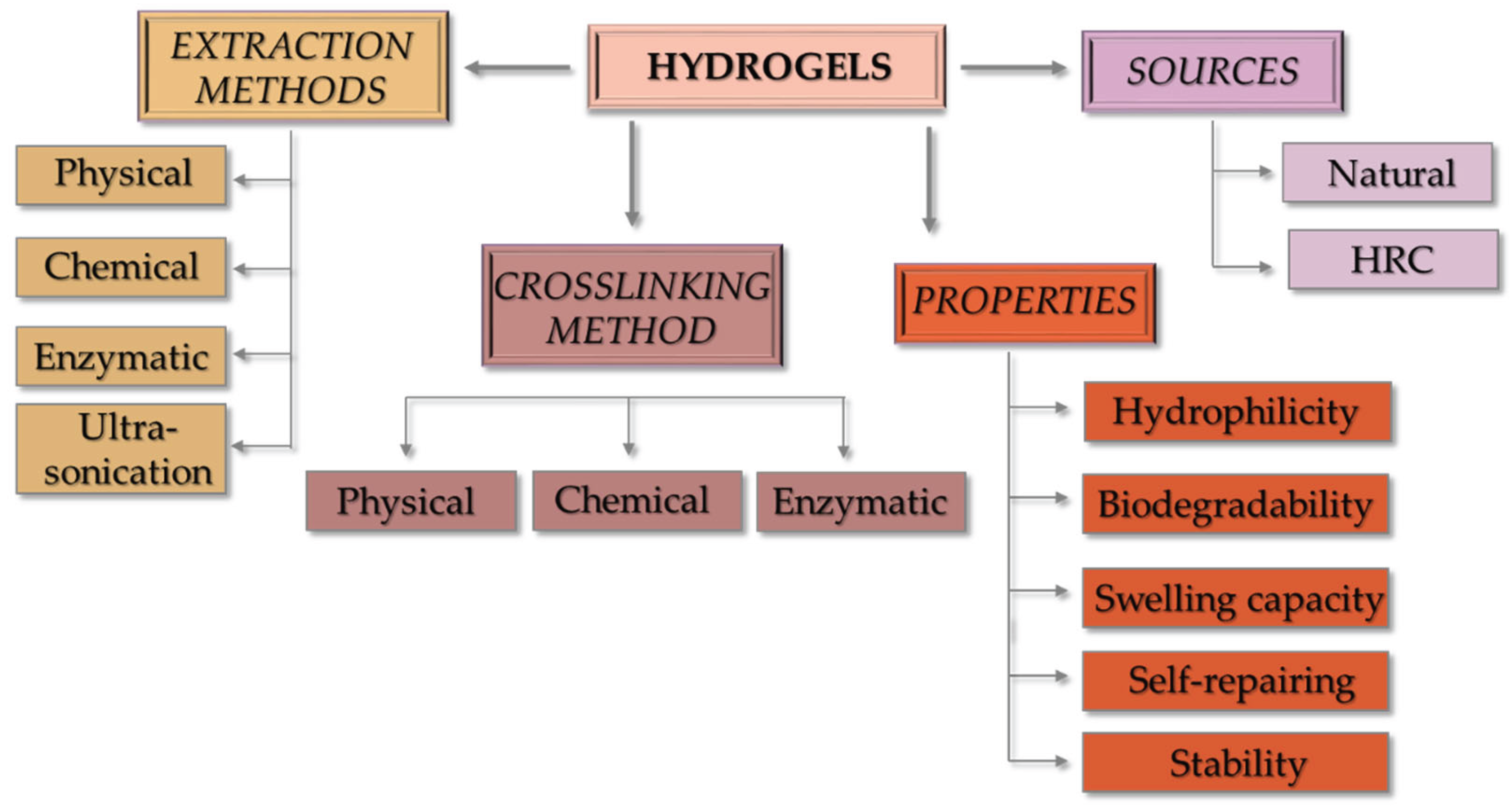
Figure 2.
The schematic illustration of the collagen-based hydrogels crosslinking processes. Physical crosslinking involves approaches such as temperature changes and UV irradiation. Chemical crosslinking requires the use of agents like glutaraldehyde, genipin, dialdehyde starch, and carbodiimides. In contrast, enzymatic crosslinking employs enzymes such as transferases (e.g., glutamine transaminase), hydrolases (e.g., lysyl oxidase), and oxidoreductases (e.g., horseradish peroxidase).
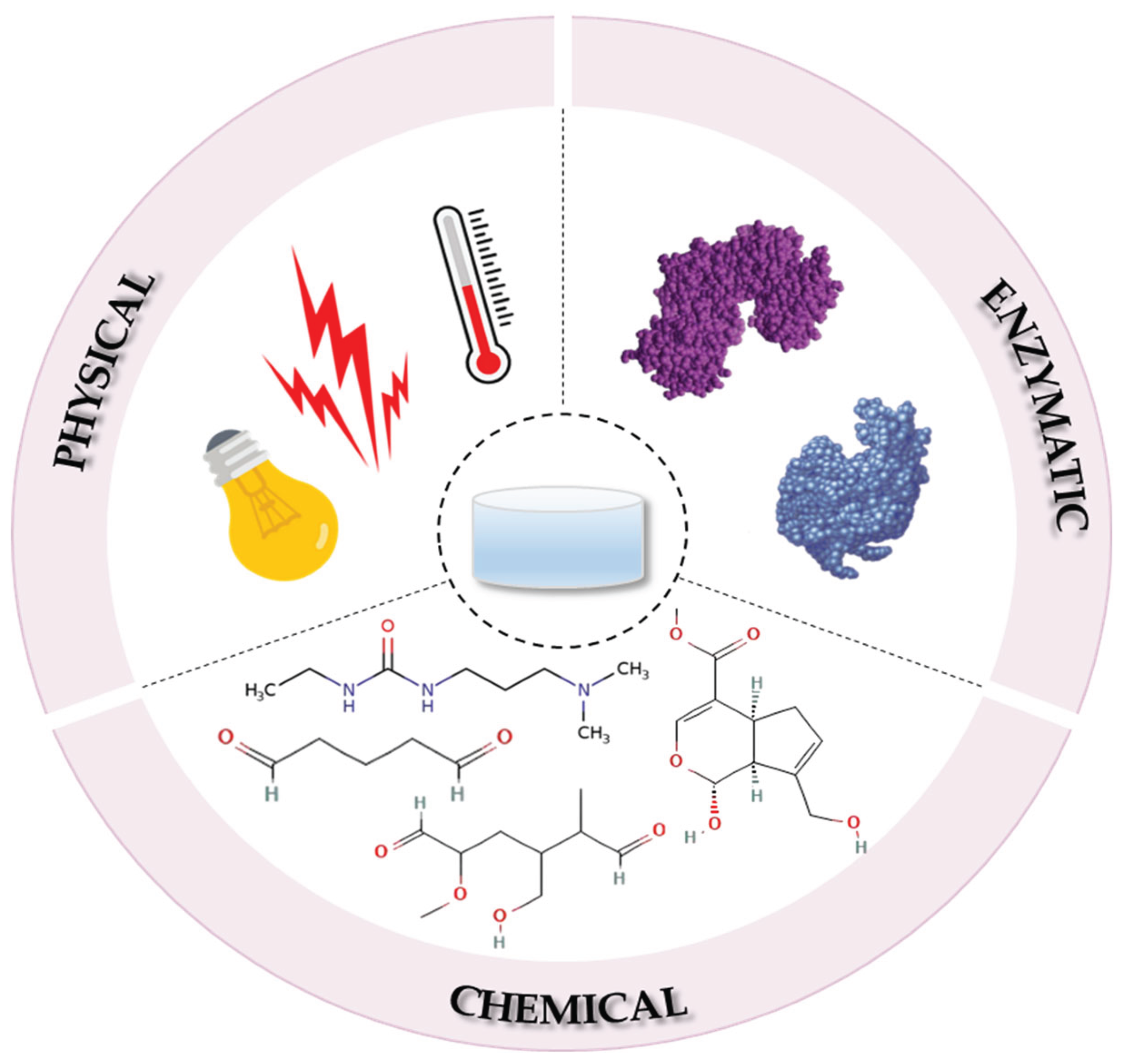
Figure 3.
The schematic illustration of the properties of collagen-based hydrogels designed for application in skin wound healing.
Figure 3.
The schematic illustration of the properties of collagen-based hydrogels designed for application in skin wound healing.
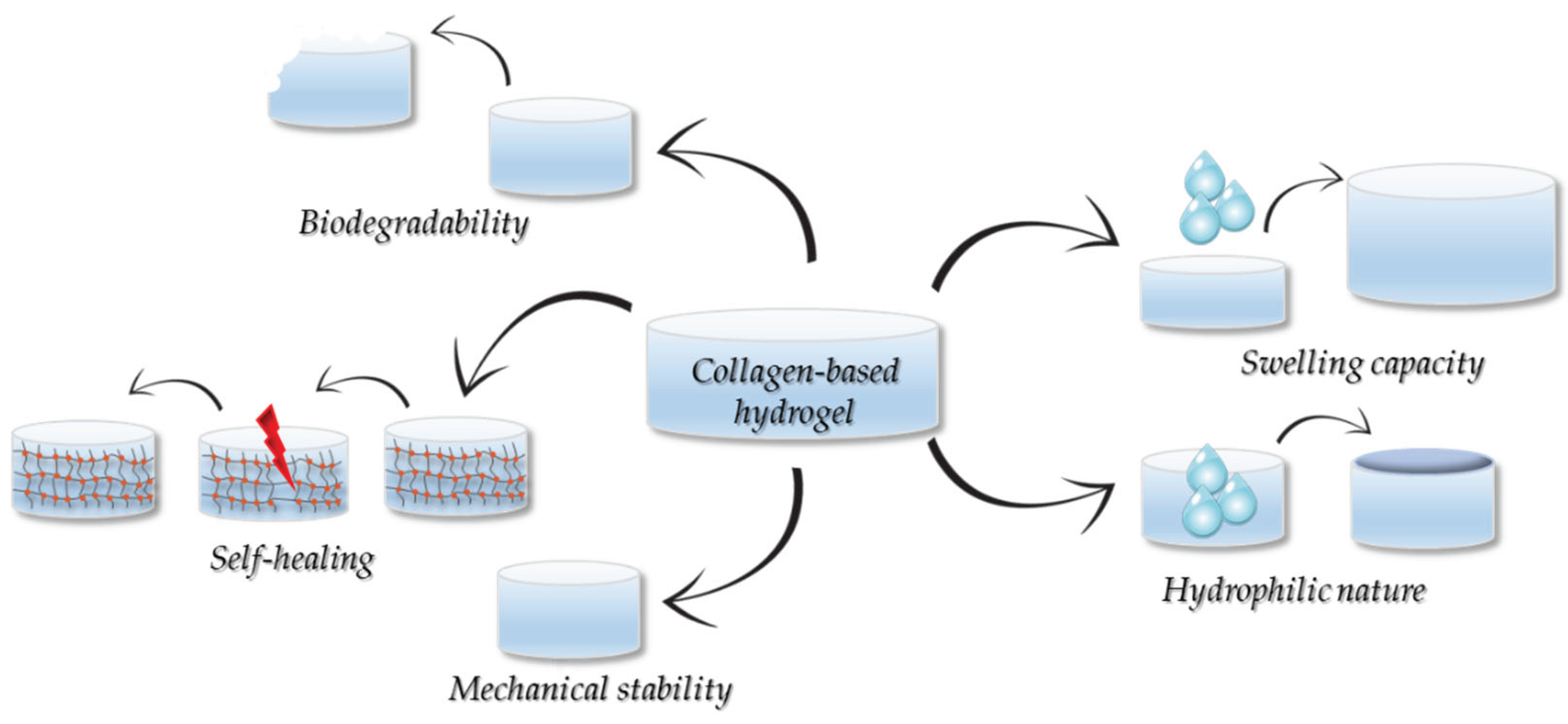
Figure 4.
The schematic illustration of the three main categories of collagen-based hydrogels designed for skin wound applications: pure collagen; collagen blends with bioactive molecules (i.e. herbal extracts, metal nanoparticles (NPs), drugs, cell-penetrating peptide, growth factors, cytokines, etc.); collagen blends with other natural and/or synthetic polymers (i.e. chitosan, hyaluronic acid, cellulose, etc.).
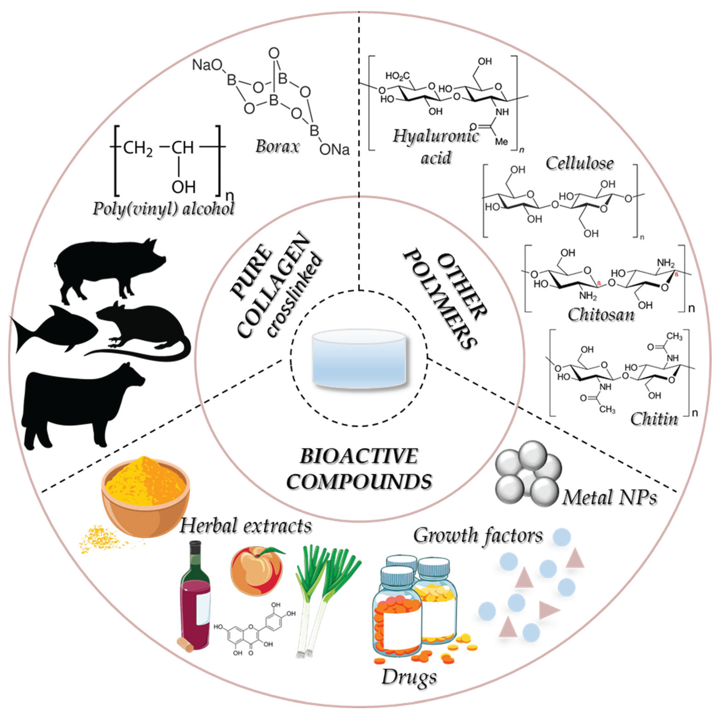
Table 1.
The phenotype for acute wounds and chronic wounds [35].
Table 1.
The phenotype for acute wounds and chronic wounds [35].
| Characteristic | Acute wounds | Chronic wounds |
|---|---|---|
| Bacterial invasion | Minimal bacterial invasion | Bacterial colonization |
| Inflammatory activity | Reduced levels of inflammatory cytokines | High concentrations of inflammatory cytokines |
| Levels of ROS/proteases | Reduced levels of proteases and ROS | High levels of ROS and proteases |
| ECM functionality | Functional ECM | Degraded ECM |
| Cell activity | Proliferating cells | Senescent cells |
| Crosslinking process | Method | Advantages | Disadvantages |
|---|---|---|---|
|
Physical |
UV radiation temperature |
lack of crosslinker improved hydrogel degradation |
phototoxicity issues structural changes in COL due to high temperature |
| Chemical | glutaraldehyde dialdehyde starch |
good stability very effective |
the crosslinker cannot be removed completely high toxicity |
| carbodiimides | optimal biocompatibility high water solubility mild reaction conditions |
reduced crosslinking strength | |
| phenolic compounds |
high water retention eco-friendly optimal pore size |
risk of self-aggregation low hydrogel extensibility |
|
| genipin | lack of toxicity favourable biocompatibility |
slow crosslinking reaction high-costs |
|
| Enzymatic | glutamine transaminase, lysyl oxidase, horseradish peroxidase |
lack of crosslinkers easily controlled conditions |
biocompatibility concerns high-costs |
Disclaimer/Publisher’s Note: The statements, opinions and data contained in all publications are solely those of the individual author(s) and contributor(s) and not of MDPI and/or the editor(s). MDPI and/or the editor(s) disclaim responsibility for any injury to people or property resulting from any ideas, methods, instructions or products referred to in the content. |
© 2025 by the authors. Licensee MDPI, Basel, Switzerland. This article is an open access article distributed under the terms and conditions of the Creative Commons Attribution (CC BY) license (http://creativecommons.org/licenses/by/4.0/).
Copyright: This open access article is published under a Creative Commons CC BY 4.0 license, which permit the free download, distribution, and reuse, provided that the author and preprint are cited in any reuse.


