
Submitted:
14 May 2025
Posted:
15 May 2025
You are already at the latest version
Abstract
This study aimed to evaluate the microbiological quality of colostrum on three dairy farms with different colostrum management hygiene practices and to compare it with current colostrum quality guidelines. On farm A, colostrum was fed raw, while on farms B and C it was heat-treated. On farms A and B feeding equipment were cleaned manually, while on farm C an automated cleaning system was used. Samples were collected from the calf feeding equipment and submitted for microbial culture: total plate (TPC), total coliforms (TCC), E. coli, enterococci, staphylococci (STAP), and lactic acid bacteria counts; in addition, pH, aW, and Brix were analysed. Colostrum quality was defined as: good quality (GQ): TPC < 100,000, TCC < 10,000, STAP < 50,000 cfu/mL, and Brix ≥ 21%; or excellent quality (EQ): TPC < 20,000, TCC < 100, STAP < 5000 cfu/mL, and Brix ≥ 22%. Approximately 57 (GQ) and 38% (EQ) of samples met the defined criteria. Lower microbial counts were found on farm C. Colostrum from farm B had lower TPC, TCC, and E. coli counts than farm A. These results showed that a considerable proportion of calves were fed colostrum with suboptimal quality, especially when less rigorous hygiene practices were implemented.
Keywords:
Colostrum management
; brix
; heat treatment
; hygiene
1. Introduction
Colostrum contains both beneficial and pathogenic bacteria, that can be derived from the dam by direct shedding from the mammary gland and via the entero-mammary pathway, or contaminated from the environment [1,2,3]. While beneficial bacteria, such as lactic acid bacteria (LAB), promote gut health [4,5], pathogenic bacteria can hinder it, reducing immunoglobulin absorption [6]. Newborn calves are highly susceptible to infections caused by various pathogens, including bacteria (e.g., Escherichia coli, and Salmonella spp.), parasites (e.g., Cryptosporidium parvum and Eimeria spp.), and viruses (e.g., rotavirus and coronavirus) [3,7,8]. Therefore, calves must ingest colostrum promptly, as it contains immunoglobulins and other immune factors that protect them during early life [9].
Proper hygiene practices are essential to minimize bacterial contamination [3]. The absence of well-established standards for bacterial counts in colostrum [10], makes it challenging to define specific hygiene protocols for colostrum management, a frequently overlooked aspect of calf care. On the other hand, immunoglobulin concentration can be easily estimated on-site using a Brix refractometer. Brix values ≥ 22% usually indicate a good quality colostrum (IgG ≥ 50 g/L) [11]. Current standards for colostrum bacterial contamination are often based on dairy industry milk standards [12] and the guidelines established by McGuirk and Collins, which typically involve a total plate count (TPC) < 100,000 cfu/mL and total coliform count (TCC) < 10,000 cfu/mL [13]. While McGuirk and Collins [13] also suggest targets for Gram-negative non-coliform count (NCC) < 50,000 cfu/mL, streptococci non-ag. < 50,000 cfu/mL, and coagulase negative staphylococci < 50,000 cfu/mL, other studies propose more strict limits, such as TPC < 20,000 cfu/mL, TCC < 100 cfu/mL [10,11], and NCC < 5,000 cfu/mL [10,14].
To achieve these goals, dairy farms must implement rigorous colostrum management protocols, emphasizing strict and consistent hygiene practices [12]. Dairy cows should be negative for pathogenic microorganisms transmissible through lacteal secretions, such as Mycobacterium paratuberculosis, Salmonella spp., Mycoplasma bovis, Staphylococcus aureus, and bovine diarrhoea and bovine leukaemia viruses [13]. Feeding colostrum from the dam to the calf (one-to-one feeding) can minimize the risk of transmitting infectious agents compared to pooled colostrum [15]. Udders should be thoroughly cleaned before colostrum harvesting and all materials and equipment used for collection, storage, and feeding colostrum must be adequately sanitized [13]. Heat treatment of colostrum (i.e., 60 °C for 60 min) is another commonly recommended practice that may reduce bacterial load [16]; however, its effect on beneficial bacteria, such as LAB, is not entirely clear [4]. Unfed colostrum should be promptly stored at 4°C for up to two days or frozen for longer-term storage [3,11]. The addition of preservatives, like potassium sorbate, can extend the refrigeration period [3]. While there is a growing body of research on colostrum management, there remains a significant gap in knowledge regarding specific hygiene practices [8,17,18]. The study by Hyde et al. [19] on British dairy farms highlights the importance of improving hygiene practices during colostrum collection and feeding, to reduce bacterial contamination. In addition, better hygiene practices were associated with higher calf serum IgG levels [18], different methods of colostrum administration affected bacterial contamination [19,20], and that the type of disinfectant used to clean the feeding apparatus affected the odds of having a TPC < 100,000 cfu/mL [15]. The aim of this study was to compare the microbiological quality of colostrum fed to calves on three dairy farms with different hygiene practices and to compare the results with current guidelines.
2. Materials and Methods
2.1. Study Design
The study was approved by the Ethics Committee for Animal Welfare (ORBEA) at Universidade de Trás-os-Montes e Alto Douro (UTAD, Portugal) under reference 2664-e-DZ-2023. Three dairy farms from the Alentejo region of Portugal (Table 1) were included in this study, based on their colostrum management protocols, in particular their colostrum hygiene practices.
Milking systems on all three farms were cleaned and sanitized similarly: post-partum cows were milked at the end of the milking session, followed by a mechanical cleaning process involving a warm water rinse (38-50 °C), a hot water wash (>60 °C) with sodium hypochlorite disinfectant, and a final cold-water rinse. Colostrum-related hygiene practices (Table 2) were as follows:
Farm A: Colostrum was collected in a bucket and then frozen in cleaned 1.5 L polyethylene bottles until use. At calving, the colostrum was thawed at 40 °C and fed to the newborn calf. Materials used for collecting, storing, and feeding the calves were washed by hand with hot water (40 – 50 °C), a non-ionic surfactant detergent (Jodel — Hygiene Products Manufacturing, Azambuja, Portugal) and with sodium hypochlorite (NaOCl) solution (2-3 % activated chlorine) (Clorosol - Comércio e Indústria de Detergentes, Lda, Vila Nova de Famalicão, Portugal) and left to dry at room temperature.
Farm B: Milked colostrum was heat treated at 60 °C for 60 min with a commercial batch pasteurizer system (Coloquick, Calvex A/S, Denmark) and frozen until need in 4 L Coloquick colostrum bags. At calving, the colostrum was thawed at 40°C and fed to the newborn calf. Materials used to collect and feed the calves were washed by hand with hot water (40 – 50 °C) and a surfactant detergent with benzenesulfonic acid, C10-13-alkyl derivs. and sodium salts (MasterChef, Lisbon, Portugal) and left to dry at room temperature.
Farm C: Milked colostrum was heat treated at 60 °C for 60 min with a commercial batch pasteurizer system (Coloquick, Calvex A/S, Denmark) and frozen until need in 4 L Coloquick colostrum bags. At calving, the colostrum was thawed at 40°C and fed to the newborn calf. Materials used to collect and feed the calves were washed with hot water (60 °C), and sodium hypochlorite (≥13 % of activated chlorine) (Ipoclorix® PWG, QuimiTécnica.com, S.A, Portugal) with an automated washing system and left to dry at room temperature.
2.2. Sample Collection
Samples of colostrum (n = 68) were collected from three dairy farms (22 from farm A, 13 from farm B, and 33 from farm C) to 100 mL sterile recipients directly from the feeding apparatus used to feed the calf (either a nipple bottle or an oesophageal tube), just before feeding. The fed colostrum was collected from the farm’s colostrum bank or from the dam. Colostrum samples were from both primiparous (n = 33) and multiparous (n = 35) Holstein-Friesian cows, milked within 12 h after birth. A sub sample (n = 6) was collected from farm C to analyse the effect of heat treatment, before (raw) and after heat treatment (HT). After collection all samples were frozen immediately at –24 °C. Samples were later transported in a cold atmosphere to the Microbiology Laboratory-MED, University of Évora for analysis. Samples were defrosted at 4 °C overnight before laboratory analysis.
2.3. Laboratory Analysis
Brix values were measured using a digital refractometer (ORF-E, Kern, Albstadt, Germany) to estimate IgG concentrations as an indicator of immunological quality. From each sample of colostrum 1 mL was diluted into 9 mL of buffered peptone water (BDH chemicals, VWR, PA, USA) and decimals solutions were prepared. One mL was pour-plated with 9 mL of each culture medium. This procedure was done in duplicates for each sample. Incubation for each plate was done as follows: mesophilic bacteria in Plate Count Agar (PCA; BDH chemicals, VWR, PA, USA) at 30 °C for 48 h; coliforms in Chromogenic Coliform Agar (BDH chemicals, VWR, PA, USA) at 37 °C for 24 h; LAB in de Man, Rogosa and Sharpe Agar (MRS; BDH chemicals, VWR, PA, USA) at 30 °C for 48 h under anaerobic conditions in an AnaeroJar (Oxoid, Hampshire, United Kingdom) using an Anaerocult® A sachet (Sigma-Aldrich, Missouri, USA), anaerobiosis condition was confirmed with a Anaerotest® stripe (Sigma-Aldrich, Missouri, USA); enterobacteria (ENTB) in Violet Red Bile Glucose Agar (VRBG; BDH chemicals, VWR, PA, USA) at 30 °C for 48 h; staphylococci (STAP) in Mannitol Salt Agar (MSA; BDH chemicals, VWR, PA, USA) at 37 °C for 48 h; yeasts and moulds in Rose Bengal Chloramphenicol Agar (RBC; BDH chemicals, VWR, PA, USA) at 25 °C for 48 h. E. coli colonies (blue and violet colonies) were confirmed from Chromogenic Coliform Agar plates by performing a Gram-negative staining and oxidase negative test. Clostridium spp. was detected by heating samples at 80 °C for 15 min, followed by plating in Sulphite Polymyxin Sulfadiazine (SPS) Agar (MERCK, Darmstadt, Germany). Salmonella spp. presence was identified with Salmonella Supp Tab kit (Biomérieux, Marcy l'Etoile, França), where 25 mL of the sample was diluted in a sterile bottle with 225 mL peptone water at 37 °C and with 25 g of Salmonella growth supplement, then the content was thoroughly mixed and incubated at 41.5 °C for 24 h, finally the reading was made with a immunodetection kit (miniVIDAS, BioMérieux, Marcy l'Etoile, France) according to the manufacturer specifications. The presence of Listeria monocytogenes was tested on ALOA® plates (Biomérieux, Marcy l'Etoile, France), and colonies were counted after an incubation period of 24 h at 37 °C. The pH (pH 1100L, pHenomenal®, VWR, Radnor, PA, USA) and aW (Hygrolab, Rotronic, Bassersdorf, Switzerland) were measured in duplicates.
2.4. Statistical Analysis
Data was analysed using the software IBM SPSS Statistics (v27, Armonk, NY). The sample size was calculated considering differences between farms with an α = 0.05 and an 85% power, setting TPC from raw and HT colostrum [21] as the main variable to perform the calculation.
The general linear model procedure was used to estimate the least square means and standard errors for TPC, TCC, LAB, STAP, Brix, pH, and aW, with Farm (A, B, and C) as a fixed factor and Plate Duplicate as a random factor. Differences between farms were tested with Tukey HSD; results are presented as LSM (SEM). Kruskal-Wallis’ test was used to compare differences in ENTB, and E. coli counts between treatments. Adjusted p-values were calculated using Bonferroni correction for multiple comparisons; results are presented as median (95% CI). The effect of heat treatment was analysed with a general linear model for repeated measures and results are reported as LSM (SEM).
Microbial counts were log10 transformed. Homoscedasticity was assessed with Levene’s Test, and normality of the residuals was analysed by inspection of normal probability plots and Kolmogorov–Smirnov test. Significant values were considered as P < 0.05.
Overall colostrum quality was defined according to current guidelines in two criteria: good quality (GQ): TPC < 100,000, TCC < 10,000, STAP < 50,000 cfu/mL, and Brix ≥ 21%; or excellent quality (EQ): TPC < 20,000, TCC < 100, STAP < 5000 cfu/mL, and Brix ≥ 22%.
3. Results
The descriptive statistics of all samples are shown in Table 3, as well as the percentage of samples that met current goals for TPC, TCC and STAP. The percentage of samples meeting the thresholds for TPC, TCC, and STAP was 69 (47/68), 91 (62/68), and 90% (61/68), for GQ colostrum, and 53 (36/68), 54 (37/68), and 69% (47/68), for EQ, respectively. Nevertheless, samples with very low to non-existent counts (e.g., TCC < detection limit cfu/mL) and samples with very high counts were found (e.g., TPC = 13,200,000.00 cfu/mL). Samples tested negative for Salmonella spp., Listeria monocytogenes, and for yeasts and moulds. Brix measurements showed that 84 (57/68) and 75% (51/68) of the samples had a Brix ≥ to 21 and 22%, respectively. As an overall quality, 57% (39/68) and 38% (26/68) of samples met the criteria for GQ and EQ, respectively.
Microbiological counts, pH, aW (Table 4), and Brix (Figure 1) were different between farms. Significantly lower microbial counts were found in farm C compared to farm A and B (P ≤ 0.002). Farm B had lower TCC, LAB and E. coli counts than farm A (P < 0.001). Significant differences were observed in colostrum pH between farm A and C (P < 0.001), and colostrum aW was higher on farm C compared to farms A and B (P = 0.032). Brix values were higher on farm C than on farms A and B (P = 0.003).
The number of samples that had a TPC < 100,000 cfu/mL was 46 (10/22), 39 (5/13), and 97% (32/33) on farm A, B, and C, respectively (Figure 2a). For a TCC < 10,000 cfu/mL, 77 (17/22), 92 (12/13), and 100% (33/33) of the samples met this threshold on farm A, B, and C, respectively. On farm A, B, and C, 82 (18/22), 85 (11/13), and 97% (32/33) of samples had a STAP < 50,000 cfu/mL, respectively. When considering lower thresholds (EQ), differences results were obtained (Figure 2b). On farm A, B, and C, respectively, 18 (4/22), 8 (1/13), and 94% (31/33) of samples had a TPC < 10,000 cfu/mL, 9 (2/22), 46 (6/13), and 88% (29/33) of samples had a TCC < 100 cfu/mL, and 32 (7/22), 69 (9/13), and 94% (31/33) had a STAPH < 5000 cfu/mL. The frequency of samples with Brix ≥ 21 and ≥ 22% was 59 (13/22), 100 (13/13), and 94% (31/33) on farm A, B, and C, respectively, and 50 (11/22), 69 (9/13), and 94% (31/33) on farm A, B, and C, respectively. When considering an overall GQ (i.e., TPC < 10,000, TCC < 10,000, STAP < 50,000 cfu/mL and Brix ≥ 21%), 18 (4/22), 39 (5/13) and 91% (30/33) of the samples on farm A, B, and C, respectively, met these criteria. When considering an overall EQ (i.e., TPC < 20,000, TCC < 100, STAP < 5000 cfu/mL, and Brix ≥ 22%), 0 (0/22), 0 (0/13), and 79% (26/33) of the samples met these criteria on farm A, B, and C, respectively.
A significant reduction in all microbial counts was observed after HT (Figure 3). However, a less pronounced effect was observed with STAP (P = 0.016), when compared with the remaining microbial counts (P < 0.001). Brix (21.5 and 21.5% SEM 1.09, for raw and HT, respectively) did not changed with HT (P = 0.500). After HT, a decrease in the pH was observed (6.43 and 6.40 SEM 0.02, for raw and HT, respectively, P = 0.017).
4. Discussion
While the important role of colostrum management in the transfer passive immunity is well-established [22], empirical research examining the impact of on-farm hygiene management practices on colostrum microbiological content remains limited. In this study, we sought to compare the microbial counts in colostrum from three dairy farms with divergent hygiene practices during colostrum management. The results obtained revealed significant variations in microbial counts among the three farms, with lower counts being associated with higher levels of hygiene. However, colostrum samples were not collected at the time of harvesting due to the fact that the majority of the colostrum administered to the calves was sourced from the farm's colostrum bank. The intention was to analyse precisely what the calves were consuming. Consequently, the comparisons between the farms must be interpreted with caution. Previous research has primarily focused on microbial contamination during the harvesting process [14,23], but contamination can occur at any stage between milking and feeding [3]. Therefore, understanding the actual microbial content of colostrum that calves are consuming is essential for animal welfare [15,24].
The results of this study showed a wide range of values with samples from only three farms (Table 2). A similar variability was found in other studies that sampled colostrum from a higher pool of farms [14,15,24]. To the authors' knowledge, no previous studies have evaluated the microbiological quality of colostrum on Portuguese dairy farms, limiting direct comparisons to findings in other countries. Compared to Czech dairy farms, our study found lower TPC values, leading to a higher proportion of samples meeting quality standards, while TCC values were similar, with an identical proportion of samples below 10,000 cfu/mL [14]. However, the Czech study only analysed raw colostrum samples collected directly from milking buckets, which may explain the higher TPC values. A study conducted on Colombian farms reported that 82% of fresh colostrum samples met the TPC < 100,000 cfu/mL standard and 76% met the TCC < 10,000 cfu/mL standard [20]. While the TPC was higher in the Colombian study, the TCC was lower compared to our findings. Additionally, Morril et al. [25] reported that in the United States only 54.8% of the samples had a TPC < 100,000 cfu/mL. This shows the great variability that exists in hygiene practices across farms. The stage at which samples are collected may account for the aforementioned variability between studies, but Stweart et al. [3] found no additional contamination from the milking bucket to the recipient where the calf would consume colostrum. This may not always be the case and highly depends on the hygiene practices. For example, Cummins et al. [26] reported very similar results with raw colostrum samples collected from the milking bucket, however, heat-treated colostrum had a much lower TPC.
There are many factors that can influence the microbial load of colostrum. Guzman-Carazo et al. [20] identified a lower microbiological quality in naturally fed calves compared to artificially fed calves, but Hyde et al. [19] observed that the feeding equipment was associated with higher TPC than the cow’s teat. The main differences between these two farms are the HT of colostrum on farm B and the use of a disinfectant in the farm A. Several studies have reported a decrease in bacteria after HT [16,27], but after HT, additional contamination can occur if proper hygiene conditions are not met. This may explain the lack of differences in TPC between farm A and B. Fecteau et al. [12] highlighted the importance of farm staff and the concept of hygiene, which can vary from person to person. In the same manner, the hygiene level during milking process and the time between collection and storage or consumption can also vary within the same farm [3,13]. Another possibility is related to the fact that only detergent was used to clean the feeding materials in farm B, whereas in farm A both detergent and disinfectant were used. It has been reported that cleaning feeding materials with chlorhexidine gluconate or bleach increased the odds of meeting the recommended thresholds, curiously the cleaning frequency of feeding materials did not [15]. In another study it was also found that using hot water to clean the feeding equipment and peracetic acid or hypochlorite to clean feeding collection equipment was associated with lower TPC, when compared to using only cold water [19]. Nevertheless, TCC, LAB, and E. coli were lower in farm B than farm A, which indicates that some of the coliform bacteria may have been reduced with HT. On the other hand, results from farm C suggest that heat treating colostrum and cleaning the feeding material in an automated equipment with hot water and disinfectant (i.e., sodium hypochlorite) effectively reduced the microbiological load. Coliforms were almost eliminated on farm C, showing that with high hygiene standards it is possible to meet more rigorous goals and consistent results [10,11]. One of the most abundant genera in bovine colostrum is STAP [4] which seems to be more resistant to HT than other common bacteria found in colostrum [28]. Staphylococci are one of the most common isolates from bovine mastitis [29], and therefore may be harmful when ingested with colostrum. Nevertheless, according to Elizondo-Salazar et al. [27], STAP are not common calf pathogens and HT reduced almost 1 log10, which did not occurred in our study (Figure 3). Nonetheless, in samples where HT was part of the protocol (i.e., farm B and C) the majority were < 5,000 cfu/mL, meeting the suggested goal, however when considering a higher threshold (i.e., 50,000 cfu/mL) the three farms had more than 80% of samples bellow the threshold. However, HT greatly reduced microbial counts of all other cultures (Figure 3), but Brix was not affected, in accordance with previous results [16,27]. Brix levels were significantly higher on farm C. However, this is unlikely to be related to microbial counts as other factors such as dam age, pre partum diet, length of dry period, volume of colostrum, and vaccination may also affect the nutritional and immunological quality of colostrum [11]. A reduction in colostrum LAB with HT was reported by Trujillo et al. [30]. The decrease in LAB with HT and the lower values observed in farm C appear to be unfavourable outcomes. However, HT appears to promote the growth of beneficial bacteria at the intestinal level, as the calves' intestinal tract is already colonised by bacteria such as Bifidobacterium at birth [31]. Furthermore, HT colostrum has been shown to improve their growth, probably due to bioactive components, such as oligosaccharides, present in colostrum [31]. Santos et al. [23] reported counts for LAB in raw colostrum samples from Brazil, with levels higher than the ones from this study. The analysis of the effect of HT (Figure 3) showed that raw samples from this farm had already lower LAB counts than the other farms, which may have influenced the counts after HT. We cannot explain this variation, but it has been shown that antimicrobials used during the dry period can affect the colostrum microbiota [32]. Cow’s gut can be populated by LAB with antimicrobial activity against certain pathogens present in the same environment [33]. The relationship between the colostrum LAB and the cow's gut microbiome remains unclear. However, it was shown that some of the bacteria present in colostrum were detected in the cow’s rectal content, including strictly anaerobic bacteria commonly present in the rumen and intestinal tract [34]. Colostrum has the capacity to modulate the newborn’s calf immune system and gut microbiome [4], and translocation of gut bacteria to colostrum can occur [1], therefore it would be plausible to think that these bacteria are important for the calf’s defences and gut colonization. Nevertheless, there is still a lack of information on the relationship between colostrum microbiome and the calf’s intestinal colonization.
Overall, only on farm C optimal results regarding both microbiological (i.e., TPC and TCC) and immunological quality (Brix) were found, even when considering more restrict thresholds. These findings align with those of Phipps et al. [15] and Morril et al. [25] who reported that only 23% and 39.4% of samples, respectively, met the established criteria. In Santos et al. [23], a similar approach was used and 22.6% of the samples met the defined criteria (ENTB < 100,000 cfu/mL and IgG > 50 mg/mL). Therefore, a great proportion of calves from farm A and B received suboptimal colostrum. It is important to note that the fact that colostrum samples were frozen before culture may underestimate the percentage of colostrum not meeting the current recommendations.
5. Conclusions
This study has demonstrated the variability in microbial composition between farms, highlighting the benefit of stricter hygiene practices to reduce microbial contamination of colostrum. It also showed that when more challenging thresholds were used, a considerable proportion of colostrum samples were not considered to meet the criteria for high microbiological quality. This indicates that the identification of better hygiene practices and the establishment of strict hygiene protocols in colostrum management are necessary to ensure that high microbiological quality colostrum is administered to the calf.
Author Contributions
Conceptualization, F.G.S., M.L.; S.R.S.; C.C. and J.L.C.; methodology, F.G.S., M.L. and J.L.C.; validation, M.L., C.C. and J.L.C.; formal analysis, F.G.S. and M.L.; investigation, F.G.S., M.L.; S.R.S.; C.C. and J.L.C; resources, M.L., S.R.S. and C.C.; data curation, F.G.S., M.L.; S.R.S.; C.C. and J.L.C.; writing—original draft preparation, F.G.S.; writing—review and editing, M.L.; S.R.S.; C.C. and J.L.C.; visualization, F.G.S., M.L.; S.R.S.; C.C. and J.L.C.; supervision, M.L.; S.R.S.; C.C. and J.L.C.; project administration, S.R.S.; C.C. and J.L.C.; funding acquisition, F.G.S., M.L.; S.R.S.; C.C. and J.L.C. All authors have read and agreed to the published version of the manuscript.
Funding
This work is funded by National Funds through FCT—Foundation for Science and Technology under the Projects CECAV UIDB/00772/2020 (https://doi.org/10.54499/UIDB/00772/2020), MED UIDB/05183/2020 (https://doi.org/10.54499/UIDB/05183/2020; https://doi.org/10.54499/UIDP/05183/2020); CHANGE (https://doi.org/10.54499/LA/P/0121/2020), CISAS UIDP/05937/2020 (https://doi.org/10.54499/UIDP/05937/2020; https://doi.org/10.54499/UIDB/05937/2020) and F.G.S. PhD grant UI/BD/150834/2021 (https://doi.org/10.54499/UI/BD/150834/2021).
Institutional Review Board Statement
The study was approved by the Ethics Committee for Animal Welfare (ORBEA) at Universidade de Trás-os-Montes e Alto Douro (UTAD, Portugal) under reference 2664-e-DZ-2023.
Data Availability Statement
The data generated/analysed during the current study are available from the corresponding author upon reasonable request.
Acknowledgments
The authors want to thank Guilhermina Pias for technical support with microbiological analyses.
Conflicts of Interest
The authors declare no conflicts of interest.
References
- Addis, M.F.; Tanca, A.; Uzzau, S.; Oikonomou, G.; Bicalho, R.C.; Moroni, P. The Bovine Milk Microbiota: Insights and Perspectives from -Omics Studies. Mol. Biosyst. 2016, 12, 2359–2372. [Google Scholar] [CrossRef] [PubMed]
- Lima, S.F.; Teixeira, A.G.V.; Lima, F.S.; Ganda, E.K.; Higgins, C.H.; Oikonomou, G.; Bicalho, R.C. The Bovine Colostrum Microbiome and Its Association with Clinical Mastitis. J. Dairy Sci. 2017, 100, 3031–3042. [Google Scholar] [CrossRef] [PubMed]
- Stewart, S.; Godden, S.; Bey, R.; Rapnicki, P.; Fetrow, J.; Farnsworth, R.; Scanlon, M.; Arnold, Y.; Clow, L.; Mueller, K.; et al. Preventing Bacterial Contamination and Proliferation during the Harvest, Storage, and Feeding of Fresh Bovine Colostrum. J. Dairy Sci. 2005, 88, 2571–2578. [Google Scholar] [CrossRef] [PubMed]
- Silva, F.G.; Silva, S.R.; Pereira, A.M.F.; Cerqueira, J.L.; Conceição, C. A Comprehensive Review of Bovine Colostrum Components and Selected Aspects Regarding Their Impact on Neonatal Calf Physiology. Animals 2024, 14, 1130. [Google Scholar] [CrossRef]
- Otte, J.-M.; Podolsky, D.K. Functional Modulation of Enterocytes by Gram-Positive and Gram-Negative Microorganisms. Am. J. Physiol. Liver Physiol. 2004, 286, G613–G626. [Google Scholar] [CrossRef]
- Malik, M.I.; Rashid, M.A.; Raboisson, D. Heat Treatment of Colostrum at 60°C Decreases Colostrum Immunoglobulins but Increases Serum Immunoglobulins and Serum Total Protein: A Meta-Analysis. J. Dairy Sci. 2022, 105, 3453–3467. [Google Scholar] [CrossRef]
- Barry, J.; Bokkers, E.A.M.; De Boer, I.J.M.; Kennedy, E. Pre-Weaning Management of Calves on Commercial Dairy Farms and Its Influence on Calf Welfare and Mortality. Animal 2020, 14, 2580–2587. [Google Scholar] [CrossRef]
- Abuelo, A.; Havrlant, P.; Wood, N.; Hernandez-Jover, M. An Investigation of Dairy Calf Management Practices, Colostrum Quality, Failure of Transfer of Passive Immunity, and Occurrence of Enteropathogens among Australian Dairy Farms. J. Dairy Sci. 2019, 102, 8352–8366. [Google Scholar] [CrossRef]
- Chase, C.C.L.; Hurley, D.J.; Reber, A.J. Neonatal Immune Development in the Calf and Its Impact on Vaccine Response. Vet. Clin. North Am. - Food Anim. Pract. 2008, 24, 87–104. [Google Scholar] [CrossRef]
- Heinrichs, J.; Jones, C. Composition and Hygiene of Colostrum on Modern Pennsylvania Dairy Farms. Available online: https://extension.psu.edu/composition-and-hygiene-of-colostrum-on-modern-pennsylvania-dairy-farms (accessed on 21 July 2024).
- Godden, S.M.; Lombard, J.E.; Woolums, A.R. Colostrum Management for Dairy Calves. Vet. Clin. North Am. - Food Anim. Pract. 2019, 35, 535–556. [Google Scholar] [CrossRef]
- Fecteau, G.; Baillargeon, P.; Higgins, R.; Paré, J.; Fortin, M. Bacterial Contamination of Colostrum Fed to Newborn Calves in Québec Dairy Herds. Can. Vet. J. 2002, 43, 523–527. [Google Scholar] [PubMed]
- McGuirk, S.M.; Collins, M. Managing the Production, Storage, and Delivery of Colostrum. Vet. Clin. North Am. - Food Anim. Pract. 2004, 20, 593–603. [Google Scholar] [CrossRef] [PubMed]
- Šlosárková, S.; Pechová, A.; Staněk, S.; Fleischer, P.; Zouharová, M.; Nejedlá, E. Microbial Contamination of Harvested Colostrum on Czech Dairy Farms. J. Dairy Sci. 2021, 104, 11047–11058. [Google Scholar] [CrossRef] [PubMed]
- Phipps, A.J.; Beggs, D.S.; Murray, A.J.; Mansell, P.D.; Stevenson, M.A.; Pyman, M.F. Survey of Bovine Colostrum Quality and Hygiene on Northern Victorian Dairy Farms. J. Dairy Sci. 2016, 99, 8981–8990. [Google Scholar] [CrossRef]
- Godden, S.; McMartin, S.; Feirtag, J.; Stabel, J.; Bey, R.; Goyal, S.; Metzger, L.; Fetrow, J.; Wells, S.; Chester-Jones, H. Heat-Treatment of Bovine Colostrum. II: Effects of Heating Duration on Pathogen Viability and Immunoglobulin G. J. Dairy Sci. 2006, 89, 3476–3483. [Google Scholar] [CrossRef]
- Van Driessche, L.; Santschi, D.E.; Paquet, É.; Renaud, D.; Charbonneau, É.; Gauthier, M. Lou; Chancy, A.; Barbeau-Grégoire, N.; Buczinski, S. Hygiene Management Practices and Adenosine Triphosphate Luminometry of Feeding Equipment in Preweaning Calves on Dairy Farms in Quebec, Canada. J. Dairy Sci. 2023, 106, 8885–8896. [Google Scholar] [CrossRef]
- Barry, J.; Bokkers, E.A.M.; Berry, D.P.; de Boer, I.J.M.; McClure, J.; Kennedy, E. Associations between Colostrum Management, Passive Immunity, Calf-Related Hygiene Practices, and Rates of Mortality in Preweaning Dairy Calves. J. Dairy Sci. 2019, 102, 10266–10276. [Google Scholar] [CrossRef]
- Hyde, R.M.; Green, M.J.; Hudson, C.; Down, P.M. Quantitative Analysis of Colostrum Bacteriology on British Dairy Farms. Front. Vet. Sci. 2020, 7, 1–12. [Google Scholar] [CrossRef]
- Guzman-Carazo, V.; Reyes-Vélez, J.; Elsohaby, I.; Olivera-Angel, M. Factors Associated with Microbiological Quality of Bovine Colostrum in Colombian Dairy Herds. Int. Dairy J. 2020, 105, 104670. [Google Scholar] [CrossRef]
- Donahue, M.; Godden, S.M.; Bey, R.; Wells, S.; Oakes, J.M.; Sreevatsan, S.; Stabel, J.; Fetrow, J. Heat Treatment of Colostrum on Commercial Dairy Farms Decreases Colostrum Microbial Counts While Maintaining Colostrum Immunoglobulin G Concentrations. J. Dairy Sci. 2012, 95, 2697–2702. [Google Scholar] [CrossRef]
- Raboisson, D.; Trillat, P.; Cahuzac, C. Failure of Passive Immune Transfer in Calves: A Meta-Analysis on the Consequences and Assessment of the Economic Impact. PLoS One 2016, 11, 1–19. [Google Scholar] [CrossRef] [PubMed]
- Santos, G.; Silva, J.T.; Santos, F.H.R.; Bittar, C.M.M. Nutritional and Microbiological Quality of Bovine Colostrum Samples in Brazil. Rev. Bras. Zootec. 2017, 46, 72–79. [Google Scholar] [CrossRef]
- McAloon, C.G.; Doherty, M.L.; Donlon, J.; Lorenz, I.; Meade, J.; O’Grady, L.; Whyte, P. Microbiological Contamination of Colostrum on Irish Dairy Farms. Vet. Rec. 2016, 178, 474. [Google Scholar] [CrossRef] [PubMed]
- Morrill, K.M.; Conrad, E.; Lago, A.; Campbell, J.; Quigley, J.; Tyler, H. Nationwide Evaluation of Quality and Composition of Colostrum on Dairy Farms in the United States. J. Dairy Sci. 2012, 95, 3997–4005. [Google Scholar] [CrossRef]
- Cummins, C.; Berry, D.P.; Murphy, J.P.; Lorenz, I.; Kennedy, E. The Effect of Colostrum Storage Conditions on Dairy Heifer Calf Serum Immunoglobulin G Concentration and Preweaning Health and Growth Rate. J. Dairy Sci. 2017, 100, 525–535. [Google Scholar] [CrossRef]
- Elizondo-Salazar, J.A.; Jayarao, B.M.; Heinrichs, A.J. Effect of Heat Treatment of Bovine Colostrum on Bacterial Counts, Viscosity, and Immunoglobulin G Concentration. J. Dairy Sci. 2010, 93, 961–967. [Google Scholar] [CrossRef]
- Mann, S.; Curone, G.; Chandler, T.L.; Moroni, P.; Cha, J.; Bhawal, R.; Zhang, S. Heat Treatment of Bovine Colostrum: I. Effects on Bacterial and Somatic Cell Counts, Immunoglobulin, Insulin, and IGF-I Concentrations, as Well as the Colostrum Proteome. J. Dairy Sci. 2020, 103, 9368–9383. [Google Scholar] [CrossRef]
- Pyörälä, S.; Taponen, S. Coagulase-Negative Staphylococci—Emerging Mastitis Pathogens. Vet. Microbiol. 2009, 134, 3–8. [Google Scholar] [CrossRef]
- Trujillo, A.J.; Castro, N.; Quevedo, J.M.; Argüello, A.; Capote, J.; Guamis, B. Effect of Heat and High-Pressure Treatments on Microbiological Quality and Immunoglobulin G Stability of Caprine Colostrum. J. Dairy Sci. 2007, 90, 833–839. [Google Scholar] [CrossRef]
- Malmuthuge, N.; Chen, Y.; Liang, G.; Goonewardene, L.A.; Guan, L.L. Heat-Treated Colostrum Feeding Promotes Beneficial Bacteria Colonization in the Small Intestine of Neonatal Calves. J. Dairy Sci. 2015, 98, 8044–8053. [Google Scholar] [CrossRef]
- Patangia, D. V.; Grimaud, G.; Linehan, K.; Ross, R.P.; Stanton, C. Microbiota and Resistome Analysis of Colostrum and Milk from Dairy Cows Treated with and without Dry Cow Therapies. Antibiotics 2023, 12, 1–16. [Google Scholar] [CrossRef] [PubMed]
- Adeniyi, B.A.; Adetoye, A.; Ayeni, F.A. Antibacterial Activities of Lactic Acid Bacteria Isolated from Cow Faeces against Potential Enteric Pathogens. Afr. Health Sci. 2015, 15, 888–895. [Google Scholar] [CrossRef] [PubMed]
- Chen, B.; Tang, G.; Guo, W.; Lei, J.; Yao, J.; Xu, X. Detection of the Core Bacteria in Colostrum and Their Association with the Rectal Microbiota and with Milk Composition in Two Dairy Cow Farms. Animals 2021, 11, 3363. [Google Scholar] [CrossRef] [PubMed]
Figure 1.
Boxplots of Brix (%) values across farms (Farm A, n=22, Farm B, n=13, and Farm C, n=33). Different letters represent significant differences (P = 0.003).
Figure 1.
Boxplots of Brix (%) values across farms (Farm A, n=22, Farm B, n=13, and Farm C, n=33). Different letters represent significant differences (P = 0.003).
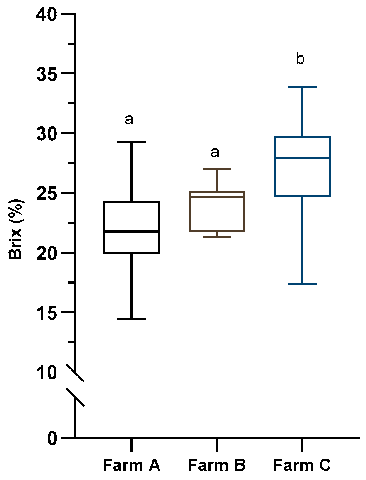
Figure 2.
Percentages of colostrum samples meeting the goal for good quality (a) total plate counts (TPC) < 100,000 cfu/mL, for total coliform counts (TCC) < 10,000 cfu/mL, and for staphylococci (STAP) < 50,000 cfu/mL; and for excellent quality (b) total plate counts (TPC) < 10,000 cfu/mL, total coliform counts (TCC) < 100 cfu/mL, and staphylococci (STAP) < 5,000 cfu/mL, on each farm (Farm A, n=22, Farm B, n=13, and Farm C, n=33).
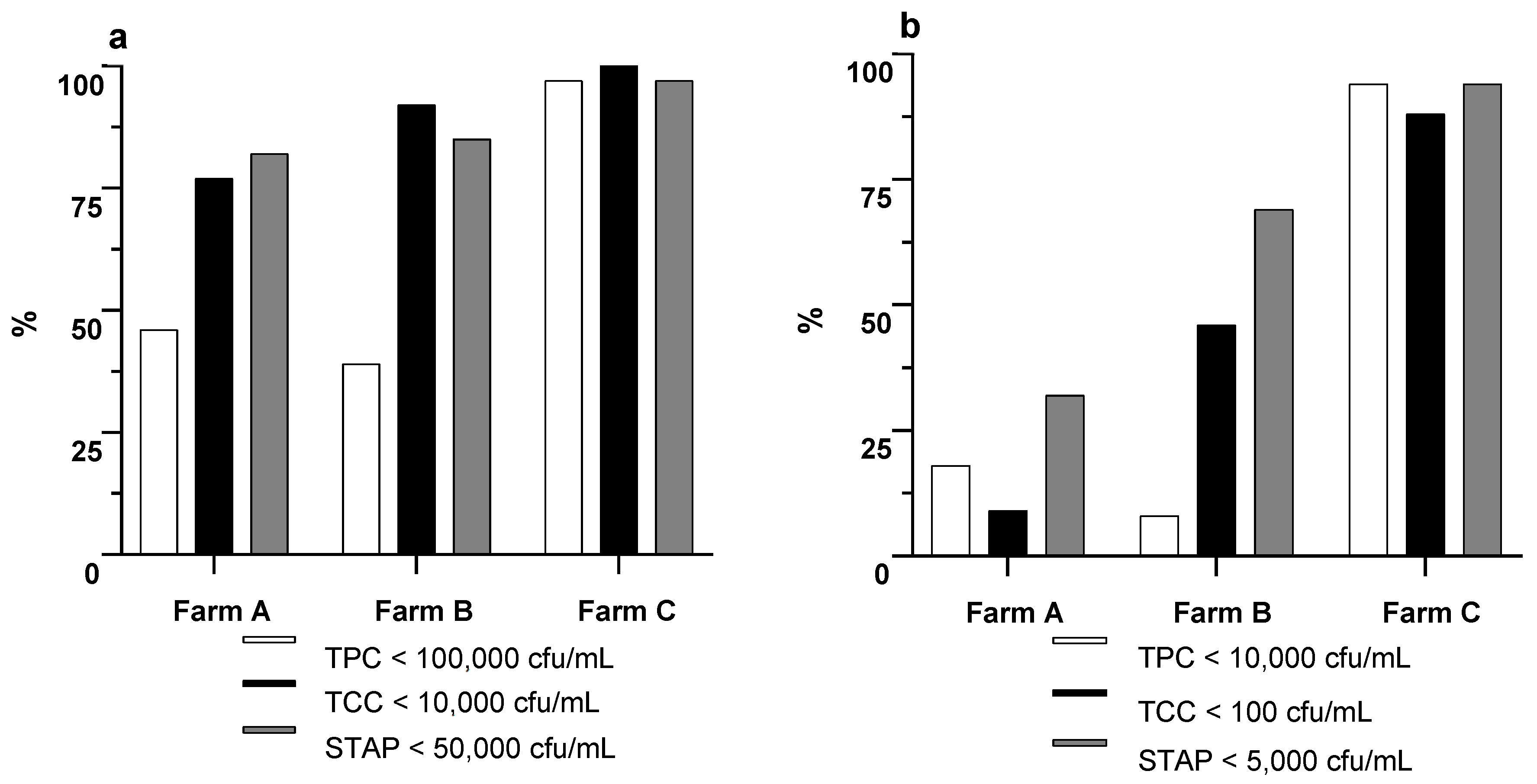
Figure 3.
Microbiological counts before (Raw) and after heat treatment (HT; 60 ˚C for 60 min) of Farm C colostrum (n = 6). TPC, total plate counts; TCC, total coliform counts; LAB, lactic acid bacteria; STAP, staphylococci; ENTB, enterobacteria, E. coli, Escherichia coli. *** P < 0.001, ** P < 0.01, * P < 0.05.
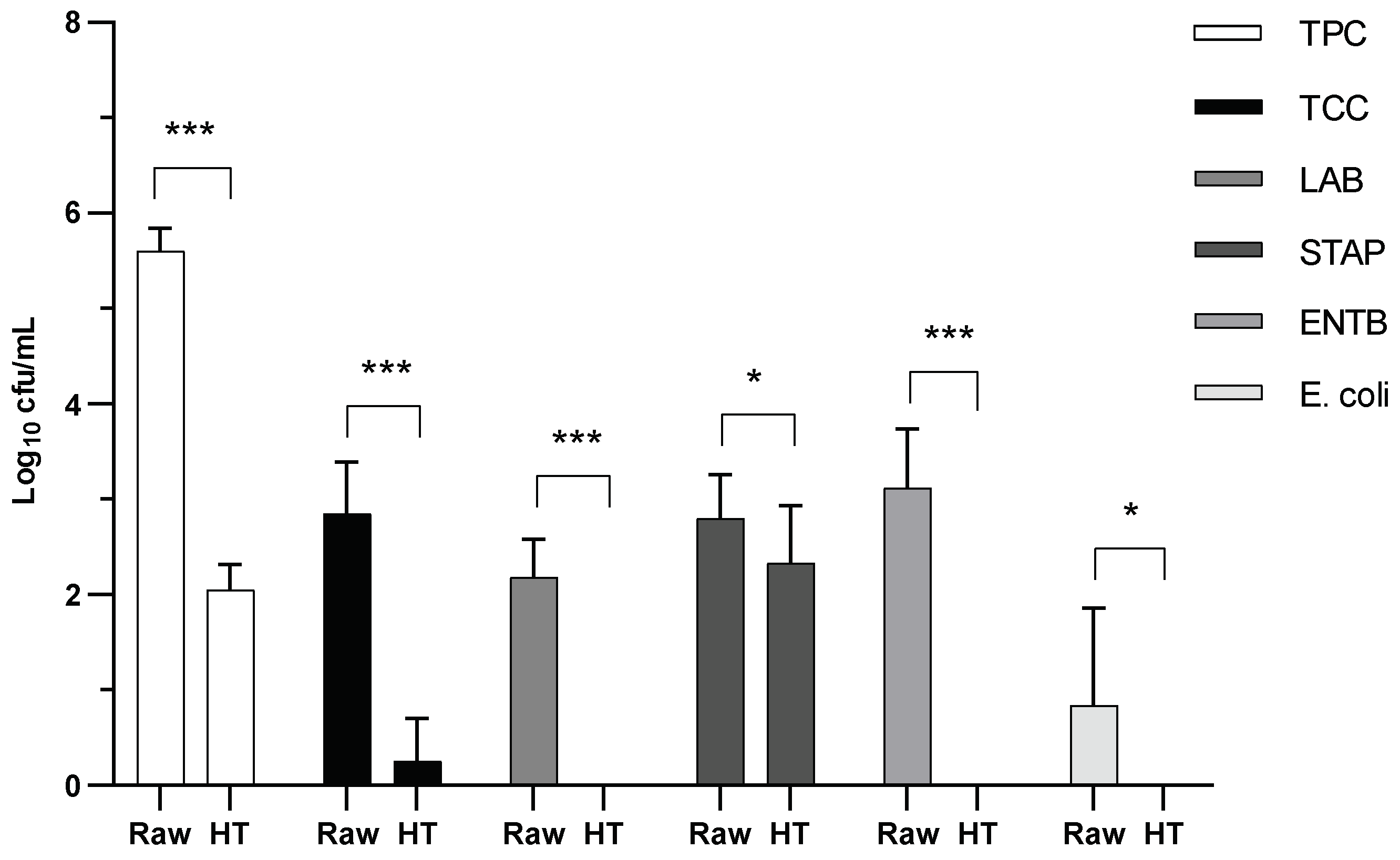

Table 1.
Herd size and production characteristics of the three dairy farms included in the study.
| Farm | Cows in milking | Average lactation nº | Milk production 305 d | Milk fat 305 d | Milk protein 305 d |
|---|---|---|---|---|---|
| A | 21 | 2.7 | 8813 | 4.46 | 3.43 |
| B | 484 | 2.6 | 13634 | 4.2 | 3.31 |
| C | 783 | 2.1 | 12118 | 4.11 | 3.22 |
Table 2.
Description of the hygiene practices used during the handling and delivery of colostrum.
| Farm | Cleaning of feeding apparatus | ||||
| HT1 | Hot water | Detergent | Disinfectant | Automated washing | |
| A | No | 40 – 50 °C | A | Yes | No |
| B | Yes | 40 – 50 °C | B | No | No |
| C | Yes | 60 °C | No | Yes | Yes |
1 Heat treatment of colostrum at 60 ˚C for 60 min.
Table 3.
Descriptive statistics for microbiological counts in colostrum samples (n = 68) on three Portuguese dairy farms and proportions of samples meeting current standards (Met).
Table 3.
Descriptive statistics for microbiological counts in colostrum samples (n = 68) on three Portuguese dairy farms and proportions of samples meeting current standards (Met).
| Counts | Mean | Median | SD | Minimum | First Q | Third Q | Maximum | Met (%) |
|---|---|---|---|---|---|---|---|---|
| TPC | 398,936.57 | 16,800.00 | 1,547,386.70 | 40.00 | 475.00 | 206,500.00 | 13,200,000.00 | 69.9a; 53.7b |
| TCC | 11,706.44 | 70.00 | 75,103.84 | < dl | < dl | 2,130.00 | 637,260.00 | 91.21a; 54.4b |
| LAB | 64,629.56 | 580.00 | 236,961.96 | < dl | 35.00 | 19,050.00 | 1,580,000.00 | |
| STAP | 17,720.67 | 1,460.00 | 50,892.69 | < dl | 350.00 | 14,750.00 | 350,000.00 | 89.7a; 69.1b |
| ENTB | 12,958.83 | 40.00 | 44,750.21 | < dl | < dl | 730.00 | 252,000.00 | |
| E. coli | 482.06 | < dl | 2,117.68 | < dl | < dl | 145.00 | 17,100.00 |
Table 4.
Microbiological counts in colostrum (expressed in log10 cfu/mL) regarding treatment (Farm A, n=22, Farm B, n=13, and Farm C, n=33).
Table 4.
Microbiological counts in colostrum (expressed in log10 cfu/mL) regarding treatment (Farm A, n=22, Farm B, n=13, and Farm C, n=33).
| Counts | Farm A | Farm B | Farm C | P-value |
|---|---|---|---|---|
| TPC1 | 5.13 (0.11) a | 5.26 (0.15) a | 2.80 (0.09) b | <0.001 |
| TCC1 | 3.47 (0.15) a | 2.12 (0.19) b | 0.71 (0.12) c | <0.001 |
| LAB1 | 4.31 (0.17) a | 3.18 (0.21) b | 1.41 (1.14) c | <0.001 |
| STAP1 | 4.02 (0.12) a | 3.55 (0.16) a | 2.46 (0.10) b | 0.002 |
| ENTB2 | 2.89 (1.60 – 5.40) a | 2.20 (< dl – 5.23) a | < dl (< dl – 3.13) b | <0.001 |
| E. coli2 | 2.18 (0.00 – 3.74) a | 0.00 (< dl – 4.23) b | < dl (< dl – 3.34) b | <0.001 |
| pH1 | 6.29 (0.02) a | 6.27 (0.03) ab | 6.22 (0.01) b | <0.001 |
| aw1 | 92.76 (0.13) a | 92.67 (0.17) a | 93.34 (0.09) c | 0.032 |
1Results reported as LSM (SEM). 2Results reported as Median (95% CI). Different superscript letters (a,b,c) represent significant differences (P < 0.05) between farms. TPC = Total Plate Count = TCC, Total Coliform Count = LAB, Lactic Acid Bacteria; STAP = staphylococci; ENTB = enterobacteria; dl = detection limit.
Disclaimer/Publisher’s Note: The statements, opinions and data contained in all publications are solely those of the individual author(s) and contributor(s) and not of MDPI and/or the editor(s). MDPI and/or the editor(s) disclaim responsibility for any injury to people or property resulting from any ideas, methods, instructions or products referred to in the content. |
© 2025 by the authors. Licensee MDPI, Basel, Switzerland. This article is an open access article distributed under the terms and conditions of the Creative Commons Attribution (CC BY) license (http://creativecommons.org/licenses/by/4.0/).
Copyright: This open access article is published under a Creative Commons CC BY 4.0 license, which permit the free download, distribution, and reuse, provided that the author and preprint are cited in any reuse.


