
Submitted:
06 May 2025
Posted:
07 May 2025
You are already at the latest version
Abstract
Background and aim: Colorectal cancer remains a leading cause of cancer-related mortality, with growing interest in nanotechnology-driven immunotherapeutics. Gold nanoparticles (AuNPs) offer a promising platform due to their biocompatibility, functional versatility, and immunomodulatory potential. Carcinoembryonic antigen (CEA), highly expressed in colorectal tumors, provides an ideal target for antigen-specific immune activation. The aim of this study was to evaluate the immunogenicity, biodistribution and therapeutic efficacy of a CEA-functionalised gold nanoparticle (CEA-AuNP) construct in a mouse model of colorectal cancer following oral administration via a customised capsular delivery system Methods: An in vivo study was conducted after a 30-day oral administration in BALB/c mice at escalating doses (5–50 mg/kg) of priorly validated in vitro CEA-functionalized gold nanoparticles (CEA-AuNPs). Histological, hyperspectral, and ELISA-based cytokine analyses were performed to assess tissue integrity, biodistribution, and immune responses. Results: CEA-AuNPs demonstrated high biocompatibility and effective macrophage internalization, inducing IL-10-dominant immune responses in the spleen and controlled pro-inflammatory cytokine shifts in the liver. Histological findings confirmed dose-dependent splenic and hepatic accumulation without systemic toxicity. Multifocal splenic amyloidosis and extramedullary hepatic hematopoiesis were noted at high doses, consistent with sustained immune activation. Conclusions: CEA-functionalized AuNPs represent a promising nanovaccine candidate for colorectal cancer prophylaxis. The construct induced tissue-specific immune responses, favoring cellular immunity and antigen processing without significant toxicity. These results support further investigation into CEA-AuNP nanoconstructs as oral immunotherapeutics in cancer prevention.
Keywords:
nanoparticle
; colorectal cancer
; vaccine
; carcinembrionic antigen (CEA)
; gold-nanoparticle
; immunoprophylaxis
1. Introduction
Colorectal cancer remains one of the most prevalent and deadly malignancies worldwide, with limited success from conventional treatment modalities such as surgery, chemotherapy, radiotherapy and emerging immunotherapies [1]. While often effective in early-stage disease, these therapies are often associated with significant limitations, such as adverse effects, limited efficacy in metastatic disease, and tumor immune evasion [2]. Consequently, the development of innovative strategies capable of inducing a potent, tumor-specific immune response has become a major focus of contemporary oncological research [3].
Cancer immunoprophylaxis, including the use of therapeutic or preventive vaccines, has emerged as a promising alternative capable of engaging the host immune system to recognize and eliminate neoplastic cells [4]. Tumor-associated antigens (TAAs) represent the molecular basis for these approaches, with carcinoembryonic antigen (CEA) being a particularly attractive target [5]. CEA is a glycoprotein overexpressed in a wide range of epithelial malignancies, including colorectal cancer, while showing restricted expression in normal adult tissues [6]. This expression profile, combined with its immunogenic properties, supports the rationale for CEA-based vaccination platforms aimed at both treatment and disease prevention.
However, effective antigen delivery remains a major challenge in the development of cancer vaccines. Traditional formulations are often limited by rapid antigen degradation, inefficient uptake by antigen-presenting cells (APCs), and insufficient trafficking to secondary lymphoid organs, resulting in suboptimal immune activation [7]. Addressing these limitations requires innovative delivery systems that preserve antigen structure, enhance cellular internalization, and sustain immunostimulatory signaling.
Nanotechnology offers a compelling solution, with gold nanoparticles (AuNPs) standing out as highly versatile carriers for vaccine design. AuNPs possess a favorable safety profile, tunable size and surface chemistry, and unique optical properties that allow for functionalization with a wide range of biomolecules [8]. When conjugated with tumor antigens such as CEA, these nanoconstructs can improve antigen stability, facilitate multivalent presentation to immune cells, and enhance the magnitude and duration of the immune response [9]. Moreover, their compatibility with oral delivery systems may enable mucosal priming and systemic immunity, offering a minimally invasive strategy for cancer prevention [10].
The aim of this study was to evaluate the translational potential of a CEA-functionalized gold nanoparticle (CEA-AuNP) platform for oral cancer immunoprophylaxis. Specifically, we sought to characterize the in vivo safety profile, biodistribution, and immunomodulatory effects of orally administered CEA-AuNPs in a murine model. Building on prior in vitro validation of antigen loading, cytocompatibility, and macrophage uptake, this investigation focuses on the immunological impact within key organs such as the spleen and liver. The study further assesses cytokine modulation and histological changes associated with nanoparticle exposure, establishing the foundation for future therapeutic development against colorectal cancer.
2. Materials and Methods
CEA-functionalized gold nanoparticles (CEA-AuNPs) were synthesized via citrate reduction and peptide conjugation, followed by physicochemical characterization using UV-Vis spectroscopy and dynamic light scattering (DLS), as previously described (unpublished data). Prior in vitro assessments confirmed high cytocompatibility and efficient macrophage internalization, supporting progression to in vivo evaluation.
Study Design for In Vivo Experiments
A subacute toxicity and biodistribution study was conducted on 30 laboratory mice divided into five groups (n = 6 per group). Animals received daily oral administration of CEA-AuNP formulations by gavage for 30 consecutive days, Phosphate-Buffered Saline (PBS) for the control group, and escalating doses of 5 mg, 10 mg, 20 mg, and 50 mg/kg /day for the test groups. Nanoparticle suspensions were freshly prepared from a stock solution at 50 µg/mL prior to each administration. All procedures were approved by the Veterinary Sanitary Directorate of Cluj County (Approval No. 287/19.01.2022).
Tissue Collection and Processing
Following euthanasia, tissues including liver, spleen, mesenteric lymph nodes, and small intestine were immediately collected for histological and optical analyses. For detailed comparative evaluation, samples from the control group and the highest-dose group were prioritized.
Histopathological Evaluation
Collected tissues were fixed in 10% neutral-buffered formalin, processed using standard paraffin-embedding protocols, and sectioned at 5 µm thickness. Sections were stained with hematoxylin and eosin (H&E) for general histological evaluation, and Congo red for amyloid detection, with and without potassium permanganate (KMnO₄) pretreatment according to Prophet et al [11].
Hyperspectral Dark-Field Microscopy
Biodistribution of CEA-AuNPs was assessed using hyperspectral dark-field microscopy. Optical images and spectral data were acquired using a CytoViva hyperspectral imaging system (CytoViva Inc., Auburn, USA) mounted on an Olympus BX43 light microscope equipped with an automated x-y motorized stage. Images were captured using a DageXL camera and processed with Exponent 7 and ENVI 4.8 software for spectral mapping of gold nanoparticle accumulation.
Biochemical and Immunological Analysis
To validate the immunological impact of CEA-AuNP exposure, cytokine levels were quantified in serum and organ homogenates (spleen and liver) using commercially available ELISA kits (Elab Biosciences) according to the manufacturer’s protocols. The cytokines measured included Interleukin-1β (IL-1β), Interleukin-4 (IL-4), Interleukin-10 (IL-10), Interleukin-12 (IL-12), Tumor necrosis factor-α (TNF-α), and Interferon-γ (IFN-γ), selected to evaluate both pro-inflammatory and anti-inflammatory responses.
3. Results
The results presented below derive from a sub-acute toxicity study in a murine model (n=30) in which CEA-AuNPs were administered orally, daily for 30 days, in single doses ranging from 5 to 50 mg/kg. The analysis focused on the evaluation of histopathologic changes, tissue distribution and immunologic response associated with sustained administration of the test compound.
The experimental groups were systematically organized, with each cohort receiving a specific treatment regimen as outlined in Table 1. The dosages were calibrated according to the assigned group, ensuring a controlled and reproducible exposure protocol.
3.1. Macroscopic Findings
Administration of CEA-AuNPs at subacute doses over a 30-day period to the 30 laboratory mice revealed dose-dependent macroscopic alterations, most notably in the spleen. A marked splenomegaly was observed in the group receiving the highest dose (50 mg/kg/day), with spleen length measurements reaching 31.25 mm compared to 20.78 mm in the control group (administered PBS only), indicating significant organ enlargement associated with high-concentration exposure.
3.2. Histopathological Findings
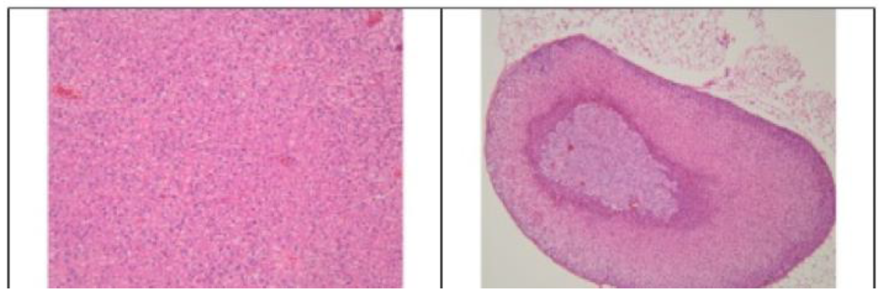
Low-dose groups (5–10 mg/kg): Hematoxylin-eosin staining revealed no significant tissue alterations in the myocardium, stomach, lungs, kidneys, or other major organs including the spleen, liver, adrenal glands, and cerebral cortex (Figure 1A-E). This indicates a favorable biocompatibility profile at low concentrations.
Figure 1A.
10 mg AuNP/kg/day. Left: Spleen – no significant changes (HE ob x10). Right: cerebral cortex, no significant changes (HE ob x20).
Figure 1A.
10 mg AuNP/kg/day. Left: Spleen – no significant changes (HE ob x10). Right: cerebral cortex, no significant changes (HE ob x20).
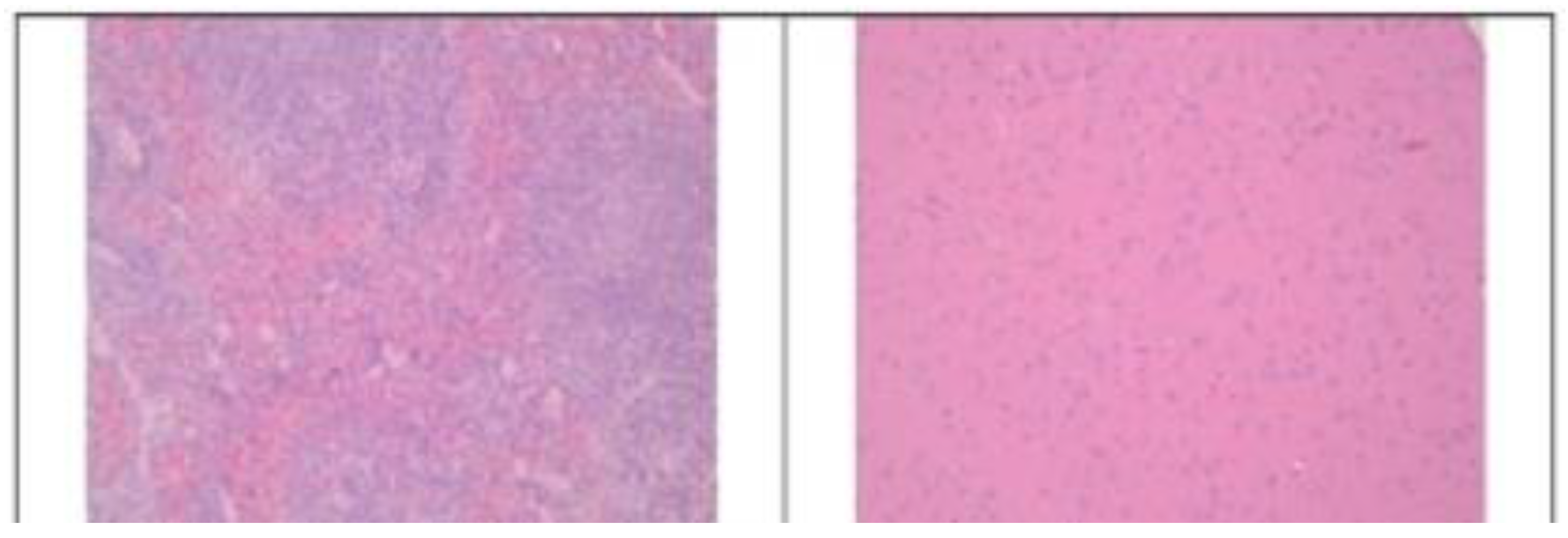
Figure 1B.
10 mg AuNP/kg/day. Myocardium: no significant changes. HE, obj x20 (left image), HE, obj x40 (right image).
Figure 1B.
10 mg AuNP/kg/day. Myocardium: no significant changes. HE, obj x20 (left image), HE, obj x40 (right image).
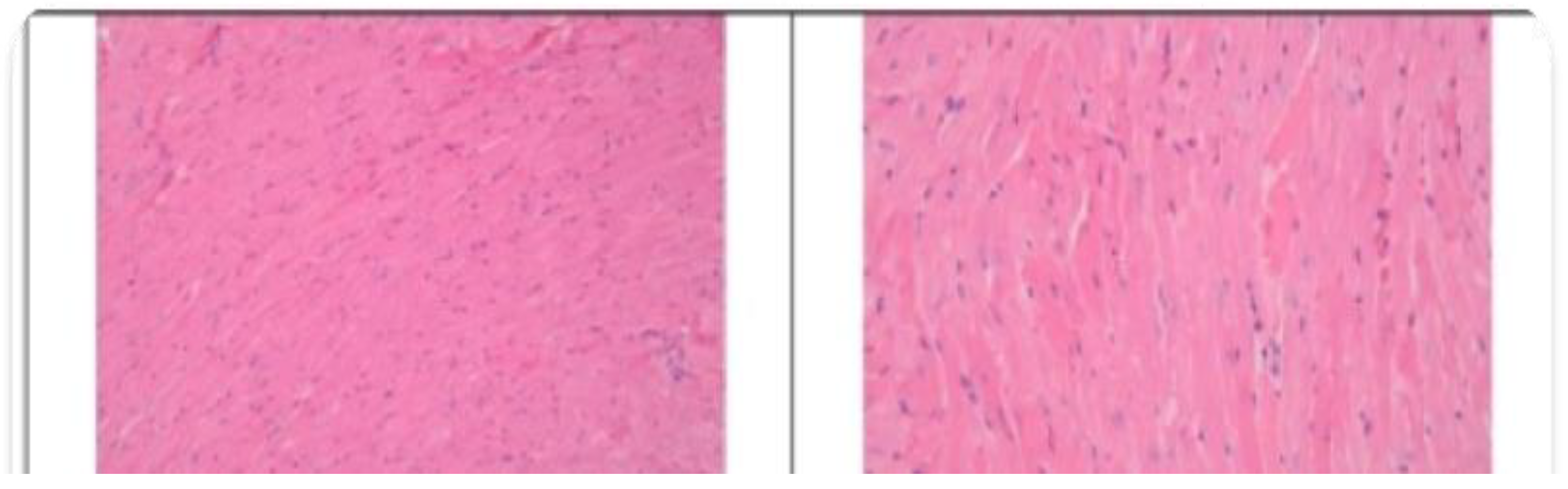
Figure 1C.
Stomach (transition area between fermentative and glandular segments): no significant changes. Rare yeast cells present in the superficial gastric mucosa. HE, obj x10 (left image), HE, obj x40 (right image).
Figure 1C.
Stomach (transition area between fermentative and glandular segments): no significant changes. Rare yeast cells present in the superficial gastric mucosa. HE, obj x10 (left image), HE, obj x40 (right image).
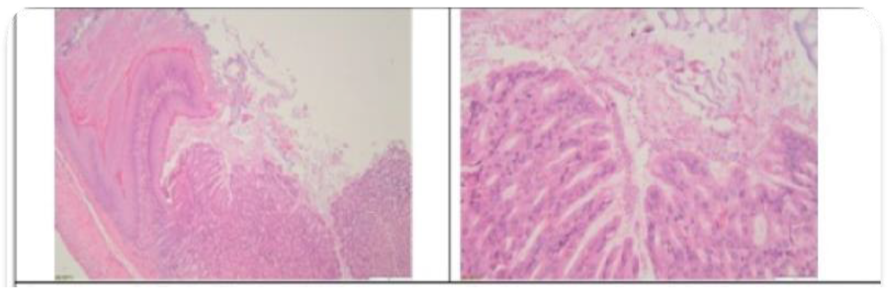
Figure 1D.
10 mg AuNP/kg/day. Left: Lung: no significant changes. HE, obj x10. Right: Renal cortex: no significant changes. HE, obj x10.
Figure 1D.
10 mg AuNP/kg/day. Left: Lung: no significant changes. HE, obj x10. Right: Renal cortex: no significant changes. HE, obj x10.
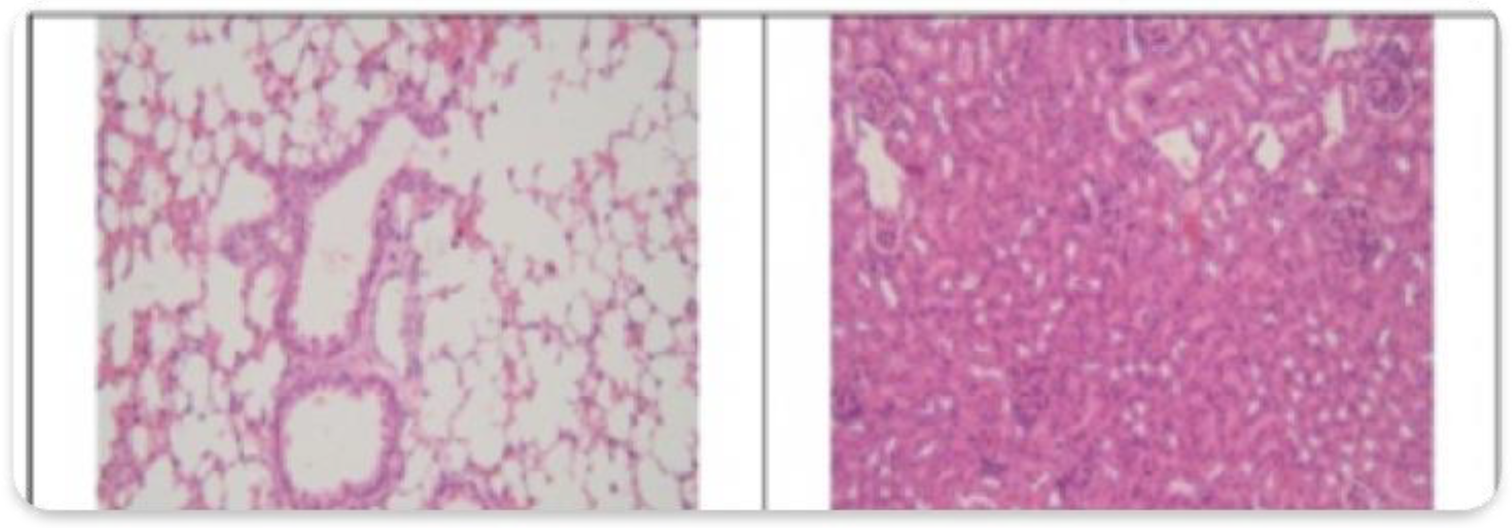
Figure 1E.
10 mg AuNP/kg/day. Left: Liver: no significant changes. HE, obj x10. Right: Adrenal gland: no significant changes. HE, obj x10.
Figure 1E.
10 mg AuNP/kg/day. Left: Liver: no significant changes. HE, obj x10. Right: Adrenal gland: no significant changes. HE, obj x10.
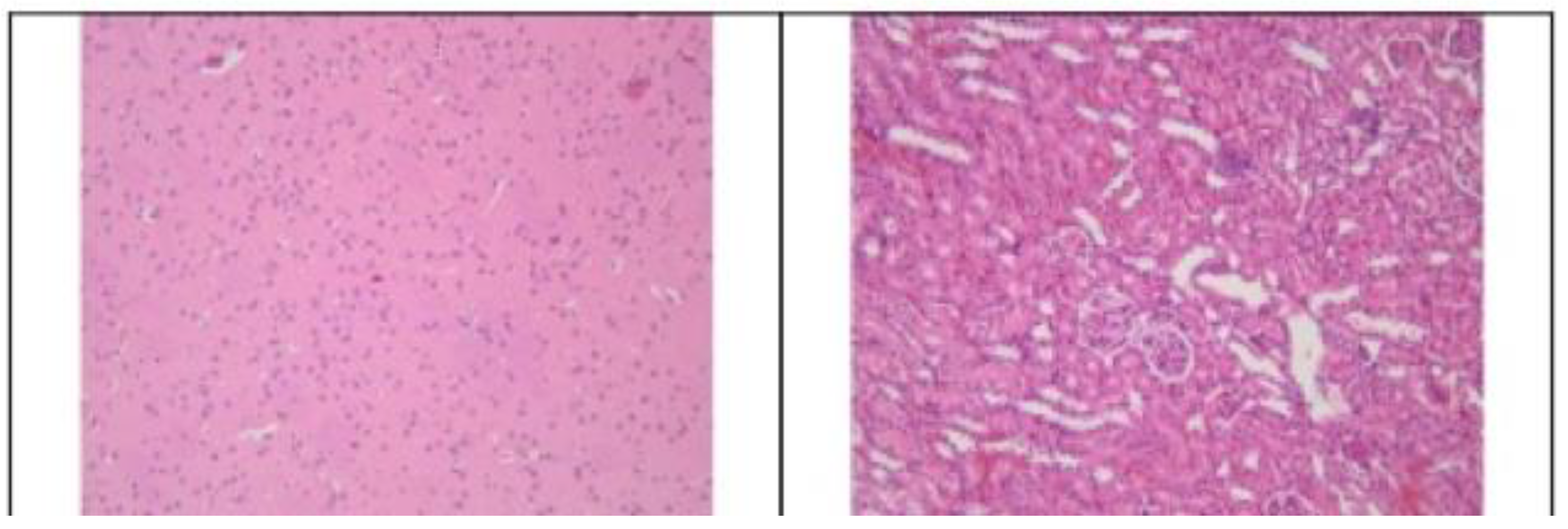
At the intermediate exposure level (20 mg/kg), histological examination revealed no significant structural abnormalities in the heart, stomach, or brain, suggesting minimal off-target effects in these organs (Figure 2A-C). Nonetheless, distinct histopathological features were observed in select tissues. In the renal cortex granular brown deposits were observed, likely representing transit or partial retention of administered material within tubular structures.
Figure 2A.
Histological Analysis – 20 mg CEA-AuNP/kg/day. (a) Myocardium: no changes. HE, obj x20. (b) Stomach (glandular-fermentative transition zone): no changes. HE, obj x4.
Figure 2A.
Histological Analysis – 20 mg CEA-AuNP/kg/day. (a) Myocardium: no changes. HE, obj x20. (b) Stomach (glandular-fermentative transition zone): no changes. HE, obj x4.
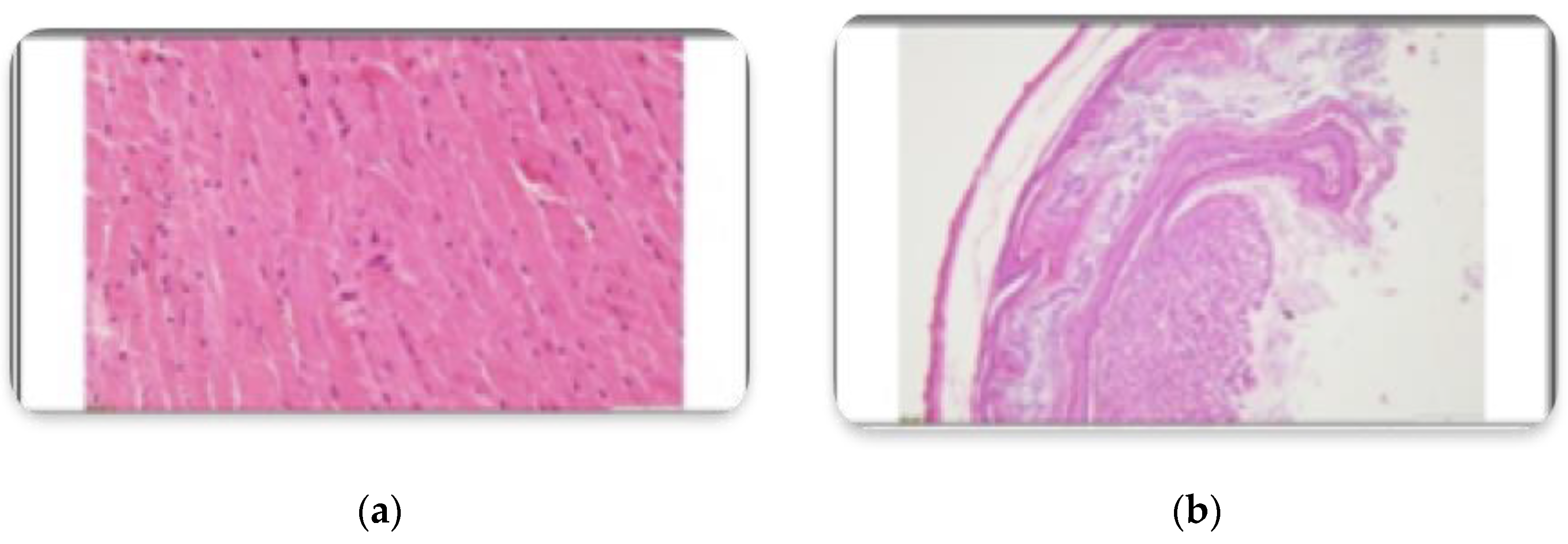
Figure 2B.
Histological Analysis – 20 mg CEA-AuNP/kg/day. Left: Renal cortex – multifocal, basophilic renal epithelial cells with brown granular appearance (possible lipofuscin). HE, obj x40. Right: Brain (cortex) – no significant changes. HE, obj x20.
Figure 2B.
Histological Analysis – 20 mg CEA-AuNP/kg/day. Left: Renal cortex – multifocal, basophilic renal epithelial cells with brown granular appearance (possible lipofuscin). HE, obj x40. Right: Brain (cortex) – no significant changes. HE, obj x20.
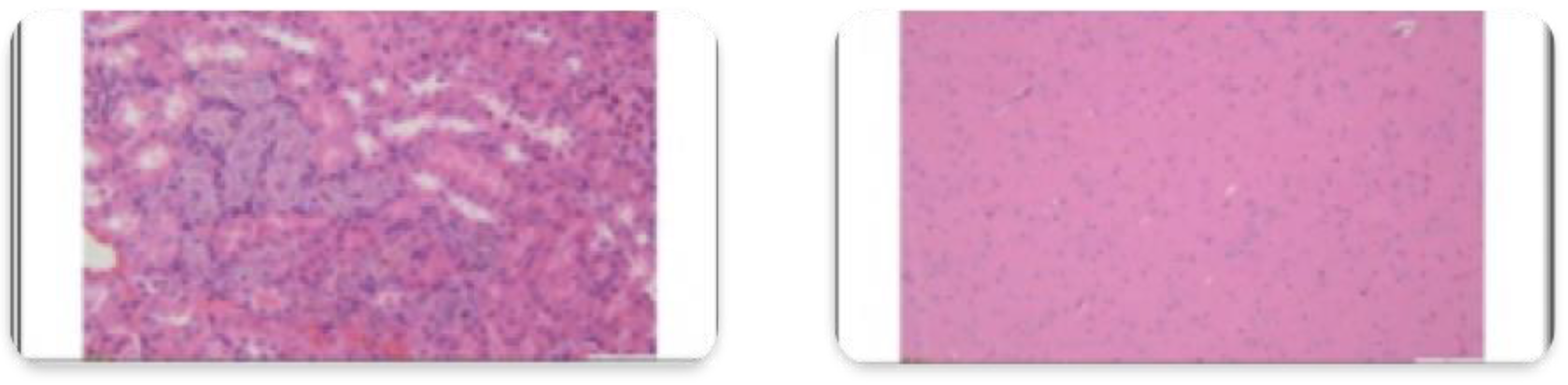
Figure 2C.
Histological Analysis – 20 mg CEA-AuNP/kg/day. Left: Liver: no significant changes. Foci of extramedullary changes HE, obj x20. Right: Spleen, no significant changes. Foci of extramedullary (regular) hematopoiesis of the erythroid and myeloid lines, in mixture with megakaryocytes, HE, ob x 20.
Identification of focal extramedullary hematopoiesis in the liver may be indicative of compensatory hematologic activity. The splenic tissues demonstrated physiologic hematopoietic activity along both erythroid and myeloid lineages, with interspersed megakaryocytes, consistent with an active yet unperturbed hematopoietic microenvironment.
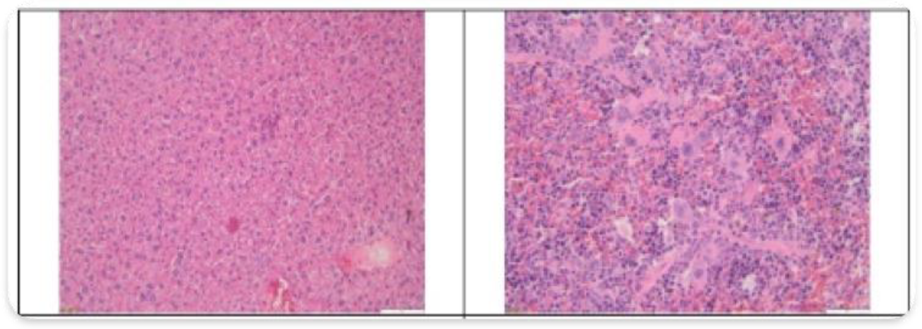
At the highest administered dose (50 mg/kg), histological evaluation revealed no detectable tissue damage in the brain, kidney, stomach, or intestinal mucosa, indicating a favorable safety profile in these organs (Figure 3A-E). However, notable histopathological alterations were observed in specific reticuloendothelial tissues: in the liver evidence of focal hepatocellular alterations, accompanied by mild periportal inflammatory infiltration, suggestive of early hepatic stress or immune activation. In the spleen, multifocal amyloid deposits were predominantly localized in the red pulp and marginal zones.
Figure 3A.
Histological Analysis – 50 mg CEA-AuNP/kg/day. Left: Myocardium: no significant changes. HE, obj x20. Right: Lung: no significant changes. HE, obj x20.
Figure 3A.
Histological Analysis – 50 mg CEA-AuNP/kg/day. Left: Myocardium: no significant changes. HE, obj x20. Right: Lung: no significant changes. HE, obj x20.
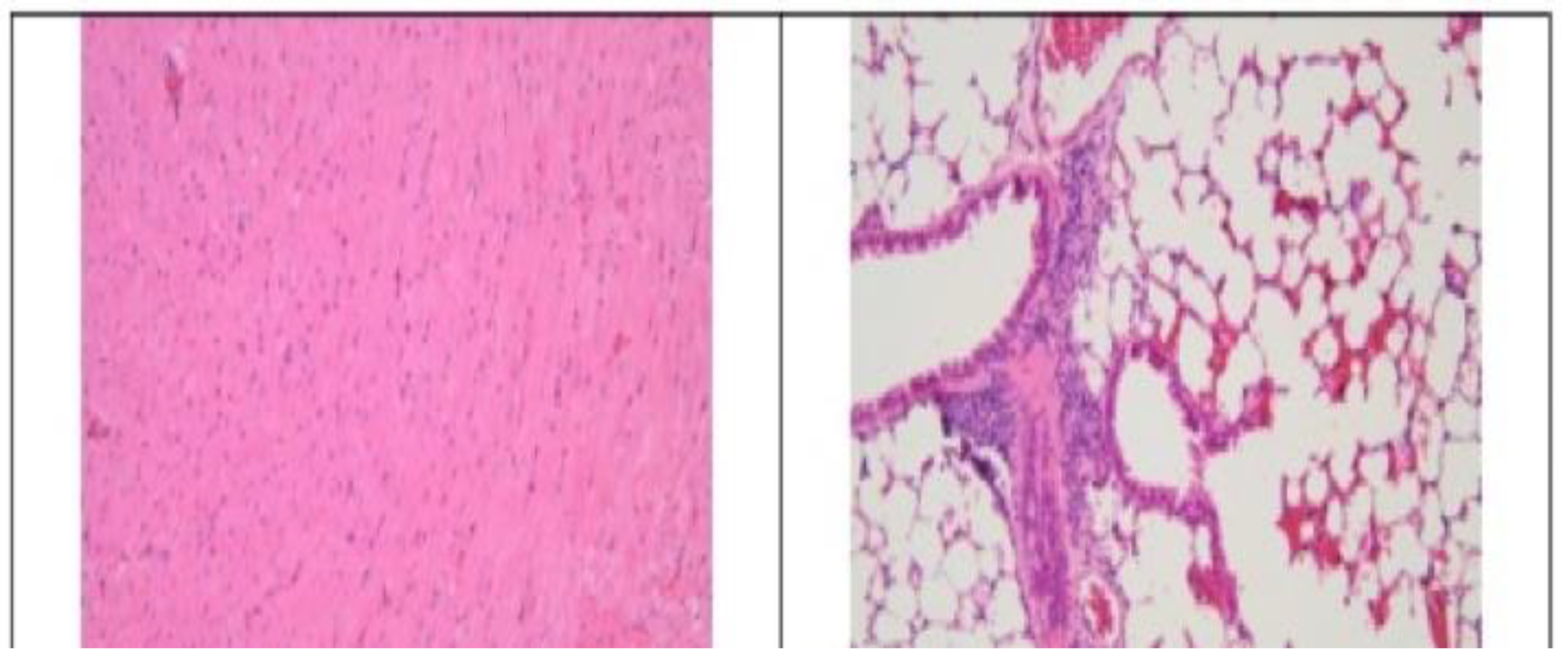
Figure 3B.
Histological Analysis – 50 mg CEA-AuNP/kg/day. Spleen: multifocal amyloidosis, predominantly in the perifollicular areas, with moderate compression atrophy of the peripheral zonal tissue. HE, obj x10 (left) and x40. (right).
Figure 3B.
Histological Analysis – 50 mg CEA-AuNP/kg/day. Spleen: multifocal amyloidosis, predominantly in the perifollicular areas, with moderate compression atrophy of the peripheral zonal tissue. HE, obj x10 (left) and x40. (right).
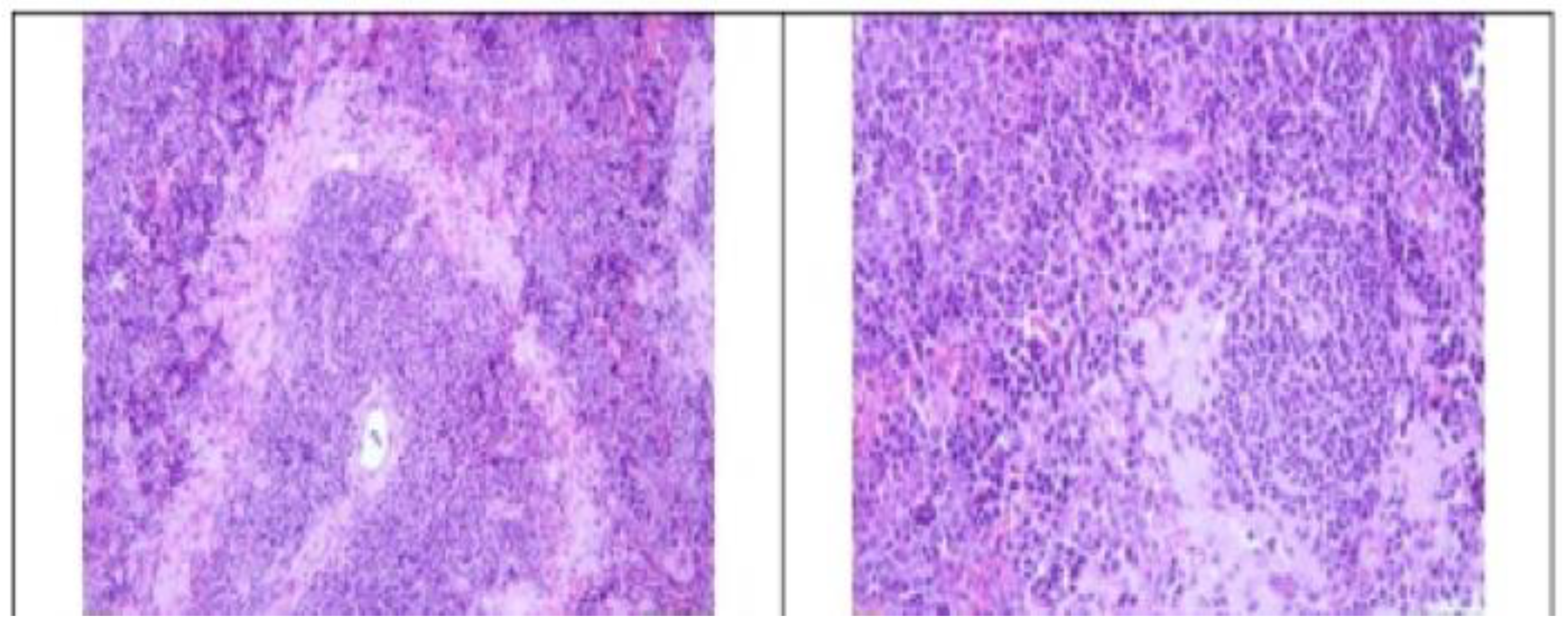
Figure 3C.
Histological Analysis – 50 mg CEA-AuNP/kg/day. Liver: hepatic amyloidosis, accumulation of eosinophilic extracellular material in the space of Disse, with moderate compression atrophy of the parenchyma. HE, obj x40.
Figure 3C.
Histological Analysis – 50 mg CEA-AuNP/kg/day. Liver: hepatic amyloidosis, accumulation of eosinophilic extracellular material in the space of Disse, with moderate compression atrophy of the parenchyma. HE, obj x40.
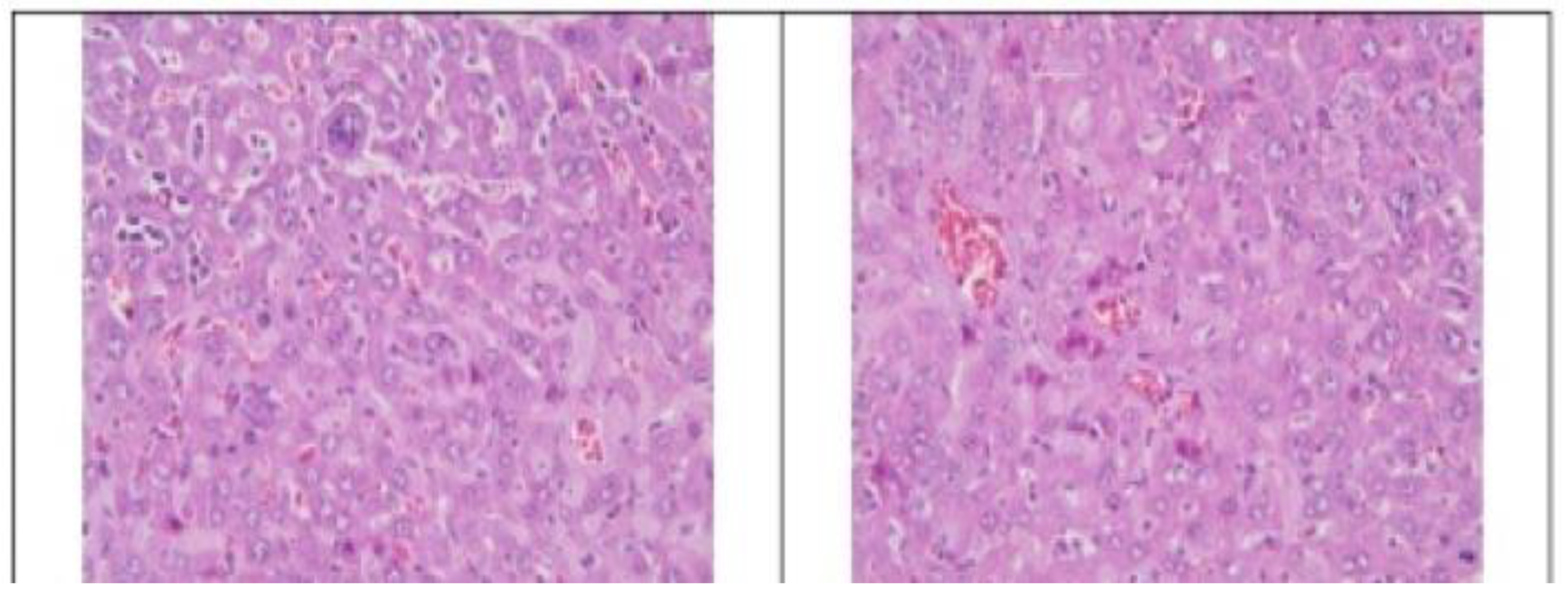
Figure 3D.
Histological Analysis – 50 mg CEA-AuNP/kg/day. Left: Stomach (transition zone glandular to fermentative stomach): no significant changes.HE. ob x 10. Right: Intestine: no significant changes. HE, ob x 20.
Figure 3D.
Histological Analysis – 50 mg CEA-AuNP/kg/day. Left: Stomach (transition zone glandular to fermentative stomach): no significant changes.HE. ob x 10. Right: Intestine: no significant changes. HE, ob x 20.
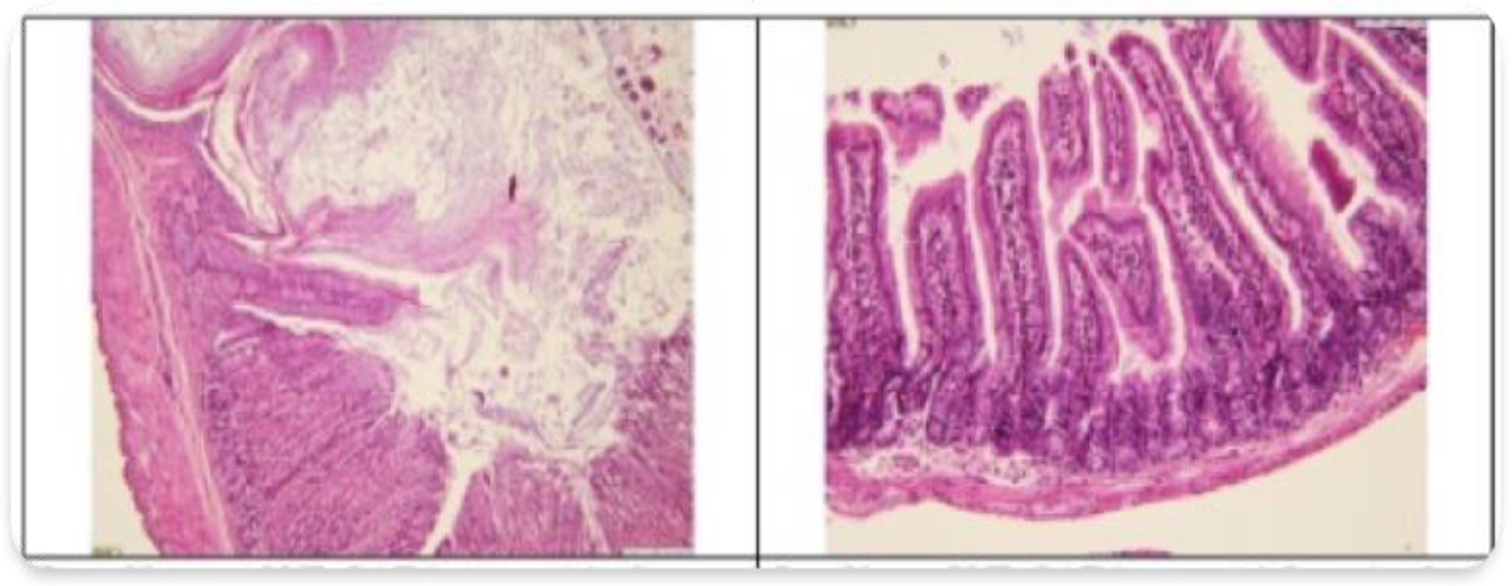
Figure 3E.
Histological Analysis – 50 mg CEA-AuNP/kg/day. Left: Cerebral cortex, no significant changes. HE, ob x 20. Right: Renal cortex: no significant changes. HE, ob x 10.
Figure 3E.
Histological Analysis – 50 mg CEA-AuNP/kg/day. Left: Cerebral cortex, no significant changes. HE, ob x 20. Right: Renal cortex: no significant changes. HE, ob x 10.
These features are consistent with chronic immune stimulation, potentially reflecting the spleen’s role in nanoparticle clearance and immune modulation. These findings highlight a tissue-specific response to high-dose exposure, with pronounced effects in organs involved in immune processing and detoxification.
3.3. Hyperspectral Dark-Field Microscopy
Hyperspectral dark-field microscopy enabled high-resolution spectral mapping of gold-based nanomaterials across multiple tissue types. Characteristic white light reflectance signatures, attributable to the optical properties of the nanoparticles, were detected with varying intensities depending on organ type and dosage.
Nanoparticle-associated spectral signals were prominently detected in the spleen (Figure 4), indicating substantial nanoparticle accumulation. This accumulation was consistent with prior in vitro internalization patterns (unpublished data).
Moderate presence in the liver (Figure 5) and myocardium, suggests partial uptake. CEA-AuNPs presence was minimal to undetectable in the kidneys, lungs, and cortical brain tissue, implying limited or no nanoparticle retention in these regions. Quantitative analysis demonstrated a dose-dependent relationship between signal intensity and nanoparticle accumulation, reinforcing the biodistribution profile inferred from organ-specific reflectance patterns.
3.4. Biochemical and Immunological Analysis
Cytokine profile analysis revealed organ-specific immune modulation following 30 days of CEA-AuNP administration, with distinct patterns emerging between lymphoid and parenchymal tissues. In the spleen, analysis demonstrated a predominantly anti-inflammatory response, characterized by a significant elevation of IL-10 levels relative to IL-12. This cytokine shift suggests macrophage activation and a cell-mediated immunological polarization, consistent with an immunostimulatory effect potentially supportive of a vaccine-like antitumoral response (Figure 6).
In contrast, the hepatic cytokine profile exhibited a more pro-inflammatory tendency, particularly at higher nanoparticle doses. Although both IL-10 and IL-12 levels were elevated, the absence of IL-10 predominance, as observed in the spleen, indicated a more balanced pro- and anti-inflammatory environment. Additionally, sporadic increases in TNF-α and IL-1β levels were noted, suggesting localized immune activation and early signs of hepatic functional stress. (Figure 7) These alterations were dose-dependent, with the most pronounced effects occurring in the 50 mg/kg group.
Collectively, these findings indicate that CEA-AuNP exposure leads to targeted immune activation within the spleen, conducive to an antitumoral nanovaccine effect, while maintaining controlled inflammatory responses within the liver at higher concentrations without evidence of systemic toxicity. Moreover, the results confirm a dose-dependent biodistribution profile and associated histological changes, particularly within reticuloendothelial tissues. A summary of cytokine modulation is presented in Table 2.
4. Discussion
The present in vivo study evaluated the safety, biodistribution, and immunological impact of CEA-AuNPs administered subacutely over 30 days in a murine model. Key findings include dose-dependent histological changes, splenic immune activation, and limited organ toxicity, suggesting a favorable immunostimulatory profile for potential vaccine development. Building upon these in vivo observations, the Discussion considers how prior preclinical validations and the present findings collectively support the immunological potential and biocompatibility of the CEA-AuNP nanovaccine platform.
Prior to in vivo testing, the CEA-AuNP nanoconstruct underwent extensive physicochemical and biological validation to ensure its suitability as a nanovaccine candidate (unpublished data). The successful synthesis and functionalization of gold nanoparticles (AuNPs) with CEA-derived peptides, confirmed through UV-Vis spectroscopy and dynamic light scattering (DLS) analyses, established a stable, monodisperse nanoplatform with favorable physicochemical properties [12,13]. These biophysical characteristics—specifically the plasmonic peak at 522 nm, post-functionalization hydrodynamic size of approximately 80 nm, and retention of colloidal stability—are consistent with literature describing effective peptide conjugation to metallic nanoparticles [14,15].
In vitro validation further confirmed the biocompatibility and functional capacity of the CEA-AuNP construct. Cell viability remained above 85% even at the highest tested dose (50 µg/mL), with a modest, dose-dependent decline likely reflecting adaptive cellular responses rather than toxicity [16,17]. The observed upregulation of caspase-3 activity suggested a controlled apoptotic response, a phenomenon beneficial in enhancing antigen presentation by macrophages [18]. Additionally, effective cellular internalization and perinuclear localization of the nanoconjugates were consistent with antigen trafficking toward proteasomal pathways essential for MHC-mediated immune activation [19].
Following this preclinical validation, the in vivo administration of CEA-AuNPs revealed consistent and promising immunological effects, particularly in splenic architecture, cytokine modulation, and systemic safety. The logical progression from in vitro validation to in vivo efficacy strengthens the translational relevance of the CEA-AuNP nanoconstruct. These in vivo findings provide important confirmation that the physicochemical integrity and immunological function observed in vitro are retained and effectively translated into a living system, supporting further development of the platform for cancer immunoprophylaxis.
The spleen exhibited the most pronounced response, both morphologically and functionally. Significant splenomegaly and histological alterations—such as lymphoid hyperplasia, active hematopoiesis, and the presence of megakaryocytes—were particularly evident in animals treated with intermediate and high doses of CEA-AuNPs. These changes indicate stimulation of both innate and adaptive immune compartments, consistent with an active immunological response to the nanoconstructs and CEA antigen exposure [20].
One of the notable histological findings was the development of splenic amyloidosis in the group exposed to the highest dose (50 mg/kg). Amyloidosis is characterized by extracellular deposition of amorphous proteinaceous material in multiple tissues, particularly within the red pulp and marginal zones of the spleen, as observed here. In murine models, inflammation-induced amyloidosis is predominantly mediated by serum amyloid A (SAA), an acute-phase reactant produced during systemic inflammatory responses [21]. Given the relatively young age of the experimental mice, the observed amyloid deposits are likely secondary to sustained immune activation triggered by prolonged nanoparticle exposure, rather than senile amyloidosis [22].
The detection of extramedullary hematopoiesis in both the spleen and liver at intermediate and high doses further supports systemic hematologic adaptation, possibly reflecting increased demands for immune cell production [23]. According to the literature, such reactive changes are non-neoplastic and occur in response to persistent immune stimulation [24,25].
Despite these histological changes, no overt tissue damage was detected in renal, pulmonary, or cerebral tissues across all groups. Minor findings, such as brown granular deposits in the renal cortex at intermediate doses, were isolated and did not indicate significant organ impairment. Hyperspectral imaging confirmed that gold nanoparticle accumulation was primarily localized to the spleen and liver, with lower presence in the heart and negligible accumulation in the kidneys and brain, consistent with the known biodistribution of nanoparticles favoring reticuloendothelial system [26,27].
This biodistribution pattern, coupled with the absence of major tissue damage, prompted a deeper evaluation of the immunological microenvironment through cytokine profiling. Cytokine analysis revealed a marked upregulation of IL-10 compared to IL-12 in the spleen, particularly at higher doses, suggesting a shift toward a regulatory macrophage phenotype (M2-like) and an anti-inflammatory immune environment [28]. Although IL-10 is often associated with immune tolerance, in the context of vaccine platforms, controlled IL-10 elevation may enhance antigen presentation and limit excessive inflammation, possibly leading to effective T-cell activation [9,29].
Similar trends were observed in hepatic tissue, where a slight increase in pro-inflammatory cytokines (TNF-α, IL-1β) was noted at higher doses. However, these effects were mild and sporadic, and the balanced IL-10/IL-12 ratio in the liver suggests organ-specific immunomodulation rather than overt toxicity [30].
The efficient localization of nanoparticles within macrophage-rich organs and the associated cytokine profiles align with prior studies on nanoparticle-based vaccine platforms, where gold nanoparticles facilitated enhanced antigen uptake and promoted robust cellular immune responses [10,31,32].
Taken together, the findings support the relative biocompatibility and immunogenic potential of CEA-AuNPs when administered at low and intermediate doses. The observed splenic immune activation, without evidence of systemic toxicity at these doses, highlights the promise of these nanoconstructs as carriers for tumor-associated antigens. Such platforms could improve antigen delivery, processing, and presentation, ultimately stimulating cytotoxic T-cell responses against CEA-expressing malignancies, including colorectal, pancreatic, and lung carcinomas [6,33,34].
The development of amyloidosis at high doses emphasizes the need for careful dose optimization in future applications. Further studies in tumor-bearing models are necessary to validate these findings, assess long-term efficacy and immunological memory, and explore potential synergy with immune checkpoint inhibitors [35]. Additionally, mechanistic investigations into antigen cross-presentation pathways (e.g., via MHC class I) will be critical for advancing translational development.
5. Conclusions
This present study demonstrates that CEA-functionalized gold nanoparticles exhibit favorable biodistribution, immunogenic activation, and a strong safety profile at low to intermediate doses in vivo. These findings, supported by prior in vitro validation, highlight the potential of CEA-AuNPs as a promising nanovaccine platform for colorectal cancer immunoprophylaxis. Further studies focusing on therapeutic efficacy, dose optimization, and combination immunotherapy strategies are warranted to advance this platform toward clinical translation.
Author Contributions
All authors have contributed equally to this work. Their involvement includes the conception and design of the study, data acquisition, analysis, and interpretation, as well as drafting the manuscript and critically revising it for important intellectual content. All authors have reviewed and approved the final version for publication. Each author accepts full responsibility for every aspect of the work, ensuring the accuracy and integrity of the entire study. Should any questions arise regarding the reliability or validity of any component, all authors are committed to promptly investigating and resolving such issues.
Funding
This research received no external funding.
Acknowledgments
The authors used artificial intelligence (AI)-based tools, including ChatGPT (OpenAI), DeepL Translator, and Perplexity AI, to assist with language editing, phrasing suggestions, and improving manuscript clarity. No AI tool was used for data analysis, scientific interpretation, or decision-making. All intellectual content, analysis, and conclusions are the original work of the authors, who assume full responsibility for the integrity and accuracy of the manuscript.
Conflicts of Interest
The authors declare no conflicts of interest.
Abbreviations
The following abbreviations are used in this manuscript:
| AuNP | Gold nanoparticle |
| CEA | Carcinoembrionic Antigen |
| CEA-AuNP | CEA functionalized gold nanoparticles |
| HE | Hematoxilin & Eozine |
References
- Granados-Romero JJ, Valderrama-Treviño AI, Contreras-Flores EH, Barrera-Mera B, Herrera Enríquez M, Uriarte-Ruíz K, et al. Colorectal cancer: a review. Int J Res Med Sci. 2017;5(11):4667. [CrossRef]
- Stintzing S. Management of colorectal cancer. F1000prime reports. 2014;6:108. [CrossRef]
- Mármol I, Sánchez-de-Diego C, Pradilla Dieste A, Cerrada E, Rodriguez Yoldi MJ. Colorectal carcinoma: a general overview and future perspectives in colorectal cancer. International journal of molecular sciences. 2017;18(1):197. [CrossRef]
- Fan T, Zhang M, Yang J, Zhu Z, Cao W, Dong C. Therapeutic cancer vaccines: advancements, challenges and prospects. Signal Transduction and Targeted Therapy. 2023;8(1):450. [CrossRef]
- Bhagat A, Lyerly HK, Morse MA, Hartman ZC. CEA vaccines. Hum Vaccin Immunother. 2023;19(3):2291857.
- Hammarström S, editor The carcinoembryonic antigen (CEA) family: structures, suggested functions and expression in normal and malignant tissues. Seminars in cancer biology; 1999: Elsevier. [CrossRef]
- Kaczmarek M, Poznańska J, Fechner F, Michalska N, Paszkowska S, Napierała A, et al. Cancer Vaccine Therapeutics: Limitations and Effectiveness-A Literature Review. Cells. 2023;12(17). [CrossRef]
- Almeida JP, Figueroa ER, Drezek RA. Gold nanoparticle mediated cancer immunotherapy. Nanomedicine. 2014;10(3):503–14. [CrossRef]
- Ferrando RM, Lay L, Polito L. Gold nanoparticle-based platforms for vaccine development. Drug Discovery Today: Technologies. 2020;38:57–67. [CrossRef]
- Dykman L, Khlebtsov N. Gold nanoparticles in biomedical applications: recent advances and perspectives. Chemical Society Reviews. 2012;41(6):2256–82. [CrossRef]
- Prophet EB. Laboratory methods in histotechnology: American Registry of Pathology; 1992.
- Ren H, Jia W, Xie Y, Yu M, Chen Y. Adjuvant physiochemistry and advanced nanotechnology for vaccine development. Chemical Society Reviews. 2023;52(15):5172–254. [CrossRef]
- Zaccariotto GdC, Bistaffa MJ, Zapata AMM, Rodero C, Coelho F, Quitiba JoVBo, et al. Cancer Nanovaccines: Mechanisms, Design Principles, and Clinical Translation. ACS Nano. 2025. [CrossRef]
- Zhang Y, Huang R, Zhu X, Wang L, Wu C. Synthesis, properties, and optical applications of noble metal nanoparticle-biomolecule conjugates. Chinese Science Bulletin. 2012;57:238–46. [CrossRef]
- Rai M, Ingle AP, Gupta I, Brandelli A. Bioactivity of noble metal nanoparticles decorated with biopolymers and their application in drug delivery. International journal of pharmaceutics. 2015;496(2):159–72. [CrossRef]
- Zhang Q, Pi J, Woods CG, Jarabek AM, Clewell III HJ, Andersen ME. Hormesis and adaptive cellular control systems. Dose-Response. 2008;6(2):dose–response. 07–028. Zhang.
- Zhang Q, Bhattacharya S, Pi J, Clewell RA, Carmichael PL, Andersen ME. Adaptive posttranslational control in cellular stress response pathways and its relationship to toxicity testing and safety assessment. Toxicological Sciences. 2015;147(2):302–16. [CrossRef]
- Byrne HJ, Maher MA. Numerically modelling time and dose dependent cytotoxicity. Computational Toxicology. 2019;12:100090. [CrossRef]
- Wieczorek M, Abualrous ET, Sticht J, Álvaro-Benito M, Stolzenberg S, Noé F, et al. Major histocompatibility complex (MHC) class I and MHC class II proteins: conformational plasticity in antigen presentation. Frontiers in immunology. 2017;8:292. [CrossRef]
- Roy AA, Pokale R, Mukharya A, Jadhav SR, Naik GARR, Rachana S, et al. Innovative Organic Core–Nonmagnetic Shell Nanoconstructs: Pioneering Precision in Cancer Theranostics. Core-Shell Nano Constructs for Cancer Theragnostic: Current Scenario, Challenges and Regulatory Aspects: Springer; 2025. p. 415–49.
- Ge F, Yao J, Fu X, Guo Z, Yan J, Zhang B, et al. Amyloidosis in transgenic mice expressing murine amyloidogenic apolipoprotein A-II (Apoa2c). Lab Invest. 2007;87(7):633–43. [CrossRef]
- Kakinen A, Javed I, Davis TP, Ke PC. In vitro and in vivo models for anti-amyloidosis nanomedicines. Nanoscale Horizons. 2021;6(2):95–119. [CrossRef]
- Cenariu D, Iluta S, Zimta A-A, Petrushev B, Qian L, Dirzu N, et al. Extramedullary Hematopoiesis of the Liver and Spleen. Journal of Clinical Medicine. 2021;10(24):5831. [CrossRef]
- Kim, CH. Homeostatic and pathogenic extramedullary hematopoiesis. Journal of blood medicine. 2010:13–9. [CrossRef]
- Yamamoto K, Miwa Y, Abe-Suzuki S, Abe S, Kirimura S, Onishi I, et al. Extramedullary hematopoiesis: Elucidating the function of the hematopoietic stem cell niche. Molecular medicine reports. 2016;13(1):587–91. [CrossRef]
- Li S-D, Huang L. Pharmacokinetics and biodistribution of nanoparticles. Molecular pharmaceutics. 2008;5(4):496–504. [CrossRef]
- Li S-D, Huang L. Nanoparticles evading the reticuloendothelial system: role of the supported bilayer. Biochimica et Biophysica Acta (BBA)-Biomembranes. 2009;1788(10):2259–66. [CrossRef]
- Saraiva M, O’garra A. The regulation of IL-10 production by immune cells. Nature reviews immunology. 2010;10(3):170–81. [CrossRef]
- Mosser DM, Edwards JP. Exploring the full spectrum of macrophage activation. Nature reviews immunology. 2008;8(12):958–69. [CrossRef]
- Ma X. TNF-α and IL-12: a balancing act in macrophage functioning. Microbes and Infection. 2001;3(2):121–9. [CrossRef]
- He J-s, Liu S-j, Zhang Y-r, Chu X-d, Lin Z-b, Zhao Z, et al. The application of and strategy for gold nanoparticles in cancer immunotherapy. Frontiers in Pharmacology. 2021;12:687399. [CrossRef]
- Sani A, Cao C, Cui D. Toxicity of gold nanoparticles (AuNPs): A review. Biochemistry and biophysics reports. 2021;26:100991. [CrossRef]
- Finn, OJ. Cancer immunology. New England Journal of Medicine. 2008;358(25):2704–15. [CrossRef]
- Chen DS, Mellman I. Elements of cancer immunity and the cancer–immune set point. Nature. 2017;541(7637):321–30. [CrossRef]
- Thomas EM, Wright JA, Blake SJ, Page AJ, Worthley DL, Woods SL. Advancing translational research for colorectal immuno-oncology. British Journal of Cancer. 2023;129(9):1442–50. [CrossRef]
- Gambinossi F, Mylon SE, Ferri JK. Aggregation kinetics and colloidal stability of functionalized nanoparticles. Advances in colloid and interface science. 2015;222:332–49. [CrossRef]
- Shafiee MAM, Asri MAM, Alwi SSS. Review on the In Vitro Cytotoxicity Assessment in Accordance to the International Organization for Standardization (ISO). Malaysian Journal of Medicine & Health Sciences. 2021;17(2).
- Wang Y, Pi C, Feng X, Hou Y, Zhao L, Wei Y. The influence of nanoparticle properties on oral bioavailability of drugs. International Journal of Nanomedicine. 2020:6295–310. [CrossRef]
- den Haan JM, Arens R, van Zelm MC. The activation of the adaptive immune system: cross-talk between antigen-presenting cells, T cells and B cells. Immunology letters. 2014;162(2):103–12. [CrossRef]
Figure 4.
Hyperspectral microscopy images (Cytoviva) from the splenic level. Left: Control. Center: 5 mg CEA-AuNP/kg/day. Right: numerous white light reflectance spots are observed for the 50 mg CEA-AuNP/kG body/day batch.
Figure 4.
Hyperspectral microscopy images (Cytoviva) from the splenic level. Left: Control. Center: 5 mg CEA-AuNP/kg/day. Right: numerous white light reflectance spots are observed for the 50 mg CEA-AuNP/kG body/day batch.
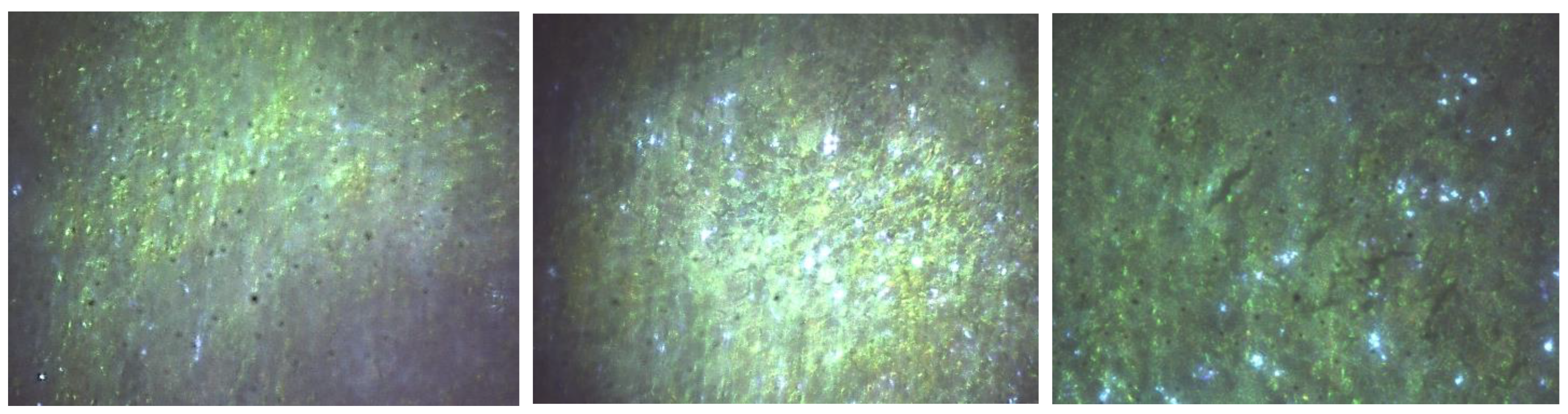
Figure 5.
Hyperspectral microscopy images (Cytoviva) of the liver. Left: Control. Center: 5 mg CEA-AuNP/kg/day. Right: numerous white light reflectance spots are observed for the 50 mg CEA-AuNP/kG body/day batch.
Figure 5.
Hyperspectral microscopy images (Cytoviva) of the liver. Left: Control. Center: 5 mg CEA-AuNP/kg/day. Right: numerous white light reflectance spots are observed for the 50 mg CEA-AuNP/kG body/day batch.
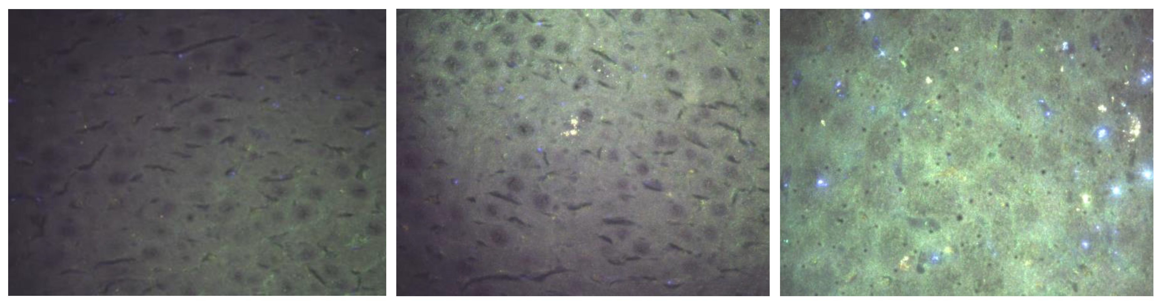
Figure 6.
Splenic cytokine expression following CEA-AuNP administration at increasing doses. Notable trends include a dose-dependent increase in IL-10 levels, suggesting anti-inflammatory and immunomodulatory effects. IL-4 levels decreased at intermediate doses but rose sharply at 50 mg/kg. IL-12 peaked at 5 mg/kg and declined at higher doses, indicating a shift in immune activation balance. Pro-inflammatory markers IL-1β and TNF-α showed variable responses, while IFN-γ increased progressively with dose. These profiles support a dynamic immunological response modulated by nanoparticle dosage.
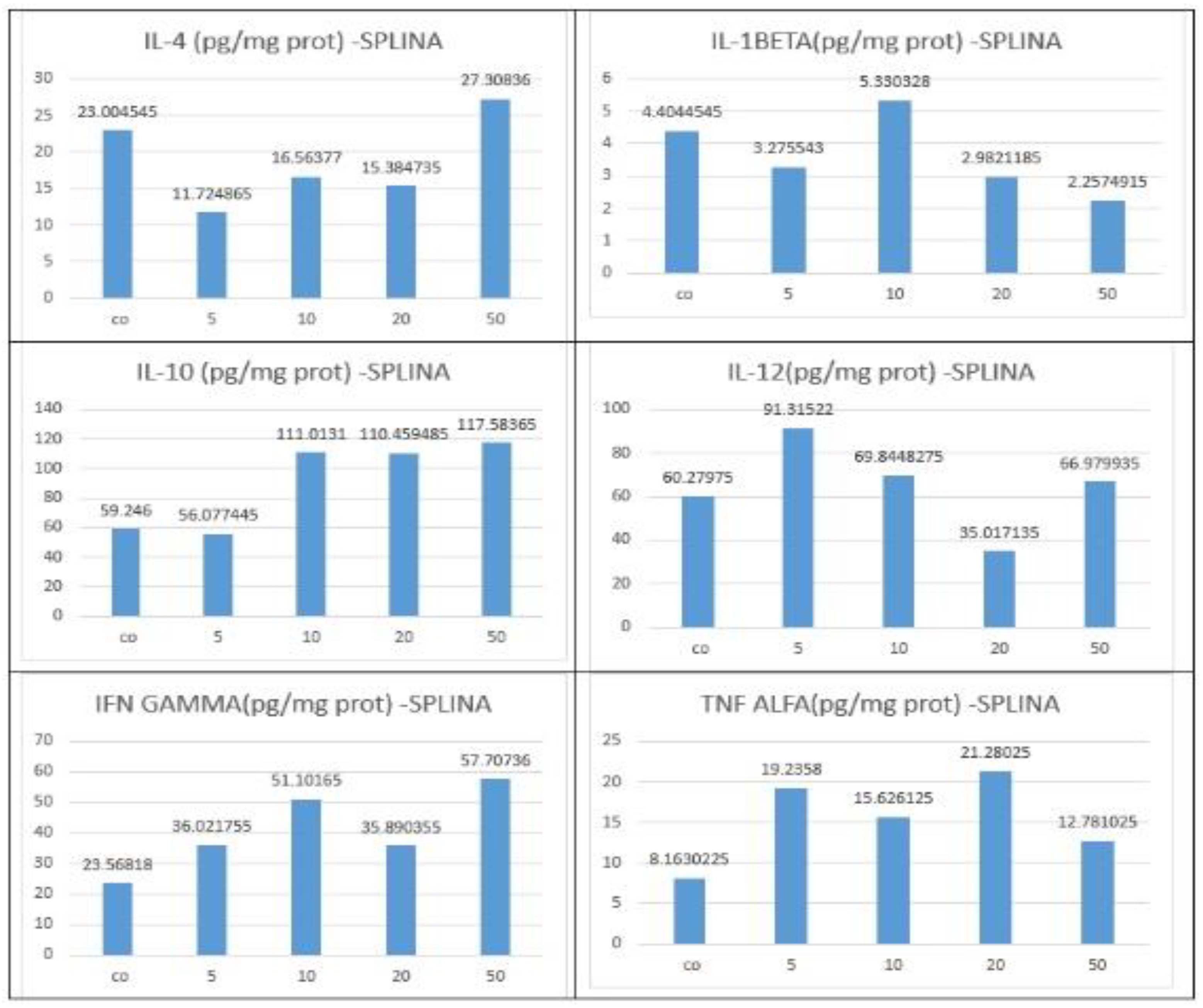
Figure 7.
Hepatic cytokine expression following CEA-AuNP administration at increasing doses. IL-10 levels remained elevated across all doses, peaking at 10 mg/kg, indicating sustained anti-inflammatory signaling. IL-4 expression decreased at intermediate doses but rebounded by 50 mg/kg. IL-12 and IL-1β levels declined at 20 and 50 mg/kg, suggesting suppression of some pro-inflammatory pathways. In contrast, TNF-α exhibited a strong dose-dependent increase, with the highest expression at 50 mg/kg. IFN-γ responses were variable, with a transient spike at 5 mg/kg. These hepatic cytokine dynamics suggest a more balanced or mildly pro-inflammatory immune profile, particularly at higher doses, potentially reflecting localized immune activation associated with hepatic nanoparticle processing.
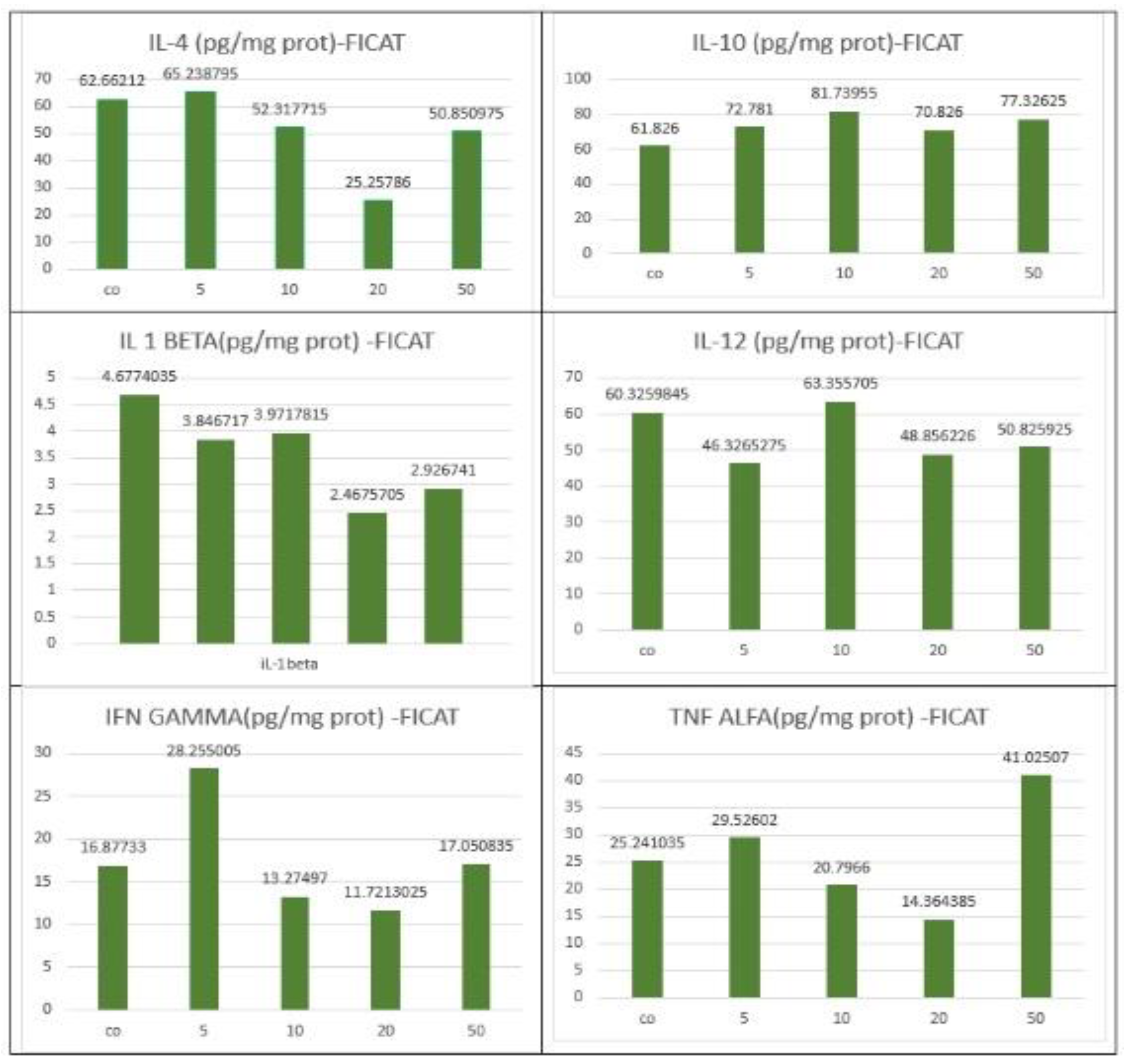

Table 1.
The composition of the experimental groups.
| Group | Treatment | Dose |
| Group 1, n=6 | CEA-AuNPs in PBS | 50 mg/kg |
| Group 2, n=6 | CEA-AuNPs in PBS | 20 mg/kg |
| Group 3, n=6 | CEA-AuNPs in PBS | 10 mg/kg |
| Group 4, n=6 | CEA-AuNPs in PBS | 5 mg/kg |
| Control Group, n=6 | PBS (vehicle control) | 0 mg/kg |
Table 2.
Summary of Cytokine Profile Changes.
| Organ | Cytokine | Observed Change | Dose Dependency | Interpretation |
|---|---|---|---|---|
| Spleen | IL-10 | Increased significantly vs IL-12 | Dose-dependent | Anti-inflammatory activation (M2-like) |
| Spleen | IL-12 | Mild increase | Present but less pronounced | Balanced Th1 response |
| Liver | IL-10 | Increased | Moderate at higher doses | Partial anti-inflammatory response |
| Liver | IL-12 | Increased | Parallel to IL-10 | Mixed immune activation |
| Liver | TNF-α | Sporadic increase | Most evident at 50 mg/kg | Early signs of hepatic immune stress |
| Liver | IL-1β | Sporadic increase | Not consistent at lower doses | Localized inflammatory activation |
Disclaimer/Publisher’s Note: The statements, opinions and data contained in all publications are solely those of the individual author(s) and contributor(s) and not of MDPI and/or the editor(s). MDPI and/or the editor(s) disclaim responsibility for any injury to people or property resulting from any ideas, methods, instructions or products referred to in the content. |
© 2025 by the authors. Licensee MDPI, Basel, Switzerland. This article is an open access article distributed under the terms and conditions of the Creative Commons Attribution (CC BY) license (http://creativecommons.org/licenses/by/4.0/).
Copyright: This open access article is published under a Creative Commons CC BY 4.0 license, which permit the free download, distribution, and reuse, provided that the author and preprint are cited in any reuse.


