
Submitted:
06 May 2025
Posted:
07 May 2025
You are already at the latest version
Abstract
Intervertebral disc disease is the most common disease of the spine in dogs and a main cause of pain and neurologic dysfunction. This article reviews fundamental aspects of the pathophysiology, clinical presentation, diagnosis, and treatment of disc extrusions. Chondroid metaplasia of the nucleus pulposus is the central process in disc degeneration. The clinical presentation varies considerably depending on breed, the location of the disc extrusion and the degree of neurological damage. Advanced imaging techniques, such as computed tomography and magnetic resonance imaging, have greatly improved diagnosis, being magnetic resonance considered the gold standard. As for treatment, both medical and surgical management are effective options depending on the degree of neurological damage and the initial response to conservative treatment. This comprehensive analysis underlines the importance of a multidisciplinary approach to optimize the quality of life of patients affected by intervertebral disc disease.
Keywords:
chondroid metaplasia
; computed tomography
; disc extrusion
; dog
; fenestration
; intervertebral disc disease
; magnetic resonance
; myelomalacia
; neurosurgery
; spinal cord injury
1. Introduction
Intervertebral disc disease (IVDD) is the most common disease of the spinal cord in dogs and is considered one of the main causes of pain and neurological dysfunction in this species. This disease is characterized by the progressive degeneration of the intervertebral discs, crucial structures for shock absorption, flexibility and stability of the spine [1,2].
Intervertebral disc (IVD) degeneration is characterized by chondroid metaplasia of the nucleus pulposus, a process that occurs in both chondrodystrophic and non-chondrodystrophic dogs. However, in chondrodystrophic canine breeds, expression of a fibroblast growth factor 4 retrogene (FGF4) on chromosome 12 is associated with a significant acceleration of disc degeneration [3,4]. As a consequence of IVD degeneration, mineralized disc material can extrude into the spinal canal, where it causes compression of the spinal cord and nerve roots. This phenomenon, known as disc extrusion or Hansen type I intervertebral disc herniation, is the most common form of disc herniation in dogs and constitutes one of the main causes of neurological signs in these animals [5,6,7].
Because of contusion and compression of the spinal cord and nerve roots, disc extrusions can cause pain, ataxia, paresis or plegia, and loss of sphincter control, depending on the severity of the lesion. The impact of this disease on the animal's quality of life is significant, and its management requires prompt and appropriate intervention to minimize neurological damage and alleviate clinical signs [6,7,8].
The diagnosis of IVDD has improved considerably with the advancement of imaging techniques. Currently, computed tomography (CT) and magnetic resonance imaging (MRI) are the most widely used diagnostic tools, as they allow detailed visualization of the spine and disc lesions, facilitating accurate localization and assessment of the degree of compression of nerve structures. However, definitive diagnosis usually requires a multimodal approach combining clinical findings, neurological evaluation and imaging studies [9].
The treatment of dogs affected by IVDD can be medical or surgical, depending on several factors, such as the degree of neurological damage or the degree of clinical response of the dog to conservative treatment, among others. The prognosis varies considerably depending on the degree of neurological damage and the speed with which the disease is diagnosed and treated. In cases where intervention is early and effective, the results can be very positive, with complete recovery of neurological function in many cases. However, in severely injured dogs, the prognosis for recovery of ambulatory function and sphincter control can be guarded to severe [1,2].
The aim of this review article is to provide a comprehensive analysis of the fundamental aspects of IVDD in dogs, focusing on the pathophysiology, clinical presentation, the most effective diagnostic strategies and current treatments for disc extrusions (Hansen type I herniations). Through this review, we seek to provide a comprehensive and updated view of the disease, based on the most recent scientific literature, in order to improve the knowledge and clinical management of this pathology in veterinary practice.
2. Topic Presentation
2.1. Pathophysiology
Degeneration of the IVD is defined by chondroid metaplasia of the nucleus pulposus, a process that occurs in both chondrodystrophic and non-chondrodystrophic dogs. During the initial stages of this degenerative process, the nucleus pulposus undergoes remarkable cellular changes, where clusters of notochordal cells are replaced by chondrocytes and their corresponding extracellular matrix. This matrix presents similar characteristics to hyaline cartilage and is mainly composed of disorganized collagen fibers. Simultaneously, a decrease in the content of glycosaminoglycans is observed, while the proportion of collagen increases (Figure 1) [10].
To assess the degree of IVD degeneration, Bergknut et al. (2013) proposed a classification scheme based on postmortem analysis of complete intervertebral segments. Subsequently, Kranenburg et al. (2013) adapted this model for application in surgical biopsies, facilitating its usefulness in the clinical setting [11,12]. Recently, a tool to assess the degree of disc degeneration with T2-weighted imaging using the Pfirrmann classification scheme has been reported [13,14,15].
This metaplasia and chondroid calcification may eventually lead to a type of IVD herniation, known as disc extrusion or Hansen type I herniation, in which the degenerated nucleus pulposus extrudes acutely through a ruptured fibrous ring into the spinal canal [16]. The other types of herniations described in the canine species, which we will not address in this review, are: Hansen type II disc herniation, acute non-compressive nucleus pulposus extrusion, acute hydrated nucleus pulposus extrusion, acute intradural/intramedullary nucleus pulposus extrusion, acute nucleus pulposus extrusion with extensive epidural hemorrhage, traumatic intervertebral disc extrusion [5].
In cases where disc material is extruded into the epidural space, it is common to observe an inflammatory response accompanied by hemorrhage. This inflammation is characterized by predominant infiltration of lymphocytes and macrophages into the extruded disc material. It has been suggested that this inflammatory response is triggered by the exposure of antigenic components of the nucleus pulposus which, upon herniation into the vertebral canal, activate the immune system [17].
However, inflammatory infiltrates have not been identified in histological specimens of complete functional spinal cord units, which explains their exclusion in the previously mentioned classification schemes. It has been proposed that acute inflammation could exacerbate clinical signs through extradural tissue swelling generated by inflammatory edema and the resulting compression on the dura mater. Furthermore, in the event that the dura is compromised, inflammatory mediators could directly affect the spinal cord [12].
It is relevant to note that no correlation has been established between the amount of extruded disc material and the degree of IVD degeneration with the degree of neurological injury of the IVDD. This suggests that current histological classification systems have limitations in predicting the functional consequences of IVDD, since they do not integrate factors such as trauma, neural compression, or inflammatory response, necrosis and hemorragy (Figure 2) [11,12].
In this context, the authors of this article are developing an investigation in which a significant correlation between the degree of inflammation and the degree of degeneration has been identified in a canine population with disc extrusions. This finding suggests that it would be necessary to study the possible usefulness of including the degree of inflammation in the histological classification schemes of the IVD to improve the predictive capacity regarding the functional impact of the disease, opening new perspectives for the selection of the recommended drugs for its treatment.
2.2. Clinical Presentation
The overall prevalence of intervertebral disc herniation in dogs is estimated at 2%, with its clinical presentation varying according to different factors, among them there is considerable variability depending on the breed analyzed [18,19]. The most commonly affected chondrodystrophic dog breeds include the Dachshund, Pekingese, French bulldog, and Beagle, among others. These breeds, particularly young and middle-aged individuals, are at increased risk of developing intervertebral disc extrusions due to their genetic predisposition and early degeneration of the nucleus pulposus. Although less frequent, this type of herniation can also occur in non-chondrodystrophic dogs. In chondrodystrophic breeds, the incidence of disc herniation peaks between 3 and 7 years old, while in non-chondrodystrophic breeds, it peaks between 6 and 8 years old [1,20].
Several studies have indicated that neutered males and females are at increased risk of developing IVD herniation compared to intact females, although the results vary between investigations. However, the risk of disc extrusion does not appear to be associated with factors such as body weight, physical condition, or activity level of the animal [21].
Disc extrusion can occur in any segment of the spine, most commonly between T11-T12 and L2-L3, which varies across studies. Clinical signs depend on the location of the herniation and their severity varies from mild spinal pain without neurological deficits to complete paralysis with loss of sensation (view supplementary material). To assess the severity of the neurological injury, the modified Frankel scale is commonly used, which classifies patients from grade 1 (hyperesthesia without neurological deficits) to grade 5 (plegia without deep pain sensation) (Table 1). The typical clinical presentation of disc extrusion includes acute-onset myelopathy, with progression of neurological deficits and characterized by significant pain [1,22,23].
Cervical disc extrusion occurs in 12.9% to 25.4% of cases of IVD herniation in dogs. Between 15% and 61% of patients with cervical herniation exhibit clinical signs of cervical hyperesthesia, often without further neurologic deficits. This lower incidence of neurologic deficits, compared with thoracolumbar hernias, is attributed to the larger diameter of the vertebral canal in the cervical region. In cases of cervical herniation, unilateral or bilateral lameness caused by nerve root compression (called root sign) is seen in 15% to 50% of cases. Although rare, serious complications may include profound sensory loss and respiratory distress. The most frequently affected disc spaces in small-breed dogs are C2-C3, while in large breeds C6-C7 predominate. In general, C5-C6 and C6-C7 are the disc spaces most commonly involved in cervical herniation of the IVD [25,26,27,28].
Thoracolumbar extrusions account for 66% to 87% of cases of IVD herniation in dogs. The most commonly affected disc spaces in chondrodystrophic breeds are T12-T13 and T13-L1, while in non-chondrodystrophic breeds, T13-L1 and L1-L2. These hernias can generate a wide range of clinical signs, from mild hyperesthesias to paraplegia with or without profound sensory loss. Lesions at the nerve root level may also occur in the lumbosacral region, and usually present with lameness. In some acute cases, they may present with spinal shock, in which spinal reflexes caudal to a severe acute injury may be diminished or absent, or with Schiff-Sherrington’s posture, which are of no prognostic value. Progressive myelomalacia (PMM) is the most severe complication that can occur in dogs with thoracolumbar disc extrusions. Its incidence is between 10-17.5% of paraplegic dogs with absence of deep sensibility, and can reach 33% in French Bulldogs. In this breed, this complication is more frequent in caudal lumbar location. Although it is a clinical diagnosis in many cases, it is important to recognize its characteristics on MRI, which will be discussed later [1,2,29-31].
As mentioned at the beginning of this section, the clinical presentation is highly variable between breeds [32,33]. Thus, it is more interesting to study each breed separately. In our center, we have reviewed the clinical presentation of disc extrusions in a population of 186 French Bulldogs with disc extrusions. In this population, hernias were more frequent in males (122) than in females 64 (34.4%), with a mean age of 4.8 years. A high proportion of cervical disc extrusions was observed (48.9%), with the C3-C4 intervertebral space being the most affected. In addition, cervical lesions tended to have lower neurological grades (57.1% grade 1) compared to thoracolumbar lesions. In thoracolumbar and lumbar disc extrusions, the most affected intervertebral space was T13-L1, followed by L1-L2 and L3-L4. Hernias were more frequent in the L1-L5 vertebral segment than in T12-L1. Unlike other chondrodystrophic breeds, French Bulldogs have a higher prevalence of cervical and caudal lumbar disc herniations. In addition, similar to what was previously described, cervical lesions had a lower degree of neurological damage than thoracolumbar lesions. These findings highlight the clinical variability of intervertebral disc herniation depending on breed and location, underscoring the importance of tailoring clinical management to the specific characteristics of each patient.
2.3. Diagnosis
The diagnosis of IVDD has undergone significant advances in recent years, positioning CT and MRI as the most accurate tools. These modalities have proven superior to traditional techniques, such as plain radiographs and myelography [34,35].
Plain radiographs (Figure 3) are a commonly used tool in the veterinary clinic, although they have notable limitations in the evaluation of disc extrusions. Radiographic findings associated with extrusions include reduction of the IVD space, narrowing of the articular facets, opacity of the intervertebral foramen, presence of mineralized disc material in the vertebral canal, and vacuum phenomenon. Although radiographs achieve an accuracy of between 51% and 94.7% in the identification of the herniated disc space, they do not provide sufficient information on the extent of extrusion, lateralization, nor on the degree of spinal cord compression. Therefore, they are not suitable as the only diagnostic tool and their usefulness is for ruling out other spinal diseases [36].
Myelography is a technique that has been displaced by advanced imaging tests. This technique can be combined with CT. The following criteria have been proposed for the diagnosis of disc extrusions: thinning and deviation of the contrast columns, discontinuity or mild to severe thinning of the contrast columns, diffuse thinning beyond the limits of the affected disc, and asymmetric distribution of contrast cranial or caudal to the injured disc. The diagnostic sensitivity of myelography varies between 53% and 97%. However, its accuracy in determining the lateralization of extrusion is lower compared to other advanced techniques and, in patients with severe spinal cord inflammation, contrast columns may not be correctly visualized, making surgical planning difficult. In addition, it carries associated risks, such as temporary neurological deterioration and post-myelographic seizures, especially in large dogs or those receiving high volumes of intrathecal contrast [37,38].
Before myelography is performed, cerebrospinal fluid (CSF) is usually collected for analysis. In dogs with disc extrusions, pleocytosis has been detected in 51% of cases, being more prevalent in thoracolumbar (61%) compared to cervical (23%) lesions. The presence of pleocytosis was associated with greater spinal cord damage in cases of thoracolumbar IVDD. Also, a higher percentage of macrophages was observed in dogs without profound sensibility that failed to recover the ability to walk. Protein concentration was increased more frequently in cervical extrusions (60%) compared to thoracolumbar extrusions (16%). On the other hand, in dogs examined more than 7 days after the onset of clinical signs, lymphocytes predominated, suggesting the presence of inflammatory changes secondary to chronic spinal cord injury [39].
CT (Figure 4) is a rapid and lower cost diagnostic tool compared to MRI. It offers a sensitivity of 81% to 100%, particularly in chondrodystrophic dogs with mineralized discs. Typical features of disc extrusion seen on CT include: hyperattenuating material within the vertebral canal, loss of epidural fat, and distortion of the spinal cord. CT also allows distinguishing between acute and chronic mineralized disc material, a capability limited on MRI. However, this technique is less accurate in older (>5 years) or small dogs (<7 kg) and does not provide details on the severity of intramedullary lesions, limiting its prognostic utility [36,40].
MRI (Figure 5) is currently considered the gold standard for the diagnosis of IVDD in both veterinary and human medicine. With a diagnostic sensitivity of over 98.5%, it provides detailed information on spinal cord compression and intramedullary lesions [41]. Characteristic findings include: extradural compression of the spinal cord, visible as loss of hyperintense signal on T2-weighted images and presence of a hypointense mass (extruded nucleus pulposus) on T1 and T2. The degree of spinal cord compression can be classified (on both CT and MRI) as mild (<25%), moderate (25-50%) or severe (>50%) according to the percentage reduction in spinal cord diameter. In addition, certain parameters observed on MRI images have been associated with unfavorable prognoses. Intramedullary T2 hyperintensity (more than 6 times the sagittal L2 length), T2 hypointensity and CSF signal attenuation on HASTE/T2* sequences have been variably associated with worse locomotor outcome and the development of PMM. Despite its many advantages, MRI has certain limitations, such as high cost, long acquisition times, and lower availability compared to other imaging modalities [36,41].
CT is considered a first-line option in dogs with acute disc extrusion and degenerated discs because of its speed and affordability. It is particularly useful in young to middle-aged chondrodystrophic dogs. On the other hand, MRI is preferable in complex cases, as it identifies intramedullary lesions and concomitant pathologies, fundamental for a comprehensive diagnosis and accurate surgical planning. In conclusion, the selection of the imaging modality should be based on the clinical characteristics of the patient, the availability of the equipment and the specific diagnostic objectives [42,43].
2.4. Treatment
Disc extrusions can be managed medically or surgically, with specific variations depending on the location of the herniation (cervical or thoracolumbar) [44]. The particularities of each location will be discussed below.
Thoracolumbar disc extrusions:
Medical treatment includes restriction of physical activity and analgesia. Surgical treatment consists mainly of decompression of the spinal cord by hemilaminectomy, with or without fenestration of the intervertebral disc. Recurrence rates associated with medical management range from 15% to 66%, in contrast to significantly lower rates in dogs undergoing hemilaminectomy combined with fenestration [45].
Ambulatory dogs can be successfully treated conservatively, although the risk of recurrence must be considered. In young active dogs, especially those with multiple mineralized discs and recurrent episodes, surgery may be a preferable option. Surgical decompression is also indicated in cases of progressive neurologic signs, lack of improvement, or persistent pain despite adequate medical management [43,46].
For nonambulatory paraparetic or paraplegic dogs with profound sensibility, medical management can be effective; however, success rates, recovery, and reduction of recurrences are generally superior with surgery, so the latter is recommended. In paraplegic patients with absence of profound sensibility, the success of medical management is low and an increased incidence of PMM is observed, justifying surgical treatment [43,47].
The main components of medical management include activity restriction (at least 4 weeks) to promote healing of the annulus fibrosus, pain management, treatment of urinary incontinence, and prevention of pressure ulcers. During this period, it is recommended to keep the animal in a confined area, except for rehabilitation exercises or physiological needs [45].
Regarding to medical treatment, corticosteroids are not routinely recommended in the acute phase. In the chronic phase, a short course of anti-inflammatory doses may be useful in some cases. NSAIDs are recommended for 5-7 days, when there are no contraindications. Persistent analgesia beyond this period may indicate the need for reevaluation and potential surgical management. Medications for pain management include NSAIDs, gabapentin or pregabalin (for neuropathic pain) and muscle relaxants, such as diazepam or methocarbamol. In cases of severe pain requiring opioids, hospitalization is suggested until patient stabilization [43].
Brief hospitalization (1-2 days) is recommended in medically treated dogs with progressive signs, allowing close monitoring for neurologic deterioration. In situations where surgical treatment is not feasible, medical management can be considered for any degree of injury, except in cases with clinical evidence of PMM [43,48]. In a recent publication, it has been reported that a high proportion of dogs (96% and 48% with and without profound sensibility, respectively) treated conservatively regained ambulation after conservative management [49].
In the surgical management of thoracolumbar extrusions, the most common techniques include: hemilaminectomy, minihemilaminactomy/pediculectomy, dorsal laminectomy, intervertebral disc fenestration with or without concurrent laminectomy, and laminectomy with durotomy. In recent years, various minimally invasive techniques have also been reported. Hemilaminectomy (Figure 7) and minihemilaminectomy, with or without concurrent fenestration, are usually the preferred surgical approaches because of their efficacy in accessing and removing disc material [43,50]. On the other hand, durotomy may improve outcomes in dogs with severe neurological signs and decrease the risk of PMM [51].
The literature supports the early performance of surgical decompression in dogs with significant neurological deficits, but does not establish a fixed schedule for its urgency. In this context, it is important to emphasize that surgery should not be refused in dogs that have been paralyzed for a prolonged period, as recovery of ambulation is possible even in those without profound sensibility and with paralysis of more than one week of evolution [52,53,54,55].
Prophylactic fenestration consists of the removal of disc material in situ to prevent future extrusions. Originally, this technique was used to treat disc extrusions without the need for laminectomy; however, it is now more frequently performed at the site of extrusion, along with decompression, to reduce the risk of further extrusion through the ruptured annulus fibrosus in the early postoperative period [56].
Fenestration of the herniated disc space at the time of surgical decompression is recommended, thus minimizing the likelihood of recurrence at the affected site. It can also be performed on adjacent discs, typically between T11 and L4, as a preventive measure. In predisposed breeds, such as Dachshunds and French bulldogs, fenestration is recommended even in non-mineralized discs [43,57].
The decision to fenestrate should take into account factors such as the patient's clinical status and surgical time. Multi-site fenestration has a low complication rate when performed by an experienced surgeon. Complications, although infrequent, are usually mild and do not affect the patient's mobility or life, except in discs caudal to L4-5, where the risk increases and routine fenestration is not recommended. Fenestration is also not suggested at T10-11 and more cranially due to the low incidence of extrusions at these locations [58,59].
Following surgery, dogs may experience discomfort at the surgical site for up to six weeks, and a small percentage may develop chronic neuropathic pain. Confinement and activity restriction for at least four weeks are essential components of postoperative care, although they should be combined with rehabilitative exercises. The recommended analgesic protocol includes: opioids for 24-48 hours postoperatively (or longer if necessary), fentanyl patch for 3-5 days, NSAIDs for 7 days as an alternative or adjunct to the fentanyl patch, and gabapentin or pregabalin to manage neuropathic pain. Additionally, interventions such as erector spinae muscle blockade, epidural morphine, and pulsed electromagnetic field therapy have been shown to be effective in reducing intraoperative and postoperative pain [43].
One of the most challenging consequences of thoracolumbar IVDD is the loss of voluntary urination and incontinence. Paraplegic dogs cannot urinate voluntarily, and although they may regain this ability, bladder emptying may be incomplete for weeks, increasing the risk of urinary tract infections. Even dogs that recover deep sensation and motor function may have suboptimal continence. Management of these cases may include the use of alpha-adrenergic antagonists (ex. Prazosin) to relax the internal urethral sphincter, with or without muscle relaxants, such as diazepam for the external sphincter. Techniques to empty the bladder include manual compression, intermittent catheterization, or the use of indwelling catheters, although the latter should be limited in the short term because of the risk of infection [45].
PMM is a clinical syndrome characterized by progressive necrosis, ischemia and hemorrhage of the spinal cord that expands cranially and caudally from the initial injury site. It develops within 24 hours to 14 days after an acute injury associated with a thoracolumbar spinal cord extrusion. In paraplegic dogs without profound sensibility, its prevalence ranges from 10% to 33%. Although French bulldogs seem to be more prone, previous studies did not control for factors that might influence the incidence [60].
Although histopathologic examination is the gold standard for the diagnosis of PMM, the presumptive diagnosis of PMM includes the combination of clinical findings such as ascending paralysis, loss of spinal reflexes, cranial migration of the cutaneous truncal reflex, hypoventilation, Horner's syndrome, diffuse pain, and thermodysregulation. A combination of clinical findings can support a high level of suspicion for PMM and, when combined with imaging findings (discussed previously) and serum biomarkers (GFAP, pNfH), may be even more suggestive of the condition. It has been reported that focal or extensive hemilaminectomy and durotomy may decrease the risk of PMM development in dogs presenting paraplegic without profound sensibility [61,62]. However, other publications have shown that durotomy is ineffective in improving functional outcome for severe acute thoracolumbar spinal cord injury in dogs [63].
Although several prognostic markers have been investigated, the absence of deep sensibility is the main unfavorable factor in daily clinical practice. Dogs with deep pain sensation have a good prognosis, especially if treated surgically. In contrast, the prognosis is guarded for those without deep pain sensation, with recovery rates after surgery ranging from 0% to 76%. In general, the prognosis is poor if no recovery of sensibility is observed within the first 2 to 4 weeks postoperatively [64,65,66,67,68,69].
Cervical disc extrusions:
Medical treatment for cervical extrusions is similar to that for thoracolumbar extrusions. Surgical treatment is usually recommended in dogs presenting with severe cervical pain, neurologic deficits, recurrence or deterioration of clinical signs after medical treatment, or in patients with a chronic history. Surgical techniques include ventral fenestration, hemilaminectomy, dorsal laminectomy and ventral slot (Figure 8). The latter is considered a safe and effective procedure, as it allows access to the ventrally located extruded nucleus pulposus. Performing multiple ventral slots does not affect the prognosis, and recurrence of clinical signs such as cervical hyperesthesia or tetraparesis is documented in only 0 % to 17 % of cases (Figure 6). Postoperative management does not differ with respect to thoracolumbar extrusions [1,70].
Other therapies that have been reported for the treatment of disc extrusions include low-level laser therapy, electromagnetic fields or oscillating electric fields, systemically or locally applied hypothermia, neuroprotective chemicals, physical rehabilitation, hyperbaric oxygen therapy, electroacupuncture, electrical stimulation of the spinal cord or specific peripheral nerves, nerve grafting strategies, 4-aminopyridine, chondroitinase ABC, and cell transplantation. Although there are reports on the potential benefits of acupuncture, it is not currently recommended as an alternative to surgical management. On the other hand, basic physical rehabilitation exercises are recommended as a complement to medical treatment. As for the other therapies, more controlled studies are still needed to demonstrate their efficacy [71,72,73].
3. Conclusions
IVDD in dogs is one of the most relevant pathologies in veterinary practice, given its prevalence and significant impact on the quality of life of patients. This review article addresses the most recent advances in the pathophysiology, clinical presentation, diagnosis and treatment of disc extrusions.
Intervertebral disc degeneration, marked by chondroid metaplasia of the nucleus pulposus, is central to the development of IVDD. The clinical presentation varies considerably depending on breed, the location of the disc extrusion and the degree of neurological damage, underscoring the need for individualized approaches in the diagnosis and treatment of each case.
Advanced imaging techniques, such as CT and MRI, have greatly improved diagnostic accuracy, allowing detailed evaluation of disc extrusions and their neurological consequences. MRI is established as the gold standard, although the choice of diagnostic modality should be tailored to the available resources and clinical needs of each patient.
As for treatment, both medical and surgical management have their place in the treatment of IVDD, depending on the degree of neurological damage and the initial response to conservative treatment. Surgical techniques, such as hemilaminectomy, fenestration and ventral slot, offer favorable results in patients with severe neurological deficits, while medical management may be sufficient in less severe cases.
This comprehensive analysis underscores the importance of a multidisciplinary approach in the management of IVDD in dogs, in order to improve clinical outcomes and quality of life of affected patients.
Author Contributions
All authors contributed equally to this work.
Funding
This research received no external funding.
Informed Consent Statement
Not applicable.
Conflicts of Interest
The authors declare no conflicts of interest.
References
- Brisson, B. A. (2010). Intervertebral disc disease in dogs. The Veterinary Clinics of North America. Small Animal Practice, 40(5), 829–858. [CrossRef]
- Griffin, J. F., Levine, J., Kerwin, S., & Cole, R. (2009). Canine thoracolumbar invertebral disk disease: Diagnosis, prognosis, and treatment. Compendium (Yardley, PA), 31(3), E3.
- Brown, E. A., Dickinson, P. J., Mansour, T., Sturges, B. K., Aguilar, M., Young, A. E., Korff, C., Lind, J., Ettinger, C. L., Varon, S., Pollard, R., Brown, C. T., Raudsepp, T., & Bannasch, D. L. (2017). FGF4 retrogene on CFA12 is responsible for chondrodystrophy and intervertebral disc disease in dogs. Proceedings of the National Academy of Sciences of the United States of America, 114(43), 11476–11481. [CrossRef]
- Dickinson, P. J., & Bannasch, D. L. (2020). Current Understanding of the Genetics of Intervertebral Disc Degeneration. Frontiers in Veterinary Science, 7, 431. [CrossRef]
- Fenn, J., Olby, N. J., & Canine Spinal Cord Injury Consortium (CANSORT-SCI). (2020). Classification of Intervertebral Disc Disease. Frontiers in Veterinary Science, 7, 579025. [CrossRef]
- Hansen, H. J. (1951). A pathologic-anatomical interpretation of disc degeneration in dogs. Acta Orthopaedica Scandinavica, 20(4), 280–293. [CrossRef]
- Hansen, H. J. (1952). A pathologic-anatomical study on disc degeneration in dog, with special reference to the so-called enchondrosis intervertebralis. Acta Orthopaedica Scandinavica. Supplementum, 11, 1–117. [CrossRef]
- Jeffery, N. D., Levine, J. M., Olby, N. J., & Stein, V. M. (2013). Intervertebral disk degeneration in dogs: Consequences, diagnosis, treatment, and future directions. Journal of Veterinary Internal Medicine, 27(6), 1318–1333. [CrossRef]
- da Costa, R. C., & Samii, V. F. (2010). Advanced imaging of the spine in small animals. The Veterinary Clinics of North America. Small Animal Practice, 40(5), 765–790. [CrossRef]
- Bergknut, N., Smolders, L. A., Grinwis, G. C. M., Hagman, R., Lagerstedt, A.-S., Hazewinkel, H. A. W., Tryfonidou, M. A., & Meij, B. P. (2013). Intervertebral disc degeneration in the dog. Part 1: Anatomy and physiology of the intervertebral disc and characteristics of intervertebral disc degeneration. Veterinary Journal (London, England: 1997), 195(3), 282–291. [CrossRef]
- Bergknut, N., Meij, B. P., Hagman, R., de Nies, K. S., Rutges, J. P., Smolders, L. A., Creemers, L. B., Lagerstedt, A. S., Hazewinkel, H. a. W., & Grinwis, G. C. M. (2013). Intervertebral disc disease in dogs - part 1: A new histological grading scheme for classification of intervertebral disc degeneration in dogs. Veterinary Journal (London, England: 1997), 195(2), 156–163. [CrossRef]
- Kranenburg, H.-J. C., Grinwis, G. C. M., Bergknut, N., Gahrmann, N., Voorhout, G., Hazewinkel, H. A. W., & Meij, B. P. (2013). Intervertebral disc disease in dogs - part 2: Comparison of clinical, magnetic resonance imaging, and histological findings in 74 surgically treated dogs. Veterinary Journal (London, England: 1997), 195(2), 164–171. [CrossRef]
- Niemeyer, F., Galbusera, F., Beukers, M., Jonas, R., Tao, Y., Fusellier, M., Tryfonidou, M. A., Neidlinger-Wilke, C., Kienle, A., & Wilke, H.-J. (2024). Automatic grading of intervertebral disc degeneration in lumbar dog spines. JOR Spine, 7(2), e1326. [CrossRef]
- Bergknut, N., Auriemma, E., Wijsman, S., Voorhout, G., Hagman, R., Lagerstedt, A.-S., Hazewinkel, H. A. W., & Meij, B. P. (2011). Evaluation of intervertebral disk degeneration in chondrodystrophic and nonchondrodystrophic dogs by use of Pfirrmann grading of images obtained with low-field magnetic resonance imaging. American Journal of Veterinary Research, 72(7), 893–898. [CrossRef]
- Harder, L., Ludwig, D., Galindo-Zamora, V., Wefstaedt, P., & Nolte, I. (2014). [Classification of canine intervertebral disc degeneration using high-field magnetic resonance imaging and computed tomography]. Tierarztliche Praxis. Ausgabe K, Kleintiere/Heimtiere, 42(6), 374–382. [CrossRef]
- Smolders, L. A., Bergknut, N., Grinwis, G. C. M., Hagman, R., Lagerstedt, A.-S., Hazewinkel, H. A. W., Tryfonidou, M. A., & Meij, B. P. (2013). Intervertebral disc degeneration in the dog. Part 2: Chondrodystrophic and non-chondrodystrophic breeds. Veterinary Journal (London, England: 1997), 195(3), 292–299. [CrossRef]
- Willems, N., Tellegen, A. R., Bergknut, N., Creemers, L. B., Wolfswinkel, J., Freudigmann, C., Benz, K., Grinwis, G. C. M., Tryfonidou, M. A., & Meij, B. P. (2016). Inflammatory profiles in canine intervertebral disc degeneration. BMC Veterinary Research, 12, 10. [CrossRef]
- Pilkington, E. J., De Decker, S., Skovola, E., Cloquell Miro, A., Gutierrez Quintana, R., Faller, K. M. E., Aguilera Padros, A., & Goncalves, R. (2024). Prevalence, clinical presentation, and etiology of myelopathies in 224 juvenile dogs. Journal of Veterinary Internal Medicine, 38(3), 1598–1607. [CrossRef]
- Rossi, G., Stachel, A., Lynch, A. M., & Olby, N. J. (2020). Intervertebral disc disease and aortic thromboembolism are the most common causes of acute paralysis in dogs and cats presenting to an emergency clinic. The Veterinary Record, 187(10), e81. [CrossRef]
- Hansen, T., Smolders, L. A., Tryfonidou, M. A., Meij, B. P., Vernooij, J. C. M., Bergknut, N., & Grinwis, G. C. M. (2017). The Myth of Fibroid Degeneration in the Canine Intervertebral Disc: A Histopathological Comparison of Intervertebral Disc Degeneration in Chondrodystrophic and Nonchondrodystrophic Dogs. Veterinary Pathology, 54(6), 945–952. [CrossRef]
- Doeven, L., Cardy, T., & Crawford, A. H. (2024). Investigation of neutering status and age of neutering in female Dachshunds with thoracolumbar intervertebral disc extrusion. The Journal of Small Animal Practice, 65(8), 637–641. [CrossRef]
- Suiter, E., Grapes, N., Martin-Garcia, L., De Decker, S., Gutierrez-Quintana, R., & Wessmann, A. (2023). MRI and clinical findings in 133 dogs with recurrent deficits following intervertebral disc extrusion surgery. The Veterinary Record, 193(5), e2992. [CrossRef]
- Mateo, I., Lorenzo, V., Foradada, L., & Muñoz, A. (2011). Clinical, Pathologic, and Magnetic Resonance Imaging Characteristics of Canine Disc Extrusion Accompanied by Epidural Hemorrhage or Inflammation. Veterinary Radiology & Ultrasound, 52(1), 17–24. [CrossRef]
- Levine, G. J., Levine, J. M., Budke, C. M., Kerwin, S. C., Au, J., Vinayak, A., Hettlich, B. F., & Slater, M. R. (2009). Description and repeatability of a newly developed spinal cord injury scale for dogs. Preventive Veterinary Medicine, 89(1–2), 121–127. [CrossRef]
- Aikawa, T., Miyazaki, Y., Kihara, S., Muyama, H., & Nishimura, M. (2024). Cervical intervertebral disc disease in 307 small-breed dogs (2000-2021): Breed-characteristic features and disc-associated vertebral instability. Australian Veterinary Journal, 102(5), 274–281. [CrossRef]
- Bersan, E., McConnell, F., Trevail, R., Behr, S., De Decker, S., Volk, H. A., Smith, P. M., & Gonçalves, R. (2015). Cervical intervertebral foraminal disc extrusion in dogs: Clinical presentation, MRI characteristics and outcome after medical management. The Veterinary Record, 176(23), 597. [CrossRef]
- Olender, M., Couturier, J., Gatel, L., & Cauvin, E. (2023). Cervical jerks as a sign of cervical pain or myelopathy in dogs. Journal of the American Veterinary Medical Association, 261(4), 510–516. [CrossRef]
- Schachar, J., Bocage, A., Nelson, N. C., Early, P. J., Mariani, C. L., Olby, N. J., & Muñana, K. R. (2024). Clinical and imaging findings in dogs with nerve root signature associated with cervical intervertebral disc herniation. Journal of Veterinary Internal Medicine, 38(2), 1111–1119. [CrossRef]
- Crawford, A. H., & De Decker, S. (2017). Clinical presentation and outcome of dogs treated medically or surgically for thoracolumbar intervertebral disc protrusion. The Veterinary Record, 180(23), 569. [CrossRef]
- Alcoverro, E., Schofield, I., Spinillo, S., Tauro, A., Ruggeri, M., Lowrie, M., & Gomes, S. A. (2024). Thoracolumbar hydrated nucleus pulposus extrusion and intervertebral disc extrusion in dogs: Comparison of clinical presentation and magnetic resonance imaging findings. Veterinary Journal (London, England: 1997), 306, 106178. [CrossRef]
- Silva, S., Guevar, J., José-López, R., De Decker, S., Brocal, J., de la Fuente, C., Durand, A., Forterre, F., Olby, N., & Gutierrez-Quintana, R. (2022). Clinical signs, MRI findings and long-term outcomes of foraminal and far lateral thoracolumbar intervertebral disc herniations in dogs. The Veterinary Record, 190(12), e1529. [CrossRef]
- Aikawa, T., Shibata, M., Asano, M., Hara, Y., Tagawa, M., & Orima, H. (2014). A comparison of thoracolumbar intervertebral disc extrusion in French Bulldogs and Dachshunds and association with congenital vertebral anomalies. Veterinary Surgery: VS, 43(3), 301–307. [CrossRef]
- Poli, F., Calistri, M., Meucci, V., DI Gennaro, G., & Baroni, M. (2022). Prevalence, clinical features, and outcome of intervertebral disc extrusion associated with extensive epidural hemorrhage in a population of French Bulldogs compared to Dachshunds. The Journal of Veterinary Medical Science, 84(9), 1307–1312. [CrossRef]
- Parry, A. T., Harris, A., Upjohn, M. M., Chandler, K., & Lamb, C. R. (2010). Does choice of imaging modality affect outcome in dogs with thoracolumbar spinal conditions? The Journal of Small Animal Practice, 51(6), 312–317. [CrossRef]
- Harder, L. K. (2016). [Diagnostic imaging of changes of the canine intervertebral disc]. Tierarztliche Praxis. Ausgabe K, Kleintiere/Heimtiere, 44(5), 359–371. [CrossRef]
- da Costa, R. C., De Decker, S., Lewis, M. J., Volk, H., & Canine Spinal Cord Injury Consortium (CANSORT-SCI). (2020). Diagnostic Imaging in Intervertebral Disc Disease. Frontiers in Veterinary Science, 7, 588338. [CrossRef]
- Robertson, I., & Thrall, D. E. (2011). Imaging dogs with suspected disc herniation: Pros and cons of myelography, computed tomography, and magnetic resonance. Veterinary Radiology & Ultrasound: The Official Journal of the American College of Veterinary Radiology and the International Veterinary Radiology Association, 52(1 Suppl 1), S81-84. [CrossRef]
- Dennison, S. E., Drees, R., Rylander, H., Yandell, B. S., Milovancev, M., Pettigrew, R., & Schwarz, T. (2010). Evaluation of different computed tomography techniques and myelography for the diagnosis of acute canine myelopathy. Veterinary Radiology & Ultrasound: The Official Journal of the American College of Veterinary Radiology and the International Veterinary Radiology Association, 51(3), 254–258. [CrossRef]
- Srugo, I., Aroch, I., Christopher, M. M., Chai, O., Goralnik, L., Bdolah-Abram, T., & Shamir, M. H. (2011). Association of cerebrospinal fluid analysis findings with clinical signs and outcome in acute nonambulatory thoracolumbar disc disease in dogs. Journal of Veterinary Internal Medicine, 25(4), 846–855. [CrossRef]
- Emery, L., Hecht, S., & Sun, X. (2018). Investigation of parameters predicting the need for diagnostic imaging beyond computed tomography in the evaluation of dogs with thoracolumbar myelopathy: Retrospective evaluation of 555 dogs. Veterinary Radiology & Ultrasound: The Official Journal of the American College of Veterinary Radiology and the International Veterinary Radiology Association, 59(2), 147–154. [CrossRef]
- Noyes, J. A., Thomovsky, S. A., Chen, A. V., Owen, T. J., Fransson, B. A., Carbonneau, K. J., & Matthew, S. M. (2017). Magnetic resonance imaging versus computed tomography to plan hemilaminectomies in chondrodystrophic dogs with intervertebral disc extrusion. Veterinary Surgery: VS, 46(7), 1025–1031. [CrossRef]
- Cooper, J. J., Young, B. D., Griffin, J. F., Fosgate, G. T., & Levine, J. M. (2014). Comparison between noncontrast computed tomography and magnetic resonance imaging for detection and characterization of thoracolumbar myelopathy caused by intervertebral disk herniation in dogs. Veterinary Radiology & Ultrasound: The Official Journal of the American College of Veterinary Radiology and the International Veterinary Radiology Association, 55(2), 182–189. [CrossRef]
- Olby, N. J., Moore, S. A., Brisson, B., Fenn, J., Flegel, T., Kortz, G., Lewis, M., & Tipold, A. (2022). ACVIM consensus statement on diagnosis and management of acute canine thoracolumbar intervertebral disc extrusion. Journal of Veterinary Internal Medicine, 36(5), 1570–1596. [CrossRef]
- Moore, S. A., Early, P. J., & Hettlich, B. F. (2016). Practice patterns in the management of acute intervertebral disc herniation in dogs. The Journal of Small Animal Practice, 57(8), 409–415. [CrossRef]
- Moore, S. A., Tipold, A., Olby, N. J., Stein, V., Granger, N., & Canine Spinal Cord Injury Consortium (CANSORT SCI). (2020). Current Approaches to the Management of Acute Thoracolumbar Disc Extrusion in Dogs. Frontiers in Veterinary Science, 7, 610. [CrossRef]
- Langerhuus, L., & Miles, J. (2017). Proportion recovery and times to ambulation for non-ambulatory dogs with thoracolumbar disc extrusions treated with hemilaminectomy or conservative treatment: A systematic review and meta-analysis of case-series studies. Veterinary Journal (London, England: 1997), 220, 7–16. [CrossRef]
- Lewis, M. J., Jeffery, N. D., Olby, N. J., & Canine Spinal Cord Injury Consortium (CANSORT-SCI). (2020). Ambulation in Dogs With Absent Pain Perception After Acute Thoracolumbar Spinal Cord Injury. Frontiers in Veterinary Science, 7, 560. [CrossRef]
- Klesty, A., Forterre, F., & Bolln, G. (2019). [Outcome of intervertebral disk disease surgery depending on dog breed, location and experience of the surgeon: 1113 cases]. Tierarztliche Praxis. Ausgabe K, Kleintiere/Heimtiere, 47(4), 233–241. [CrossRef]
- Khan, S., Jeffery, N. D., & Freeman, P. (2024). Recovery of ambulation in small, nonbrachycephalic dogs after conservative management of acute thoracolumbar disk extrusion. Journal of Veterinary Internal Medicine, 38(5), 2603–2611. [CrossRef]
- Skytte, D., & Schmökel, H. (2018). Relationship of preoperative neurologic score with intervals to regaining micturition and ambulation following surgical treatment of thoracolumbar disk herniation in dogs. Journal of the American Veterinary Medical Association, 253(2), 196–200. [CrossRef]
- Takahashi, F., Honnami, A., Toki, M., Dosaka, A., Fujita, Y., Hara, Y., & Yamaguchi, S. (2020). Effect of durotomy in dogs with thoracolumbar disc herniation and without deep pain perception in the hind limbs. Veterinary Surgery: VS, 49(5), 860–869. [CrossRef]
- Ferreira, A. J. A., Correia, J. H. D., & Jaggy, A. (2002). Thoracolumbar disc disease in 71 paraplegic dogs: Influence of rate of onset and duration of clinical signs on treatment results. The Journal of Small Animal Practice, 43(4), 158–163. [CrossRef]
- Immekeppel, A., Rupp, S., Demierre, S., Rentmeister, K., Meyer-Lindenberg, A., Goessmann, J., Bali, M. S., Schmidli-Davies, F., & Forterre, F. (2021). Investigation of timing of surgery and other factors possibly influencing outcome in dogs with acute thoracolumbar disc extrusion: A retrospective study of 1501 cases. Acta Veterinaria Scandinavica, 63(1), 30. [CrossRef]
- Martin, S., Liebel, F. X., Fadda, A., Lazzerini, K., & Harcourt-Brown, T. (2020). Same-day surgery may reduce the risk of losing pain perception in dogs with thoracolumbar disc extrusion. The Journal of Small Animal Practice, 61(7), 442–448. [CrossRef]
- Upchurch, D. A., Renberg, W. C., Turner, H. S., & McLellan, J. G. (2020). Effect of Duration and Onset of Clinical Signs on Short-Term Outcome of Dogs with Hansen Type I Thoracolumbar Intervertebral Disc Extrusion. Veterinary and Comparative Orthopaedics and Traumatology: V.C.O.T, 33(3), 161–166. [CrossRef]
- Jeffery, N. D., & Freeman, P. M. (2018). The Role of Fenestration in Management of Type I Thoracolumbar Disk Degeneration. The Veterinary Clinics of North America. Small Animal Practice, 48(1), 187–200. [CrossRef]
- Aikawa, T., Fujita, H., Shibata, M., & Takahashi, T. (2012). Recurrent thoracolumbar intervertebral disc extrusion after hemilaminectomy and concomitant prophylactic fenestration in 662 chondrodystrophic dogs. Veterinary Surgery: VS, 41(3), 381–390. [CrossRef]
- Aikawa, T., Fujita, H., Kanazono, S., Shibata, M., & Yoshigae, Y. (2012). Long-term neurologic outcome of hemilaminectomy and disk fenestration for treatment of dogs with thoracolumbar intervertebral disk herniation: 831 cases (2000-2007). Journal of the American Veterinary Medical Association, 241(12), 1617–1626. [CrossRef]
- Brisson, B. A., Holmberg, D. L., Parent, J., Sears, W. C., & Wick, S. E. (2011). Comparison of the effect of single-site and multiple-site disk fenestration on the rate of recurrence of thoracolumbar intervertebral disk herniation in dogs. Journal of the American Veterinary Medical Association, 238(12), 1593–1600. [CrossRef]
- Balducci, F., Canal, S., Contiero, B., & Bernardini, M. (2017). Prevalence and Risk Factors for Presumptive Ascending/Descending Myelomalacia in Dogs after Thoracolumbar Intervertebral Disk Herniation. Journal of Veterinary Internal Medicine, 31(2), 498–504. [CrossRef]
- Castel, A., Olby, N. J., Ru, H., Mariani, C. L., Muñana, K. R., & Early, P. J. (2019). Risk factors associated with progressive myelomalacia in dogs with complete sensorimotor loss following intervertebral disc extrusion: A retrospective case-control study. BMC Veterinary Research, 15(1), 433. [CrossRef]
- Castel, A., Olby, N. J., Mariani, C. L., Muñana, K. R., & Early, P. J. (2017). Clinical Characteristics of Dogs with Progressive Myelomalacia Following Acute Intervertebral Disc Extrusion. Journal of Veterinary Internal Medicine, 31(6), 1782–1789. [CrossRef]
- Jeffery, N. D., Rossmeisl, J. H., Harcourt-Brown, T. R., Granger, N., Ito, D., Foss, K., & Chase, D. (2024). Randomized Controlled Trial of Durotomy as an Adjunct to Routine Decompressive Surgery for Dogs With Severe Acute Spinal Cord Injury. Neurotrauma Reports, 5(1), 128-138. [CrossRef]
- Jeffery, N. D., Barker, A. K., Hu, H. Z., Alcott, C. J., Kraus, K. H., Scanlin, E. M., Granger, N., & Levine, J. M. (2016). Factors associated with recovery from paraplegia in dogs with loss of pain perception in the pelvic limbs following intervertebral disk herniation. Journal of the American Veterinary Medical Association, 248(4), 386–394. [CrossRef]
- Mayhew, P. D., McLear, R. C., Ziemer, L. S., Culp, W. T. N., Russell, K. N., Shofer, F. S., Kapatkin, A. S., & Smith, G. K. (2004). Risk factors for recurrence of clinical signs associated with thoracolumbar intervertebral disk herniation in dogs: 229 cases (1994-2000). Journal of the American Veterinary Medical Association, 225(8), 1231–1236. [CrossRef]
- Olby, N. J., da Costa, R. C., Levine, J. M., Stein, V. M., & Canine Spinal Cord Injury Consortium (CANSORT SCI). (2020). Prognostic Factors in Canine Acute Intervertebral Disc Disease. Frontiers in Veterinary Science, 7, 596059. [CrossRef]
- Svensson, G., Simonsson, U. S. H., Danielsson, F., & Schwarz, T. (2017). Residual Spinal Cord Compression Following Hemilaminectomy and Mini-Hemilaminectomy in Dogs: A Prospective Randomized Study. Frontiers in Veterinary Science, 4, 42. [CrossRef]
- Wang-Leandro, A., Siedenburg, J. S., Hobert, M. K., Dziallas, P., Rohn, K., Stein, V. M., & Tipold, A. (2017). Comparison of Preoperative Quantitative Magnetic Resonance Imaging and Clinical Assessment of Deep Pain Perception as Prognostic Tools for Early Recovery of Motor Function in Paraplegic Dogs with Intervertebral Disk Herniations. Journal of Veterinary Internal Medicine, 31(3), 842–848. [CrossRef]
- Woelfel, C. W., Robertson, J. B., Mariani, C. L., Muñana, K. R., Early, P. J., & Olby, N. J. (2021). Outcomes and prognostic indicators in 59 paraplegic medium to large breed dogs with extensive epidural hemorrhage secondary to thoracolumbar disc extrusion. Veterinary Surgery: VS, 50(3), 527–536. [CrossRef]
- Guo, S., Lu, D., Pfeiffer, S., & Pfeiffer, D. U. (2020). Non-ambulatory dogs with cervical intervertebral disc herniation: Single versus multiple ventral slot decompression. Australian Veterinary Journal, 98(4), 148–155. [CrossRef]
- Lewis, M. J., Granger, N., Jeffery, N. D., & Canine Spinal Cord Injury Consortium (CANSORT-SCI). (2020). Emerging and Adjunctive Therapies for Spinal Cord Injury Following Acute Canine Intervertebral Disc Herniation. Frontiers in Veterinary Science, 7, 579933. [CrossRef]
- Prager, J., Fenn, J., Plested, M., Escauriaza, L., Merwe, T. van der, King, B., Chari, D., Wong, L.-F., & Granger, N. (2022). Transplantation of encapsulated autologous olfactory ensheathing cell populations expressing chondroitinase for spinal cord injury: A safety and feasibility study in companion dogs. Journal of Tissue Engineering and Regenerative Medicine, 16(9), 788–798. [CrossRef]
- Hodgson, M. M., Bevan, J. M., Evans, R. B., & Johnson, T. I. (2017). Influence of in-house rehabilitation on the postoperative outcome of dogs with intervertebral disk herniation. Veterinary Surgery: VS, 46(4), 566–573. [CrossRef]
Figure 1.
Sections of a canine intervertebral disc extrusion, with a high degree of degeneration, stained with: (a) Hematoxylin-Eosin, (b) Masson’s Trichrome and (c) Alcian Blue-PAS (10x).
Figure 1.
Sections of a canine intervertebral disc extrusion, with a high degree of degeneration, stained with: (a) Hematoxylin-Eosin, (b) Masson’s Trichrome and (c) Alcian Blue-PAS (10x).
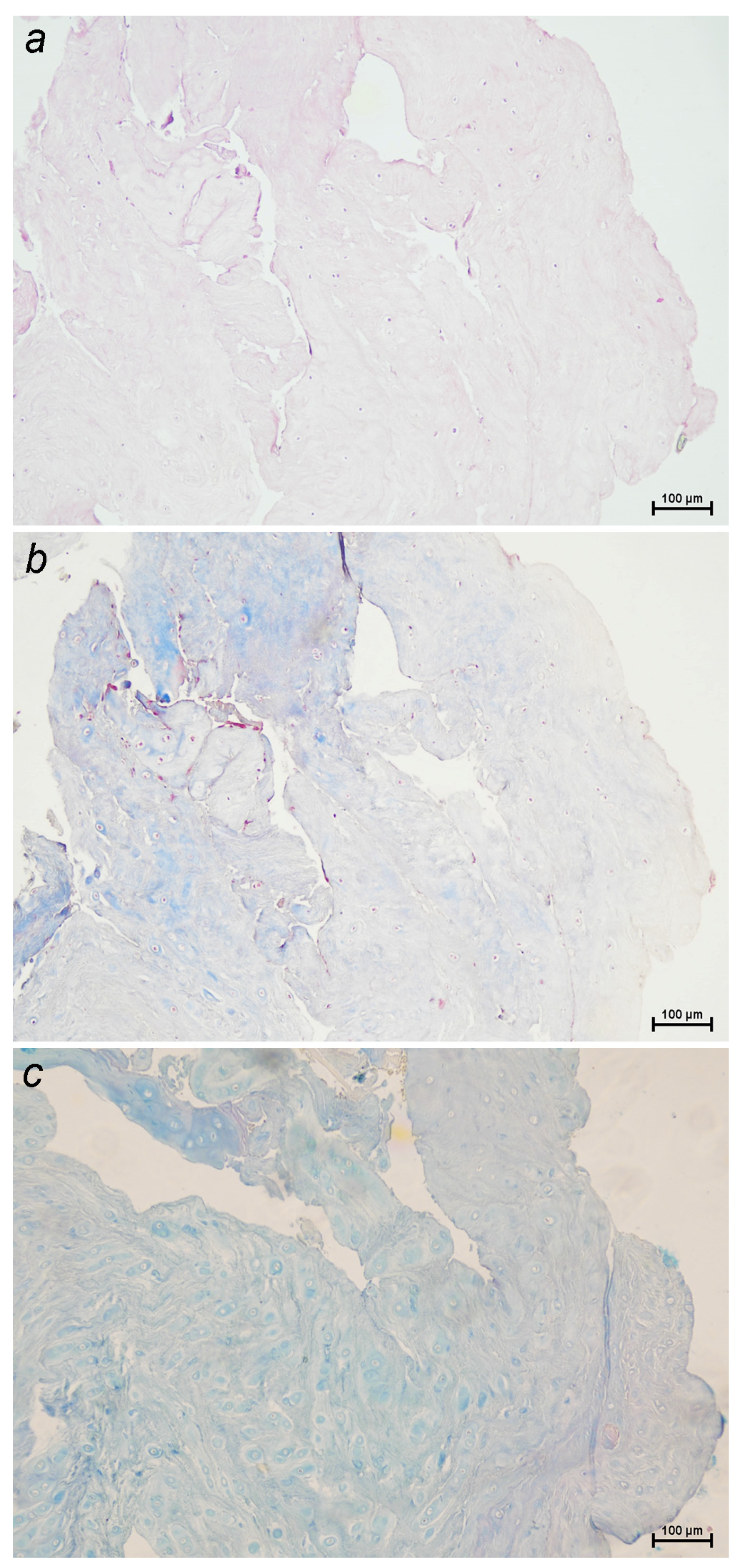
Figure 2.
Sections of a canine intervertebral disc extrusion stained with: (a) Hematoxylin-Eosin, (b) Masson’s Trichrome and (c) Alcian Blue-PAS (10x). A severe inflammatory reaction with necrosis and hemorragy are seen (more than 75% of the sample).
Figure 2.
Sections of a canine intervertebral disc extrusion stained with: (a) Hematoxylin-Eosin, (b) Masson’s Trichrome and (c) Alcian Blue-PAS (10x). A severe inflammatory reaction with necrosis and hemorragy are seen (more than 75% of the sample).
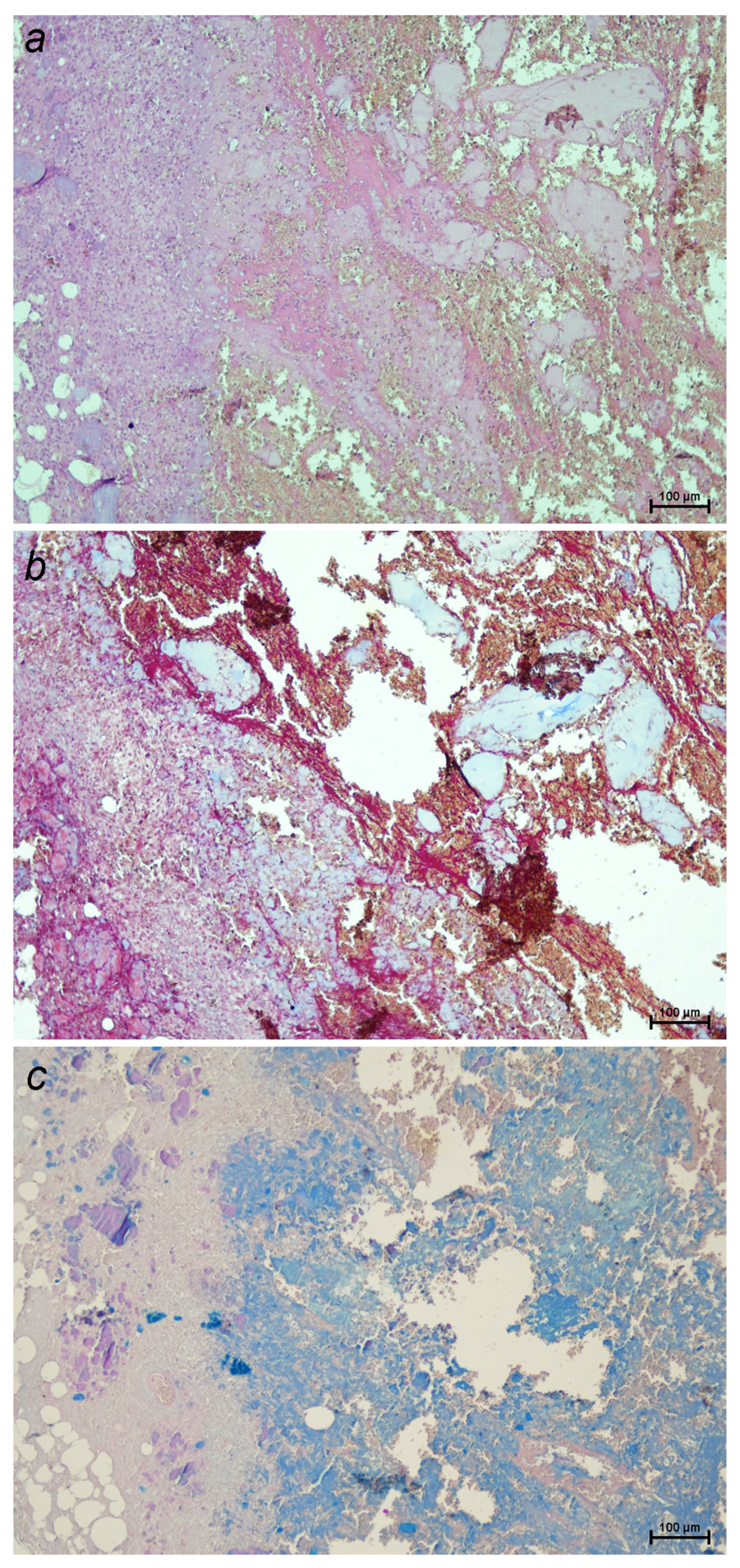
Figure 3.
Right-lateral radiograph of the lumbosacral spine of a dog spine in a dog with a surgically confirmed disc extrusion at L5-L6 (white arrow). Note the reduction of the interverbertebral disc space and the presence of mineral disc material in the vertebral canal.
Figure 3.
Right-lateral radiograph of the lumbosacral spine of a dog spine in a dog with a surgically confirmed disc extrusion at L5-L6 (white arrow). Note the reduction of the interverbertebral disc space and the presence of mineral disc material in the vertebral canal.
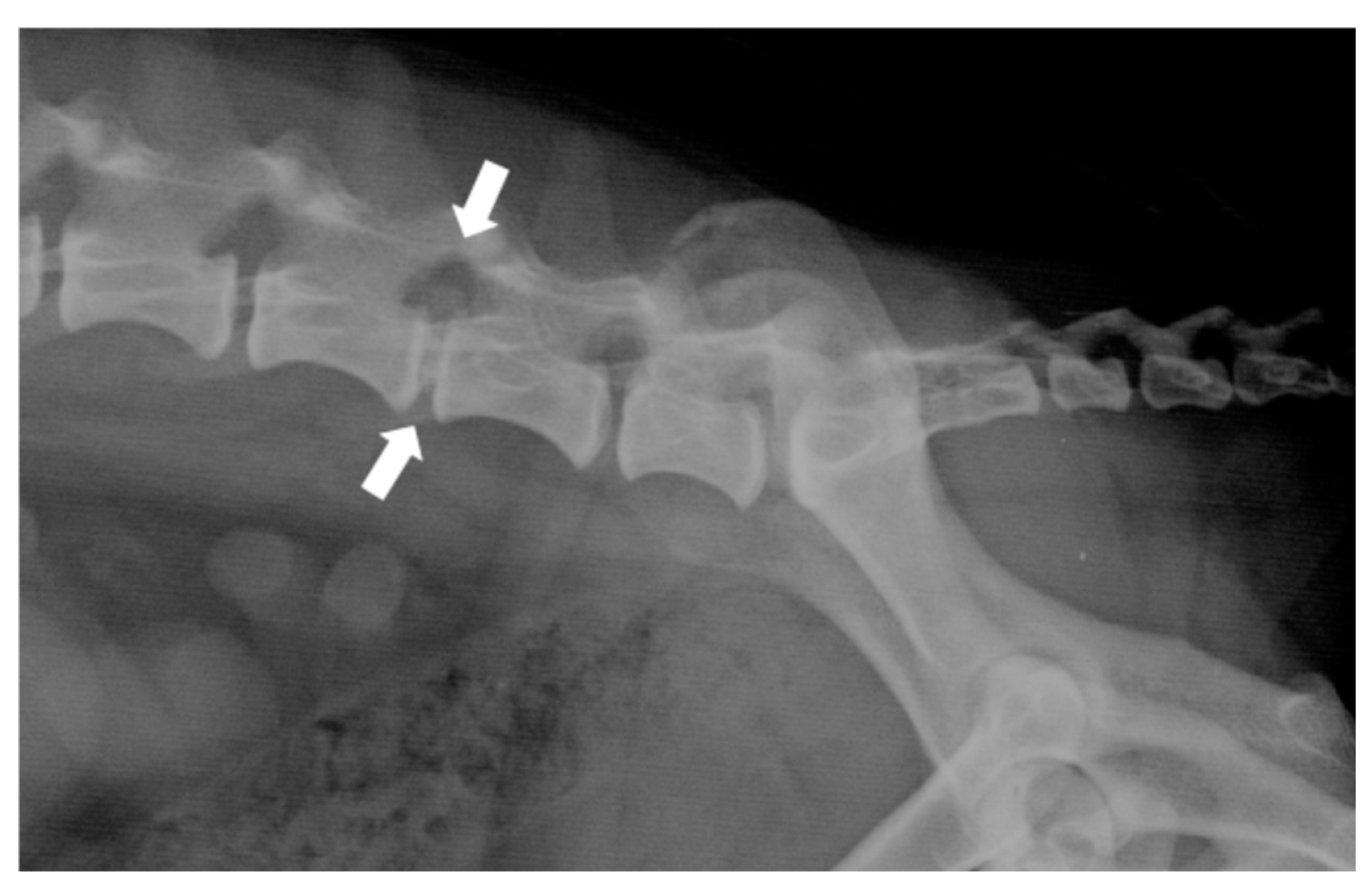
Figure 4.
Computed tomographic (CT) images showing several examples of the appearance of mineralized intervertebral disc extrusion in the vertebral canal. (A) Sagittal reconstructed non-contrast CT image showing a hyperattenuating mass suggestive of extruded disc material into the vertebral canal between C2-3 (arrow). (B) Transverse non-contrast CT image of the same patient showing a large hyperattenuating mass into the vertebral canal at the intervertebral disc level C2-3 with a mild lateralization to the left (arrow). (C) Sagittal reconstructed post-contrast CT image showing an extensive heterogeneously hyperattenuating material (compared to the spinal cord) suggestive of extruded degenerated disc mixed with epidural haemorrhage, compressing the spinal cord along the vertebral bodies of T13 and L1. (D) Transverse non-contrast CT image showing a large hyperattenuating mass disc extrusion occupying most of the vertebral canal at the intervertebral disc level L1-L2.
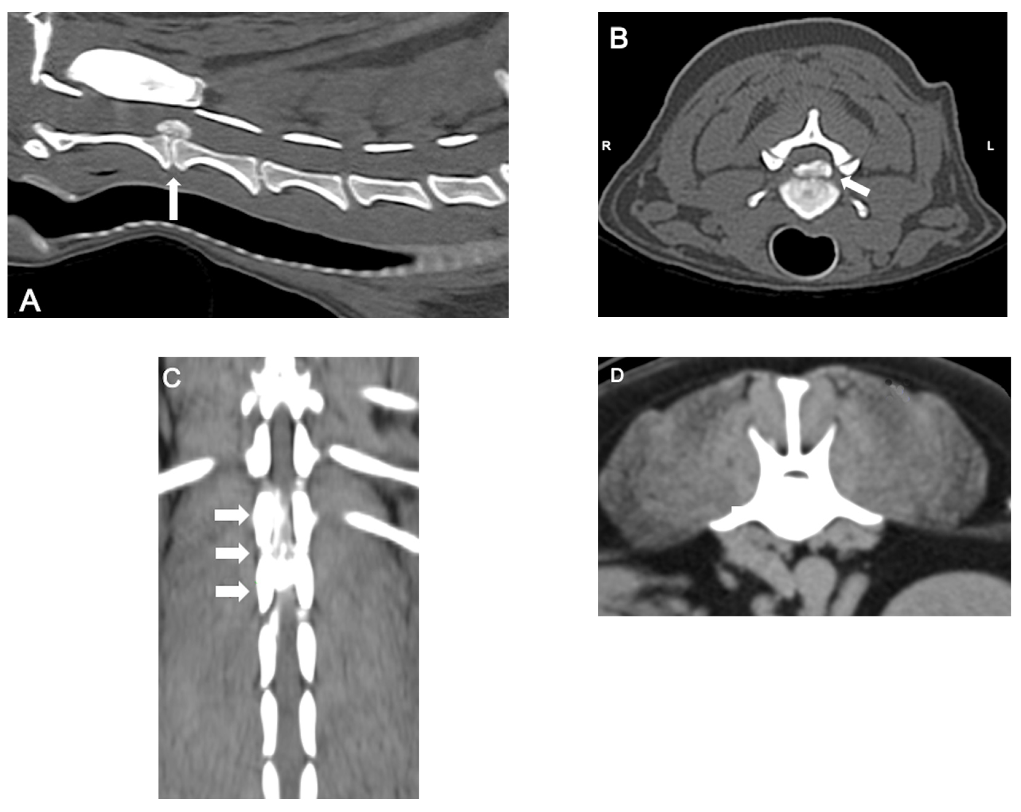
Figure 5.
Magnetic resonance imaging (MRI) images showing two examples of the appearance of mineralized intervertebral disc extrusion in the vertebral canal (A) Dorsal T1W image showing the hypointense compressive material (arrows) at L2-3. (B) Sagittal T2W image showing a severe ventral extradural compression in the ventral aspect of the vertebral canal at the intervertebral level T13-L1 (arrows).
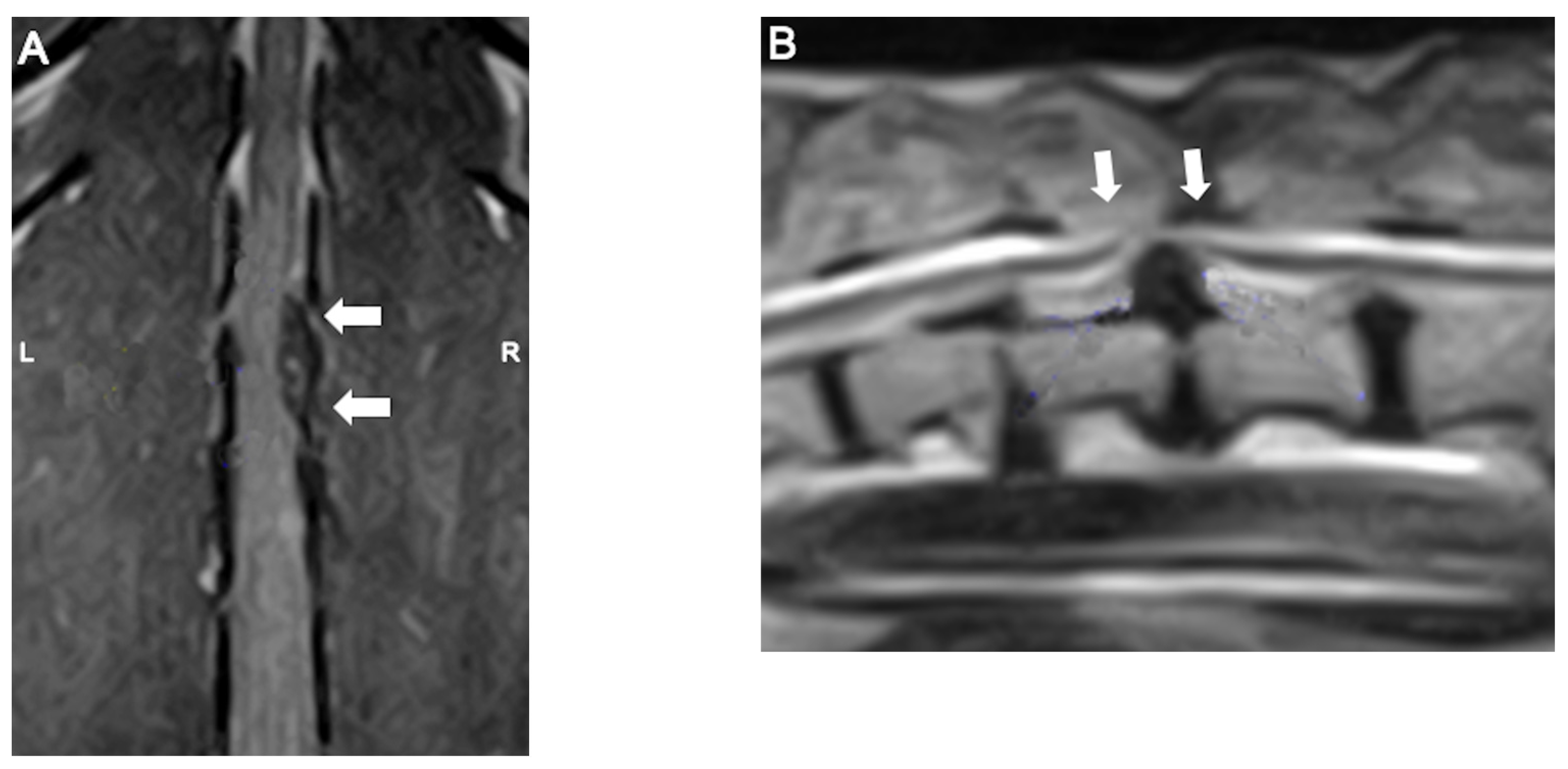
Figure 6.
Sagittal reconstructed non-contrast CT images of a dog presenting with two simultaneous cervical disc herniations (C3-4 and C5-6) (arrows). (A) Pre-surgical and (B) post-surgical.
Figure 6.
Sagittal reconstructed non-contrast CT images of a dog presenting with two simultaneous cervical disc herniations (C3-4 and C5-6) (arrows). (A) Pre-surgical and (B) post-surgical.
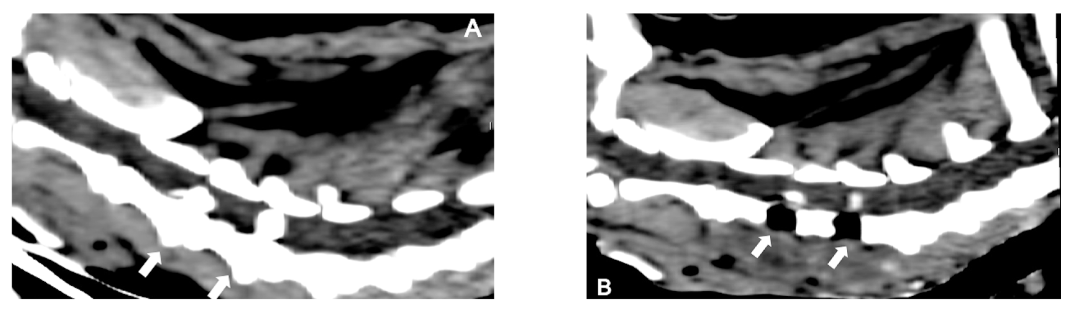

Table 1.
Modified Frankel Score [24].
Table 1.
Modified Frankel Score [24].
| Grade 1 | Spinal hyperaesthesia |
| Grade 2 | Ambulatory paraparesis |
| Grade 3 | Non-ambulatory paraparesis |
| Grade 4 | Paraplegic with intact pain sensation |
| Grade 5 | Paraplegic with absent deep pain sensation |
Disclaimer/Publisher’s Note: The statements, opinions and data contained in all publications are solely those of the individual author(s) and contributor(s) and not of MDPI and/or the editor(s). MDPI and/or the editor(s) disclaim responsibility for any injury to people or property resulting from any ideas, methods, instructions or products referred to in the content. |
© 2025 by the authors. Licensee MDPI, Basel, Switzerland. This article is an open access article distributed under the terms and conditions of the Creative Commons Attribution (CC BY) license (http://creativecommons.org/licenses/by/4.0/).
Copyright: This open access article is published under a Creative Commons CC BY 4.0 license, which permit the free download, distribution, and reuse, provided that the author and preprint are cited in any reuse.


