
Submitted:
07 May 2025
Posted:
07 May 2025
You are already at the latest version
Abstract
This study evaluated the encapsulation of Lactiplantibacillus fabifermentans BAL-27 ITTG and phenolic compounds from coffee husks using alginate beads. The research considered variables such as alginate concentration (1.5% and 3%), crosslinking time (8 and 20 minutes), and the inclusion of chitosan. A 2³ factorial design was employed, and the effects were analyzed using ANOVA (p < 0.05). The encapsulation efficiency for the probiotic exceeded 80%, and its viability following gastrointestinal simulation ranged from 73.65% to 85.34%. Phenolic compounds achieved encapsulation efficiencies of up to 20%. In yogurt, the alginate beads maintained probiotic viability at approximately 9 Log₁₀ CFU/g and preserved the stability of antioxidant compounds over 28 days. Moreover, the incorporation of beads did not adversely affect the physicochemical properties or sensory acceptance of the yogurt, supporting their potential application in functional foods.
Keywords:
Lactobacillus fabifermentans BAL-27 ITTG
; functional food
; husk coffee extract
1. Introduction
Currently, interest in functional foods and beverages has increased significantly due to growing awareness of the importance of proper nutrition and health, as well as increasing scientific evidence supporting their efficacy [1]. Among these foods, the roles of probiotics and phenolic compounds have been widely studied, as both have demonstrated health benefits when consumed in adequate amounts. However, the incorporation of these bioactive compounds into food matrices may alter their physicochemical and organoleptic properties or reduce their bioactivity during processing. To address these challenges, researchers have implemented microencapsulation techniques to protect and stabilize these compounds in a way that preserves both the sensory characteristics of the food and the functional properties of the encapsulated ingredients during storage. Over the years, various technologies have been developed to extend the shelf life of these bioactives, with encapsulation emerging as one of the most employed strategies.
For the encapsulation of lactic acid bacteria, spray drying, lyophilization, and encapsulation in alginate beads are among the most prominent techniques. However, the latter presents significant challenges, as the viability of probiotics tends to decline rapidly during storage, as reported by Silva et al. [2], De Prisco et al. [3], and Kumar and Kumar [4]. This decrease in viability is often attributed to the physicochemical properties of the beads and their water activity (Aw). Another critical factor is the ability of probiotics to withstand the encapsulation conditions, prompting ongoing efforts to identify more resilient strains. In our laboratories, Lactiplantibacillus fabifermentans BAL-27 ITTG has shown promising probiotic potential [5], suggesting its suitability for food applications.
Another group of important bioactive components includes phenolic compounds (antioxidants), which are highly susceptible to degradation by high temperatures, light, and oxygen during processing and storage. The antioxidant capacity of these compounds is associated with various health benefits, including anti-inflammatory, anticancer, antiatherosclerotic properties, and improved gut microbiota health [6]. Several studies have reported promising encapsulation efficiencies for phenolic compounds, distinct from those observed for probiotics. For example, Belščak et al. [7] and Li et al. [8] reported encapsulation efficiencies exceeding 57% for caffeine and phenolic compounds in green tea. These studies highlighted the crucial role of chitosan in achieving high encapsulation efficiency. Given these favorable outcomes, researchers are actively exploring new sources of phenolic compounds, particularly from agricultural by-products.
The coffee cherry has long been utilized; however, due to its nutritional potential, its by-products have attracted growing global interest. One such by-product is the pericarp (outer skin), which contains phenolic compounds at concentrations ranging from 9.2 to 13 mg GAE/g, as reported by Ballesteros et al. [9], Heeger et al. [10], and Ribeiro et al. [11]. These phenolic compounds are particularly valued for their antimicrobial and antioxidant properties, making their extraction and application especially appealing.
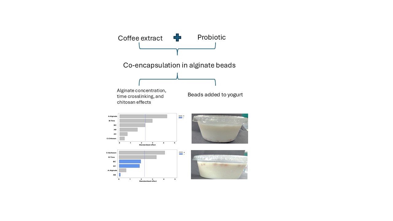
Despite numerous reports on the individual encapsulation of probiotics and phenolic compounds, there is limited information on their co-encapsulation. This is largely due to the antimicrobial nature of phenolic compounds, which may inhibit beneficial microorganisms. Therefore, in the present study, L. fabifermentans was co-encapsulated with phenolic compounds extracted from coffee pericarp using alginate beads. The effects of alginate concentration, crosslinking time, and the presence or absence of chitosan were evaluated. Additionally, the impact of incorporating these capsules into yogurt was assessed by monitoring probiotic survival, survival under simulated gastrointestinal conditions, and the physicochemical and sensory properties of the final product.
2. Materials and Methods
2.1. Materials and Microorganisms
Lactiplantibacillus fabifermentans BAL-27 ITTG was provided by the National Technological Institute of Mexico/Technological Institute of Tuxtla Gutiérrez, Chiapas, Mexico. The microorganism was reactivated in de Man, Rogosa, and Sharpe (MRS; Difco) broth and incubated at 37 °C for 8 h. Subsequently, the culture was centrifuged at 4,500 rpm and 4 °C for 30 min. The resulting cell pellet was washed with 0.9% (w/v) saline solution and centrifuged again under the same conditions. Finally, the cell pellet was stored at 4 °C until further use. All unspecified reagents were purchased from Sigma-Aldrich (USA).
2.2. Extraction of Phenolic Compounds
Ripe coffee cherries (Coffea arabica) were supplied by Rilly de Lievano Co. (Chiapas, Mexico). The cherries were washed with soap and water, disinfected by immersion in chlorinated water (100 ppm), rinsed thoroughly, and shade-dried to remove surface moisture. The pericarp was manually separated from undamaged, fully ripe cherries. For the extraction of phenolic compounds, the pericarp was immersed in distilled water at a 1:5 (w/v) ratio and stirred at 60 °C for 24 h. The resulting mixture was filtered through Whatman No. 4 paper, and the filtrate was lyophilized using a freezing dryer (Labconco, Cole-Parmer, USA) at −40 °C and 0.03 mbar of pressure [12]. The resulting coffee cherry pericarp extract (hereafter referred to as “coffee extract”) was stored in the dark at −18 °C until further use.
2.3. Encapsulation of Bioactive Compounds
Alginate beads were obtained via ionic gelation method using two concentrations of sodium alginate (1.5 and 3% w/v), two crosslinking times (8 and 20 min), and with or without chitosan coating (molecular weight: 100,000–300,000) (Sigma‒Aldrich, USA).
To prepare the beads, hydrated alginate (12 h under refrigeration) was mixed with the cell pellet of L. fabifermentans and the resuspended lyophilized coffee extract to final concentration of 10 Log10 CFU/mL and 2% (w/v), respectively. The mixture was homogenized at 1,000 rpm for 10 minutes and then added dropwise into a sterile 5% (w/v) calcium chloride solution. After, the beads were immersed in the CaCl2 solution for 8 or 20 minutes, according to the experimental design. A batch of beads was subsequently coated by immersion in a 0.4% (w/v) chitosan solution according to the methodology described by Krasaekoopt et al. [13]. Previously, low-molecular-weight chitosan was dissolved in 1% (v/w) acetic acid. The pH of the solution was subsequently adjusted to 5.7 ± 0.3 with 1 M NaOH. The mixture was filtered with filter paper (Whatman # 4) and sterilized at 121 °C for 15 minutes prior to immersion of the beads. Fifteen g of beads were immersed in 100 mL of the chitosan solution and stirred at 100 rpm for 40 min. Finally, the beads were removed from the chitosan solution, washed twice with sterile distilled water and stored under refrigeration until use. Importantly, the beads were used for no more than 5 hours for future determinations.
2.4. Determination of the Viability of Lactobacillus Fabifermentans
To evaluate the viability of microorganisms, 1 g of beads was mixed with 5 mL of 2% sodium citrate (p/v). The mixture was homogenized at 3,200 rpm for 5 minutes using an Ultra-Turrax homogenizer (IKA T25, USA) following the protocol described by De Prisco et al. [3]. The viable cell concentration was determined by plating on MRS agar according to Ramírez-Pérez et al. [5]. The encapsulation efficiency (EE) was determined via Equation (1):
where Ni is the concentration (CFU/g) of cells encapsulated in the beads and No is the concentration (CFU/g) of cells in the encapsulation mixture.
EE(%) = (Log10 Ni/Log No) x 100
2.5. Gastrointestinal Simulation
Gastrointestinal simulation was performed according to Ramírez-Pérez et al. [5]. Briefly, the tolerance of the microorganisms to acidic conditions was evaluated by exposing them to a pH of 1.9 for 30 minutes. The passage through the small intestine was subsequently simulated via intestinal juices (pH 7.5), and the beads were incubated for 6 hours. The resistance gastrointestinal simulation (RGS) was calculated by Equation 2.
where Ni represents the cell concentration present at the end of the intestinal stage and No represents the initial cell concentration.
RGS (%) = (Log10 Ni/Log10 No) x 100
2.6. Quantification of Phenolic Compounds in the Beads
The phenolic compound content in the alginate beads was quantified by dissolving 1 g of the beads, as described in Section 2.3. The concentration of phenolic compounds was determined using the Folin‒Ciocalteu spectrophotometric method [14]. Briefly, 1 mL of the sample was mixed with 4.2 mL of distilled water, followed by the addition of 0.5 mL of 2 N Folin–Ciocalteu reagent and 1 mL of 20% (w/v) sodium carbonate. The mixture was incubated at room temperature for 2 hours. The absorbance was measured at 765 nm using a spectrophotometer (Cole-Parmer UV-2100, USA). A standard calibration curve was prepared using gallic acid (GA). The results are expressed in mg gallic acid equivalents per gram of bead (mg GAE/g).
The encapsulation efficiency of phenolic compounds (PCEE) was determined by Equation (3):
where CFi represents the concentration of phenolic compounds encapsulated in the beads and CFo corresponds to the initial concentration of phenolic compounds in the mixture before encapsulation.
PCEE (%) = (CFi/CFo) x 100
2.7. Preparation of Yogurt
A commercial starter culture from the BIOPROX brand (Mexico) was used, activated in sterile distilled water, and incubated at 37 °C for 1 h according to the manufacturer’s instructions. For inoculum preparation, whole milk powder was reconstituted to a concentration of 12% (w/v) and sterilized at 121 °C for 15 minutes. The starter culture was subsequently inoculated at 1% (v/v) and incubated at 35 °C for 8 h [15].
Yogurt preparation was conducted following the methodology described by Morales-Ruiz [15], with slight modifications. Milk was standardized by dissolving 6 g of powdered whole milk (Nido, Mexico) and 7 g of sugar in 100 mL of liquid whole milk (Pradel, Mexico). The mixture was subjected to low-temperature pasteurization (73 °C for 20 s), followed by inoculation with the activated culture at 10% (v/v). The mixture was stirred for 20 minutes, and 30 mL aliquots were transferred into 40 mL polyethylene containers containing alginate beads at a 1:10 (w/v) ratio. Fermentation was carried out at 40 °C for 2 h, after which the samples were refrigerated (approximately 5 °C) to allow for gelation.
The yogurt was stored at 5 °C for 28 days, and samples were collected every 7 days to assess the viability of the microorganisms, as well as the physicochemical and sensory properties of the product. In addition, the phenolic compound content, and the release of L. fabifermentans under simulated gastrointestinal conditions were quantified at the end of the storage period.
2.7.1. Viability of Lactobacillus Fabifermentans in Yogurt
To determine the viability of the probiotic on the beads within the yogurt, the beads were removed and carefully washed with sterile water to remove excess yogurt. The determination of the viability of microorganisms was then carried out as described in Section 2.4.
2.7.2. Physicochemical Analysis of Yogurt
Syneresis
The syneresis of the yogurt was determined according to Zainoldin and Hi Baba [16]. For this purpose, 20 g of yogurt was weighed in 50 mL tubes and centrifuged at 4,500 rpm for 20 min at 4 °C. The supernatant was subsequently weighed, and the percentage of syneresis was calculated using Equation (4).
Syneresis (%) = ((weight of the supernatant)/(weight of the sample)) * 100
pH
The pH of the yogurt was measured during storage using a digital pH meter (Deluxe pH meter, India).
Titratable acidity
The titratable acidity, expressed as a percentage of lactic acid, was determined according to NOM-243-SSA1-2010 [17]. Eighteen grams of yogurt were mixed with 36 g of distilled water. Ten drops of 1% phenolphthalein (w/v) were added and titrated with 0.1 N NaOH. The acidity was calculated using Equation (5).
where V is the volume (mL) of NaOH used in the titration, N is the normality of NaOH (0.1 N), and M is the weight of the sample.
Acidity (%) = (V * N * 9)/M
Sensory analysis
The degree of global acceptance, as well as the properties of odor, color, flavor and texture, was evaluated using a structured seven points hedonic test (1: dislike extremely; 7: like extremely). The evaluation was performed by a panel of 60 untrained judges (aged 18-24 years). Assessment was conducted at the beginning and end (28 days) of yogurt shelf-life.
2.8. Experimental Design and Statistical Analysis
A completely randomized 2³ factorial experimental design was implemented with three replicates. The following factors were evaluated: alginate concentration (1.5% and 3%), immersion time (8 and 20 minutes), and the presence or absence of chitosan. All data were analyzed using analysis of variance (ANOVA) at a significance level of p < 0.05. Mean comparisons were performed using Tukey’s test, with the aid of the Statgraphics Centurion XVI software. For the sensory analysis, yogurt without alginate beads was used as the control.
3. Results
3.1. Encapsulation of Lactobacillus Fabifermentans BAL-27-ITTG and Phenolic Compounds
3.1.1. Viability of Encapsulated Lactobacillus Fabifermentans BAL-27-ITTG
The viability of L. fabifermentans microencapsulated with coffee extract had a range of 8.83 to 9.47 Log10 CFU/g of beads (Table 1). The highest alginate concentration (3%) combined with the longest crosslinking time (20 minutes) resulted in the greatest probiotic viability (9.45 and 9.47 Log₁₀ CFU/g), regardless of the presence or absence of a chitosan coating. In contrast, treatments T1 and T5 presented the lowest content of viable microorganisms in the beads (8.83 and 8.91 Log10 CFU/g, respectively). This is related to the low alginate concentration (1.5% w/v) and short crosslinking time (8 min), regardless of the presence or absence of chitosan.
Although achieving high cell viability within the beads is one of the main goals of encapsulation, it is also essential to ensure a high microbial encapsulation efficiency (MEE). Treatments T3 and T4 exhibited the highest MEE values, at 84.70% and 84.84%, respectively (Table 1). This outcome may be influenced by the alginate concentration and the crosslinking time, as illustrated in the Pareto diagram (Figure 1a). The diagram indicates that both factors had a positive effect on MEE, whereas the addition of chitosan showed no significant influence. MEE increased with higher alginate concentrations and longer crosslinking times. In contrast, treatments T5 and T6 exhibited the lowest MEE values, at 81.00% and 81.79%, respectively (Table 1). Although the mean comparison test revealed statistically significant differences among treatments, the actual differences in MEE values were small. The higher MEE observed with increased alginate concentration may be attributed to a reduction in osmotic stress, ionic imbalance, and protein denaturation, thereby protecting the integrity of the probiotic cell membrane [18].
3.1.2. Encapsulated Phenolic Compounds
The encapsulation of phenolic compounds is another important criterion, as it directly affects their stability, bioavailability, and biological activity in the consumer. These compounds play a crucial role in preventing oxidative stress, as their consumption is associated with reduced cellular damage, enhanced immune defense, and a lower risk of developing diseases related to free radicals [19]. The concentration of phenolic compounds in the beads ranged from 0.47 to 0.51 mg GAE/g across all treatments (Table 1), corresponding to phenolic compound encapsulation efficiencies (PCEE) between 14% and 20%.
Analysis of variance revealed that while the factors studied did not significantly affect the concentration of encapsulated phenolic compounds, they did have a significant influence on PCEE (Figure 1b). The lowest PCEE values were observed in treatments T1 and T2 (14.22% and 17.02%, respectively), both of which used an 8-minute crosslinking time. Conversely, the addition of chitosan contributed to higher PCEE values, even under short crosslinking conditions, as seen in treatments T1 and T2 (Table 1).
3.1.3. Viability of Lactobacillus Fabifermentans BAL-27-ITTG During Gastrointestinal Simulation
The resistance of a probiotic to gastrointestinal simulation is key to ensuring its arrival in the large intestine and ability to colonize it. The highest resistance to gastrointestinal simulation was 85.34% for Treatment T3, compared with treatments T5 and T8, with values of 75.51 and 76.11%, respectively (Table 1). This may be because treatment T3 contained a higher concentration of alginate (3%) without the addition of chitosan than did treatments T5 and T8, which presented the lowest concentration of alginate (1.5%) supplemented with chitosan in both treatments.
3.2. Evaluation of the Quality of Alginate Beads During Yogurt Storage
To demonstrate the functionality of yogurt as a functional food, it was essential to ensure the viability of the encapsulated probiotic during storage. Therefore, the viability of the probiotic and the stability of the phenolic compounds encapsulated within the beads incorporated into the yogurt were evaluated. The beads were carefully extracted from the yogurt, washed, and analyzed weekly over a 28-day storage period.
3.2.1. Viability of Lactobacillus Fabifermentans BAL-27-ITTG
Beads prepared with 1.5% (w/v) sodium alginate and crosslinked for 8 or 20 minutes, both with and without chitosan, maintained a probiotic concentration of approximately 9 Log₁₀ CFU/g throughout the 28-day storage period. The viability kinetics (Figure 2a) showed that from week 0 to week 3, the concentration of viable microorganisms remained stable. However, a significant increase in viability was observed between weeks 3 and 4. A similar trend was observed with higher alginate concentrations (3%), as shown in Figure 2b.
3.2.2. Phenolic Compounds in Beads During Storage
At the beginning of the storage period, no significant differences were observed in the phenolic compound content among the treatments (p > 0.05). However, by the end of the storage period, treatments T1, T2, T3, and T6 showed significant differences (p < 0.05), indicating that storage time had a measurable effect on these treatments (Table 2).
The treatments with chitosan (T4, T5, T7 and T8) did not significantly differ (p < 0.05) during storage, which suggests that the chitosan layer also functioned as a protective barrier, limiting the release and diffusion of phenolic compounds.
3.2.3. Viability of Lactobacillus Fabifermentans BAL-27-ITTG from Alginate Beads During Gastrointestinal Simulation After Four Weeks of Yogurt Storage
After four weeks of yogurt storage, the viability of Lactobacillus fabifermentans under simulated gastrointestinal conditions ranged from 71% to 89% (Table 3). The highest survival rate was observed in treatment T1, which used 1.5% alginate, an 8-minute immersion time, and no chitosan coating. In contrast, the lowest survival was recorded in treatment T8, which involved 1.5% alginate, a 20-minute immersion time, and chitosan coating.
3.3. Effects of the Addition of Lactobacillus Fabifermentans and Encapsulated Phenolic Compounds on the Physicochemical and Sensory Properties of Yogurt
Once the yogurt was confirmed to function as a functional food, it was necessary to evaluate whether its physicochemical and sensory properties were affected. For the analysis of syneresis, pH, and titratable acidity, the encapsulated beads were removed from the yogurt prior to measurement. However, for the sensory evaluation, the beads were left intact.
3.3.1. Determination of the Physicochemical and Microbiological Properties of Yogurt
Yogurt Syneresis
Syneresis is a key physical parameter used to assess yogurt quality, as it reflects the instability of the gel network and its inability to retain the serum phase within the matrix [20]. Elevated levels of syneresis are generally undesirable from a consumer perspective. The incorporation of alginate beads significantly increased (p < 0.05) the syneresis of the yogurt compared to the control sample (yogurt without beads) (Table 4). The highest syneresis percentages (22–29%) were observed in treatments T4, T5, T7, and T8, all of which included chitosan.
On the other hand, treatments T1, T2, T3, and T6, which did not contain chitosan, also exhibited significantly higher syneresis (p < 0.05) compared to the control yogurt. The presence of beads in yogurt from the beginning of fermentation disrupted the homogeneity of the product, as illustrated in Figure 3. This disruption interfered probably with the formation of a uniform casein network, thereby contributing to the increased syneresis observed. The syneresis of the yogurt during storage (Table 4) increased even for the yogurt without beads. Table 4 shows that the chitosan treatments presented the highest percentage of syneresis, which was above 35%.
Titratable pH and acidity
The initial pH of the yogurt ranged from 4.26 to 4.34 (Table 5), values that are close to those of the control yogurt, which did not contain beads, and are within the maximum allowable limit (pH ~ 4.50) according to NOM-243-SSA1-2010 [17]. During yogurt production, the initial milk pH of 6.2 decreased due to the activity of the starter culture, as approximately 20–40% of the lactose is converted into lactic acid, as reported by Cheng [21]. Throughout storage, the pH of the yogurt decreased, with statistically significant differences (p < 0.05) observed between treatments. At the end of storage, the pH of the yogurt ranged from 3.93 to 4.0, with a significant difference (p < 0.05) between the control yogurt and the yogurt stored without chitosan (treatments T1, T2, T3, and T6). This variation could be attributed to the potential release of L. fabifermentans into the yogurt. The titratable acidity of yogurt supplemented with alginate beads increased slightly during storage, which accounts for the observed decrease in pH (Table 6). A similar decrease in pH was also observed in the control yogurt without beads. According to NOM-243-SSA1-2010 [17], the minimum allowable titratable acidity is 0.50%, and all treatments met this standard.
To demonstrate the potential release of L. fabifermentans from the beads, the concentration of L. fabifermentans in the yogurt was determined (Table 7). The concentration of L. fabifermentans was found to be extremely low (between ~ 10³ and ~ 10⁴ CFU/g) in treatments T1, T2, T3, and T6, which did not contain chitosan. This value was negligible compared to the concentration of the starter culture (9 Log₁₀ CFU/g yogurt).
Sensory analysis of yogurt
A hedonic test was conducted to assess the palatability of the yogurt containing beads. The sensory evaluation was performed on freshly produced yogurt as well as after 28 days of storage, with 60 untrained judges aged between 18 and 24 years. In general (Table 8), the sensory attributes were slightly affected by the addition of beads compared to the control yogurt (without beads). However, the observed decrease was minimal, corresponding to only one point on the hedonic scale for each attribute. Although this comparison was made, it does not imply that the product is undesirable, as the products are inherently different.
The color of a product is a crucial indicator, as it is the first visual cue for consumers when selecting a product [22]. At the beginning of storage, the color (Table 8) was significantly different (p < 0.05) between the treated yogurt and the control yogurt. However, yogurt containing chitosan beads did not show a significant difference (p > 0.05), indicating that this attribute was favorable. The chitosan beads gave the yogurt a shinier appearance, which was considered an improvement.
Smell is another key property that distinguishes yogurt. On day zero, the acceptance scores ranged from 5.18 to 5.80 across treatments (Table 8). By week four, no significant difference (p > 0.05) was observed between the yogurt from the treatment groups and the control group. These results are favorable, suggesting that the addition of beads did not negatively impact on the product’s aroma, and the yogurt remained acceptable at the end of storage.
Similarly, taste did not significantly differ (p > 0.05) between the treated yogurt and the control yogurt at the end of storage (Table 8). These findings align with those of Kailasapathy [23], who reported that the use of encapsulated beads in yogurt did not alter color, acidity, or flavor properties. During fermentation, milk protein proteolysis generates peptides and amino acids, which contribute to the formation of flavor compounds [24], while short-chain fatty acids are also produced [5]. Furthermore, although the addition of beads to the yogurt matrix can affect its texture, the yogurt with added beads showed no significant change in texture at either the beginning or the end of storage (Table 8).
Finally, in terms of general acceptance at the end of storage, treatments T1, T4, T5, T7, and T8 did not show significant differences (p > 0.05) compared to the control yogurt (Table 8). Therefore, the addition of alginate beads to yogurt did not affect its overall acceptance. The sensory attributes of taste, odor, texture, and general acceptance were not adversely affected by the incorporation of alginate beads. These results suggest that the decrease in pH during storage and the increase in acidity did not influence the sensory outcomes, due to the low concentration of lactic acid.
4. Discussion
A key aspect of this study was the evaluation of the survival of Lactiplantibacillus fabifermentans and the encapsulation efficiency of phenolic compounds under different formulation conditions. The survival of L. fabifermentans ranged from 8.83 to 9.47 Log₁₀ CFU/g, while the encapsulation efficiency of phenolic compounds (PCEE) was approximately 20%. An increase in alginate concentration (3%) and crosslinking time (20 minutes) led to improved encapsulation efficiency. These findings are consistent with those reported by Abbaszadeh et al. [25], who demonstrated that higher alginate concentrations enhanced the encapsulation efficiency of Lactobacillus rhamnosus. This behavior is attributed to the increased rigidity provided by the alginate matrix, which limits the release of microorganisms during the hardening and coating stages [25,26].
Regarding crosslinking time, longer durations favor enhanced intermolecular interactions between the guluronic acid residues in sodium alginate, leading to a more cohesive gel network [26,27]. In contrast, shorter crosslinking times may result in incomplete gelation and reduced encapsulation efficiency [26]. Additionally, during the gelation process, alginate forms a more stable crosslinked matrix in the presence of calcium ions, thereby supporting probiotic survival [18].
Nevertheless, prior to gelation, alginate may interact with both the phenolic compounds from the coffee extract and the probiotic cell wall, creating potential competition for calcium ion (Ca²⁺) binding. Caffeine and chlorogenic acid—two of the principal phenolic compounds identified in coffee husks [28]—may engage in hydrogen bonding with alginate. Specifically, interactions may occur between the hydroxyl groups of chlorogenic acid, the carbonyl groups of caffeine, and the carboxyl groups of alginate. Such interactions could potentially influence the structure and functionality of the encapsulation matrix.
These mechanisms may significantly influence the stability and encapsulation efficiency of phenolic compounds within the alginate matrix, as reported by Machado et al. [29]. Additionally, the low PCEE observed in this study could be attributed to the diffusion of phenolic compounds into the calcium chloride solution during the crosslinking process, thereby reducing their retention within the encapsulating matrix. The physical entrapment of these compounds in the "egg-box" structure formed by calcium–alginate interactions may also be insufficient to ensure complete encapsulation, ultimately compromising both stability and efficiency.
These findings are particularly noteworthy considering the report by Khochapong et al. [12], which indicated that aqueous coffee husk extract inhibits the growth of microorganisms belonging to the genus Lactobacillus. Despite this, the viability of L. fabifermentans remained above 6 Log₁₀ CFU/g, the minimum threshold recommended by the FAO/WHO [30] for probiotics to confer health benefits through food consumption. Therefore, the successful co-encapsulation of L. fabifermentans with phenolic compounds from coffee husk supports the potential development of a novel functional food product.
The PCEE values obtained in this study are comparable to those reported by other authors. For instance, Arriola et al. [31] reported a PCEE of 20% in alginate beads containing dandelion extract, while Flamminii et al. [32] observed a PCEE of 21% when encapsulating olive leaf extract using a combination of alginate and pectin. However, our encapsulation efficiencies were lower than those reported by Belščak et al. [7], who achieved 68.94% encapsulation efficiency for caffeine using an alginate–chitosan matrix, and those reported by Li et al. [8], who obtained 57.76% efficiency for phenolic compounds from green tea using the same encapsulation system.
These discrepancies may be primarily attributed to methodological differences among the studies. In the present work, beads containing the coffee extract were immersed in the chitosan solution after gelation, whereas in previous studies, chitosan was incorporated directly into the mixture of phenolic compounds and CaCl₂ prior to the crosslinking process. This alternative strategy may have enhanced encapsulation efficiency by promoting earlier interaction and stabilization of the phenolic compounds within the polymer matrix.
Although the Pareto diagram (Figure 1b) indicates that both the addition of chitosan and extended crosslinking time had a statistically significant and positive effect on PCEE, the magnitude of this increase appears to be constrained by the nature of intermolecular interactions among phenolic compounds, sodium alginate, microbial cell wall components, and calcium chloride during gelation. Given the hydrophilic nature of phenolic compounds, they are more likely to remain in the aqueous phase, thereby reducing their entrapment within the polymer matrix and subsequently limiting the PCEE [32].
The observed increase in PCEE may be attributed to the cationic nature of chitosan, which can form an external membrane around alginate beads by establishing electrostatic interactions with the carboxyl groups of alginate. These interactions help to stabilize the matrix and reduce the diffusion and loss of phenolic compounds during the encapsulation process [33].
During the gastrointestinal simulation, the encapsulated probiotic demonstrated a certain degree of protection. It is plausible that under simulated gastric conditions (pH 1.9), the chitosan layer was partially solubilized, contributing to the degradation of the polymeric network. As a result, the probiotic was exposed to the gastric solution containing chitosan, which may have exerted a synergistic antimicrobial effect and reduced probiotic viability. Indeed, Barbosa et al. [34] reported antimicrobial activity of chitosan under acidic conditions.
In contrast, Ramírez-Pérez et al. [5] reported a viability of 90% for L. fabifermentans in gastrointestinal simulations when no encapsulation system was applied. Although higher viability may be observed in the absence of encapsulation under short-term conditions, in the long term, encapsulation remains essential to protect the microorganism from environmental stressors and to ensure its stability during prolonged storage.
On the other hand, during the initial stages of gastric simulation, the volume of the alginate beads gradually increased, allowing for the controlled release of L. fabifermentans. At the end of the gastric phase and throughout the intestinal simulation, the beads continued to swell without undergoing structural disintegration. This volumetric expansion is likely related to the increase in internal pH caused by exposure to intestinal fluid, which promotes bead swelling. Under acidic conditions, the amino groups of chitosan are ionized, while the hydroxyl groups of phenolic compounds become deprotonated, leading to matrix expansion and facilitating the release of encapsulated microorganisms [33,35].
The survival rates observed in this study are consistent with those reported by Azam et al. [36], who documented approximately 80% viability of L. rhamnosus encapsulated in sodium alginate beads. According to these authors, the internal structure of alginate beads contains intramolecular voids that enable bile salt diffusion, which in turn contributes to the increased bead volume during intestinal simulation.
Throughout the storage period, L. fabifermentans maintained its viability within the encapsulation system. The "egg-box" structure of the calcium-alginate matrix likely played a crucial role in retaining both the microorganisms and phenolic compounds, while simultaneously reducing water availability within the beads. This decrease in water activity (Aw) likely limited microbial metabolism, rendering the probiotic less susceptible to environmental stress.
Moreover, as noted by Murthy and Naidu [37], coffee husk contains approximately 12% sugars, which may have served as a carbon source for L. fabifermentans, supporting minimal growth and survival over the 28-day storage period despite limited nutrient and water mobility inside the matrix. Similarly, Machado et al. [38] demonstrated that sugars extracted from coffee husks promoted the growth of L. paracasei, suggesting that these carbohydrates exert a beneficial effect on probiotic viability.
These findings are particularly encouraging, as they confirm the sustained viability of the encapsulated probiotic in yogurt. In contrast, previous studies by Silva et al. [2], De Prisco et al. [3], and Chaikha [39] reported a decline of approximately three logarithmic cycles in the viability of encapsulated probiotics during storage. Likewise, Kumar and Kumar [4] observed a decrease in Lactobacillus rhamnosus viability from 8.8 to 4.35 Log10 CFU/g over a 15-day period using beads composed of 2% alginate and 2% CaCl₂.
The retention of gastrointestinal survival (RGS) of probiotics after four weeks of storage is particularly relevant; however, these values decreased over time. This reduction in probiotic viability may be attributed to prolonged storage of the beads in yogurt. As storage progresses, the acidity of the yogurt increases, potentially leading to the partial dissolution of the chitosan-coated beads under acidic conditions. This process facilitates the release of the encapsulated probiotic. Once released, chitosan may interact with the positively charged free amino groups present in the probiotic cell wall, causing structural damage and, consequently, cell death [40]. In contrast, beads composed solely of alginate as the encapsulating agent did not exhibit this detrimental effect, thereby allowing for higher survival rates of the probiotic throughout the four-week storage period.
Regarding phenolic compounds, a decline in their concentration was observed at the end of the storage period. This reduction may be attributed to the diffusion of phenolic compounds into the yogurt matrix. As previously described by Chan et al. [41] and Gombotz and Wee [42], water-soluble bioactive compounds can readily diffuse from the encapsulation matrix into the surrounding medium, driven by a concentration gradient.
Nevertheless, the presence of chitosan appeared to mitigate this diffusion, functioning as a protective barrier. The addition of chitosan likely reduced the release of phenolic compounds due to the formation of electrostatic interactions between the carboxyl groups of alginate and the amino groups of chitosan. These interactions lead to the development of a semi-permeable membrane around the beads, which restricts the outward diffusion of the encapsulated compounds [43].
At this stage, we confirmed both the high survival rate of L. fabifermentans and the retention of phenolic compounds within the encapsulated beads. However, it was also necessary to assess the impact of these beads on the yogurt matrix. Syneresis was evaluated, and higher levels were observed in formulations containing chitosan-coated beads. This phenomenon may be attributed to the behavior of the beads during milk fermentation, wherein the chitosan-coated beads tended to sink to the bottom of the container, as illustrated in Figure 3.
During fermentation, milk acidification leads to the formation of a gel, altering the density and allowing denser chitosan-coated beads to settle. As fermentation proceeds, this downward movement may disrupt the structural integrity of the gel, contributing to water release and consequently increasing syneresis. The sinking behavior of the beads may also be due to the formation of a polyelectrolyte complex between the amino groups of chitosan and the carboxylate groups of alginate, which results in a denser polymeric network [44] compared to uncoated beads. Moreover, increased syneresis may be partially explained by the acid-induced contraction of the protein network. Higher acidity promotes protein hydration and gel shrinkage, ultimately leading to enhanced syneresis [45]. Among the physicochemical parameters, pH and acidity are essential indicators of yogurt quality. The initial drop in pH is primarily due to lactic acid production by the starter culture. However, the continued decrease in pH during storage can be attributed to the gradual release of L. fabifermentans from the beads, which ferment lactose into organic acids, mainly lactic and acetic acids [5,46].
Several studies have examined the incorporation of alginate beads into yogurt and their influence on the food matrix. For example, Pourjafar et al. [47] and Budianto et al. [48] reported that the addition of alginate beads did not significantly alter the pH of yogurt compared to control formulations, and the pH remained stable throughout storage. These findings contrast with our results, where a noticeable decrease in pH was observed during storage, presumably due to probiotic release and metabolic activity.
Considering the slight acidification observed, a sensory evaluation was conducted using a hedonic test to assess consumer acceptability. The results indicated that none of the treatments were negatively affected by the addition of beads. Even at the end of the storage period, the yogurt was rated as acceptable by the panelists. Notably, despite the release of L. fabifermentans and phenolic compounds, the sensory attributes of the yogurt—such as taste, texture, and appearance—remained unchanged. This finding contrasts with previous observations by Rahmani et al. [49], who reported that the direct addition of free phenolic compounds could negatively impact sensory properties. Nonetheless, the overall acceptability in their study remained above the threshold of consumer acceptance.
5. Conclusions
An increase in the concentration of sodium alginate and chitosan, as well as the gelation time during ionic gelation, positively influenced the encapsulation efficiency of coffee extract and the viability of Lactobacillus fabifermentans in alginate beads. This ionic gelation process enabled the encapsulation of phenolic compounds and L. fabifermentans in stable gel beads, which were successfully maintained in yogurt for 28 days of storage. The concentration of phenolic compounds and the viability of L. fabifermentans in chitosan-coated beads remained stable throughout the four-week storage period in yogurt. Although the physicochemical properties of yogurt supplemented with alginate beads were significantly different from those of the control yogurt at the end of storage, these changes did not negatively impact consumer acceptance, as assessed through sensory evaluation. Yogurt containing L. fabifermentans and coffee extract encapsulated in sodium alginate beads exhibited acceptable physicochemical and organoleptic properties, making it a promising functional food product. Given its beneficial characteristics, this formulated yogurt offers potential applications within the food industry.
Author Contributions
Conceptualization, M.A-A.; methodology, CM-A., LMCV-C., and MCL-H. ; software, MAR-C. and AG-L.; investigation, AVT-G.; resources, MA-A.; data curation, MA-A.; and MAR-C. ; writing — original draft preparation, MA-A., CM-A., MAR-C., EBE-D; writing — review and editing, MA-A., GP-P, CM-A., and MAR-C. ; supervision, MA-A. All authors have read and agreed to the published version of the manuscript.
Funding
This research received financial support from Tecnologico Nacional de México.
Institutional Review Board Statement
Not applicable.
Informed Consent Statement
Not applicable.
Data Availability Statement
The original contributions presented in this study are included in the article. Further inquiries can be directed to the corresponding author.
Acknowledgments
A.V.T.-G. is grateful to SECIHTI (Mexico) for scholarship.
Conflicts of Interest
The authors declare that they have no conflicts of interest.
Abbreviations
| The following abbreviations are used in this manuscript | . |
| EE | Encapsulation Efficiency |
| GAE | Gallic Acid Equivalents |
| MEE | Microbial Encapsulation Efficiency |
| PCEE | Phenolic Compounds Encapsulation Efficiency |
| RGS | Resistance of Gastrointestinal Simulation |
References
- Santeramo, F.G.; Carlucci, D.; De Devitiis, B.; Seccia, A.; Stasi, A.; Viscecchia, R.; Nardone, G. Emerging trends in European food, diets and food industry. Food Res Inter. 2018, 104, 39-47. [CrossRef]
- Silva, M.P.; Tulini, F.L.; Martins, E.; Penning, M.; Favaro-Trindade, C.S.; Poncelet, D. Comparison of extrusion and co-extrusion encapsulation techniques to protect Lactobacillus acidophilus LA3 in simulated gastrointestinal fluids. LWT Food Sci Technol. 2018, 89, 392-399. [CrossRef]
- De Prisco, A.; Van, H.J.; Flogiano, V.; Mauriello, G. Microencapsulated starter culture during yogurt manufacturing, effect on technological features. Food Bioprocess Technol. 2017, 10(10), 1767-1777. [CrossRef]
- Kumar, A.; Kumar, D. Development of antioxidant rich fruit supplemented probiotic yogurts using free and microencapsulated Lactobacillus rhamnosus culture. J Food Sci Technol. 2016, 53(1), 667-675. [CrossRef]
- Ramírez-Pérez, J.I.; Álvarez-Gutiérrez, P.E.; Luján-Hidalgo, M.C.; Ovando-Chacón, S.L.; Soria-Guerra, R.E.: Ruiz-Cabrera, M. A.; Grajales-Lagunes, A.; Abud-Archila, M. Effect of linear and branched fructans on growth and probiotic characteristics of seven Lactobacillus spp. isolated from an autochthonous beverage from Chiapas, Mexico. Arch Microbiol 2022, 364. [CrossRef]
- Bartoszek, M.; Polak, J. An electron paramagnetic resonance study of antioxidant properties of alcoholic beverages. Food Chem. 2012, 132(4), 2089-2093. [CrossRef]
- Belščak, A.; Komez, D.; Karlović, S.; Djaković, S.; Špolijarć, I.; Mršić, G.; Ježek, D. Improving the controlled delivery formulation of caffeine in alginate in alginate hidrogel beads combined with pectin, carrageenan, chitosan and psyllium. Food Chem. 2015, 167, 378-386. [CrossRef]
- Li, Q. ; Duan, M.; Hou, D.; Chen, X.; Shi, J.; Zhou, W. Fabrication and characterization of Ca (II)-alginate-based beads combined with different polysaccharides as vehicles for delivery, release and storage of tea polyphenols. Food Hydrocoll. 2021, 112, 106274. [CrossRef]
- Ballesteros, L.F.; Teixeira, J.A.; Mussatto, S.I. Selection of the solvent and extraction conditions for maximum recovery of antioxidant phenolic compounds from coffee silverskin. Food Bioprocess Technol. 2014. 7(5), 1322-1332. [CrossRef]
- Heeger, A.; Konsińka, A.; Cantergiani, E.; Andlauer, W. Bioactives of coffee Cherry pulp and its utilisation for production of cascara beverage. Food Chem. 2017, 221, 969-975. [CrossRef]
- Ribeiro, E.F.; Luzia, D.M.M.; Jorge, N. Antioxidant compounds extraction from coffe husk: the influence of solvent type and ultrasound exposure time. Acta Sci Technol. 2019, 41(1), 36451. [CrossRef]
- Khochapong, W.; Ketnawa, S.; Ogawa, Y.; Punbusayakul, N. Effect of in vitro digestion on bioactive compunds, antioxidant and antimicrobial activities of coffee (Coffea arabica L.) pulp aqueous extract. Food Chem. 2021, 328, 1-6. [CrossRef]
- Krasaekoopt, W.; Bhandari, B.; Deeth, H. The influence of coating materials on some properties of alginate beads and survivability of microencapsulated probiotic bacteria. Int Dairy J. 2004, 14(8), 737-743. [CrossRef]
- Singleton, V.L.; Orthofer, R.; Lamuela-Raventós, R.M. Analysis of total phenols and other oxidation substrates and antioxidants by means of folin-ciocalteu reagent. Methods Enzymol. 1999, 299, 152-178. [CrossRef]
- Morales-Ruiz, M.C. Fortificación de yogurt con fitonanopartículas de ZnO y Lactiplantibacillus fabifermentans BAL-27-ITTG. [ Tesis de maestría en Ciencias de Ingeniería Bioquímica, Instituto Tecnológico Nacional de Tuxtla Gutiérrez]. 2022.
- Zainoldin, K.; Hj Baba, A. The Effect of Hylocereus polyrhizus and Hylocereus undatus on physicochemical, proteolysis, and antioxidant activity in yogurt. World Acad Sci Eng Technol. 2009, 60, 361-366.
- Norma Oficial Mexicana NOM-243-SSA1-2010. Productos y servicios. Leche, fórmula láctea, producto lácteo combinado y derivados lácteos. Disposiciones y especificaciones sanitarias. Métodos de prueba. [Consultado el 10 de Julio de 2023]. http://dof.gob.mx/normasOficiales/4156/salud2a/salud2a.htm.
- Phùng, T.; Dinh, H.; Ureña, M.; Oliete, B.; Denimal, E.; Dupont, S.; Beney, L.; Karbowiak, T. Sodium Alginate as a promising encapsulating material for extremely-oxygen sensitive probiotics. Food Hydrocoll. 2025, 160. [CrossRef]
- Das, A.B.; Goud, V.V.; Das, C. Phenolic compounds as functional ingredients in beverages. In Value-added ingredients and enrichments of beverages. Academic Press. 2019.
- Izadi, Z.; Nasirpour, A.; Garoosi, G.A.; Tamjidi, F. Rheological and physical properties of yogurt enriched with phytosterol during storage. J Food Sci Technol. 2015, 52, 5341-5346. [CrossRef]
- Cheng H. Volatile flavor compounds in yogurt: a review. Crit Rev Food Sci Nutr. 2010, 50(10),938–950. [CrossRef]
- Burton, E.; Arief, I.I.; Taufik, E. Formulasi yoghurt probiotik karbonasi dan potensi sifat fungsionalnya. Jurnal Ilmu Produksi dan Teknologi Hasil Peternakan, 2014, 2(1), 213-218.
- Kailasapathy, K. Survival of free and encapsulated probiotic bacteria and their effect on the sensory properties of yoghurt. LWT-Food Sci Technol. 2006, 39(10), 1221-1227. [CrossRef]
- Sert, D.; Mercan, E.; Dertli, E. Characterisation of lactic acid bacteria from yogurtlike product fermented with pine cone and determination of their role on physicochemical, textural and microbiological properties of product. LWT Food Sci Technol. 2017, 78, 70-76. [CrossRef]
- Abbaszadeh, S.; Gandomi, H.; Misaghi, A.; Bokaei S.; Noori, N. The effect of alginate and chitosan concentrations on some properties of chitosan-coated alginate beads and survivability of encapsulated Lactobacillus rhamnosus in simulated gastrointestinal conditions and during heat processing. J Sci Food Agric. 2014, 94, 2210–221. [CrossRef]
- Łętocha, A.; Miastkowska, M.; Sikora, E. Preparation and characteristics of alginate microparticles for food, pharmaceutical and cosmetic applications. Polym. 2022, 14(18), 1-32. [CrossRef]
- Gedam, S.; Jadhav, P.; Talele, S.; Jadhav, A. Effect of crosslinking agent on development of gastroretentive mucoadhesive microspheres of risedronate sodium. Int J Appl Pharm. 2018, 10, 133–140. [CrossRef]
- Abud-Archila, M.; Mendoza, C. Jícama mínimamente procesada fortificada con probióticos y compuestos fenólicos de café verde microencapsulados. Biotecnia, 2024, 26, e2350. [CrossRef]
- Machado, A.R.; Silva, P.M.P.; Vicente, A.A.; Souza-Soares, L.A.; Pinheiro, A.C.; Cerqueira, M.A. Alginate particles for encapsulation of phenolic extract from Spirulina sp. LEB-18: physicochemical characterization and assessment of in vitro gastrointestinal behavior. Polym, 2022, 14(21), 4759. [CrossRef]
- FAO, OMS Organización de las Naciones Unidad para la Alimentación y la Agricultura y Organización Mundial de la Salud. 2001. https://www.fao.org/3/a0512s/a0512s.pdf.
- Arriola, N.D.A.; Chater, P.I.; Wilcox, M.; Lucini, L.; Rocchetti, G.; Dalmina, M.; Pearson, J.P.; Amboni, R.D.D.M.C. Encapsulation of Stevia rebaudiana Bertoni aqueous crude extracts by ionic gelation–Effects of alginate blends and gelling solutions on the polyphenolic profile. Food Chem. 2019, 275, 123-134. [CrossRef]
- Flamminii, F.; Di Mattia, C.D.; Nardella, M.; Chiarini, M.; Valbonetti, L.; Neri, L.; Difonzo, G; Pittia, P. Structuring alginate beads with different biopolymers for the development of functional ingredients loaded with olive leaves phenolic extract. Food Hydrocoll. 2020, 108, 105849. [CrossRef]
- Martinović, J.; Lukinac, J.; Jukić, M.; Ambrus, R.; Planinić, M.; Šelo, G.; Klarić, A.-M.; Perković, G.; Bucic-Kojic, A. Physicochemical characterization and evaluation of gastrointestinal in vitro behavior of alginate-based microbeads with encapsulated Grape pomace extracts. Fharmaceutics. 2023, 15, 1-28. [CrossRef]
- Barbosa, M.; Gonçalves, I.; Moreno, P.; Gonçalves, R.; Santos, S.; Pêgo, A.; Amaral, I. Chitosan. Comprehensive Biomaterials II. 2017, 2nd Edition.
- Liang, J.; Yan, H.; Puligundla, P.; Gao, X.; Zhou, Y.; Wan, X. Applications of chitosan nanoparticles to enhance absorption and bioavailability of tea polyphenols: A review. Food Hydrocoll. 2017, 69, 286–292. [CrossRef]
- Azam, M.; Saeed, M.; Pasha, I.; Shahid, M. A prebiotic-based biopolymeric encapsulation system for improved survival of Lactobacillus rhamnosus. Food Biosci. 2020, 37, 100679. [CrossRef]
- Murthy, P.S.; Madhava Naidu, M. Sustainable management of coffee industry byproducts and value addition - A review. Resour Conserv Recycl. 2012, 66, 45–58. [CrossRef]
- Machado, M.; Galrinho, M.; Passos, C.; Espírito, L.; Simona, M.; Ranga, F.; Puga, H.; Palmeira, J.; Coimbra, M.; Oliveira, M.; Ferreira, H.; Alves, R. Prebiotic potential of a coffee silverskin extract obtained by ultrasound-assisted extraction on Lacticaseibacillus paracasei subsp. paracasei. J Funct Foods. 2024, 120. [CrossRef]
- Chaikham, P. Stability of probiotics encapsulated with Thai herbal extracts in fruit juices and yogurt during refrigerated storage. Food Biosci. 2015, 12, 61-66. [CrossRef]
- Ahmed, H.; Rahaman, A.; Uddin, E.; Rafid, M.; Hosen, S.; Layek, K. Development and characterization of chitosan-based antimicrobial films: A sustainable alternative to plastic packaging. Cleaner Chem Eng. 2025, 11. [CrossRef]
- Chan, E.S.; Lee, B.B.; Ravindra, P.; Poncelet, D. Prediction models for shape and size of ca-alginate macrobeads produced through extrusion–dripping method. J. Colloid Interface Sci. 2009, 338(1), 63-72. [CrossRef]
- Gombotz, W.R.; Wee, S.F. Protein release from alginate matrices. Adv Drug Delivery Rev. 2012, 64, 194-205. [CrossRef]
- Belščak-Cvitanović, A.; Đorđević, V.; Karlović, S.; Pavlović, V.; Komes, D.; Ježek, D.; Bugarsky, B.; Nedović, V. Protein-reinforced and chitosan-pectin coated alginate microparticles for delivery of flavan-3-ol antioxidants and caffeine from green tea extract. Food Hydrocoll. 2015, 51. [CrossRef]
- Pasparakis, G.; Bouropoulos, N. Swelling studies and in vitro release of verapamil from calcium alginate and calcium alginate–chitosan beads. Int J Pharm. 2006, 323(1-2), 34-42. [CrossRef]
- Romero del Castillo S.; Mestres Lagarriga, J. Productos lácteos: Tecnología. Editorial Up. 159 pp. 2004.
- Feng, Y; Niu, L.; Li, D.; Zeng, Z.; Sun, C.; Xiao, J. Effect of calcium alginate/collagen hydrolysates beads encapsulating high-content tea polyphenols on quality characteristics of set yogurt during cold storage. LWT-Food Sci and Technol. 2024, 191, 1-12. [CrossRef]
- Pourjafar, H.; Noori, N.; Gandomi, H.; Basti, A.A.; Ansari, F. Viability of microencapsulated and non-microencapsulated Lactobacilli in a commercial beverage. Biotechnol Rep. 2020, 25, e00432. [CrossRef]
- Budianto, E.; Saepudin, E.; Nasir, M. The encapsulation of Lactobacillus casei probiotic bacteria based on sodium alginate and chitosan. In IOP Conference Series: Earth and Environmental Science, 483(1), 012043. IOP Publishing, 2020.
- Rahmani, F.; Gandomi, H.; Noori, N.; Faraki, A.; Farzaneh, M. Microbial, physiochemical and functional properties of probiotic yogurt containing Lactobacillus acidophilus and Bifidobacterium bifidum enriched by green tea aqueous extract. Food Sci Nutr. 2021, 9(10), 5536-5545. [CrossRef]
Figure 1.
Pareto diagram showing the effects of the sodium alginate concentration, time and application of chitosan: MEE of L. fabifermentans (A) and PCEE (B).
Figure 1.
Pareto diagram showing the effects of the sodium alginate concentration, time and application of chitosan: MEE of L. fabifermentans (A) and PCEE (B).
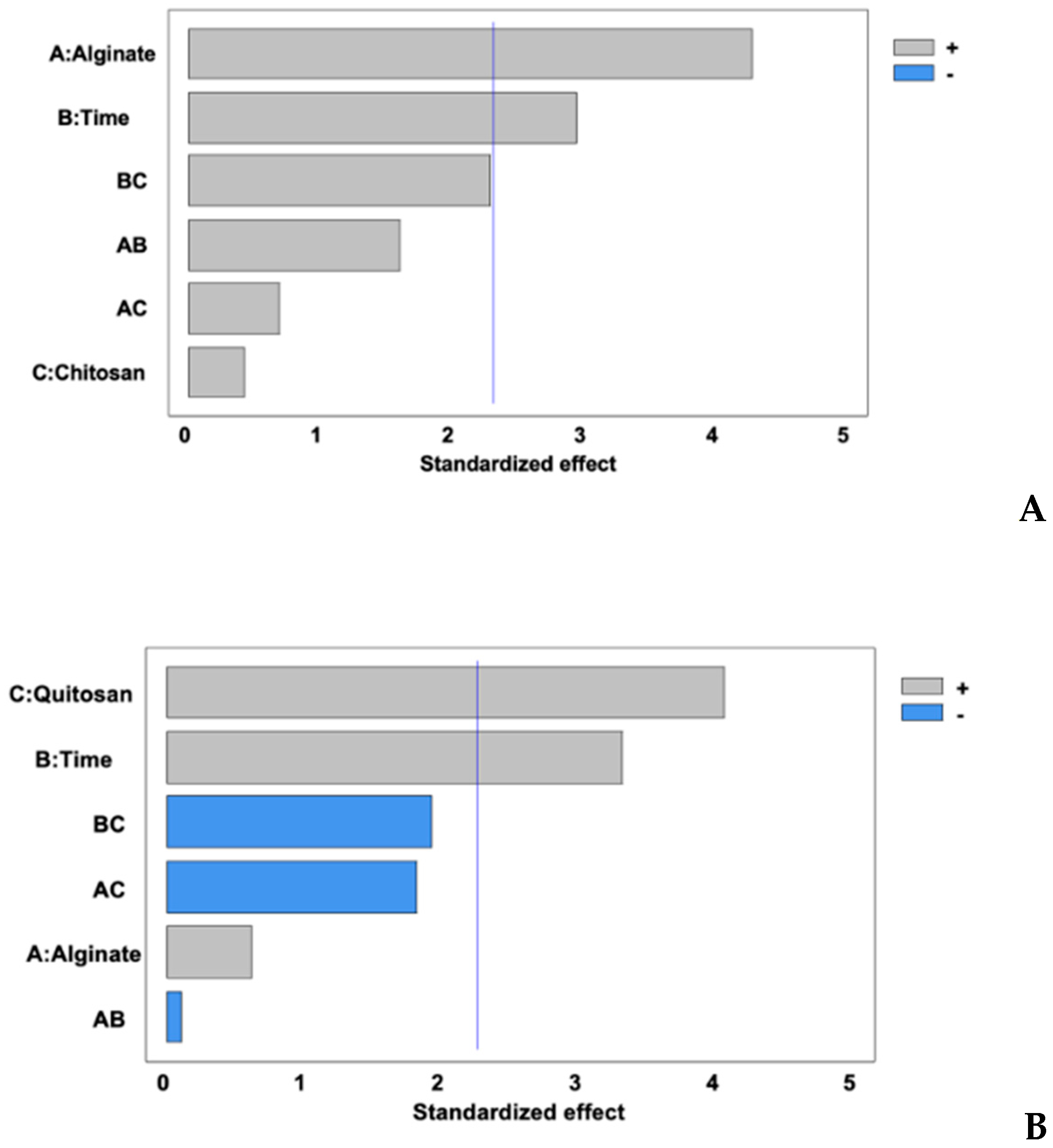
Figure 2.
Chitosan effect of alginate beads concentration 1.5% (A), 3% (B), during immersion time of 8 min on viability of probiotic for 4 weeks. Similar lowercase showed no significant statistical difference between treatments (p ˃ 0.05). Without chitosan (•) with chitosan (▪). Red line is the value (6 Log10 CFU/g) reported by the FAO/WHO [30] for food to have a beneficial effect.
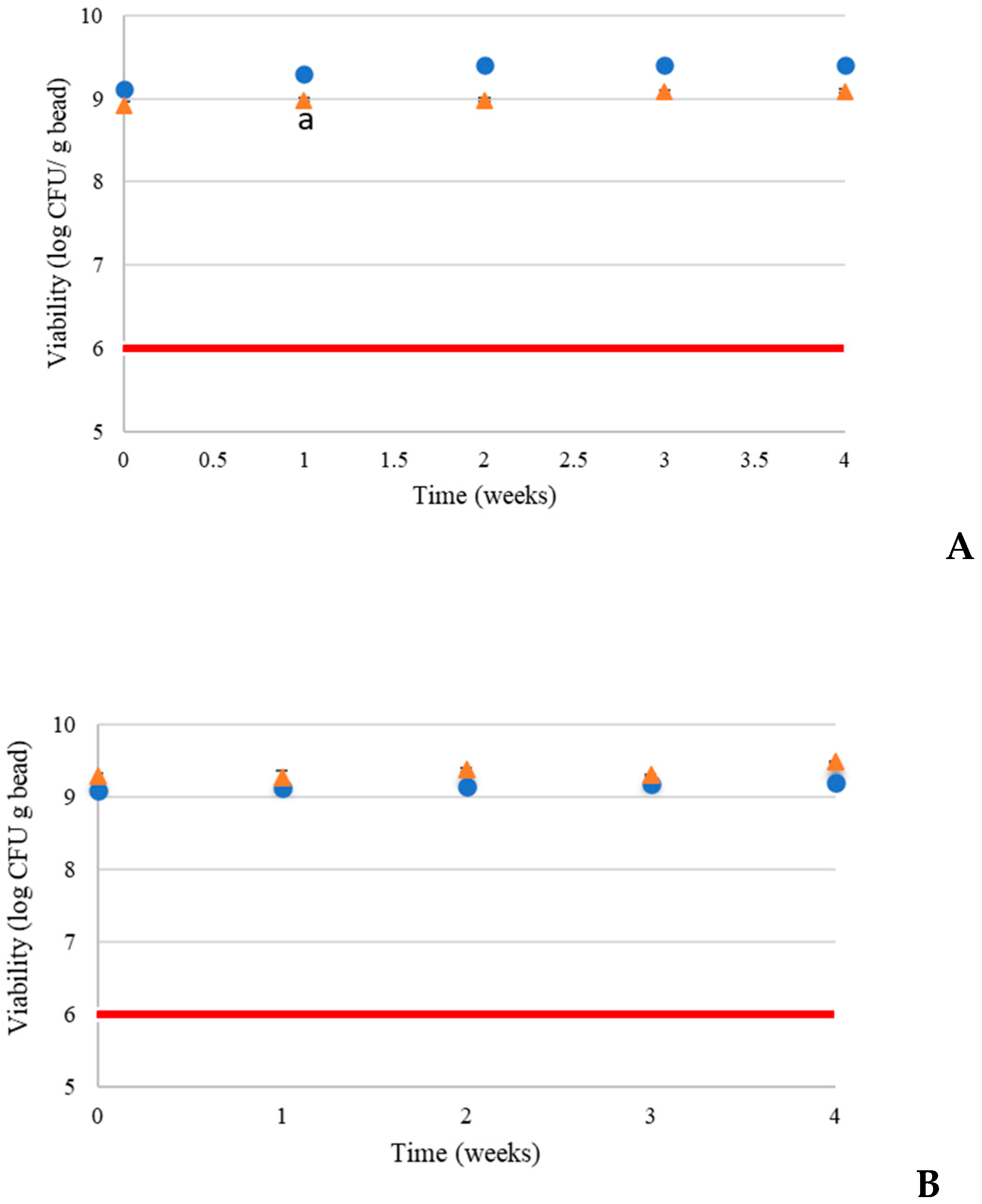
Figure 3.
Yogurt with added alginate beads.
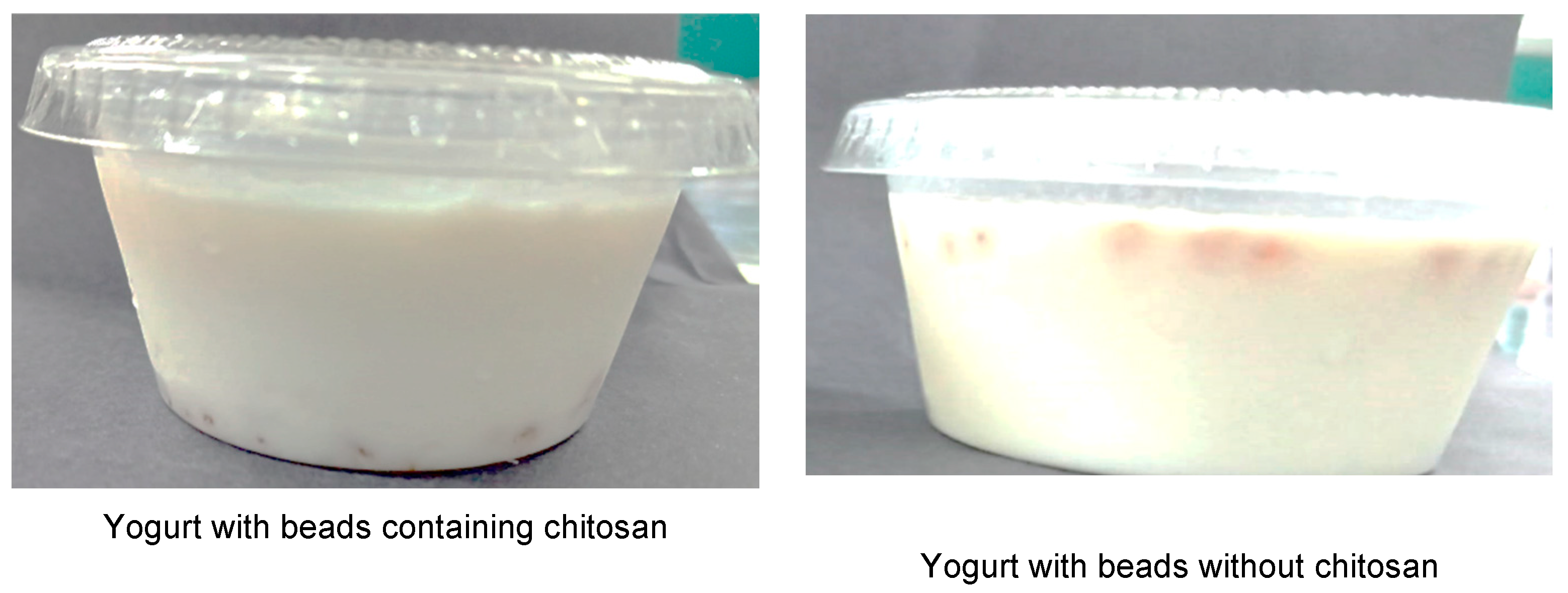

Table 1.
Viability and MEE of L. fabifermentans and phenolic compounds. PCEE and RGS of L. fabifermentans encapsulated in beads.
Table 1.
Viability and MEE of L. fabifermentans and phenolic compounds. PCEE and RGS of L. fabifermentans encapsulated in beads.
| Treatment |
Viability (Log10 CFU/g bead) |
MEE (%) |
PC (mg GAE/g bead) |
PCEE |
RGS (%) |
| T1 | 8.83c | 82.97b | 0.48a | 14.22c | 84.38 ab |
| T2 | 9.09abc | 82.86b | 0.49a | 17.02bc | 83.82 b |
| T3 | 9.45a | 84.70a | 0.47a | 19.75ab | 85.34 a |
| T4 | 9.47a | 84.84a | 0.51a | 20.99a | 73.65 e |
| T5 | 8.91bc | 81.00c | 0.48a | 20.86a | 75.51 d |
| T6 | 9.00abc | 81.79c | 0.52a | 19.12ab | 84.08 ab |
| T7 | 9.29abc | 83.47b | 0.52a | 19.06ab | 77.94 c |
| T8 | 9.38ab | 83.31b | 0.52a | 20.89a | 76.11 d |
| Tukey | 0.532 | 1.01 | 0.07 | 3.03 | 1.50 |
MEE = microbial encapsulation efficiency, PC= phenolic compound, PCEE = phenolic compound encapsulation efficiency, RGS = resistance gastrointestinal simulation, T1 = 1.5% (w/v) alginate, 8 min, without chitosan. T2 = 3.0% (w/v) alginate, 8 min, without chitosan. T3 = 3.0% (w/v) alginate, 20 min, without chitosan. T4 = 3.0% (w/v) alginate, 20 min, with chitosan. T5 = 1.5% (w/v) alginate, 8 min, with chitosan. T6 = 1.5% (w/v) alginate, 20 min, without chitosan. T7 = 3.0% (w/v) alginate, 8 min, with chitosan. T8 = 1.5% (w/v) alginate, 20 min, with chitosan. Similar lowercase letters in the same column indicate no significant difference between treatments (p ˃ 0.05).
Table 2.
Concentration of phenolic compounds (mg EAG/g bead) in alginate beads during yogurt storage.
Table 2.
Concentration of phenolic compounds (mg EAG/g bead) in alginate beads during yogurt storage.
| Treatment | Storage time (weeks) | Tukey | |
| 0 | 4 | ||
| T1 | 0.48Aa | 0.43Bb | 0.04 |
| T2 | 0.49Aa | 0.45Bb | 0.04 |
| T3 | 0.47Aa | 0.41Babc | 0.08 |
| T4 | 0.51Aa | 0.52Aa | 0.07 |
| T5 | 0.48Aa | 0.47Aabc | 0.13 |
| T6 | 0.52Aa | 0.48Babc | 0.02 |
| T7 | 0.52Aa | 0.52Aa | 0.04 |
| T8 | 0.52Aa | 0.50Aab | 0.02 |
| Tukey | 0.07 | 0.04 | |
T1 = 1.5% (w/v) alginate, 8 min, without chitosan. T2 = 3.0% (w/v) alginate, 8 min, without chitosan. T3 = 3.0% (w/v) alginate, 20 min, without chitosan. T4 = 3.0% (w/v) alginate, 20 min, with chitosan. T5 = 1.5% (w/v) alginate, 8 min, with chitosan. T6 = 1.5% (w/v) alginate, 20 min, without chitosan. T7 = 3.0% (w/v) alginate, 8 min, with chitosan. T8 = 1.5% (w/v) alginate, 20 min, with chitosan. Similar lowercase letters in the same column indicate no significant difference between treatments (p ˃ 0.05). A similar capital letter in the same row indicates no significant difference between treatments (p ˃ 0.05).
Table 3.
Survival of L. fabifermentans on alginate beads during gastrointestinal simulation after 4 weeks of storage
Table 3.
Survival of L. fabifermentans on alginate beads during gastrointestinal simulation after 4 weeks of storage
| Treatment |
RGS (%) |
| T1 | 89.82a |
| T2 | 83.82c |
| T3 | 84.22c |
| T4 | 79.86d |
| T5 | 73.25f |
| T6 | 88.09b |
| T7 | 77.23e |
| T8 | 71.88g |
| Tukey | 2.8104 |
RGS = Resistance Gastrointestinal Simulation, T1 = 1.5% (w/v) alginate, 8 min, without chitosan. T2 = 3.0% (w/v) alginate, 8 min, without chitosan. T3 = 3.0% (w/v) alginate, 20 min, without chitosan. T4 = 3.0% (w/v) alginate, 20 min, with chitosan. T5 = 1.5% (w/v) alginate, 8 min, with chitosan. T6 = 1.5% (w/v) alginate, 20 min, without chitosan. T7 = 3.0% (w/v) alginate, 8 min, with chitosan. T8 = 1.5% (w/v) alginate, 20 min, with chitosan. Similar lowercase letters in the same column indicate no significant difference between treatments (p ˃ 0.05).
Table 4.
Syneresis (%) of yogurt supplemented with alginate beads during storage
| Treatment | Time (weeks) | ||||||
| 0 | 1 | 2 | 3 | 4 | Tukey | ||
| T1 | 20.65Ac | 23.40Bc | 25.52Cc | 25.29Cc | 28.48Dc | 1.42 | |
| T2 | 21.03Acd | 24.74Bc | 24.74Bc | 24.91Bc | 25.09Bb | 0.97 | |
| T3 | 21.84Ade | 24.1Bc | 24.21Bc | 25.41Cc | 25.91Cb | 1.01 | |
| T4 | 29.70Ag | 31.37Ad | 34.57Bf | 39.26Cf | 39.93Cf | 1.88 | |
| T5 | 22.42Ae | 24.45Bc | 27.37Cd | 29.91Dd | 35.45Ed | 1.50 | |
| T6 | 17.60Ab | 18.84Ab | 21.33Bb | 23.36Cb | 25.56Db | 1.88 | |
| T7 | 23.73Af | 27.69Bd | 29.65Ce | 30.46Cd | 38.33De | 1.90 | |
| T8 | 23.06Af | 25.65Bcd | 28.52Cde | 32.86De | 37.54Ee | 0.86 | |
| Control | 13.63Aa | 15.29Ba | 17.71Ca | 20.14Da | 20.58Da | 1.33 | |
| Tukey | 1.00 | 2.77 | 1.95 | 1.04 | 1.02 | ||
T1 = 1.5% (w/v) alginate, 8 min, without chitosan. T2 = 3.0% (w/v) alginate, 8 min, without chitosan. T3 = 3.0% (w/v) alginate, 20 min, without chitosan. T4 = 3.0% (w/v) alginate, 20 min, with chitosan. T5 = 1.5% (w/v) alginate, 8 min, with chitosan. T6 = 1.5% (w/v) alginate, 20 min, without chitosan. T7 = 3.0% (w/v) alginate, 8 min, with chitosan. T8 = 1.5% (w/v) alginate, 20 min, with chitosan. Similar lowercase letters in the same column indicate no significant difference between treatments (p ˃ 0.05). A similar capital letter in the same row indicates no significant difference between treatments (p ˃ 0.05).
Table 5.
pH of yogurt fermented with alginate beads for four weeks.
| Treatment | Time (weeks) | |||||
| 0 | 1 | 2 | 3 | 4 | Tukey | |
| T1 | 4.34Aa | 4.09Bd | 4.00BCc | 3.97Cb | 3.97Ccd | 0.08 |
| T2 | 4.32Aab | 4.09Bd | 4.03Bc | 3.96Cb | 3.93Cd | 0.06 |
| T3 | 4.26Aabc | 4.13Bcd | 4.02Cc | 3.97CDb | 3.96Dcd | 0.05 |
| T4 | 4.29Aabc | 4.15Bbc | 4.09Bb | 4.00Cab | 4.00Cabc | 0.08 |
| T5 | 4.26Aabc | 4.17Bbc | 4.09Cb | 4.01Dab | 4.00Dabc | 0.04 |
| T6 | 4.26Aabc | 4.18ABb | 4.09BCb | 4.03Cab | 3.98Cbcd | 0.13 |
| T7 | 4.27Aabc | 4.17Bbc | 4.10Cb | 4.04Dab | 4.04Dab | 0.05 |
| T8 | 4.27Aabc | 4.15Bbc | 4.10Cb | 4.04Dab | 4.00Eabc | 0.03 |
| Control | 4.34Aa | 4.27Ba | 4.20Ca | 4.11Da | 4.05Ea | 0.03 |
| Tukey | 0.08 | 0.04 | 0.05 | 0.12 | 0.06 | |
T1 = 1.5% (w/v) alginate, 8 min, without chitosan. T2 = 3.0% (w/v) alginate, 8 min, without chitosan. T3 = 3.0% (w/v) alginate, 20 min, without chitosan. T4 = 3.0% (w/v) alginate, 20 min, with chitosan. T5 = 1.5% (w/v) alginate, 8 min, with chitosan. T6 = 1.5% (w/v) alginate, 20 min, without chitosan. T7 = 3.0% (w/v) alginate, 8 min, with chitosan. T8 = 1.5% (w/v) alginate, 20 min, with chitosan. Similar lowercase letters in the same column indicate no significant difference between treatments (p ˃ 0.05). A similar capital letter in the same row indicates no significant difference between treatments (p ˃ 0.05).
Table 6.
Titratable acidity (% lactic acid) of yogurt supplemented with alginate beads during four weeks of storage.
Table 6.
Titratable acidity (% lactic acid) of yogurt supplemented with alginate beads during four weeks of storage.
| Treatment | Time (weeks) | |||||
| 0 | 1 | 2 | 3 | 4 | Tukey | |
| 1 | 0.96Abc | 0.98Aab | 0.99Aab | 1.04Bab | 1.11Cabc | 0.03 |
| 2 | 0.94Aab | 0.96Aa | 1.04Bab | 1.09BCab | 1.12Cbc | 0.06 |
| 3 | 0.94Aab | 1.06Bc | 1.06Bb | 1.14Cb | 1.14Cc | 0.02 |
| 4 | 0.94Aab | 0.99ABab | 1.01ABab | 1.03ABa | 1.08Ba | 0.10 |
| 5 | 0.92Aa | 0.95Ba | 0.98Ca | 1.05Dab | 1.09Eab | 0.01 |
| 6 | 0.96Abc | 1.03ABbc | 1.05BCb | 1.12Cab | 1.14Cc | 0.08 |
| 7 | 0.95Abc | 1.02Bbc | 1.06Cb | 1.10Dab | 1.12Eab | 0.02 |
| 8 | 0.98Ac | 1.03ABbc | 1.06BCb | 1.11Cab | 1.09BCab | 0.07 |
| Control | 0.92Aa | 0.96ABa | 1.00Bab | 1.05Cab | 1.09Cab | 0.04 |
| Tukey | 0.02 | 0.05 | 0.02 | 0.05 | 0.04 | |
T1 = 1.5% (w/v) alginate, 8 min, without chitosan. T2 = 3.0% (w/v) alginate, 8 min, without chitosan. T3 = 3.0% (w/v) alginate, 20 min, without chitosan. T4 = 3.0% (w/v) alginate, 20 min, with chitosan. T5 = 1.5% (w/v) alginate, 8 min, with chitosan. T6 = 1.5% (w/v) alginate, 20 min, without chitosan. T7 = 3.0% (w/v) alginate, 8 min, with chitosan. T8 = 1.5% (w/v) alginate, 20 min, with chitosan. Similar lowercase letters in the same column indicate no significant difference between treatments (p ˃ 0.05). A similar capital letter in the same row indicates no significant difference between treatments (p ˃ 0.05).
Table 7.
Concentration of L. fabifermentans and starter culture in yogurt at the end of storage (week 4)
Table 7.
Concentration of L. fabifermentans and starter culture in yogurt at the end of storage (week 4)
| Treatment | L. fabifermentans (CFU/g yogurt) | Starter culture (CFU/g yogurt) |
| T1 | ~104 | ~109 |
| T2 | ~103 | ~109 |
| T3 | ~104 | ~109 |
| T4 | ND | ~109 |
| T5 | ND | ~109 |
| T6 | ~103 | ~109 |
| T7 | ND | ~109 |
| T8 | ND | ~109 |
ND = NOT DETECTED.
Table 8.
Results obtained in the seven-point hedonic test performed on days 0 and 28 of storage
| Time (days) | Treatment | ||||||||||
| Control | T1 | T2 | T3 | T4 | T5 | T6 | T7 | T8 | Tukey | ||
| Color | 0 | 6.26a | 5.25cd | 5.08d | 5.56bcd | 6.05ab | 5.38cd | 5.66bcd | 6.25ab | 5.80abc | 0.56 |
| 28 | 6.50a | 5.46c | 5.57c | 5.89abc | 6.39ab | 6.16abc | 5.7bc | 6.36ab | 6.40ab | 0.77 | |
| Smell | 0 | 6.05a | 5.36b | 5.41b | 5.71ab | 5.48ab | 5.18b | 5.78ab | 5.80ab | 5.50ab | 0.63 |
| 28 | 6.14a | 5.46a | 5.67a | 5.82a | 5.82a | 5.56a | 5.50a | 5.70a | 5.86a | 0.90 | |
| Flavor | 0 | 6.29a | 4.73d | 5.21cd | 5.3bcd | 5.38bcd | 5.70abc | 5.55bc | 5.91ab | 5.51bc | 0.69 |
| 28 | 6.28a | 5.35ab | 5.42ab | 5.67ab | 6.0ab | 5.60ab | 5.16ab | 5.66ab | 6.16a | 0.98 | |
| Texture | .0 | 5.85a | 4.6c | 4.91bc | 5.15abc | 5.16abc | 4.56c | 4.98bc | 5.43ab | 5.11bc | 0.84 |
| 28 | 6.17a | 5.07bc | 4.28c | 5.10bc | 5.75abc | 5.33ab | 5.33abc | 5.76ab | 5.86ab | 0.95 | |
| Global Appearance | 0 | 6.25a | 5.01b | 5.2c | 5.40bc | 5.41bc | 5.50bc | 5.45bc | 5.95ab | 5.46bc | 0.11 |
| 28 | 6.42a | 5.42abc | 5.17cd | 5.5bcd | 5.96abc | 5.73abcd | 5.10d | 5.76abcd | 6.06ab | 0.84 | |
T1 = 1.5% (w/v) alginate, 8 min, without chitosan. T2 = 3.0% (w/v) alginate, 8 min, without chitosan. T3 = 3.0% (w/v) alginate, 20 min, without chitosan. T4 = 3.0% (w/v) alginate, 20 min, with chitosan. T5 = 1.5% (w/v) alginate, 8 min, with chitosan. T6 = 1.5% (w/v) alginate, 20 min, without chitosan. T7 = 3.0% (w/v) alginate, 8 min, with chitosan. T8 = 1.5% (w/v) alginate, 20 min, with chitosan. A similar lowercase letter in the same row indicates no significant difference between treatments for each descriptor and time (p ˃ 0.05).
Disclaimer/Publisher’s Note: The statements, opinions and data contained in all publications are solely those of the individual author(s) and contributor(s) and not of MDPI and/or the editor(s). MDPI and/or the editor(s) disclaim responsibility for any injury to people or property resulting from any ideas, methods, instructions or products referred to in the content. |
© 2025 by the authors. Licensee MDPI, Basel, Switzerland. This article is an open access article distributed under the terms and conditions of the Creative Commons Attribution (CC BY) license (http://creativecommons.org/licenses/by/4.0/).
Copyright: This open access article is published under a Creative Commons CC BY 4.0 license, which permit the free download, distribution, and reuse, provided that the author and preprint are cited in any reuse.


