
Submitted:
25 April 2025
Posted:
25 April 2025
You are already at the latest version
Abstract
Freshwater has been polluted with plastics for decades, but only a few scientific papers have described it until now. This is the first study to investigate the presence of meso- and microplastic particles in the digestive tract of fish from an inland river in Romania. Fish specimens representing the ichthyocenosis of the Oituz River were sampled by electrofishing at different sampling sites (upstream to downstream) in 2004, 2008 and 2015, and the results were compared. Plastic particles were classified and counted according to shape as: fibers, films, and fragments (in decreasing order) and their size measured, and according to color (eight groups were recorded, blue being the most numerous, followed by transparent and red). There was no correlation between length and number of plastic particles. There was a positive correlation between weight and number of plastic particles. All fish species investigated are omnivorous, so no differences in feeding habits were observed. Feeding habitats were compared and demersal species accumulated more plastic particles than the benthopelagic ones. Scanning Electron Microscopy (SEM) was used to observe the surface morphology of the found microplastics. The main polymers were identified by FTIR spectroscopy and most common type of polymer was polyamide. Older samples contained a higher number of micro- and mesoplastics (mainly polyamide and polyethylene terephthalate), with the recent lower amounts likely due to pollution control measures through the recycling of both PET bottles and other plastics that previously reached the riverbed.
Keywords:
freshwater fishes
; ingestion
; plastic particles
; polyamide
; polyethylene terephthalate
1. Introduction
Fresh waters provide the necessary drinking water, water used by agriculture, industry, services, and urban development. They represent, at the same time, a significant energy resource, an important transport route, and a substantial food reservoir through the fish fauna. These aquatic ecosystems have suffered in recent decades due to anthropogenic pressure: pollution, eutrophication, urban and industrial development, hydrotechnical works. All this is reflected on the aquatic biodiversity [1,2].
Microplastics (MPs) are a class of environmental contaminants of emerging concern. Emerging pollutants can appear as a direct result of irresponsible human activities, such as poor management of toxic waste and excessive consumption of certain chemicals. Currently, microplastics are considered one of the main causes of pollution, having serious effects on both natural habitats and the living beings that inhabit them. Microplastics are pervasive in almost all types of aquatic habitats and occupy the same size range as sediments and some planktonic organisms, which render these particles bioavailable to a wide range of aquatic organisms, including fish [3]. The causality is given by the expansion of urbanization and industrialization, but also their durability. Microplastics can enter rivers from primary and secondary sources, directly through the discharge of wastewater or derived from the decomposition of larger plastic materials [4]. Plastics that end up in the environment will persist and will fragment into smaller and smaller particles under the action of different environmental factors. Plastics can be classified, depending on their size, into macroplastics > 25 mm, mesoplastics 5-25 mm, microplastics < 5 mm and nanoplastics ≤ 0.1 µm [4]. Fish ingest microplastics in the aquatic environment, generally in the form of fibers, fragments, and films. Ingestion can be direct, from water or sediments, or indirect through the ingestion of organisms that have previously ingested microplastics. Also, due to their small size and similarity to natural food, fish may ingest them accidentally or intentionally [5]. Polyethylene terephthalate (PET), high-density polyethylene (HDPE), polyvinyl chloride (PVC), low-density polyethylene (LDPE), polypropylene (PP), polystyrene (PS), and miscellaneous plastics are the most important plastic products produced [6]. In many studies, the most common is polystyrene, but there are also studies where polyethylene is the most common polymer observed [7].
Global plastic production from 1975 to 2016, according to statistics, increased substantially, reaching more than 335 million tons per year [8,9]. Rivers, urban and industrial sewage, drains are among the most important pathways for the transfer of plastic debris to aquatic ecosystems [10,11]. The concerns of the last period about MP pollution have brought this problem not only to the attention of the general public but also to decision-makers. International organizations are providing assistance for additional research on microplastic pollution and its impacts, to develop better regulations and policies worldwide [12]. Better control of the sources of plastic waste, through applying the principles of the 3 Rs (Reduce, Re-use, Recycle), and improving the overall management of plastics via the circular economy, represents the most efficient and cost-effective way of reducing the quantity of plastic objects and microplastic particles accumulating in the ocean [13].
Polyamide is considered as the main microplastic in laundry wastewater and as a considerable source of pollution of marine ecosystems [14]. Studies have shown the dominant presence of polyethylene terephthalate (PET) and polyamide (PA) microplastics in water, and also in the digestive tract of marine fish, and one of the sources of polyamide pollution has been identified as the fibers in the composition of fishing nets [15]. The presence of polyamide microplastics was also highlighted in freshwater [16]. Studies have shown that, in addition to the direct toxicological effect of polyamide particles, they are also vectors for other chemical compounds, including drug compounds, with a cationic structure [17,18] and the degradation of polyamide waste leads to the release of gases and liquids with a polluting effect on the aquatic environment [19].
Fish species occupy varied positions (depending on species and development stage) in the trophic structure of the ecosystem, influencing the stability, sustainability, and dynamics of trophic networks [20]. Studies on fish have shown that microplastics and associated toxins bioaccumulate and cause problems such as intestinal damage and altered metabolic profiles [21].
Since the feeding ecology of a species is closely related to its population dynamics, knowledge of its trophic spectrum contributes to the understanding of subjects related to prey selection, competition, and energy transfer between ecosystems, but also within them, habitat preferences, predation. In conservation strategies whose main element is the protection of ecosystems and species, this information is essential for achieving the proposed objectives [22].
The species regularly consumed by humans as a result of fishing activities are also affected [23]. The size of microplastics is a relevant factor in their ingestion, because the smaller they are, the easier they can be consumed by a greater variety of organisms [24], being confused with prey or accidentally consumed together with it [25]. In the studies carried out until now, microplastics appear in all or only some of the fish analyzed.
Compared with marine species, freshwater fishes have been less studied [3].Romania's inland waters have been polluted with plastics for decades, but very few scientific papers have described it until now. The study aimed at an evaluation of the feeding behavior of eight species of fish, as well as the analysis of the microplastic found in their stomach contents. We investigated 260 stomachs belonging to the species: chub - Squalius cephalus (Linnaeus, 1758) (S.c.), Eurasian minnow - Phoxinus phoxinus (Linnaeus, 1758) (Ph.ph.), Romanian barbel - Barbus petenyi Heckel, 1852 (B.p.), stone loach - Barbatula barbatula (Linnaeus, 1758) (B.b.), schneider - Alburnoides bipunctatus (Bloch, 1782) (A.b.), balcan spined loach - Sabanejewia balcanica (Karaman, 1922) (S.b.), european bitterling - Rhodeus amarus (Bloch, 1782) (Rh.a.), and gudgeon - Gobio obtusirostris Valenciennes, 1842 (G.o.). Fishes were captured in the summer of 2004, 2008, and 2015 from the tributary of the Siret River, the Oituz River, Romania. The objective was to investigate the components of the diet of each fish species, respectively the characteristics of the ingested plastics, in relation to the specific feeding behavior and the location of sampling. We started from the following hypotheses: (1) the most recent samples should contain a higher amount of microplastics than the oldest ones due to the expansion of plastic production and spillage into the environment, and (2) we will find microplastics in all the investigated samples. We found that older samples contained a higher number of micro- and mesoplastics, mainly consisting of polyamide and polyethylene terephthalate. This may be due to national pollution control measures by recycling both PET bottles and other plastics that previously ended up in the riverbed. Secondly, not all specimens investigated ingested plastics, but all investigated species did.
2. Materials and Methods
2.1. Study Area
This study analyzed fish caught in the Oituz River basin. The Oituz River is a right tributary of the Trotuș River, which is a right tributary of the Siret River. The Oituz River originates in the Vrancea Mountains at an altitude of 1400 meters. The length of the water course is 62 km, the flow rate is 3.43 m3/second, and the total area of the Oituz basin is 337 km2. The climate is temperate continental. It is part of the main tributaries of the Trotuș River, along with Șulta, Uz, Slănic, Cașin and Tazlău [26] (Figure 1).
2.2. Field and Laboratory Methods
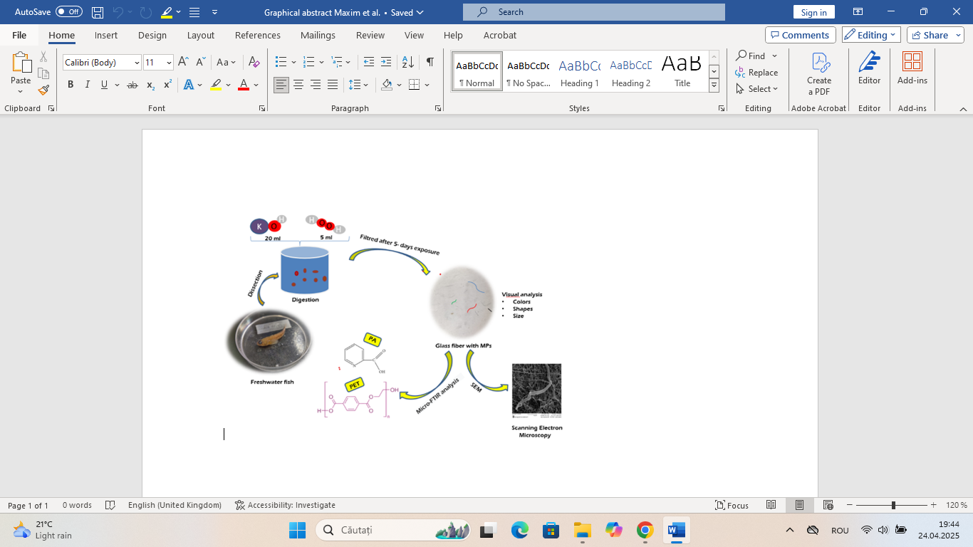
The fish were captured by a team of specialists from the "Vasile Alecsandri" University of Bacău using the electrofishing method [27,28,29,30,31] at ten sampling points along the river course, from the spring to the confluence with the Trotuș River. The principle of operation: the anode and cathode, by immersion in water, produce an electric field that causes a muscular response in the fish, forcing them to orient themselves towards the anode. Used regularly, it caused temporary paralysis of the fish. After the fish were caught and identified, they were fixed in 4% formalin and then dissected, and the stomach contents removed. In the laboratory, the contents were preserved in 70% technical alcohol, water, and glycerin in a ratio of 1:2:0.25 [30,31]. The intestinal contents of certain fish were examined under a stereomicroscope with digital camera, Zeiss Discovery V.8.
The implemented protocol was approved by the Ethics Commission of the Faculty of Biology, "Alexandru Ioan Cuza" University of Iasi, under registration number 5581/12.12.2024.
2.3. Micro-and Mesoplastics Isolation, Identification, and Characterization
We used the method of digestion with potassium hydroxide, KOH [32,33]. We added 20 ml of 1 M KOH solution to the contents of the samples and waited for 72 h at room temperature, in the dark. After this period, 5 mL of hydrogen peroxide (30%) was added [34]. Depending on the amount of organic material in the sample, the reaction period varies from 48 hours to 5 days at room temperature. The resulting solution was filtered through a 1.2 µm pores, 47 mm diameter glass fiber filtering membrane by a vacuum pump. The resulting filters were placed in Petri dishes and were visually inspected under a stereomicroscope equipped with a digital camera for adequate detection of categories of shape, size, and color [35]. Then, microplastics were identified, measured, and counted with the help of a Zeiss Discovery V.8 stereomicroscope, that has a built-in camera, using ZEN software.
Statistical analyses were performed using JMP14. Initially, the data were tested for normality using the Shapiro-Wilk test. Comparisons between groups were made using one-way ANOVA. Student's t-test was used as post-hoc test when necessary. A Pearson’s correlation test was performed to test the relation between fish size/weight and number of plastic particles found in gastrointestinal tracts. The influence of the feeding habits and habitat was also considered.
Scanning Electron Microscopy (SEM) was used to observe the surface features of the found microplastics. Gold metallization was performed (layer of approximately 10-15 nanometers) with EMS 550x metallizer. Then, the samples were visualized with a TESCAN VEGA II SBH scanning electron microscope, using a high vacuum and an acceleration voltage of 30 kV.
The analysis of the functional groups of the chemical compounds in the composition of the microplastics retained on the surface of the glass fiber filter was carried out by FTIR Microscopy in ATR (Attenuated total reflection) mode and a square aperture of 200 x 200 μm, selected from the options provided by the LUMOS II (Bruker Optik GmbH, Ettlingen, Germany): 4500-500 cm-1 spectral range with 4 cm-1 spectral resolution and microscope field view of 2.2x2.0 cm2 at 4.25 micrometers spatial resolution.. The software used was OPUS 8.7.41, designed for LUMOS II.
2.4. Quality Control and Quality Assurance Measures
Quality control protocols was followed in this study to ensure the real availably of the results [36]. In this regard, all materials used in extractions were non-plastic (glass, metal) and thoroughly washed with distilled water to ensure that the samples were not contaminated, and the results are accurate. Cotton lab coats was used by all the laboratory personnel during experiments. In addition, a filter used for filtering the NaCl solution without any sample was prepared and placed close to the working bench to test for airborne contamination during the extractions used as blank sample and the data were later harmonized without influence. The mean concentration detected in blank samples was 1.5 ± 0.7 MPs, most likely originating from textile fibers not fully covered by the lab coat. Observation and counting of microplastics were performed by the same researcher to ensure the consistency of the statistical standards.
2.5. Quantification of MPs from Different Matrices
Fibers collected from regular cloths labeled as made of 100% polyamide composition and plastic foil of polyethylene cut from regular plastic bag were analyzed with Micro-FTIR spectroscopy in the same conditions as the samples of microplastics collected from the fish stomachs.
3. Results
In this study, eight fish species were analyzed, with a total of 215 individuals, caught in different years/decades from the Oituz River. Investigating the fish, we found that microplastics, mostly in the form of fibers, were found in each species but not in all specimens. A small percentage (1.86%) presented empty stomachs. There were five plastic fragments, one transparent in Barbus petenyi from 2008 (Table 2), two transparent fragments in two Phoxinus phoxinus from 2008 (Table 4), and two in Sabanejewia balcanica. More than two microplastics were present in all fish individuals except for one Barbus petenyi from 2008 in which a single microplastic fiber was found and in a Squalius cephalus from 2015 in which there were two fibers/items. Microplastics represented by sizes <5 millimeters, as well as mesoplastics that are characterized as having sizes between 5-25 mm were found in all eight fish species (Figure 2).
The fewest mesoplastics, 1, belong to Sabanejewia balcanica and Rhodeus amarus, and the most mesoplastics belong to Barbus petenyi from 2008. The microplastics shaped as fibers were the most numerous, in each species, the most found in Squalius cephalus specimens from 2004, with 478 micro- and mesoplastics (Table 1).
Compared to the chub from 2015, where there were 295 micro- and mesoplastics (14.75 items/individual), the chub from 2004 has more fibers: 478 (23.9 items/individual), but the average size of the microplastic is smaller, 1.01 mm, compared to the average from 2015, which is 1.47 mm. In chub (40 specimens investigated), the three most common colors were (in descending order) blue, transparent, and red, the blue ones dominating both in 2004 (71.96%) and in 2015 (68,47%).
This section may be divided by subheadings. It should provide a concise and precise description of the experimental results, their interpretation, as well as the experimental conclusions that can be drawn.

Table 1.
Characteristics of plastic items identified in Squalius cephalus (n=40).
| Species | Squalius cephalus (2004) | Squalius cephalus (2015) | |
|---|---|---|---|
| Plastic characteristics | No. of items | No. of items | |
| Shape | Fibers | 478 | 295 |
| Size | 0-0.9 mm | 318 | 132 |
| 1-1.9 mm | 95 | 107 | |
| 2-2.9 mm | 36 | 32 | |
| 3-4.9 mm | 19 | 15 | |
| 5-10 mm | 10 | 8 | |
| >10 mm | 0 | 1 | |
| Average size | 1.01 mm | 1.47 mm | |
| Color | Blue | 344 | 202 |
| Transparent | 45 | 56 | |
| Red | 34 | 17 | |
| Black | 10 | 4 | |
| Brown | 24 | 9 | |
| Green | 1 | 2 | |
| Yellow | 0 | 1 | |
| Purple | 20 | 4 | |
| Plastics’ totals | 478 | 295 | |
Through the analysis of 40 mediterranean barbel stomachs, we identified 671 micro- and mesoplastics, of which 374 were in the stomachs from 2008 (average 18.7 items/individual) and 297 in the stomachs from 2015 (average 14.85 items/individual) (Table 2). In both periods/decades, blue and transparent fibers dominated, followed by red ones in the case of the specimens caught in 2015 and brown ones in those from 2008. In both cases, most microplastics were sized between 0 and 1.9 mm.
Table 2.
Characteristics of plastic items identified in Barbus petenyi (n=40).
| Species | Barbus petenyi (2008) | Barbus petenyi (2015) | |
|---|---|---|---|
| Plastic characteristics | No. of items | No. of items | |
| Shape | Fibers | 373 | 297 |
| Fragments | 1 | 0 | |
| Size | 0-0.9 mm | 142 | 69 |
| 1-1.9 mm | 113 | 115 | |
| 2-2.9 mm | 64 | 71 | |
| 3-4.9 mm | 38 | 32 | |
| 5-10 mm | 15 | 10 | |
| >10 mm | 2 | 0 | |
| Average size | 1.84 mm | 1.87 mm | |
| Color | Blue | 181 | 172 |
| Transparent | 113 | 93 | |
| Red | 28 | 18 | |
| Black | 3 | 10 | |
| Brown | 39 | 1 | |
| Green | 1 | 0 | |
| Yellow | 1 | 0 | |
| Purple | 7 | 3 | |
| Plastics’ totals | 374 | 297 | |
In 40 stomachs of Barbatula barbatula we identified 612 micro- and mesoplastics. Comparing the microplastics found in these two periods, we found that in 2008 (average 19.6 items/individual) the number is almost double that in 2015, but the fiber size is smaller than in 2015 (average 11 items/individual) (Table 3). All these microplastics are represented by fibers, mostly blue, followed by transparent, and red ones. Besides these, black, brown, green, yellow, and purple fibers were also found.
Table 3.
Characteristics of plastic items identified in Barbatula barbatula (n=40).
| Species | Barbatula barbatula (2008) |
Barbatula barbatula (2015) |
|
|---|---|---|---|
| Plastic characteristics | No. of items | No. of items | |
| Shape | Fibers | 392 | 220 |
| Size | 0-0.9 mm | 158 | 64 |
| 1-1.9 mm | 149 | 91 | |
| 2-2.9 mm | 46 | 36 | |
| 3-4.9 | 32 | 24 | |
| 5-10 mm | 7 | 5 | |
| Average size | 1.46 mm | 1.69 mm | |
| Color | Blue | 243 | 152 |
| Transparent | 118 | 31 | |
| Red | 13 | 13 | |
| Black | 3 | 8 | |
| Brown | 3 | 2 | |
| Green | 2 | 1 | |
| Yellow | 2 | 5 | |
| Purple | 8 | 8 | |
| Plastics’ totals | 392 | 220 | |
Out of 647 micro- and mesoplastics found in the stomachs of Phoxinus phoxinus (40 specimens investigated), 645 were shaped as fibers and two as fragments. Most fibers were sized between 1 and 1.9 mm, more being present in the specimens caught in 2008 (average 20.45 items/individual) (Table 4) than in 2015 (average 11.9 items/individual). Regarding the color of plastics, blue fibers dominated in both periods, followed by transparent, and red ones.
Table 4.
Characteristics of plastic items identified in Phoxinus phoxinus (n=40).
| Species | Phoxinus phoxinus (2008) | Phoxinus phoxinus (2015) | |
|---|---|---|---|
| Plastic characteristics | No. of items | No. of items | |
| Shape | Fibers | 407 | 238 |
| Fragments | 2 | 0 | |
| Size | 0-0.9 mm | 109 | 78 |
| 1-1.9 mm | 153 | 102 | |
| 2-2.9 mm | 89 | 33 | |
| 3-4.9 | 45 | 18 | |
| 5-10 mm | 11 | 7 | |
| >10 mm | 2 | 0 | |
| Average size | 1.94 mm | 1.67 mm | |
| Color | Blue | 213 | 179 |
| Transparent | 144 | 35 | |
| Red | 24 | 19 | |
| Black | 2 | 2 | |
| Brown | 15 | 2 | |
| Green | 2 | 0 | |
| Yellow | 1 | 0 | |
| Purple | 8 | 1 | |
| Plastics’ totals | 409 | 238 | |
Of 176 micro- and mesoplastics found in Alburnoides bipunctatus stomachs (20 specimens investigated), 159 were fibers and 17 films (Table 5) (average 6.8 items/individual in 2008, and average 10.8 items/individual in 2015). Most fibers were sized between 1 and 1.9 mm. Most were present in the specimens caught in 2015. Regarding the color of microplastics, blue fibers dominated in both periods, followed by transparent, and red ones.
We identified 145 micro- and mesoplastics (average 7.25 items/individual), 143 fibers, and two fragments, in the digestive tracts of 20 specimens of Sabanejewia balcanica. Four individuals had their stomachs empty. The blue plastics dominated, followed by the transparent ones, and most were sized between 1 and 1.9 mm. In the digestive tract of Rhodeus amarus (6 specimens investigated), we identified 58 microplastics (9.66 items/individual), 53 fibers, and 5 films (Table 6). In this case, the number of blue fibers dominated, followed by the transparent ones. In Gobio obtusirostris (9 specimens investigated) we found 132 microplastics (14.66 items/individual), of which 124 fibers and 8 films. The blue color dominated with 81 fibers, followed by the transparent fibers with 35. In this species, many fibers were sized between 1 and 1.9, followed by the ones sized between 0 and 0.9.
We found that in all eight species investigated, fibers dominated, followed by films and fragments (Figure 3). The reason for the reduced sizes found in 2004 and 2008 may consist of the continuous fragmentation of larger fibers in the natural environment.
The predominant colors are the same in all species, represented by blue, transparent and red. Brown, black, purple, green, and yellow fibers were also identified (Figure 4).
Analysis of the relations between fish size/weight, feeding way, habitat, and number of plastic particles found in gastrointestinal tracts.
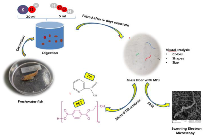
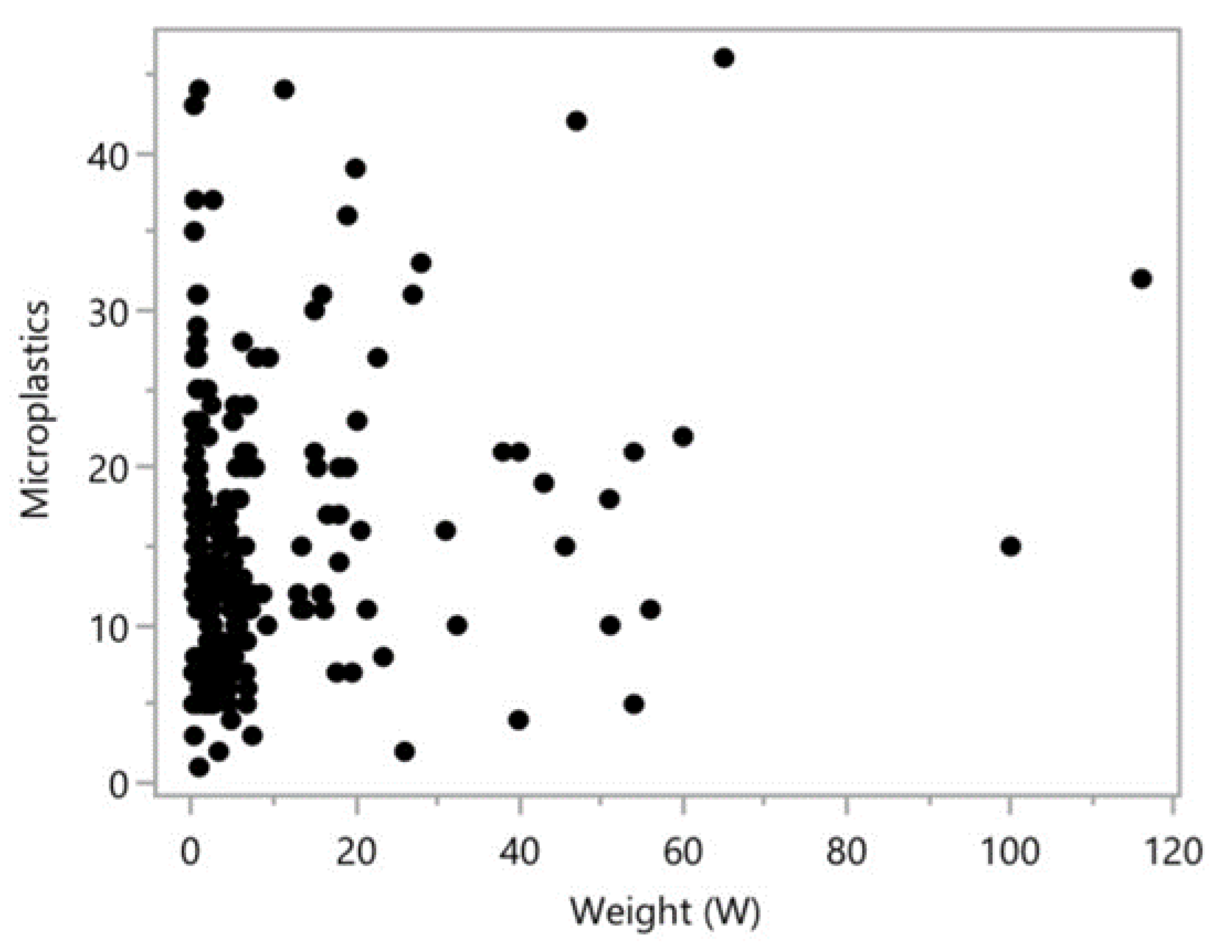
There was no correlation between length and number of plastic particles. There was a positive correlation between weight and number of plastic particles. All fish species investigated are omnivorous, so no differences in feeding habits were observed. Feeding habitats were compared and demersal species accumulated more plastic particles than the benthopelagic ones.
We applied an ANOVA analysis to determine whether there were statistically significant differences in the number of microplastics among the species studied. We analyzed which species differ significantly in terms of microplastic accumulation. R² = 0.150, which means that species explains only 15% of the variation in microplastic accumulation, indicating a moderate influence. The differences between species are statistically significant p < 0.05, suggesting that species plays an important role in microplastic accumulation. Squalius cephalus and Barbus petenyi have the highest accumulations of microplastics. Squalius cephalus accumulates much more microplastics than Alburnoides bipunctatus. Barbus petenyi has a higher accumulation than Phoxinus phoxinus. The differences between Gobio obtusirostris and Rhodeus amarus are slightly significant. Sabanejewia balcanica and Barbatula barbatula do not show notable differences. There are significant differences between some species, confirmed by statistical tests.
Figure 6.
Distribution of microplastics by fish weight.
To assess whether fish weight influences microplastic accumulation, we analyzed the correlation between these two variables. A p-value of 0.0028 is much lower than the typical threshold of 0.05. This suggests that there is a statistically significant relationship between the number of microplastics and the weight of the fish. Larger fish are exposed to contaminated water for a longer period and consume a larger volume of food, which may favor the bioaccumulation of microplastics in the digestive tract. Also, larger fish might have a more variate diet, exposing them to more sources of contamination.
Figure 7.
Distribution of microplastic by habitat, performed using one-way ANOVA.
The ANOVA test indicated a significant difference with p < 0.05 between the two habitats. Fish living close to the bottom accumulate more microplastics, while benthopelagic fish living between the bottom and the water column accumulate fewer microplastics. This difference can be explained by the concentration of microplastics in the sediments. Still, the differences may also be related to the feeding site and sources of microplastic contamination of the water. Fish that feed directly on the substrate have a higher risk of ingestion. Currents can redistribute microplastics, making them more accessible to demersal fish.
The surface characteristics of microplastics found in the gastrointestinal tract of fish, obtained using scanning electron microscopy (SEM), are shown in Figure 8.
For evaluating the chemical composition of the microplastics collected from the fish stomach, the analysis was performed with the micro-FTIR technique in ATR mode that allows the visualization of the sample during the measurements and the acquisition of spectra at the points of interest, avoiding interference with other materials such as the filter fiber as in this case. The spectral data were acquired in the points shown in the microscope images gained during micro-FTIR analysis (Figure 9).
After long exposure to variable environmental conditions, such as temperature, pressure, and chemical/enzymatic interactions, microplastics are in a certain degree of degradation and decomposition, which makes it difficult to be recognized by a software and identification in a database, which is also the case for this study. For this reason, in our study we evaluated the chemical composition of microplastics by assigning the peaks in the spectra to the functional groups [37] and, based on the identified functional groups, we estimated the materials and proceeded to use polyamide (PA) and polyethylene terephthalate (PET) as control samples.
Therefore, in the spectra presented in Figure 10, the amides are denoted by the double peak in the 3300-3200 cm-1 range for NH stretching vibrations, 1700-1600 cm-1 range for C=O stretching and around 1590 cm-1 for NH bending vibrations (S.c.08; S.b.08; G.o.15; Ph.ph.15; Ph.ph.08; A.b.15; B.p.08; Rh.a.15; S.c.15). Their aliphatic structure is indicated by the peaks at about 2900 cm-1 for CH asymmetric stretching and 2800 cm-1 for CH symmetric stretching. The CH vibrations in the range 3200-3000 cm-1 show aromatic amides. Therefore, the spectra of the microplastics samples S.c.08; S.b.08; G.o.15; Ph.ph.15; Ph.ph.08; A.b.15; B.p.08; Rh.a.15; S.c.15, compared to the polyamide fiber used as control, indicate polyamide microfibers.
In the fingerprint area of all spectra, considering the fact that the measurement of the spectrum of the polyamide fibers (PA) used as control was carried out under the same conditions as for the microplastic samples - i.e., on a glass fiber filter support - and no interference was recorded with the vibrations specific to the functional groups of the glass fiber, the vibrations in the 1400-900 cm-1 range resulted in the samples spectra can be assigned to functional groups other than Si-O characteristic of some chemical compounds incorporated in microplastics by phenomena of adsorption, absorption and/or adhesion to the polyamide microfibers. Polyamide is known for these sorption and adhesion properties which make it suitable to fabricate membranes for removing impurities from water or from other liquids [38]. The strong vibrations in the 1275 - 1040 cm-1 range are thus attributed to the C-O and CO-O groups which may be the result of the partial decomposition of the polyamide, but also of the interaction with the peroxide used for the cleaning treatments with the material taken from the fish stomach. They can also belong to other compounds, such as alcohols, esters absorbed from the environment traveled by microfibers (wastewater, air, etc.) and to polyethylene terephthalate microplastics (PET) as noticed in the PET control spectrum analyzed in the same conditions (on glass fiber filter support). Finally, in the same range, vibrations of Si-O groups of the sand particles adhering to the analyzed microplastics may occur.
The stronger peaks (enhanced transmittance) of the S.c.08, S.b.08, G.o.15, Ph. ph. 15, Ph.ph.08, A.b.15, Rh.a.15, A.b.08 samples compared to the polyamide control fiber show that the microplastics are loaded with other chemicals by sorption processes from the environment they crossed, such intaking phenomenon being already reported by research [17,18].
The B.p.15 samples exhibit enlarged peak of beard-like shape in the 3500 - 3100 cm-1 range which can be assigned to carboxyl groups. In the spectra of the microplastics samples B.p.15 (Figure ii), the large round peak in the 3366 - 3171 cm-1 range is assigned to OH stretching vibrations and the 1702 cm-1 band is for C=O in the carboxyl group (COOH). These peaks indicate polyamide degradation by hydrolysis into amines and carboxylic acids due to the long-time exposure in water at significant depths, as well as to the chemical environment in the fishes’ stomachs where pH and lake of oxygen enhanced the process [39]. These types of vibration bands are also specific to amino acids/proteins and show adsorption of blood during microplastics removal from the fish stomach.
The A.b.08 microplastics present in their IR spectra (Figure ii A.b.08) asymmetric and symmetric stretching vibrations characteristic to aliphatic CH groups at 2929 cm-1 and 2883 cm-1, respectively. The C=O stretchings in A.b.08 are assigned to the vibrations at 1652, 1631 cm-1. The C=O groups indicate carboxyl together with the large round peak in the 3366 - 3097 cm-1 range. In the 1400 cm-1 - 832 cm-1 of the fingerprint area, C-O and CO-O groups are denoted by the 1029-983 cm-1 skeletal vibrations. The strong peak at 1390 cm-1 is assigned to CH3 group bending. The spectra of the B.b.15 and A.b.08 samples are very similar to the PET control spectrum, indicating that these group of microfibers is sourced in polyethylene terephthalate materials.
The sources of microplastics collected from the stomachs of different fish species for this study may be threads or cords used for technical purposes and made of mixtures of polyamide, polyethylene terephthalate and glass fibers. In such a situation, based on the IR spectra, different environmental conditions, including water and enzymes, could contribute to decomposition processes. This is a possible explanation for the fingerprint area, the same in all samples analyzed and overlapped on the vibration bands in the glass fiber filter. It also explains the enhanced PA decay in the B.p.15 sample and possibly in the A.b.08 sample, where the PET spectrum is observed as primary vibrations. It is also important to note that for fiber mixtures some of the peaks of the mixture components may overlap the others, the shape of the most intense ones being visible in terms of transmittance. The advanced degradation of PA into acids and amines in B.p.15 sample, could be assigned to the feeding behavior of the fish known as omnivorous meaning if it fed with other organisms contaminated with microplastics of PA and the hydrolysis already started.
Differences in the intensity of transmittance within the spectra of the microplastics collected from the same fish are assigned to the yield and kind of the chemicals adsorbed or absorbed.
Also, the samples of microplastics are seen in the FTIR microscope images (Figure 10) as being of different colors. We noticed that the color or, sometimes, the lack of color cannot be related to the transmittance intensity. Therefore, the different color may be due to the thickness of the layer of substances adsorbed on the surface of the microfibers.
4. Discussion
No microplastics were detected in any fish prior to 1950. From mid-century to 2018, microplastic concentrations showed a significant increase when data from all fish were considered together. All detected particles were fibers, and represented plastic polymers (e.g., polyester) along with mixtures of natural and synthetic textiles. Museum specimens are an overlooked source for assessing historical patterns of microplastic pollution, and for predicting future trends in freshwater fish, thereby helping to sustain the health of commercial and recreational fisheries worldwide [40].
Rivers are implicated as major pathways of microplastic transport to marine and lake ecosystems, and microplastic ingestion by freshwater biota is a risk associated with microplastic contamination, but there is little research on microplastic ecology within freshwater ecosystems. Microplastic uptake by fish is likely affected by environmental microplastic abundance and aspects of fish ecology, but these relationships have rarely been addressed [41]. Differences in MPs loading may be related to the level of water pollution, MPs characteristics, and the behavioral and feeding characteristics of the fish species. The deformability of MP fibers makes them similar to worms and eggs, which are prone to accidental ingestion by fish. Once ingested, large MPs are difficult to eliminate by fish digestive systems. This may be one reason why most of the MPs found in fish in this study were fibers. Color may have some effect on MP ingestion by fish, as some of them are visual predators and prefer to ingest white, yellow, and blue foods [25]. On the other side, it is believed that mechanisms of ingestion and egestion effectively result in a mass balance that does not engender internal accumulation [42].
Squalius cephalus, Barbus petenyi, Alburnoides bipunctatus, Rhodeus amarus and Gobio obtusirostris are benthopelagic, which live and feed on the bottom but in the open water column as well. Barbatula barbatula, Phoxinus phoxinus and Sabanejewia balcanica are demersal and, consequently, feed on the bottom of the water.
There is limited research on the trophic spectrum of fish species in Romania compared to international studies. However, Romanian researchers have studied the ichthyofauna and trophic spectrum of fish in both flowing and stagnant water ecosystems, such as the Buzău River and its tributaries [43], the Argeș River, and the Dâmbovița River [29,30], as well as Dorobanț, Aroneanu, and Venetia lakes on the Ciric River [44], dam lakes on the Jiu River [45], and the Budeasa-Golesti area of the Argeș River in Pitești [46]. Additionally, topics such as diet, prey characteristics, and feeding behavior have also been researched by various authors [28,47,48,49,50,51,52].
Squalius cephalus is one of the most common and widespread cyprinid species in Europe [53]. Favorable habitats for rheophilic species, such as the chub, are those with clean and transparent water, sandy substrate, and banks intensely shaded by trees [54]. The reason why this species has a wide geographical distribution is that it has high ecological tolerance, and its population migrates to rivers to spawn in spring and early summer [55]. It is most abundant in small rivers and large streams, but also occurs in slow-flowing lowland rivers and very small mountain streams [53]. The European chub is benthopelagic and potamodromous [55]. Adults are solitary and juveniles are gregarious [56]. The species is omnivorous due to the length and the shape of the digestive tract [57] . It feeds on a variety of aquatic and terrestrial animals and plant material. Large individuals prey predominantly on fishes. In a study carried out in Lake Tӧdürge, Turkey, the chub's population had a varied food regime, such as insects, phytoplankton, zooplankton, fish, nematodes, macrophytes, plant and animal detritus, in which zooplankton (69.2%) prevailed [57]. A study found as predominant the insects' families Chironomidae (Diptera order) and Formicidae (Hymenoptera order), alongside with Araneidae family of spiders [30]. Another research [58] identified 9 macroinvertebrate groups: Nematoda and Oligochaeta worms, Crustacea, insects as Ephemeroptera, Odonata, Hemiptera (aphids), Lepidoptera, Coleoptera, Diptera, where Coleoptera and Nematoda were the most common. The highest dominance index was calculated for the Coleoptera group with 60.68% followed by Nematoda with 28.8%. In 20% of the stomachs investigated were found fish remains, represented by: scales, vertebrae, radii, heads, and even larger pieces of undigested or digested fish. In addition to these, were also identified: feathers, plant fibers, pebbles, fat drops, and microplastics. A recent study [59] identified in the chub diet: Collembola, Ephemeroptera (family Baetidae), Coleoptera, Diptera (mainly family Chironomidae) and Hymenoptera (family Formicidae), the Chironomidae group being the best represented in terms of relative abundance and frequency of occurrence. The microplastics sizes that dominated in Squalius cephalus were between 2-2.9 mm (47%) [59].
Barbus peteny lives and moves near the bottom of the fast-flowing, clear, and well-oxygenated waters water. It has an omnivorous diet and prefers benthic invertebrates (annelid worms, gammarid crustaceans, insect larvae) and plant material [29,60]. It usually consumes the most resources available in the environment, in addition to chironomids and oligochaetes, which are more abundant as a resource, ephemeropterans and trichopterans whose abundance in the site is lower [49]. Along with insects, nematodes, oligochaetes, mites, and individuals of Phoxinus phoxinus were found [29].
Barbatula barbatula is usually found in flowing stretches of streams and medium-sized rivers with gravel to stone bottom, but also in a variety of other habitats, including sandy canals and lake shores. It is an omnivorous species, and adults consume relatively large benthic invertebrates such as gammarids, chironomids, insect larvae. Barbatula barbatula exploits resources in proportion to their accessibility in the environment. Dominant in the diet are chironomids, followed by Ephemeroptera, Trichoptera, Plecoptera, Hirudinea and oligochaetes [50]. The results of a study on the trophic spectra of the fish species inhabiting the Oituz and Caşin rivers demonstrated that the stone loach showed a constant feeding preference for Ephemeroptera (Baetidae family) and avoided Trichoptera [27]. On the other hand, another study from the same year, but carried out on fish species from the Buzău River, reported that the diet of the stone loach contains, in addition to Ephemeroptera larvae, also Trichoptera larvae. Additional, larvae of Plecoptera, Hymenoptera, Diptera (families Chironomidae, Limoniidae, Simuliidae), and nematodes were found [43]. Following the analysis of the stomach contents, despite of the mentioned taxa, macroinvertebrates belonging to Ephemeroptera (Oligoneuriidae family) were identified [29].
Phoxinus phoxinus is found in a variety of habitats, from well-oxygenated waters of small, fast-flowing streams to large lowland rivers and large oligotrophic lakes. Feeds on algae, plant debris (in rivers), zooplankton, worms, mollusks, crustaceans, and insect larvae. The Eurasian minnow is an omnivorous and opportunistic fish [61] with a generalist feeding mode [62]. A study on the feeding pattern of Phoxinus phoxinus found that smaller specimens consumed mainly benthic prey (mostly Diptera larvae) and less terrestrial invertebrates [62]. Another study showed that it prefers the larvae of Ephemeroptera, Hymenoptera, Coleoptera, larvae of Trichoptera and Chironomidae [29]. P. phoxinus caught in Lake Ånnsjön generally ingested more zooplankton, compared to those caught in adjacent streams, which ingested macroinvertebrates and terrestrial insects to a greater extent [63].
Alburnoides bipunctatus is a species inhabiting streams and rivers in foothills with well oxygenated, fast-flowing water; found also in rivers with very calm waters. It is susceptible to human activities, and the number of habitats suitable for spawning is limited [64]. It spawns in small groups, deep in gravel, on the bottom of waters with rapid currents. It feeds on insect larvae (Ephemeroptera) and dead insects, as well as on isopod crustaceans such as Asellus aquaticus, annelid worms (Oligochaeta) and algae.
Sabanejewia balcanica a demersal freshwater fish. During the day they stay in sand or gravel and descend up to 1.5 m into the water. During daylight, adults are found burrowing into sand, sometimes in gravel on hill streams with clear water. Also found in moderate current with few plants at water depths up to 1.5 m. Have also been observed in large rivers.
Rhodeus amarus is a species of Community interest. It reproduces in clear, slow-moving water, often on muddy bottoms. It is a small-sized omnivorous fish species and usually feeds on plant detritus, and green algae, less often on worms, crustaceans, and insect larvae [65]. In European bitterling’s diet were identified: Nematoda, Ephemeroptera (Baetidae family), Coleoptera, and Diptera (mainly Chironomidae family), the Chironomidae group being the best represented in terms of relative abundance and frequency of occurrence [59]. The microplastics sizes that dominated in Rhodeus amarus were between 0-0.9 mm (69%). Rhodeus amarus had more microplastics in its stomach contents than the chub and, in addition, had films and not just fibers. Some fish also contained vegetation and solid materials (seeds, sand, and pebbles of different sizes) in the stomach [59].
Gobio obtusirostris breeds in shallow water, over rocks, sand, or vegetation. The most important species of insects that are consummated by Gobio obtusirostris are Diptera, Coleoptera, Heteroptera, and Trichoptera. Besides these, vegetal remains were also found [66].
In 215 fish stomachs analyzed, we identified a total of 3214 microplastics, most of which are represented by fibers with 98.91%, films with 0.94% and fragments with 0.15% (Figure 3). Regarding the color of the microplastics found, blue dominated in all samples with a total percentage of 61.44%, followed by transparent 23.95%, red 6.53%, brown 3.57%, purple 1.83%, black 1.80%, green 0.57% and yellow 0.31% (Figure 4). From the combined data of all samples, the size range of microplastics in fish was from 0-10 mm, with a few exceptions >10 mm. The dominated size range is 0-1.9 mm, 72.12%, followed by 2-4.9 mm, 17.89% and >5 mm with 9.99% (Figure 2). The smaller the microplastics, the greater the possibility of ingestion.
Our results are in accordance with those published by S. Wang et al. [33]. In this study, 47 freshwater fish with different feeding habits were obtained from the Beijiang River and Pearl River Delta regions. Microplastics were present in all samples. The abundance of microplastics in fish samples from the Beijiang region ranged from 1.0 to 15.0 (average 5.6) items/individual, whereas the values for fish from the Pearl River Delta region were 1.0–14.0 (average 4.8) items/individual. The values for omnivorous fish samples from the two regions were 6.8 and 6.2 items/individual, respectively. The common colors of microplastics found were white, black, and yellow. Other colors were red, blue, and green.
A high percentage of microplastics was found in the stomachs of the fish (96.4%) caught by local fishermen around Ubolratana Reservoir, Thailand. Most of the microplastics were around 0.5 mm, blue, and fiber-shaped [67]. High percentages of microplastic ingestion, some reaching 100% were found in a study of freshwater fish from the Río de la Plata estuary: 87 fish belonging to 11 species and four feeding habits were investigated, and all accumulated microplastic particles. The fibres represented the 96% of MPs found [68]. Microplastic was detected in fish (11 species) from all 3 major tributaries of Lake Michigan, USA., but there was no correlation between microplastic concentrations in fish and surface waters. Rather, there was a significant effect of functional feeding group on microplastic concentration in fish. Neogobius melanostomus (a zoobenthivore) had the highest concentration of gut microplastic (19 particles fish−1) and had a positive linear relationship between body size and number of microplastic particles. In this study, 85% of fish had microplastic in their digestive tissues with an average of approximately 13 particles fish−1. There were some links between fish body size and trophic position, and the abundance of microplastic in digestive tissue. Fibers comprised approximately 97–100% of all microplastic found. Fragments were rare and accounted for approximately 2.5–3% of the microplastic. Clear and blue fibers were predominant; small (<1.5 mm) fibers were the most common size [41]. A review since 2021 found that 49% of all fish sampled globally for microplastic ingestion had plastic (average of 3.5 pieces per fish), with fish from North America ingesting more plastic than fish from other regions. Research from marine environments dominated (82% of species) but freshwater fish ingested more plastic, as did detritivores, fish in deeper waters and those from aquaculture sources [69]. The highest concentration of microplastics and other anthropogenic microparticles ever reported in bony fish was documented for Lake Ontario: 12,442 anthropogenic microparticles across 212 fish (8 species), Humber River: 943 across 50 fish (1 species), and Lake Superior: 3094 across 119 fish (7 species). Fish from Lake Ontario had the greatest mean abundance of anthropogenic microparticles in their gastrointestinal tracts (59 particles/fish), with up to 915 microparticles in a single fish. Fish from Lake Superior contained a mean of 26 particles/fish, and fish from Humber River contained 19 particles/fish. Most particles were microfibers. Overall, ≥90% of particles were anthropogenic, of which 35-59% were microplastics. Polyethylene (24%), polyethylene terephthalate (20%), and polypropylene (18%) were the most common microplastics [70].
Other studies have also found micro- and mesoplastics in all investigated 6 fish species [71] and as in our case the abundance of microplastics was higher than that of mesoplastics, representing 55.92.3% of the total plastic items in each species. The average abundance of microplastics ranged from 1.1 to 7.2 items per individual. The average abundance of mesoplastics ranged from 0.2 to 3.0 items per individual. The dominant ones were: fiber in shape, transparent in color and cellophane in composition. MPs were found in all fish samples (30 fishes, representing 15 species) in Gehu Lake, Changzhou. The average abundance of MPs in fish was 10.7 items/fish Ingested microplastic consisted primarily of fibers, mostly transparent or blue. The dominant size of MPs observed in this study was 0.1–0.5 mm. PES, man-made fiber, and PP were the dominant types; acrylic acid, polyvinyl chloride, polyamide, rayon, polyethylene, and polyethylene terephthalate were also found [72].
In our study, the dominant form of microplastic is the fiber, followed by the film and fragment. Similar results, in which the number of fibers exceeds that of the fragments, were also reported in other studies such as [24,35,73] Siddique et al., 2022; [74] and others. Of 64 specimens of roach sampled, 33% contained at least one microplastic particle. The majority of particles were fibres (75%), with fragments and films also seen (22.7% and 2.3% respectively). Polymers identified were polyethylene, polypropylene, and polyester. The size of fish correlated with the actual quantity of microplastics in the gut, and females ingested more MPs than males, probably with food, due to increased energy requirements for spawning [75]. Of 202 gudgeons and 187 roaches collected, 54.5%, respectively 53.9% had ingested MP like particles. Most particles were fibres (99.8%), and only one fragment. The length of fibres ranged from 0.5 to 5 mm and the dimension of the fragment was 0.5 × 1 mm. The colours of the identified MP-like particles were diverse (blue, black, transparent, green, red, sky-blue, violet, brown, orange and white) [76]. Microplastics were found in 72% of the 58 brown trout sampled along the River Slaney catchment, Ireland, being present in 66% of guts (1.88 ± 1.53 MPs fish⁻1) and 28% of stomachs (1.31 ± 0.48 MPs fish⁻1). Fibres were the dominant shape, followed by fragments. No difference in median microplastic burden was observed between fish collected in high and low exposure sites. Microplastic burden was unrelated to fish length, same as in our case. 11 polymers were identified, polystyrene being the main polymer found, followed by polyester urethane in guts or aramid in stomachs [77] (O’Connor et al., 2020) A recent study [58] found in the stomachs of the 60 specimens of chub, 124 microplastics in the form of microfibers of different colors and sizes: 42 purple, 38 blue, 19 red, 13 black, and 12 brown. The reason for fibers being so common has been attributed to their diverse origin. Fibers may result from the degradation of clothing items, furniture, and fishing gear. Washing a single item of synthetic clothing may release approximately 2000 fibers [78]. The predation tendency of the aquatic organisms for MP shape, size, and color were investigated in 105 fishes from 14 different species from the Yangtze River estuary. Main polymers were cellulose and polyethylene terephthalate characterized by black and gray fibers and lengths ranging from 0.1 mm to 1 mm. Previous studies also confirmed by previous studies that fibers were the elementary MP shape preyed on by both macroinvertebrates and fish. This may be due to the high abundance of fiber in the habitat (water and sediment). Household laundry in coastal residential areas is a crucial source of high abundance microfibers. It is estimated that millions of fibers (e.g., cotton, rayon, and semi-synthetics) can be released from a single garment during a typical household wash. They are discharged into offshore waters via sewage pipes, which increases the risk of ingestion by aquatic organisms [79].
In the Pichavaram mangroves, India, seasonal trends, and consumption of microplastics by several fish species were examined. MPs abundance was higher during the monsoon (45%), suggesting seasonal runoff and increased plastic pollution during heavy rains as key contributors. Microplastic contamination was dominated by fibers (71%-73%) and fragments (27%-29%). A significant proportion (32%) of MPs were smaller than 1 mm in all seasons. The fibers were predominant blue (40%) and red (13%) across all seasons, followed by transparent, brown, and yellow. Polypropylene was the most prevalent polymer, with significant contributions from Polyethylene and Polyethylene Terephthalate [80].
Contrary to our classification regarding the shape of the found microplastics, other authors had the fragment as the dominant shape [33,81,82]. A study on 84 (4 species) typical wild fishes in the Lijiang River identified MPs in 81.0% of specimens with an average abundance of 0.6 ± 0.6 items/individual. Blue MPs had the largest proportion in fishes, accounting for 10%-2.7%. Seven polymer types of MPs were identified, and PET was the most abundant. Large-sized (>0.3mm) and colored MPs in morphotypes of flakes and fibers dominated in wild fishes [25]. A comprehensive study of microplastic contamination in southern Germany investigated 1167 individual fish of 22 different species sampled from 11 rivers and 6 lakes, and plastic particles were found in 18.8% of individuals. Overall plastic abundance was 0.2 ± 0.5 particles per fish and intensity ranged from 1 to 4 particles (mean: 1.2 ± 0.5). Of the ingested plastic particles, 97.7% were smaller than 5 mm and were thus categorized as microplastics. Particles longer than 5 mm consisted exclusively of fibres. Of all the microplastics detected, most particles comprised fragments, at 54%, with fibres making up a further 39%. Plastic films and beads were rare, accounting for 2% and 3%, respectively. In decreasing order were: transparent, black, blue, white, red, grey, yellow, and green [83].
Present work also showed that blue is the most common color, followed by transparent and red. Blue was the dominant color also following other studies, but preceded by black, white, and red in [84], transparent and black [73], black and red in [85]. Other researchers showed that white was the dominant color followed by yellow and black [33]. It was stated that the color can indicate the source of the microplastics. Thus, the colored fibers come from the household sewage discharge that contains the water used for washing clothes, while most of the transparent fibers result from fishing activities. Therefore, the ingestion of microplastics could cause serious consequences for aquatic fauna [32] This fact was also mentioned in [86] study, where was demonstrated the passage of microplastics from one organism to another within the food chains, causing negative effects on the analyzed individuals. 80 perch specimens from 4 lakes in Italy were analysed and MPs fish-1 ranged from 1.24 in Lake Orta to 5.59 ± 2.61 MPs fish-1 in Lake Garda, with microplastics a higher presence occurrence higher in empty stomachs. The isolated particles, on average smaller than 400 μm, were mainly fragments, with polymers as polyethylene, polyethylene terephthalate, polystyrene, polyamide, and polycarbonate, but films were more abundant in Lake Como. The vast majority of MPs presented black/grey or blue colours. The colour of MPs can be an indicator of origin and/or typology and can represent different levels of risk to organisms. The colour of particles influences predatory activity and thus could increase the likelihood of accidental ingestion of MPs by predators due to possible confusion with their prey [34].
In a study of freshwater fish from the Chi River in Thailand, the abundance, size, color, and shape of microplastics were monitored. 8 fish species were investigated, as in our study. The results showed that 72.9% of the collected fish were contaminated with microplastics. The majority of the microplastics were larger than 0.5 mm, and of these, 86.9% were in the form of fibers, and 56.9% were blue, followed by red, black, white, transparent, and brown [87]. Similar results were presented in a study on 48 specimens of Hoplosternum littorale in Brazil, where 83% of the fish examined had microplastics in their gut, most of them in the form of fibers (46.6%) smaller than 5 mm in size. The ingestion of microplastics was negatively correlated with the diversity of other food items in the gut of individual fish [88].
According to the study that investigated 8 commercial freshwater fish species from Bangladesh, microplastics were found in 73.3% of the fish analyzed, and the majority of microplastics were in the 500 μm-1 mm size range; transparent fiber was the most dominant MP. Most MPs were composed of EVA, PP-PE, and HDPE polymers. Demersal fish had a higher MP ingestion rate than pelagic and benthopelagic fish, similar to our findings. [89]. A similar percentage of microplastic occurrence: 73.5% was also reported in 5 fish species (181 specimens sampled, 1 to 20 MPs per fish: fibers (89.5%), fragments (10.2%), and less than 1% beads) in an urban stream in prairie, Saskatchewan, Canada [90] and in bluegill and sunfish (436 specimens in all) from the Brazos River basin in Texas, USA, with percentages from 45% to 75% [91].
A frequency of microplastics of 69.7% was observed in a study of 8 commercial fish species (109 individuals) collected from a municipal water supply lake in Nigeria. On average, 1-6 microplastics were present per fish, with sizes ranging from 124 μm to 1.53 mm [92]. In another study, when six freshwater fish species with different feeding habits were considered, only 40% of the fish ingested microplastics. Most of these were between 1 and 5 mm in size [93]. A lack of relationship among number of MPs ingested and feeding habit are indicative that other factors are involved in fish quantitative MPs intake [21]. Another study on 13 fish species from a reservoir in the Three Gorges, China, showed as most abundant microplastics in the 1-5 mm range, accounting for 71.4%. Fibers and fragments were the most common forms. The predominant color was blue, followed by red, green, white, yellow, and black [94] .
In a review based on 34 articles published in a decade, 64 fish species were analyzed. The results show that the majority accumulated microplastics in the form of fibers and fragments at 68.1% and 21%, respectively, the predominant color being black at 22.4%, followed by blue at 21.3% [95]. There are other studies in which blue fiber color was second with a percentage of 17%, after white with 43%. In smaller quantities, fibers of other colors, such as black, gray, yellow, and red, were also found [96]. Same for microplastics found in two economic fish from Guangdong province (43.4% occurence). The plastics were dominated by white in color (61%), fragment in shape (67%), and less than 1 mm in size (74%). Fibers and pellets were also identified. Various colors of MPs were found as white, blue, green, red, yellow, black, and others [97].
Other researchers had similar results to ours in terms of dominant polymer: polyamide (PA) 64%, followed by polypropylene (PP) 15%, polystyrene (PES) 12%, polyvinyl chloride (PVC) 5%, and polyethylene (PE) 4% [98] . In another study, polyamide (PA) is also dominant, but this time followed by polyethylene polystyrene (PS) and polymethyl methacrylate (PMMA) [99].
Some studies have shown the dominance of polymers other than the ones we found. Various researchers have found that among the most common types of polymers found in freshwater fish is polyethylene terephthalate (PET), followed by PP [48], PES, and PE [100]. A broader variety of polymers was observed in Oreochromis farmed fish (44% occurrence, against 75% in natural source fish) [100]. The edible part of fish presented a lower prevalence of MPs compared to gill and stomach/gut, the last displaying a higher diversity of MPs. Fragments were the dominant type in all tissues. There were also studies where PET was in third place after PE and PP [101]. In another study, rayon and PES were predominant [102]. Polyethylene dominating, besides polypropylene, polystyrene, and polyamide were identified in commercial fish species in southern coastal region of India [103]. 17 commercial fish species (220 specimens) had a total of 1115 MPs particles. MPs most frequently found were fibers, transparent MPs, and tiny MPs (0.5 mm). Most of the fish had high MP levels, and all samples showed a surplus of secondary MPs, with fragments and fibers making up 36% and 32%, respectively, of the total MPs.
A review that considered 79 specialized articles demonstrated that most polymers were of the type PE, PS, PP, PET, but rayon, nylon, cellophane and acrylonitrile were also found [104]. In another review [105] the most common polymers were PE, PP, PS, and PET. Similar results, in which PE and PP are the most abundant, also appear in other studies [106,107].
75.9% of Nile tilapia (n = 29) and 78.6% of catfish (n = 14), purchased from local sellers in Cairo contained MPs in their digestive tract [78]. The most abundant were fibers (65%), followed by films (26.5%), and fragments. Polyethylene (PE), polyethylene terephthalate (PET) and polypropylene (PP) were identified. Black and red colored MPs were most abundant for fibers and films (black > red > blue > green > other > transparent for fibers and black > red > transparent > green > blue for films). Fragments were predominantly blue (blue > black > transparent). The difference between 7.5 ± 4.9 and 4.7 ± 1.7 items per fish was significant. The omnivorous diet of the tilapia which contains plankton may mean that it is more likely to mistake plastic items for food.
5. Conclusions
Contrary to the initial hypothesis that recent samples should contain more plastics than older ones, we found that older samples contained a higher number of micro- and mesoplastics, mainly consisting of polyamide and polyethylene terephthalate; this may be due to national pollution control measures by recycling both PET bottles and other plastics that previously ended up in the riverbed. Secondly, not all specimens investigated ingested plastics, but all investigated species did. The largest species (chub and Romanian barbel) accumulated the most plastic materials. The number of particles (micro- and mesoplastics) found in the gastrointestinal tract was positively correlated with weight and differences between habitats (and feeding area) were assessed: demersal species accumulated more microplastics than benthopelagic ones.
The IR spectroscopy performed with the micro-FTIR in ATR mode, provided information on the chemical composition of the microplastics collected from the stomach of the analyzed fishes and on their transformation during the exposure to the freshwater and biological environments, with specific physico-chemical conditions, leading to loading substances on the microplastics and/or to induce decomposition such as polyamide hydrolysis into carboxylic acids and amines. The resulted acids and amines were detected in the IR spectroscopic analysis, but one can assume that some of the detected decomposing products had been further degraded into small molecules, including gas. The most widespread type of polymer was polyamide (PA) with a percentage of 84.61%, followed by polyethylene terephthalate (PET) with 15.39%, a possible explanation being that PET and PA have higher specific densities compared to other polymers that may be present in benthic waters and, thus, can be more easily ingested by the fish studied. However, the sources of microplastic pollution analyzed in this study can be textile fabrics and threads, as well as technical cords and twine, including fishing nets. Plastic materials widespread occurrence and ingestion indicate that future research is needed for an ample range of species and habitats to fully establish the potential effects of microplastics in the aquatic environment. Extensive studies are needed to provide information on the specific influences on the chemical changes of water-polluting microplastics and to identify the source of these pollutants.
Author Contributions
Conceptualization, M.N. and R.J.; methodology, R.J., M.N. and I.C.; software, S.-M.O., A.C., S.G.; validation, R.J., I.C.; investigation, R.M., A.R., S.-M.O., A.C., I.D.C., I.D.C. and A.C.; writing—original draft preparation, R.M., A.R. and I.C.; visualization, validation, S.-M.O., A.C., S.G.; writing—review and editing, M.N., I.C. and D.U.; supervision, M.N., D.U.; project administration, M.N., D.U. and S.G.; funding acquisition, M.N., S.G. and I.C. All authors have read and agreed to the published version of the manuscript.
Funding
This research received no external funding.
Institutional Review Board Statement
The animal study protocol was approved by the Ethical Commission from the Faculty of Biology, ”Alexandru Ioan Cuza” University of Iasi, with registration number 5581/12.12.2024.
Informed Consent Statement
Not applicable.
Data Availability Statement
Data will be made available on reasonable request from the corresponding author.
Acknowledgments
Acknowledgment is given to infrastructure support from the Operational Program Competitiveness 2014–2020, Axis 1, under POC/448/1/1 Research infrastructure projects for public R&D institutions/Sections F 2018, through the Research Center with Integrated Techniques for Atmospheric Aerosol Investigation in Romania (RECENT AIR) project, under grant agreement MySMIS no. 127324.
Conflicts of Interest
The authors declare that they have no known competing financial interests or personal relationships that could have appeared to influence the work reported in this paper.
Abbreviations
| MPs | microplastics |
| PA PP PS |
polyamide polypropylene polyethylene polystyrene |
| PET | polyethylene terephthalate |
| PES PVC PE PMMA |
polystyrene polyvinyl chloride polyethylene polymethyl methacrylate |
References
- Burlacu, L.; Deak, G.; Boboc, M.; Raischi, M.; Holban, E.; Sadîca, I.; Jawdhari, A. Understanding the Ecosystem Carrying Capacity for Romanichthys valsanicola, a Critically Endangered Freshwater Fish Endemic to Romania, with Considerations upon Trophic Offer and Behavioral Density. Diversity 2023, 15, 748. [Google Scholar] [CrossRef]
- Vlăduţu, A.-M. The Impact of The Anthropic Factors on Disrupting and Destabilizing the Ecological Balance of The Vâlsan River. Current Trends in Natural Sciences 2013, 2, 30–35. [Google Scholar]
- Wang, W.; Ge, J.; Yu, X. Bioavailability and toxicity of microplastics to fish species: A review. Ecotoxicology and Environmental Safety 2020, 189, 109913. [Google Scholar] [CrossRef] [PubMed]
- Strungaru, S.A.; Jijie, R.; Nicoara, M.; Plavan, G.; Faggio, C. Micro- (nano) plastics in freshwater ecosystems: Abundance, toxicological impact and quantification methodology. TrAC - Trends in Analytical Chemistry 2019, 110, 116–128. [Google Scholar] [CrossRef]
- Collard, F.; Gasperi, J.; Gabrielsen, G.W.; Tassin, B. Plastic Particle Ingestion by Wild Freshwater Fish: A Critical Review. Environmental Science and Technology 2019, 53, 12974–12988. [Google Scholar] [CrossRef]
- Liu, K.; Wu, T.; Wang, X.; Song, Z.; Zong, C.; Wei, N.; Li, D. Consistent Transport of Terrestrial Microplastics to the Ocean through Atmosphere. Environmental Science and Technology 2019, 53, 10612–10619. [Google Scholar] [CrossRef]
- Neves, D.; Sobral, P.; Ferreira, J.L.; Pereira, T. Ingestion of microplastics by commercial fish off the Portuguese coast. Marine Pollution Bulletin 2015, 101, 119–126. [Google Scholar] [CrossRef]
- Alimba, C.G.; Faggio, C. Microplastics in the marine environment: Current trends in environmental pollution and mechanisms of toxicological profile. In Environmental Toxicology and Pharmacology 2019, 68, 61–74. [Google Scholar] [CrossRef]
- Shahul Hamid, F.; Bhatti, M.S.; Anuar, N.; Anuar, N.; Mohan, P.; Periathamby, A. Worldwide distribution and abundance of microplastic: How dire is the situation? Waste Management and Research 2018, 36, 873–897, SAGE Publications Ltd. [Google Scholar] [CrossRef]
- Guzzetti, E.; Sureda, A.; Tejada, S.; Faggio, C. Microplastic in marine organism: Environmental and toxicological effects. Environmental Toxicology and Pharmacology 2018, 64, 164–171. [Google Scholar] [CrossRef]
- Monteiro, C.A.; Cannon, G.; Moubarac, J.C.; Levy, R.B.; Louzada, M.L.C.; Jaime, P.C. The UN Decade of Nutrition, the NOVA food classification and the trouble with ultra-processing. Public Health Nutrition 2018, 21, 5–17, Cambridge University Press. [Google Scholar] [CrossRef] [PubMed]
- WHO. WHO calls for more research into microplastics and a crackdown on plastic pollution. WHO Newsroom, Geneva, Switzerland, 2019.
- GESAMP. Sources, fate and effects of microplastics in the marine environment: a global assessment. (Kershaw, P.J., ed.). (IMO/FAO/UNESCO-IOC/UNIDO/WMO/IAEA/UN/UNEP/UNDP Joint Group of Experts on the Scientific Aspects of Marine Environmental Protection). Rep. Stud. GESAMP No. 90, 2015.
- Hu, Y.; Zhou, L.; Zhu, J.; Gao, J. Efficient removal of polyamide particles from wastewater by electrocoagulation. Journal of Water Process Engineering 2023, 51, 103417. [Google Scholar] [CrossRef]
- Choy, C.A.; Robison, B.H.; Gagne, T.O.; Erwin, B.; Firl, E.; Halden, R.U.; Hamilton, J.A.; Katija, K.; Lisin, S.E.; Rolsky, C.; Van Houtan, K.S. The vertical distribution and biological transport of marine microplastics across the epipelagic and mesopelagic water column. Scientific Reports 2019, 9, 7843. [Google Scholar] [CrossRef]
- Michler-Kozma, D.N.; Kruckenfellner, L.; Heitkamp, A.; Ebke, K.P.; Gabel, F. Uptake and Transfer of Polyamide Microplastics in a Freshwater Mesocosm Study. Water 2022, 14, 887. [Google Scholar] [CrossRef]
- Alijagic, A.; Kotlyar, O.; Larsson, M.; Salihovic, S.; Hedbrant, A.; Eriksson, U.; Karlsson, P.; Persson, A.; Scherbak, N.; Farnlund, K.; Engwall, M.; Sarndahl, E. Immunotoxic, genotoxic, and endocrine disrupting impacts of polyamide microplastic particles and chemicals. Environment International 2024, 183, 108412. [Google Scholar] [CrossRef]
- Wagstaff, A.; Lawton, L.A.; Petrie, B. Polyamide microplastics in wastewater as vectors of cationic pharmaceutical drugs. Chemosphere 2022, 288, 132578. [Google Scholar] [CrossRef]
- Zheng, L.; Wang, M.; Li, Y.; Xiong, Y.; Wu, C. Recycling and Degradation of Polyamides. Molecules 2024, 29, 1742. [Google Scholar] [CrossRef]
- Iușan, C.; Câmpan, K.T. Guide to fish (ichthyofauna) in the Rodna Mountains National Park, Exclus Publishing, Bucuresti, Romania, 2013, 150 p. (in Romanian).
- Li, J.; Liu, H.; Chen, J.P. Microplastics in freshwater systems: A review on occurrence, environmental effects, and methods for microplastics detection. Water Research 2018, 137, 362–374. [Google Scholar] [CrossRef]
- Braga, R.R.; Bornatowski, H.; Vitule, J.R.S. Feeding ecology of fishes: An overview of worldwide publications. Reviews in Fish Biology and Fisheries 2012, 22, 915–929. [Google Scholar] [CrossRef]
- Rochman, C.M.; Hoh, E.; Hentschel, B.T.; Kaye, S. Long-term field measurement of sorption of organic contaminants to five types of plastic pellets: Implications for plastic marine debris. Environmental Science and Technology 2013, 47, 1646–1654. [Google Scholar] [CrossRef]
- Koongolla, J.B.; Lin, L.; Pan, Y.F.; Yang, C.P.; Sun, D.R.; Liu, S.; Xu, X.R.; Maharana, D.; Huang, J.S.; Li, H.X. Occurrence of microplastics in gastrointestinal tracts and gills of fish from Beibu Gulf, South China Sea. Environmental Pollution 2020, 258. [Google Scholar] [CrossRef] [PubMed]
- Zhang, L.; Xie, Y.; Zhong, S.; Liu, J.; Qin, Y.; Gao, P. Microplastics in freshwater and wild fishes from Lijiang River in Guangxi, Southwest China. Science of the Total Environment 2021, 755, 142428. [Google Scholar] [CrossRef] [PubMed]
- Atlas of the Romanian Water Cadastre, Ed. Ministerul Mediului, Aquaproiect S.A., Bucuresti, Romania, 1992, 694 p. (in Romanian).
- Nicoara, M.; Ureche, D.; Plavan, G.; Erhan, M. Comparative analyses of the food spectra of the fish populations living in the rivers Oituz and Casin. Analele Ştiinţifice ale Universităţii „AL. I. CUZA” Iaşi 2006, Tom LII (Biologie animală), 87–93.
- Rău, M.A.; Plavan, G.; Strungaru, Ș.A.; Nicoara, M.; Moglan, I.; Ureche, D. Feeding Ecology of Two Sympatric Fish Species in a River Ecosystem. Analele Științifice ale Universității „Alexandru Ioan Cuza” din Iași 2015, Tom LXI, 11–18.
- Ureche, D.; Nicoara, M.; Ureche, C. A comparative analysis of food components in fish populations from the Buzau River basin (Romania). Oceanological and Hydrobiological Studies 2008, XXXVII(I), 121–132. [Google Scholar]
- Ureche, D.; Ureche, C.; Nicoara, M.; Plavan, G. The role of macroinvertebrates in diets of fish in River Dambovita, Romania. SIL Proceedings 2010, 30, 30–1582. [Google Scholar] [CrossRef]
- Ureche, D.; Ureche, C. Study of Fish Communities in the Siret River, and Some Tributaries (Bacau – Racaciuni Section, 2012-2016). International symposium ”Functional ecology of animals” 2021, 479-481. [CrossRef]
- Saad, D.; Chauke, P.; Cukrowska, E.; Richards, H.; Nikiema, J.; Chimuka, L.; Tutu, H. First biomonitoring of microplastic pollution in the Vaal river using Carp fish (Cyprinus carpio) “as a bio-indicator”. Science of the Total Environment 2022, 836. [Google Scholar] [CrossRef]
- Wang, S.; Zhang, C.; Pan, Z.; Sun, D.; Zhou, A.; Xie, S.; Wang, J.; Zou, J. Microplastics in wild freshwater fish of different feeding habits from Beijiang and Pearl River Delta regions, south China. Chemosphere 2020, 258, 127345. [Google Scholar] [CrossRef]
- Galafassi, S.; Sighicelli, M.; Pusceddu, A.; Bettinetti, R.; Cau, A.; Temperini, M.E.; Gillibert, R.; Ortolani, M.; Pietrelli, L.; Zaupa, S.; Volta, P. Microplastic pollution in perch (Perca fluviatilis, Linnaeus 1758) from Italian south-alpine lakes. Environmental Pollution 2021, 288, 117782. [Google Scholar] [CrossRef]
- Siddique, M.A.M.; Uddin, A.; Rahman, S.M.A.; Rahman, M.; Islam, M.S.; Kibria, G. Microplastics in an anadromous national fish, Hilsa shad Tenualosa ilisha from the Bay of Bengal, Bangladesh. Marine Pollution Bulletin 2022, 174, 113236. [Google Scholar] [CrossRef]
- Akkan, T.; Gedik, K.; Mutlu, T. Protracted dynamicity of microplastics in the coastal sediment of the Southeast Black Sea. Marine Pollution Bulletin 2023, 188, 114722. [Google Scholar] [CrossRef]
- Pretsch, E.; Buhlmann, P.; Badertscher, M. Structure Determination of Organic Compounds. Tables of Spectral Data, 4th ed.; Revised and Enlarged Edition, Springer: Berlin/Heidelberg, Germany, 2009.
- Elhady, S.; Bassyouni, M.; Mansour, R.A.; Elzahar, M.H.; Abdel-Hamid, S.; Elhenawy, Y.; Saleh, M.Y. Oily Wastewater Treatment Using Polyamide Thin Film Composite Membrane Technology. Membranes 2020, 10, 84. [Google Scholar] [CrossRef]
- Deshoulles, Q.; Le Gall, M.; Dreanno, C.; Arhant, M.; Priour, D.; Le Gac, P.-Y. Modelling pure polyamide 6 hydrolysis: Influence of water content in the amorphous phase. Polymer Degradation and Stability 2021, 183, 109435. [Google Scholar] [CrossRef]
- Hou, L.; McMahan, C.D.; McNeish, R.E.; Munno, K.; Rochman, C.M.; Hoellein, T.J. A fish tale: a century of museum specimens reveal increasing microplastic concentrations in freshwater fish. Ecological Applications 2021, 31, e2320. [Google Scholar] [CrossRef] [PubMed]
- McNeish, R.E.; Kim, L.H.; Barrett, H.A.; Mason, S.A.; Kelly, J.J.; Hoellein, T.J. Microplastic in riverine fish is connected to species traits. Scientific Reports 2018, 8, 11639. [Google Scholar] [CrossRef] [PubMed]
- Gouin, T. Toward an Improved Understanding of the Ingestion and Trophic Transfer of Microplastic Particles: Critical Review and Implications for Future Research. Environmental Toxicology and Chemistry 2020, 39, 1119–1137. [Google Scholar] [CrossRef]
- Nicoara, M.; Ureche, D.; Ureche, C.; Nicoara, A. Macroinvertebrate presence in food of fish population from River Buzau (Romania). Internationale Vereinigung Für Theoretische Und Angewandte Limnologie: Verhandlungen 2006, 29, 22256–22258. [Google Scholar] [CrossRef]
- Nicoară, M.; Erhan, M.; Plavan, G.; Cojocaru, I.; Davideanu, A.; Nicoară, A. The Ecological Complex Role of the Macroinvertebrate Fauna From the River Ciric, Analele Științifice ale Universității „Al. I. Cuzaˮ Iași, s. Biologie Animală 2009, Tom LV, 125–132.
- Dumitrașcu, O.C.; Mitrea, I. Data upon the ichthyofauna of three reservoirs from the Jiu River, Romania. South Western Journal of Horticulture, Biology and Environment 2012, 3, 1–8. [Google Scholar]
- Truță, A.-M.; Dumitru, R. Research on Argeș river fish fauna in Budeasa-Golești area. Current Trends in Natural Sciences 2015, 4, 95–105. [Google Scholar]
- Bănăduc, D. Fish associations ‒ habitats quality relation in the Târnave rivers ecological assessment. Transylvanian Review of Systematical and Ecological Research 2005, 2, 123–136. [Google Scholar]
- Bănăduc, D. Species and subspecies of the genus Gobio (Gobioninae, Cyprinidae, Pisces) in Romania - Analysis of the state of knowledge. Brukenthal Acta Musei 2006, I(3), 125–144. (in Romanian). [Google Scholar]
- Bănăduc, D.; Oprean, L.; Bogdan, A.; Curtean-Bănăduc, A. The analyse of the trophic resources utilisation by the congeneric species Barbus barbus (Linnaeus, 1758) and Barbus meridionalis Risso, 1827 in Tarnava River basin. Transylvanian Review of Systematical and Ecological Research 2011, 12, 101–118. [Google Scholar]
- Curtean-Bănăduc, A.; Bănăduc, D. Cibin River fish communities structural and functional aspects. Studii Şi Cercetări Ştiinţifice-Seria Biologie 2004, 93–102. [Google Scholar]
- Popa, G.O.; Dudu, A.; Bănăduc, D.; Curtean-Bănăduc, A.; Burcea, A.; Ureche, D.; Nechifor, R.; Georgescu, S.E.; Costache, M. Genetic analysis of populations of brown trout (Salmo trutta L.) from the Romanian Carpathians, Aquatic Living Resources 2019. [CrossRef]
- Rau, M.A.; Plavan, G.; Strungaru, S.A.; Nicoara, M.; Rodriguez-Lozano, P.; Mihu-Pintilie, A.; Ureche, D.; Klimaszyk, P. The impact of amur sleeper (Perccottus glenii Dybowsky, 1877) on the riverine ecosystem: Food selectivity of amur sleeper in a recently colonized river. Oceanological and Hydrobiological Studies 2017, 46, 96–107. [Google Scholar] [CrossRef]
- Nyeste, K.; Dobrocsi, P.; Czeglédi, I.; Czédli, H.; Harangi, S.; Baranyai, E.; Simon, E.; Nagy, S.A.; Antal, L. Age and diet-specific trace element accumulation patterns in different tissues of chub (Squalius cephalus): Juveniles are useful bioindicators of recent pollution. Ecological Indicators 2019, 101, 1–10. [Google Scholar] [CrossRef]
- Bulat, D. Ichthyofauna of the Republic of Moldova: threats, trends, and rehabilitation recommendations. Acad. de Științe a Moldovei, Inst. de Zoologie al Acad. de Științe a Moldovei, Chișinău, Republica Moldova, 2017, 344 p. (in Romanian).
- Sasi, H.; Ozay, G.G. Age, Growth, Length-Weight Relationship and Reproduction of Chub, Squalius cephalus (L., 1758) in Upper Akcay River, Turkey. Pakistan Journal of Zoology 2017. [Google Scholar] [CrossRef]
- Collard, F.; Gasperi, J.; Gilbert, B.; Eppe, G.; Azimi, S.; Rocher, V.; Tassin, B. Anthropogenic particles in the stomach contents and liver of the freshwater fish Squalius cephalus. Science of the Total Environment 2018, 643, 1257–1264. [Google Scholar] [CrossRef]
- Ünver, B.; Erk’akan, F. Diet composition of chub, Squalius cephalus (Teleostei: Cyprinidae), in Lake Tödürge, Sivas, Turkey. Journal of Applied Ichthyology 2011, 27, 1350–1355. [Google Scholar] [CrossRef]
- Maxim, R.; Rusu, A.; Plavan, G.-I.; Ureche, D.; Cojocaru, I.; Nicoara, M. Feeding ecology of Squalius cephalus population from river Oituz, Scientific Study & Research. Biology 2024, 33, 31–34. [Google Scholar] [CrossRef]
- Rusu, A.; Maxim, R.; Ureche, D.; Plavan, G.-I.; Cojocaru, I.; Nicoară, M. The trophic spectra of Squalius cephalus (Linnaeus, 1758) and Rhodeus amarus (Bloch, 1782) fish species from Dâmbovnic River (Neajlov/Danube Basin). Transylvanian Review of Systematical and Ecological Research 27.1 The Wetlands Diversity 2025, 25–32. [Google Scholar]
- Piria, M.; Treer, T.; Aničić, I.; Safner, R.; Odak, T. The natural diet of five cyprinid fish species. Agriculturae Conspectus Scientificus 2005, 70, 21–28. [Google Scholar]
- Huusko, A.; Sutela, T. Minnow predation on vendace larvae: intersection of alternative prey phenologies and size-based vulnerability. Journal of Fish Biology 1997, 50, 965–977. [Google Scholar] [CrossRef]
- Oscoz, J.; Leunda, P.M.; Miranda, R.; Escala, M.C. Summer feeding relationships of the co-occurring Phoxinus phoxinus and Gobio lozanoi (Cyprinidae) in an Iberian river. Folia Zoologica, 2006, 55, 418–432. [Google Scholar]
- Scharnweber, K. Morphological and trophic divergence of lake and stream minnows (Phoxinus phoxinus). Ecology and Evolution 2020, 10, 8358–8367. [Google Scholar] [CrossRef] [PubMed]
- Treer, T.; Piria, M.; Aničić, I.; Safner, R.; Tomljanović, T. Diet and growth of spirlin, Alburnoides bipunctatus in the barbel zone of the Sava River. Folia Zoologica 2006, 55, 97–106. [Google Scholar]
- Bănărescu, P.M.; Bănăduc, D. Habitats Directive (92/43/EEC) Fish species (Osteichthyes) on the Romanian territory. Acta Ichtiologica Romanica 2007, II, 43–78. [Google Scholar]
- Vornicu, B.; Ion, I. Nutrition of Some Species of Fish in the Middle Basin of Moldova River. Analele Ştiinţifice ale Universităţii „Al.I.Cuza” Iaşi, s. Biologie animală 2004, Tom L, 223-227.
- Kasamesiri, P.; Thaimuangpho, W. Microplastics ingestion by freshwater fish in the Chi River, Thailand. International Journal of GEOMATE 2020, 18, 114–119. [Google Scholar] [CrossRef]
- Pazos, R.S.; Maiztegui, T.; Colautti, D.C.; Paracampo, A.H.; Gómez, N. Microplastics in gut contents of coastal freshwater fish from Río de la Plata estuary. Marine Pollution Bulletin 2017, 122, 85–90. [Google Scholar] [CrossRef]
- Wootton, N.; Reis-Santos, P.; Gillanders, B.M. Microplastic in fish – A global synthesis. Reviews in Fish Biology and Fisheries 2021, 31, 753–771. [Google Scholar] [CrossRef]
- Munno, K.; Helm, P.A.; Rochman, C.; George, T.; Jackson, D.A. Microplastic contamination in Great Lakes fish. Conservation Biology 2021, 1–11. [Google Scholar] [CrossRef]
- Jabeen, K.; Su, L.; Li, J.; Yang, D.; Tong, C.; Mu, J.; Shi, H. Microplastics and mesoplastics in fish from coastal and fresh waters of China. Environmental Pollution 2017, 221, 141–149. [Google Scholar] [CrossRef]
- Xu, X.; Zhang, L.; Xue, Y.; Yu Gao, Y.; Wang, L.; Peng, M.; Jiang, S.; Zhang, Q. Microplastic pollution characteristic in surface water and freshwater fish of Gehu Lake, China. Environmental Science and Pollution Research 2021, 28, 67203–67213. [Google Scholar] [CrossRef]
- Gago, J.; Carretero, O.; Filgueiras, A.V.; Viñas, L. Synthetic microfibers in the marine environment: A review on their occurrence in seawater and sediments. Marine Pollution Bulletin 2018, 127, 365–376. [Google Scholar] [CrossRef] [PubMed]
- Wright, S.L.; Thompson, R.C.; Galloway, T.S. The physical impacts of microplastics on marine organisms: a review. Environmental Pollution 2013, 178, 483–492. [Google Scholar] [CrossRef] [PubMed]
- Horton, A.A.; Jürgens, M.D.; Lahive, E.; van Bodegom, P.M.; Vijver, M.G. The influence of exposure and physiology on microplastic ingestion by the freshwater fish Rutilus rutilus (roach) in the River Thames, UK. Environmental Pollution 2018, 236, 188–194. [Google Scholar] [CrossRef] [PubMed]
- Kuśmierek, N.; Popiołek, M. Microplastics in freshwater fish from Central European lowland river (Widawa R., SW Poland). Environmental Science and Pollution Research 2020, 27, 11438–11442. [Google Scholar] [CrossRef]
- O’Connor, J.D.; Murphy, S.; Lally, H.T.; O’Connor, I.; Nash, R.; O’Sullivan, J.; Bruen, M.; Heerey, L.; Koelmans, A.A.; Cullagh, A.; Cullagh, D.; Mahon, A.M. Microplastics in brown trout (Salmo trutta Linnaeus, 1758) from an Irish riverine system. Environmental Pollution 2020, 267. [Google Scholar] [CrossRef]
- Khan, F.R.; Shashoua, Y.; Crawford, A.; Drury, A.; Sheppard, K.; Stewart, K.; Sculthorp, T. ‘The Plastic Nile’: First Evidence of Microplastic Contamination in Fish from the Nile River (Cairo, Egypt). Toxics 2020, 8, 22. [Google Scholar] [CrossRef]
- Li, Z.; Chao, M.; He, X.; Lan, X.; Tian, C.; Feng, C.; Shen, Z. Microplastic bioaccumulation in estuary-caught fishery resource. Environmental Pollution 2022, 306, 119392. [Google Scholar] [CrossRef]
- Bhattacharya, S.; Kolandhasamy, P.; Mandal, A.; Rajaram, R.; Darbha, G.K. Ecological risk assessment and ingestion of microplastics in edible finfish and shellfish species collected from tropical mangrove forest, Southeastern India. Chemosphere 2025, 377, 144308. [Google Scholar] [CrossRef]
- McGoran, A.R.; Clark, P.F.; Morritt, D. Presence of microplastic in the digestive tracts of European flounder, Platichthys flesus, and European smelt, Osmerus eperlanus, from the River Thames. Environmental Pollution 2017, 220, 744–751. [Google Scholar] [CrossRef]
- Uurasjärvi, E.; Sainio, E.; Setälä, O.; Lehtiniemi, M.; Koistinen, A. Validation of an imaging FTIR spectroscopic method for analyzing microplastics ingestion by Finnish lake fish (Perca fluviatilis and Coregonus albula). Environmental Pollution 2021, 288. [Google Scholar] [CrossRef]
- Roch, S.; Walter, T.; Ittner, L.D.; Friedrich, C.; Brinker, A. A systematic study of the microplastic burden in freshwater fishes of south-western Germany - Are we searching at the right scale? Science of The Total Environment 2019, 689, 1001–1011. [Google Scholar] [CrossRef] [PubMed]
- Fonte, E.; Ferreira, P.; Guilhermino, L. Temperature rise and microplastics interact with the toxicity of the antibiotic cefalexin to juveniles of the common goby (Pomatoschistus microps): Post-exposure predatory behaviour, acetylcholinesterase activity and lipid peroxidation. Aquatic Toxicology 2016, 180, 173–185. [Google Scholar] [CrossRef] [PubMed]
- Nel, H.A.; Froneman, P.W. A quantitative analysis of microplastic pollution along the south-eastern coastline of South Africa. Marine Pollution Bulletin 2015, 101, 274–279. [Google Scholar] [CrossRef] [PubMed]
- da Costa Araújo, A.P.; Malafaia, G. Microplastic ingestion induces behavioral disorders in mice: A preliminary study on the trophic transfer effects via tadpoles and fish. Journal of Hazardous Materials 2021, 401. [Google Scholar] [CrossRef]
- Kasamesiri, P.; Meksumpun, C.; Meksumpun, S.; Ruengsorn, C. Assessment on Microplastics Contamination in Freshwater Fish: A Case Study of the Ubolratana Reservoir, Thailand. International Journal of GEOMATE 2021, 20, 62–68. [Google Scholar] [CrossRef]
- Silva-Cavalcanti, J.S.; Silva, J.D.B.; de França, E.J.; de Araújo, M.C.B.; Gusmão, F. Microplastics ingestion by a common tropical freshwater fishing resource. Environmental Pollution 2017, 221, 218–226. [Google Scholar] [CrossRef]
- Parvin, F.; Jannat, S.; Tareq, S.M. Abundance, characteristics and variation of microplastics in different freshwater fish species from Bangladesh. Science of the Total Environment 2021, 784. [Google Scholar] [CrossRef]
- Campbell, S.H.; Williamson, P.R.; Hall, B.D. Microplastics in the gastrointestinal tracts of fish and the water from an urban prairie creek. Facets 2017, 2, 395–409. [Google Scholar] [CrossRef]
- Peters, C.A.; Bratton, S.P. Urbanization is a major influence on microplastic ingestion by sunfish in the Brazos River Basin, Central Texas, USA. Environmental Pollution 2016, 210, 380–387. [Google Scholar] [CrossRef]
- Adeogun, A.O.; Ibor, O.R.; Khan, E.A.; Chukwuka, A.V.; Omogbemi, E.D.; Arukwe, A. Detection and occurrence of microplastics in the stomach of commercial fish species from a municipal water supply lake in southwestern Nigeria. Environmental Science and Pollution Research 2020, 27, 31035–31045. [Google Scholar] [CrossRef]
- Sarijan, S.; Azman, S.; Said, M.I.M.; Lee, M.H. Ingestion of microplastics by commercial fish in Skudai River, Malaysia. EnvironmentAsia 2019, 12, 75–84. [Google Scholar] [CrossRef]
- Zhang, K.; Xiong, X.; Hu, H.; Wu, C.; Bi, Y.; Wu, Y.; Zhou, B.; Lam, P.K.S.; Liu, J. Occurrence and Characteristics of Microplastic Pollution in Xiangxi Bay of Three Gorges Reservoir, China. Environmental Science and Technology 2017, 51, 3794–3801. [Google Scholar] [CrossRef] [PubMed]
- Azizi, N.; Khoshnamvand, N.; Nasseri, S. The quantity and quality assessment of microplastics in the freshwater fishes: A systematic review and meta-analysis. Regional Studies in Marine Science 2021, 47, 101955. [Google Scholar] [CrossRef]
- Roch, S.; Brinker, A. Rapid and Efficient Method for the Detection of Microplastic in the Gastrointestinal Tract of Fishes. Environmental Science and Technology 2017, 51, 4522–4530. [Google Scholar] [CrossRef]
- Sun, D.; Wang, J.; Xie, S.; Tang, H.; Zhang, C.; Xu, G.; Zou, J.; Zhou, A. Characterization and spatial distribution of microplastics in two wild captured economic freshwater fish from north and west rivers of Guangdong province. Ecotoxicology and Environmental Safety 2021, 207, 111555. [Google Scholar] [CrossRef]
- Rivas-Mena, G.; Sánchez-Guerrero-Hernández, M.J.; Yeste, M.P.; Ramos, F.; González-Ortegón, E. Microplastics in the stomach content of the commercial fish species Scomber colias in the Gulf of Cadiz, SW Europe. Marine Pollution Bulletin 2024, 200, 0–7. [Google Scholar] [CrossRef]
- Karlsson, T.M.; Vethaak, A.D.; Almroth, B.C.; Ariese, F.; van Velzen, M.; Hassellöv, M.; Leslie, H.A. Screening for microplastics in sediment, water, marine invertebrates and fish: Method development and microplastic accumulation. Marine Pollution Bulletin 2017, 122, 403–408. [Google Scholar] [CrossRef]
- Garcia, A.G.; Suárez, D.C.; Li, J.; Rotchell, J.M. A comparison of microplastic contamination in freshwater fish from natural and farmed sources. Environmental Science and Pollution Research 2021, 28, 14488–14497. [Google Scholar] [CrossRef]
- Collard, F.; Gilbert, B.; Eppe, G.; Parmentier, E.; Das, K. Detection of Anthropogenic Particles in Fish Stomachs: An Isolation Method Adapted to Identification by Raman Spectroscopy. Archives of Environmental Contamination and Toxicology 2015, 69, 331–339. [Google Scholar] [CrossRef]
- Pradit, S.; Noppradit, P.; Goh, B.P.; Sornplang, K.; Ong, M.C.; Towatana, P. Occurrence of microplastics and trace metals in fish and shrimp from Songkhla lake, Thailand during the COVID-19 pandemic. Applied Ecology and Environmental Research 2021, 19, 1085–1106. [Google Scholar] [CrossRef]
- Harikrishnan, T.; Janardhanam, M.; Sivakumar, P.; Sivakumar, R.; Rajamanickam, K.; Raman, T.; Thangavelu, M.; Muthusamy, G.; Singaram, G. Microplastic contamination in commercial fish species in southern coastal region of India. Chemosphere 2023, 313, 137486. [Google Scholar] [CrossRef] [PubMed]
- Galafassi, S.; Campanale, C.; Massarelli, C.; Uricchio, V.F.; Volta, P. Do freshwater fish eat microplastics? A review with a focus on effects on fish health and predictive traits of mps ingestion. Water (Switzerland) 2021, 13, 1–20. [Google Scholar] [CrossRef]
- Li, C.; Busquets, R.; Campos, L.C. Assessment of microplastics in freshwater systems: A review. Science of the Total Environment 2020, 707, 135578. [Google Scholar] [CrossRef]
- Garcia-Torné, M.; Abad, E.; Almeida, D.; Llorca, M.; Farré, M. Assessment of Micro- and Nanoplastic Composition (Polymers and Additives) in the Gastrointestinal Tracts of Ebro River Fishes. Molecules 2023, 28. [Google Scholar] [CrossRef]
- Ma, H.; Pu, S.; Liu, S.; Bai, Y.; Mandal, S.; Xing, B. Microplastics in aquatic environments: Toxicity to trigger ecological consequences. Environmental Pollution 2020, 261, 114089. [Google Scholar] [CrossRef]
Figure 1.
Electrofishing sampling sites (1-10) across Oituz River and its tributary Leșunțu Mare River (source Google Earth).
Figure 1.
Electrofishing sampling sites (1-10) across Oituz River and its tributary Leșunțu Mare River (source Google Earth).
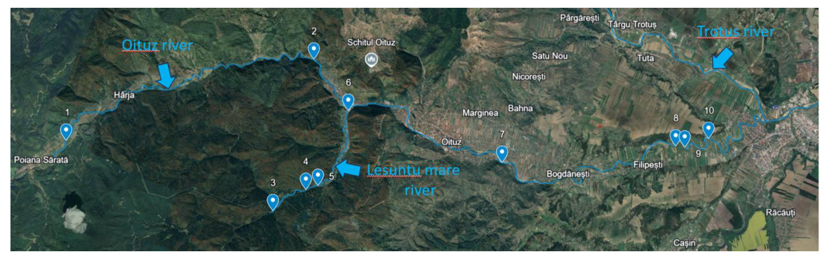
Figure 2.
Clusters of plastics' size expressed as percentages (n=215).
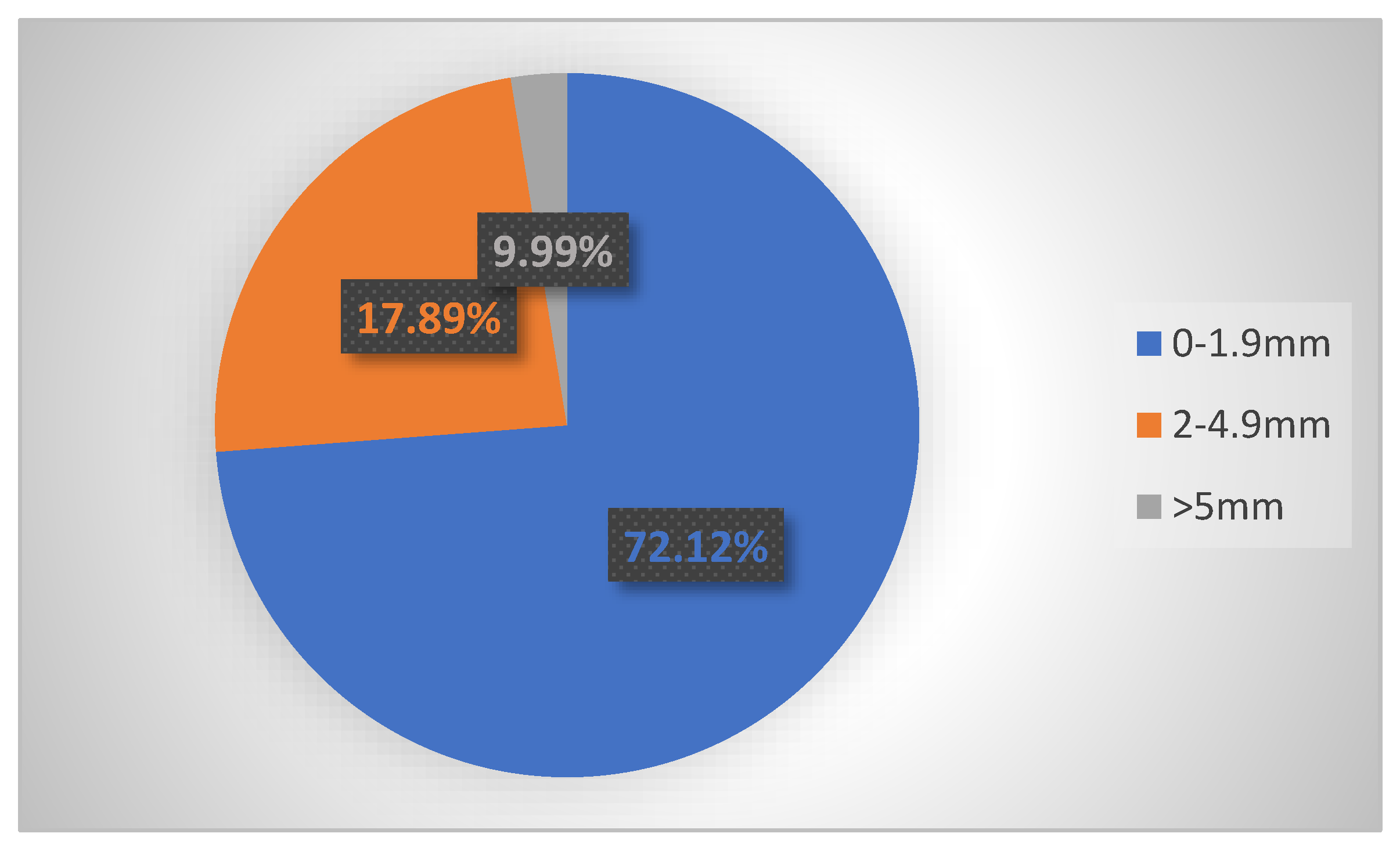
Figure 3.
Clusters of plastics' shape expressed as percentages (n=215).
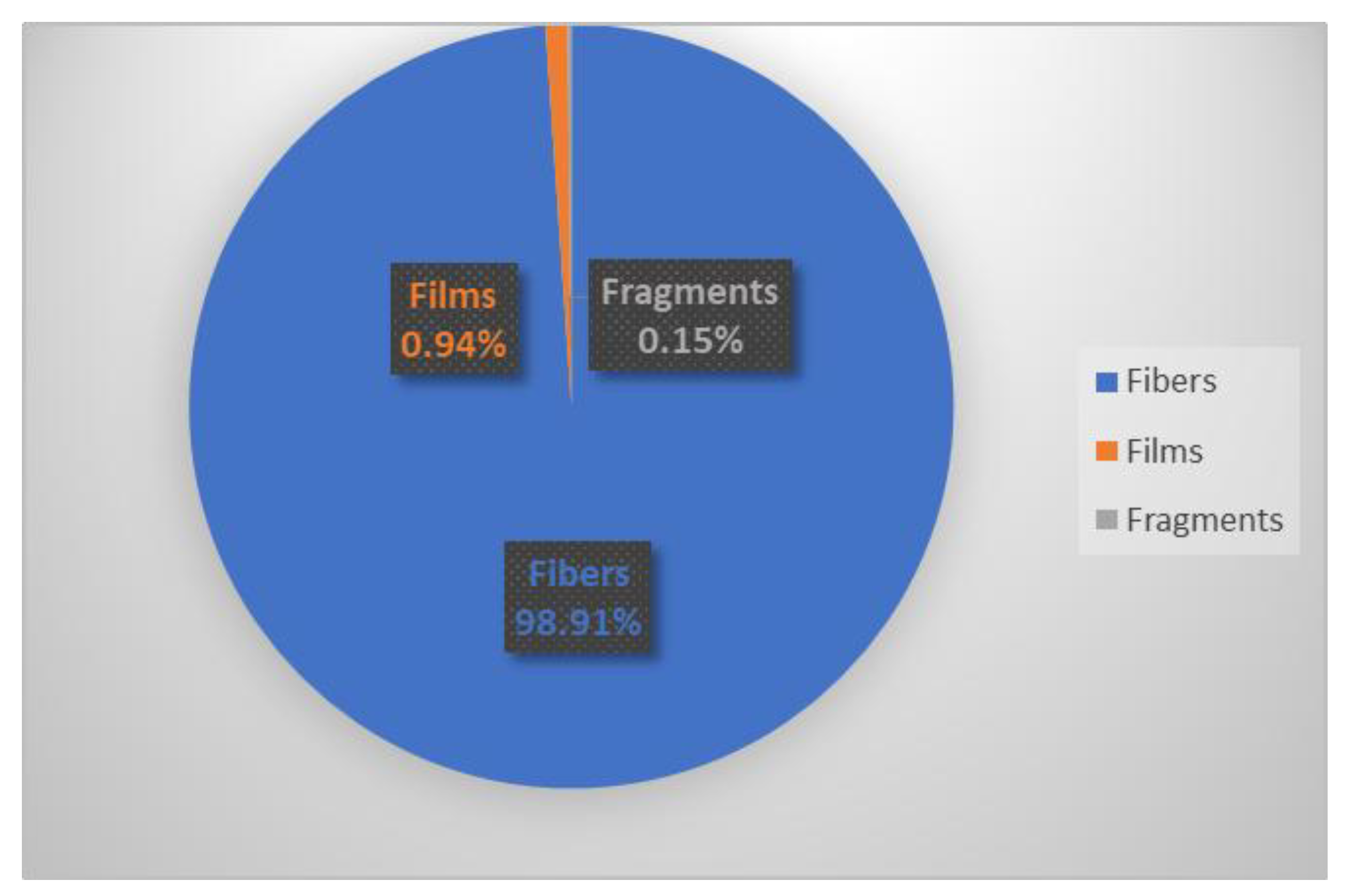
Figure 4.
Plastics' colors expressed as percentages (n=215).
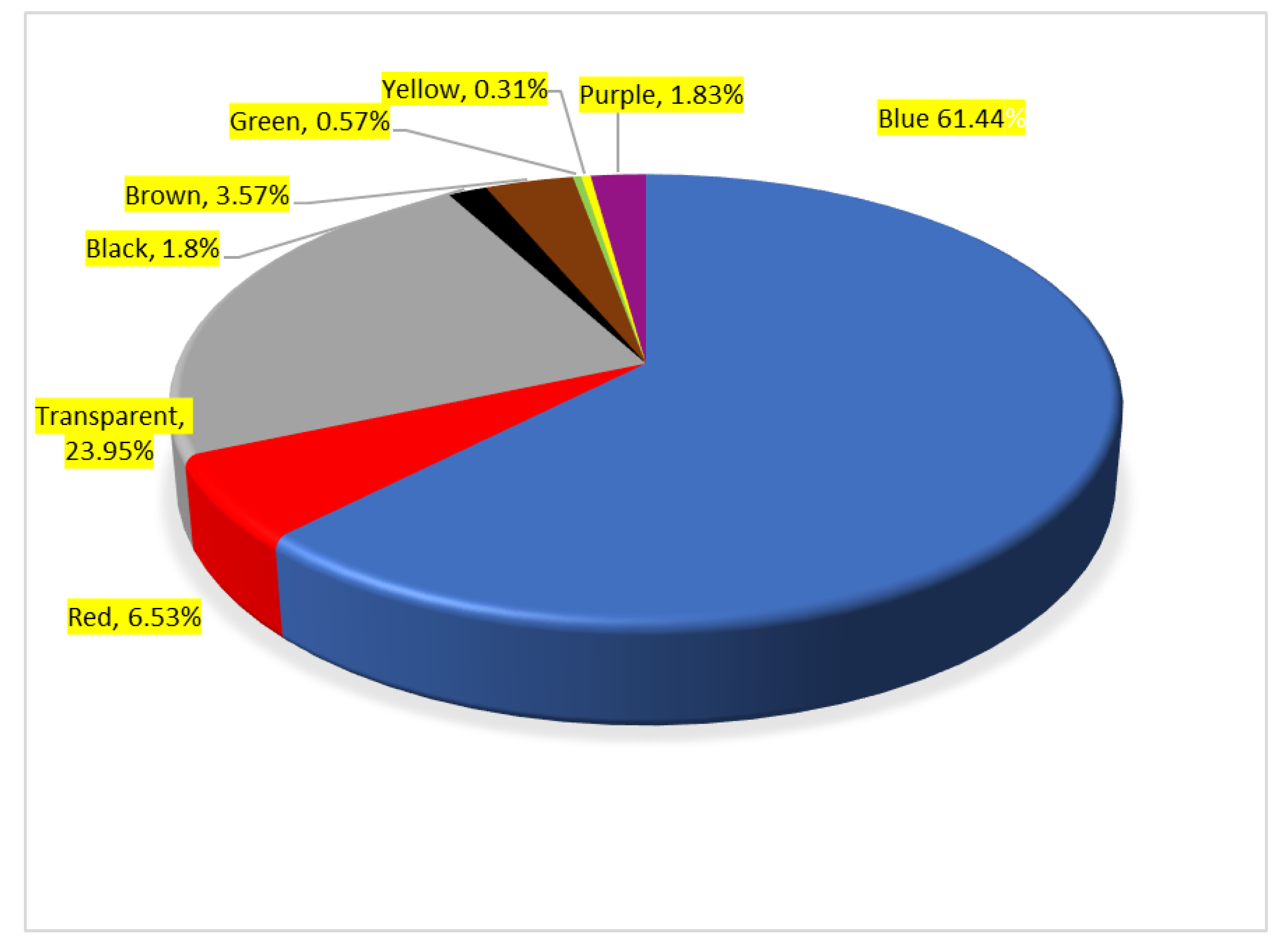
Figure 5.
Distribution of microplastics by species, performed using one-way ANOVA.
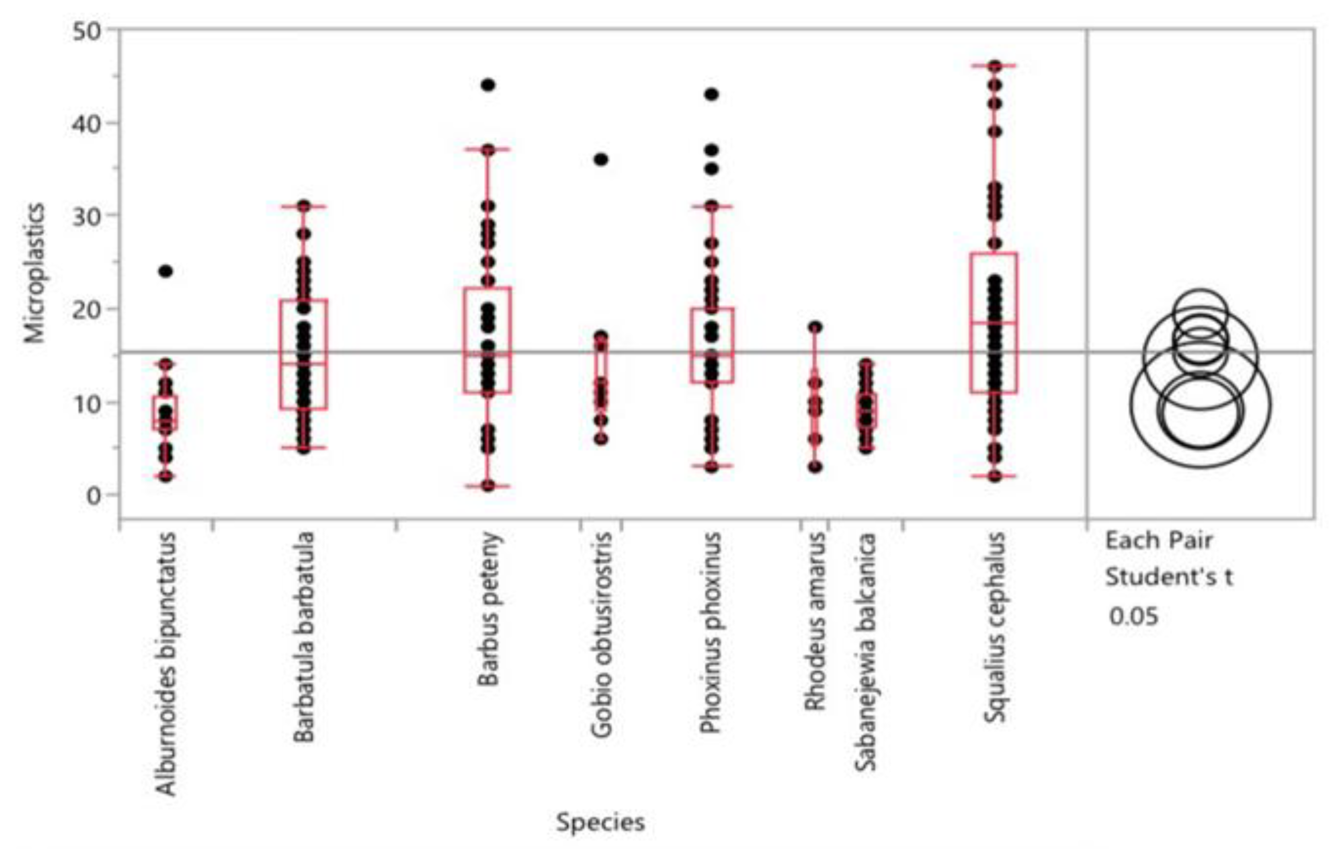
Figure 8.
Scanning Electron Microscopy (SEM) images.
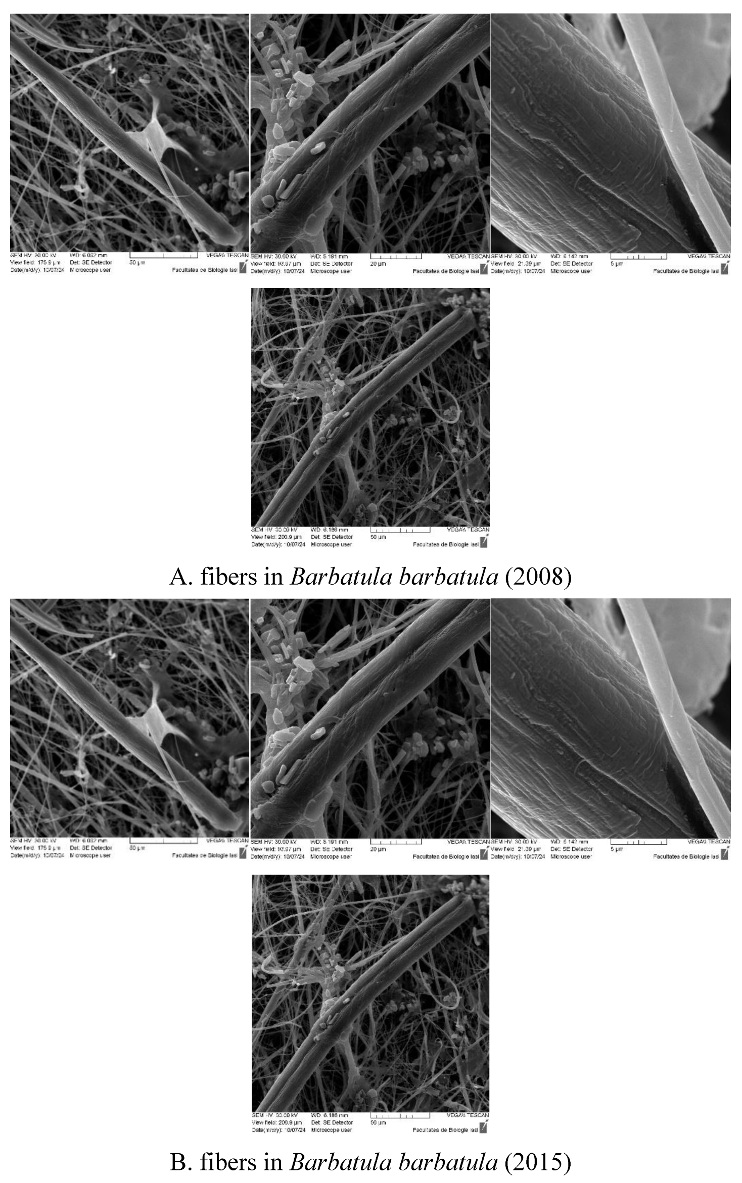
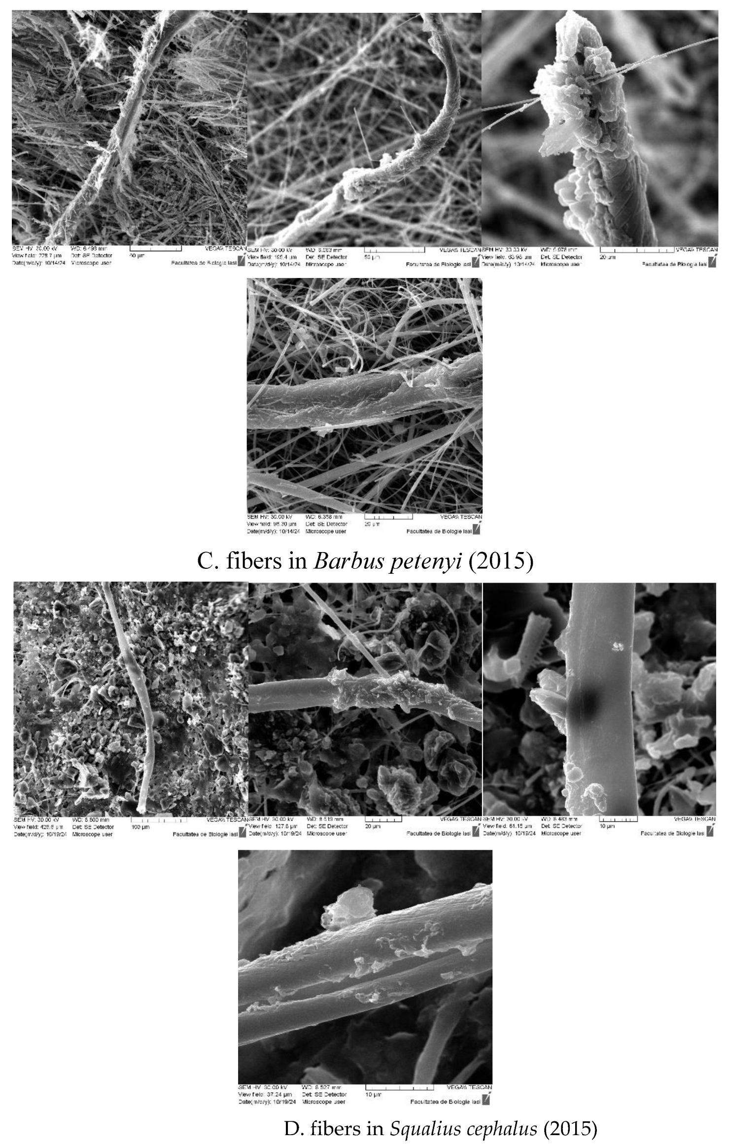
Figure 9.
Microscopic images of the ATR-FTIR spectral data acquiring points on the microplastics collected from the stomach of the fishes S.c.08, S.b.08, G.o.15, B.b.15, Ph.ph.15, Ph.ph.08, B.b.08, A.b.15, B.p.08, B.p.15, Rh.a.15, A.b.08, S.c.15.
Figure 9.
Microscopic images of the ATR-FTIR spectral data acquiring points on the microplastics collected from the stomach of the fishes S.c.08, S.b.08, G.o.15, B.b.15, Ph.ph.15, Ph.ph.08, B.b.08, A.b.15, B.p.08, B.p.15, Rh.a.15, A.b.08, S.c.15.
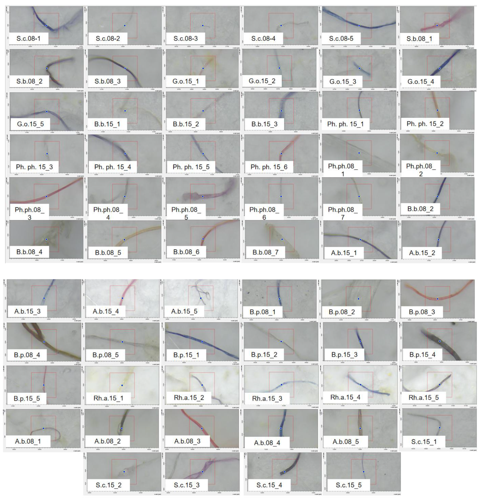
Figure 10.
IR spectra of the microplastics collected from the stomach of the fishes S.c.08, S.b.08, G.o.15, B.b.15, Ph.ph.15, Ph.ph.08, B.b.08, A.b.15, B.p.08, B.p.15, Rh.a.15, A.b.08, S.c.15, compared to the polyamide (PA) and polyethylene (PET) control spectra.
Figure 10.
IR spectra of the microplastics collected from the stomach of the fishes S.c.08, S.b.08, G.o.15, B.b.15, Ph.ph.15, Ph.ph.08, B.b.08, A.b.15, B.p.08, B.p.15, Rh.a.15, A.b.08, S.c.15, compared to the polyamide (PA) and polyethylene (PET) control spectra.
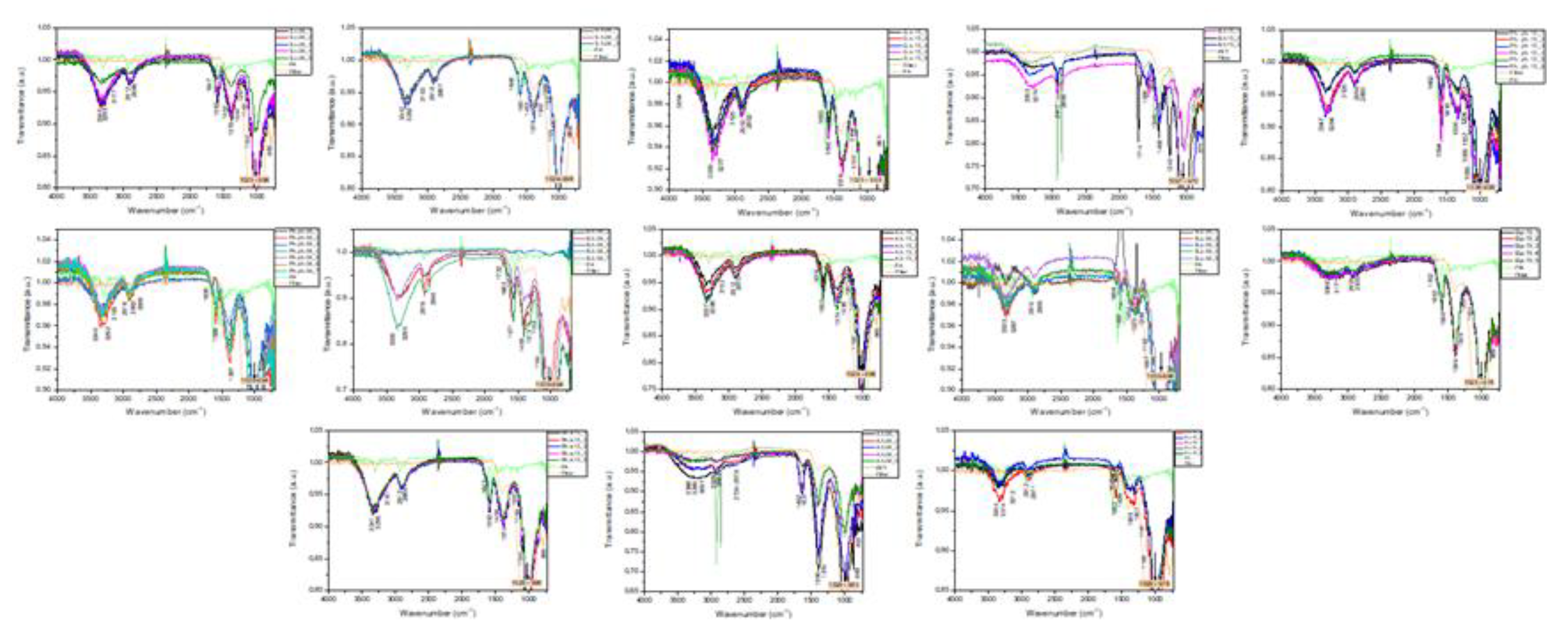
Table 5.
Characteristics of plastic items identified in Alburnoides bipunctatus (n=20).
| Species |
Alburnoides bipunctatus (2008) |
Alburnoides bipunctatus (2015) |
|
|---|---|---|---|
| Plastic characteristics | No. of items | No. of items | |
| Shape | Fibers | 68 | 91 |
| Films | 0 | 17 | |
| Size | 0-0.9 mm | 13 | 27 |
| 1-1.9 mm | 26 | 33 | |
| 2-2.9 mm | 15 | 27 | |
| 3-4.9 | 14 | 18 | |
| 5-10 mm | 0 | 3 | |
| Average size | 2.0 mm | 2.0 mm | |
| Color | Blue | 39 | 52 |
| Transparent | 7 | 40 | |
| Red | 6 | 14 | |
| Black | 8 | 0 | |
| Brown | 5 | 2 | |
| Green | 3 | 0 | |
| Plastics’ totals | 68 | 108 | |
Table 6.
Characteristics of micro- and mesoplastics identified in Sabanejewia balcanica (n=20), Rhodeus amarus (n=6), and Gobio obtusirostris (n=9).
Table 6.
Characteristics of micro- and mesoplastics identified in Sabanejewia balcanica (n=20), Rhodeus amarus (n=6), and Gobio obtusirostris (n=9).
| Species |
Sabanejewia balcanica (2008) |
Rhodeus amarus (2015) |
Gobio obtusirostris (2015) |
|
|---|---|---|---|---|
| Plastic characteristics | No. of items | No. of items | No. of items | |
| Shape | Fibers | 143 | 53 | 124 |
| Films | 0 | 5 | 8 | |
| Fragments | 2 | 0 | 0 | |
| Size | 0-0.9 mm | 50 | 19 | 34 |
| 1-1.9 mm | 55 | 19 | 47 | |
| 2-2.9 mm | 22 | 7 | 26 | |
| 3-4.9 | 17 | 12 | 17 | |
| 5-10 mm | 1 | 0 | 8 | |
| >10 mm | 0 | 1 | 0 | |
| Average size | 1.5 mm | 1.9 mm | 2.0 mm | |
| Color | Blue | 85 | 32 | 81 |
| Transparent | 34 | 19 | 35 | |
| Red | 11 | 4 | 9 | |
| Black | 7 | 0 | 1 | |
| Brown | 7 | 2 | 4 | |
| Green | 1 | 1 | 2 | |
| Plastics’ totals | 145 | 58 | 132 | |
Disclaimer/Publisher’s Note: The statements, opinions and data contained in all publications are solely those of the individual author(s) and contributor(s) and not of MDPI and/or the editor(s). MDPI and/or the editor(s) disclaim responsibility for any injury to people or property resulting from any ideas, methods, instructions or products referred to in the content. |
© 2025 by the authors. Licensee MDPI, Basel, Switzerland. This article is an open access article distributed under the terms and conditions of the Creative Commons Attribution (CC BY) license (http://creativecommons.org/licenses/by/4.0/).
Copyright: This open access article is published under a Creative Commons CC BY 4.0 license, which permit the free download, distribution, and reuse, provided that the author and preprint are cited in any reuse.


