Submitted:
27 February 2025
Posted:
28 February 2025
You are already at the latest version
Abstract
The aim of this work was a detailed radiological and histopathological evaluation of a solid tumour, presumably cancerous in nature, diagnosed in a 2-year-old goose (Anser domesticus). The radiograph examination showed an osseous change with the involvement of cervical vertebrae. A tumour measuring 15x10x9 cm was dense and had well-defined borders, suggesting the presence of calcified bone tissue. Histopathology revealed a well-defined benign neoplasm derived from bone and consisted large area of irregular, disorganized bone trabeculae, surrounded by a layer of osteoblasts. The tumour has been classified as an osteoma, which originates from the body of the vertebrae. Osteoma is a benign, well-differentiated tumour with a structure that resembles bone tissue. It presents as a well-demarcated, hard, single tumour that can grow to a considerable size. The aetiology of osteomas in birds remains unclear, because of the small number of cases described. Therefore, the influence of factors such as age, breed or sex, trauma, embryonic malformation, infection, developmental disorders, genetic factors on the development of this type of tumour has not been established. Trauma seems to be the most obvious cause of growth in this case. This work provides valuable information about osteomas in birds, important for understanding these neoplasms.
Keywords:
osteoma
; goose
; tumour
1. Introduction
Neoplasms in birds are mainly found in wild birds, pet birds, exotic birds and, to a lesser extent, in farming poultry [1]. Tumours described in birds include descriptions of neoplasms of the skin, internal organs (liver, kidney), reproductive system, brain, vascular system, connective tissue, bones and eyes. Neoplastic diseases of the skeletal system are rare (6.66%) and the available literature is limited [1]. Bone tumours include osteomas and osteosarcomas, and in the available literature there are descriptions of osteomas in the Peach-faced lovebird [2,3], Blue-Fronted Amazon Parrot [4], a owl [5,6], canary [1,7], hen [8], broiler chicken [9], domestic duck, budgerigar [1,10] and eclectus parrot [11].
Osteosarcoma has been described in in the Brown Leghorn hen [12], 30-week-old hen [8], free-range chicken [13], broiler chicken [9], a Japanese quail, cockatoo, dove [10], budgerigar, mallard [1] and the foot of a goose [14].
Benign neoplasia may originate from tarsometatarsus, plantar footpad, cranium [10], scapula, wing [2,5-7], nasal cavity [4], cutis [3], long bone [9] and sternum [11].
Bone tumours are very rarely described in breeding birds due to the short fattening period or the large number of birds in breeding flocks where proliferative changes may be overlooked. Neoplasia in geese is still a poorly understood and described area of avipathology, and therefore an area for further analysis. The presented case of a tumour in a two-year-old gander is an interesting research material that can contribute to the knowledge and development of the diagnosis of tumours in birds. The main suspicion was that the trauma had been the cause of the growth of the tumour.
The aim of this work is the radiological and histopathological evaluation of a solid lesion diagnosed in geese, which is suspected to be neoplastic in nature.
2. Materials and Methods
During a clinical examination of a goose farm, the veterinarians noticed the presence of a 2 years old male showing symptoms of disorders. The clinical examination revealed the presence of a large mass in the neck and spine, which probably contributed to causing neurological disorders. The clinicians, together with the breeder, decided to euthanise the bird on humanitarian grounds, then secured the body and transferred it for further research. The carcass of a male goose (Anser domesticus) was delivered to the Department of Pathological Anatomy of the University of Warmia and Mazury in Olsztyn. A radiological examination and a detailed necropsy were carried out. The entire lesion was removed, fixed in 10% formalin and decalcified. The collected and fixed specimen was then subjected to routine histopathological examination (haematoxylin and eosin staining was performed - HE).
3. Results
3.1. Results of Radiological Examination
The radiograph examination was taken in two projections, dorsal and lateral (Figure 1 and Figure 2) and in the area of the 8-13 cervical vertebra, a osseous change with the involvement of other cervical vertebrae was noted. The change was a well-circumscribed hyperechogenic mass extending into the thoracic cavity. A tumour measuring 15 cm in length and 10 cm in width was dense and had well-defined borders, suggesting the presence of calcified bone tissue. The tumour was spherical with a homogeneous surface, indicating slow and mild growth. It showed the signs of calcification without causing visible destruction of the surrounding soft tissues and adjacent structures. The tumour slightly pushed the trachea and oesophagus at the sides of the neck but did not affect their patency.
3.2. Results of Macroscopic Examination
An autopsy revealed a solid tumour measuring 15 x 10 x 9 cm, located in the cervical vertebrae (Figure 3). The tumour extended into the thoracic cavity (Figure 4), weighed 2 kg and consisted of hard, compact bone tissue that was tightly fused to the spine - derived from the vertebrae in the area of the 8-13 cervical vertebrae (Figure 5). There was no evidence of metastasis of bone tumour to other organs and tissues. Despite its size, the tumour did not infiltrate the trachea or oesophagus, but moved them to the side of the neck. The surface of the tumour was slightly rough, but the surrounding soft tissue and skin showed no signs of infiltration. The macroscopic evaluation of the organs and tissues did not reveal any pathological changes.
3.3. Results of Microscopic Examination
Histopathological examination confirmed the presence of a well-defined benign neoplasm derived from bone tissue arose from the body of the cervical vertebrae (Figure 6). The tumour was well circumscribed and consisted large area of irregular, disorganized bone trabeculae, usually surrounded by a layer of neoplastic osteoblasts (Figure 7 and Figure 8). Between the trabeculae well-vascularised connective tissue was observed, suggesting intense metabolic activity within the cancer. Histopathological examination of the tumour showed that the change was mild and derived from bone tissue. The tumour has been classified as an osteoma. The microscopic evaluation of the organs and tissues did not reveal any pathological changes or metastasis.
4. Discussion
Radiological and histopathological examination revealed the presence of an osteoma. To the best of our knowledge, this is the first case of a benign bone tumour originating from a cervical vertebra. Benign bone cancers are rare in birds and the aetiology is poorly understood [1].
Osteoma is a benign, well-differentiated tumour with a structure that resembles bone tissue. It usually presents as a well-demarcated, hard, single tumour that can grow to a considerable size. Osteomas are characterised by slow growth that can last for many months, after which the tumour reaches a large size and often stops growing. These tumours can remain dormant for a long time, often for years, without causing any clinical symptoms, until the tumour grows again.
The case described was a large tumour, and previous cases of osteomas in birds have not grown to this size. The osteoma described in 24-year-old Amazon Parrot in the left nostril was 5 mm in diameter, total excision of the lesion resulted in complete recovery [4]. Cowan et al. [11] described interesting findings in an Eclectus Parrot, where an osteoma 1 cm in diameter was diagnosed in the sternum. The tumour exhibits well-differentiated trabecular bone containing osteocytes, osteoblasts and osteoclasts with spaces of bone marrow. The case of osteoma in the canary on the left wing reached a size of 12x8x6 mm, and the histopathological changes did not demonstrate such wide areas of osseous trabeculae as in our case [7]. Cagnini et al. [2] showed an osteoma in the same location with larger dimensions (22x20x22 mm) and the histological image revealed a wider area of mature trabeculated bone formation. In lovebird, bilateral cutis osteomas at the junction of the ulnar radius and humerus, measuring 3.4x2.2 cm and 2.4x2.7 cm, showed bone spicules with osteoblastic/osteocytic proliferation also with the presence of mineralised woven bone matrix. Together with the tumour content, active inflammatory changes with cellular infiltrations were noted [3]. No inflammatory changes were diagnosed in our study. Hahn et al. [6] described a case of osteoma in a barred owl with a larger size on the left radius (7x5x4.5cm) and the body of the tumour consisted of multiple large coalescing islands of well-differentiated cartilage separated by small spicules of mature bone or thin connective tissue. The presence of the tumour caused muscle atrophy and skin ulceration. In our case, such changes were not seen. A tumour of similar size was reported in a great horned owl (5x4.5x3.5 cm) attached to the left ulna [5]. Histopathological examination revealed thick trabeculae of lamellar and woven bone from the periosteum, supported by abundant loose fibrovascular stroma. After the tumour was completely resected, there was no evidence of regrowth. Although our described tumour was large, it did not exert pressure on the spinal cord, but it grew, compressed the surrounding tissues, displaced other organs (trachea, oesophagus), but did not cause a direct threat to life. In addition, no atrophic changes and no inflammatory response were observed in the tissues that were examined.
The aetiology of osteomas in birds remains unclear, mainly because of the small number of cases described. Various authors have pointed out that the cause is still unclear, and therefore, the influence of factors such as age, breed or sex, trauma, embryonic malformation, infection, developmental disorders, genetic factors on the development of this type of tumour have been suggested [1,4,6,7]. More reliable information is provided by the work of Cowan et al. [11], in which trauma was the main cause of osteoma in the Eclectus parrot. Three months after the injury, a tumour was found in the sternum. Similar observations are confirmed by Pinzon-Osorio et al. [3], where in the lovebird, owner-reported traumatic wings injuries (1 month prior) were the most likely cause of tumour growth. In the case described, the neck of the birds, especially the geese, is long and uncovered and it is very likely that the injury was the cause of the growth of the tumour. We do not know if other birds were involved or if there was a human factor - mistreatment of birds on the farm or a deliberate attack. In the early stages, the tumour grows slowly and can only be diagnosed by radiograph examination in a small number of cases and only when it causes visible clinical symptoms or when it is visible. In the case of pet birds that are in direct contact with their owners, even minor changes and clinical signs may be observed at an earlier stage. In farm animals, such changes are rarely seen and therefore the tumour grew rapidly at a later stage, which has been confirmed in other studies [6]. This confirms our suspicion that in a 2-year-old gander, the trauma may have occurred much earlier, perhaps even at a young age, and the tumour may have started to grow over time.
5. Conclusions
Bone derived neoplasms not associated with infectious factors are rare in aviopathology. The confirmation of the case of the clinical occurrence of osteoma in the domestic goose can be a valuable material for further studies in this field.
Author Contributions
Conceptualization, M.G., A.M. and E.Ł.; methodology, M.G. and A.M.; investigation, M.G.; writing—original draft preparation, M.G.; writing—review and editing, M.G.; visualization, M.G. and A.M.; funding acquisition, M.G. All authors have read and agreed to the published version of the manuscript.
Funding
Funded by Minister of Science under the Reginal Initiative of Excellence Program.
Institutional Review Board Statement
Not applicable.
Informed Consent Statement
Not applicable.
Data Availability Statement
Data will be provided on request.
Conflicts of Interest
The authors declare no conflicts of interest.
References
- Sánchez-Godoy, F.; Ledesma-Ramírez, I.; Morales-Salina, E. A retrospective study of neoplasms in ornamental and pet birds diagnosed at the Hospital de Aves of the Universidad Nacional Autónoma de México (2007-2014). Braz. J. Vet. Pathol. 2020, 13, 1–11. [Google Scholar] [CrossRef]
- Cagnini, D.Q.; do Carmo Moraes, W.K.; Dias, M.C.; Bonfim, L.S.; Franca, F.M.; Regalin, D. Osteoma in Agapornis roseicollis. Acta Vet. Bras. 2022, 16, 342–345. [Google Scholar] [CrossRef]
- Pinzón-Osorio, C.A.; Gomez, A.P.; Alvarez-Mira, D.M. Bilateral osteoma cutis in a Peach-Faced Lovebird (Agapornis roseicollis). J. Vet. Med. Sci 2020, 82, 536–540. [Google Scholar] [CrossRef] [PubMed]
- Cardoso, J.F.R.; Levy, M.G.B.; Liparisi, F.; Romao, M.A.P. Osteoma in a blue-fronted amazon parrot (Amazona aestiva). J. Avian Med. Surg. 2013, 27, 218–221. [Google Scholar] [CrossRef] [PubMed]
- Barbosa, L.; Tom, D.; Han, S. Successful resection of an osteoma in a great horned owl (Bubo virginianus) with subsequent lack of reginal feather regrowth. Vet Rec Case Rep. 2019, 7, e000887. [Google Scholar] [CrossRef]
- Hahn, K.A.; Jones, M.P.; Petersen, M.G.; Patterson, M.M. Clinical and pathological characterization of an osteoma in a barred owl. Avian Pathol. 1998, 27, 306–308. [Google Scholar] [CrossRef] [PubMed]
- Javdani, M.; Hashemnia, M.; Nikousefat, Z.; Ghasemi, M. Extraskeletal osteoma in a canary (Serinus canaria). Vet. Res. Forum. 2017, 8, 265–268. [Google Scholar] [PubMed]
- Reece, R.L. Some observations on naturally occurring neoplasms in domestic fowl in the state of Victoria, Australia. Avian Pathol. 1996, 25, 407–447. [Google Scholar] [CrossRef] [PubMed]
- Campbell, J.G; Appleby, E.C. Tumours in young chickens bred for rapid body growth (broiler chickens): A study of 351 cases. J. Pathol. Bacteriol. 1966, 92, 77–90. [Google Scholar] [CrossRef] [PubMed]
- Reece, R.L. Observations on naturally occurring neoplasms in birds in the state of Victoria, Australia. Avian Pathol. 1992, 2, 3–32. [Google Scholar] [CrossRef] [PubMed]
- Cowan, M.L; Yang, P.J.; Monks, D.J.; Raidal, S.R. Suspected Osteoma in an Eclectus Parrot (Eclectus roratus roratus). J. Avian Med. Surg. 2011, 25, 281–285. [Google Scholar] [CrossRef] [PubMed]
- Siller, W.G. An osteogenic sarcoma in the fowl. Br. J. Cancer. 1959, 13, 642–646. [Google Scholar] [CrossRef] [PubMed]
- Dittmer, K.E; French, A.F; Thompson, D.J.; Buckle, K.N.; Thompson, K.G. Primary bone tumors in birds: A review and description of two new cases. Avian Dis. 2012, 56, 422–426. [Google Scholar] [CrossRef] [PubMed]
- Mawdesley-Thomas, L.E; Solden, D.H. Osteogenic sarcoma in a domestic goose (Anser anser). Avian Dis. 1967, 11, 365–370. [Google Scholar] [CrossRef] [PubMed]
Figure 1.
Ventral-dorsal radiograph of the cervical region in goose. Large hyperechogenic mass tightly fused to the spine - derived from the vertebrae.
Figure 1.
Ventral-dorsal radiograph of the cervical region in goose. Large hyperechogenic mass tightly fused to the spine - derived from the vertebrae.

Figure 2.
Lateral radiograph of the cervical region in goose. The tumour has pushed the trachea to the sides of the neck without altering its patency.
Figure 2.
Lateral radiograph of the cervical region in goose. The tumour has pushed the trachea to the sides of the neck without altering its patency.
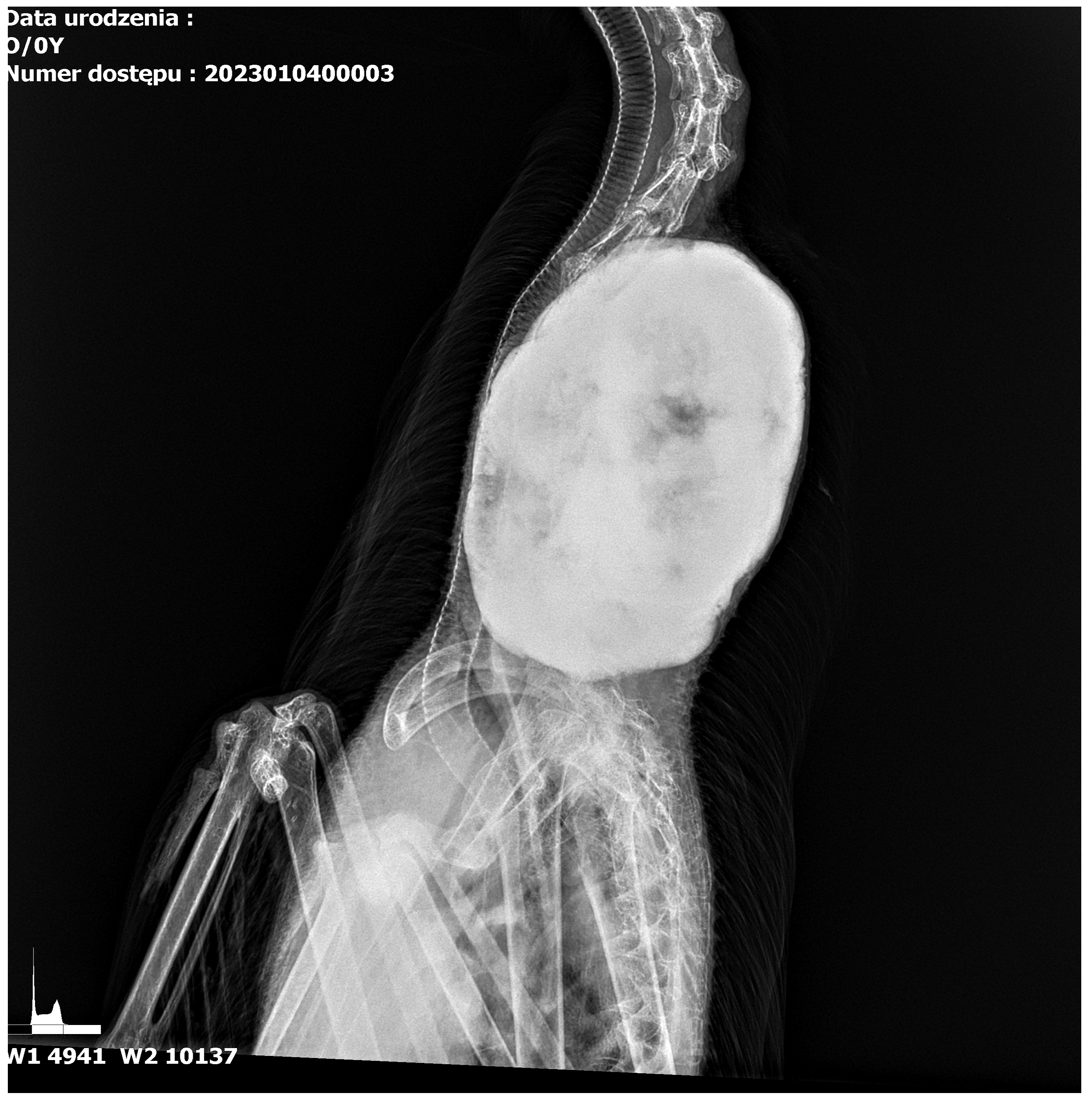
Figure 3.
Domestic goose. Large tumour in the distal part of the cervical region without skin lesions and ulcerations.
Figure 3.
Domestic goose. Large tumour in the distal part of the cervical region without skin lesions and ulcerations.
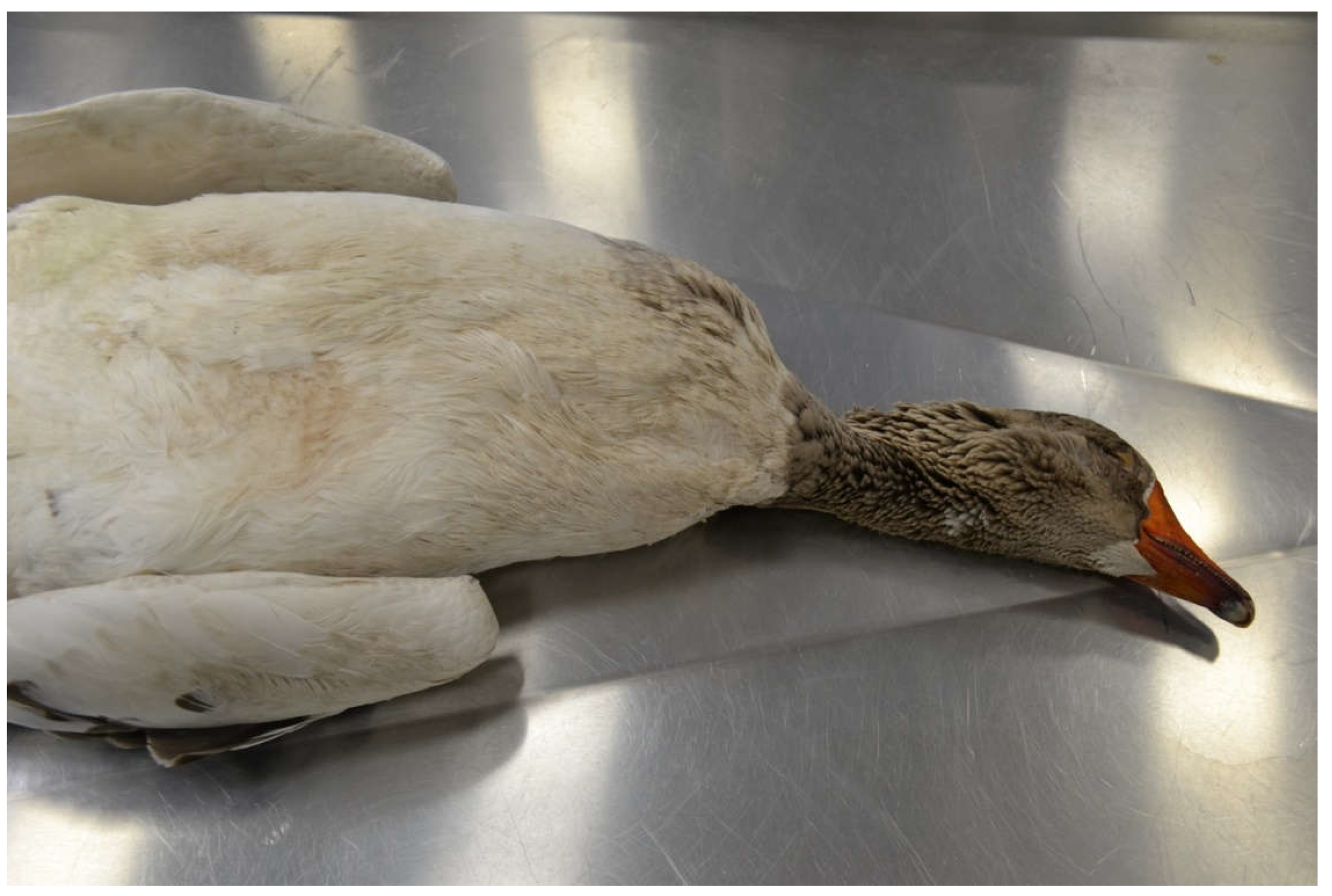
Figure 4.
Domestic goose. The well-demarcated tumour has pushed the trachea and oesophagus to the sides of the neck without altering its patency.
Figure 4.
Domestic goose. The well-demarcated tumour has pushed the trachea and oesophagus to the sides of the neck without altering its patency.
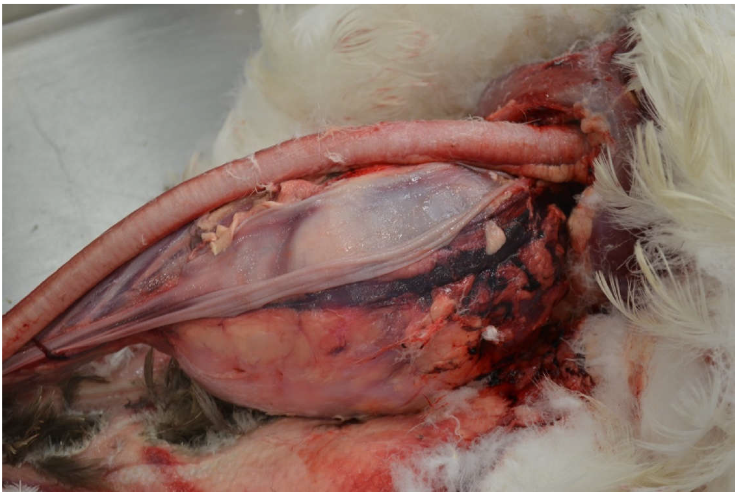
Figure 5.
Domestic goose. The cross section of the tumour with visible present of cervical vertebrae (circle).
Figure 5.
Domestic goose. The cross section of the tumour with visible present of cervical vertebrae (circle).
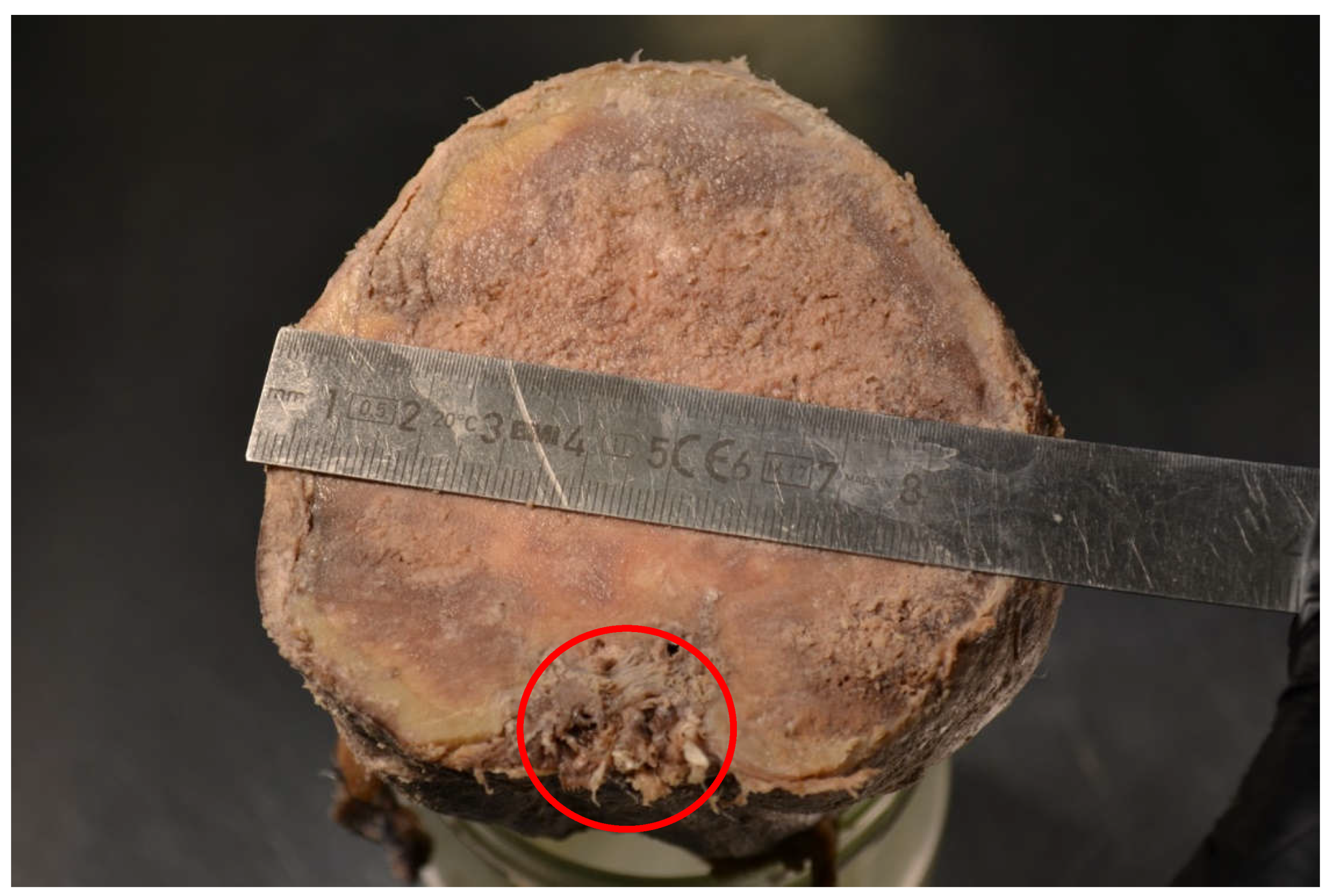
Figure 6.
Domestic goose. Cervical vertebra with tumour. Visible central canal with spinal cord (red asterisk). Below, visible tumour mass originating from the body of the cervical vertebra (black asterisks). HE staining.
Figure 6.
Domestic goose. Cervical vertebra with tumour. Visible central canal with spinal cord (red asterisk). Below, visible tumour mass originating from the body of the cervical vertebra (black asterisks). HE staining.
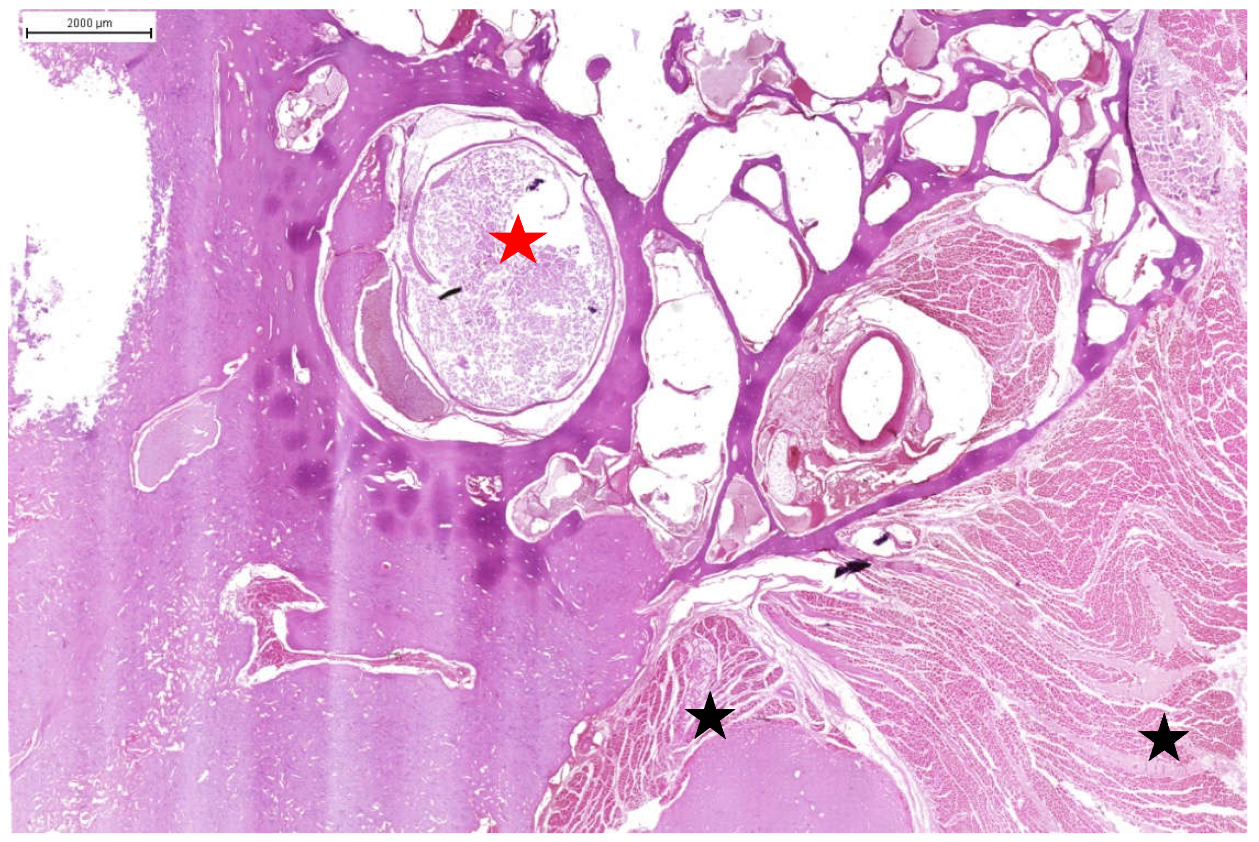
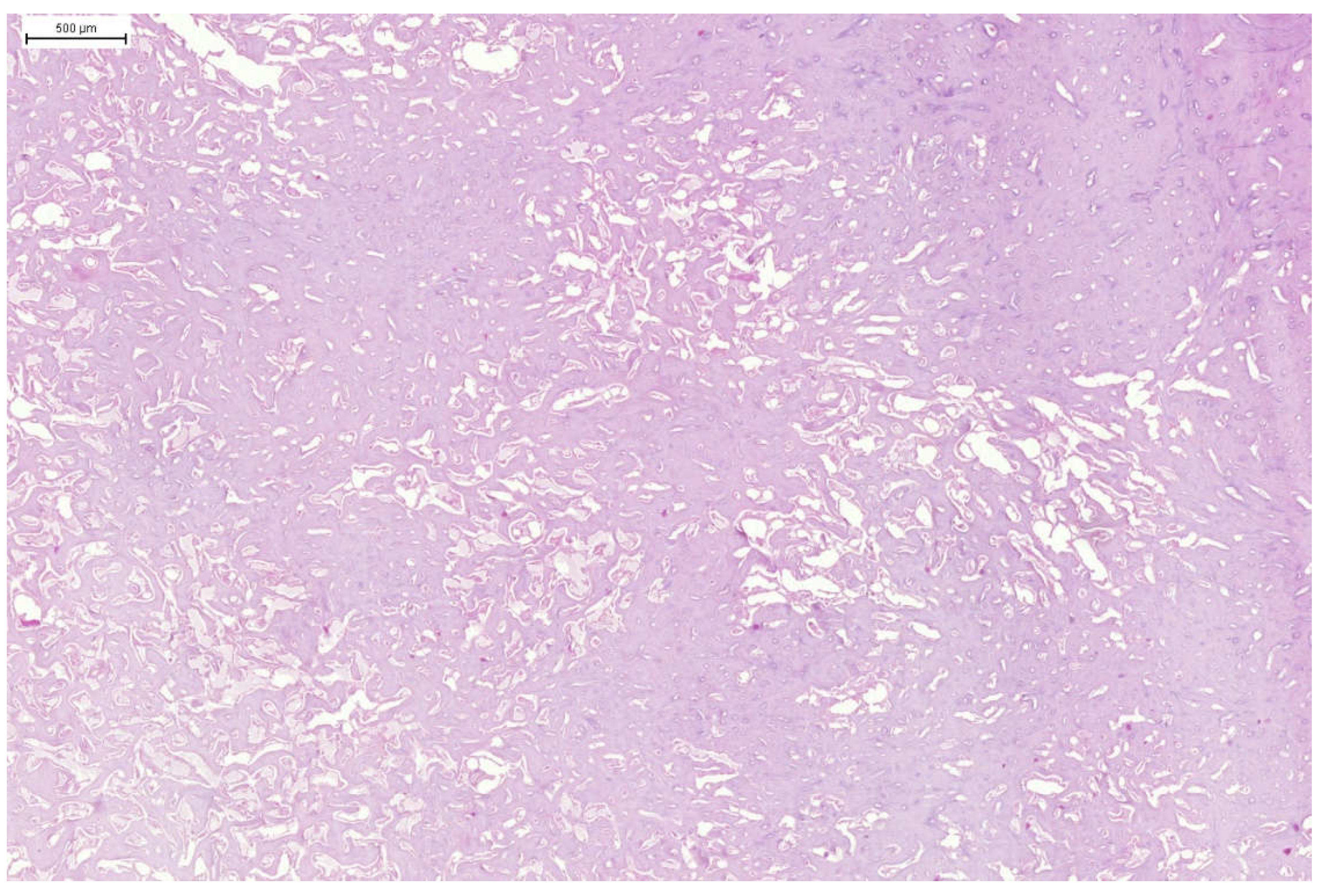
Figure 7.
Domestic goose. Tumour mass. Large dense area of trabecular bone with visible irregular holes containing blood vessels and neoplastic osteoblasts. HE staining.
Figure 7.
Domestic goose. Tumour mass. Large dense area of trabecular bone with visible irregular holes containing blood vessels and neoplastic osteoblasts. HE staining.

Figure 8.
Domestic goose. Tumour mass. Large area of trabecular bone with visible irregular holes containing blood vessels and osteoblasts. HE staining.
Figure 8.
Domestic goose. Tumour mass. Large area of trabecular bone with visible irregular holes containing blood vessels and osteoblasts. HE staining.
Disclaimer/Publisher’s Note: The statements, opinions and data contained in all publications are solely those of the individual author(s) and contributor(s) and not of MDPI and/or the editor(s). MDPI and/or the editor(s) disclaim responsibility for any injury to people or property resulting from any ideas, methods, instructions or products referred to in the content. |
© 2025 by the authors. Licensee MDPI, Basel, Switzerland. This article is an open access article distributed under the terms and conditions of the Creative Commons Attribution (CC BY) license (http://creativecommons.org/licenses/by/4.0/).
Copyright: This open access article is published under a Creative Commons CC BY 4.0 license, which permit the free download, distribution, and reuse, provided that the author and preprint are cited in any reuse.


