Submitted:
27 February 2025
Posted:
27 February 2025
You are already at the latest version
Abstract
Background: Cancer remains one of the leading causes of mortality worldwide, while natural antioxidants have emerged as promising therapeutic agents in cancer treatment. Although fucoidan from brown algae shows anticancer potential, its efficacy is limited by bioavailability challenges, and the synergistic effects of combining it with gold nano-particles remain unexplored. Methods: Fucoidan was extracted from Sargassum cinereum and Turbinaria decurrens using acid precipitation. Gold nanoparticles (AuNPs) were manufactured by a green technique that employed fucoidan as both a reducing and stabilizing agent. The nanoparticles were analyzed utilizing UV-Vis spectroscopy, FTIR, TEM, XRD, and zeta potential assessment. Antioxidant activities were evaluated utilizing DPPH and FRAP assays. Cytotoxicity was assessed against HepG2, THP-1, and BNL cells utilizing MTT and SRB tests. Flow cytometry was utilized for cell cycle analysis, and molecular docking was applied to examine interactions with cancer-associated proteins. Results: T. decurrens yielded higher fucoidan extraction (235.9 mg/g dry weight) and demonstrated superior antioxidant activity in Ferric Reducing Antioxidant Power (FRAP) (9.21 μg Trolox Equivalents /mg) and 2, 2-Diphenyl-1-Picryl-Hydrazyl-Hydrate (DPPH) (4.48 μg Trolox Equivalents /mg) assays compared to S. cinereum. Molecular docking revealed strong binding of fucoidan to cancer-related proteins, particularly COX-2 (-7.1 kcal/mol) and TERT (-5.4 kcal/mol), while the fucoidan-gold nanoparticle complex (F-AuNPs) showed enhanced cellular uptake and improved cytotoxicity against HepG2 cells (IC50: 392.81 μg/mL and 459.75 μg/mL for S. cinereum and T. decurrens formulations, respectively). Conclusions: These findings suggest a promising synergistic approach for enhancing fucoidan-gold nanoparticle therapeutic potential in cancer treatment through combined in vitro and in silico analyses.
Keywords:
Fucoidan
; Gold Nanoparticles
; Anticancer Activity
; Antioxidant Properties
; Molecular Docking
; Brown seaweed
; Cytotoxicity
1. Introduction
The usage of biological materials for various treatments and preventive measures has been a common tradition worldwide. This trend has accelerated in recent years, with many pharmaceutical companies working to extract and utilize potent bioactive diseases, chronic diseases, and even cancer over chemically synthesized alternatives due to their significantly reduced or nonexistent side effects in the process [1]. The plant and marine materials used for cancer treatments belong to the family of polyphenols, polysaccharides, peptides, and other bioactive constituents [2,3].
Seaweeds are a particularly rich source of bioactive compounds such as polyphenols, polysaccharides, and peptides, which have shown promise in cancer treatment. A review by Pereira & Valado [4] highlights the potential of seaweed-derived polysaccharides and phlorotannins in drug discovery, demonstrating their applications in anticancer, antimicrobial, and anti-inflammatory therapies. Studies have also demonstrated the antiproliferative effects of seaweed extracts. For instance, aqueous extracts from Ecklonia maxima and Ulva rigida have been shown to induce apoptosis in human liver cancer cells by promoting reactive oxygen species (ROS) production and disrupting mitochondrial membranes [5]. Another study reviewed the role of seaweed oligosaccharides in disease treatment, noting their therapeutic potential against cancer, diabetes, and obesity [6]. Furthermore, a study on Caulerpa lentillifera emphasized its bioactive components, including phenolic compounds and polysaccharides, which exhibit antioxidant and anti-inflammatory properties beneficial for cancer therapy [7].
Cancer is a group of diseases characterized by the uncontrolled growth of cells. The major types of cancer include breast, lung, colon, melanoma, and lymphoma [8]. However, cancer remains a complex and challenging disease for researchers, not only in medicine but also in the fields of biology, chemistry, physics, and even computer science. As the nature of cancer becomes better understood, treatment strategies continue to evolve, including chemotherapy, radiotherapy, and surgery. However, these conventional therapies have long-term side effects, such as weakness, fatigue, malnutrition, hair loss, nerve and muscle problems, and mental health issues. Due to these side effects, many natural materials have been explored as alternative treatments for cancer, though their effectiveness varies [9].
Among the promising natural compounds, Phaeophyceae species are extensively studied for their bioactive composition, particularly their fucoidan content. These species are processed due to the bioavailability and bioactive properties of fucoidan. Because a large number of additional assets are deposited in the intracellular environment, Sargassum and Turbinaria algae have demonstrated anticancer and antioxidant capacities [10]. Hence, an anticancer agent and a potent natural radical scavenger and antioxidant can potentially be extracted from these algae. Fucoidans are natural sulfated polysaccharides found in marine brown algae, characterized by unique chemical structures with varying sulfation patterns, molecular weights, sugar compositions, and uronic acid content depending on their sources [11]. Best practices in cancer therapy include four critical factors: anticancer effects, chemical structure, antioxidant activity, and immune stimulation effects. Remarkably, fucoidan has demonstrated anticancer and antioxidant properties in several case studies [12]. The sulfated polysaccharides have the ability to interact with a wide variety of proteins, including cytokines, enzymes, and growth factors, thereby regulating cell-dependent events [13,14,15]. Additionally, fucoidan can reduce the concentration of reactive oxygen species and enhance the activity of various antioxidative enzymes, allowing for its potential use in drug design, particularly for cancer treatment and neuroprotection [16]. However, the interaction between fucoidan and gold nanoparticles remains an area that requires further scientific exploration [17].
On the other hand, gold nanoparticles (GNPs) possess unique properties, including inherent low toxicity and various synthesis methods. GNPs have been prepared without exposure to hazardous toxic materials and characterized without the need for a technician or sophisticated instruments. Among the four methods applied to synthesize GNPs, the use of neutral aqueous herbal extracts resulted in the most stable nanoparticles, demonstrating potential antioxidant and antitumor properties [18]. However, the bioactivity potential of fucoidan combined with gold metal nanoparticles is still not well understood [19].
In the present study, gold nanoparticles are synthesized using marine polysaccharides from Sargassum cinereum and Turbinaria decurrens, followed by their characterization. Their potential capabilities as antioxidant and anticancer agents are studied through comprehensive analysis. The analysis and docking results from this study provide essential insights into how polysaccharides extracted from these marine sources, in combination with gold nanoparticles, can be effectively utilized as anticancer and antioxidant agents [20].
Furthermore, the study investigates the applications of gold nanoparticles synthesized using marine polysaccharides from Sargassum cinereum and Turbinaria decurrens in enhancing the antioxidant and anticancer activities of fucoidan-gold nanoparticles. These effects are evaluated through both in silico and in vitro analyses to assess their therapeutic potential.
2. Materials and Methods
2.1. Collection and Identification of Macroalgae Samples
Two species of fresh brown seaweeds were handpicked at 1 m depth from submerged rocks from Hurghada city, Red Sea (27° 17′ 13″ N, 33° 46′ 21″ E), during summer 2023 as the most abundant species in that time. Algal samples were rinsed with seawater to remove sand, stored in plastic bags with seawater to prevent evaporation, and transported on ice to the Taxonomy and Biodiversity Laboratory, NIOF, Alexandria for identification. The samples were cleaned of epiphytes and debris, rinsed with fresh water, and partially preserved in 5% formalin for taxonomic identification. The rest were shade-dried at 25–30°C for 4–7 days, crushed into coarse powder and stored in airtight bottles at −20°C.
2.2. Extraction of Fucoidan
Fucoidan was extracted using the method of Tako [26], ten grams of powdered dry algae were suspended in 200 mL of 0.05 M HCl and magnetically stirred for 2 hours at room temperature. The extract was centrifuged at 4000 rpm for 20 minutes, and the supernatant was filtered. The filtrate was neutralized with 0.5 M NaOH and then precipitated by adding ethanol in a 2:1 volume ratio. The precipitated fucoidan was freeze-dried using a rotary evaporator. The fucoidan yield was expressed as a percentage of the initial dry weight of the algae.
2.3. Preparation of Fucoidan Solution
Two grams of the extracted fucoidan powder were dissolved in 20 mL of distilled water with magnetic stirring until a homogeneous solution was obtained. The mixture was then filtered using Whatman GD/X syringe filters (1.2 μm pore size; Whatman, UK) to remove undissolved particles. The filtered solution was store at 4℃ for further use.
2.4. Biosynthesis of Gold Nanoparticles (AuNPs)
A 10 mL aliquot of the fucoidan solution was added to 90 mL of 1 mM gold chloride solution (HAuCl₄·3H2O) and stirred magnetically at room temperature. The color change during the reaction, indicating of AuNP formation, was monitored using a UV-Vis spectrophotometer (Cintra 303). After formation, the AuNP solution was centrifuged at 10,000 rpm (Qiagen Sigma 4-15C) for 15 minutes, and the collected AuNPs were used for subsequent applications.
2.5. Characterization of Au-NPs
The formation of AuNPs was confirmed using a UV-Vis spectrophotometer (UV 2101/PC) operating in the range of 200–900 nm. The Fourier Trsnsform Infrared Spectroscopy FTIR spectrum was recorded using a Perkin Elmer 1430 FTIR spectrophotometer. A pellet was prepared by mixing 5 mg of the material with 200 mg of FTIR-grade KBr. The pellet was placed in the sample holder, and FTIR spectra were measured between 400 and 4000 cm-¹ [27].
X-ray diffraction (XRD) analysis was performed using an XRD-6000 (SHIMADZU) with an incident beam (λ = 1.540598 Å) in a 2θ range of 5.1° to 80.1° to determine the diffraction pattern of the AuNPs.
Transmission electron microscopy (TEM) imaging was conducted using Field Emission Transmission Electron Microscopy (NRC QUANTA FEG250). For observation, 10–20 μL of the prepared AuNP solution was dried on a copper grid. Zeta potential, particle size, and size distribution were analyzed using dynamic light scattering (DLS) with a Zetasizer Nano ZN instrument at a fixed scattering angle of 173° and a constant temperature of 25 ± 0.1°C to ensure the stability of the synthesized AuNPs.
2.6. In Vitro
2.6.1. Antioxidant Activity
DPPH (2,2-Diphenyl-1-Picryl-Hydrazyl-Hydrate) Free Radical Scavenging Assay
The DPPH free radical scavenging assay was conducted according to Boly et al. [28] with modifications from Elkhloly et al. [29]. A Trolox standard was used, and the AuNP solution was prepared at a concentration of 2.5 mg/mL in distilled water. In a 96-well plate, 100 μL of freshly prepared 0.1% DPPH reagent in methanol was mixed with 100 μL of the sample (n=3). The reaction mixture was incubated in the dark at 50–70°C for 30 minutes. After incubation, the reduction in DPPH color intensity was measured at 540 nm. The percentage of inhibition was calculated using the following equation:
Ferric Reducing Antioxidant Power (FRAP) Assay
The FRAP assay was performed as described by Benzie & Strain [30] with samples prepared at 12.5 mg/mL in distilled water. Trolox standards were prepared in methanol at concentrations ranging from 25 to 400 μg/mL. The FRAP reagent consisted of 300 mM acetate buffer (pH 3.6), 10 mM TPTZ (2,4,6-tripyridyl-s-triazine) in 40 mM HCl, and 20 mM FeCl3 mixed in a 10:1:1 volumetric ratio. For this assay, 190 μL of FRAP reagent was added to 10 μL of sample in a 96-well plate (n=3). The plate was incubated at room temperature in darkness for 30 minutes, and absorbance was measured at 593 nm using a FluoStar Omega microplate reader. Results were expressed as μg Trolox equivalents per mg of sample.
2.6.2. Cytotoxicity and Anticancer Effect
Cell Culture and General Conditions
Mouse liver cells (BNL), hepatocellular carcinoma cells (HepG2), and acute monocytic leukemia cells (THP1) were obtained commercially (Signa aldrech) and cultured in the appropriate media. BNL and HepG2 cells were maintained in DMEM medium, while THP1 cells were cultured in RPMI medium. Both media were supplemented with 100 mg/mL streptomycin, 100 units/mL penicillin, and 10% heat-inactivated fetal bovine serum. All cultures were incubated at 37°C in a humidified environment with 5% CO2.
SRB Assay for Normal BNL Cells
The viability of BNL cells was assessed using the SRB assay. Cells (5 × 10³/well) were seeded into 96-well plates in 100 μL of medium and incubated for 24 hours. Subsequently, 100 μL of medium containing AuNPs at various concentrations was added. After the treatment period, cells were fixed with 150 μL of 10% trichloroacetic acid (TCA) at 4°C for 1 hour, rinsed five times with distilled water, and stained with 70 μL of 0.4% (w/v) SRB solution. After a 10-minute incubation in the dark at room temperature, excess dye was removed by washing three times with 1% acetic acid. Plates were air-dried overnight, and the bound dye was solubilized with 150 μL of 10 mM TRIS buffer. Absorbance was measured at 540 nm using an Infinite F50 microplate reader (TECAN, Switzerland). Results were expressed as a percentage of the control group.
MTT Assay for HepG2 Cells
HepG2 cell viability was evaluated using the MTT assay (3-[4,5-dimethylthiazol-2-yl]-2,5 diphenyl tetrazolium bromide). Cells (5 × 10³/well) were seeded in 96-well plates and incubated for 24 hours. Subsequently, the medium was replaced with 100 μL of fresh medium containing different concentrations of AuNPs. After 48 hours, the medium was removed, and 20 μL of 1 mg/mL MTT solution in PBS was added. Following a 4-hour incubation at 37°C, formazan crystals were solubilized in 100 μL of DMSO. Absorbance was measured at 570 nm using a FLUOstar Omega microplate reader (BMGLABTECH, Germany).
WST-1 Assay for THP1 Cells
THP1 cell viability was assessed using the WST-1 assay (Abcam® kit, ab155902). Cells (5 × 10³/well) were seeded in 96-well plates and incubated for 24 hours. Subsequently, cells were treated with 50 μL of fresh medium containing AuNPs at different concentrations. After 24 hours, 10 μL of WST-1 reagent was added to each well, and absorbance at 450 nm was measured after 1 hour using an Infinite F50 microplate reader (TECAN, Switzerland).
2.6.3. Determination of Anticancer Effects
Cell Cycle Analysis by Flow Cytometry
HepG2 cells (1 × 10⁵) were seeded and treated with AuNPs for 48 hours. After trypsinization, cells were washed twice with ice-cold PBS (pH 7.4) and fixed in 2 mL of 60% ice-cold ethanol at 4°C for 1 hour. Fixed cells were washed twice with PBS and resuspended in 1 mL of PBS containing 50 μg/mL RNAase A and 10 μg/mL propidium iodide (PI). After a 20-minute incubation in the dark at 37°C, DNA content was analyzed using flow cytometry (ACEA Novocyte™) with the FL2 detector (λex/em 535/617 nm). Data were processed using ACEA NovoExpress™ software, with 12,000 events per sample analyzed. Cell cycle phase distribution was quantified.
2.7. In-Silico Study
2.7.1. Target Prediction
SwissTargetPrediction (http://www.swisstargetprediction.ch/) [31] was utilized to predict potential biological targets of fucoidan in the Rat model. SwissTargetPrediction is a web-based tool that predicts the most probable macromolecular targets of small molecules based on 2D and 3D similarity measures with known ligands.
2.7.2. Protein Preparation
The three-dimensional structures of the ALOX (P12527), COX-2 (P35355), H3R (Q9QYN8), TERT (Q673L6) and TYMS (UniProt ID: P45352) were obtained. The proteins were prepared for docking using the AutoDockTools 1.5.6 suite [32]. All non-protein atoms, including crystallographic waters and ligands, were removed. Polar hydrogen atoms were added, and the protein structures were cleaned by merging non-polar hydrogen atoms and assigning Gasteiger charges.
2.7.3. Ligand Preparation
The three-dimensional structure of Fucoidan (PubChem: 204) and Au gold was prepared using Avogadro 1.2.0 [33]. All ligand structures were prepared by adding hydrogen atoms, assigning partial charges (Gasteiger method), and performing energy minimization using the UFF force field.
2.7.4. Binding Site Prediction
The potential binding site on the STAT3 protein was predicted using the CB-Dock web server [34], which employs a deep learning-based approach for identifying putative binding pockets and cavities.
2.7.5. Molecular Docking
Molecular docking simulations were performed using AutoDock 4.2 [35] with the genetic algorithm. The AutoGrid 4 program was used to calculate grid maps for the predicted binding site with a grid box size of 20 Å x 20 Å x 20 Å and a grid spacing of 0.375 Å. The following docking parameters were used:
ga_pop_size: 500 (number of individuals in the population)
ga_num_evals: 2500000 (maximum number of energy evaluations)
ga_num_generations: 27000 (maximum number of generations)
The docking calculations were performed with a maximum of 10 conformations for the ligand, and the results were ranked based on the estimated free energy of binding (predicted binding affinity).
2.8. Statistical analysis
The statistical analysis was conducted using SPSS software (version 23, IBM Corp., Armonk, NY, USA). All experiments were performed with three independent replicates. The analysis of variance (ANOVA) was employed to determine the statistical significance of the differences among the experimental groups. A post hoc analysis was carried out using the Tukey test to compare the means between the groups. Statistical significance was set at p < 0.05.
3. Results
3.1. Identification of Macroalgae Samples
3.2. Extraction of Fucoidan
The extraction of fucoidan from the two brown algal species, T. decurrens yields a higher quantity of fucoidan (235.9 mg/g dry weight) than S. cinereum (195.8 mg/g DW), suggesting it as a more efficient source Figure 2.
3.3. Synthesis of Fucoidan Derived Gold Nano (F-AuNPs)
Morphological Visual Observations
The gradually occurring color change suggested the formation of F-AuNPs over time in both T. decurrens and S. cinereum fucoidan extracts, showing a transition from yellow to reddish-brown as shown in Figure 3A,B.
3.4. Characterization of Au-NPs
3.4.1. UV–vis Spectral Studies
When gold nanoparticles are present, they exhibit a brown coloration due to surface plasmon resonance (SPR). This was demonstrated in studies of F-AuNPs derived from T. decurrens and S. cinereum, which showed characteristic SPR peaks at 549 nm and 539 nm respectively Figure 4A, B.
3.4.2. Fucoidan-Gold Nanoparticle FT-IR Spectroscopy Analysis
The FT-IR spectra of fucoidan-synthesized gold nanoparticles (F-AuNPs) from T. decurrens and S. cinereum were analyzed to confirm the presence of functional groups and binding patterns in Figure 5. The spectra of both species exhibited characteristic absorption bands that indicate successful functionalization of the gold nanoparticles with fucoidan. In the T. decurrens F-AuNPs spectrum, prominent absorption bands were observed across several regions. The broad band around 3400 cm-¹ corresponds to O-H stretching vibrations, indicating the presence of hydroxyl groups. The peaks in the 2900-3000 cm-¹ region are attributed to C-H stretching vibrations of the pyranoid ring structure. Notable absorption bands were also detected at approximately 1635 cm-¹ (C=O stretching) and 1420 cm⁻¹ (symmetric COO⁻ stretching), confirming the presence of carboxylate groups. The characteristic sulfate group signals were observed at 1240 cm⁻¹ (S=O stretching) and 850 cm⁻¹ (C-O-S bending), indicating the retention of sulfated polysaccharide structure in the nanoparticle complex Figure 5A. Similarly, the S. cinereum F-AuNPs spectrum displayed comparable functional group signatures, though with some variations in peak intensities. The broad hydroxyl band was centered around 3420 cm-¹, while C-H stretching vibrations appeared at 2918 cm⁻¹. The carboxylate group signals were observed at 1640 cm-¹ and 1425 cm-¹, while sulfate group absorptions were detected at 1235 cm-¹ and 845 cm-¹. The presence of these characteristic bands confirms the successful coating of gold nanoparticles with fucoidan polysaccharides. The FTIR analysis demonstrates successful functionalization while maintaining the essential molecular characteristics of the polysaccharide coating Figure 5B.
3.4.3. X-Ray Diffraction Analysis of F-AuNPs
The crystalline nature of the synthesized fucoidan-gold nanoparticles (F-AuNPs) from T. decurrens and S. cinereum was confirmed through XRD analysis as illustrated in Figure 6. The XRD pattern of T. decurrens F-AuNPs revealed distinct diffraction peaks at 2θ values of 13.87°, 28.44°, 31.84°, 38.36°, 40.53°, 44.53°, 64.87°, and 77.8°. Among these, the peaks at 38.36°, 44.53°, 64.87°, and 77.8° can be indexed to the (111), (200), (220), and (311) Bragg’s reflections of the face-centered cubic (fcc) structure of metallic gold, respectively. The additional peaks at lower angles (13.87°, 28.44°, 31.84°, and 40.53°) may be attributed to the crystalline nature of the fucoidan matrix Figure 6A.
Similarly, S. cinereum F-AuNPs exhibited characteristic peaks at 2θ values of 11.49°, 28.11°, 31.1°, 31.77°, 38.24°, 40.16°, 44.57°, 64.86°, and 77.76°. The peaks at 38.24°, 44.57°, 64.86°, and 77.76° correspond to the (111), (200), (220), and (311) crystallographic planes of gold nanoparticles Figure 6B.
These XRD patterns confirm that both seaweed species yielded crystalline gold nanoparticles with a typical face-centered cubic structure, according to the Joint Committee on Powder Diffraction Standards (JCPDS file no: ICDD-PDF2). The presence of additional peaks suggests a complex interaction between the fucoidan molecules and the gold nanoparticle surface, which could influence the stability and biological properties of the synthesized F-AuNPs.
3.4.4. Transmission Electron Microscopy (TEM) Analysis of F-AuNPs
The morphological characteristics and size distribution of the fucoidan-functionalized gold nanoparticles (F-AuNPs) synthesized from T. decurrens and S. cinereum were examined using TEM at different magnifications Figure 7.
The (TEM) micrographs of T. decurrens F-AuNPs revealed predominantly spherical particles with some irregular shapes. Images taken at different scales (200 nm and 100 nm) demonstrated that the nanoparticles were well-dispersed with some degree of aggregation. Particle size measurements indicated dimensions ranging from approximately 4.5 nm to 59.47 nm, showing relatively uniform size distribution Figure 7A.
Similarly, S. cinereum F-AuNPs exhibited mostly spherical morphology with occasional irregular shapes. The TEM images at various magnifications (200 nm and 100 nm) showed well-defined particles with some clustering. The particle size analysis revealed dimensions ranging from 4.95 nm to 78.16 nm, indicating a slightly broader size distribution compared to T. decurrens F-AuNPs Figure 7B.
In both cases, the TEM analysis confirms the successful synthesis of gold nanoparticles with sizes ranging from very small (4-8 nm) to relatively large particles (>50 nm). This size distribution pattern suggests that while both seaweed species effectively facilitated the formation of gold nanoparticles, the growth and aggregation processes resulted in a heterogeneous size population. The presence of both smaller and larger particles might influence the overall properties and potential applications of these F-AuNPs. Interestingly, the SAED pattern of AuNPs was also seen in Figure 7. The SAED pattern highlights variations in crystallinity and grain size among the samples. Polycrystalline characteristics are observed through the concentric rings. T. decurrens F-AuNPs exhibit more prominent and distinct rings, indicating smaller and uniform grains, while S. cinereum F-AuNPs display broader rings, suggesting larger or heterogeneous particle sizes. In the centre right corner, the SAED structure is displayed.
3.4.5. Zeta Potential Analysis
The results of Zeta potential analysis showed that the net charge of F-AuNPs from T. decurrens was -24.4 mV, with a standard deviation of 5.23 mV and a conductivity of 2.01 mS/cm. In comparison, the net charge of F-AuNPs from S. cinereum was -20.7 mV, with a standard deviation of 3.65 mV and a conductivity of 1.53 mS/cm. Moderate stability was expected for both F-AuNPs due to the presence of ionic species and electrostatic repulsion. Turbinaria exhibited less variability in zeta potential under the same conditions compared to Sargassum and showed slightly more stability over time. This may be attributable to the magnitude of its zeta potential, despite a higher standard deviation and thus variability in charge distribution, as shown in Figure 8A,B.
3.5. Antioxidant activity
3.5.1. DPPH Free Radical Scavenging Assay
The DPPH assay results showed that the F-AuNPs produced by T. decurrens exhibited an antioxidant activity of 3.44 µg Trolox equivalents per mg of sample (± 0.11). On the other hand, the F-AuNPs produced by S. cinereum showed higher antioxidant activity of 4.48 µg Trolox equivalents per mg of material (± 0.42). This indicates that the F-AuNPs produced by S. cinereum possess an enhanced capacity to neutralize free radicals, emphasizing their prospective use in antioxidant-related applications Table 2. The IC50 of trolox was 16.75, while IC50 of T. decurrens F-AuNPs was 18.31, S. cinereum F-AuNPs IC50 was 20.09.
3.5.2. Ferric Reducing Antioxidant Power (FRAP) Assay
The FRAP assay results showed that F-AuNPs produced by Turbinaria decurrens exhibited a ferric-reducing antioxidant power of 9.21 µg Trolox equivalents per mg sample (± 0.30), whereas F-AuNPs produced by S. cinereum exhibited a slightly lower activity of 8.42 µg Trolox equivalents per mg sample (± 0.27). The results indicate that T. decurrens exhibits a greater ability to decrease ferric ions Table 3.
3.6. SRB Assay for Normal BNL Cells
The cytotoxic effects of the two formulations on BNL (mouse normal liver) cells were evaluated using the SRB assay. The results revealed that, both formulations had IC50 values of more than 300 µg/mL, indicating low cytotoxicity against normal liver cells. This indicates that the tested formulations are minimally toxic within the tested concentration range and are not likely to harm normal liver cells at these doses Figure 9A-D.
3.7. MTT Assay for HepG2 Cells and SRB Assay for THP1
The MTT test is a sensitive colorimetric technique measuring determining the total number of viabilities in cell multiplication and cytotoxic activity experiments.
Fucoidan-derived gold nanoparticles (F-AuNPs) synthesized from T. decurrens and S. cinereum were assessed for their cytotoxicity towards hepatocellular carcinoma cells (HepG2) via the MTT assay and acute monocytic leukaemia cells (THP1) using the WST-1 assay. It was found that F-AuNPs of T. decurrens (459.75 µg/mL) and S. cinereum (392.81 µg/mL) displayed prominent cytotoxicity against HepG2 cells. Thus, these observations illustrate that F-AuNPs, especially from S. cinereum, have promise as an anticancer agent against HepG2 cells Figure 10A & B.
In contrast to this, the two formulations exhibited IC50 values above 400 µg/mL on THP1 cells, sugesting low cytotoxicity within the tested concentration range. These results suggest that the formulations are relatively non-toxic to THP1 cells and warrant further exoloration for safety profiles and potential therapeutic applications Figure 10C &D.
3.8. Cell Cycle Analysis by Flow Cytometry
Flow cytometry analysis evaluated the effect of F-AuNPs synthesized from T.decurrens and S. cinereum on the cell cycle distribution of hepatocellular carcinoma cells (HepG2) in different phases of the cell cycle (G0/G1, S, and G2/M) to assess potential antiproliferative effects. The control group’s cell distribution was as follows: G0/G1 accounted for (54.90%) of the total, S phase (17.58%), and G2/M for about (27.52%). In contrast, cells exposed to formulation T exhibited a significant increase in the G0/G1 phase (67.08%), alongside a notable decrease in the S phase (10.06%), G2/M about (22.69%), and a minor Sub-G1 population, indicating minimal apoptotic activity. These results indicate that Formulation T induces G0/G1 phase arrest, highlighting its antiproliferative effect by halting the progression of cells into the S phase.
For Formulation S, a comparable effect was observed resulting in an increased G0/G1 phase population (67.89%) and a decreased S phase population (11.77%), with G2/M comprising about 20.23%, along with a minor Sub-G1 population. The results indicate that Formulation S, similar to Formulation T, produces G0/G1 phase arrest, thereby inhibiting the transition of cells into the S phase. The data collectively indicate that both formulations possess antiproliferative effects via modifying the normal cell cycle distribution in HepG2 cells, mainly by affecting the G0/G1 phase Figure 11A-C and Figure 12.
3.9. Molecular Docking
Molecular docking was performed to investigate the interaction between F-AuNPs with target proteins to predict inhibition, with the results presented in Figure 13 A-Fand Table 4.
The molecular docking study illustrated interactions between fucoidan-gold nanoparticles (F-AuNPs) and target proteins, as illustrated in Fig. 14. The F-AuNPs exhibited notable binding affinities for multiple target proteins. Arachidonate 5-lipoxygenase (ALOX5) exhibited a binding energy of (-4.07 kcal/mol). The dioxygenase is crucial in leukotriene biosynthesis and lipid peroxidation, significantly affecting inflammation and cell death mechanisms, such as apoptosis, pyroptosis, and ferroptosis.
These results suggest that F-AuNPs may inhibit oxidative stress and inflammation by targeting ALOX5 and COX-2, thereby reducing lipid peroxidation and pro-inflammatory mediators. This mechanism correlates with the observed antioxidant activity in DPPH and FRAP assays, highlighting the enhanced free radical scavenging capabilities of F-AuNPs derived from S. cinereum and T. decurrens. Additionally, the interaction with TERT suggests the potential of F-AuNPs to inhibit telomerase activity, a critical factor in cancer cell proliferation and immortality, which is supported by the observed G0/G1 cell cycle arrest in HepG2 cells. Furthermore, interactions with TYMS and CDKs indicate an inhibition of DNA synthesis and cell cycle progression, reinforcing the antiproliferative effects noted in the cytotoxicity assays. The strong binding affinity to H3R suggests an immunomodulatory role, whichh may complement the anticancer effects of F-AuNPs. The conjugation of fucoidan with gold nanoparticles likely enhances cellular uptake and stability, as demonstrated by lower IC50 values and superior antioxidant activity compared to fucoidan alone. These findings bridge the in silico and in vitro analyses, providing a robust mechanistic foundation for the therapeutic potential of F-AuNPs in oxidative stress-related diseases and cancer.
4. Discussion
The variations in fucoidan yield may be attributed to differences in species-specific cell wall compositions, polysaccharide structures, or environmental factors influencing algal growth [36]. Previous studies have reported that extraction conditions, including pH, temperature, and solvent ratio, significantly impact the yield and purity of fucoidan [37]. T. decurrens yielding 23.8% of its dry weight appears exceptionally high, surpassing S. cinereum yields. This highlights its strong potential as a commercially viable fucoidan source, provided extraction parameters are optimized. A similar study on Turbinaria sp. from Indonesia reported the highest yield among three tested brown seaweeds [38].
UV-visible spectroscopy is a widely used analytical method that employs light wavelengths between 400-800 nm to characterize metal nanoparticles ranging from 2-100 nm in size [39]. For specific metals, distinct wavelength ranges are used: silver nanoparticles are typically characterized at 400-450 nm [40], while gold nanoparticles are analyzed at 500-550 nm [41]. When gold nanoparticles are present, they exhibit a brown coloration due to surface plasmon resonance (SPR). This was demonstrated in studies of F-AuNPs derived from T. decurrens and S. cinereum, which showed characteristic SPR peaks at 549 nm and 539 nm respectively. The SPR absorption is highly dependent on several factors including the particles' nature, dimensions, geometric configuration, and their spacing relative to each other [42].
In the T. decurrens F-AuNPs spectrum, prominent absorption bands were observed across several regions indicating the retention of sulfated polysaccharide structure in the nanoparticle complex. This is similar to observations reported by El-Sheekh et al. [37] and Lee et al. [43].
Similarly, the spectrum of S. cinereum F-AuNPs displayed comparable functional group signatures, although with some variations in peak intensities. The presence of these characteristic bands confirms the successful coating of gold nanoparticles with fucoidan polysaccharides.
The crystalline nature of the synthesized fucoidan-gold nanoparticles (F-AuNPs) from T. decurrens and S. cinereum was confirmed through XRD analysis. The XRD patterns revealed characteristic peaks that are consistent with the face-centered cubic (fcc) crystalline structure of gold nanoparticles [44]. The XRD pattern of T. decurrens F-AuNPs revealed distinct diffraction peaks, while the additional peaks likely represent the crystalline structure of the fucoidan coating [39].
The core diameter of the AuNPs was 18.5 nm semi-spherical [45] and almost uniformly-sized. The addition of sodium citrate made it possible to achieve the homogeneity in the size and shape of AuNPs [42,43].
The size of nanogold particles varies depending on synthesis methods, stabilizers, and biological sources. Transmission electron microscopy (TEM) analysis of T. decurrens F-AuNPs revealed predominantly spherical particles with some irregular shapes, ranging from 4.5 nm to 59.47 nm, while S. cinereum F-AuNPs exhibited a similar morphology with a size range of 4.95 nm to 78.16 nm. Comparable studies on gold nanoparticles synthesized from Rhaphidophora aurea extracts reported spherical particles with diameters of 10 nm to 35 nm [46].
In another study, a lycopene-gold nanoemulsion showed nanogold particle sizes averaging 3.3 nm to 4.7 nm, as determined by TEM [41]. Similarly, Au nanoparticles used in catalysis displayed sizes influenced by dopant modifications, with TEM confirming the changes [47]. Most of the nanoparticles formed by this green method are well separated, and have a diameter in the range of 10–50 nm with an average particle size of 39 ± 2 nm [48]. These findings indicate that nanogold particle sizes fall within the observed range of T. decurrens and S. cinereum F-AuNPs, confirming their nanoscale properties. Further optimization in synthesis methods can influence size distribution and morphology for targeted applications.
SAED pattern highlights variations in crystallinity and grain size among the samples. NPs are semi-spherically shaped with smooth geometry. The ultrasonic power is responsible for the formation and collapse of the bubbles. These bubbles produce hot spots, which have intense local temperatures (~5000 K) and pressures (~1000 atm), sometimes identified as a micro-reactors [49]. Moreover, the collapse of the bubble leads to an intense flow of power within the solution. The size of the nanoparticles is therefore formed under crucial conditions through ultrasonic power via the cavitation process [44].
According to the face-centered cubic structure of biosynthesized gold nanoparticles, the spots in the selected area electron diffraction (SAED) pattern are indexed and reveal that the particles are single, and crystalline in nature [48].
The results of Zeta potential showed that the net charge of F-AuNPs from T. decurrens was -24.4 mV, with a standard deviation of 5.23 mV and a conductivity of 2.01 mS/cm, in comparison, the net charge of F-AuNPs from S.argassum cinereumwas -20.7 mV, with a standard deviation of 3.65 mV and a conductivity of 1.53 mS/cm, respectively.
For biomedical applications, nanoparticles must be stable and homogeneously dispersed in biological media [50]. Zeta potential values ranging from 0 to ±5 mV indicate rapid agglomeration and suspension precipitation, ±10 to ±30 mV are incipient instability, ±30 to ±40 mV denote moderate stability, while ±40 to ±60 mV denote good suspension stability [51,52]
The negative sign of the zeta potential is attributed to adsorbed negative ions such as hydroxide anions (OH−) derived from the aqueous medium [50]. This negative charge can be attributed to the absorption of citrate onto the surface of Au [51]. These high negative values of zeta potential for AuNPs confirmed the presence of negatively charged carboxylate groups on the surface of the nanoparticles. The repulsion force between suspended particles is caused by zeta potential, which increases with the increase in surface charge of the particles suspended in the solution [45].
The sonochemical method can be a valuable tool for the preparation of AuNPs because the high energy generated by the radiation waves prevents cluster agglomeration and results in stable dispersion [53].
The MTT test is a highly sensitive colorimetric technique used for determining the total number of viable cell multiplication and cytotoxicity activity experiments. It was found that F-AuNPs of T. decurrens (459.75 µg/mL) and S. cinereum (392.81 µg/mL) displayed prominent cytotoxicity against HepG2 cells. In contrast, the two formulations exhibited IC50 values on THP1 cells that were higher than 400 µg/mL, indicating low cytotoxicity within the concentration range tested. Nanoparticles with cell viability above 70% have been reported to be suitable as biocompatible material [54]. Therefore, the synthesized AuNPs have the potential to be used as biocompatible diagnostic nanoprobes [44]. This study suggested that AuNPs could be cytotoxic to HepG2 cells. AuNPs smaller than 10 nm have effectively entered and accumulated in tumors cells and thereby induced cell death [55,56]. Previous studies have also reported the antiproliferative potential of AuNPs in the colon, [57] breast, [58] cholangiocarcinoma,[59] cervical, and ovarian [60] cancer cell lines and our current results are consistent with the above studies. These studies show that regardless of the mode of synthesis (chemical, phyto, and microbial), AuNPs have the potential to inhibit the proliferation of cancer cells [61]. The experimental results proved the excellent anticancer activity of gold nanoparticles against the HeLa cell line [62,63]. Applying AuNPs for medical purposes, Hamed as well as some co-workers showed anticancer properties versus carcinoma cell lines [64].
The antioxidant potential of F-AuNPs synthesized from T. decurrens and S. cinereum was evaluated using both the DPPH free radical scavenging assay and the FRAP assay. The DPPH assay revealed that S. cinereum-derived F-AuNPs exhibited a higher free radical scavenging activity (4.48 µg Trolox equivalents/mg) compared to T. decurrens-derived F-AuNPs (3.44 µg Trolox equivalents/mg). This suggests that S. cinereum F-AuNPs may be more effective in neutralizing free radicals, making them promising candidates for applications requiring strong radical-scavenging properties, such as pharmaceutical and cosmetic formulations.
In contrast, the FRAP assay showed that T. decurrens F-AuNPs exhibited a greater ferric-reducing power (9.21 µg Trolox equivalents/mg) compared to S. cinereum (8.42 µg Trolox equivalents/mg). This suggests that while S. cinereum may be superior in free radical scavenging, T. decurrens exhibits a better capacity for electron donation, which is crucial in redox-based applications. The observed differences in antioxidant activity between T. decurrens and S. cinereum F-AuNPs could be linked to variations in fucoidan structure. Studies have shown that fucoidan can enhance the bioactivity of gold nanoparticles by improving their stability and radical-scavenging potential.
Research by Barakat et al. [65] demonstrated that ulvan, another sulfated polysaccharide from Ulva fasciata, contributed to strong antioxidant activity in synthesized nanoparticles. Similarly, fucoidan extracted from Sargassum and Turbinaria species may play a crucial role in the antioxidant performance of their respective F-AuNPs by providing functional groups that facilitate electron donation and free radical neutralization. Previous studies have reported that F-NPs exhibit enhanced DPPH scavenging and FRAP activity due to their polysaccharide-mediated surface modifications [66].
These findings suggest that fucoidan could be a key factor in modulating the antioxidant capacity of T. decurrens and S. cinereum F-AuNPs. Further studies on the structural characterization of fucoidan in these species and its specific role in nanoparticle synthesis could provide deeper insights into optimizing their antioxidant potential for biomedical and pharmaceutical applications.
The molecular docking study demonstrated interactions between fucoidan-gold nanoparticles (F-AuNPs) and target proteins. The F-AuNPs exhibited notable binding affinities for multiple target proteins. Arachidonate 5-lipoxygenase (ALOX5) exhibited a binding energy of (-4.07 kcal/mol).Dioxygenase is essential in leukotriene biosynthesis and lipid peroxidation, significantly affecting inflammation and cell death mechanisms, such as apoptosis, pyroptosis, and ferroptosis. ALOX5 activity is modulated by factors including protein phosphorylation, redox state, and metal ions, indicating its potential as a therapeutic target in conditions associated with dysregulated inflammation and cell death [67]. A study conducted by that examined significant findings within their research domain.
Cyclooxygenase-2 (COX-2, -4.15 kcal/mol) is crucial role in cancer progression by promoting facilitates chronic inflammation and the production of prostaglandin E2 (PGE2). This process stimulates angiogenesis, cellular proliferation, and metastasis via pathways including VEGF, MAPK, and PI3K/AKT [68]. Telomerase Reverse Transcriptase (TERT, -3.98 kcal/mol), TERT/telomerase is essential in cancer as it facilitates cellular immortalization by stabilizing telomere length, thereby circumventing the barrier of replicative senescence. In addition to its role in telomere maintenance, TERT contributes to tumor progression by promoting cell proliferation, increasing resistance to apoptosis, facilitating genomic instability, and modulating oncogenic signaling pathways. Therefore, TERT serves as a critical driver in cancer development and progression [69].
Thymidylate Synthase (TYMS, -4.06 kcal/mol) Thymidylate synthase (TS) is an essential enzyme in DNA synthesis, promoting the conversion of deoxyuridine monophosphate (dUMP) to deoxythymidine monophosphate (dTMP), which is vital for DNA replication and repair. TS is often overexpressed in neoplastic cells, increasing uncontrolled proliferation and contributing to treatment resistance [70], Histamine H3 Receptor (H3R, -5.07 kcal/mol) The histamine H₃ receptor (H₃R) is essential in cancer progression, particularly in prostate cancer, where the overexpression of it facilitates tumor growth, invasion, and metastasis. It influences essential pathways, including androgen receptor (AR) signaling, which is vital for the development of prostate cancer cells. Activation of H₃R increases cell survival through enhancing the expression of anti-apoptotic proteins such as BCL-2 and inhibiting apoptosis [71]. Cyclin-Dependent Kinases (CDKs,-1.99kcal/mol). CDK11 regulates the cell cycle, specifically during the G2/M phase, facilitating accurate cell division. It additionally facilitates transcription and RNA processing. The overexpression of CDK11 in cancer facilitates tumor proliferation, rendering it a potential target for pharmacological intervention [72].
5. Conclusions
These results suggest that F-AuNPs may inhibit oxidative stress and inflammation by targeting ALOX5 and COX-2, thereby reducing lipid peroxidation and pro-inflammatory mediators. This mechanism correlates with the observed antioxidant activity in DPPH and FRAP assays, highlighting the enhanced free radical scavenging capabilities of F-AuNPs derived from S. cinereum and T. decurrens. Additionally, the interaction with TERT suggests the potential of F-AuNPs to inhibit telomerase activity, a critical factor in cancer cell proliferation and immortality, which is supported by the observed G0/G1 cell cycle arrest in HepG2 cells. Furthermore, interactions with TYMS and CDKs indicate an inhibition of DNA synthesis and cell cycle progression, reinforcing the antiproliferative effects noted in the cytotoxicity assays. The strong binding affinity to H3R suggests an immunomodulatory role, which may complement the anticancer effects of F-AuNPs. The conjugation of fucoidan with gold nanoparticles likely enhances cellular uptake and stability, as demonstrated by lower IC50 values and superior antioxidant activity compared to fucoidan alone. These findings bridge the in silico and in vitro analyses, providing a robust mechanistic foundation for the therapeutic potential of F-AuNPs in oxidative stress-related diseases and cancer.
Author Contributions
Conceptualization, ASE and SFG; methodology, ASE and MMI; software, MEE; validation, SFG, MMI and MEE; formal analysis, MEE; investigation, MMI; resources, DA and MMAA; data curation, MMI; writing—original draft preparation, ASE; writing—review and editing, SFG, MMI and MEE.; supervision, SFG, MMI and MEE; project administration, SFG; funding acquisition, DA and MMAA All authors have read and agreed to the published version of the manuscript.
Funding
This project was funded by the Researchers Supporting Project (number RSPD2025R716), King Saud University, Riyadh, Saudi Arabia.
Institutional Review Board Statement
Not applicable
Informed Consent Statement
Not applicable
Data Availability Statement
Data supporting these results are available on reasonable request from the corresponding author.
Acknowledgments
the authors extent there appreciation to the Researchers Supporting Project (number RSPD2025R716), King Saud University, Riyadh, Saudi Arabia for funding this project.
Conflicts of Interest
The authors declare no conflicts of interest
References
- K. Dzobo, “The role of natural products as sources of therapeutic agents for innovative drug discovery,” Compr. Pharmacol., 2022,p. 408. [CrossRef]
- S. A. Khalifa et al., “Marine natural products: A source of novel anticancer drugs,” Mar. Drugs, 2019, vol. 17, no. 9, p. 491. [CrossRef]
- M. E. Osman et al., “The seasonal fluctuation of the antimicrobial activity of some macroalgae collected from Alexandria Coast, Egypt,” Salmonella-Distrib. Adapt. Control Meas. Mol. Technol. InTech, 2012, pp. 173–186.
- L. Pereira and A. Valado, “Harnessing the power of seaweed: Unveiling the potential of marine algae in drug discovery,” Explor. Drug Sci., 2023,vol. 1, no. 6, pp. 475–496. [CrossRef]
- T. A. Olasehinde and A. O. Olaniran, “Antiproliferative and apoptosis-inducing effects of aqueous extracts from Ecklonia maxima and Ulva rigida on HepG2 cells,” J. Food Biochem., 2022, vol. 46, no. 12, p. e14498.
- J. Cao et al., “Advances in the research on micropropagules and their role in green tide outbreaks in the Southern Yellow Sea,” Mar. Pollut. Bull.,2023, vol. 188, p. 114710. [CrossRef]
- X. Chen, Y. Sun, H. Liu, S. Liu, Y. Qin, and P. Li, “Advances in cultivation, wastewater treatment application, bioactive components of Caulerpa lentillifera and their biotechnological applications,” PeerJ, 2019, vol. 7, p. e6118.
- B. Koul, Herbs for cancer treatment. Springer Nature, 2020.
- B. Liu, H. Zhou, L. Tan, K. T. H. Siu, and X.-Y. Guan, “Exploring treatment options in cancer: tumor treatment strategies,” Signal Transduct. Target. Ther., 2024,vol. 9, no. 1, p. 175.
- R. Al Monla, Z. Dassouki, N. Sari-Chmayssem, H. Mawlawi, and H. Gali-Muhtasib, “Fucoidan and alginate from the brown algae Colpomenia sinuosa and their combination with vitamin C trigger apoptosis in colon cancer,” Molecules, 2022,vol. 27, no. 2, p. 358. [CrossRef]
- B. Li, F. Lu, X. Wei, and R. Zhao, “Fucoidan: structure and bioactivity,” Molecules, 2008, vol. 13, no. 8, pp. 1671–1695.
- F. Atashrazm, R. M. Lowenthal, G. M. Woods, A. F. Holloway, and J. L. Dickinson, “Fucoidan and cancer: A multifunctional molecule with anti-tumor potential,” Mar. Drugs,2015, vol. 13, no. 4, pp. 2327–2346. [CrossRef]
- K. A. Sanjeewa and Y.-J. Jeon, “Fucoidans as scientifically and commercially important algal polysaccharides,” Mar. Drugs, 2021, vol. 19, no. 6, p. 284.
- S. Huo et al., “A preliminary study on polysaccharide extraction, purification, and antioxidant properties of sugar-rich filamentous microalgae Tribonema minus,” J. Appl. Phycol., 2022, vol. 34, no. 6, pp. 2755–2767. [CrossRef]
- K. M. Barakat, M. M. Ismail, H. E. Abou El Hassayeb, N. A. El Sersy, and M. E. Elshobary, “Chemical characterization and biological activities of ulvan extracted from Ulva fasciata (Chlorophyta),” Rendiconti Lincei Sci. Fis. E Nat., 2022, vol. 33, no. 4, pp. 829–841.
- K. Senthilkumar, P. Manivasagan, J. Venkatesan, and S.-K. Kim, “Brown seaweed fucoidan: biological activity and apoptosis, growth signaling mechanism in cancer,” Int. J. Biol. Macromol.,2013, vol. 60, pp. 366–374.
- P. Manivasagan et al., “Doxorubicin-loaded fucoidan capped gold nanoparticles for drug delivery and photoacoustic imaging,” Int. J. Biol. Macromol., 2016, vol. 91, pp. 578–588.
- P. Elia, R. Zach, S. Hazan, S. Kolusheva, Z. Porat, and Y. Zeiri, “Green synthesis of gold nanoparticles using plant extracts as reducing agents,” Int. J. Nanomedicine, 2014,pp. 4007–4021.
- I. H. Hameed, H. J. Altameme, and G. J. Mohammed, “Evaluation of antifungal and antibacterial activity and analysis of bioactive phytochemical compounds of Cinnamomum zeylanicum (Cinnamon bark) using gas chromatography-mass spectrometry,” Orient. J. Chem., 2016,vol. 32, no. 4, p. 1769. [CrossRef]
- G. Corso, A. Deng, B. Fry, N. Polizzi, R. Barzilay, and T. Jaakkola, “Deep confident steps to new pockets: Strategies for docking generalization,” ArXiv, 2024.
- F. Taylor, “The taxonomy and relationships of red tide flagellates,” Toxic Dinoflagelletas, 1985,pp. 11–26.
- A. Aleem, “Contributions to the study of the marine algae of the Red Sea,” Bull Fac Sci KAU Jeddah, 1978, vol. 2, pp. 99–118.
- A. A. Aleem, The marine algae of Alexandria, Egypt. 1993.
- R. M. Oza and S. Zaidi, “A revised checklist of Indian marine algae,” CSMCRI Bhavnagar, 2001,vol. 296.
- Guiry, M. D. and Guiry, G. M. (2022). In., “AlgaeBase. https://www.algaebase.org/.”.
- M. Tako, “Rheological characteristics of fucoidan isolated from commercially cultured Cladosiphon okamuranus,” 2003.
- C. G. Boeriu, D. Bravo, R. J. Gosselink, and J. E. van Dam, “Characterisation of structure-dependent functional properties of lignin with infrared spectroscopy,” Ind. Crops Prod., 2004,vol. 20, no. 2, pp. 205–218.
- R. Boly, T. Lamkami, M. Lompo, J. Dubois, and I. Guissou, “DPPH free radical scavenging activity of two extracts from Agelanthus dodoneifolius (Loranthaceae) leaves,” Int. J. Toxicol. Pharmacol. Res., 2016,vol. 8, no. 1, pp. 29–34.
- N. S. Elkholy, M. L. M. Hariri, H. S. Mohammed, and M. W. Shafaa, “Lutein and β-carotene characterization in free and nanodispersion forms in terms of antioxidant activity and cytotoxicity,” J. Pharm. Innov., 2023,vol. 18, no. 4, pp. 1727–1744. [CrossRef]
- I. F. Benzie and J. J. Strain, “The ferric reducing ability of plasma (FRAP) as a measure of ‘antioxidant power’: the FRAP assay,” Anal. Biochem., 1996,vol. 239, no. 1, pp. 70–76.
- D. Gfeller, A. Grosdidier, M. Wirth, A. Daina, O. Michielin, and V. Zoete, “SwissTargetPrediction: a web server for target prediction of bioactive small molecules,” Nucleic Acids Res., 2014,vol. 42, no. W1, pp. W32–W38.
- J. Eberhardt, D. Santos-Martins, A. F. Tillack, and S. Forli, “AutoDock Vina 1.2. 0: New docking methods, expanded force field, and python bindings,” J. Chem. Inf. Model., 2021,vol. 61, no. 8, pp. 3891–3898.
- M. D. Hanwell, D. E. Curtis, D. C. Lonie, T. Vandermeersch, E. Zurek, and G. R. Hutchison, “Avogadro: an advanced semantic chemical editor, visualization, and analysis platform,” J. Cheminformatics,2021, vol. 4, pp. 1–17.
- X. Liu et al., “The direct STAT3 inhibitor 6-ethoxydihydrosanguinarine exhibits anticancer activity in gastric cancer,” Acta Mater. Medica, 2022,vol. 1, no. 3, pp. 365–380.
- G. M. Morris et al., “AutoDock4 and AutoDockTools4: Automated docking with selective receptor flexibility,” J. Comput. Chem., 2009, vol. 30, no. 16, pp. 2785–2791.
- M. T. Ale and A. S. Meyer, “Fucoidans from brown seaweeds: An update on structures, extraction techniques and use of enzymes as tools for structural elucidation,” Rsc Adv., 2013,vol. 3, no. 22, pp. 8131–8141.
- M. El-Sheekh, E. A. Alwaleed, W. M. Kassem, and H. Saber, “Optimizing the fucoidan extraction using Box-Behnken Design and its potential bioactivity,” Int. J. Biol. Macromol., 2024,vol. 277, p. 134490.
- F. N. L. LUTFIA, A. Isnansetyo, R. A. Susidarti, and M. Nursid, “Chemical composition diversity of fucoidans isolated from three tropical brown seaweeds (Phaeophyceae) species,” Biodiversitas J. Biol. Divers., 2020, vol. 21, no. 7.
- B. Sadeghi, M. Mohammadzadeh, and B. Babakhani, “Green synthesis of gold nanoparticles using Stevia rebaudiana leaf extracts: Characterization and their stability,” J. Photochem. Photobiol. B,2015 ,vol. 148, pp. 101–106.
- N. Kaur et al., “Lycium shawii mediated green synthesis of silver nanoparticles, characterization and assessments of their phytochemical, antioxidant, antimicrobial properties,” Inorg. Chem. Commun., vol. 2024,159, p. 111735.
- J. Huang et al., “Biosynthesis of silver and gold nanoparticles by novel sundried Cinnamomum camphora leaf,” Nanotechnology, 2007,vol. 18, no. 10, p. 105104.
- P. Mulvaney, “Surface plasmon spectroscopy of nanosized metal particles,” Langmuir,1996, vol. 12, no. 3, pp. 788–800.
- H.-G. Lee et al., “Structural characterization and anti-inflammatory activity of fucoidan isolated from Ecklonia maxima stipe,” Algae, 2022,vol. 37, no. 3, pp. 239–247.
- M. Ali Dheyab, A. Abdul Aziz, M. S. Jameel, P. Moradi Khaniabadi, and A. A. Oglat, “Rapid sonochemically-assisted synthesis of highly stable gold nanoparticles as computed tomography contrast agents,” Appl. Sci.,2020, vol. 10, no. 20, p. 7020.
- P. K. Sahoo, D. Wang, and P. Schaaf, “Tunable plasmon resonance of semi-spherical nanoporous gold nanoparticles,” Mater. Res. Express, 2014,vol. 1, no. 3, p. 035018. [CrossRef]
- M. J. Firdhouse and P. Lalitha, “Biogenic silver nanoparticles–synthesis, characterization and its potential against cancer inducing bacteria,” J. Mol. Liq.,2016, vol. 222, pp. 1041–1050.
- X. Sun et al., “Au Nanoparticles Supported on Hydrotalcite-Based MMgAlOx (M= Cu, Ni, and Co) Composite: Influence of Dopants on the Catalytic Activity for Semi-Hydrogenation of C2H2,” Catalysts,2024, vol. 14, no. 5, p. 315.
- P. Manivasagan and J. Oh, “Production of a novel fucoidanase for the green synthesis of gold nanoparticles by Streptomyces sp. and its cytotoxic effect on HeLa cells,” Mar. Drugs,2015, vol. 13, no. 11, pp. 6818–6837.
- H. N. Kim and K. S. Suslick, “The effects of ultrasound on crystals: Sonocrystallization and sonofragmentation,” Crystals,2018, vol. 8, no. 7, p. 280.
- K. G. Paul, T. B. Frigo, J. Y. Groman, and E. V. Groman, “Synthesis of ultrasmall superparamagnetic iron oxides using reduced polysaccharides,” Bioconjug. Chem., 2004,vol. 15, no. 2, pp. 394–401.
- A. Kumar and C. K. Dixit, “Methods for characterization of nanoparticles,” in Advances in nanomedicine for the delivery of therapeutic nucleic acids, Elsevier, 2017, pp. 43–58.
- M. A. Dheyab, A. A. Aziz, M. S. Jameel, O. A. Noqta, P. M. Khaniabadi, and B. Mehrdel, “Simple rapid stabilization method through citric acid modification for magnetite nanoparticles,” Sci. Rep., 2020,vol. 10, no. 1, p. 10793.
- S. Bagheri, H. Aghaei, M. Ghaedi, A. Asfaram, M. Monajemi, and A. A. Bazrafshan, “Synthesis of nanocomposites of iron oxide/gold (Fe3O4/Au) loaded on activated carbon and their application in water treatment by using sonochemistry: Optimization study,” Ultrason. Sonochem., 2018.vol. 41, pp. 279–287.
- M. Mahmoudi, A. Simchi, A. Milani, and P. Stroeve, “Cell toxicity of superparamagnetic iron oxide nanoparticles,” J. Colloid Interface Sci.,2009, vol. 336, no. 2, pp. 510–518.
- S. S. Agasti, A. Chompoosor, C.-C. You, P. Ghosh, C. K. Kim, and V. M. Rotello, “Photoregulated release of caged anticancer drugs from gold nanoparticles,” J. Am. Chem. Soc., 2009, vol. 131, no. 16, pp. 5728–5729.
- H. Sun, J. Jia, C. Jiang, and S. Zhai, “Gold nanoparticle-induced cell death and potential applications in nanomedicine,” Int. J. Mol. Sci., 2018,vol. 19, no. 3, p. 754. [CrossRef]
- M. Vairavel, E. Devaraj, and R. Shanmugam, “An eco-friendly synthesis of Enterococcus sp.–mediated gold nanoparticle induces cytotoxicity in human colorectal cancer cells,” Environ. Sci. Pollut. Res.,2020, vol. 27, no. 8, pp. 8166–8175.
- M. Kamala Priya and P. R. Iyer, “Anticancer studies of the synthesized gold nanoparticles against MCF 7 breast cancer cell lines,” Appl. Nanosci.,2015, vol. 5, no. 4, pp. 443–448.
- N. Rattanata et al., “Gold nanoparticles enhance the anticancer activity of gallic acid against cholangiocarcinoma cell lines,” Asian Pac. J. Cancer Prev., 2015, vol. 16, no. 16, pp. 7143–7147.
- A. A. Kajani, A.-K. Bordbar, S. H. Z. Esfahani, and A. Razmjou, “Gold nanoparticles as potent anticancer agent: green synthesis, characterization, and in vitro study,” RSC Adv., 2016,vol. 6, no. 68, pp. 63973–63983.
- J. T. Nandhini, D. Ezhilarasan, and S. Rajeshkumar, “An ecofriendly synthesized gold nanoparticles induces cytotoxicity via apoptosis in HepG2 cells,” Environ. Toxicol., 2021,vol. 36, no. 1, pp. 24–32.
- M. Islam, Y. Kusumoto, and M. A. Al-Mamun, “Cytotoxicity and cancer (HeLa) cell killing efficacy of aqueous garlic (Allium sativum) extract,” J. Sci. Res., 2011,vol. 3, no. 2, pp. 375–382.
- D. Raghunandan et al., “Anti-cancer studies of noble metal nanoparticles synthesized using different plant extracts,” Cancer Nanotechnol., 2011,vol. 2, pp. 57–65.
- M. M Hamed and L. S Abdelftah, “Biosynthesis of gold nanoparticles using marine Streptomyces griseus isolate (M8) and evaluating its antimicrobial and anticancer activity,” Egypt. J. Aquat. Biol. Fish., 2019,vol. 23, no. 1, pp. 173–184.
- K. M. Barakat, M. M. Ismail, H. E. Abou El Hassayeb, N. A. El Sersy, and M. E. Elshobary, “Chemical characterization and biological activities of ulvan extracted from Ulva fasciata (Chlorophyta),” Rendiconti Lincei Sci. Fis. E Nat., 2022, vol. 33, no. 4, pp. 829–841. [CrossRef]
- J. Venkatesan, S. S. Murugan, and G. H. Seong, “Fucoidan-based nanoparticles: Preparations and applications,” Int. J. Biol. Macromol., 2022,vol. 217, pp. 652–667.
- Q.-Y. Sun, H.-H. Zhou, and X.-Y. Mao, “Emerging roles of 5-lipoxygenase phosphorylation in inflammation and cell death,” Oxid. Med. Cell. Longev., 2019, vol. 2019, no. 1, p. 2749173.
- L. Y. Pang, E. A. Hurst, and D. J. Argyle, “Cyclooxygenase-2: A role in cancer stem cell survival and repopulation of cancer cells during therapy,” Stem Cells Int., 2016,vol. 2016, no. 1, p. 2048731.
- X. Yuan, C. Larsson, and D. Xu, “Mechanisms underlying the activation of TERT transcription and telomerase activity in human cancer: old actors and new players,” Oncogene, 2019,vol. 38, no. 34, pp. 6172–6183.
- E. Chu, M. A. Callender, M. P. Farrell, and J. C. Schmitz, “Thymidylate synthase inhibitors as anticancer agents: from bench to bedside,” Cancer Chemother. Pharmacol., 2003, vol. 52, pp. 80–89.
- J. Chen and X.-Y. Hu, “Inhibition of histamine receptor H3R suppresses prostate cancer growth, invasion and increases apoptosis via the AR pathway,” Oncol. Lett., 2018, vol. 16, no. 4, pp. 4921–4928.
- S. Bhambri and P. C. Jha, “Targeting cyclin-dependent kinase 11: a computational approach for natural anti-cancer compound discovery,” Mol. Divers.,2025, pp. 1–17.
Figure 1.
Photo of Morphological photographs of seaweeds collected from the shores of the Red Sea. (A) T. decurrens Bory; (B) S. cinereum J. Agardh.
Figure 1.
Photo of Morphological photographs of seaweeds collected from the shores of the Red Sea. (A) T. decurrens Bory; (B) S. cinereum J. Agardh.
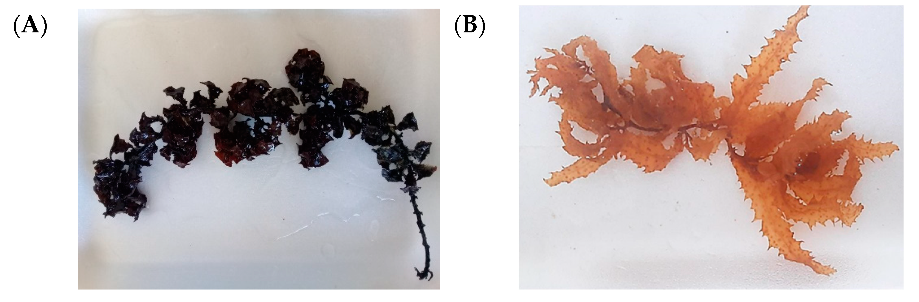
Figure 2.
Yield of fucoidan (mg/g dry weight) extracted from S. cinereum and T. decurrens.
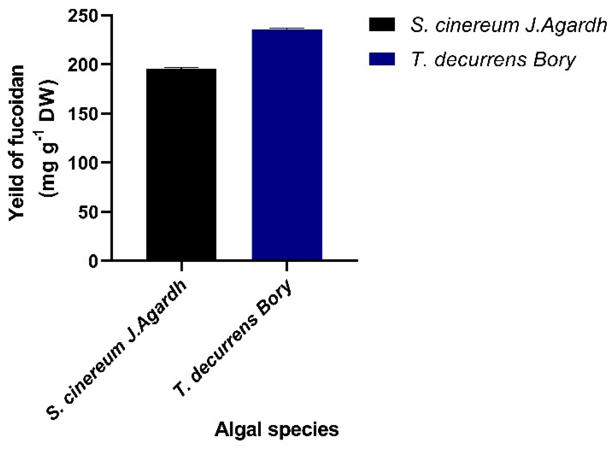
Figure 3.
Formation of F-AuNPs using: (A) S. cinereum and; (B) T. decurrens fucoidan, showing a gradual color shift from yellow to reddish-brown over 2 hours.
Figure 3.
Formation of F-AuNPs using: (A) S. cinereum and; (B) T. decurrens fucoidan, showing a gradual color shift from yellow to reddish-brown over 2 hours.
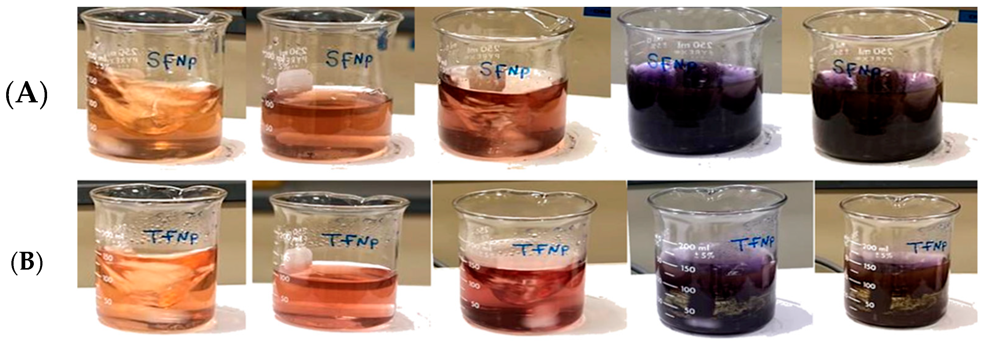
Figure 4.
UV absorption spectrum curve analysis for F-AuNPs (A) T. decurrens; (B) and S. cinereum.
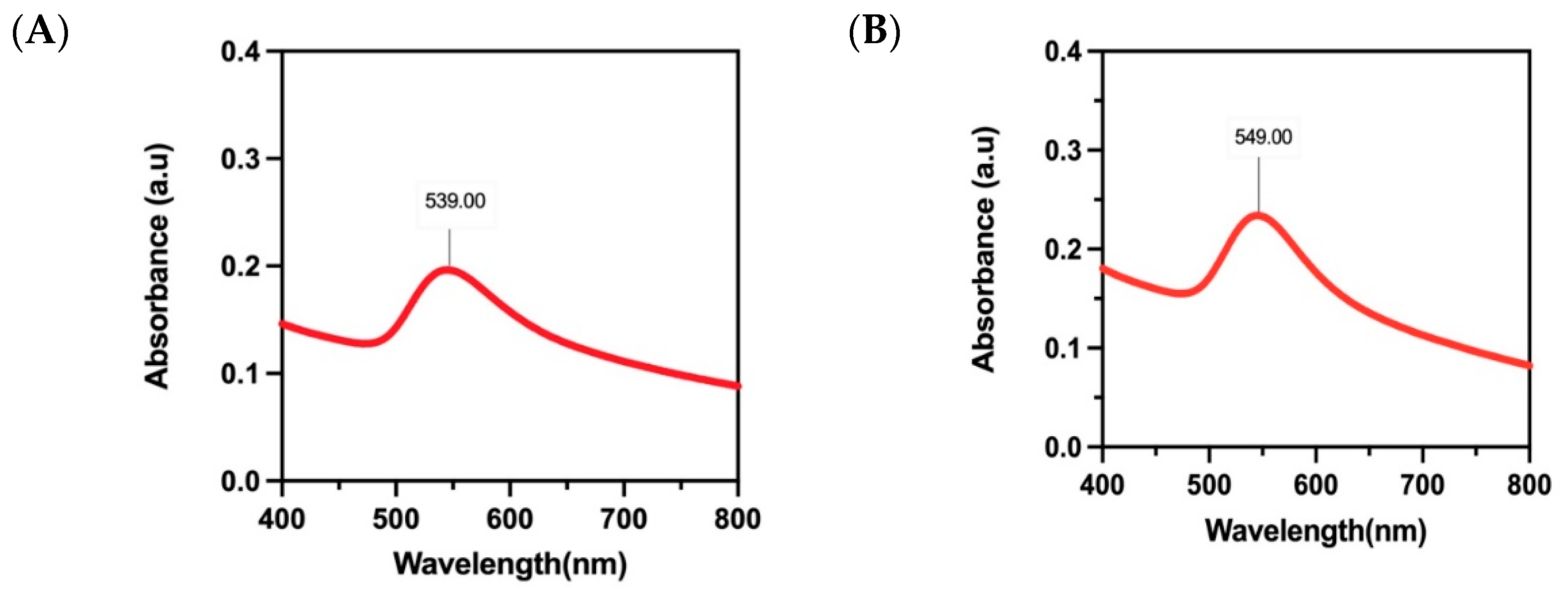
Figure 5.
FTIR Spectroscopy Analysis of F-AuNPs: (A) T. decurrens; (B) and S. cinereum.
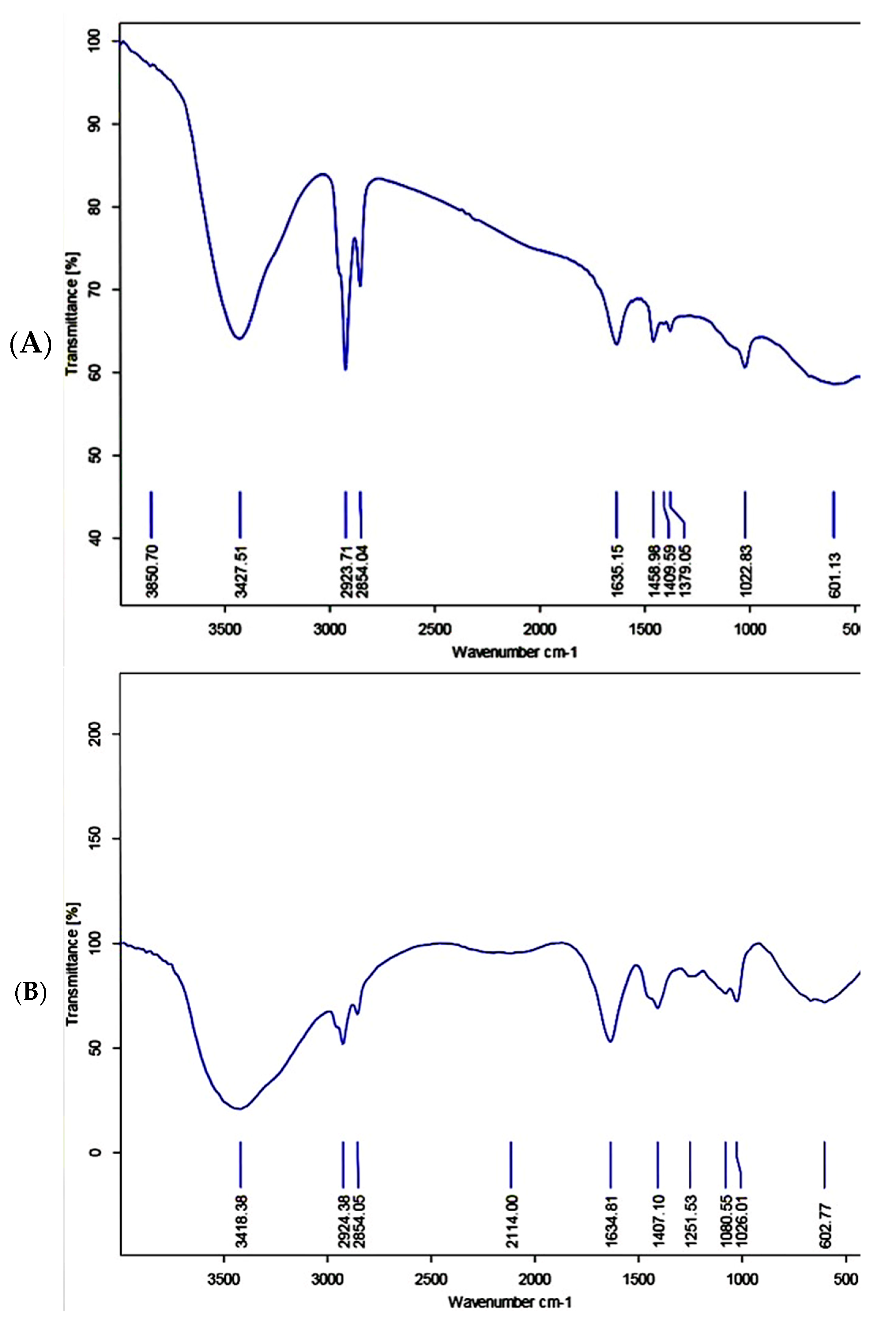
Figure 6.
XRD pattern of F-AuNPs: (A) T. decurrens F-AuNPs; (B) and S. cinereum.
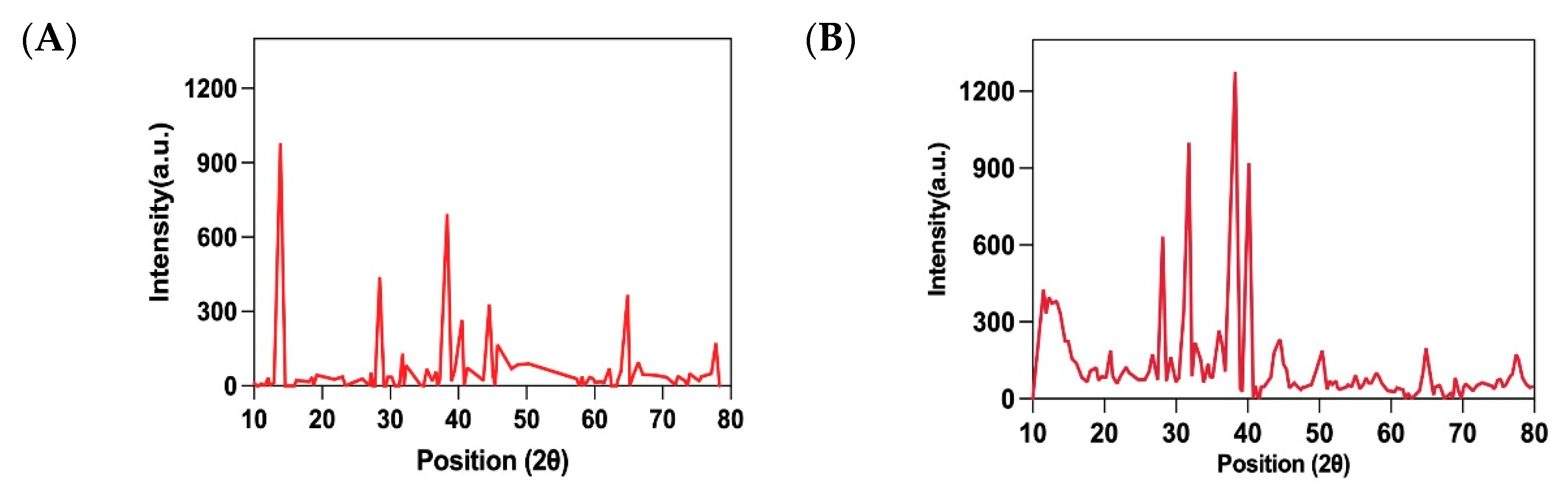
Figure 7.
TEM images for F-AuNPs: (A) T. decurrens; (B) and S. cinereum and selected area electron diffraction (SAED) pattern.
Figure 7.
TEM images for F-AuNPs: (A) T. decurrens; (B) and S. cinereum and selected area electron diffraction (SAED) pattern.
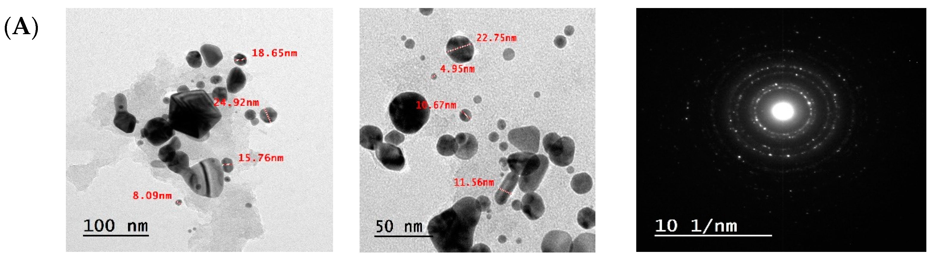
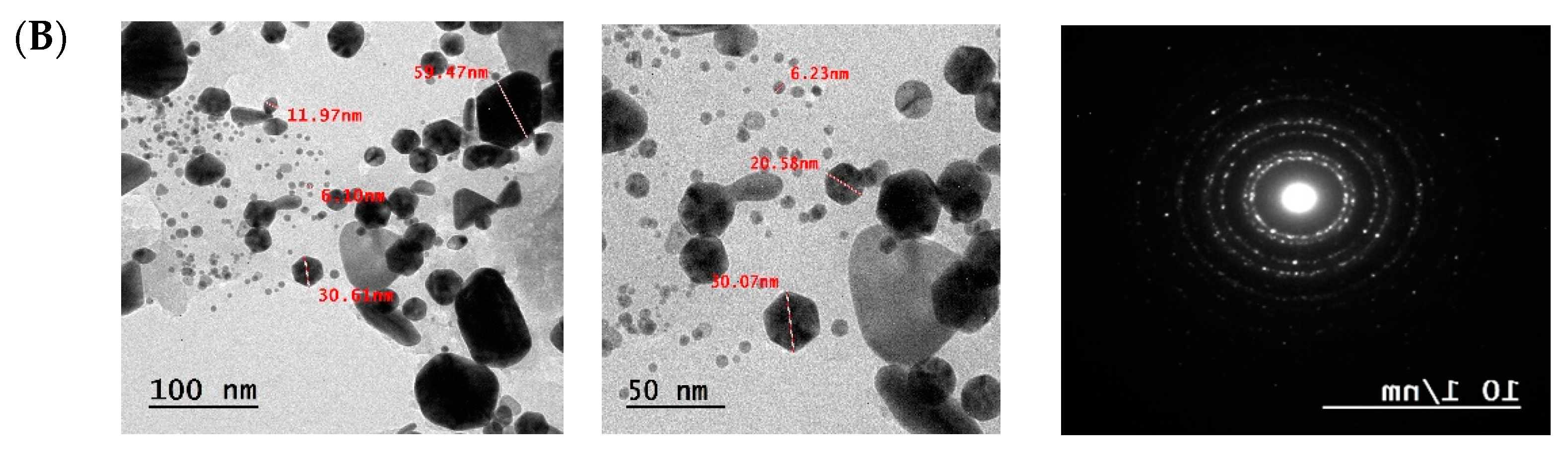
Figure 8.
Zeta potential analysis of F-AuNPs for: (A) T. decurrens; (B) and S. cinereum.
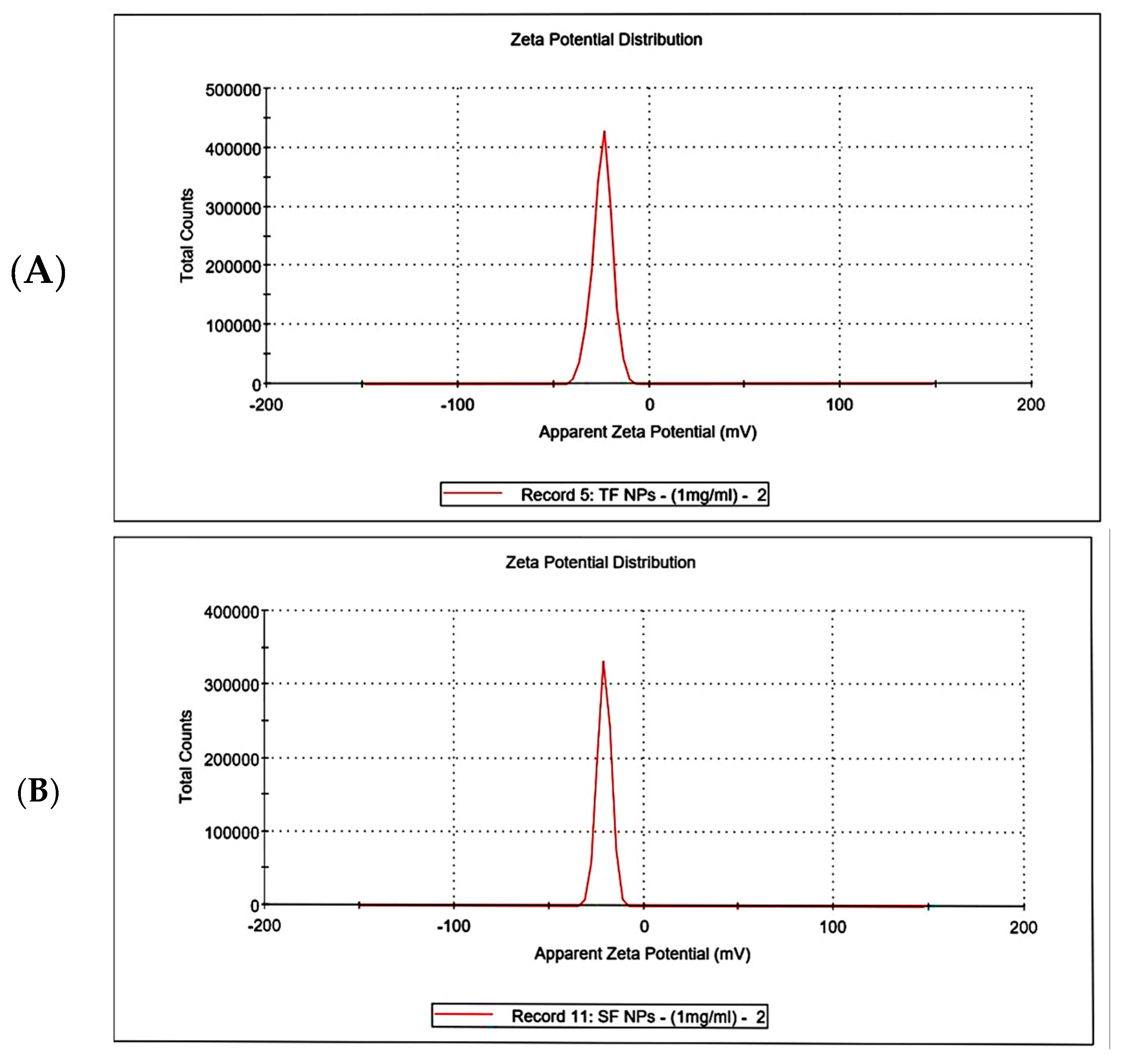
Figure 9.
SRB Assay for Normal BNL Cells: (A) T. decurrens; (B) S. cinereum (C) IC50 of F-AuNPs T. decurrens and (D) IC50 of F-AuNPs S. cinereum. .
Figure 9.
SRB Assay for Normal BNL Cells: (A) T. decurrens; (B) S. cinereum (C) IC50 of F-AuNPs T. decurrens and (D) IC50 of F-AuNPs S. cinereum. .
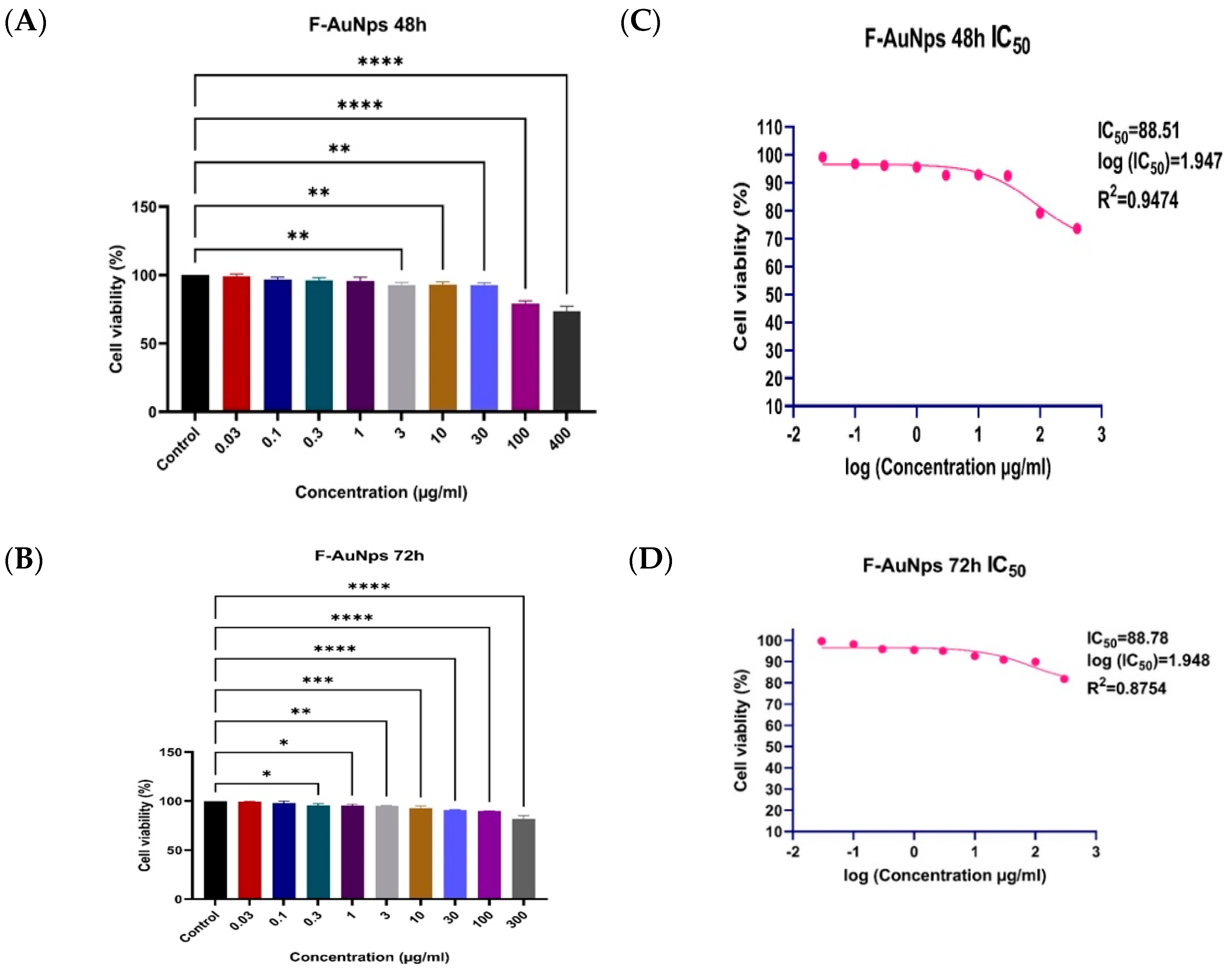
Figure 10.
HepG1 (MTT) assay for: (A) T. decurrens F-AuNPs; (B) S. cinereum F-AuNPs; (C) THP1 (SRB) assay of F-AuNPs T. decurrens; and (D) F-AuNPs S. cinereum.
Figure 10.
HepG1 (MTT) assay for: (A) T. decurrens F-AuNPs; (B) S. cinereum F-AuNPs; (C) THP1 (SRB) assay of F-AuNPs T. decurrens; and (D) F-AuNPs S. cinereum.

Figure 11.
Flow cytometry analysis: (A) control (B); T. decurrens; and (C) S. cinereum.
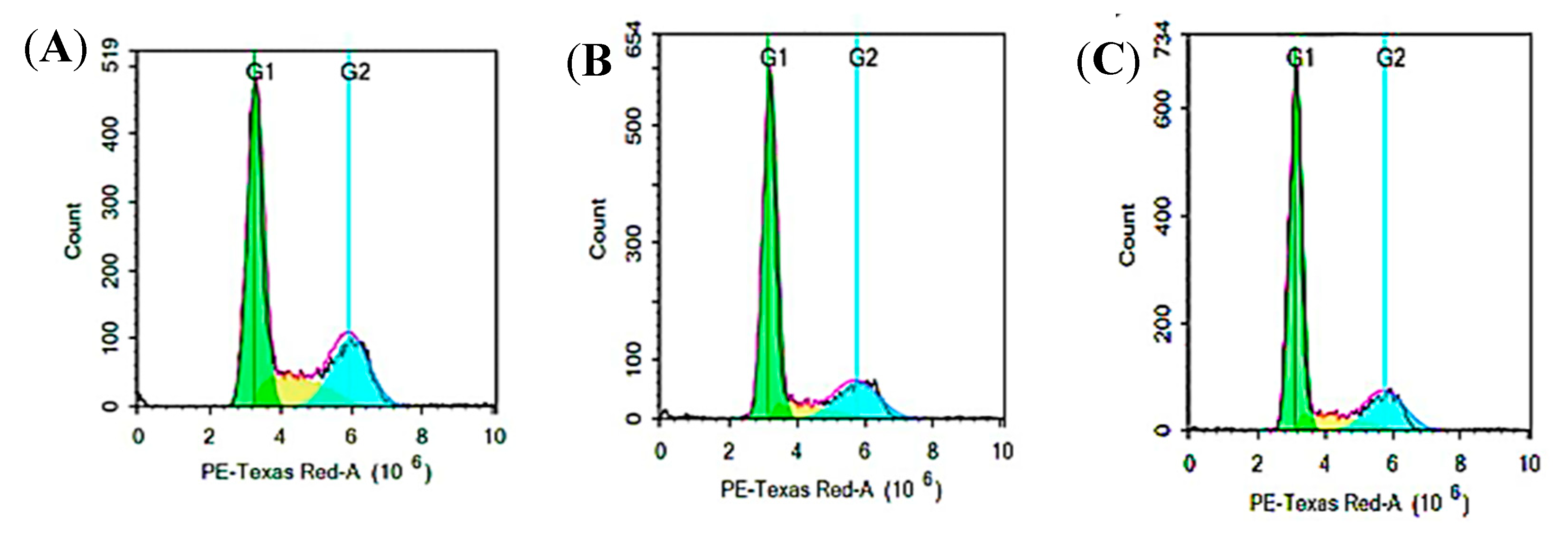
Figure 12.
Comparison of cell cycle distribution across different phases (G1, S, and G2/M) in HepG2 cells treated with Formulations T and S, compared to the control.
Figure 12.
Comparison of cell cycle distribution across different phases (G1, S, and G2/M) in HepG2 cells treated with Formulations T and S, compared to the control.
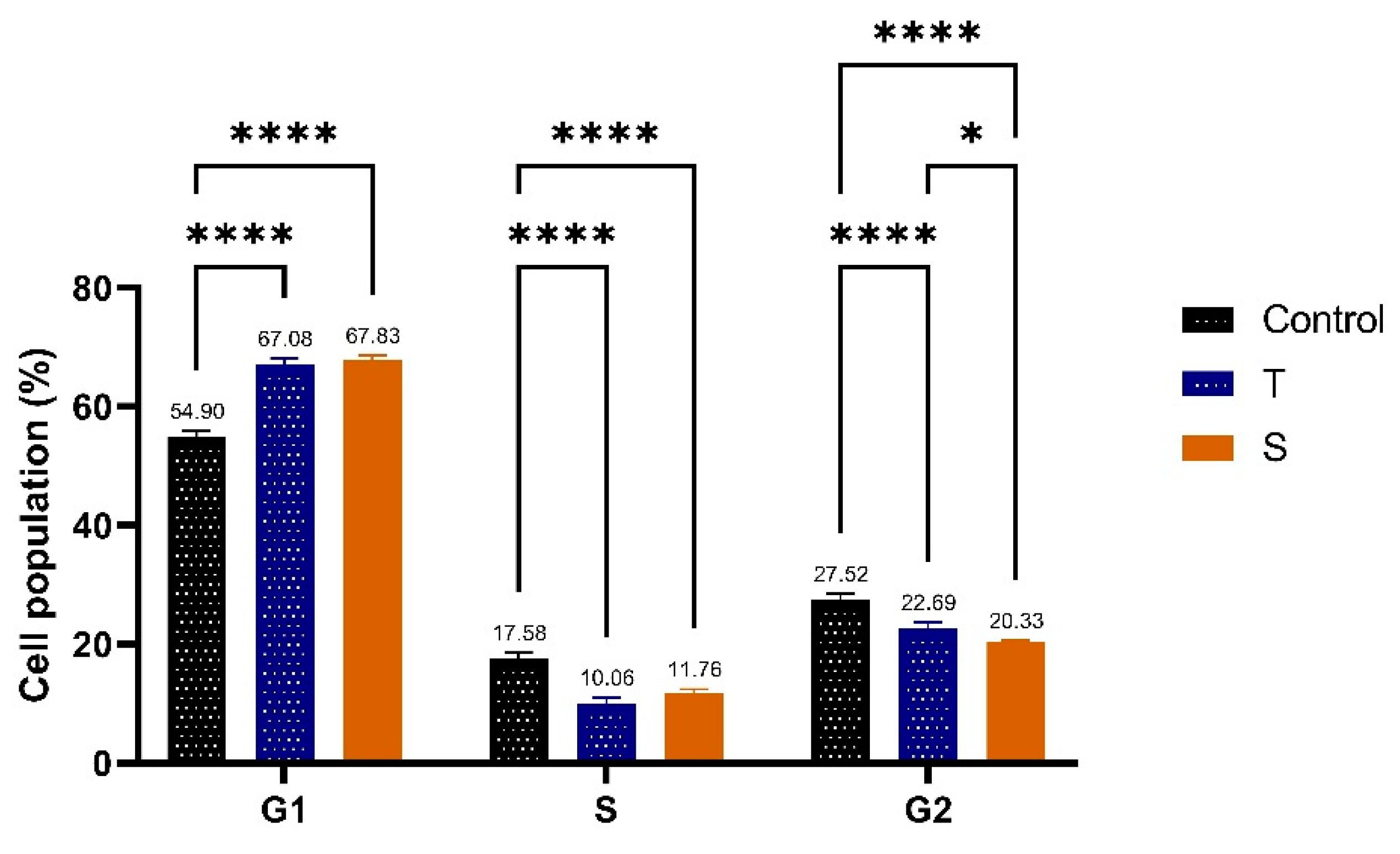
Figure 13.
Molecular docking analysis: (A) (2D and 3D) Binding of F-Au NAPs with the active sites of Arachidonate 5-lipoxygenase; (B) (2D and 3D) Binding of F-Au NAPs with the active sites transcriptase; (C) (2D and 3D) Binding of F- Au NAPs with the active sites of Telomerase reverse transcriptase. (D) 2D and 3D) Binding of F- Au NAPs with the active sites of Thymidylate synthase; (E) (2D and 3D) Binding of F- Au NAPs with the active sites of Histamine H3 receptor; (F) (2D and 3D) Binding of F- Au NAPs with the active sites of Cyclin-Dependent Kinases.
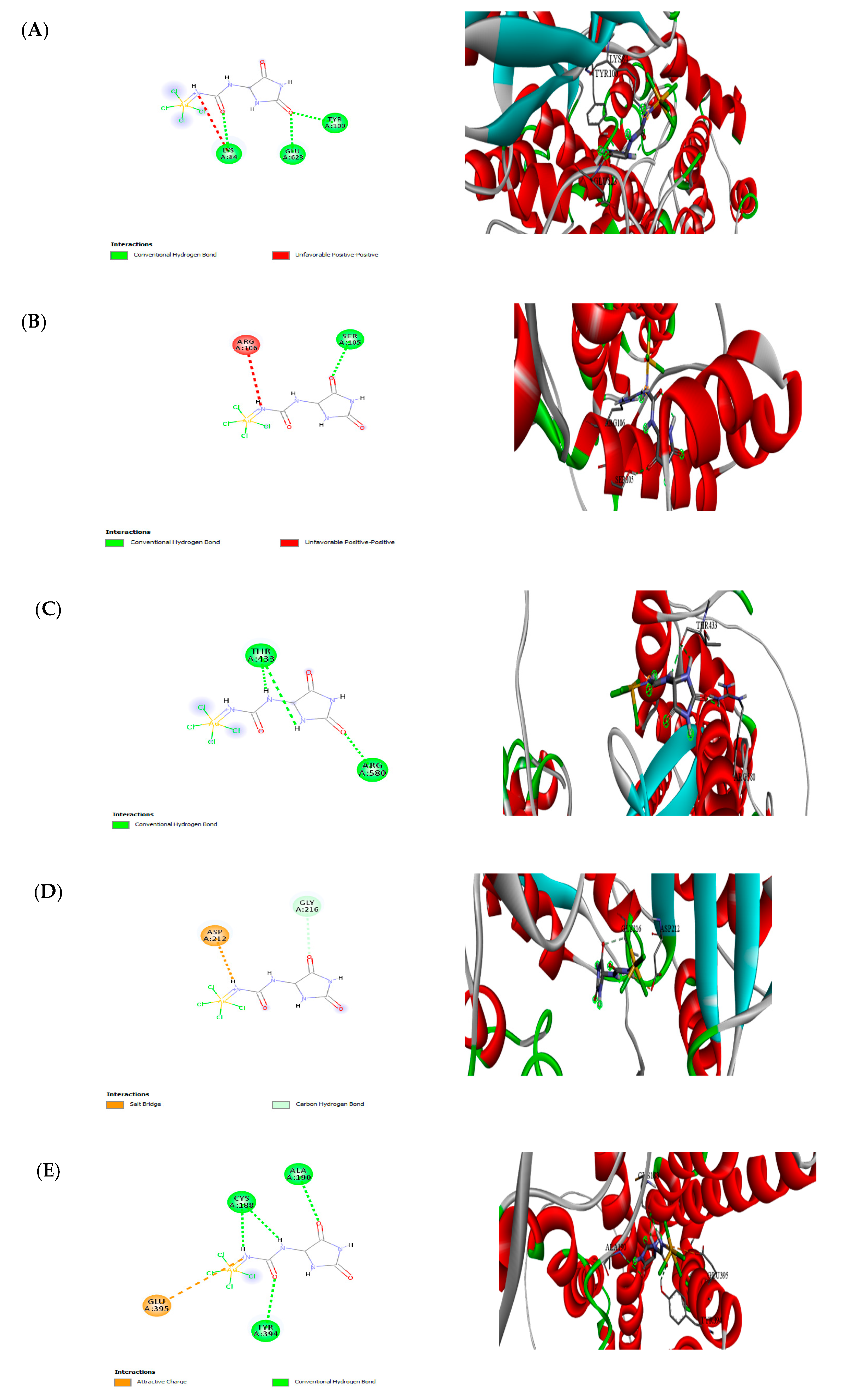
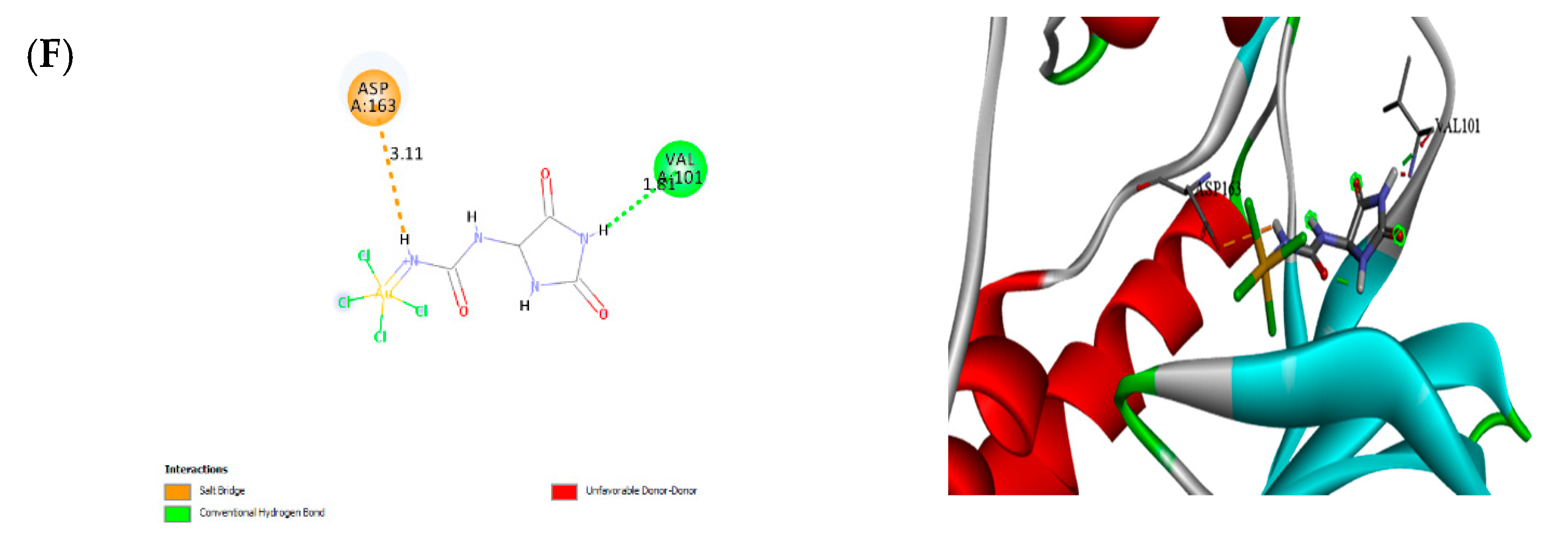

Table 1.
Identification of the collected seaweeds.
| Empire: Eukaryota | Empire: Eukaryota |
| Kingdom: Chromista | Kingdom: Chromista |
| Subkingdom: Harosa | Subkingdom: Harosa |
| Phylum: Ochrophyta | Phylum: Ochrophyta |
| Class: Phaeophyceae | Class: Phaeophyceae |
| Subclass: Fucophycidae | Subclass: Fucophycidae |
| Order: Fucales | Order: Fucales |
| Family: Sargassaceae | Family: Sargassaceae |
| Genus: Sargassum | Genus: Turbinaria |
Table 2.
The DPPH assay of fucoidan gold nanoparticles(F-AuNPs) for two brown seaweeds.
| Inhibition (%) | |||
|---|---|---|---|
| Concentrations (µ gm/ml) | Trolox | S. cinereum F-AuNPs | T. decurrens F-AuNPs |
| 3.90 | 11.73±0.21 | 10.82±0.11 | 12.05±0.41 |
| 7.81 | 25.92±039 | 21.61±0.08 | 24.07±0.36 |
| 15.60 | 47.15±0.42 | 43.23±0.13 | 39.06±0.12 |
| 25.00 | 68.64±0.15 | 66.19±0.09 | 62.49±0.31 |
| 31.25 | 83.80±0.18 | 80.49±0.19 | 78.11±0.31 |
Table 3.
FRAP assay of fucoidan gold nanoparticles(F-AuNPs) for two brown seaweeds.
| Inhibition (%) | |||
|---|---|---|---|
| Concentrations (µ gm/ml) | Trolox | S. cinereum F-AuNPs | T. decurrens F-AuNPs |
| 25 | 0.09±0.17 | 0.07±0.27 | 0.07±0.30 |
| 50 | 0.15±0.28 | 0.14±0.32 | 0.14±0.11 |
| 100 | 0.34±0.16 | 0.28±0.45 | 0.28±0.17 |
| 200 | 0.65±0.08 | 0.56±0.31 | 0.57±0.12 |
| 400 | 1.40±0.15 | 1.12±0.28 | 1.14±0.19 |
Table 4.
Molecular docking results in terms of the binding energy of F-Au NAPs with protein receptors.
Table 4.
Molecular docking results in terms of the binding energy of F-Au NAPs with protein receptors.
| No. | Receptors | Nanoparticles | Binding affinity (Kcal/mol) |
|---|---|---|---|
| 1 | Arachidonate 5-lipoxygenase | F-Au NAPs | -4.07 |
| 2 | Cyclooxygenase-2 | F-Au NAPs | -4.15 |
| 3 | Telomerase reverse transcriptase | F-Au NAPs | -3.98 |
| 4 | Thymidylate synthase | F-Au NAPs | -4.06 |
| 5 | Histamine H3 receptor | F-Au NAPs | -5.07 |
| 6 | Cyclin-Dependent Kinases | F-Au NAPs | -1.99 |
Disclaimer/Publisher’s Note: The statements, opinions and data contained in all publications are solely those of the individual author(s) and contributor(s) and not of MDPI and/or the editor(s). MDPI and/or the editor(s) disclaim responsibility for any injury to people or property resulting from any ideas, methods, instructions or products referred to in the content. |
© 2025 by the authors. Licensee MDPI, Basel, Switzerland. This article is an open access article distributed under the terms and conditions of the Creative Commons Attribution (CC BY) license (http://creativecommons.org/licenses/by/4.0/).
Copyright: This open access article is published under a Creative Commons CC BY 4.0 license, which permit the free download, distribution, and reuse, provided that the author and preprint are cited in any reuse.


