Preprint
Article

This version is not peer-reviewed.

The Nasal Microbiome of Sows in the Federal District of Brazil Has Revealed a Diverse Phenotypic Resistance Profile

Submitted:

19 February 2025

Posted:

20 February 2025

You are already at the latest version

Abstract

Antimicrobial resistance is a universal threat and is leading to a new awareness of antimicrobial use. The colonization of tissues by some microorganisms carrying resistance genes may pose a risk of spreading resistance to pathogens. Antimicrobials may induce an unstable microbiome that compromises the animal's immunity. Indeed, dysbiosis has been linked to many alterations in the immune response. Here, we isolated bacterial colonizers from the nasal microbiota of sows to describe the phenotypic resistance profile on different health managements. One hundred and thirty-two strains isolated from 50 nasal swabs collected from sows were tested against up to 23 antimicrobial agents by disk diffusion. Overall, the nasal communities showed 55% antimicrobial resistance (1605/2888 tests). Resistance was detected for all tested antimicrobials. The antimicrobial showing higher rate of resistance was bacitracin (92%), while the lowest was found in the aminoglycosides. Actinobacillus suis was one of the species with highest rate of resistance. The time and number of drugs used in feed influenced the resistance rate in the isolates. Vaccination was confirmed as a strategic protocol for disease control Studies on antimicrobial resistance in commensals may allow the identification of microorganisms to be used in surveillance and may constitute a tool for evaluating strategies to reduce antimicrobial use in pig herds.

Keywords: 
;  ;  

1. Introduction

Four decades ago, antimicrobials (AMB) at low dosages used to be part of swine herd management to improve animal weight [1]. Nowadays, the use of growth promoters and metaphylactic treatment is still a practice in broiler chickens and pigs [2]. In Brazil, AMBs are also used for enhancing animal health by reducing the burden of pathogenic microbes and, therefore, disease outbreaks in swine herd [3].
Nonetheless, the widespread use of AMBs has increased the selection pressure on the microbiome and promoted the spread of resistance genes among animals, humans and the environment. The emergence of AMB resistant microorganisms is a consequence of the continued use of AMBs, increasing costs and mortality rates in the production system, and causing a global health problem [3,4]. Antimicrobial resistance (AMR) occurs when a microorganism develops mechanisms that allow the growth in the presence of AMBs. A microorganism that has acquired resistance to at least one agent in three or more AMB categories is characterized as multidrug resistant (MDR) [5]. Thus, antimicrobial use (AMU) puts pressure on a given bacterial population and selects resistant variants [6], not only in the target pathogens but also in commensals from the microbiota [7]. AMR hinders treatment efficacy, limiting therapeutic options [8] and favors colonization by MDR microorganisms [5].
Therefore, AMB treatment should only be used when recommended by veterinarians at an appropriate dose and duration when deemed necessary and effective during an outbreak, which is important for animal welfare [6,7,8]. Strategies for reduction of AMU in veterinary medicine include vaccination protocols and promotion of a balanced microbiota through beneficial microbial colonization [9]. Instead, on some Brazilian farms, pigs are still fed AMBs in their feed to prevent infections [10]. AMU should be optimized to reduce the selection and spread of AMR [11]. It is important to improve managers’ understanding of effective strategies to prevent AMU [12] and to maintain a stable microbiota community [13].
The microbiota community plays an important role, with the nasal microbiota contributing to respiratory health [13]. One of the most important functions of symbiotic microorganisms is to protect against pathogens, mainly through pathogen exclusion and immune system stimulation [14,15], provided by a diverse microbial community [16]. One of the strategies to ensure beneficial microbial colonization of animals is to promote a balance between colonization and immunity [7,9].
Nevertheless, metaphylactic AMB treatments have the potential to disrupt the beneficial microbial communities of the microbiota, leading to dysbiosis [7,16]. This unbalanced microenvironment is more susceptible to pathogen invasion, raising the risk for disease development [17]. Furthermore, if the microbiota members possess resistance genes, these can be transferred to the pathogens [18], potentially undermining disease management. Bacteria have the potential of genetic modification, and this will enable them, sooner or later, to neutralize the action of newly invented AMBs [19].
Notably, the circulation of resistant strains can occur in sick or healthy animals. Antimicrobial resistance (AMR) of Streptococcus suis in clinically healthy sows has been reported previously in China, and transmission is likely to occur between healthy carrier sows and their offspring [20].
Increasingly, networked and well-informed consumers are putting pressure on production chains to adopt high standards of sustainability [21]. Ensuring welfare for animals can strengthen the immune system and reduce the need for AMU. Good animal husbandry meets the behavioral, environmental and physiological needs of animals, resulting in less stress on the herd and less susceptibility to disease [22]. In this context, biosecurity should also be improved by reviewing management practices [23] rather than relying on metaphylactic treatments, which can lead to loss of drug efficacy [24]. Biosecurity is the primary measure to manage biological risks, designed to prevent the introduction (external biosecurity) and spread (internal biosecurity) of disease within populations, herds, or groups of animals. In addition, the governments of many countries have adopted actions on the basis of the One Health approach. These restrictive measures are part of a broader strategy to combat AMR, in line with international guidelines from the World Health Organization (WHO), the Food and Agriculture Organization (FAO) and the World Organization for Animal Health (WOAH). Meanwhile, in Brazil the manufacture, marketing and distribution of AMB products for veterinary use have been strictly regulated. Mandatory veterinary prescriptions are being implemented for the purchase of AMBs, as indicated in the resistance monitoring program, known as PAN BR AGRO, legislation.
Public authorities are encouraging alternative practices among pig producers through comparative results based on scientific data and educational programs [9,10,22,23]. Since 2018, Brazil has established procedures for the production and use of veterinary drugs in feed, including the obligation to inform AMU in feed formulation [25,26]. It is recognized that Brazil has been advancing for the more conscious use of AMB in livestock farming (Brazil, 2020). The use of tetracyclines, macrolides, avoparcin and penicillin as growth promoters is prohibited, but significant progress remains to be made. The PAN BR AGRO is monitoring bacterial pathogens from animal-derived foods [25]. Government policies are fostering alternatives to AMU, enforcing animal welfare and good production practices, and a new culture that comes with microbial diversity [8]. AMR surveillance programs have become essential to control the use of AMBs in food production and to evaluate the strategies adopted. In this way, AMB susceptibility testing is recommended for the appropriate use of AMBs and to identify MDR [27].
Here, we report the isolation of bacterial colonizers from the nasal microbiota of sows and describe the phenotypic resistance profile in different sanitary management practices in the Federal District, Brazil.

2. Materials and Methods

2.1. Ethical Approval

All samples were collected according to ISO/TS 34700:2016 with the permission of the farm owners. The study was approved by the Ethical Committee for the Use of Animals (CEUA) of the University of Brasília nº 23106.022976/2023-55.

2.2. Sampling, Swine Farms and Sows

From March 2022 to October 2023, sows were screened in 10 sow farms (A to J) located in the Federal District, Midwest of Brazil. The Federal District is a central area of Brazil, where the capital Brasilia is located, with only 5,761 km², while Brazil has 8,510,000 km². However, it is very important in terms of governmental affairs, and the surrounding rural area has developed in agribusiness contributing to the gross domestic product (GDP). There are 24 farms in the Federal District and 10 of them are sow farms, classified as two-site herds (1 farm), farrow-to-finish (3 farms), and one-site herd (6 farms). The health management varied from each farm by vaccination protocols and metaphylactic treatments in sow feed (some farms alternate different AMB with amoxicillin - Table 3), the hygiene practices (some use neutral soap before disinfectant to clean the empty lots, others not), the sanitary downtime (vary from 3 to 5 days), welfare status (only farm C was Animal Welfare Certified), and biosecurity measures which was evaluated by a Bio Score (Biosecurity Data Collection). Herd sizes ranged from 50 to 4,273 sows. Five sows, aged between 14 and 48 months, showing symptoms of coughing and prostration, were selected for sampling based on convenience. In the absence of clinical signs, but in the same batch housing the clinical case, apparently healthy sows were selected to achieve 5 sows sampled. The sow population in the batches varies from 20 to 60 among farms. Sows treated with marbofloxacin, gentamicin or amoxicillin with clavulanic acid (injectable) up to seven days prior to the farm visits were not sampled. Therefore, a total of 50 nasal swabs in duplicate were placed in plain Falcon tubes for PCR assays and in 5 mL BHI, KASVI® tubes for bacterial culture.
Surveillance routine screening at the slaughterhouse, performed independently from the study, reported the presence of lung lesions and their cause in pigs from the farms studied.

2.3. Biosecurity Data Collection

The farms varied in animal welfare and hygiene management. Biosecurity was scored using 10 questions that assessed the risk of disease entering and spreading in the herd, by assessing the preventive measures in place on the pig farms. The higher the score, the better the preventive measures in place. The main measures rated were isolation of the farm, safe distance from other pig herds and roads, replacement of breeding stock, quarantine, possible sources of vectors, type of feed, mode of transport and registration of access by vehicles and people. The questionnaire used for this study was based on the official form of the Ministério da Agricultura, Pecuária e Abastecimento (MAPA, Brazil). In the farms under study, the biosecurity (Bio) score ranged from 6 to 9 (median value: 7.1) on a scale of 1 to 10 points (Table 1).
The same researcher interviewed and collected data in all participating herds. The cross-sectional study included 463 sows housed in the sampled batches. The total population of sows in the Federal District farms was 9,544. The farms used different AMBs in the sow feed, but all added amoxicillin to sow diets, especially from June to August, due to the dry season, high daily temperature amplitude (hot during the day and cold at night) and increased dust. Of note, at the time the samples were collected, national legislation restricting medicated feed was not updated. Recently, Brazil aligned the AMB policies with global standards and implemented measures to effectively monitor AMU and reduce AMR.
The vaccination protocols in the farms included commercial and autogenous vaccines for several swine diseases. Piglets were weaned between 21 and 24 days, then the pen was emptied for 3 to 5 days, followed by rigorous cleaning. This variation was due to farm management and the number of farrowing sows. Cleaning was done with high pressure water and only farm F used detergent. After cleaning and removal of visible organic material, quaternary ammonia or bleach was applied. Although there are no mandatory vaccines, the farms used commercial and autogenous vaccines with different protocols.

2.4. DNA Extraction and PCR Assays

Genomic DNA was extracted from each sample using the Genomic DNA Extraction Kit® (Biogene, Madison, WI, USA) according to the manufacturer’s instructions. PCR assays were performed as in previous work [28]. We detected Actinobacillus pleuropneumoniae, Glaesserella parasuis, and Pasteurella multocida by multiplex PCR assay and Mycoplasma hyopneumoniae by nested PCR [28]. The positive control samples were kindly provided by the Federal University of Viçosa - UFV and the negative control was ultrapure water.

2.5. Culture and Bacterial Isolation

Standard microbiological techniques were used to isolate bacterial colonies at the Veterinary Microbiological Laboratory of the University of Brasília (UnB). Nasal swabs were plated on blood agar and incubated overnight at 37°C. Morphology, hemolytic activity, Gram stain, catalase and oxidase tests were performed individually. Contaminating Gram+ bacilli with the presence of spores and yeasts were discarded. All colonies were classified according to the results of oxidative/fermentative (OF), methyl red, and Vöges-Proskauer (Vm/Vp) tests, followed by other biochemical tests according to the protocols established in the Standard Operating Procedure of the Veterinary Microbiology Laboratory of the UnB. In addition to Vm/Vp, staphylococci were cultured on salt-mannitol agar (BD®) and differentiated using the coagulase test. For enterobacteria, the characterization followed the established differentiation protocol: indol, citrate, urea and TSI (triple sugar iron) and consumption of sugars and proteins. After bacterial characterization [29], each strain was individually plated on blood agar and frozen using BHI with 20% glycerol.

2.6. Antimicrobial Susceptibility Test

Isolates were subjected to antimicrobial susceptibility testing using the disk diffusion Kirby-Bauer method to evaluate phenotypic resistance [30]. This method classifies bacterial isolates as susceptible (S): bacteria are inhibited by concentrations of the AMB at standard doses; intermediate (I): a higher dose of the AMB may be required to achieve efficacy; or resistant (R): the drug is ineffective against microorganisms. The categories are based on the size of the zones of inhibition around the AMB-impregnated discs. These classifications, along with regularly updated cut-off values, ensure that AMBs remain effective and safe while minimizing the development of resistance. To date, (I) results are considered susceptible with increased exposure, meaning that higher than standard doses or prolonged exposure may be required. Intermediate isolates are a “gray area” where efficacy depends on increased exposure, which can be quantified through PK/PD modeling and adjusted dosing regimens. A higher dose of the AMB is required to achieve efficacy, which is reflected in (S) results. The cut-offs for susceptible, intermediate and resistant are determined based on diameter zones. Here, the inhibition zone sizes around the AMB discs were read with a pachymeter. Interpretation of the resistance profiles was performed according to CLSI (Clinical and Laboratory Standards Institute) standards [31], where available, and following the AMB manufacturer’s instructions. The products have analytical sensitivities at their respective concentrations adjusted to McFarland’s Standard Turbidity Scale 0.5. The 23 AMBs used are listed in Table 2.

2.7. Statistical Analyses

Graphs were generated with RStudio (2024.04.0) [32] using the ggplot2 package [33]. Statistical analyses were performed using SAS software (version v9.4, Cary, North Carolina, USA). For each AMB, the proportion of resistant isolates was calculated by dividing the number of resistant isolates by the total number of isolates tested. We chose a nonparametric test, Chi-Square, to estimate correlations between AMB resistance and health management variables, such as vaccination protocols. Fisher’s exact test was used when the sample size was small, resulting in expected frequencies below 5. Pearson correlation was used to assess correlation between years of use and number of AMBs or bio score with overall resistance in each farm. The level of resistance among bacterial species or among farms was compared using Krustal-Wallis with post-hoc Dunn’s test.

3. Results

3.1. Swine Farms and Sows’ Health Management

Samples were collected from sows of different parity, with 42% (21/50) in first parity, 36% (18/50) between 2nd and 4th parity and 22% (11/50) in 5th parity or above. Fifty-four percent (27/50) of the sows were in maternity stalls and 46% were in gestation stalls. Forty-four percent (22/50) of the sows showed no clinical signs of infectious disease, but a history of infectious disease was reported in 34% (17/50) of the sows. During the visits, 22% (11/50) of the sows were coughing, sneezing and/or had purulent or sanguinolent nasal secretions. The number of workers varied from 1 to 96 depending on the herd size, but the number of sows per worker varied from 27 to 65.75 among the herds, with an average of 46.3 sows per worker. The smallest herd of 50 sows was managed by only one worker, while the largest herd of 4,273 sows was managed by 96 workers. Some farms used in-feed amoxicillin continuously, while others used AMBs on a rotational basis in the sow feed.
The farms used medicated feed to acclimate the sows as a preventive strategy to prevent atrophic rhinitis in sows and their offspring and thus control the spread of infectious diseases by reducing shedding. However, some farms added florfenicol, penicillin or tylosin, alternating the AMBs with each use, as recommended by the veterinarian. Farm managers used 800 mg/ton of amoxicillin to achieve 20 mg/kg per lactating sow. Amoxicillin is a broad-spectrum penicillin AMB that is active against many Gram-positive and some Gram-negative bacteria, including Pasteurella multocida, a major pathogen in atrophic rhinitis and Bordetella bronchiseptica. Farm A frequently included clindamycin, tetracycline, enrofloxacin, and oxytetracycline in the feed rotation for metaphylactic purposes, and Farm J included clindamycin in an attempt to increase the effectiveness of treatment. Occasionally, marbofloxacin, gentamicin, or amoxicillin with clavulanic acid was injected for therapeutic treatment. The use of amikacin was not reported by any of the farm managers or staff. The vaccination protocols varied among the farms. Commercial vaccines were used in 7/10 farms and were directed against Mycoplasma hyopneumoniae (Myo), porcine circovirus type II (PCV2), Pasteurella multocida, Bordetella bronchiseptica, Salmonella ser. Typhimurium, Streptococcus suis infection diseases. Autogenous vaccines were used in 8/10 herds to prevent outbreaks caused by P. multocida, Glaesserella parasuis, S. Typhimurium, E. coli and, S. suis. Table 3 details health management practices among swine farms, including metaphylactic feed and vaccine protocols adopted by each farm.
Table 3. Farm, antimicrobial agents in metaphylactic treatment, vaccines protocols and production type.
Farm AMB Agents Vaccines Production Type
A AMO, CLI, TET, ENO, OXY M. hyopneumoniae, circovirus, P. multocida, S. ser. Typhimurium One-site-herd: piglet unit production
B AMO, FLF, PEN M. hyopneumoniae, circovirus, G. parasuis, S. suis. One-site-herd: piglet unit production
C AMO, FLF P. multocida, B. bronchiseptica, G. parasuis, S. ser. Typhimurium, E. coli. Two-site-herd: piglet and gilt and young boar production
D AMO, FLF G. parasuis One-site-herd: piglet unit production
E AMO None Farrow-to-finish: piglet to hog-finished production
F AMO, FLF, PEN P. multocida, S. ser. Typhimurium, S. suis. One-site-herd: piglet unit production
G AMO None Farrow-to-finish: piglet to hog-finished production
H AMO, FLF M. hyopneumoniae, circovirus, P. multocida, S. ser. Typhimurium, S. suis One-site-herd: piglet unit production
I AMO, FLF, TYL M. hyopneumoniae, circovirus, P. multocida, G. parasuis, S. ser. Typhimurium One-site-herd: piglet unit production
J AMO, CLI P. multocida, G. parasuis Farrow-to-finish: piglet to hog-finished production
AMO: amoxicillin, CLI: clindamycin, TET: tetracycline, ENO: enrofloxacin, OXY: oxytetracycline, PEN: penicillin, FLF: florfenicol, TYL: tylosin.

3.2. Lung Lesions and Pathogen Detection

Detection of pathogens is summarized in Table 4. P. multocida was detected in 6 out of 10 farms and G. parasuis in 4 out of 10 farms. G. parasuis and P. multocida were detected together in one sow. A. pleuropneumoniae was detected only in farm J. M. hyopneumoniae was not detected in any of the 50 samples. Farm C had Mhyo-free status, but we cannot exclude the presence of M. hyopneumoniae in the other farms that tested negative by nested PCR. G. parasuis and A. pleuropneumoniae were only detected by PCR, as they cannot grow on blood agar plates. A. pleuropneumoniae was also detected in the slaughterhouse in samples from farm J.
Lung lesions caused by P. multocida were reported by the slaughterhouse in the herds of farms B, D and G, which did not use the corresponding vaccine. In nasal samples, P. multocida was detected by PCR and culture in farms A, B, E, G, H, and PCR but not culture in farm D. As shown in Table 3, six of the 10 farms applied P. multocida vaccines: five used autogenous vaccines and one a commercial one (farm C). Only one of them, farm J, reported lung lesions at slaughter (although isolation showed A. pleuropneumoniae in the lesions). The use of an autogenous vaccine against P. multocida acted as a protective factor against lung lesions, as this vaccine was significantly associated with a reduction in pulmonary lesions (p-value of 0.048).

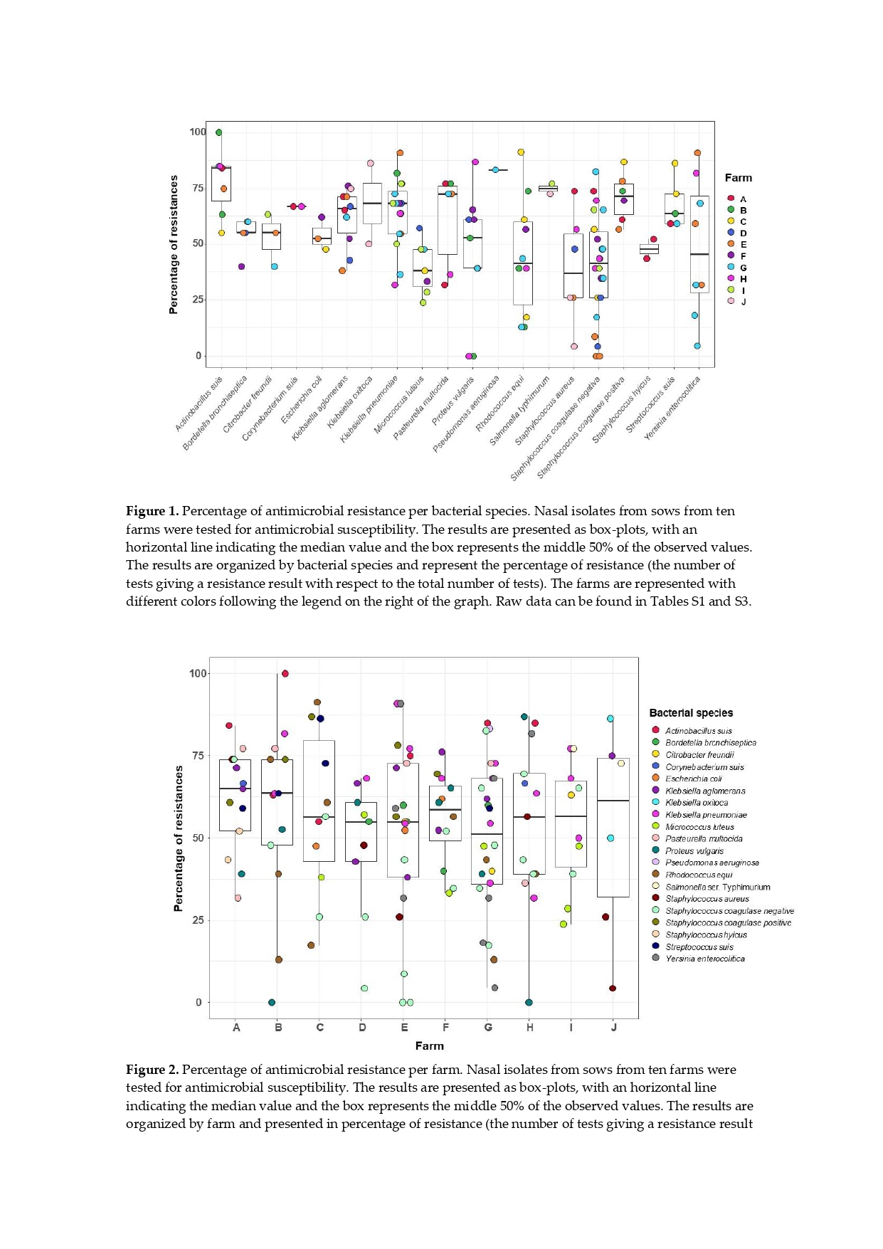
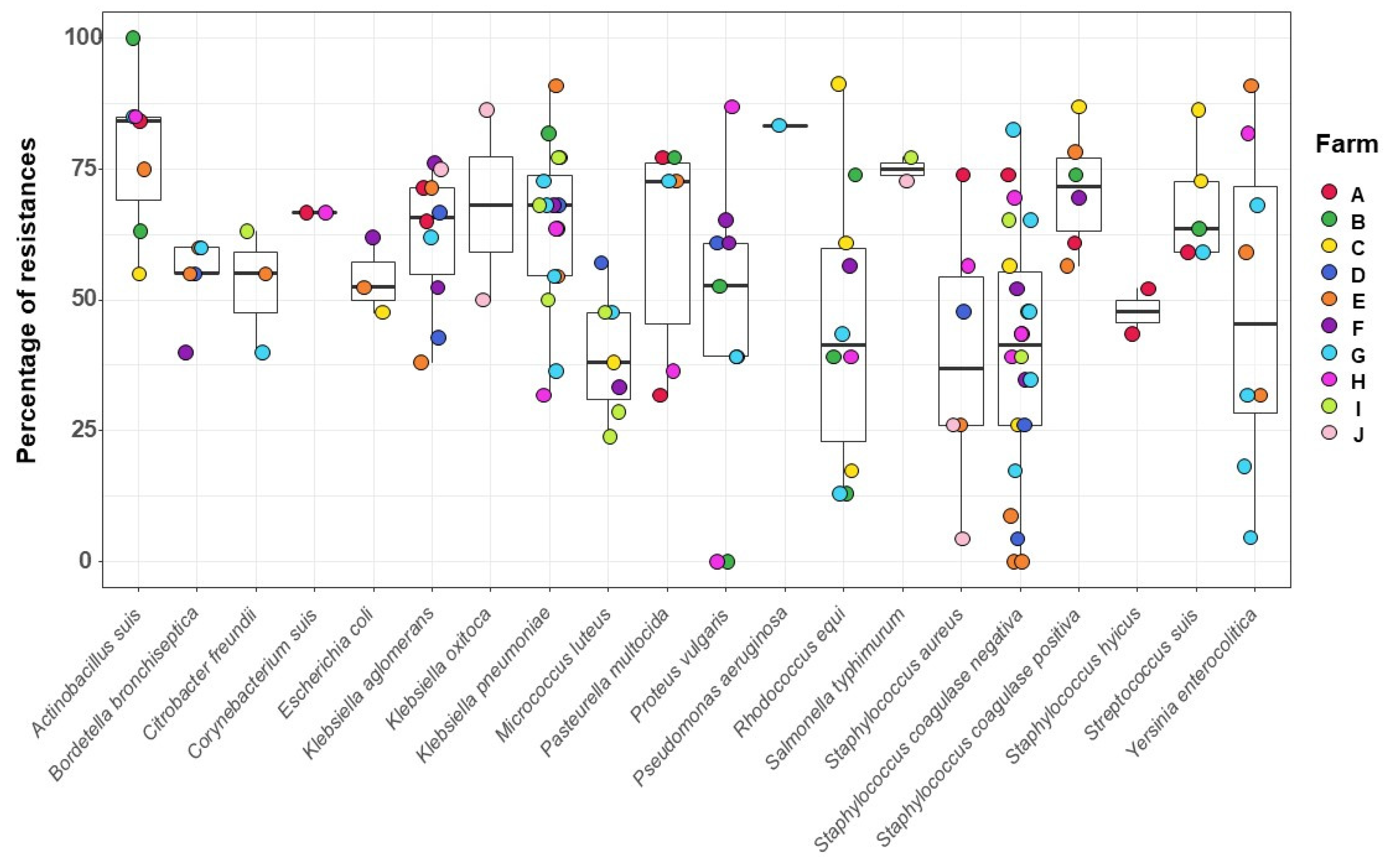
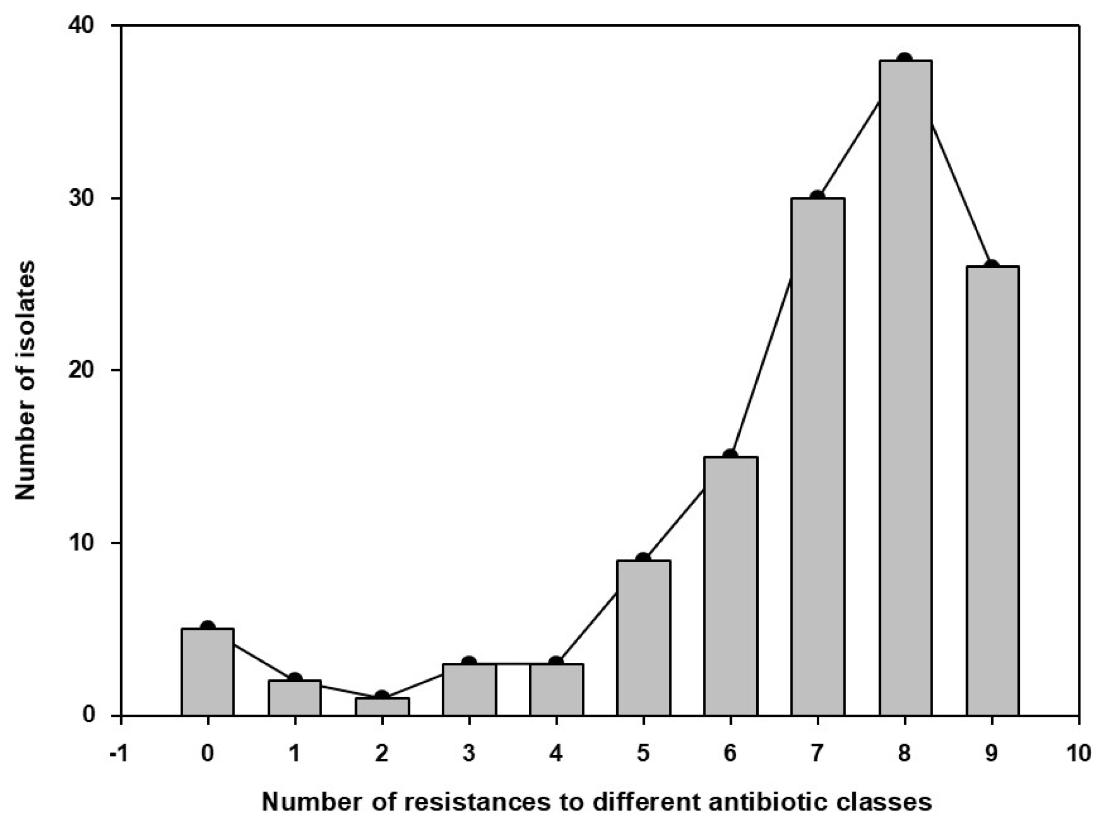
3.3. Bacterial Isolates from the Sows` Nasal Microbiota and Resistance Profile

A total of 132 strains belonging to 20 bacterial species were isolated from the 50 sows. Staphylococcus coagulase negative, typical commensals of the nasal microbiota was the most prevalent (22/50; 44%). Although Enterobacteriaceae are commonly associated with the gut microbiota, herein Klebsiella, Pantoea, Proteus, Yersinia, Escherichia, and Salmonella were isolated from the sow nasal swabs. A statistically significant association was observed between the administration of penicillin and the isolation of Proteus vulgaris (Chi-Square, p = 0.0317). Pathogens such as P. multocida, B. bronchiseptica, K. pneumoniae, P. aeruginosa, S. aureus, Y. enterocolitica, S. Typhimurium and S. suis were isolated from sows without respiratory diseases. But, as expected, A. pleuropneumoniae was isolated from the sow with fever from farm J. Lastly, the administration of an autogenous Streptococcus suis vaccine was statistically associated with the isolation of Rhodococcus equi (Chi-Square, p = 0.0410).
To investigate AMB resistance in the isolates, tests with 23 AMB agents were performed (Supplementary Table S1). Overall, the results showed 55.6% (1605/2888) AMR (Supplementary Tables S1 andS2). The AMB class showing less resistance was the aminoglycosides (16.7-39.4%; Table S2), while the polypeptide bacitracin showed the highest rate (92.4%; Tables S1 and S2). In the macrolides, tulathromycin showed a lower degree of resistance, 33.6%, than erythromycin and tylosin, with 57.4 and 72.3%, respectively. In the beta lactams, the highest rate of resistance was observed for penicillin (79.2%). It is noteworthy to point out that the addition of clavulanic acid to amoxicillin reduced the resistance from 65.2% (with amoxicillin only) to 27.3% (Tables S1 and S2). It is also of note to highlight the presence of resistance to ceftiofur (a third-generation cephalosporin) in several taxa, including A. suis, S. aureus and S. suis.
Considering the history of use of each antibiotic reported, the longest reported period of antibiotic use was nine years for bacitracin and the shortest was one year for amoxicillin with clavulanic acid, marbofloxacin, and gentamicin. We observed a correlation between the number of years that the AMB was used and the percentage of global resistance observed for that AMB (Pearson correlation; r = 0.88, p-value <0.001; Table S2). Surprisingly, we observed some resistance to amikacin, which was never used in the herds. A correlation between the rate of resistance in each farm and the number of AMBs used in the farm was found (Pearson correlation; p = 0.02). On the other hand, no correlation was detected between the level of resistance in a farm and its corresponding Bio score (Pearson correlation; p = 0.578).
The phenotypic profile of antimicrobial resistance varied from 39% to 83% among bacterial species (Supplementary Table S3 and Figure 1) and from 47% to 65% among farms (Supplementary Table S3 and Figure 2).
We observed heterogeneity in AMR frequency within each bacterial species (Figure 1). As an example, Staphylococcus coagulase negative isolates showed wide diversity in the number of resistances: the isolate from farm A showed AMR of 74% (17/23); in farm E, 2 isolates showed no resistances, one isolate 8.7% (2/23) and the fourth isolate 43.5% (10/23); isolates from farm F showed 52% (12/23) and 35% (8/23) of antimicrobial resistance; isolates from farm G showed resistance varying from 17.4% (4/23) to 82.6% (19/23). In addition, the rate of resistance in the different bacterial species was significantly different (Kruskal-Wallis, p-value 0.0015; Figure 1). This global difference was due to the differences between A. suis and M. luteus (z = 3.5, p = 0.037), and A. suis versus and Staphylococcus coagulase negative (z = 3.8, p = 0.014). In the case of S. Typhimurium (isolated only from Farms I and J) and P. aeruginosa (isolated only from Farm G), which showed high percentage of resistances, it is possible that the low number of observations may have limited the test’s ability to detect differences.
Isolates with varying degree of resistance were found in the different farms, but no differences were detected in the overall resistance levels among the farms (Krustal-Wallis, p = 0.94; Figure 2).
When the resistance to multiple AMB families was examined, the majority of isolates showed multiresistance to 7-9 AMB classes (Supplementary Table S1; Figure 3). On the other hand, only 5 isolates did not show any resistance (2 Proteus and 3 Staphylococcus), 2 isolates showed resistance to just one AMB family, and one isolate to 2 families. Some of the more frequent MDR profiles included resistance to bacitracin and penicillin combined with tetracycline (60% of isolates), doxycycline (57%), florfenicol (56%) or tylosin (55%).

4. Discussion

This cross-sectional study evaluates, for the first time, the antimicrobial resistance of bacterial isolates collected from the nasal cavities of sows in the Federal District, Brazil. We screened for the most common respiratory pathogens in swine production [34,35,36,37,38,39]. However, a high level of AMR was observed not only in pathogens, but also in commensals. Globally, the rate of resistance to an AMB correlated with the number of years using the drug, and resistances in a farm correlated with the number of AMBs used. In agreement with a previous report showing that biosecurity did not correlate with AMU [10], in this study, AMR was not associated with the biosecurity score.
Noteworthy, we found S. suis, A. suis, S. aureus isolates that were resistant to ceftiofur. In a study in Spain by Blanco-Fuertes et al. [15], treatment with ceftiofur administered to sows resulted in higher levels of resistance genes in weaned piglets. In Canada, ceftiofur was a routine treatment for disease prevention in suckling pigs in some herds [40]. Ceftiofur administration had a longer effect on the nasal microbiota composition of piglets when administered to their sows before farrowing than when applied directly to the piglets at birth [15]. The findings of cephalosporin-resistant S. suis, A. suis, S. aureus are concerning since third and fourth-generation cephalosporins are considered critically important AMBs in human medicine [34]. In addition, resistance to ceftiofur has been described in P. multocida from cattle origin [41], and P. multocida isolates of wildlife origin [42], reinforcing the fact that AMR is a multifactorial problem, with intrinsic links in the human, animal and environmental interface. Furthermore, Pasteurellaceae isolates from wild and domestic animals in an alpine ecosystem in northeastern Spain exhibited similar levels of resistance for macrolides [42]. Here, we also found a high level of frequency of resistance to macrolides, particularly tylosin. Tylosin was used in the medicated feed for sows on farm I and all isolates from farm I showed resistance to this antimicrobial.
Medicated feed including oxytetracycline was used by Farm A. Mou et al. [43]. [] found that oral oxytetracycline had a greater effect on the diversity and disruption of the microbiota than the intramuscular route. They described different dosing regimens of oxytetracycline associated with shifts in the nasal microbiota [43]. In addition to resistance, antimicrobials could be a significant cause of dysbiosis in the offspring. Bonillo-Lopez et al. [44] showed that sow treatment reduces the nasal bacterial load of sows and alters the composition of the nasal microbiota of piglets, showing unusual taxa in their nasal microbiota. In this study we found a potential link between penicillin supplementation and the presence of P. vulgaris, in the nasal cavity of the studied population. AMBs can alter microbiota, reducing competition and allowing the proliferation of opportunistic bacteria [14]. Besides, Proteus is known for its ability to acquire resistance genes through plasmids, transposons and other mobile genetic elements, facilitating their survival in environments with AMB pressure [5,6,7,8]. If other susceptible bacteria are eliminated, pathobionts can proliferate [14]. Since amoxicillin and penicillin, being β-lactams, target preferentially Gram-positive bacteria, Gram-negative bacteria may gain a competitive advantage in the microbiome environment [12,13]. Still, some farm managers in this survey alternated penicillin, florfenicol, tylosin, clindamycin, tetracycline, enrofloxacin and oxytetracycline with amoxicillin in the feed, disregarding that metaphylactic treatments can be avoided without negatively impacting the production [15].
In this context, the new regulation implemented in 2024 (Decree No. 12.031/2024; [46]) introduced an updated regulatory framework for feed production, including medicated feeds. It improves control mechanisms to ensure the product quality and safety of feeds containing veterinary medicines, with the aim of preventing misuse, safeguarding animal health and mitigating antimicrobial resistance. Currently, authorization and licensing are key requirements for the manufacture of medicated feed. In addition, facilities must be registered and licensed by MAPA and only veterinary drugs approved by MAPA, with continued emphasis on the prohibition of certain antimicrobials, may be use. Medicated feeds require a veterinarian’s prescription detailing species, diagnosis, dosage, duration of treatment and withdrawal periods. Labels must contain comprehensive information, and production records should be meticulously maintained for traceability in accordance with the latest regulatory standards. Ongoing government initiatives are aimed at educating veterinarians and producers on the responsible use of medicated feed, reflecting the latest legislative changes [46].
Medicated feed is still a common strategy in many countries to control the occurrence of Mycoplasma, Pasteurella, Glaesserella [9] and S. suis [37], although colonization by respiratory pathogens does not always lead to disease, since mechanisms involved in the early local innate immune response might favor colonization without clinical illness development [45]. All farms in this study used metaphylactic amoxicillin for sows, an AMB commonly used to treat S. suis infections, a zoonotic pathogen widely distributed in pig farms [47]. Although in S. suis beta-lactam resistant strains are primarily found in commensal sites [48], most S. suis isolated from the nasal swabs in this study were sensible to amoxicillin, probably due to the lack of widespread production of beta-lactamases by this bacterium [49]. On the other hand, S. suis in this study presented AMR to several classes of AMBs, including quinolone, cephalosporin and tetracycline. Being a ubiquitous component of the microbiota of the upper respiratory tract [48], pigs are usually colonized by more than one serotype, but only a few strains can produce disease [37]. Although, the majority of clinical S. suis isolates remains sensitive to amoxicillin [37], recent reports found higher levels of resistances in S. suis strains isolated from clinically healthy sows in China for tetracycline (91.7%), sulfametoxazol (86.7%), erythromycin (67.2%), and trimethoprim/sulfamethoxazole (59.1%) [20]. Also, S. suis isolates from Australia showed high resistance frequencies for tetracycline (99.3%) and erythromycin (83.8%) [34]. Here, all nasal S. suis isolates showed resistance to florfenicol and clindamycin, which is higher than previously reported [20,37].
It is well known that it is essential to control the spread of pathogenic lineages of S. suis through pig populations [47,48], yet, controlling the transmission of S. suis is a challenge [35,37]. Meanwhile, in this study, the use of autogenous vaccine against S. suis has been positive associated with Rhodoccocus equi isolation. While the statistical result between S. suis vaccine and R. equi isolation is intriguing, future studies about microbiota shifts may help to better understand the underlying mechanisms. If the vaccine affects the composition of the microbiota, potentially shifting the balance of bacterial populations, it may indirectly affect R. equi colonization. S. suis is commonly found in the upper respiratory tracts of pigs [47], and reducing its prevalence could reduce competition, allowing R. equi to expand. On the other hand, vaccines enhance immunity. In this study, autogenous P. multocida reduce the incidence of lung lesions reported by slaughterhouse among the sow farms. This protective effect highlights the potential role of targeted vaccination strategies in managing respiratory disease and reducing AMU.
The problem of AMR is not limited to clinical isolates. Indeed, in line with a recent report, there are significant antimicrobial resistances among commensals [20]. Herein we found multidrug resistance pattern in P. agglomerans and S. coagulase negative. Also, Brazil has been already characterized MDR Staphylococcus coagulase-negative involved in subclinical mastitis [50]. In China, a meta-analysis was conducted to investigate the epidemiology and antimicrobial resistance rates of Staphylococcus coagulase-negative, associated with bovine mastitis, and found the majority of isolates to be resistant to beta-lactams [51]. In that regard, the level of use of antimicrobials correlates to the level of antimicrobial resistance [52]. In this study, all sows were fed with amoxicillin for metaphylactic purposes, and we found a high frequency of resistance to beta-lactam in the isolates obtained here. Notably, overall resistance was associated with the exposition period. Foreseeable, those antimicrobials most used in the sow herd presented higher resistance frequency.
Surprisingly, in this study, Enterobacteriaceae, Escherichia, Klebsiella, Pantoea, Proteus, Salmonella and Yersinia, were isolated from the sow nasal swabs. Enterobacteriaceae family members, together with other gut species, have been demonstrated to be active in the pig nasal microbiota [53] and are likely to play specific roles in the upper respiratory tract. Therefore, characterizing the composition of the nasal microbiota, in addition to detecting possible resistance genes in the respiratory microbiota [54] can help understand the role and beneficial interactions within the members of the nasal community [15].
Although interactions between bacterial species are not clear, members of the nasal microbiota may be involved in protecting against diseases by preventing colonization by pathogens [14]. Indeed, G. parasuis establishes a deferential network involving complex interactions. On this context, Mahmmod et al. [55] estimated statistic significant association for G. parasuis colonization, where Bacteroidaceae, and Mycoplasmataceae in the nasal mucosa of piglets were likely to prevent virulent G. parasuis colonization, whereas Chitinophagaceae and Streptococcaceae were associated with a higher likelihood of colonization by virulent G. parasuis. Similarly, pig carriers or non-carriers of S. aureus presented a distinct nasal microbiome [56].
Given its importance, commensal microbiota should not act as a reservoir of resistance genes [15,57]. Testing commensal communities can be a tool for AMR surveillance. While bacterial isolation and antimicrobial susceptibility testing are time-consuming and may not be suitable for use in current farm practices [29], multiplex PCR has the potential to be a faster technique implemented for a national antimicrobial resistance surveillance program [58]. In any case, susceptibility tests [30,31] will help the farm manager to choose the appropriate AMB in the event of disease outbreaks [59].

5. Conclusions

In this study, the time and number of drugs in feed medications influenced the resistance rate of colonizers in the sow nasal microbiota. Resistance was detected for all tested antimicrobials, including multi-drug-resistant bacteria. Our results support the use of nasal swabs from sows to complement the monitoring of AMR in pigs. Vaccination was confirmed as a strategic protocol for disease control.

Supplementary Materials

The following supporting information can be downloaded at the website of this paper posted on Preprints.org, Table: S1, S2, S3.

Author Contributions

Conceptualization, S.P., and LR.; methodology, S.P., and LR.; software, B.D., and P.O-G; validation, M.R., B.D, and R.F.; formal analysis, F.S.; investigation, S.P., and LR.; resources, S.P. and LR; data curation, R.F.; writing—original draft preparation, L.R.; writing—review and editing, V.A, analysis, visualization and editing, F.S.; supervision, S.P.; project administration, S.P and F.S.; funding acquisition, F.S,. All authors have read and agreed to the published version of the manuscript.

Funding

LR was supported by Coordenação de Aperfeiçoamento de Pessoal de Nível Superior (88881.933625/2024-01) | Fundação de Amparo à Pesquisa do Distrito Federal (01/2021 and 01/2023). POG is supported by a FPU fellowship (FPU19/02126) funded by MCIU/AEI/ 10.13039/501100011033 from the Spanish Government.

Data Availability Statement

The data that support the findings of this study are available from the corresponding author upon reasonable request.

Acknowledgments

The author would like to thank the University of Brasilia and the Secretary of Agriculture of the Federal District, Brazil. The author also appreciates the well-established communication with all the farm owners, employees, and veterinarians who allowed access to the studied herds for their support.

Conflicts of Interest

The authors declare no conflicts of interest.

References

  1. Prescott, JF. History and current use of antimicrobial drugs in veterinary medicine. Microbiol Spectr 2017, 5, 1–16. [Google Scholar] [CrossRef]
  2. Zeon O, Kibe LW. Antimicrobial Drug Resistance and Antimicrobial Resistant Threats. Phys Assist Clin 2023, 8, 411–20. [Google Scholar] [CrossRef]
  3. Thompson NT, Kitzenberg DA, Kao DJ. Persister-mediated emergence of antimicrobial resistance in agriculture due to antibiotic growth promoters. AIMS Microbiol 2023, 9, 738–56. [Google Scholar] [CrossRef]
  4. Letek, M. Alexander Fleming. The discoverer of the antibiotic effects of penicillin. Front Young Minds 2020, 8, 159. [Google Scholar] [CrossRef]
  5. Magiorakos AP, Srinivasan A, Carey RB, Carmeli Y, Falagas ME, Giske CG, et al. Multidrug-resistant, extensively drug-resistant and pandrug-resistant bacteria: an international expert proposal for interim standard definitions for acquired resistance. Clin Microbiol Infect 2012, 18, 268–81. [Google Scholar] [CrossRef] [PubMed]
  6. Hughes, D. Selection and evolution of resistance antimicrobial drugs. IUBMB Life 2014, 66, 521–9. [Google Scholar] [CrossRef] [PubMed]
  7. Collignon PJ, McEwen SA. One Health—its importance in helping to better control antimicrobial resistance. Trop Med Infect Dis 2019, 4, 22. [Google Scholar] [CrossRef]
  8. Salam MA, Al-Amin MY, Salam MT, Pawar JS, Akhter N, Rabaan AA, et al. Antimicrobial Resistance: A Growing Serious Threat for Global Public Health. Healthcare 2023, 11, 1946. [Google Scholar] [CrossRef]
  9. Costa-Hurtado M, Barba-Vidal E, Maldonado J, Aragon V. Update on Glässer’s disease: how to control the disease under restrictive use of antimicrobials. Vet Microbiol 2020, 242, 108595. [Google Scholar] [CrossRef]
  10. Dutra MC, Moreno LZ, Dias RA, Moreno AM. Antimicrobial Use in Brazilian Swine Herds: Assessment of Use and Reduction Examples. Microorganisms 2021, 9, 881. [Google Scholar] [CrossRef]
  11. Hopman NEM, Van Dijk MAM, Broens EM, Wagenaar JA, Heederik DJJ, Van Geijlswijk IM. Quantifying antimicrobial use in Dutch companion animals. Front Vet Sci 2019, 6, 158. [Google Scholar] [CrossRef] [PubMed]
  12. Ferdinand AS, Coppo MJC, Howden BP, Browning GF. Tackling antimicrobial resistance by integrating One Health and the Sustainable Development Goals. BMC Glob Public Health 2023, 1, 11. [Google Scholar] [CrossRef]
  13. Pirolo M, Espinosa-Gongora C, Bogaert D, Guardabassi L. The porcine respiratory microbiome: recent insights and future challenges. Anim Microbiome 2021, 3, 9. [Google Scholar] [CrossRef]
  14. Caballero-Flores G, Pickard JM, Núñez G. Microbiota-mediated colonization resistance: mechanisms and regulation. Nat Rev Microbiol 2023, 21, 347–360. [Google Scholar] [CrossRef] [PubMed]
  15. Blanco-Fuertes M, Sibila M, Franzo G, Obregon-Gutierrez P, Illas F, Correa-Fiz F, Aragón V. Ceftiofur treatment of sows results in long-term alterations in the nasal microbiota of the offspring that can be ameliorated by inoculation of nasal colonizers. Anim Microbiome 2023, 5, 53. [Google Scholar] [CrossRef]
  16. Correa-Fiz F, Gonçalves JMS, Illas F, Aragón V. Antimicrobial removal on piglets promotes health and higher bacterial diversity in the nasal microbiota. Sci Rep 2019, 9, 6545. [Google Scholar] [CrossRef]
  17. Elgamal Z, Singh P, Geraghty P. The upper airway microbiota, environmental exposures, inflammation, and disease. Medicina (Kaunas) 2021, 57, 823. [Google Scholar] [CrossRef]
  18. Pan Z, Liu J, Zhang Y, Chen S, Ma J, Dong W, Wu Z, Yao H. A novel integrative conjugative element mediates the transfer of multi-drug resistance between Streptococcus suis strains of different serotypes. Vet Microbiol 2019, 229, 110–6. [Google Scholar] [CrossRef]
  19. Lin J, Nishino K, Roberts MC, Tolmasky M, Aminov RI, Zhang L. Mechanisms of antibiotic resistance. Front Microbiol 2015, 6, 5–8. [Google Scholar] [CrossRef]
  20. Zhang C, Ning Y, Zhang Z, Song L, Qiu H, Gao H. In vitro antimicrobial susceptibility of Streptococcus suis strains isolated from clinically healthy sows in China. Vet Microbiol 2008, 131, 386–92. [Google Scholar] [CrossRef]
  21. Lebret B, Čandek-Potokar M. Review: Pork quality attributes from farm to fork. Part I. Carcass and fresh meat. Animal. 2022, 16 (Suppl 1). [Google Scholar] [CrossRef]
  22. Bhilegaonkar KN, Kolhe RP, Kumar MS. Good animal husbandry practices. In: Encyclopedia of Food Safety. 2nd ed. Academic Press; 2024. p. 407–15. [CrossRef]
  23. Albernaz-Gonçalves R, Antillón GO, Hötzel MJ. Linking Animal Welfare and Antibiotic Use in Pig Farming-A Review. Animals (Basel) 2022, 12, 216. [Google Scholar] [CrossRef]
  24. Caselani, K. Resíduos de medicamentos veterinários em alimentos de origem animal. Arq Cienc Vet Zool UNIPAR 2014, 17, 189–97. [Google Scholar] [CrossRef]
  25. Brasil. Ministério da Saúde, Secretaria de Vigilância em Saúde, Departamento de Vigilância das Doenças Transmissíveis. Plano de ação nacional de prevenção e controle da resistência aos antimicrobianos no âmbito da saúde única 2018-2022 (PAN-BR) [Internet]. 2018 [cited 2024 Jun 8]. Available from: http://bvsms.saude.gov.br/bvs/publicacoes/.
  26. Ministério da Agricultura Pecuária e Abastecimento. Instrução Normativa SDA/MAPA nº113 de 16 de dezembro de 2020 [Internet]. Available from: https://www.gov.br/agricultura/pt-br/assuntos/sanidade-animal-e-vegetal/saude-animal/programas-de-saude-animal/sanidade-suidea/legislacao-suideos/2020IN113de16dedezembroBPMeBEAgranjasdesunoscomerciais.pdf/view.
  27. Guardabassi L, Apley M, Olsen JE, Toutain PL, Weese S. Optimization of antimicrobial treatment to minimize resistance selection. Microbiol Spectr 2018, 6, 1–36. [Google Scholar] [CrossRef]
  28. Brombilla T, Ogata RA, Nassar AFC, Cardoso MV, Ruiz VLA, Fava CD. Effect of bacterial agents of the porcine respiratory disease complex on productive indices and slaughter weight. Cienc Anim Bras 2019, 20, e51615. [Google Scholar] [CrossRef]
  29. Markey, BK. Clinical Veterinary Microbiology. 2nd ed. Edinburgh: Elsevier; 2013.
  30. Hudzicki, J. Kirby-Bauer Disk Diffusion Susceptibility Test Protocol. Americ Soc 2016, 1, 23, https://asm.org/getattachment/2594ce26‐bd44‐47f6‐8287‐0657aa9185ad/Kirby‐Bauer‐Disk‐Diffusion‐ Susceptibility‐Test‐Protocol‐pdf.pdf. [Google Scholar]
  31. Clinical and Laboratory Standards Institute (CLSI). M100-S15 Vol. 25 No. 1. Available from: https://clsi.org/standards/products/microbiology/documents/m100/.
  32. RStudio Team. RStudio: Integrated development for R. Boston: RStudio, PBC; 2020. Available from: http://www.rstudio.com/.
  33. Wickham, H. ggplot2: Elegant graphics for data analysis. New York: Springer-Verlag; 2009.
  34. Dayao DAE, Gibson JS, Blackall PJ, Turni C. Antimicrobial resistance in bacteria associated with porcine respiratory disease in Australia. Vet Microbiol. 2014, 171, 232–5. [Google Scholar] [CrossRef]
  35. Correa-Fiz F, Neila-Ibáñez C, López-Soria S, et al. Feed additives for the control of post-weaning Streptococcus suis disease and the effect on the faecal and nasal microbiota. Sci Rep 2020, 10, 20354. [Google Scholar] [CrossRef]
  36. Hadjirin, N.F. , Miller, E.L., Murray, G.G.R. et al. Large-scale genomic analysis of antimicrobial resistance in the zoonotic pathogen Streptococcus suis. BMC Biol 2021, 19, 191. [Google Scholar] [CrossRef]
  37. Segura M, Aragon V, Brockmeier SL, Gebhart C, Greeff A, Kerdsin A, et al. Update on Streptococcus suis research and prevention in the era of antimicrobial restriction: 4th International Workshop on S. suis. Pathogens 2020, 9, 374. [CrossRef]
  38. Guitart-Matas J, Gonzalez-Escalona N, Maguire M, Vilaró A, Martinez-Urtaza J, Fraile L, et al. Revealing genomic insights of the unexplored porcine pathogen Actinobacillus pleuropneumoniae using whole genome sequencing. Microbiol Spectr 2022, 10, e01185–22. [Google Scholar] [CrossRef]
  39. Espinosa Gongora C, Larsen N, Schønning K, Fredholm M, Guardabassi L. Differential analysis of the nasal microbiome of pig carriers or non-carriers of Staphylococcus aureus. PLoS One 2016, 11, e0160331. [Google Scholar] [CrossRef]
  40. Bosman AL, Deckert AE, Carson CA, Poljak Z, Reid-Smith RJ, McEwen SA. Antimicrobial use in lactating sows, piglets, nursery, and grower-finisher pigs on swine farms in Ontario, Canada during 2017 and 2018. Porc Health Manag 2022, 8, 17. [Google Scholar] [CrossRef] [PubMed]
  41. Klima CL, Holman DB, Cook SR, Conrad CC, Ralston BJ, Allan N, et al. Multidrug resistance in Pasteurellaceae associated with bovine respiratory disease mortalities in North America from 2011 to 2016. Front Microbiol 2020, 11, 606438. [Google Scholar] [CrossRef]
  42. Torres-Blas I, Fernández Aguilar X, Cabezón O, Aragon V, Migura-García L. Antimicrobial resistance in Pasteurellaceae isolates from Pyrenean Chamois (Rupicapra pyrenaica) and domestic sheep in an alpine ecosystem. Animals (Basel) 2021, 11, 1686. [Google Scholar] [CrossRef]
  43. Mou KT, Allen HK, Alt DP, Trachselb J, Haua SJ, Coetzeec JF, Holmand DB, Kellnerb S, Lovingb CL, Brockmeier SL Shifts in the nasal microbiota of swine in response to different dosing regimens of oxytetracycline administration. Vet Microbiol 2019, 237, 108386.
  44. Bonillo-Lopez L, Obregon-Gutierrez P, Huerta E, Correa-Fiz F, Sibila M, Aragon V. Intensive antibiotic treatment of sows with parenteral crystalline ceftiofur and tulathromycin alters the composition of the nasal microbiota of their offspring. Vet Res 2023, 54, 112. [Google Scholar] [CrossRef]
  45. Neila-Ibáñez C, Brogaard L, Pailler-García L, Martínez J, Segalés J, Segura M, et al. Piglet innate immune response to Streptococcus suis colonization is modulated by the virulence of the strain. Vet Res 2021, 52, 145. [Google Scholar] [CrossRef]
  46. Ministério da Agricultura, Pecuária e Abastecimento. Decreto Nº 12.031, de 2024. Diário Oficial da União. Available from: https://www.gov.br/agricultura.
  47. Hopkins D, Poljak Z, Farzan A, Friendship R. Factors contributing to mortality during a Streptococcus suis outbreak in nursery pigs. Can Vet J 2018, 59, 623–30. [Google Scholar]
  48. Murray GGR, Hossain ASMM, Miller EL, Bruchmann S, Balmer AJ, Matuszewska M, et al. The emergence and diversification of a zoonotic pathogen from within the microbiota of intensively farmed pigs. Proc Natl Acad Sci USA 2023, 120, e2307773120. [Google Scholar] [CrossRef]
  49. Hadjirin, N.F. , Miller, E.L., Murray, G.G.R. et al. Large-scale genomic analysis of antimicrobial resistance in the zoonotic pathogen Streptococcus suis. BMC Biol 2021, 19, 191. [Google Scholar] [CrossRef] [PubMed]
  50. Francisco MS, Rossi CC, Brito MAVP, Laport MS, Barros EM, Giambiagi-de Marval M. Characterization of biofilms and antimicrobial resistance of coagulase-negative Staphylococcus species involved with subclinical mastitis. J Dairy Res 2021, 88, 179–84. [Google Scholar] [CrossRef]
  51. Deng J, Liu K, Wang K, Yang B, Xu H, Wang J, et al. The prevalence of coagulase-negative Staphylococcus associated with bovine mastitis in China and its antimicrobial resistance rate: a meta-analysis. J Dairy Res 2023, 90, 158–63. [Google Scholar] [CrossRef]
  52. Schwarz, S. , Kehrenberg, C.,Walsh, T.R. Use of antimicrobial agents in veterinary medicine and food animal production. International Journal of Antimicrobial Agents 2001, 17, 431–437. [Google Scholar] [CrossRef] [PubMed]
  53. Obregon-Gutierrez P, Bonillo-Lopez L, Correa-Fiz F, Sibila M, Segalés J, Kochanowski K, et al. Gut-associated microbes are present and active in the pig nasal cavity. Sci Rep 2024, 14, 8470. [Google Scholar] [CrossRef]
  54. López-Serrano S, Galofré-Milà N, Costa-Hurtado M, et al. Heterogeneity of Moraxella isolates found in the nasal cavities of piglets. BMC Vet Res 2020, 16, 28. [Google Scholar] [CrossRef]
  55. Mahmmod YS, Correa-Fiz F, Aragon V. Variations in association of nasal microbiota with virulent and non-virulent strains of Glaesserella (Haemophilus) parasuis in weaning piglets. Vet Res 2020, 51, 7. [Google Scholar] [CrossRef] [PubMed]
  56. Espinosa Gongora C, Larsen N, Schønning K, Fredholm M, Guardabassi L. Differential analysis of the nasal microbiome of pig carriers or non-carriers of Staphylococcus aureus. PLoS One 2016, 11, e0160331. [Google Scholar] [CrossRef]
  57. Penders J, Stobberingh EE, Savelkoul PH, Wolffs PF. The human microbiome as a reservoir of antimicrobial resistance. Front Microbiol 2013, 4, 87. [Google Scholar] [CrossRef]
  58. Scalisi N, Kuhnert P, Amado MEV, Overesch G, Stärk KDC, Ruggli N, et al. Seroprevalence of Mycoplasma hyopneumoniae in sows fifteen years after implementation of a control programme for enzootic pneumonia in Switzerland. Vet Microbiol 2022, 270, 109455. [Google Scholar] [CrossRef]
  59. Aguilar-Vega C, Scoglio, C, Clavijo MJ, Robbins R, Karriker L, Liu X, Martínez-López B. A tool to enhance antimicrobial stewardship using similarity networks to identify antimicrobial resistance patterns across farms. Sci Rep. 2023, 13, 2931. [Google Scholar] [CrossRef] [PubMed]
Figure 1. Percentage of antimicrobial resistance per bacterial species. Nasal isolates from sows from ten farms were tested for antimicrobial susceptibility. The results are presented as box-plots, with an horizontal line indicating the median value and the box represents the middle 50% of the observed values. The results are organized by bacterial species and represent the percentage of resistance (the number of tests giving a resistance result with respect to the total number of tests). The farms are represented with different colors following the legend on the right of the graph. Raw data can be found in Tables S1 and S3.
Preprints 149939 g001
Figure 2. Percentage of antimicrobial resistance per farm. Nasal isolates from sows from ten farms were tested for antimicrobial susceptibility. The results are presented as box-plots, with an horizontal line indicating the median value and the box represents the middle 50% of the observed values. The results are organized by farm and presented in percentage of resistance (the number of tests giving a resistance result with respect to the total number of tests in each bacterial species). The bacterial species are represented with different colors following the legend on the right of the graph. Raw data can be found in Tables S1 and S3.
Preprints 149939 g002
Figure 3. Distribution of number of resistances, including MDR (multi-drug resistance), in the bacterial isolates from the nasal cavities of sows obtained in this study.
Preprints 149939 g003
Table 1. Farm biosecurity evaluation by scoring preventive measures.
Farm Farm isolation Swine herds distance Road distance Breeders reposition Quaran-tine Vectors control Type of Feed Feed Transport Vehicle disinfection Human Access Bio score*
A 1 0 0.5 1 0.5 1 1 1 1 1 8
B 0.75 1 0.75 1 0 0.25 1 1 0.25 1 7
C 1 0.75 0.25 1 1 1 1 1 1 1 9
D 0.5 1 0.75 0.5 0 0.5 1 1 0.75 1 7
E 1 1 1 0 0 1 1 1 0 0 6
F 0.5 1 0.75 1 0 0.5 1 1 0.25 1 7
G 1 1 1 0 0 1 1 1 0.5 0.5 7
H 0.25 1 0.75 1 0 1 1 1 0.5 0.5 7
I 1 1 0.25 1 0 1 1 1 0.25 0.5 7
J 1 0 1 0 0 1 1 1 0.5 0.5 6
*Biosecurity score is composed of biosecurity modules (columns). Scale (0, 0.25, 0.5, 1): 0=None, 0.25=Low, 0.5=Moderate, 1=High. Bioscore (0–10) is the sum of scores across categories, with higher values for greater biosecurity.
Table 2. Antimicrobials used for susceptibility testing and their concentration.
Pharmacologic Class Antimicrobials Concentration disk
Aminoglycoside Amikacin (AMI) 30 µg
Gentamicin (GEN) 10 µg
Neomycin (NEO) 30 µg
Amphenicol Florfenicol (FLF) 30 µg
β-lactamase Amo + Clavulanic acid (AMC) 20 µg
Amoxicillin (AMO) 30 µg
Ampicillin (AMP) 10 µg
Penicillin Penicillin (PEN) 30 µg
Cephalosporine Cephalothin (CFL) 30 µg
Cephalexin (CFE) 30 µg
Ceftiofur (CFT) 30 µg
Quinolones Enrofloxacin (ENO) 5µg
Marbofloxacin (MBO) 5µg
Norfloxacin (NOR) 10 µg
Lincosamide Clindamycin (CLI) 2 µg
Macrolide Erythromycin (ERI) 15 µg
Tylosin (TLS) 60 µg
Tulathromycin (TUL) 30 µg
Tetracycline Tetracycline (TET) 30 µg
Doxycycline (DOX) 30 µg
Polypeptide Bacitracin (BC) 10 µg
Sulphonamide sulfametoxazol (SUL) 300µg
sulfametoxazol-trimetoprim (SUT) 25 µg
Table 4. Pathogen detection by PCR or bacterial culture in nasal samples from sows.
Farm PCR Culture
A P. multocida A. suis; P. multocida; S. suis.
B P. multocida; G. parasuis A. suis; P. multocida; S. suis.
C G. parasuis A. suis; S. suis
D P. multocida B. bronchiseptica
E P. multocida A. suis; B. bronchiseptica; P. multocida
F G. parasuis B. bronchiseptica
G P. multocida A. suis; B. bronchiseptica;P. multocida; S. suis
H P. multocida A. suis; P. multocida
I G. parasuis A. suis; S. suis
J A. pleuropneumoniae S. ser. Typhimurium; S. aureus
Disclaimer/Publisher’s Note: The statements, opinions and data contained in all publications are solely those of the individual author(s) and contributor(s) and not of MDPI and/or the editor(s). MDPI and/or the editor(s) disclaim responsibility for any injury to people or property resulting from any ideas, methods, instructions or products referred to in the content.
Copyright: This open access article is published under a Creative Commons CC BY 4.0 license, which permit the free download, distribution, and reuse, provided that the author and preprint are cited in any reuse.
Prerpints.org logo

Preprints.org is a free preprint server supported by MDPI in Basel, Switzerland.

Subscribe

Disclaimer

Terms of Use

Privacy Policy

Privacy Settings

© 2025 MDPI (Basel, Switzerland) unless otherwise stated