Preprint
Article

This version is not peer-reviewed.

Genetic Results and Clinical Pregnancy Outcomes Following Preimplantation Genetic Testing: A Retrospective Analysis of 2577 Embryo Biopsies

Submitted:

18 February 2025

Posted:

19 February 2025

You are already at the latest version

Abstract

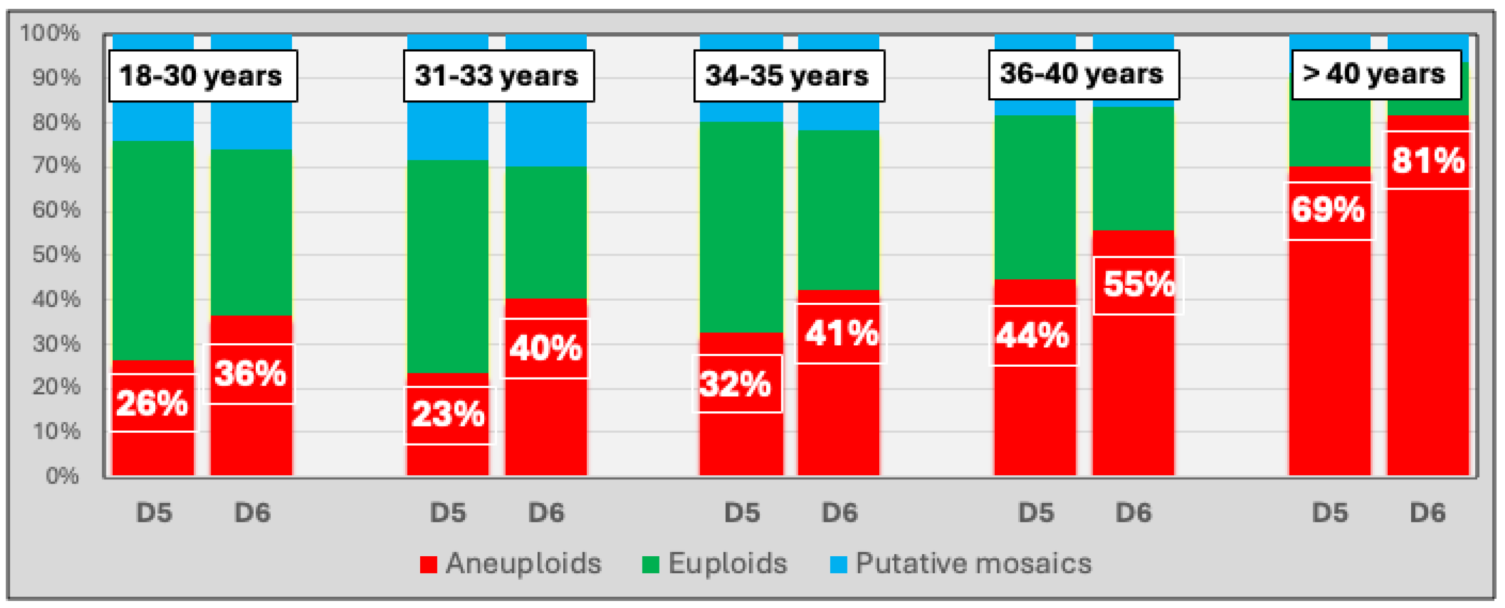
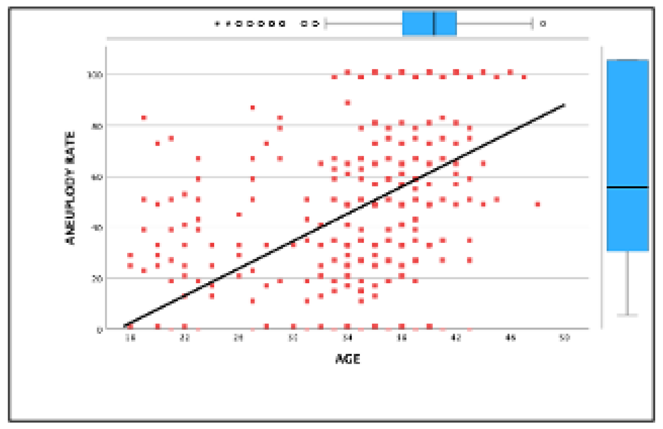
Background and Objectives: This study investigates the impact of maternal age and blastocyst development stage on aneuploidy rates. It evaluates the effectiveness of preimplantation genetic testing for aneuploidy (PGT-A) in improving clinical outcomes in in vitro fertilization (IVF). While PGT-A is often recommended for older patients, this study highlights its value across all maternal age groups in optimizing embryo selection. Methods: A retrospective observational study was conducted, analyzing 691 IVF cycles with PGT-A and 2,577 biopsied blastocysts between January 2019 and December 2023 at a single reproductive center. Patients were stratified into five age groups (<30, 31–33, 34–35, 36–40, >40 years), and blastocyst biopsies were performed on days 5 or 6 for genetic testing. Primary outcomes included euploidy and aneuploidy rates, while secondary outcomes assessed embryo availability and pregnancy complications. Results: The overall euploidy rate was 34.5%, declining with age from 43.6% (<30 years) to 15.9% (>40 years), while aneuploidy rates peaked at 75.43% (>40 years). Blastocysts biopsied on day 5 showed higher euploidy rates than on day 6 (40.16% vs. 27.92%, p<0.001). PGT-A cycles demonstrated superior ongoing pregnancy rates compared to cycles without genetic testing, with the most significant benefit observed in patients aged 36–40 (OR: 2.16, 95% CI: 1.07–4.35). However, all age groups benefited from PGT-A in reducing failed transfers due to non-viable embryos. Conclusions: This study underscores the universal utility of PGT-A in IVF, demonstrating its effectiveness in enhancing clinical outcomes and embryo selection, not only among older patients but across all maternal age groups. These findings highlight PGT-A as a valuable tool for optimizing IVF success regardless of patient age.

Keywords: 
;  ;  ;  ;  ;  

1. Introduction

A critical challenge in reproductive medicine is the development and implementation of reliable tools for embryonic selection that can predict successful implantation while excluding embryos with limited developmental potential. Embryo aneuploidy, a condition characterized by an abnormal number of chromosomes, is one of the primary causes of reproductive failure and implantation failure in assisted reproductive technologies (ART), especially among women of advanced maternal age [1,2].
Preimplantation genetic testing for aneuploidy (PGT-A) has emerged as a valuable tool for identifying chromosomally normal embryos during in vitro fertilization (IVF) cycles [3]. This technique involves biopsying a few cells from a blastocyst on day 5 or 6 of development, followed by genetic analysis to determine the chromosomal composition. By identifying and excluding aneuploid embryos, PGT-A improves implantation rates, reduces miscarriage rates, and enhances live birth rates [4,5]. Furthermore, the use of PGT-A facilitates elective single embryo transfer, minimizing the risks associated with multiple pregnancies while maintaining high success rates [6,7,8].
Despite these benefits, the universal application of PGT-A remains contentious. Critics argue that the procedure may increase treatment costs and require significant resources and expertise. Additionally, the biopsy process poses risks to the embryo, with potential implications for survival and implantation success [9]. Some studies have also questioned the utility of PGT-A across all patient groups, noting a lack of clear benefits in younger patients or those with good ovarian reserve [10,11].
This study aims to address these controversies by evaluating the impact of maternal age and blastocyst development stage on aneuploidy rates and assessing the effectiveness of PGT-A in improving clinical outcomes. The findings provide important insights into optimizing ART practices and the role of PGT-A in enhancing reproductive success.

2. Materials and Methods

2.1. Patients

This retrospective study analyzed 691 cycles of preimplantation genetic testing for aneuploidies (PGT-A) conducted at our reproductive center between January 2019 and December 2023. A total of 2,577 blastocysts were evaluated. Patients were recommended to undergo intracytoplasmic sperm injection (ICSI) and PGT-A after receiving comprehensive counseling regarding the procedure, including its potential benefits, risks of misdiagnosis, and reported success rates. In patients over the age of 35, the acceptance rate for preimplantation genetic testing for aneuploidy (PGT-A) exceeded 95%. Conversely, in patients under the age of 35, PGT-A was utilized in 74% of the cycles.
All patients underwent diagnostic evaluations and met predefined inclusion and exclusion criteria. Only couples with normal karyotypes were included in the study. Informed consent was obtained from all participants for using their data in this research. The study was approved by our center's Institutional Review Board (IRB) and adhered to the ethical principles of the Declaration of Helsinki.
Patients were stratified into five age cohorts for analysis: <30 years, 31–33 years, 34–35 years, 36–40 years, and >40 years. The mean number of embryos produced and the euploidy rate—defined as the proportion of euploid embryos relative to the total number of tested embryos—were calculated for each patient.

2.2. Ovarian Stimulation and Embryo Procedures

All participants underwent ovarian stimulation using a short GnRH antagonist protocol as previously described [12]. On day 2 of the menstrual cycle, a single injection of Corifollitropin alpha (Elonva®) was administered, followed by recombinant FSH (Bemfola, 300 IU/day). GnRH antagonist (Ganirelix, 250 µg/day) was initiated when the lead follicle reached 14 mm. Ovulation was triggered using a GnRH agonist (Decapeptyl, 0.2 mg) once at least one follicle exceeded 18 mm in size.
Oocyte retrieval was performed 36–38 hours post-trigger using ultrasound-guided transvaginal aspiration with a double-lumen needle (Cook Medical Incorporated) and vacuum system set at 100 mmHg.

2.3. Laboratory Protocols

Cumulus cells were removed from oocytes 4 hours post-retrieval, and ICSI was performed 40–42 hours after ovulation trigger. Oocytes were cultured in a time-lapse incubator (EmbryoScope+; Vitrolife, Sweden) under controlled conditions (37°C, 5% O₂, 6% CO₂). Blastocyst biopsies were performed on days 5–7. Only expanded blastocysts with distinct inner cell masses and trophectoderm (TE) were biopsied and subsequently vitrified within one hour.

2.4. Blastocyst Grading and Biopsy Procedure

Blastocyst quality was assessed by two embryologists following ASEBIR guidelines [13]. Faster-growing embryos (biopsied on day 5) were graded separately from slower-growing embryos (biopsied on days 6–7). Blastocysts rated A, B, or C were selected for biopsy, while those graded C− were excluded.
The biopsy procedure was conducted under aseptic conditions on a heated stage using a Nikon Eclipse microscope with micromanipulation tools. A Laser RI Saturn 5™ system was used to create an opening in the zona pellucida, allowing the biopsy of 4–6 cells from the TE. Cells were transferred into PCR tubes for genetic analysis.

2.5. Frozen-Thawed Embryo Transfer Protocol

During the study period, 839 embryo transfers were performed, including both euploid and mosaic embryos identified via PGT-A. In 246 cases, patients opted for the transfer of non-biopsied embryos. All transfers involved single embryo transfer (SET), guided by morphological scoring and PGT-A results.
Selected blastocysts were thawed following Irvine Scientific's protocol, cultured until transfer, and loaded into a Sureview Wallace transfer catheter for ultrasound-guided uterine placement [14].

2.6. Endometrial Preparation

Endometrial preparation began on cycle day 2 with oral estradiol valerate (Progyluton, Bayer Hispania, Barcelona, Spain) at 4 mg/day. After 8–10 days, endometrial thickness and serum progesterone levels were assessed. If the lining exceeded 6 mm and progesterone was below 1.6 ng/mL, oral micronized progesterone (Utrogestan, SEID, Barcelona, Spain) was initiated at 600 mg/day six days before embryo transfer.

2.7. Embryo Transfer and Pregnancy Outcomes

Patients received vaginal micronized progesterone (800 mg/day, Utrogestan) following embryo transfer. For positive β-hCG tests (serum hCG >25 IU/L on day 10 post-transfer), hormonal support continued until the 10th gestational week. Clinical pregnancy was confirmed by ultrasound at six weeks, and patients were monitored through delivery.
The implantation rate was calculated as the proportion of embryos exhibiting cardiac activity per transfer. Ongoing pregnancy was defined as reaching at least 20 weeks of gestation, and all pregnant patients were followed through delivery.

2.8. Statistical Analysis

Statistical analyses were performed using IBM SPSS Statistics Version 29.0.0.0. Pearson's correlation test was applied to evaluate relationships, with correlation coefficients (r) and p-values reported, considering statistical significance at P < 0.05. Regression analysis was used to explore variable associations, and chi-square tests (P < 0.05) were employed to compare percentage differences across age groups.

3. Results

The average age of women undergoing ovarian stimulation was 35.16 ± 0.24 years (mean ± standard error of the mean). The average number of oocytes retrieved was 11.94 ± 0.24, with 10.38 ± 0.21 MII oocytes and 7.11 ± 0.15 fertilized oocytes. The average number of blastocysts analyzed per cycle was 3.73 ± 0.10. The number and percentage of embryos diagnosed as euploid, aneuploid, and potential mosaics were analyzed based on the patient’s age (Table 1) and the morphokinetic characteristics of the biopsied blastocysts (Table 2). A total of 52 cases (2,02% of the biopsied blastocysts) yielded a nonconcurrent result.
Patients were grouped by age to compare the percentage of aneuploid embryos across different age groups (Figure 1). As expected, age and aneuploidy rate had a significant positive correlation (Figure 2).
Patients with a greater number of biopsied blastocysts demonstrated an increased probability of having at least one viable euploid or mosaic embryo for transfer. For instance, a biopsy of a single blastocyst yielded a transferable embryo in only 36.36% of cases. This percentage rose with an increasing count of biopsied blastocysts, reaching nearly 100% for patients with five or more. These findings highlight the benefit of multiple blastocysts to enhance the chances of successful embryo transfer. Despite fewer blastocysts, eliminating nonviable (aneuploid) ones remains cost-effective (Table 3).
As described in the materials and methods, the embryo transfers performed during the study period were assessed. Table 4 presents the outcomes regarding positive pregnancy tests, clinical pregnancies, and ongoing pregnancies, according to the number of transfers. It also reports miscarriage and ectopic pregnancy rates based on the number of diagnosed clinical pregnancies. The table is subdivided into transfers of exclusively euploid embryos (Table 4a), transfers of embryos following PGT-A (including embryos labeled as euploid and possible mosaics) (Table 4b), and transfers of non-analyzed embryos (based on informed patient decisions) (Table 4c).
Table 5 shows the differences in pregnancy rates per transfer, stratified by age and PGT-a application and expressed as Odds Ratio and percentage improvement.

4. Discussion

The indication for PGT-A as a method of embryo selection has become widespread to increase pregnancy rates. However, its applicability in specific patient subgroups remains subject to debate [16,915]
Indeed, with the advent of technologies capable of identifying the embryo with the highest potential for implantation and development—including the exclusive transfer of blastocysts, surpassing the outdated policy of transferring embryos at earlier developmental stages—the use of artificial intelligence to select the blastocyst with the greatest potential and PGT-A to eliminate non-viable embryos makes the practice of transferring multiple embryos in an assisted reproduction procedure an approach that should be considered obsolete [17].
This study assessed the association between embryo development and chromosomal outcomes analyzed through PGT-A, encompassing women across all age groups.
This study demonstrated a significant association, previously reported [18], between age and embryonic aneuploidy rates. Similarly, there was a relationship between blastocyst expansion on day five versus day six and the percentage of aneuploid embryos. These data indicate that blastocysts' development and morphological characteristics, such as their expansion, can provide valuable insights into their chromosomal status, as stated by Santamonkunrot et al. [2]. Similarly, such an association between blastocyst development would explain the traditionally better outcomes observed after day five blastocyst transfers than day 6 [19].
Increased embryonic selection may result in a higher likelihood of lacking embryos available for transfer. Therefore, it is reasonable to infer that while such selective practices do not necessarily elevate the cumulative pregnancy rates per woman, they are likely to improve the pregnancy rates per embryo transfer.
Nonetheless, PGT-A has certain limitations. Several investigators have compared offspring from embryo transfers using IVF/ICSI with PGT-A to offspring from IVF/ICSI without PGT-A. While patients over the age of 37 who have euploid embryos for transfer may benefit from PGT-A, no benefit has been demonstrated when all age groups were assessed [1]. Desmyttere et al. [20] and He et al. [21] found no difference in adverse neonatal outcomes after the evaluation of 995 and 1,721 neonates, respectively. A retrospective study cautioned against the routine use of PGT-A in fresh in vitro fertilisation (IVF) cycles until its safety and effectiveness are established [10].
Indeed, there exist randomized controlled trials in which the application of preimplantation genetic testing for aneuploidies (PGT-A) has not demonstrated the anticipated enhancement in pregnancy rates in certain age groups. In a multicenter randomized control trial, Munne et al. [22] showed that PGT-A did not improve overall pregnancy outcomes in all women, as analyzed per embryo transfer or ITT. However, they found a significant increase in OPR per embryo transfer with the use of PGT-A in the subgroup of women aged 35–40 years who had two or more embryos that could be biopsied, but this was not significant when analyzed by ITT [22].
Preimplantation Genetic Testing for Aneuploidy (PGT-A) is unable to enhance pregnancy outcomes in a randomized controlled trial per intention-to-treat (ITT) patient, as the biopsy procedure does not enhance embryo quality. This technique fundamentally represents a selective process that seeks to maximize the implantation rate in a cohort of mixed euploid and aneuploid blastocysts [5]. Therefore, any expectation should be confined to a potential elevation in pregnancy rates per embryo transfer by reducing the number of embryos eligible for transfer.
It has been suggested that many embryos labeled aneuploid may possess significant reproductive potential [11,23], and therefore, the use of PGT-A may harm patients via the discard of reproductively competent embryos. As evidenced by Tiegs et al. in their landmark non-selection study, aneuploid embryos are incompatible with a viable pregnancy [23]. Therefore, the availability of a tool capable of identifying such embryos significantly enhances the potential for a successful pregnancy outcome following embryo transfer. Thus, PGT-A facilitates the exclusion of non-viable embryos; however, additional methodologies are required to maximize the likelihood of achieving a pregnancy with the fewest possible ET attempts.
The focus is not on selecting the "optimal" embryo but instead on eliminating the "non-viable" embryo. The lack of observed improvement, even per embryo transfer, in clinical outcomes among younger patients has been attributed to various potential factors.
The relatively low proportion of aneuploid embryos at younger ages, typically deemed unsuitable for transfer, and classifying patients into very restricted age groups may mask the potential benefits of embryo selection.
Additionally, the potential detrimental impacts of trophectoderm biopsy and subsequent embryo vitrification are considered significant contributing factors.
It is paramount to stress that the successful routine implementation of PGT-A relies on the availability of high-quality equipment and the professional training of the involved embryologists. Therefore, any potential beneficial effect of a procedure like PGT-A should be evaluated based on laboratory results with the best outcomes. Suboptimal procedural execution in a laboratory will not improve previous benchmarks.
The problem in the past has been that the process of PGT-A (trophectoderm biopsy and subsequent DNA amplification and analysis) could have been more efficient, with high loss rates of potential embryo implantations [5,11]. Therefore, the outcomes of multicenter randomized trials may be adversely affected by data from centers with inferior results, potentially diluting any beneficial effects of the procedure. As stated by the authors of the RCT mentioned above [22], substantial heterogeneity was observed among the various laboratories, characterized by divergent rates of aneuploidies across the different groups. For example, the percentage of euploid embryos after PGT-A ranged from 38% to 100% in younger patients and 17% to 75% in older patients, and the combined OPRs per transfer ranged from 30% to 60%. This variability undermines the generalizability of the overall results of the RCT.
Furthermore, the authors acknowledged two additional potential biases in the study: a selection bias favoring patients with a good prognosis (at least two blastocysts available for transfer) and the previously mentioned age factor. Although women aged 25 to 40 years were included, the mean age of the patients in both the study group and the control group was 33,7 and 33,8 years, respectively, with more than half of the patients aged <35 years, which is well below the usual age of women undergoing ART in most clinics. Additionally, patients over 40 years of age with previous implantation failures or recurrent miscarriages, who are traditionally seen as potential beneficiaries of PGT, were not included in the RCT.
Any selection method, regardless of its nature, has to be associated with a reduced availability of embryos for transfer. One of the problems argued against the widespread use of PGT-A in assisted reproduction is the lower availability of embryos for transfer. In this setting, it has been argued that among women of advanced age, the chances of getting blastocysts are lower as their ovarian reserve decreases [18].
In fact, the development process of the blastocyst stage itself means that a percentage of embryos will not be able to reach this stage. As described in the present study, as the number of blastocysts biopsied per cycle decreases, the possibility that there will be no transferable embryos increases. As an example, Deng et al. [24] reported that in patients with poor ovarian response, PGT-A cycles had less chance to reach embryo transfer compared with those not using PGT-A (13.7 vs. 70.6%, p < 0.001), and no difference was observed in the LBR per oocyte retrieval in cycles using or not using PGT-A (6.6 vs. 5.4%, p = 0.814). However, considering the high predictive value of an aneuploidy diagnosis [23], the reduction in the number of blastocysts available for transfer would occur at the expense of embryos without developmental capacity. Therefore, the number of unsuccessful transfers would be reduced.
Additionally, a selection method cannot enhance the intrinsic quality of the embryos. Therefore, it is reasonable to expect that cumulative pregnancy rates per patient or ovarian stimulation cycle remain similar. The key lies in determining whether increased embryo selection correlates with higher implantation and ongoing pregnancy rates per embryo transfer. Reducing the number of embryos to be transferred should be manageable if the embryos that are not transferred lack developmental potential. In this setting, the number of transfers required to achieve an ongoing pregnancy is reduced, thereby decreasing the time to pregnancy.
Moreover, the time to achieve pregnancy (as fewer transfers would be necessary) or to make decisions in cases where there are no transferable embryos would be shortened. Additionally, the availability of a selection method with high predictive value would eliminate the need or recommendation to transfer more than one embryo per transfer, thus avoiding multiple pregnancies [6,7,17]. Embryonic euploidy is a necessary prerequisite for establishing a viable pregnancy, but it is not sufficient on its own. While an aneuploid embryo will never lead to a viable pregnancy, the presence of a euploid embryo does not guarantee one.

Limitations of the Study#

The retrospective design inherently presents certain limitations in interpreting conclusions. Successive cycles from the same patients were included, meaning the analysis was not restricted to the first cycles per patient. The causes of infertility were not included in the analysis of the results, which may represent a limitation of the study.

Strengths of the Study:

The large number of blastocysts evaluated, including all age groups, provides robustness to the findings. In addition, the exclusive use of single embryo transfers, a unique and effective methodology, and the comprehensive follow-up of all cases through to birth constitute additional strengths.
Declaration of generative AI and AI-assisted technologies in the writing process.
During the preparation of this work the author(s) used Chat GPT 4.0 during the writing process in order to improve the readability and language of the manuscript. After using this tool, the authors reviewed and edited the content as needed and take full responsibility for the content of the published article.

5. Conclusions

Although PGT-A has been traditionally recommended for older patients, this study shows its value across all age groups, improving embryo selection and clinical outcomes by reducing failed transfers due to aneuploid embryos. Regardless of maternal age, PGT-A is an essential tool for optimizing IVF success.

Author Contributions

G.B. and E.M. conceived the study idea (Conceptualization). G.B., R.C., S.S. and J.B. participated in the design of the trial and in the recruitment of participants, and in the assessment of clinical outcomes (Investigation, Project administration). E.M., M.DLH., O.G., and O.A. were involved in the critical revision of the manuscript (Review & editing). G.B. coordinated the data collection, performed the analysis, and wrote the first draft of the manuscript (Formal analysis, Methodology, Writing, Review and editing).

Funding

“This research received no external funding”.

Attestation statement

  • The authors hereby certify that none of the subjects included in this trial were concurrently participating in any other clinical trials.
  • Additionally, data about any subjects involved in this study have not been previously published.
  • Finally, I confirm that all relevant data will be made available to the journal's editors for review or inquiry upon request.

Institutional Review Board Statement

The study was approved by our center's Institutional Review Board (IRB) and adhered to the ethical principles of the Declaration of Helsinki.

Informed Consent Statement

Informed consent was obtained from all participants involved in the study.

Abbreviations

The following abbreviations are used in this manuscript:
PGT-A Preimplantation genetic testing for aneuploidies
ET Embryo transfer

References

  1. Kang, H.J.; Melnick, A.P.; Stewart, J.D.; Xu, K.; Rosenwaks, Z. Preimplantationgenetic screening: Who benefits? Fertil Steril 2016, 106, 597–602. [Google Scholar] [CrossRef] [PubMed]
  2. Santamonkunrot, P.; Samutchinda, S.; Niransuk, P.; Satirapod, C.; Sukprasert, M. The Association between Embryo Development and Chromosomal Results from PGT-A in Women of Advanced Age: A Prospective Cohort Study. J. Clin. Med. 2024, 13, 626. [Google Scholar] [CrossRef]
  3. Chen, N.Q.; Si, C.R.; Yung, S.C.; Hon, S.K.; Arasoo, J.; Ng, S.-C. Analysis of a preimplantation genetic test for aneuploidies in 893 screened blastocysts using KaryoLite BoBs: a single-centre experience. Singap. Med J. 2024. [Google Scholar] [CrossRef]
  4. Viotti, M. Preimplantation Genetic Testing for Chromosomal Abnormalities: Aneuploidy, Mosaicism, and Structural Rearrangements. Genes 2020, 11, 602. [Google Scholar] [CrossRef]
  5. Paulson, R.J. Is preimplantation genetic testing for aneuploidy (PGT-A) getting better? How can we know and how do we counsel our patients? Fertil Steril Rep. 2023, 4, 329–330. [Google Scholar] [CrossRef] [PubMed]
  6. Forman, E.J.; Tao, X.; Ferry, K.M.; Taylor, D.; Treff, N.R.; Scott, R.T. Single embryo transfer with comprehensive chromosome screening results in improved ongoing pregnancy rates and decreased miscarriage rates. Hum. Reprod. 2012, 27, 1217–1222. [Google Scholar] [CrossRef] [PubMed]
  7. Forman, E.J.; Hong, K.H.; Franasiak, J.M.; Scott, R.T. Obstetrical and neonatal outcomes from the BEST Trial: single embryo transfer with aneuploidy screening improves outcomes after in vitro fertilization without compromising delivery rates. Am. J. Obstet. Gynecol. 2014, 210, 157.e1–157.e6. [Google Scholar] [CrossRef]
  8. Davis, O.S.; Favetta, L.A.; Deniz, S.; Faghih, M.; Amin, S.; Karnis, M.; Neal, M.S. Potential Costs and Benefits of Incorporating PGT-A Across Age Groups: A Canadian Clinic Perspective. J. Obstet. Gynaecol. Can. 2024, 46, 102361. [Google Scholar] [CrossRef]
  9. Bellver, J.; Bosch, E.; Espinós, J.J.; Fabregues, F.; Fontes, J.; García-Velasco, J.; Llácer, J.; Requena, A.; Checa, M.A. Second-generation preimplantation genetic testing for aneuploidy in assisted reproduction: a SWOT analysis. Reprod. Biomed. Online 2019, 39, 905–915. [Google Scholar] [CrossRef]
  10. Kushnir, V.A.; Darmon, S.K.; Albertini, D.F.; Barad, D.H.; Gleicher, N. Effectiveness of in vitro fertilization with preimplantation genetic screening: a reanalysis of United States assisted reproductive technology data 2011–2012. Fertil. Steril. 2016, 106, 75–79. [Google Scholar] [CrossRef]
  11. Paulson, R.J. Outcome of in vitro fertilization cycles with preimplantation genetic testing for aneuploidies: let’s be honest with one another. Fertil. Steril. 2019, 112, 1013–1014. [Google Scholar] [CrossRef] [PubMed]
  12. Barrenetxea, G.; Celis, R.; Barrenetxea, J.; Martínez, E.; Heras, M.D.L.; Gómez, O.; Aguirre, O. Intraovarian platelet-rich plasma injection and IVF outcomes in patients with poor ovarian response: a double-blind randomized controlled trial. Hum. Reprod. 2024, 39, 760–769. [Google Scholar] [CrossRef]
  13. Saiz, I.C.; Gatell, M.C.P.; Vargas, M.C.; Mendive, A.D.; Enedáguila, N.R.; Solanes, M.M.; Canal, B.C.; López, J.T.; Bonet, A.B.; Acosta, M.V.H.d.M. The Embryology Interest Group: updating ASEBIR's morphological scoring system for early embryos, morulae and blastocysts. Med Reprod Embriol Clín 2018, 5, 42–54. [Google Scholar] [CrossRef]
  14. Barrenetxea, G.; Romero, I.; Celis, R.; Abio, A.; Bilbao, M. Correlation between plasmatic progesterone, endometrial receptivity genetic assay and implantation rates in frozen-thawed transferred euploid embryos. A multivariate analysis. Eur. J. Obstet. Gynecol. Reprod. Biol. 2021, 263, 192–197. [Google Scholar] [CrossRef] [PubMed]
  15. Nuñez-Calonge, R.; Santamaria, N.; Rubio, T.; Moreno, J.M. Making and Selecting the Best Embryo in In vitro Fertilization. Arch. Med Res. 2024, 55, 103068. [Google Scholar] [CrossRef]
  16. Fernandes, S.L.E.; de Carvalho, F.A.G. Preimplantation genetic testing: A narrative review. Porto Biomed. J. 2024, 9, 262. [Google Scholar] [CrossRef]
  17. Alteri, A.; Arroyo, G.; Baccino, G.; Craciunas, L.; De Geyter, C.; Ebner, T.; Koleva, M.; Kordic, K.; Mcheik, S.; et al. ESHRE guideline: number of embryos to transfer during IVF/ICSI. Hum. Reprod. 2024, 39, 647–657. [Google Scholar] [CrossRef]
  18. Mei, Y.; Lin, Y.; Chen, Y.; Zheng, J.; Ke, X.; Liang, X.; Wang, F. Preimplantation genetic testing for aneuploidy optimizes reproductive outcomes in recurrent reproductive failure: a systematic review. Front. Med. 2024, 11, 1233962. [Google Scholar] [CrossRef]
  19. Barrenetxea, G.; de Larruzea, A.L.; Ganzabal, T.; Jiménez, R.; Carbonero, K.; Mandiola, M. Blastocyst culture after repeated failure of cleavage-stage embryo transfers: A comparison of day 5 and day 6 transfers. Fertil Steril 2005, 83, 49–53. [Google Scholar] [CrossRef]
  20. Desmyttere, S.; De Rycke, M.; Staessen, C.; Liebaers, I.; De Schrijver, F.; Verpoest, W.; et al. Neonatal follow-up of 995 consecutively born children after embryo biopsy for, P.G.D. Hum Reprod 2012, 27, 288–293. [Google Scholar] [CrossRef]
  21. He, H.; Jing, S.; Lu, C.F.; Tan, Y.Q.; Luo, K.L.; Zhang, S.P.; et al. Neonatal outcomes of live births after blastocyst biopsy in preimplantation genetic testing cycles: afollow-up of 1,721 children. Fertil Steril 2019, 112, 82–88. [Google Scholar] [CrossRef] [PubMed]
  22. Munné, S.; Kaplan, B.; Frattarelli, J.L.; Child, T.; Nakhuda, G.; Shamma, F.N.; Silverberg, K.; Kalista, T.; Handyside, A.H.; Katz-Jaffe, M.; et al. Preimplantation genetic testing for aneuploidy versus morphology as selection criteria for single frozen-thawed embryo transfer in good-prognosis patients: a multicenter randomized clinical trial. Fertil. Steril. 2019, 112, 1071–1079.e7. [Google Scholar] [CrossRef] [PubMed]
  23. Tiegs, A.W.; Tao, X.; Zhan, Y.; Whitehead, C.; Kim, J.; Hanson, B.; Osman, E.; Kim, T.J.; Patounakis, G.; Gutmann, J.; et al. A multicenter, prospective, blinded, nonselection study evaluating the predictive value of an aneuploid diagnosis using a targeted next-generation sequencing–based preimplantation genetic testing for aneuploidy assay and impact of biopsy. Fertil. Steril. 2021, 115, 627–637. [Google Scholar] [CrossRef] [PubMed]
  24. Deng, J.; Hong, H.Y.; Zhao, Q.; Nadgauda, A.; Ashrafian, S.; Behr, B.; Lathi, R.B. Preimplantation genetic testing for aneuploidy in poor ovarian responders with four or fewer oocytes retrieved. J. Assist. Reprod. Genet. 2020, 37, 1147–1154. [Google Scholar] [CrossRef]
Figure 1. Percentage of aneuploidy based on age and day of blastocyst development.
Preprints 149827 g001
Figure 2. Aneuploidy rate by age (Pearson correlation: 0,419; p<0,001).
Preprints 149827 g002
Table 1. Comprehensive PGT-A Outcomes Stratified by Maternal Age Groups.
Age
(years)
Cycles Biopsied blastocysts
n (per cycle)
Euploids
n (%)
Aneuploids
n (%)
Mosaics1
n (%)
N.D.2
n (%)
18-30 75 445 (5,93) 194 (43,60) 132 (29,66) 109 (24,49) 10 (2,25)
31-33 42 199 (4,72) 80 (40,20) 58 (29,15) 56 (28,14) 5 (2,51)
34-35 142 585 (4,12) 242 (41,37) 211 (36,07) 118 (20,17) 14 (2,39)
36-40 288 1002 (3,48) 318 (31,74) 494 (49,30) 172 (17,17) 18 (1,80)
>40 144 346 (2,41 55 (15,90) 261 (75,43) 25 (7,23) 5 (1,45)
Total 691 2577 (3,73) 889 (34,50) 1156 (44,86) 480 (18,63) 52 (2,02)
1 Possible (putative) mosaics. 2 N.D: lack of diagnoses.
Table 2. Outcomes Correlated with Blastocyst Expansion Day during Embryo Culture.
Day Biopsied blastocysts Euploids
n (%)
Aneuploids
n (%)
Mosaics1
n (%)
N.D.2
n (%)
Day 5* 1382 555 (40,16) 533 (38,57) 264 (19,10) 28 (2,03)
Day 6* 1182 330 (27,92) 617 (52,20) 211 (17,85) 24 (2,03)
D7 13 4 (30,77) 6 (46,15) 5 (38,46) 0 (0)
Total 2577 889 (34,50) 1156 (44,86) 480 (18,63) 52 (2,02)
1 Possible (putative) mosaics. 2 N.D: lack of diagnoses. * Comparison of percentages between Day 5 and Day 6. Chi-square analysis: 54.58; P=0,0001.
Table 3. Transferable embryo availability based on the number of biopsied blastocysts.
Biopsied
blastocysts
Patients
n
Embryo availability for ET
N (%)
No Yes
1 110 70 (63,64) 40 (36,36)
2 162 59 (36,42) 103 (63,58)
3 119 32 (26,89) 87 (73,11)
4 99 9 (9,09) 90 (90,91)
5 66 2 (3,03) 64 (96,87)
6 42 0 (0,00) 42 (100)
7 29 1 (3,54) 28 (96,55)
8 20 0 (0,00) 20 (100)
9 20 0 (0,00) 20 (100)
10 10 0 (0,00) 10 (100)
11 7 0 (0,00) 7 (100)
12 4 0 (0,00) 4 (100)
13 2 0 (0,00) 2 (100)
14 1 0 (0,00) 1 (100)
Table 4a. Pregnancies stratified by age and PGT results (only euploid embryos included).
PGT-A (euploid embryos)
Age
(years)
ET PPT
n (%)1
Gestations
n (%)1
Ongoing gestations
n (%)1
Miscarriages
n (%)2
Ectopic pregnancies
n (%)2
18-30 21 14 (66,67) 12 (57,14) 10 (47,62) 2 (16,67) 0 (0)
31-33 99 62 (62,63) 58 (58,59) 47 (47,47) 10 (17,24) 2 (3,45)
34-35 128 70 (54,69) 57 (44,53) 49 (38,28) 8 (14,04) 4 (7,02)
36-40 385 210 (54,55) 194 (50,39) 148 (38,44) 43 (22,16) 3 (1,55)
>40 84 44 (52,38) 26 (30,95) 20 (23,81) 6 (23,08) 0 (0)
Total 717 400 (48,40) 347 (48,40) 274 (38,21) 69 (19,88) 9 82,59)
ET: embryo transfers. PPT: positive pregnancy test. Gestations: presence of gestational sac on ultrasound examination. Ongoing gestations: evolutive pregnancies overcoming 24 weekes. 1 Percentage calculated based on the number of transfers performed. 2 Percentage based on confirmed clinical pregnancies.
Table 4b. Pregnancies stratified by age and PGT results (euploid and mosaic embryos included).
PGT-A (transferable embryos)
Age
(years)
ET PPT
n (%)1
Gestations
n (%)1
Ongoing gestations
n (%)1
Miscarriages
n (%)2
Ectopic pregnancies
n (%)2
18-30 24 14 (58,33) 12 (50,00) 10 (41,67) 2 (16,67) 0 (0)
31-33 120 75 (62,50) 69 (57,50) 56 (46,67) 12 (17,39) 2 (2,90)
34-35 151 83 (54,97) 67(44,37) 57 (37,75) 10 (14,93) 5 (7,46)
36-40 454 244 (53,74) 225 (49,56) 169 (37,22) 49 (21,78) 4 (1,78)
>40 90 46 (31,11) 28 (31,11) 22 (24,44) 6 (22,43) 0 (0)
Total 839 462 (47,79) 401 (47,79) 314 (37,43) 79 (19,70) 11 (2,74)
ET: embryo transfers. PPT: positive pregnancy test. Gestations: presence of gestational sac on ultrasound examination. Ongoing gestations: evolutive pregnancies overcoming 24 weeks. 1 Percentage calculated based on the number of transfers performed. 2 Percentage based on confirmed clinical pregnancies.
Table 4c. Pregnancies stratified by age without PGT-A.
No PGT-A
Age
(years)
ET PPT
n (%)1
Gestations
n (%)1
Ongoing gestations
n (%)1
Miscarriages
n (%)2
Ectopic pregnancies
n (%)2
18-30 61 30 (49,18) 25 (40,98) 20 (32,79) 5 (20,00)) 0 (0)
31-33 89 44 (49,44) 39 (43,82) 28 (31,46) 11 (28,21) 0 (0)
34-35 39 19 (48,72) 17 (43,59) 10 (25,64) 7 (41,18) 0 (0)
36-40 49 19 (38,78) 17 (34,69) 11 (22,45) 6 (35,29) 0 (0)
>40 8 2 (25,00) 2 (25,00) 1 (12,50) 1 (50,00) 0 (0)
Total 246 114 (46,34) 100 (28,46) 100 (28,46) 30 (30,00) 0 (0)
ET: embryo transfers. PPT: positive pregnancy test. Gestations: presence of gestational sac on ultrasound examination. Ongoing gestations: evolutive pregnancies overcoming 24 weeks. 1 Percentage calculated based on the number of transfers performed. 2 Percentage based on confirmed clinical pregnancies.
Table 5. Increase in ongoing pregnancy rates per transfer (expressed as Odds Ratio and percentage improvement), stratified by age and the application of PGT-A.
Age
(years)
Euploid* vs. No PGT-A PGT-a** vs, No PGT-A
OR (95% CI) Δ % OR (95% CI) Δ %
18-30 1,86 (0,68-5,11) +45,23% 1,46 (0,55-3,87) +27,08%
31-33 1,97 (1,08-3,57) +50,89% 1,91 (1,07-3,38) +48,35%
34-35 1,80 (0,81-4,01) +49,30% 1,72 (0,78-3,80) +45,28%
36-40 2,16 (1,07-4,35 +71,22% 2,05 (1,02-4,11) +65,79%
>40 2,19 (0,25-18,87) +90,48% 2,26 (0,26-19,4 +95,52%
Total 1,55 (1,13-2,13) +34,26% 1,50 (1,10-2,05) +31,52%
* Putative mosaics excluded. ** Putative mosaics included
Disclaimer/Publisher’s Note: The statements, opinions and data contained in all publications are solely those of the individual author(s) and contributor(s) and not of MDPI and/or the editor(s). MDPI and/or the editor(s) disclaim responsibility for any injury to people or property resulting from any ideas, methods, instructions or products referred to in the content.
Copyright: This open access article is published under a Creative Commons CC BY 4.0 license, which permit the free download, distribution, and reuse, provided that the author and preprint are cited in any reuse.
Prerpints.org logo

Preprints.org is a free preprint server supported by MDPI in Basel, Switzerland.

Subscribe

Disclaimer

Terms of Use

Privacy Policy

Privacy Settings

© 2025 MDPI (Basel, Switzerland) unless otherwise stated