Submitted:
13 February 2025
Posted:
14 February 2025
Read the latest preprint version here
Abstract
Dearth of knowledge exists on identification of the begomoviruses and distribution of cassava mosaic viruses across key cassava growing regions of Sierra Leone. The objective of the study was to identify and map the distribution of cassava mosaic disease (CMD)-associated viruses in farmers' fields of Sierra Leone. Cassava leaf samples were collected in 109 smallholder farms during a geo-referenced survey conducted from 10th May to 5th June, 2024. Molecular diagnostics were carried out to identify the viral strains associated with CMD. Findings revealed that infection by stem cutting was more predominant in the south, east, north and northwest regions than the west region; whereas infection by whitefly was predominant in the west, north and north-west regions. The PCR screening of 426 samples coupled with sequence analysis revealed the presence of African cassava mosaic-like (ACMV-like) viruses, and East African cassava mosaic-like (EACMV-like) viruses as single infections at 78.1% and 1.3%, respectively. Co-infections of ACMV-like and EACMV-like viruses were detected in 20.6% of the tested samples. In addition, 70.6% of the samples positive for EACMV-like virus (single and mixed infections) were found to be positive for East African cassava mosaic Cameroon virus (EACMCMV). The ACMV and co-infection of ACMV and EACMV viruses were present in all regions, while EACMCV was detected in all regions except the western area. The results indicate that EACMCMV variant occurrence may be more prevalent in Sierra Leone than previously thought. This study suggests use of good agricultural practices and participatory surveillance as a strategy for managing CMD in Sierra Leone.
Keywords:
Epidemiology
; Viruses
; East African Cassava Mosaic (EACMV)
; African Cassava Mosaic Virus (ACMV)
; Manihot esculenta
1. Introduction
Cassava (Manihot esculenta Crantz) is traditionally cultivated using the stem cutting technique. Cassava propagation through the stem cuttings is one of the bottlenecks for production of disease-free planting materials [1]. Cassava botanical seeds are normally dormant and germinate very slowly; consequently, farmers use the conventional stem cutting propagation technique. This method leads to viral, fungal and bacterial diseases which may contribute to decreasing cassava production and productivity as well as loss of superior genotypes [2]. Moreover, diseases and pests affect the growth and development of cassava, consequently contributing to reduction in economic yields in several production zones in Africa [3,4].
Cassava mosaic disease (CMD) is one of the key biotic constraints of cassava production in the 21st century [5,6]. The disease causes significant cassava fresh storage root yield loss in Africa [7]. In West Africa, high CMD infection can reduce cassava storage root yield by up to 90% if control intervention is not implemented [7]. The CMD is spread by whitefly vectors (Bemisia tabaci) and infected cuttings [8,9,10]. The disease is widespread in several countries in East, Central, and West Africa and towards Southern Africa [11,12,13].
Cassava mosaic disease is caused by a Cassava Mosaic Begomoviruses (CMBs) complex composed of eleven species of bipartite Begomoviruses, of which, nine have been reported in Africa [14,15]. The distribution and spread of these begomoviruses often differ from one country to another and/or from one region to another within the same country [16,17,18,19,20]. Cassava Begomovirus can infect both the primary and secondary host, cassava, and other weed species that grows around cassava fields, known as alternate hosts. This makes managing the virus and eradicating it extremely challenging.
Several epidemiological studies conducted in Sierra Leone noted the presence of CMD in the majority of cassava-growing areas [7,21,22,23]. These studies have focused on screening for resistance in different agroecosystems, incidence and severity of CMD, as well as evaluation of the impact of the disease on cassava genotypes in the country. Despite these various studies carried out in Sierra Leone, the identification and/or characterization of the begomoviruses and distribution of cassava mosaic viruses remains less documented in several provinces. The objective of the present study was to identify and map the distribution of CMD-associated viruses in farmers' fields of Sierra Leone.
2. Results
2.1. Phenotypic Detection and Distribution of Cassava Mosaic Disease
Generally, mean incidence of CMD, mean severity of CMD, percent of plants infected by cuttings or whiteflies, and number of whiteflies per plant significantly (p<0.05) varied among the regions and districts of Sierra Leone (Figure 3 and Figure 4). The CMD symptoms were found in all the 109 cassava fields surveyed with the highest proportions of incidence and severity recorded in the west region, while the lowest values were recorded in the south region (Figure 3a and b). Percent of infected plants through stem cutting was more predominant in the south, east, north and north-west regions than in the west region where the whitefly mode of infection was higher than the stem cutting (Figure 3c). The highest number of whiteflies per plant was found in the west region with the lowest proportion recorded in the south region (Figure 3d)
At the district level, the highest CMD incidence scores of almost 100% were recorded in Bombali, western rural and western urban districts, with the lowest of 70% CMD incidence found in Bonthe district. The western rural and western urban districts exhibited highest severity values of 4.8 (high infection) with the lowest of 3.2 (low infection of the disease) recorded in Bonthe district (Figure 4a and b). Percent of infected plants through stem cutting was more predominant in all the districts except western rural and western urban districts where the whitefly mode of infection was higher than the stem cutting (Figure 4c). The highest number of whiteflies per plant was found in the western urban district, followed by western rural district with the lowest of 2 whitefly per plant recorded in Bonthe district (Figure 4d)
At the district level, the highest CMD incidence scores of almost 100% were recorded in Bombali, western rural and western urban districts, with the lowest of 70% CMD incidence found in Bonthe district. The western rural and western urban districts exhibited highest severity values of 4.8 (high infection) with the lowest of 3.2 (low infection of the disease) recorded in Bonthe district (Figure 4a and b). Percent of infected plants through stem cutting was more predominant in all the districts except western rural and western urban districts where the whitefly mode of infection was higher than the stem cutting (Figure 4c). The highest number of whiteflies per plant was found in the western urban district, followed by western rural district with the lowest of 2 whiteflies per plant recorded in Bonthe district (Figure 4d).
The distribution of CMD infection in cassava fields of Sierra Leone are also shown in distribution maps (Figure 5). The results reveal that the incidence of CMD ranged from 25-50% in the south region (Moyamba, Bonthe and Bo districts) and some part of Bombali district, followed by some farms with 51-75% incidence of CMD found in the north and north-west regions. The east, north, north-west and west regions recorded the highest incidence of CMD ranging from 76-100%. The south region recorded the lowest severity scores of 2.0-3.0 (mild) and 3.1-4.0 (severe) infection of the disease. Kailahun and Kono districts in the east region had severity scores of 3.1-4.0 and 4.1-5.0, respectively. Cassava farms in Falaba and Bombali districts in the north region recorded severity scores of 3.1-4.0 and 4.1-5.0, respectively. Karene in the north-west region, and the western rural and western urban districts in the west region had the highest severity damage ranging from 4.1-5.0.
2.2. Molecular Detection and Distribution of Cassava Begomoviruses in Sierra Leone
A total of 426 cassava leaf samples were collected from 320 plants with symptoms and 106 plants without symptoms in 2024 for PCR analysis. Among the samples having observable symptoms, 5.6% (28/320) tested negative for ACMV-like virus and EACMV-like virus. On the other hand, 12.3% (13/106) and 5.7% (6/106) of symptomless samples tested positive for ACMV-like virus and mixed infection of ACMV-like virus and EACMV-like virus, respectively. Of the 426 samples, 311 (73.0%) were found positive with 115 (27.0%) recorded as negative (Table 2 and Table 3). Among the positive samples studied, the single ACMV-like virus infection was the most frequent, accounting for 78.1% (243/311) of all CMD begomovirus infection, followed by mixed infections of ACMV-like virus and EACMV-like virus with 20.6% (64/311), and single infection of EACMV-like virus with 1.3% (4/311).
The typical gel electrophoresis photo of ACMV begomoviruses detected in the study is presented in Figure 6.
The single infection of ACMV-like virus was predominant in all surveyed regions, with the highest proportion (93.1%, 27/29) in the western area, followed by the north region (87.7%, 71/81), whereas the south had the lowest of 64.4% (47/73). The mixed infection occurred in all the regions, with the highest proportions in south (35.6%, 26/73) and the lowest of 6.9% (2/29) was detected in the western area. The single infection of EACMV-like virus was found in the northwest (5.0%, 3/60), and east (13.2%, 1/68) regions, whereas single infection of EACMV-like virus was not detected in the regions of south, north and western area (Table 3). Of the 68 EACMV-like virus positive samples (single and mixed infections), 70.6% (48/68) tested positive for EACMVCM variant using the primer pair VNF031/VNF032.
Figure 7 and Figure 8 show the distribution of cassava mosaic begomoviruses detected in smallholder cassava farmers’ fields of Sierra Leone. The ACMV single infection is widely spread across the districts of Sierra Leone represented by green geo-reference points, followed by ACMV/EACMV mix infection (with red geo-reference points) which is widely spread particularly in the south and north regions. The EACMV Cameroun virus infection, illustrated by a grey color triangular shape, is widely spread in the northwest region and some parts in the south and east regions, while the EACMV single infection is only found in the east and the northwest regions with geo-reference points in yellow.
The distribution map of the CMBs (ACMV, EACMV, EACMCMV and EACMV-Ug) detected in this study is shown in Figure 8. Results revealed that, out of the 426 samples screened, five were infected by EACMV-Ug. These EACMV-Ug infected samples were from fields in Moyamba, Port Loke, Falaba and Kono districts. These districts share boarder with Guinea except the Moyamba district.
2.3. Confirmation of CMGs Identity by Sequencing
A search for related sequences in the GenBank database (NCBI, BLASTN) revealed the existence and spread of Ug-variant in four districts with red like star geo-reference points (Figure 9). The sequences of the 40 samples that tested positive for the ACMV-like virus were most closely related to ACMV, EACMV_Ug, EACMV_Cameroon variant, EACMV_Nigeria variant, and EACMV_Ghana variant. The EACMV Cameroon virus widely spread across the country with geo-reference points in yellow color distributed mostly in the south and north regions of Sierra Leone, while the blast results showed few EACMV Nigeria, Ghana and Kenyan variants.
3. Discussion
The detection and confirmations of EACMV-Ug and its spread in four different districts in Sierra Leone, creates serious threat to cassava production in the country. This virus was first reported in 2023 in Forécariah, Guinea, a town located near the border of Sierra Leone and in Kambia district, north region Sierra Leone [24]. Kambia is located at about 11 km from the Guinean border, whereas Forécariah is situated at 34.8 km from the Sierra Leone border. Given the high traffic and trade existing between Forécariah and Sierra Leone, it is probable that EACMV-Ug was introduced into the country from Forécariah through exchange of cassava planting materials. The EACMV-Ug virus was found in single and mixed infections with ACMV. Co-infection of both viruses could result in a synergistic interaction with severe impact on cassava fields leading to emergence of epidemics, as was the case in East Africa in the 1990s [25,26,27,28]. Accordingly, the blast analysis of the full-length nucleotide sequences of EACMV-Ug revealed a trans-replication between the DNA molecules circulating in Guinea and Sierra Leone, which are very similar to EACMV-Ug2 DNA-A and EACMV-Ug3 DNA-B described in Uganda [25]. The reassortant virus resulting from this association is a severe variant of EACMV-Ug that was favored over the existing less severe EACMV-Ug1 strain. This reassortant virus causes very severe CMD symptoms. Moreover, the analysis of common region of the EAMCV-Ug strain from Guinea and Sierra Leone indicated that it is closely related to the one associated with the Ugandan epidemic. This emerging is biotic constraint is a threat to the economy and resource poor cassava growers of Sierra Leone, considering the devastation caused by EACMV-Ug in cassava fields in Uganda during the 1990s epidemic. The large-scale epidemiological survey also revealed a high incidence of CMD in Sierra Leone. Such incidence could largely be attributed to the continuous cultivation of susceptible cassava varieties and instead the use of diseased free planting materials [29]. Findings showed that CMD single incidence varied greatly amongst regions. The highest CMD single infections of 87.6% observed in four districts in the north region is an indication of the risks of CMD spread to new areas. The spread is largely attributable to exchange of planting materials by farmers without consideration of their phytosanitary status [30]. The lowest CMD detection of 64.45% was found in the south region involving four districts, possibly attributable to the high level of adoption and utilization of improved cassava varieties that are resistant to CMD [31]. The east region recorded the highest of 78% ACMV-single infection for three districts compared to the northwest region which recorded 75% ACMV-single infection for three districts and the western area which had two districts recorded the highest of 93.1% ACMV. For EACMV-single infection across the five regions northwest region recorded the highest of 5% infection, followed by east region which recorded about 1.4% all the other regions recorded no EACMV-single infection. Report on mix-infection across the five regions of Sierra Leone the south region recorded the highest rates of EACMV-single infection with the 35.6% with four districts where the Uganda virus was found in Moyamba followed by east region with 20.6% while the northwest region recorded third highest rates of EACMV-single infection with a recorded of 12.3% and western area recorded the lowest EACMV-single infection across Sierra Leone with a 6.9%. The Cameroon virus infection across the five regions was wide spread across the country with the southern region recorded 100% from all cassava fields visited followed by the north region which recorded about 60% and 53.3% for both east and northwest regions (0%). Molecular analysis of the samples collected during the 2024 survey in Sierra Leone showed that the country is becoming a hot spot of cassava begomoviruses diversity in West Africa. Indeed, four begomoviruses including ACMV, EACMV, EACMCMV and EACMV-Ug, were previously found in single, double and triple infections in Guinea and Sierra Leone [24]. To our knowledge, this is the second report of such multiple associations of cassava mosaic begomoviruses in cassava plants in Sierra Leone because a recent publication has shown incident of Uganda virus in Sierra Leone [24]. It reflects the importance of CMD pressure on cassava production in Sierra Leone and raises the question of the origin of such a wide diversity of CMBs in this country. ACMV was the most detected in all the five regions of Sierra Leone as observed in almost all sub-Saharan Africa countries, where CMD occurs [32,33]. Although triple infections were found in all the regions in Sierra Leone, the southern east region and northwest region registered the highest number of plants infected by ACMV+EACMCMV+EACMV-Ug associated with very severe CMD. The synergistic action between the viruses involved in this triple infection probably contributed to the increased symptom severity as mentioned by Harimalala et al. [31]. Cases of triple infections ACMV+EACMV+EACMV-Ug were also reported in Burundi [34]. Since EACMV-Ug was reported in all the four regions of Guinea, including the town sharing border with Sierra Leone, historical samples from a survey conducted in 2022 in Sierra Leone were re-analyzed [24]. An alarming south east and northwest spread of EACMV-Ug in four different districts in Sierra Leone is similar to the southward spread that occurred in Uganda during the CMD epidemic mentioned in several studies [25,34,35]. The blast analysis of EACMV-Ug DNA-A sequences obtained in this study showed that they were closely related to those from East and Central Africa. In addition, the Guinean and Sierra Leonean EACMV-Ug sequences contain the same ACMV recombinant fragment in their coat protein (CP). These findings along with the fact that it is the same reassortant virus EACMV-Ug2 DNA-A + EACMV-Ug3 DNA-B that is circulating in Guinea and Sierra Leone, suggest that this virus was probably introduced into Guinea from East or Central African countries via infected cassava planting materials. During the 2024 fields survey, most of the cassava varieties cultivated in Sierra Leone were local varieties and these cassava varieties were all found susceptible to CMD with ACMV detected in all varieties, although, some were more susceptible than others. These results advocate for the urgent deployment of CMD management strategies in the region and CMD-resistant varieties.
4. Materials and Methods
4.1. Description of the Study Area
The entire study was carried out in Sierra Leone of which bordered by Liberia and Guinea). Sierra Leone, a country located on the west coast of Africa between latitudes 7° and 10° N and longitudes 10° and 13° W, offers a rich and complex field of study encompassing disciplines such as history, political science, anthropology, economics, environmental studies, and public health. Covering a total land area of approximately 71,740 km2, Sierra Leone boasts a diverse landscape with a wide array of geographical features important for survey studies. The western region features flat coastal plains extending inland for about 100 km, characterized by mangrove swamps and river deltas. As one moves inland, the terrain becomes more rugged, with the central and eastern parts of the country dominated by hills and mountains. The Loma Mountains, including the highest peak, Mount Bintumani at 1,945 m, provide significant elevation. Dense tropical rainforests cover much of the eastern region, contributing to biodiversity and ecological studies. Sierra Leone is bordered by Guinea to the north and northeast, with a border length of approximately 652 km, and by Liberia to the southeast, with a border length of about 306 km. The Atlantic Ocean forms the western boundary, providing a coastline of about 402 km, which is critical for marine and coastal surveys. The country experiences a tropical climate with a wet season from May to November characterized by heavy rains, receiving annual rainfall ranging from 2,000 to 3,000 mm, with the coastal areas receiving the highest amounts. The dry season, from December to April, is marked by lower precipitation and the harmattan winds, which can affect visibility and survey conditions.
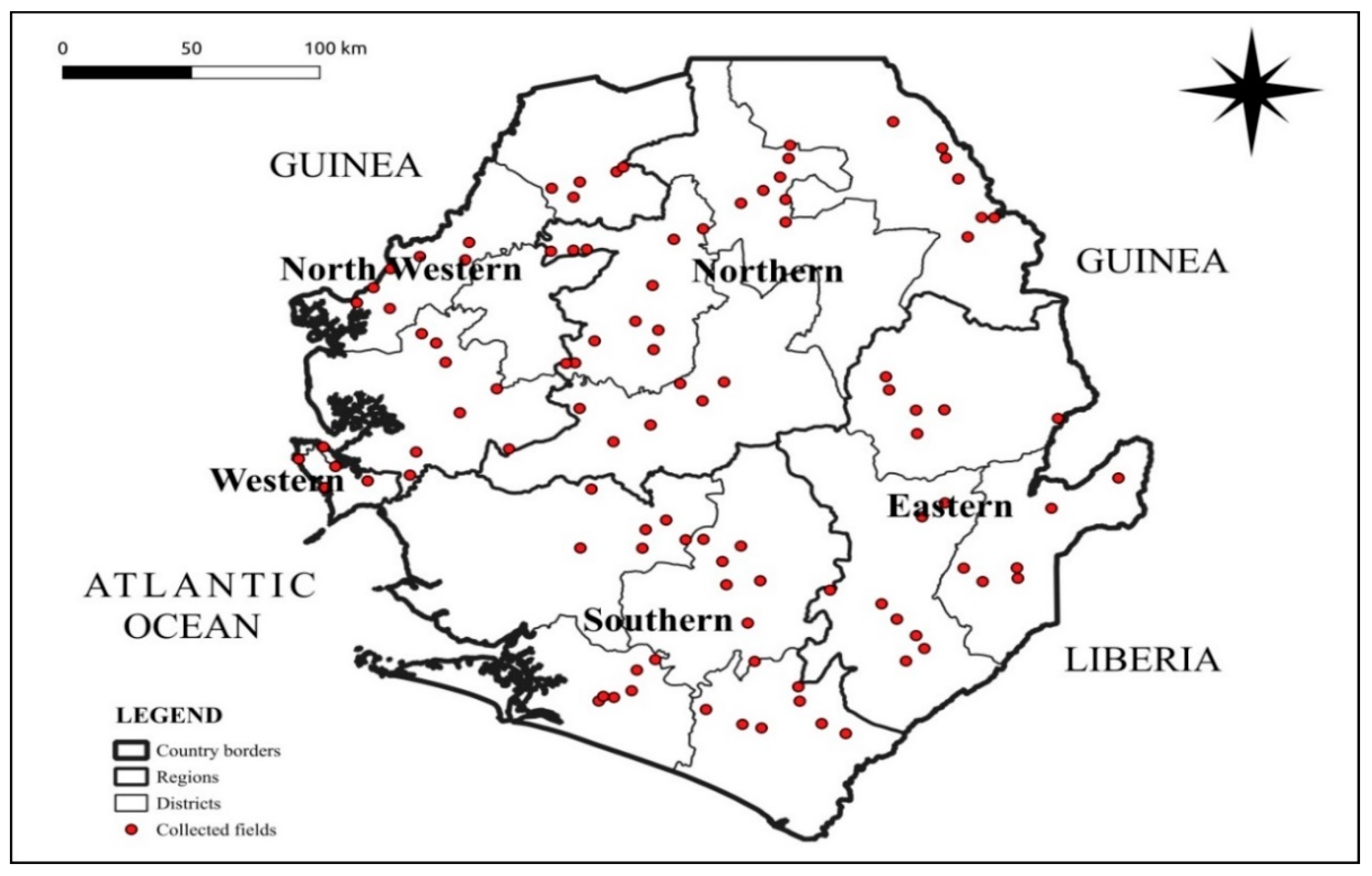
Figure 1.
Map of study area showing country borders, regions, districts and fields.
4.2. Survey Design
Cassava mosaic disease assessment survey was conducted in 2024 using the Central and West African Virus Epidemiology (WAVE) harmonized sampling and diagnostic protocols [33,36]. Assessments were done in 109 farms covering five regions, sixteen districts and agro-ecologies (rain forest, coastal plains, savannah lowlands and savannah highlands) of Sierra Leone. Cassava fields surveyed were 10 km apart and, for each farm surveyed, 30 plants were assessed randomly along two diagonals. The geolocation coordinates of each field were recorded using a global positioning device (Garmin Dakota TM 10).
4.3. Field Data Collection and Storage
Tablet with iForm Zerion (version 9.1.6) software developed by Cambridge, UK’s Epidemiology modelling group for survey was used for data collection in all West Africa virus epidemiology participating countries. Data collected included name of the village or town, the district, region, whitefly counts, cassava mosaic disease symptom observed, geographical coordinates (latitude and longitude), mode of infection and altitude. Other data on variety, date and time, field size, planting types and distance between survey site was collected. The recorded data were uploaded to iForm’s cloud-based database and then integrated into the WAVE Cube. A total of 30 cassava plants per cassava farm were assessed along two diagonals in an X shape (15 plants chosen randomly on each diagonal). Data collected on the sampled plants included CMD severity, whitefly abundance and where the plant was infected, the source of infection was determined as either from cuttings or by the vector. According to Sseruwagi et al. [13] from three to six MAP, a distinction is possible between cutting-borne and whitefly-borne infections. Symptoms appearing only on upper leaves were taken to have resulted from whitefly-transmitted infection, whereas plants that showed symptoms either only on the lower leaves or on all leaves were taken as having been infected through cassava cuttings.
The disease severity assessment was done using the 1-5 rating scale where 1 = symptom-less plants; 2 = mild chlorotic patterns affecting most leaves, mild distortions at the bases of most leaves and remaining part of the leaves are normal; 3 = pronounced chlorosis on most leaves, narrowing and distortion of the lower one-third of the leaflets; 4 = severe chlorosis and distortion of two-thirds of most leaves and general reduction of leaf size and some stunting; and 5 = most severe symptoms (severe chlorosis, leaf distortion, twisting, misshapen leaves, severe reduction of most leaves and severe plant stunting) [37]. The CMD incidence was calculated as the percentage of CMD symptomatic plants out of the total plants assessed using the formula provided by Sseruwagi et al. [13].
Mean incidence (%) = × 100. The incidence was then visually categorized into five percentage: fields with 0 incidence were recorded as healthy; >0–25% as low incidence; >25–50% as medium incidence; >50–75% as high incidence; and >75–100% as very high incidence. The whitefly population was estimated by counting the number of whiteflies on the top five fully expanded leaves of each plant. The mean of whiteflies per plant was calculated as the total number of whiteflies recorded on 30 plants divided by 30. The mode of infection in each plant was determined based on the location of the symptomatic leaves as previously described by Sseruwagi et al. [13].
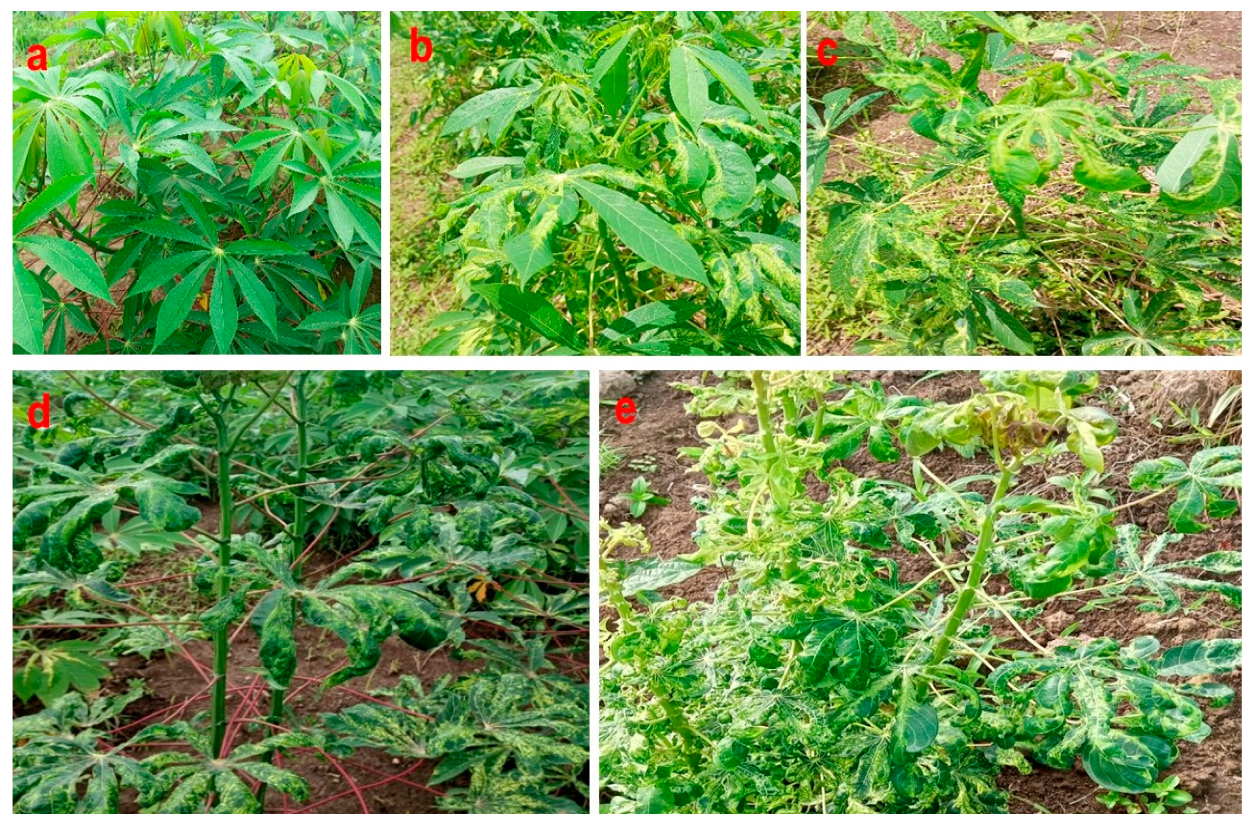
Figure 2.
Cassava showing (a) a healthy-looking plant; (b-e) cassava mosaic disease (CMD) infected plants.
Figure 2.
Cassava showing (a) a healthy-looking plant; (b-e) cassava mosaic disease (CMD) infected plants.
4.4. Molecular Detection of Cassava Begomoviruses
Total DNA was extracted from cassava leaves using the CTAB protocol as previously described [38]. The concentration of the DNA per sample was done using a NanoDrop 2000 spectrophotometer (Thermo Fisher Scientific) and adjusted to 150 ng/μl. Previous studies noted that the most cumbersome CMBs in smallholder cassava cultivation system in Sierra Leone were ACMV and East African cassava mosaic virus (EACMV) [23]. Because the current status of the incidence and severity of these two CMD causing viruses is unknown in Sierra Leone, thus, the survey was conducted across the country focusing on ACMV and EACMV. The extracted DNA was subjected to PCR using the specific primers listed in Table 1 for the detection of ACMV-like and EACMV-like viruses. The samples that were positive for EACMV-like virus were subjected to another round of PCR using specific primers for the detection of EACMVCM. The PCR mix was prepared in a final volume of 25 μl using 20.9 μl of molecular biology grade water, 2.5 µl of 10× reaction buffer, 0.5 µl of 10 mM dNTPs, 0.5 µl of 10 µM of each primer, 0.1 µl of 5 U/µl of Maximo Taq DNA polymerase (GeneON), and 150 ng DNA template of each sample. The DNA amplification was carried out in a SimpliAmp thermal cycler (Life Technologies Holdings Pte Ltd). The PCR temperature profile was set at 94°C for 4 or 5 min for initial denaturation, followed by 35 cycles of amplification at 94°C for 45 or 60 s, 55°C for 45 or 60 s, and 72°C for 55 or 60 s (depending on primers). The final elongation step was performed at 72°C for 7 or 10 min. The PCR-amplified products were subjected to 1% agarose gel electrophoresis and then stained with ethidium bromide. The electrophoresis was performed at 100 V and the gel was visualized using a Compact Digimage System, UVDI series (MS major science).
4.5. Data Analysis
4.5.1. Phenotypic Analysis
Data analysis was performed using the R software v. 3.6.1 (R Development Core Team). The normality of the variables was determined using the Shapiro–Wilk test. For variables that were not distributed according to the normal distribution, the generalized linear model was used. The difference in the number of whiteflies per plant between regions and the difference in the severity score of CMD between regions were assessed using the generalized linear model. The map of Sierra Leone showing the regions where surveys were done in 2024 was developed using QGIS software v. 2.18.26 (https://qgis.org/downloads/).
4.5.2. Molecular Analysis
The PCR products of 50 EACMV-like positive samples were sequenced in both forward and reverse orientation at Macrogen Meibergdreef 57 1105 BA, Amsterdam, the Netherlands, Europe using Sanger et al. [41] method. Contigs were assembled and edited using Geneious Prime® 2022.2.1. (Biomatters Ltd, Auckland, New Zealand.) software. Consensus sequence obtained from forward and reverse sequences for each sample was subjected to BLASTn in NCBI for preliminary species assignment and subsequently for pairwise sequence comparison [42]. The sequences were aligned with representative isolates of begomoviruses using ClustalW alignment method in MEGA X software [43].
5. Conclusions
This study identified and mapped the distribution of CMD-associated viruses (begomoviruses) in farmers' fields of Sierra Leone that could be exploited for effective phytosanitation strategies to mitigate its effect on crop yield loss. The east region, north region, northwest region and western areas were most affected by the disease. The spread of the disease is linked to the use of infected planting materials mainly through stem cuttings. Findings described for the first time an unprecedented diversity of EACMV-Ug infecting diverse local cassava varieties in Sierra Leone. The identification of EACMV-Ug in four districts, three of which share border with Guinea, presents a serious threat for cassava production in the country. This discovery suggests the need for an urgent rapid intervention through the implementation of control strategies by national and regional cassava stakeholders in Sierra Leone and West Africa. Cassava field surveys should be conducted urgently to re-assess EACMV-Ug spread in details and the use of plant village NURU APP to detect CMD severity symptom during cassava stem cutting collection for next planting season. Awareness campaigns should be done to various stakeholders including farmers, extension officers and policymakers on the importance of CMD, identification of the disease symptoms and best practices to mitigate its spread. Strict implementation of phytosanitary procedures including seed certification, and the deployment of resistant varieties must be considered to limit the spread of EACMV-Ug in Sierra Leone and West Africa regions.
Author Contributions
Conceptualization, M.D.S. and A.E.S.; methodology, A.E.S., M.D.S., E.B.T., S.Z. and W.J-L.A.; software, M.D.S., E.B.T., S.Z. and W.J-L.A.; validation, F.T. and P.E.N.; formal analysis, M.D.S., E.B.T., S.Z. W.J-L.A. and P.E.N.; investigation, M.D.S., A.E.S., J.S.P., A.E.O. and M.A.B.; resources, A.E.S. and J.S.P.; data curation, M.D.S.; writing—original draft preparation, M.D.S. and P.E.N.; writing—review and editing, M.D.S., A.E.S., J.S.P., F.T., A.O.E., M.A.B. and P.E.N.; visualization, E.B.T., S.Z., W.J-L.A. and P.E.N.; supervision, A.E.S., M.A.B. and A.O.E.; project administration, A.E.S., A.O.E. and J.S.P.; funding acquisition, A.E.S., A.O.E. and J.S.P. All authors have read and agreed to the published version of the manuscript.
Funding
This work was supported, in whole or in part, by the Bill and Melinda Gates Foundation and The United Kingdom Foreign, Commonwealth and Development Office (FCDO) under a grant with the investment record ID OPP1212988/INV-002969 to the Central and West African Virus Epidemiology (WAVE) Program for root and tuber crops—through a subgrant from the Université Félix Houphouët-Boigny (UFHB) to Njala University. Under the grant conditions of the foundation, a Creative Commons Attribution 4.0 Generic License has already been assigned to the author-accepted manuscript version that might arise from this submission.
Data Availability Statement
For ethical reasons, the corresponding author can make data from this research available upon request.
Acknowledgments
The authors thank the crop protection and cassava breeding team at Njala University, the Sierra Leone Agricultural Research Institute (SLARI), and WAVE Ivory Coast for their technical support during this study.
Conflicts of Interest
The authors declare no conflicts of interest.
Abbreviations
The following abbreviations are used in this manuscript:
| ACMV | African Cassava Mosaic Virus |
| CMD | Cassava Mosaic Disease |
| CTAB | Cetyltrimethylammonium bromide |
| DNA | Deoxyribonucleic acid |
| EACMV | East African Cassava Mosaic Virus |
| PCR | Polymerase chain reaction |
References
- Ogero, K.O.; Mburugu, G.N.; Mwangi, M.; Ombori, O.; Ngugi, M. In vitro micropropagation of cassava through low cost tissue culture. Asian J. Agric. Sci. 2012, 4, 205–209. [Google Scholar]
- Nassar, N.M.A.; Ortiz, R. A review on cassava improvement: challenges and impacts. J. Agric. Sci. 2007, 145, 163–171. [Google Scholar] [CrossRef]
- Bakelana, Z.; Boykin, L.M.; Mahungu, N.; Mavila, N.; Magole, M.; Nduandele, N.; Makuati, L.; Ndomateso, T.; Monde, G.; Pita, J.; Lema, M.L.; Tshilenge, K. First report and preliminary evaluation of cassava root necrosis in Angola. Intern. J. Agric. Environ. Biores. 2019, 4, 37–46. [Google Scholar]
- Legg, J.P.; Jeremiah, S.C.; Obiero, H.M.; Maruthi, M.N.; Ndyetabula, I.; OkaoOkuja, G.; Bouwmeester, H.; Bigirimana, S.; Tata-Hangy, W.; Gashaka, G.; Mkamilo, G.; Alicai, T.; Lava Kumar, P. Comparing the regional epidemiology of the cassava mosaic and cassava brown streak virus pandemics in Africa. Virus Res. 2011, 159, 161–170. [Google Scholar] [CrossRef] [PubMed]
- Winter, S.; Koerbler, M.; Stein, B.; Pietruszka, A.; Paape, M.; Butgereitt, A. Analysis of cassava brown streak viruses reveals the presence of distinct virus species causing cassava brown streak disease in East Africa. J. Gen. Virol. 2010, 91, 1365–1372. [Google Scholar] [CrossRef]
- Alicai, T.; Omongo, C.A.; Maruthi, M.N.; Hillocks, R.J.; Baguma, Y.; Kawuki, R.; Bua, A.; Otim-Nape, G.W.; Colvin, J. Re-emergence of Cassava Brown Streak Disease in Uganda. Plant Disease 2007, 91, 24–29. [Google Scholar] [CrossRef]
- Samura, A.E.; Massaquoi, F.B.; Mansaray, A.; Kumar, P.L.; Koroma, J.P.C.; Fomba, S.N.; Dixon, A. Status and diversity of the cassava mosaic disease causal agents in Sierra Leone. Intern. J. Agric. Forestry 2014, 4, 246–254. [Google Scholar] [CrossRef]
- Chi, Y.; Pan, L.L.; Bouvaine, S.; Fan, Y.Y.; Liu, Y.Q.; Liu, S.S.; Seal, S.; Wang, X.W. Differential transmission of Sri Lankan cassava mosaic virus by three cryptic species of the whitefly Bemisia tabaci complex. Virol. 2020, 540, 141–149. [Google Scholar] [CrossRef]
- Njoroge, M.K.; Mutisya, D.L.; Miano, D.W.; Kilalo, D.C. Whitefly species efficiency in transmitting cassava mosaic and brown streak virus diseases. Cogent Biol. 2017, 3, 1311499. [Google Scholar] [CrossRef]
- Maruthi, M.N.; Hillocks, R.J.; Mtunda, K.; Raya, M.D.; Muhanna, M.; Kiozia, H.; Rekha, A.R.; Colvin, J.; Thresh, J.M. Transmission of cassava brown streak virus by Bemisia tabaci (Gennadius). J. Phytopathol. 2005, 153, 307–312. [Google Scholar] [CrossRef]
- Chikoti, P.C.; Tembo, M.; Chisola, M.; Ntawuruhungu, P.; Ndunguru, J. Status of cassava mosaic disease and whitefly population in Zambia. African J. Biotechnol. 2015, 14, 2539–2546. [Google Scholar]
- Thresh, J.M.; Cooter, R.J. Strategies for controlling cassava mosaic virus disease in Africa. Review Article. Plant Pathol. 2005, 54, 587–614. [Google Scholar] [CrossRef]
- Sseruwagi, P.; Sserubombwe, W.S.; Legg, J.P.; Ndunguru, J.; Thresh, J.M. Methods of surveying the incidence and severity of cassava mosaic disease and whitefly vector populations on cassava in Africa: a review. Virus Res. 2004, 100, 129–142. [Google Scholar] [CrossRef] [PubMed]
- Crespo-Bellido, A.; Hoyer, J.S.; Dubey, D.; Jeannot, R.B.; Duffy, S. Interspecies recombination has driven the macroevolution of cassava mosaic begomoviruses. J. Virol. 2021, 95, e00541–21. [Google Scholar] [CrossRef]
- Maruthi, M.N.; Seal, S.; Colvin, J.; Briddon, R.W.; Bull, S.E. East African cassava mosaic Zanzibar virus – a recombinant begomovirus species with a mild phenotype. Arch. Virol. 2004, 149, 2365–2377. [Google Scholar] [CrossRef] [PubMed]
- Monde, G.; Walangululu, J.; Winter, S.; Bragard, C. Dual infection by cassava begomoviruses in two leguminous species (Fabaceae) in Yangambi, northeastern Democratic Republic of Congo. Arch. Virol. 2010, 155, 1865–1869. [Google Scholar] [CrossRef]
- Patil, B.L.; Fauquet, C.M. Cassava mosaic geminiviruses: actual knowledge and perspectives. Mol. Plant Pathol 2009, 10, 685–701. [Google Scholar] [CrossRef]
- Ndunguru, J.; Legg, J.P.; Aveling, T.A.S.; Thompson, G.; Fauquet, C.M. Molecular biodiversity of cassava begomoviruses in Tanzania: evolution of cassava geminiviruses in Africa and evidence for East Africa being a center of diversity of cassava geminiviruses. Virol. J. 2005, 2, 21. [Google Scholar] [CrossRef]
- Ariyo, O.A.; Koerbler, M.; Dixon, A.G.O.; Atiri, G.I.; Winter, S. Molecular variability and distribution of cassava mosaic begomoviruses in Nigeria. J. Phytopathol. 2005, 153, 226–231. [Google Scholar] [CrossRef]
- Were, H.K.; Winter, S.; Maiss, E. Distribution of begomoviruses infecting cassava in Africa. J. Plant Pathol. 2003, 85, 145–151. [Google Scholar]
- Samura, A.E.; Kanteh, S.M.; Norman, J.E.; Fomba, S.N. Integrated pest management options for the cassava mosaic disease in Sierra Leone. Intern. J. Agric. Innov. Res. 2016, 5, 2319–1473. [Google Scholar]
- Sesay, J.V.; Ayeh, K.O.; Norman, P.E.; Acheampong, E. Shoot nodal culture and virus indexing of selected local and improved genotypes of cassava (Manihot esculenta) from Sierra Leone. Intern. J. Biotechnol. Mol. Biol. Res. 2016, 7, 20–28. [Google Scholar]
- Sesay, J.V.; Lebbie, A.; Wadsworth, R.; Nuwamanya, E.; Bado, S.; Norman, P.E. Genetic structure and diversity study of cassava (Mannihot esculenta) germplasm for African cassava mosaic disease and fresh storage root yield. Open J Genet. 2023, 13, 23–47. [Google Scholar] [CrossRef]
- Combala, M.; Pita, J.S.; Tiendrebeogo, F.; Gbonamou, M.; Eni, A.O. First report of East African cassava mosaic virus-Uganda (EACMV-Ug) infecting cassava in Guinea, West Africa. New Dis. Rep Submitted. 2024. [Google Scholar]
- Pita, J.S.; Fondong, V.N.; Sangare, A.; Otim-Nape, G.W.; Ogwal, S.; Fauquet, C.M. Recombination, pseudorecombination and synergism of geminiviruses are determinant keys to the epidemic of severe cassava mosaic disease in Uganda. J. Gen. Virol. 2001, 82, 655–665. [Google Scholar] [CrossRef]
- Valam-zango, A.; Zinga, I.; Hoareau, M.; Mvila, A.C.; Semballa, S.; Lett, J.M. First report of cassava mosaic geminiviruses and the Uganda strain of east african cassava mosaic virus (EACMV-UG) associated with cassava mosaic disease in Equatorial Guinea. New Dis. Rep. 2015, 32, 29. [Google Scholar] [CrossRef]
- Tiendrébéogo, F.; Lefeuvre, P.; Hoareau, M.; Traoré, V.S.E.; Barro, N.; Reynaud, B.; Traoré, A.S. Occurrence of East African cassava mosaic virus -Uganda (EACMV-UG) in Burkina Faso. Plant Pathol. 2009, 58, 783. [Google Scholar] [CrossRef]
- Doyle, J.J.; Doyle, J.L. A rapid DNA isolation procedure for small quantities of fresh leaf tissue. Phytochem. Bull. 1987, 19, 11–15. [Google Scholar]
- Chikoti, P.C.; Peter, S.; Mulenga, R.M.; Tembo, M. Cassava mosaic disease: a review of a threat to cassava production in Zambia. J. Plant Pathol. 2019, 101, 467–477. [Google Scholar] [CrossRef]
- Anani, J.; Serge, S.; Pita, J.S.; Hound, S.E. Cassava mosaic disease (CMD) in Benin: Incidence, severity and its whitefly abun- dance from field surveys in 2020. Crop Prot. 2022, 158, 106007. [Google Scholar]
- Harimalala, M.; Chiroleu, F.; Giraud-Carrier, C.; Hoareau, M.; Zinga, I.; Randriamampianina, J.A.; Velombola, S.; Ranomenja-nahary, S.; Andrianjaka, A.; Reynaud, B.; Lefeuvre, P. , Lett, J.M. Molecular epidemiology of cassava mosaic disease in Madagascar. Plant Pathol. 2014, 64, 501–507. [Google Scholar] [CrossRef]
- Houngue, J.A.; Pita, J.S.; Habib, G.; Cacaï, T.; Zandjanakou-tachin, M.; Abidjo, E.A.E.; Ahanhanzo, C. Survey of farmers’ knowledge of cassava mosaic disease and their preferences for cassava cultivars in three agro-ecological zones in Benin. J. Ethnobiol. Ethnomed. 2018, 14, 29. [Google Scholar] [CrossRef] [PubMed]
- Eni, A.O.; Efekemo, O.P.; Onile-ere, O.A.; Pita, J.S. South west and north central Nigeria: Assessment of cassava mosaic disease and field status of African cassava mosaic virus and East African cassava mosaic virus. Ann. Appl. Biol. 2021, 178, 466–479. [Google Scholar] [CrossRef]
- Busogoro, J.P.; Masquellier, L.; Kummert, J.; Dutrecq, O.; Lepoivre, P.; Jijakli, M.H. Application of a simplified Molecular protocol to reveal mixed infections with begomoviruses in cassava. J. Phytopathol. 2008, 457, 452–457. [Google Scholar] [CrossRef]
- Zhou, X.; Robinson, D.J.; Harrison, B.D. Types of variation in DNA-A among isolates of East African cassava mosaic virus from Kenya, Malawi and Tanzania. J. Gen. Virol. 1998, 1998. 79, 2835–2840. [Google Scholar] [CrossRef]
- Soro, M.; Tiendrébéogo, F.; Pita, J.S.; Traoré, E.T.; Somé, K.; Tibiri, E.B.; Néya, J.B.; Mutuku, J.M.; Simporé, J.; Koné, D. Epidemiological assessment of cassava mosaic disease in Burkina Faso. Plant Pathol. 2021, 70, 2207–2216. [Google Scholar] [CrossRef]
- IITA (International Institute of Tropical Agriculture, (1990). Cassava in Tropical Africa, A reference manual. IITA, Ibadan, Nigeria. 176pp.
- Permingeat, H.R.; Romagnoli, M.V.; Sesma, J.I.; Vallejos, R.H. A simple method for isolating DNA of high yield and quality from cotton (shape Gossypium hirsutum L.) leaves. Plant Mol. Biol. Rep 1998, 16, 89–89. [Google Scholar] [CrossRef]
- Matic, S.; da Cunha, A.T.P.; Thompson, J.R.; Tepfer, M. An analysis of viruses associated with cassava mosaic disease in three Angolan provinces. J. Plant Pathol http://www.jstor.org/stable/45156055. 2012, 94, 443–450. [Google Scholar]
- Alabi, O.J.; Kumar, P.L.; Naidu, R.A. Multiplex PCR method for the detection of African cassava mosaic virus and East African cassava mosaic Cameroon virus in cassava. J. Virol. Methods 2008, 154, 111–120. [Google Scholar] [CrossRef]
- Sanger, F.; Nicklen, S.; Coulson, A.R. DNA sequencing with chain-terminating inhibitors. Proc. Nat. Acad. Sci. 1977, 74, 5463–5467. [Google Scholar] [CrossRef]
- Bao, Y.; Chetvernin, V.; Tatusova, T. Improvements to pairwise sequence comparison (PASC): a genome-based web tool for virus classification. Arch. Virol. 2014, 159, 3293–3304. [Google Scholar] [CrossRef] [PubMed]
- Kumar, S.; Stecher, G.; Li, M.; Knyaz, C.; Tamura, K. MEGA X: molecular evolutionary genetics analysis across computing platforms. Mol. Biol. Evol. 2018, 35, 1547–1549. [Google Scholar] [CrossRef] [PubMed]
Figure 3.
Epidemiological assessment of cassava mosaic diseuse (CMD) per region in Sierra Leone. (a) mean incidence of CMD, (b) mean severity of CMD, (c) percentage of plants infected by cuttings or whiteflies, (d) number of whiteflies per plant. Error bars represent standard error (±SE).
Figure 3.
Epidemiological assessment of cassava mosaic diseuse (CMD) per region in Sierra Leone. (a) mean incidence of CMD, (b) mean severity of CMD, (c) percentage of plants infected by cuttings or whiteflies, (d) number of whiteflies per plant. Error bars represent standard error (±SE).
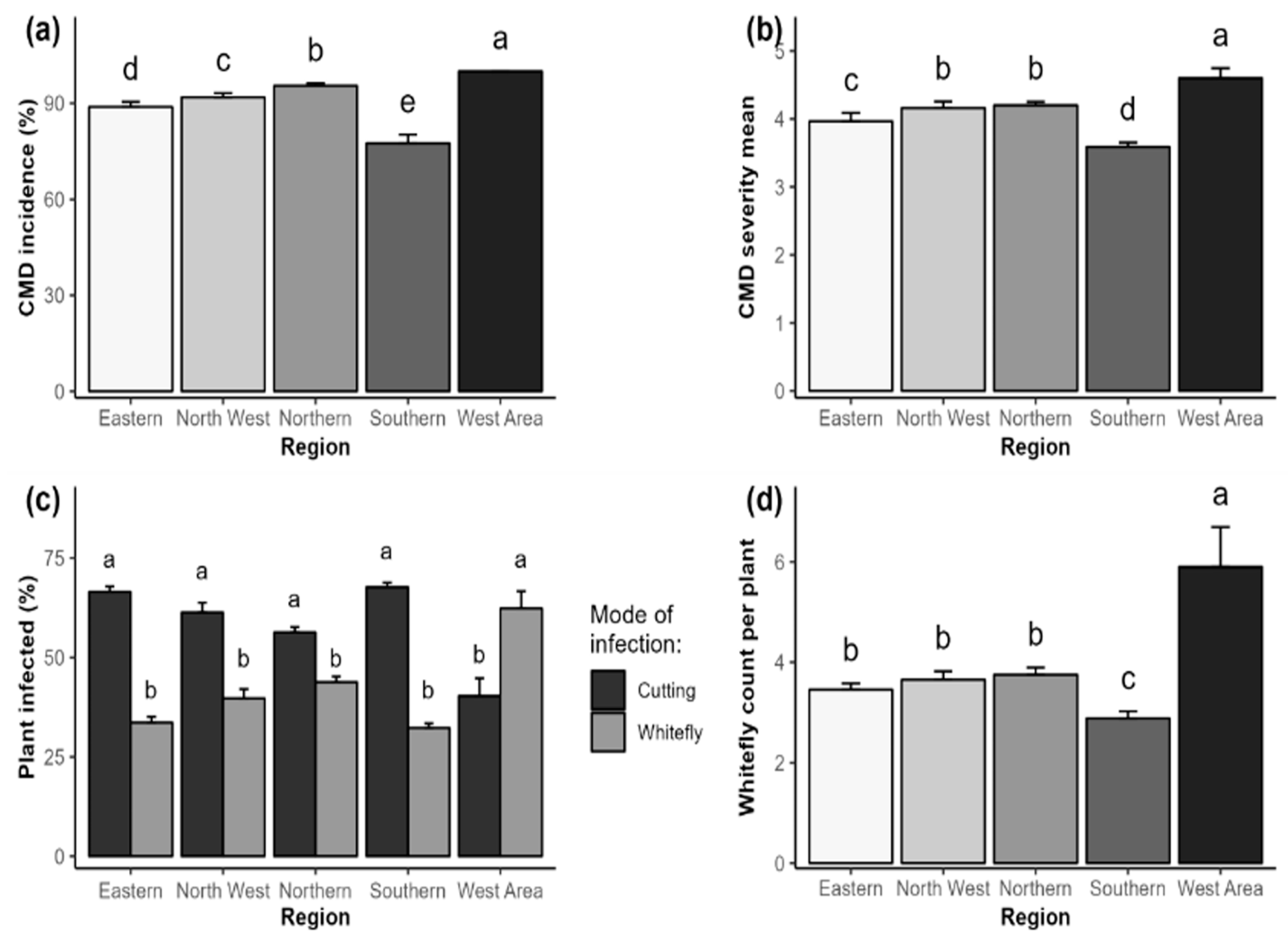
Figure 4.
Epidemiological assessment of cassava mosaic diseuse (CMD) per district in Sierra Leone. (a) mean incidence of CMD, (b) mean severity of CMD, (c) percentage of plants infected by cuttings or whiteflies, (d) number of whiteflies per plant. Error bars represent standard error (±SE).
Figure 4.
Epidemiological assessment of cassava mosaic diseuse (CMD) per district in Sierra Leone. (a) mean incidence of CMD, (b) mean severity of CMD, (c) percentage of plants infected by cuttings or whiteflies, (d) number of whiteflies per plant. Error bars represent standard error (±SE).
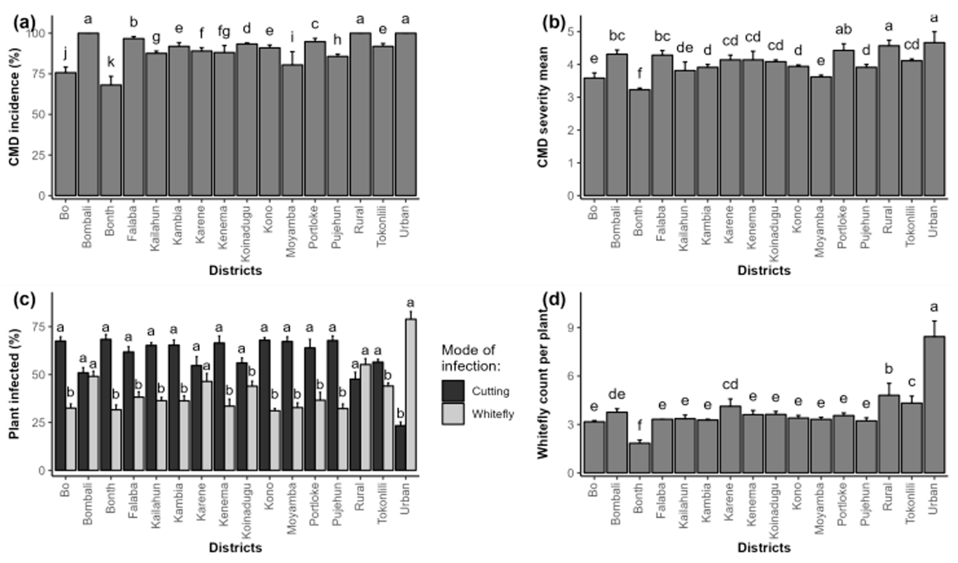
Figure 5.
Map of cassava mosaic disease (CMD) distribution across Sierra Leone.
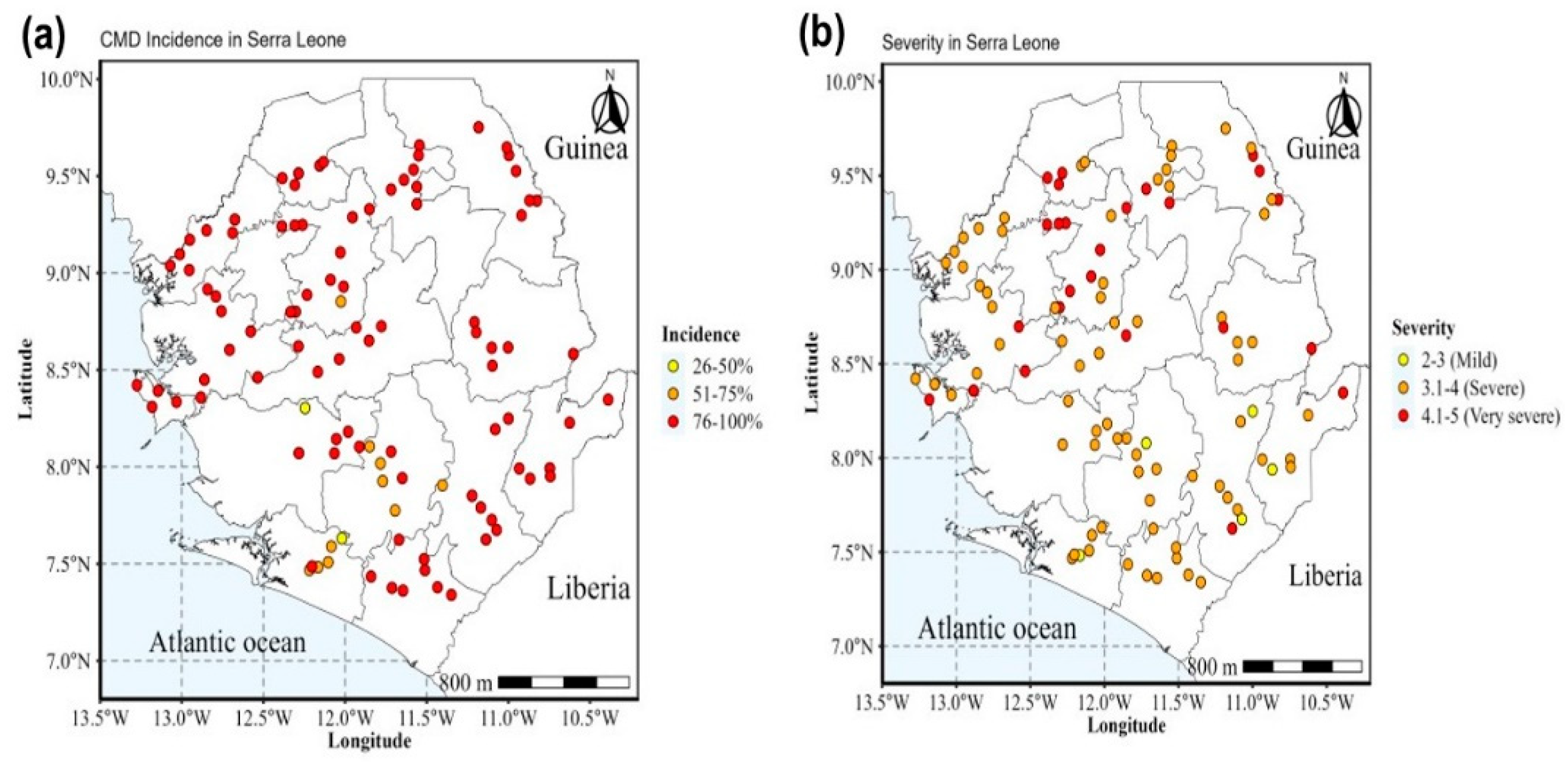
Figure 6.
PCR amplification of cassava DNA using JSP001/JSP002. ACMV M=DNA ladder, ACMV detection at 783 bp=lanes 1-4, and 6-9; positive control=lane T+; negative control=lane T-; and water=lane H20.
Figure 6.
PCR amplification of cassava DNA using JSP001/JSP002. ACMV M=DNA ladder, ACMV detection at 783 bp=lanes 1-4, and 6-9; positive control=lane T+; negative control=lane T-; and water=lane H20.
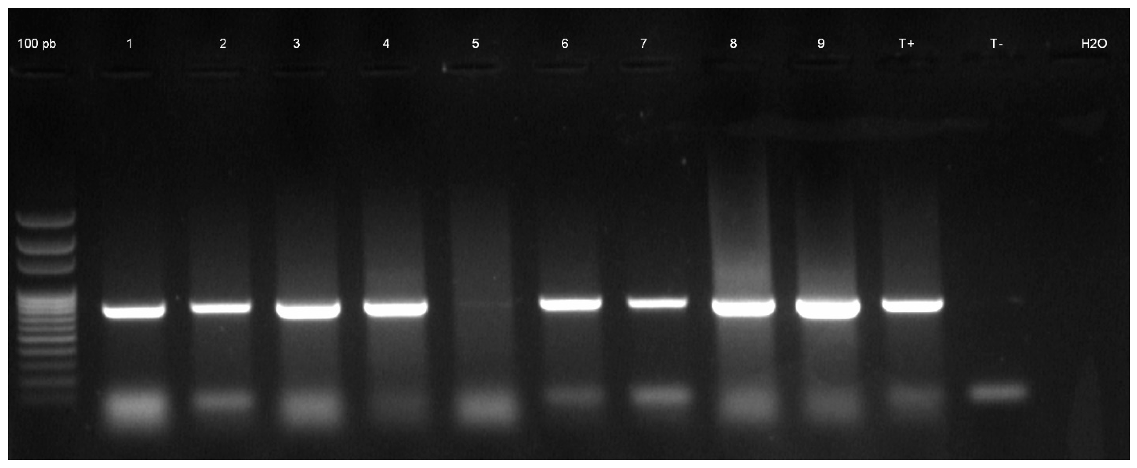
Figure 7.
Distribution of cassava mosaic begomoviruses isolates in Sierra Leone.
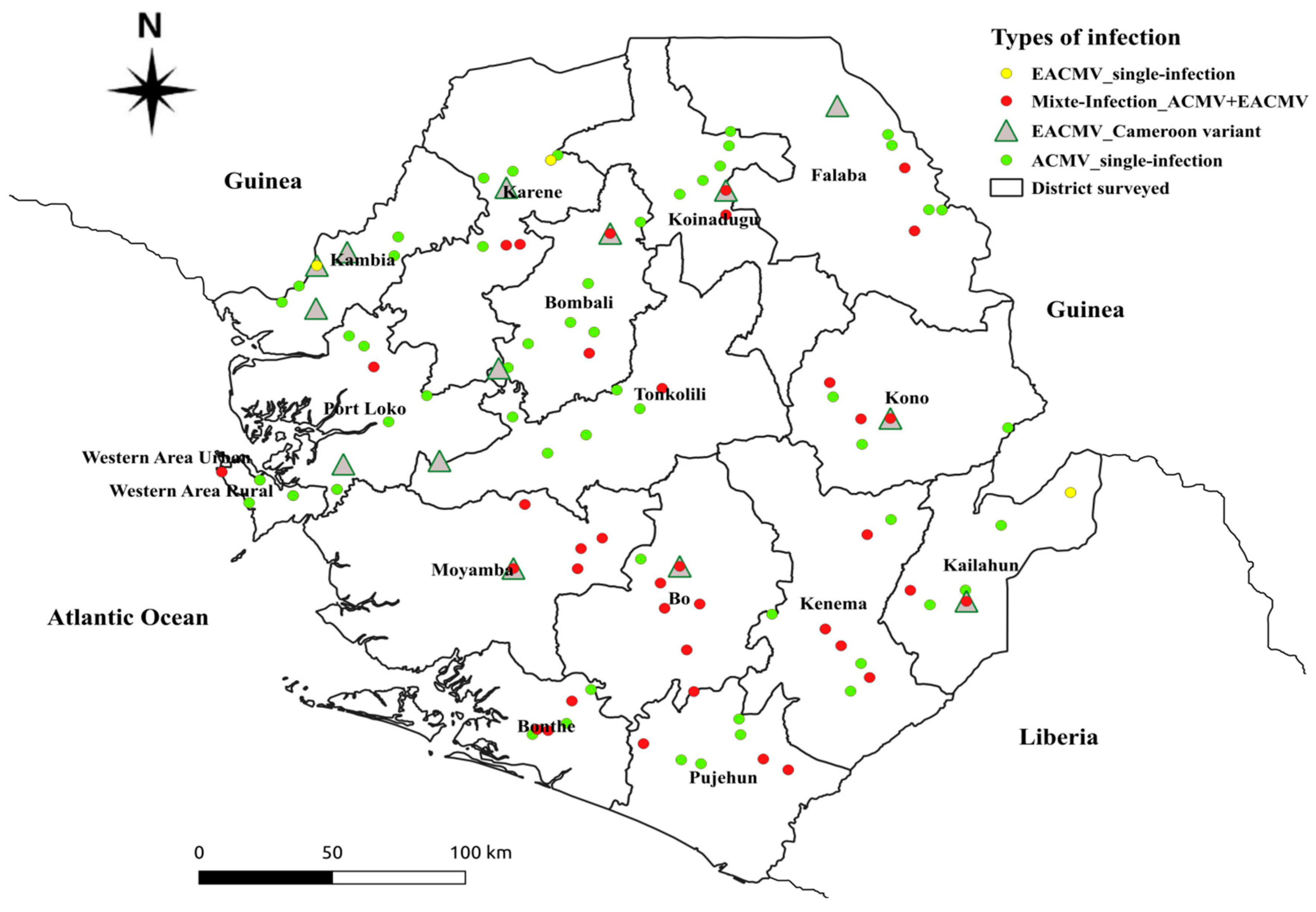
Figure 8.
Type of infection cassava mosaic disease across Sierra Leone during 2024.
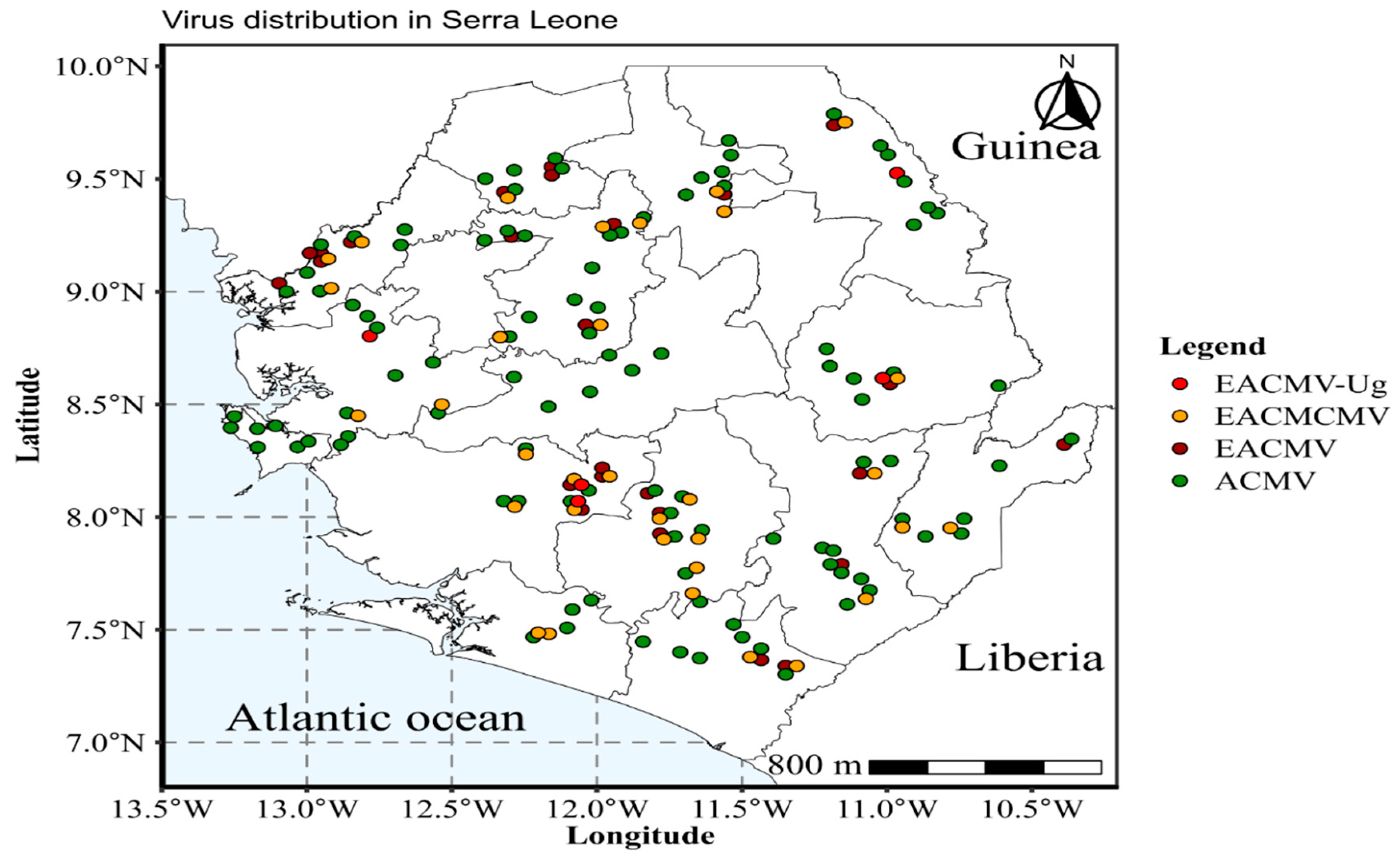
Figure 9.
Distribution of east African cassava mosaic virus variants in Sierra Leone.
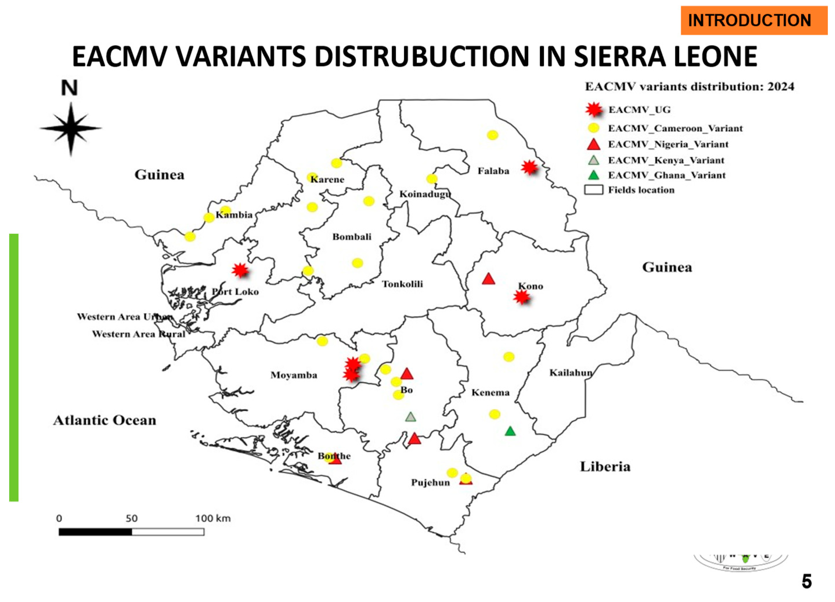

Table 2.
Viral strains associated with CMD in Sierra Leone.
|
Number of samples |
Status of samples |
Positive samples |
Negative samples |
|||
|---|---|---|---|---|---|---|
|
Detection of ACMV |
Detection of EACMV | Detection of ACMV/ EACMV | Detection of Cameroon variant | |||
| 320 CMD | 230 | 4 | 58 | 43 | 28 | |
| 426 | 106 Symptomless | 13 | 0 | 6 | 5 | 87 |
| Total | 243 | 4 | 64 | 48 | 115 | |
Values in brackets are positive single infection of ACMV after delineating for EACMV, EACMV-Cam and mixed infections. Total positive samples=311 and total negative samples=115.
Table 3.
Distribution of begomoviruses across regions of Sierra Leone.
|
Region |
Tested samples |
Positive samples |
ACMV-single infection | EACMV-single infection | Mixed infection (ACMV & EACMV) | EACMV Cameroon variant | ||||
|---|---|---|---|---|---|---|---|---|---|---|
| n | % | n | % | N | % | n | % | |||
| South | 112 | 73 | 47 | 64.4 | 0 | 0.0 | 26 | 35.6 | 26 | 100.0 |
| East | 81 | 68 | 53 | 77.9 | 1 | 1.5 | 14 | 20.6 | 8 | 53.3 |
| North | 110 | 81 | 71 | 87.7 | 0 | 0.0 | 10 | 12.3 | 6 | 60.0 |
| Northwest | 79 | 60 | 45 | 75.0 | 3 | 5.0 | 12 | 20.0 | 8 | 53.3 |
| West | 44 | 29 | 27 | 93.1 | 0 | 0.0 | 2 | 6.9 | 0 | 0.0 |
| Total | 426 | 311 | 243 | 78.1 | 4 | 1.3 | 64 | 20.6 | 48 | 70.6 |
n=sample size; %=percent.
Table 1.
List of primer pairs used to confirm the presence of ACMV, EACMV and EACMVCM.
| Primer | Sequence (5′-3′) | Target region | Expected size (bp) | Virus species | Reference |
|---|---|---|---|---|---|
| JSP001 | ATGTCGAAGCGACCAGGAGAT | DNA-A (CP) | 783 | ACMV | Pita et al. [25] |
| JSP002 | TGTTTATTAATTGCCAATACT | ||||
| ACMBVF | TCGGGAGTGATACATGCGAAGGC | DNA-B (BV1/BC1) | 628 | ACMV | Matic et al. [39] |
| ACMBVR | GGCTACACCAGCTACCTGAAGCT | ||||
| WAVE-508F | AAGGCCCATGTAAGGTCCAG | AV1/AC3 | 800 | ACMV | WAVE |
| WAVE-1307R | GAAGGAGCTGGGGATTCACA | ||||
| WAVE-177F | GATCTGCGGGCCTATCGAAT | BV1 | 800 | ACMV | WAVE |
| WAVE-197R | TTCACGCTGTGCAATACCCT | ||||
| WAVE-370F | ACAGCCCATACAGGAACCGT | AV1/AC3 | 1000 | ACMV | WAVE |
| WAVE-1369R | CGACCATTCCTGCTGAACCA | ||||
| WAVE-982F | TTCGTGTCATCTGCAGGAGA | BV1/BC1 | 800 | ACMV | WAVE |
| WAVE-1781R | GTACCATGGCAGCTGCTGTA | ||||
| JSP001 | ATGTCGAAGCGACCAGGAGAT | DNA-A (CP) | 780 | EACMV | Pita et al. [25] |
| JSP003 | CCTTTATTAATTTGTCACTGC | ||||
| CMBRepF | CRTCAATGACGTTGTACCA | DNA-A (AC1) | 650 | EACMV | Alabi et al. [40] |
| EACMVRepR | GGTTTGCAGAGAACTACATC | ||||
| WAVE-EA1875F | TGTACCAGGCGTCGTTTGAA | AC1 | 800 | EACMVCM | WAVE |
| WAVE-E2674R | TGTCCCCCGATCCAAAACG | ||||
| WAVE-EB1869F | TTCCAAGGGGAGGGTTCTGA | BC1 | 800 | EACMV | WAVE |
Disclaimer/Publisher’s Note: The statements, opinions and data contained in all publications are solely those of the individual author(s) and contributor(s) and not of MDPI and/or the editor(s). MDPI and/or the editor(s) disclaim responsibility for any injury to people or property resulting from any ideas, methods, instructions or products referred to in the content. |
© 2025 by the authors. Licensee MDPI, Basel, Switzerland. This article is an open access article distributed under the terms and conditions of the Creative Commons Attribution (CC BY) license (http://creativecommons.org/licenses/by/4.0/).
Copyright: This open access article is published under a Creative Commons CC BY 4.0 license, which permit the free download, distribution, and reuse, provided that the author and preprint are cited in any reuse.


