Submitted:
09 February 2025
Posted:
10 February 2025
You are already at the latest version
Abstract
Photoaging is common and represents one of the primary pathways for hair damage in daily life. Hydrolyzed keratin, which is usually derived from wool and consists of a series of polypeptide molecules, has been investigated as UV damage prevention ingredient for hair care. Scanning Electron Microscopy (SEM) and fluorescent penetration experiments verified that hydrolyzed keratin can deposit on the hair cuticles to form a film and partly penetrate into hair cortex. This film played as a UV blocker and helped hair resist surface damage and maintain a sleek and healthy morphology after UV radiation. Surprisingly, it was found that hydrolyzed keratin treatment combined with subsequently UV radiation could significantly improve the tensile properties of hair. For hydrolyzed keratin treated hair, tensile strength maintained after UV radiation, while as a comparison, it decreased by 11.74% for untreated hair. This phenomenon is explained by a UV-induced degradation-penetration mechanism. During UV radiation, an increase in free amino acid content and conductivity was observed for hydrolyzed keratin solution, demonstrating the photodegradation into smaller peptide and amino acid. The degradation of hydrolyzed keratin allowed it more easily to enter the interior of hair cortex, thereby enhanced the tensile properties via enhancing chemical bonds.
Keywords:
hair photoaging
; UV damage
; hydrolyzed keratin
1. Introduction
Hair damage is a common phenomenon caused by various factors which has been attracting more and more research interests in recent years. Various factors in the processes of perming, straightening and dyeing, including oxidizing agent [1,2,3], reducing agents [4] and heat [5], can cause certain damage to hair cuticle and cortex and lead to irreversible changes on hair surface and internal structure. In addition to these human-induced damages, hair damage also occurs naturally everyday due to a diverse of environmental disruptors like ash, air pollutes, and more importantly, UV radiation. Among these environmental disruptors, UV radiation from sunlight has a rather severe impact on the physical and chemical properties of hair.
According to the latest research, UVA and UVB in particular regulate the complex central and global homeostasis. This includes the rapid or slow activation of local and central neuro-immuno-endocrine responses involving the brain, endocrine, and immune systems, with secondary effects on peripheral organs. [6] UV radiation induced hair damage is also called hair photoaging, which could lead to many hair damages such as protein loss [7], amino acid and lipid oxidation [8,9], melanin degradation [10], and obvious cuticle deformation [11]. For instance, Richena et al. employed Atomic Force Microscopy (AFM) to characterize hair surface after UV irradiation and found that average step height of surface cuticle layer of the hair significantly increased from 203 nm to 338 nm after irradiation. [11] In addition to surface damage, mechanical properties and manageability of hair are also greatly influenced under UV radiation, as demonstrated in a study by Nogueira et al. It was found that after photoaging, both tensile strength and largest elongation of hair showed a noticeable decrease. [1] Photoaging would be severer for dyed hair, due to more cysteic acid created during hair coloring which provides more binding sites for metal ions, and the metal ions could accelerate the photoaging process. [12] Tang et al. observed increased levels of metal content, particularly iron and copper, which are believed to play a key role in hair photoaging through activating the production of hydroxyl radicals with dye. [13] Apart from hair dyeing, bleaching also leads to an increased copper ion adsorption of hair from tap water, which in turn results in the production of more oxidative free radicals. Naqvi et al. used the probe technology to monitor the hydroxyl radical formation in hair bleaching systems and found the level of hydroxyl radicals formed under UV radiation was proportional to the level of copper present. [14]
Aiming to protect hair from photoaging, different photoaging preventing ingredients have been proposed by many researchers which could be generally classified into three categories, including silicones, dyes and antioxidants. Silicone, which is widely used in shampoo and other hair care products, could form a film on hair surface which can effectively prevent color change after UV radiation. [15,16] In addition to silicones, some dyes are also found to be able to protect hair against photoaging. Pande et al. found that light absorption ability of the dyes can reduce the degradation of hair proteins under UV radiation and some hair dyes can make the disulfide damage reduce to 5% from 13%. [17] Utilization of antioxidants which could quench free radicals or scavenge ROS (reactive oxygen species) is another approach to slowing down lipid oxidation and reducing protein degradation in photoaging. For example, Fernández et al. found natural antioxidants obtained from artichoke and rice can prevent UV damage. Artichoke presented better performance in reducing lipid peroxidation and protein degradation while rice extract was better in preserving shine and color of dyed hair. [18] Similarly, pomegranate hydroalcoholic extract, honeysuckle extract and tea extract have also been found to be effective in protecting color of dyed hair and reducing protein loss under UV radiation, which may be related to their rich content of antioxidant active components such as tannins, flavonoids, and polyphenols. [19,20]
Among diverse hair care ingredients, protein-based materials have been attracting more and more attentions due to their excellent biocompatibility and ecological friendliness. Sahib et al. invented a peptide composition which can penetrate inside the human hair fiber to improve its strength and hydrophobicity. [21] Among protein-based materials, hydrolyzed keratin stands out as one of the most extensively studied components. This prominence is attributed not only to its readily available nature but also to its chemical similarities with the keratin found in hair, which confers a higher compatibility and affinity with hair’s own protein structure. Many studies have demonstrated that the inclusion of hydrolyzed keratin into hair care product formulations can markedly enhance the tensile strength of hair, efficiently prevent hair from breaking, and ameliorate the hygroscopic nature of hair, rendering hair better manageability. [22,23] However, UV protection performance of hydrolyzed keratin has been rarely explored.
In this work, performance of hydrolyzed keratin in protecting hair from photoaging has been evaluated in terms of surface morphology and hair tensile test. The deposition and penetration behavior of hydrolyzed keratin were confirmed by fluorescence labeling experiments and Scanning Electron Microscopy (SEM) experiments. The roles of hydrolyzed keratin as UV reducer and hair strengthener during UV radiation were demonstrated. Finally, based on the experiments results of stress relaxation test, Differential Scanning Calorimetry (DSC) analysis, a UV-induced degradation-penetration mechanism for the strengthening effect of hydrolyzed keratin on hair is proposed.
2. Experimental Method
2.1. Materials
Sodium dodecyl sulfate (SDS), disodium hydrogen phosphate (Na2HPO4·12H2O), sodium dihydrogen phosphate (NaH2PO4·2H2O), N,N-dimethylformamide(DMF), Sinopharm Chemical Reagent Co., LTD.; 6-carboxytetramethylrhodamine, succinimidyl Ester (6-TAMRA, SE), Shanghai Yuanye Biotechnology Co., LTD.; tissue freezing medium, Leica Biosystems.; virgin white Chinese hair (18 cm×1.5 cm ×1 g) was purchased from Shanghai Canyu commercial Co., Ltd., and sourced from Chinese females aged 20-60; hydrolyzed keratin(17.5%), ProSinaTM, purchased from CRODA China (Shanghai, China); bleached hair samples (23 cm×3 cm×10 g), sourced from Chinese females aged 20-60, provided by Unilever (China) Investment Co., LTD.
Hydrolyzed keratin-treated hair samples: bleached hair samples were soaked in the hydrolyzed keratin solution (2wt%, with a pH of 5.5-6.0) for 4 h, and then placed at 25°C and 60%RH for 24 h to remove the surface moisture.
Photoaging hair samples: Hair samples were prepared into hair pieces and placed in a xenon lamp aging chamber to induce aging damage, simulating the natural damage that consumers' hair experiences in daily life.
2.2. SEM Characterization
Surface morphology of hair samples was observed using a Field Emission Scanning Electron Microscope S-4800 (Hitachi, Ltd., Tokyo, Japan) using a secondary electron (SE) mode at an accelerating voltage of 3 kV and a probe current of 10 μA. Before characterization, hair samples were trimmed and attached to the sample stage using conductive adhesive.
2.3. Hydrolyzed Keratin Permeability Test
The penetration behavior of hydrolyzed keratin was observed using fluorescence labeling method, as detailed by Brunner [24]. Due to the fluorescence interference of melanin White hair was employed in the test. Damaged white hair was obtained by bleaching the virgin Chinese white hair using powdered bleacher according to Imai[2]. The bleached white hair was washed by 1% SDS solution and dried under constant temperature (25°C ± 1°C) and humidity (60%RH ± 5%) firstly. Then, hair sample was soaked in a 2wt% fluorescent-labeled hydrolyzed keratin solution for 4 hours. The obtained hair sample was rinsed twice with deionized water and dried under constant temperature (25°C ± 1°C) and humidity (60% ± 5%). Next, the dried hair sample was completely covered with a tissue freezing agent and frozen overnight at -18 °C. Leica CM1950 cryostat (Leica Biosystems, Germany) was used to cut the hair fibers into 25 μm sections. The fluorescence intensity was observed under the inverted biological microscope.
2.4. Tensile Tests
Tensile tests were conducted to investigate the restorative strength of hydrolyzed keratin solution on damaged hair using the XS (08) XT-3 fiber strength tester (Xusai Instrument, Shanghai, China). Before the test, the hair was placed in a constant temperature and humidity environment with temperature (25°C ± 1°C) and humidity (60% ± 5%) for 24 hours to equilibrate. The measurement of diameter was taking a point at the top, bottom, and middle of the stretching area to measure under the microscope SN-1200W (Sangnond Technology, Shenzhen, Guangzhou, China). Single hair fibers (70-90 μm) were selected. Tensile strength (σt),plateau load () and Young’s modulus (E) of the single hair fiber were calculated through the following formula [25,26]:
where Fb, S, Fh, Fy are breaking strength, cross-sectional area, yield strength, and yield zone endpoint strength of the fiber, and ΔL, L are Hookean region length and initial length respectively.
Results were based on 30 hair fibers and T-test was performed for each group and calculated the p value using SPSS software.
2.5. Stress Relaxation
Stress relaxation test was conducted on the same equipment as described in section 2.5 using a similar method used by Barba at al [27]. Hair fibers with diameter of 70-90 μm were selected from hair samples. The selected single hair fiber was elongated at constant ratio of 30% for 90 s and the stress was recorded as relaxation curve. The stress curve (σr) in the process is calculated using the following formula [27]:
where F is the strength at 30% strain and S is the cross-section area of the hair. Then, the stress relaxation rate proportion η was calculated based on the stress value at 30% strain σ0 and the stress value σ90 at 90 s according to the following equation:
Results were based on 30 hair fibers and T-test was performed for each group and calculated the p value using SPSS software.
2.6. DSC
DSC testing was conducted on DSC 204 F1 (NETZSCH Geraetebau GmbH, Selb, Bavaria, Germany). Before the test, hair samples were equilibrated at 25°C and 60%RH for 24 h and then were cut into small pieces (about 5 mm) and placed 10 mg into an aluminum crucible, which was perforated for easy removal of gases produced during heating. The temperature program was set to maintain a constant temperature at 60 °C for 10 minutes, followed by an increase to 260 °C at a rate of 10 °C/min, and nitrogen gas was used as the purge gas during the test. [28]
2.7. Hydrolyzed Keratin Degradation Experiment
The UV induced degradation of hydrolyzed keratin was verified by preparing a 2wt% hydrolyzed keratin solution (PH=5.5-6) and kept under 365 nm UV radiation (PL-L 18W/10/4P, Royal Philips N.V., Eindhoven, Netherlands) at 3.5W/m2 for 6 days, simulating 10 days of UV exposure for 2 h per day in Shanghai, China. [29] The conductivity and free amino acid concentration of the solution was monitored every 24 hours. Conductivity was measured using a conductivity meter DDS-307 (Yueping Scientific Instrument, shanghai, China), and free amino acid was quantified using the ninhydrin assay. [30]
3. Result and Discussion
3.1. Deposition and Penetration Behavior of Hydrolyzed Keratin
The deposition and penetration behavior of hydrolyzed keratin was investigated using SEM and fluorescence microscope. The SEM images of untreated hair and hydrolyzed keratin-treated hair samples were shown in Figure 1a and b respectively. Comparing two images, it could be observed that there was significant deposition of keratin at the edges of the hair cuticle scales, which is consistent with Malinauskyte’s finding. [30] Fluorescence micrographs shown in Figure 1c,d further verifies that hydrolyzed keratin can deposit on cuticle and penetrate into hair cortex. Considering a moderate average molecular weight about 3000 Da, penetration and deposition of hydrolyzed keratin could both occur during the treatment. Some small molecule components from the hydrolyzed keratin with higher degree of hydrolysis may permeate into the hair keratin, while the majority might accumulate and deposit on hair cuticle due to their large molecular weight. This finding is consistent with that of Malinauskyte, that mid-range MW hydrolyzed keratin can able to penetrate deeper into the cortex, and high-MW keratin peptides only penetrated outer layers of the cortex [30]
3.2. UV Protection Performance of Hydrolyzed Keratin
To investigate the photoprotective effect of hydrolyzed keratin on hair, hair sample treated with hydrolyzed keratin solution were bundled together and placed in a xenon lamp aging chamber for 6 days at 35°C and 60% humidity, and the untreated hair serves as the control group. As shown in Figure 2a,b, there was some cuticle damage caused by UV radiation, which is consistent with Richena’s study [15]. As reported in some literatures [15,35], long time explosion of hair to UV radiation could even result in significant loss of outer cuticles, make hair frizzy and prone to split. However, as shown in Figure 2c, hair sample treated with hydrolyzed keratin only presented slight hair surface damage after exposure to UV radiation. The results prove that the hydrolyzed keratin film deposited on hair surface can effectively prevent UV damage, playing the role as a UV reducer.
The protective effect of hydrolyzed keratin on hair is also demonstrated by the outcomes of tensile test. Tensile properties of hair samples were evaluated before and after UV radiation. As shown in Figure 3, photoaging lead to a significant decrease of 11.74% in tensile strength after UV radiation for hair without hydrolyzed keratin protection. As a UV reducer, hydrolyzed keratin helped prevent UV damage hence tensile strength did not decrease for treated group after UV radiation. However, surprisingly, it was found that after treatment with hydrolyzed keratin, hair sample exhibited a remarkable enhancement in plateau load strength and Young’s modulus properties after UV radiation, which present an increase of 18.51% and 27.33% respectively after UV radiation. In comparison, before UV radiation, hair treated by hydrolyzed keratin presented similar tensile properties with control group. The phenomena indicate that besides UV reducer, the hydrolyzed keratin also plays a role as hair strengthener, and this strengthen effect could protect the damaged hair from UV radiation.
3.3. Mechanism Investigation
To elucidate the mechanisms of UV-induced strengthening effect of hydrolyzed keratin on hair, stress relaxation tests were conducted on hair samples before and after UV radiation. This test is usually employed to compare the proportion of weak intermolecular forces within different hair samples. [27] As illustrated in Figure 4a, during the relaxation, weak chemical bonds including hydrogen and ionic bonds got broken within 90 seconds after the stress applied while strong chemical bonds such as covalent bond remained. As depicted in Figure 4b, treated group showed an enhancement in the proportion of weak chemical bonds by 6.56% compare with untreated group, suggesting that the deposition and penetration of hydrolyzed keratin on hair enhance the internal chemical bonds within the hair. Moreover, after UV radiation, treated group showed a further increase of 3.85% in the proportion of weak chemical bonds, indicating that UV radiation promoted the strengthening of the internal chemical bonds. Therefore, it could be concluded that hydrolyzed keratin, when used as a treatment for hair, acts as a strengthener, enhancing the hair's overall strength. Furthermore, after UV radiation exposure, hydrolyzed keratin serves as a UV reducer, effectively reducing hair damage while also increasing the proportion of weak intermolecular forces in the hair, thus further enhancing its strength.
Thermal stability tests conducted on hair using DSC have yielded comparable results, which are presented in Figure 5. It could be seen that after UV radiation, melting temperature of control group shifted from 243.41°C to 245.05°C, which indicates more intermolecular forces created by fiber brittleness caused by UV. [36] In companion, the treated group with hydrolyzed keratin protection also showed a smaller shift after UV radiation, with the peak position shifting from 243.35 °C to 244.25°C. However, fiber brittleness seems unlikely for treated group since the tensile properties didn’t show any reduction. Therefore, the peak shifting towards higher temperatures for treated group should be ascribed to the formation of new intermolecular forces after UV irradiation, which is in consist with the stress relaxation results.
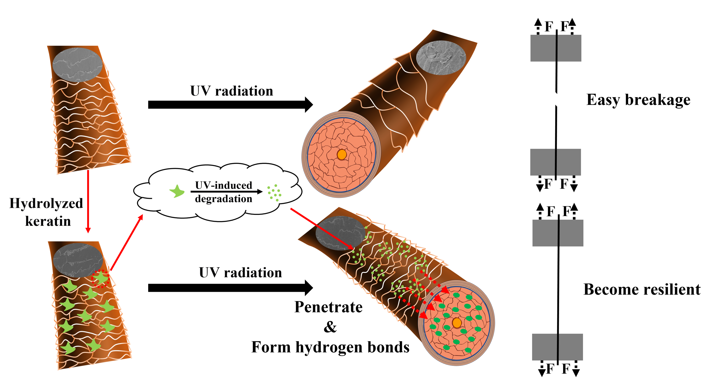
Combining the above experimental results, UV protection provided by hydrolyzed keratin and its strengthening effect on hair after UV treatment are believed to mainly contributed by the enhancement of intermolecular forces within hair fibers. This mechanism is similar with the repair mechanism of amino acids on damaged hair. Our previous study found that amino acids could improve the hair’s tensile strength and yield stress. [37] While considering the large molecule weight of hydrolyzed keratin, a UV-induced degradation-penetration mechanism is proposed. As illustrated in Figure 6, after treatment, hydrolyzed keratin could create a protective film on hair surface. This film partly consumed UV and helped to maintain hair surface morphology under UV radiation. In addition, the film underwent degradation under UV radiation, which leaded to a reduction in molecular weight and an increase in penetration ability. The degraded amino acids penetrated into hair cortex and therefore enhanced the weak bonds within hair fibers, rendering hair surprisingly superior tensile properties compared to that prior to damage.
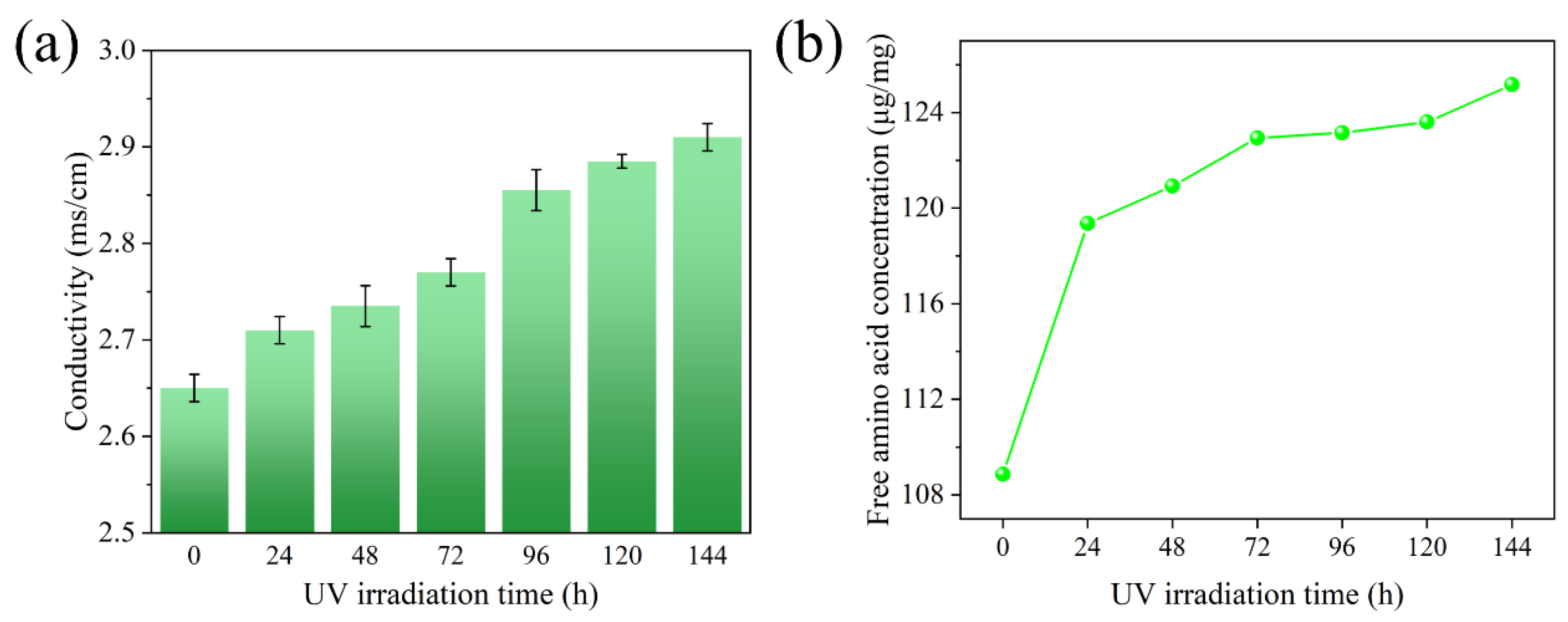
To verify this inference, hydrolyzed keratin solution was subjected to UV radiation for a period of six days, during which the electrical conductivity and free amino acid content were monitored. As shown in Figure 7a,b, both electrical conductivity and free amino acid concentration in the hydrolyzed keratin solution incrementally increased during UV irradiation, indicating the formation of electrolytes which is mainly amino acids. The result suggests that large molecule in hydrolyzed keratin molecules was degraded into smaller peptides and amino acids under UV. As demonstrated by the experiments conducted by Malinauskyte [31], proteins of low molecular weight are more likely to penetrate into hair strands. Consequently, this UV-induced degradation process not only consumed the energy of UV but also results in the formation of smaller molecular fragments that enhance penetration into the hair's interior. This, in turn, contributes to a significant improvement in the hair's tensile strength and overall resilience.
4. Conclusions
Upon the application of hydrolyzed keratin to hair, it forms a layer on the hair's surface and partially permeates into the hair cortex. This film serves to mitigate the detrimental effects of UV radiation on hair. The presence of hydrolyzed keratin acts as a shield against UV-induced damage, thereby preserving the hair's structural integrity, as well as its tensile and thermal stability characteristics.
The mechanism of protection is posited to involve the degradation of hydrolyzed keratin on the hair surface under the influence of UV radiation, resulting in the formation of smaller peptides or free amino acids. These lower molecular weight byproducts can penetrate the hair shaft, which subsequently leads to an enhancement in the hair's tensile strength. This process underscores the role of hydrolyzed keratin in conferring resilience to the hair against environmental stressors, particularly those associated with UV exposure.
References
- Nogueira, A. C. S.; Nakano, A. K.; Joekes, I. Impairment of hair mechanical properties by sun exposure and bleaching treatments. J. Cosmet. Sci. 2004, 55, 533–537. [Google Scholar] [PubMed]
- Imai, T. The influence of hair bleach on the ultrastructure of human hair with special reference to hair damage. Okajimas Folia Anat. Jpn. 2011, 88, 1–9. [Google Scholar] [CrossRef] [PubMed]
- Dyer, J. M.; Bell, F.; Koehn, H.; et al. Redox proteomic evaluation of bleaching and alkali damage in human hair. Int. J. Cosmet. Sci. 2013, 35, 555–561. [Google Scholar] [CrossRef] [PubMed]
- Contreras, F.; Ermolenkov, A.; Kurouski, D. Infrared analysis of hair dyeing and bleaching history. Anal. Methods 2020, 12, 3741–3747. [Google Scholar] [CrossRef]
- Hyun, J. W. Analysis of Morphological changes on the hair surface by heat perm treatment method. J. Korean Soc. Cosmetol. 2023, 2023 29, 449–455. [Google Scholar] [CrossRef]
- Slominski, R. M.; Chen, J. Y.; Raman, C.; et al. Photo-neuro-immuno-endocrinology: How the ultraviolet radiation regulates the body, brain, and immune system. Proc. Natl. Acad. Sci. U.S.A. 2024, 121, e2308374121. [Google Scholar] [CrossRef]
- Estibalitz, F.; Barba, C.; Alonso, C.; et al. Photodamage determination of human hair. J. Photochem. Photobiol. B: Biol. 2012, 106, 101–106. [Google Scholar]
- Robbins, C. R.; Bahl, M. K. Analysis of hair by electron spectroscopy for chemical analysis. J. Cosmet. Sci. 1984, 35, 379–390. [Google Scholar]
- Ji, J. H.; Park, T. S.; Lee, H. J.; et al. The ethnic differences of the damage of hair and integral hair lipid after ultra violet radiation. Ann. Dermatol. 2013, 25, 54–60. [Google Scholar] [CrossRef]
- Hoting, E.; Zimmermann, M.; Höcker, H. Photochemical alterations in human hair. II: Analysis of melanin. J. Cosmet. Sci. 1995, 46, 181–190. [Google Scholar]
- Richena, M.; Rezende, C. A. Effect of photodamage on the outermost cuticle layer of human hair. J. Photochem. Photobiol. B: Biol. 2015, 153, 296–304. [Google Scholar] [CrossRef] [PubMed]
- Smart, K. E.; Kilburn, M.; Schroeder, M.; et al. Copper and calcium uptake in colored hair. Int. J. Cosmet. Sci. 2009, 60, 337–45. [Google Scholar] [CrossRef]
- Ying, T.; Jolon, M.; Dyer, Santanu, D. ; et al. Trace metal ions in hair from frequent hair dyers in China and the associated effects on photo-oxidative damage. J. Photochem. Photobiol. B: Biol. 2016, 156, 35–40. [Google Scholar]
- Naqvi, K. R.; Marsh, J. M.; Godfrey, S.; et al. The role of chelants in controlling Cu(II)-induced radical chemistry in oxidative hair colouring products. Int. J. Cosmet. Sci. 2013, 35, 41–49. [Google Scholar] [CrossRef]
- Michelli, F. D.; André, R. B.; Maria, V. R. V. Effects of solar radiation on hair and photoprotection. J. Photochem. Photobiol. B: Biol. 2015, 153, 240–246. [Google Scholar]
- Schlosser, A. Silicones used in permanent and semi-permanent hair dyes to reduce the fading and color change process of dyed hair occurred by wash-out or UV radiation. J. Cosmet. Sci. 2004, 55 Suppl, S123–131. [Google Scholar] [CrossRef]
- Pande, C. M.; Albrecht, L.; Yang, B. Hair photoprotection by dyes. J. Cosmet. Sci. 2001, 52, 377–389. [Google Scholar]
- Fernández, E.; Martínez-Teipel, B.; Armengol, R.; et al. Efficacy of antioxidants in human hair. J. Photochem. Photobiol. B: Biol. 2012, 117, 146–156. [Google Scholar] [CrossRef]
- Michelli, F. D.; Richard, P.; Jordana, R. C.; et al. Efficacy of Punica granatum L. hydroalcoholic extract on properties of dyed hair exposed to UVA radiation. J. Photochem. Photobiol. B: Biol. 2013, 120, 142–147. [Google Scholar]
- Davis, S. L.; Marsh, J. M.; Kelly, C. P.; et al. Protection of hair from damage induced by ultraviolet irradiation using tea (Camellia sinensis) extracts. J. Cosmet. Dermatol. 2022, 21, 2246–2254. [Google Scholar] [CrossRef]
- Sahib, S.; Jungman, E. Aquis Hairsciences Inc. Composition for improving hair health. US2020/0069551A1.
- Cruz, C. F.; Azoia, N. G.; Matamá, T.; et al. Peptide-protein interactions within human hair keratins. Int. J. Biol. Macromol. 2017, 101, 805–814. [Google Scholar] [CrossRef] [PubMed]
- Tinoco, A.; Gonçalves, J.; Silva, C.; et al. Keratin-based particles for protection and restoration of hair properties. Int. J. Cosmet. Sci. 2018, 40, 408–419. [Google Scholar] [CrossRef] [PubMed]
- Brunner, A.; Minamitake, Y.; Göpferich, A. Labelling peptides with fluorescent probes for incorporation into degradable polymers. Eur. J. Pharm. Biopharm. 1998, 45, 265–273. [Google Scholar] [CrossRef] [PubMed]
- Antunes, E.; Cruz, C. F.; Azoia, N. G.; et al. Insights on the mechanical behavior of keratin fibrils. Int. J. Biol. Macromol. 2016, 89, 477–483. [Google Scholar] [CrossRef]
- Wortmann, F. J.; Quadflieg, J. M.; Wortmann, G. Comparing hair tensile testing in the wet and the dry state: Possibilities and limitations for detecting changes of hair properties due to chemical and physical treatments. Int. J. Cosmet. Sci. 2022, 44, 421–430. [Google Scholar] [CrossRef]
- Barba, C.; Scott, S.; Roddick-Lanzilotta, A.; et al. Restoring important hair properties with wool keratin proteins and peptides. Fibers Polym. 2010, 11, 1055–1061. [Google Scholar] [CrossRef]
- Lima, C. R. R. de C.; Machado, L. D. B.; Velasco, M. V. R.; et al. DSC measurements applied to hair studies. J. Therm. Anal. Calorim. 2018, 132, 1429–1437. [Google Scholar] [CrossRef]
- Gu, H. j.; Peng, L.; Jiang, W. C.; et al. Impact of solar ultraviolet radiation on daily outpatient visits of atopic dermatitis in Shanghai, China. Environ. Sci. Pollut. Res. 2021, 28, 18081–18088. [Google Scholar] [CrossRef]
- Abernathy, D. G.; Spedding, G.; Starcher, B. Analysis of protein and total usable nitrogen in beer and wine using a microwell ninhydrin assay. J. Inst. Brew. 2009, 115, 122–127. [Google Scholar] [CrossRef]
- Malinauskyte, E.; Shrestha, R.; Cornwell, P. A.; et al. Penetration of different molecular weight hydrolysed keratins into hair fibres and their effects on the physical properties of textured hair. Int. J. Cosmet. Sci. 2021, 43, 26–37. [Google Scholar] [CrossRef]
- Wiesche, E. S.; Körner, A.; Schäfer, K.; Wortmann, F. J. Prevention of hair surface aging. J. Cosmet. Sci. 2011, 2011 62, 237–49. [Google Scholar]
- Camargo, F. B. Jr.; Minami, M. M.; Rossan, M. R.; et al. Prevention of chemically induced hair damage by means of treatment based on proteins and polysaccharides. J. Cosmet. Dermatol. 2022, 21, 827–835. [Google Scholar] [CrossRef] [PubMed]
- Cavallaro, G.; Milioto, S.; Konnova, S.; et al. Halloysite/keratin nanocomposite for human hair photoprotection coating. ACS Appl. Mater. Interfaces 2020, 12, 24348–24362. [Google Scholar] [CrossRef] [PubMed]
- Maeda, K.; Yamazaki, J.; Okita, N.; et al. Mechanism of cuticle hole development in human hair due to UV-radiation exposure. Cosmetics 2018, 5, 24. [Google Scholar] [CrossRef]
- Chandrashekara, M. N.; Ranganathaiah, C. Chemical and photochemical degradation of human hair: A free-volume microprobe study. J. Photoch. Photobio. B 2010, 101, 286–294. [Google Scholar] [CrossRef]
- Jiayi, F.; Wenshen, Y.; Marina, B.; et al. Study on the efficacy and mechanism of an amino acid combination in hair care. China Surfactant Detergent & Cosmetics 2024, 54, 1059–1068. [Google Scholar]
Figure 1.
SEM pictures of hair cuticles (a) before and (b) after hydrolyzed keratin deposition, fluorescence pictures of hair (c) surface and (d) cross-section treated with labelled hydrolyzed keratin.
Figure 1.
SEM pictures of hair cuticles (a) before and (b) after hydrolyzed keratin deposition, fluorescence pictures of hair (c) surface and (d) cross-section treated with labelled hydrolyzed keratin.
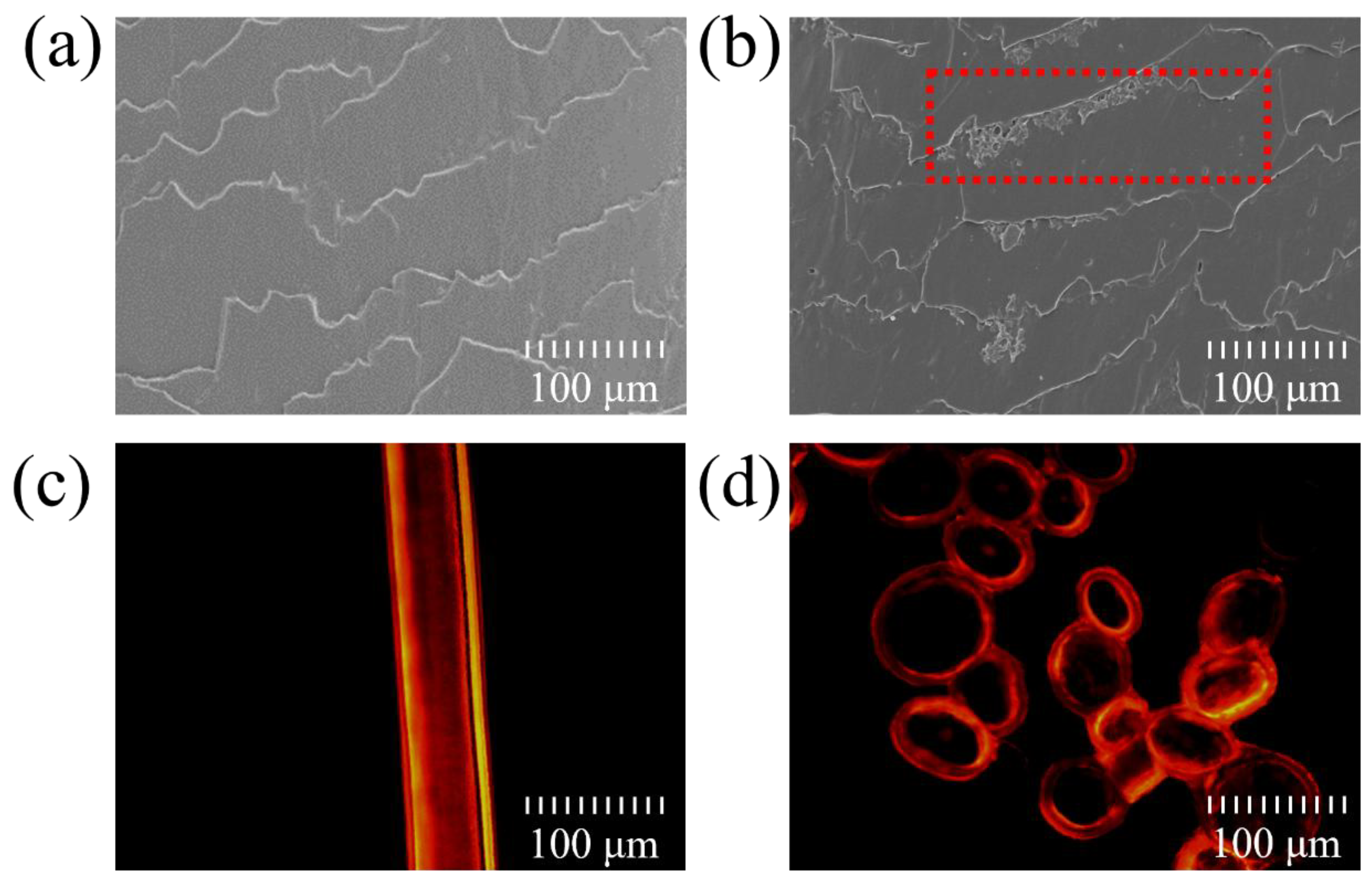
Figure 2.
morphology of cuticles characterized by SEM:(a) untreated hair before UV radiation (b) untreated hair after UV radiation (c) hair treated with hydrolyzed keratin and then exposed to UV.
Figure 2.
morphology of cuticles characterized by SEM:(a) untreated hair before UV radiation (b) untreated hair after UV radiation (c) hair treated with hydrolyzed keratin and then exposed to UV.
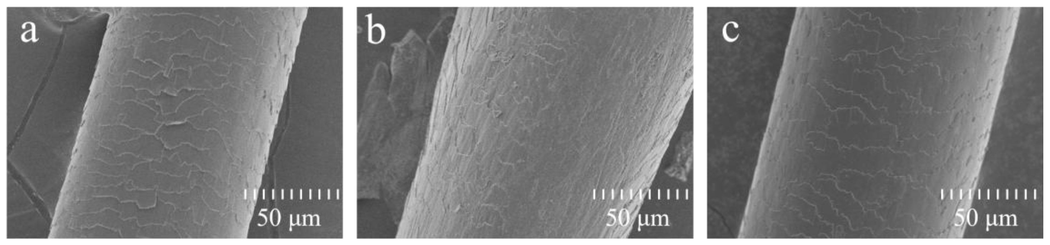
Figure 3.
(a)tensile strength, (b) plateau load and (c) Young’s module of hair samples of UV damage experiment. (p:*<0.05; **<0.01, ***<0.001).
Figure 3.
(a)tensile strength, (b) plateau load and (c) Young’s module of hair samples of UV damage experiment. (p:*<0.05; **<0.01, ***<0.001).
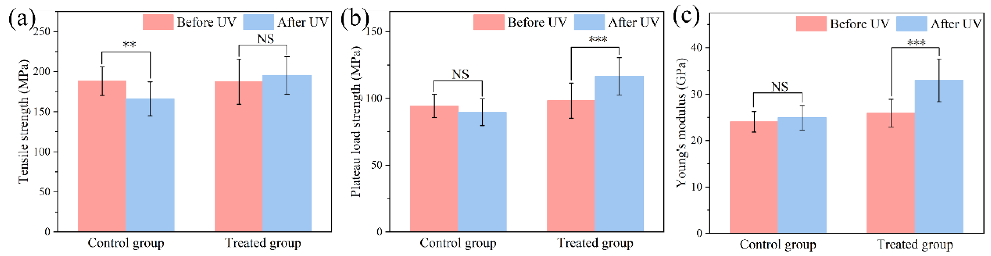
Figure 4.
(a) diagram of stress relaxation test, (b) proportion of weak interaction force (p:*<0.05; **<0.01, ***<0.001).
Figure 4.
(a) diagram of stress relaxation test, (b) proportion of weak interaction force (p:*<0.05; **<0.01, ***<0.001).
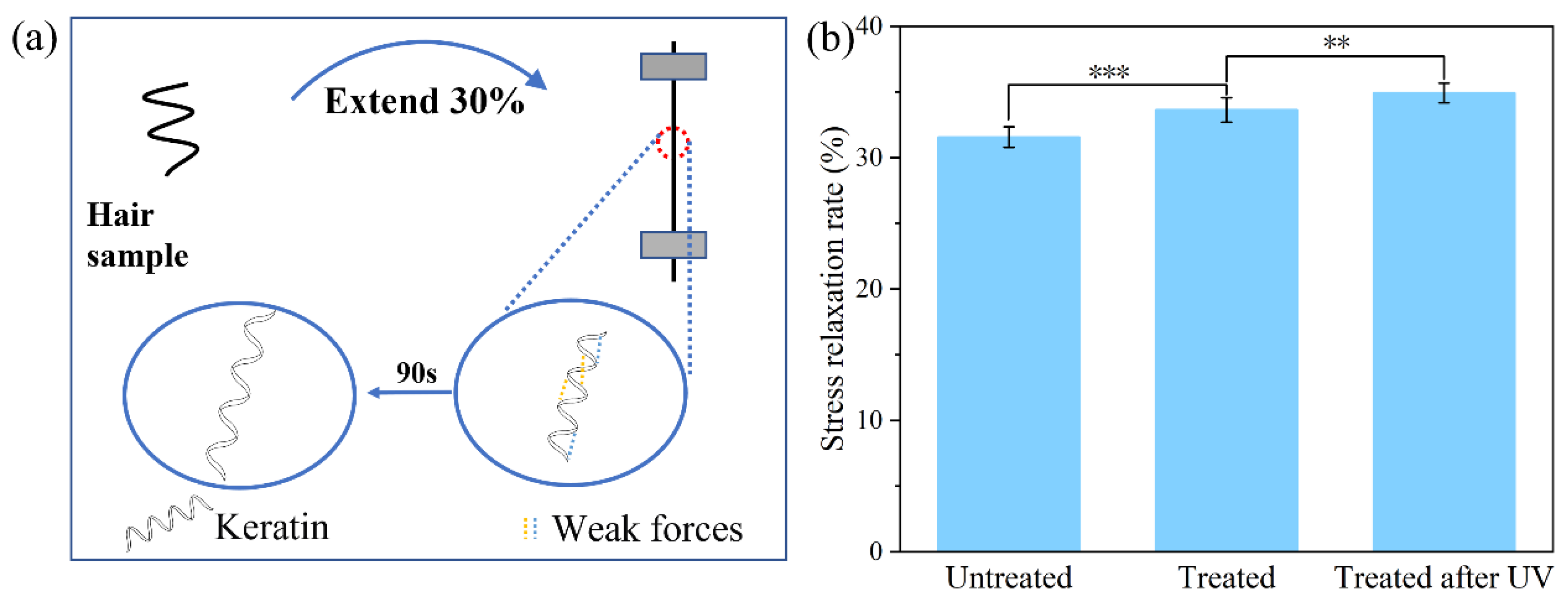
Figure 5.
DSC curves of hair samples.
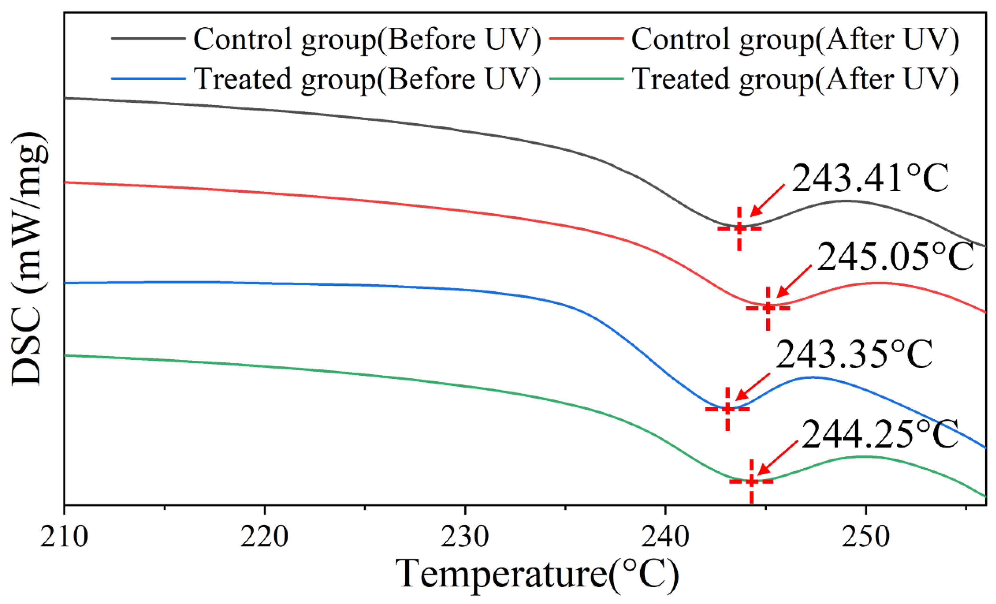
Figure 6.
mechanism diagram of hydrolyzed keratin's protection on hair.
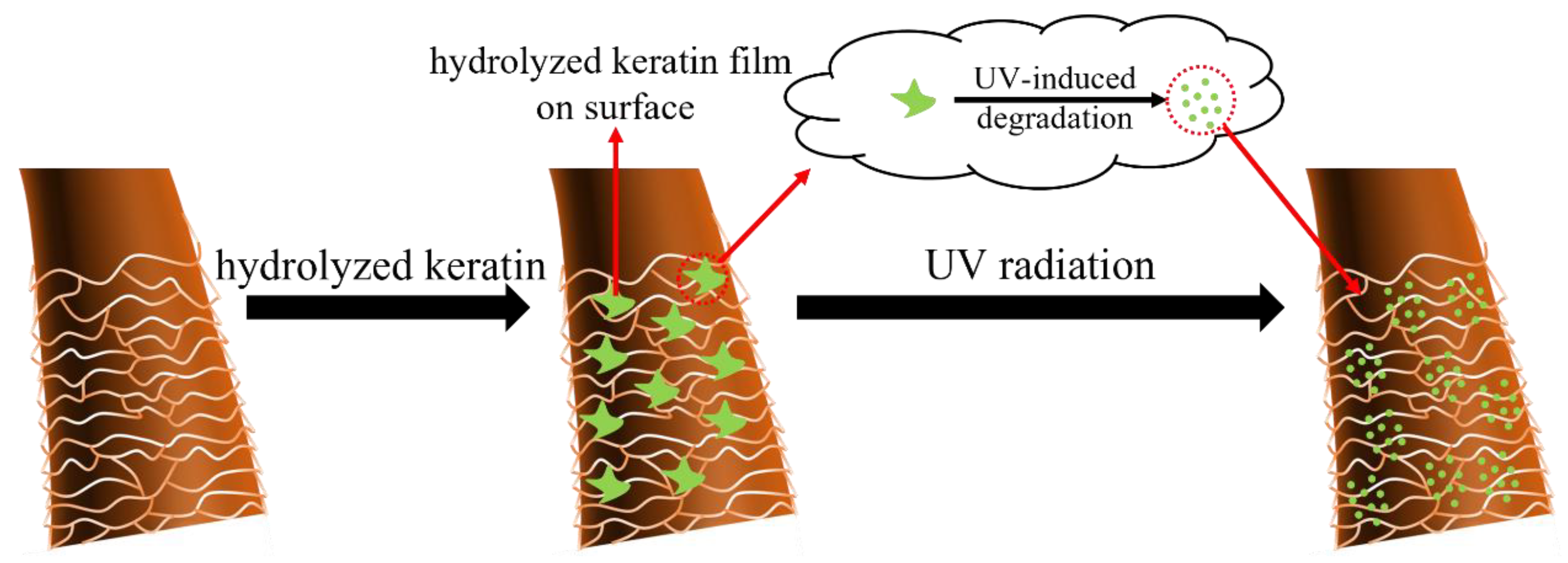
Figure 7.
(a)conductivity and (b)free amino acid concentration after treating with UV irradiation.
Disclaimer/Publisher’s Note: The statements, opinions and data contained in all publications are solely those of the individual author(s) and contributor(s) and not of MDPI and/or the editor(s). MDPI and/or the editor(s) disclaim responsibility for any injury to people or property resulting from any ideas, methods, instructions or products referred to in the content. |
© 2025 by the authors. Licensee MDPI, Basel, Switzerland. This article is an open access article distributed under the terms and conditions of the Creative Commons Attribution (CC BY) license (http://creativecommons.org/licenses/by/4.0/).
Copyright: This open access article is published under a Creative Commons CC BY 4.0 license, which permit the free download, distribution, and reuse, provided that the author and preprint are cited in any reuse.


