Preprint
Article

This version is not peer-reviewed.

EvSec22, a SNARE Protein, Regulates Hyphal Growth, Stress Tolerance, and Nematicidal Pathogenicity in Esteya vermicola

A peer-reviewed article of this preprint also exists.

Submitted:

30 January 2025

Posted:

01 February 2025

You are already at the latest version

Abstract

Bursaphelenchus xylophilus, the causative agent of pine wilt disease (PWD), poses a severe global threat to coniferous forests. Esteya vermicola, an endoparasitic nematophagous fungus, exhibits promising biocontrol potential against this pinewood nematode. The vesicular transport system, evolutionarily conserved in eukaryotes, is essential for fungal pathogenicity. Based on our genome sequence of E. vermicola CBS115803, we identified EvSec22, a gene encoding a SNARE protein implicated in vesicular transport process. This study investigates the role of EvSec22 in E. vermicola during nematode infection, utilizing our optimized gene knockout methodology. Infection assays revealed that EvSec22 deletion significantly impaired the pathogenicity of E. vermicola against B. xylophilus. Phenotypic analyses revealed that the ΔEvSec22 mutant exhibited suppressed hyphal growth, reduced conidiation, and abnormal septal spacing. Furthermore, the mutant showed significantly diminished tolerance to osmotic stress (sorbitol) and oxidative stress (hydrogen peroxide). Overall, the EvSec22 gene is associated with the virulence of E. vermicola CBS115803 against B. xylophilus, and its deletion also impacts the normal growth of E. vermicola and its tolerance to abiotic stress. This study providing new insights into SNARE protein functions in fungal biocontrol agents.

Keywords: 
;  ;  ;  ;  

1. Introduction

The pine wood nematode (Bursaphelenchus xylophilus), originally identified in 1929 from declining Pinus palustris stands in Texas, USA[1], was subsequently established in 1972 as the causative agent of pine wilt disease (PWD)–a coniferous tree epidemic commonly termed "pine tree cancer" due to its rapid lethality[2,3]." Globally, PWD is one of the most severe forest diseases, causing devastating damage to pine forest resources and ecosystems, particularly in East Asia, leading to significant economic losses[4,5,6]. Chemical nematicides are widely employed to manage B. xylophilus; however, their prolonged use frequently leads to diminished field efficacy, the development of nematode resistance, and significant environmental risks[7,8].
The use of fungi, bacteria, and actinomycetes as biological nematicides has gained increasing attention, particularly the development of nematophagous fungi for biocontrol applications[9,10]. Esteya vermicola is an endoparasitic fungus of B. xylophilus that can survive in pine trees and their resin secretions. This fungus attracts nematodes by releasing volatile compounds that mimic pine-derived chemicals. The lunate-shaped conidia of E. vermicola adhere to and subsequently infect B. xylophilus, forming penetration pegs that breach the nematode cuticle. Once inside, the fungus utilizes the nematode’s organic components for growth, eventually emerging as hyphae from the nematode carcass and producing new lunate-shaped conidia to continue the infection cycle[11]. Studies have shown that within 24 hours, 90 % of nematodes are infected by E. vermicola conidia, and complete nematode mortality occurs within 8-10 days, initiating a new infection cycle[11,12]. Due to its strong infection capability and endoparasitic nature, E. vermicola holds promise as a biocontrol agent against B. xylophilus[13,14]. Additionally, research indicates that culture media influence the production and infectivity of E. vermicola conidia. Since only lunate-shaped adhesive conidia possess infection capability, nutrient-rich potato dextrose agar (PDA) medium is suitable for large-scale fungal propagation, whereas nutrient-poor water agar (WA) medium is preferable for maximizing lunate-shaped conidia production[15].
In eukaryotic cells, most secretory proteins enter the endoplasmic reticulum (ER) lumen and are transported to the plasma membrane via vesicles or specialized carriers, progressing from the ER to the Golgi apparatus before reaching their target membranes[16]. SNAREs (soluble N-ethylmaleimide-sensitive factor attachment protein receptors) are a highly conserved class of membrane-associated proteins that play a vital role in exocytosis by facilitating vesicle-membrane fusion, a process crucial for maintaining cellular function and homeostasis[17]. Based on conserved amino acid residues within the SNARE motif, SNARE proteins are classified into v-SNAREs and t-SNAREs. v-SNAREs are typically embedded in transport vesicles, while t-SNAREs localize to target membranes[18]. In other types of cells or organisms, homologs of synaptic SNARE are involved in cytokinesis[19]. VAMP (also known as synaptobrevin) is a v-SNARE protein on the vesicle membrane, and in yeast, Snc1 and Snc2 are homologous proteins of VAMP, which are involved in the cytosolization of yeast secretory vesicles[20]. Syntaxin is a t-SNARE protein on the cytoplasmic membrane, and in yeast, Sso1p and Sso2p are homologous proteins of Syntaxin , which play a key role in the secretion process in yeast[21].
v-SNARE Snc1 is responsible for both paracrine and retrograde vesicular transport between the plasma membrane (PM) and the Golgi apparatus, and it interacts with the cytosolic SNARE (Sso 1/2, Sec 9) and the cytosolic complex to complete the cytosolic process[22]. It has been demonstrated that the ER-Golgi transport cycle operates in a COPI(Coat Protein I)-dependent manner, requiring SNARE proteins to bind membranes via their transmembrane domains (TMDs)[23]. The conservation of the TMD in SNARE proteins highlights the significance of retrograde transport pathways in cellular function[24]. Studies have shown that the deletion of SNARE protein Sec22 impairs the extracellular secretion of virulence-associated proteins required for host infection. VdSec22 in Verticillium dahliae regulates the secretion of various enzymes, such as cellulases, pectinases, and xylanases, which are involved in carbohydrate metabolism and host cell wall degradation. Loss of VdSec22 reduces V. dahliae virulence in cotton[25]. Additionally, Sec22 contributes to effector secretion via exocytosis in Colletotrichum orbiculare, facilitating primary hyphal development[26]. MoSec22 is also involved in Magnaporthe oryzae infection of rice. Deletion of MoSec22 inhibits conidiation, disrupts cell wall integrity, and impairs reactive oxygen species (ROS) production[27]. Both of which are critical for hyphal germination, appressorium formation, hyphal tip growth, and pathogenicity[28,29].
Despite its established roles in various pathogenic fungi, the function of Sec22 in E. vermicola development and infection of B. xylophilus remains unexplored. This study investigates the role of EvSec22 in E. vermicola CBS115803 through gene knockout analysis.

2. Materials and Methods

2.1. Vector Construction and Fungal Transformation

The knockout vector was constructed following our previously established gene knockout protocol[30]. Upstream and downstream DNA fragments (1.5 kb each) of the EvSec22 gene were amplified from the genome of E. vermicola CBS115803. A 2.0 kb hygromycin resistance gene cassette was amplified from the pSilent1 vector. Homologous recombination fragments were obtained via fusion PCR and then ligated into a linearized (BamH I) green fluorescent protein (GFP) expression plasmid (pKOXN) using a 2× Seamless Cloning Kit (D7010S, Beyotime Biotech Inc., China). The gene knockout plasmid (pKOXN-EvSec22) was introduced into Agrobacterium tumefaciens AGL1 via the freeze-thaw method. E. vermicola CBS115803 was transformed using Agrobacterium-mediated transformation, and knockout transformants were selected on a medium supplemented with 500 μg/mL cefotaxime and 200 μg/mL hygromycin B. The gene’s deleted strains were screened according to our published protocol. Detailed methods are provided in Supplementary Methods.
To construct the complementation vector, the full-length EvSec22 gene, including its 1,500 bp promoter and 1,500 bp terminator sequences, was amplified using gene-com F/R primers and cloned into the complementation plasmid pMD-3. The resulting construct was introduced into the corresponding gene deletion strain (ΔEvSec22). Transformants were selected using cefotaxime at a concentration of 500 μg/mL, hygromycin B at 150 μg/mL, and Basta at 50 μg/mL. Gene expression in the complemented strain was analyzed using RT-PCR and designated as ΔEvSec22comp. Primer sequences are listed in Table S1.

2.2. Infectivity Assay of B. xylophilus

Botrytis cinerea was inoculated onto PDA plates and incubated at 26°C until the fungal mycelia covered the entire 90 mm diameter plates. Subsequently, B. xylophilus nematodes were surface-sterilized by immersion in a 3% hydrogen peroxide solution and then inoculated onto B. cinerea. This process aimed to eliminate surface bacteria on the nematodes before infection experiments. After incubation at 26°C for five days, nematodes were isolated for the subsequent E. vermicola infection assay.
Simultaneously, 50 μL of conidial suspension (5×10⁶ conidia) from wild-type E. vermicola CBS115803, mutant strains, and complemented strains were evenly spread onto WA plates. These 60 mm diameter plates were incubated in the dark at 26°C for five days. A 20 μL suspension of B. xylophilus nematodes (~400 individuals) was then inoculated onto E. vermicola WA plates. Nematodes were observed every two days for up to 11 days using an inverted optical microscope. Mortality rates were determined by counting the percentage of dead nematodes in the first 100 encountered[31]. Each experiment was repeated six times.

2.3. Conidia Count

A 5 μL suspension containing 1×10⁷ conidia from wild-type E. vermicola CBS115803, mutant strains, and complemented strains was spotted onto pre-prepared PDA plates and incubated at 26°C for 14 days. Colonies were then washed with sterile water to obtain conidial suspensions. The total number of lunate and rod-shaped conidia was counted using a hemocytometer under a microscope, and the proportion of lunate conidia was calculated[32]. Each experiment was repeated three times.

2.4. Growth Assay on Solid Medium

A 5 μL suspension containing 1×10⁷ conidia from wild-type E. vermicola CBS115803, mutant strains, and complemented strains was spotted onto pre-prepared 90 mm diameter PDA plates. After incubation at 26°C for eight days, colony sizes were photographed and measured. Each experiment was repeated three times.

2.5. Hyphal Septal Distance Measurement

Wild-type E. vermicola CBS115803, mutant strains, and complemented strains were cultured on PDA plates at 26°C for eight days. A 1:1 mixture of sterile water and Calcofluor White fluorescent dye was prepared, and hyphae were stained in this solution for 0.5 hours. The hyphal septal distance was observed under a fluorescence microscope[33]. Each experiment was repeated 25 times.

2.6. Abiotic Stress Assay

PDA solid media containing 0.7 M KCl, 1 mM H₂O₂, or 1 M sorbitol were prepared, with PDA serving as the control. A 3 μL conidial suspension was spotted onto the center of each medium[34]. The plates were then incubated upside down at 26°C for 10 days. Colony sizes were photographed and measured. The measured colony diameters were analyzed and relative growth inhibition (RGI) was used to assess stress tolerance, RGI = (DC - DT)/DC × 100%, DC and DT denote the diameters of the colonies on control and stress plates, respectively[35]. Each experiment was repeated three times.

2.7. Statistical Analysis

The data were expressed as mean ± standard deviation (SD). Statistical analysis was performed using one-way analysis of variance (ANOVA) followed by Dunnett Test, utilizing GraphPad Prism 8.0 software (GraphPad, San Diego, CA, USA).

3. Result

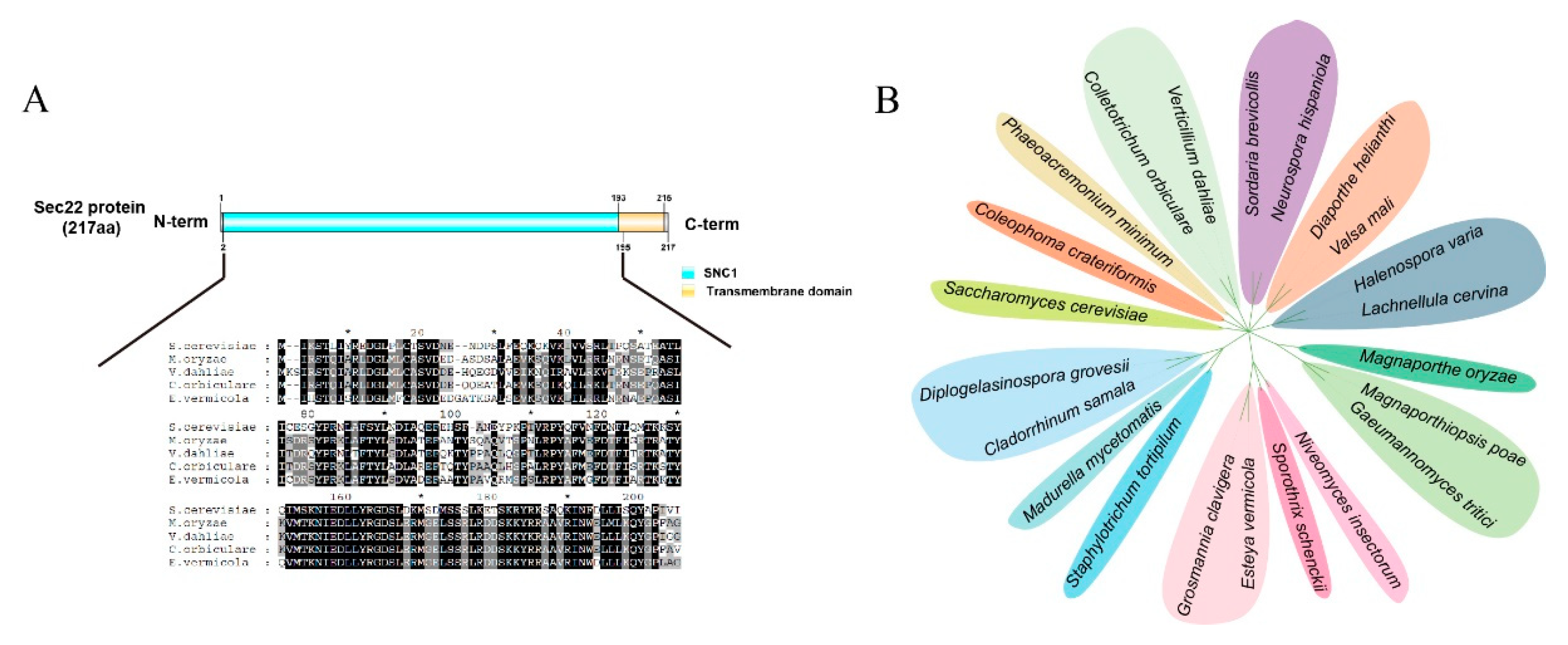
3.1. Sequence Analysis of EvSec22 in E. vermicola CBS115803

Based on the genome sequencing results of E. vermicola CBS115803 (PRJNA361544), we identified a SNARE gene, EvSec22, through homology alignment. The gene has a full length of 654 bp, lacks introns, and encodes a 217-amino acid protein (Supplementary protein and genome sequence). Conserved domain prediction revealed that the C-terminal of this protein contains a conserved SNARE domain (SNC1, residues 2-195) and a transmembrane domain (residues 193-215). The SNC1 protein, a member of a highly conserved protein family, is classified as a SNARE protein that plays a crucial role in intracellular transport and secretion processes. This functional characterization strongly suggests that the EvSec22 represents a typical SNARE-type protein associated with the secretory pathway. Homology alignment revealed that EvSec22 is highly conserved among various fungi, including Saccharomyces cerevisiae, Magnaporthe oryzae, Verticillium dahliae, and Colletotrichum orbiculare (Figure 1A). Moreover, EvSec22 is widely distributed among numerous fungal species (Figure 1B).

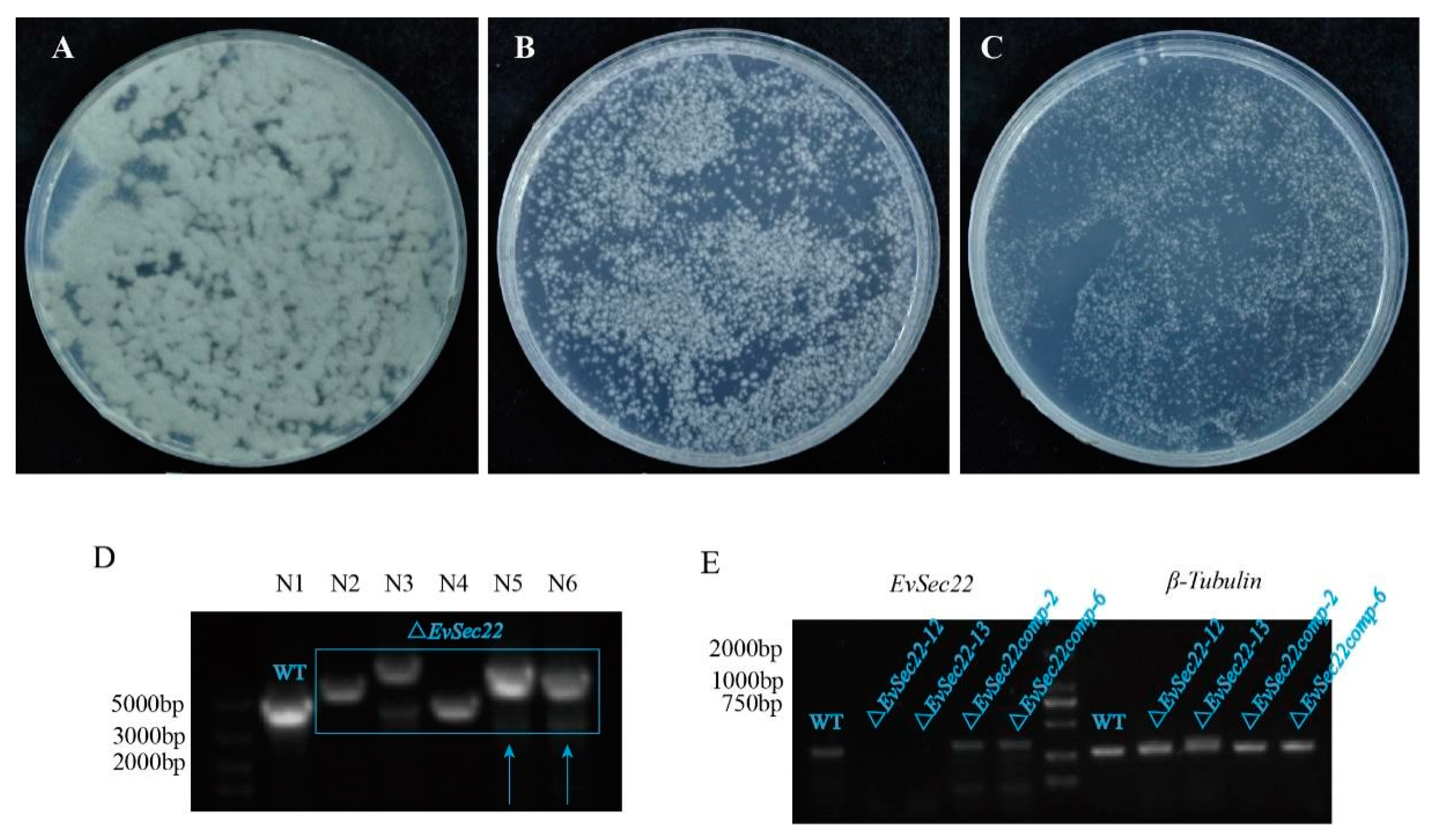
3.2. Optimization of E. vermicola Transformation Methods and Construction of EvSec22 Mutants and Complementary Strains

The robust hyphal growth of E. vermicola CBS115803 hampers the selection of single-colony transformants. Clethodim, a growth inhibitor, significantly reduced hyphal growth at 0.01% and 0.02% concentrations. In this study, a concentration of 0.02% clethodim in PDA medium was employed to enhance the efficiency of single-colony isolation (Figure 2A-C).
EvSec22 knockout mutants were screened using fluorescence exclusion (Figure S2), flank-specific PCR validation, and gene expression analysis (Figure 2D). Expression analysis confirmed the successful recovery of EvSec22 in complemented strains (Figure 2E).

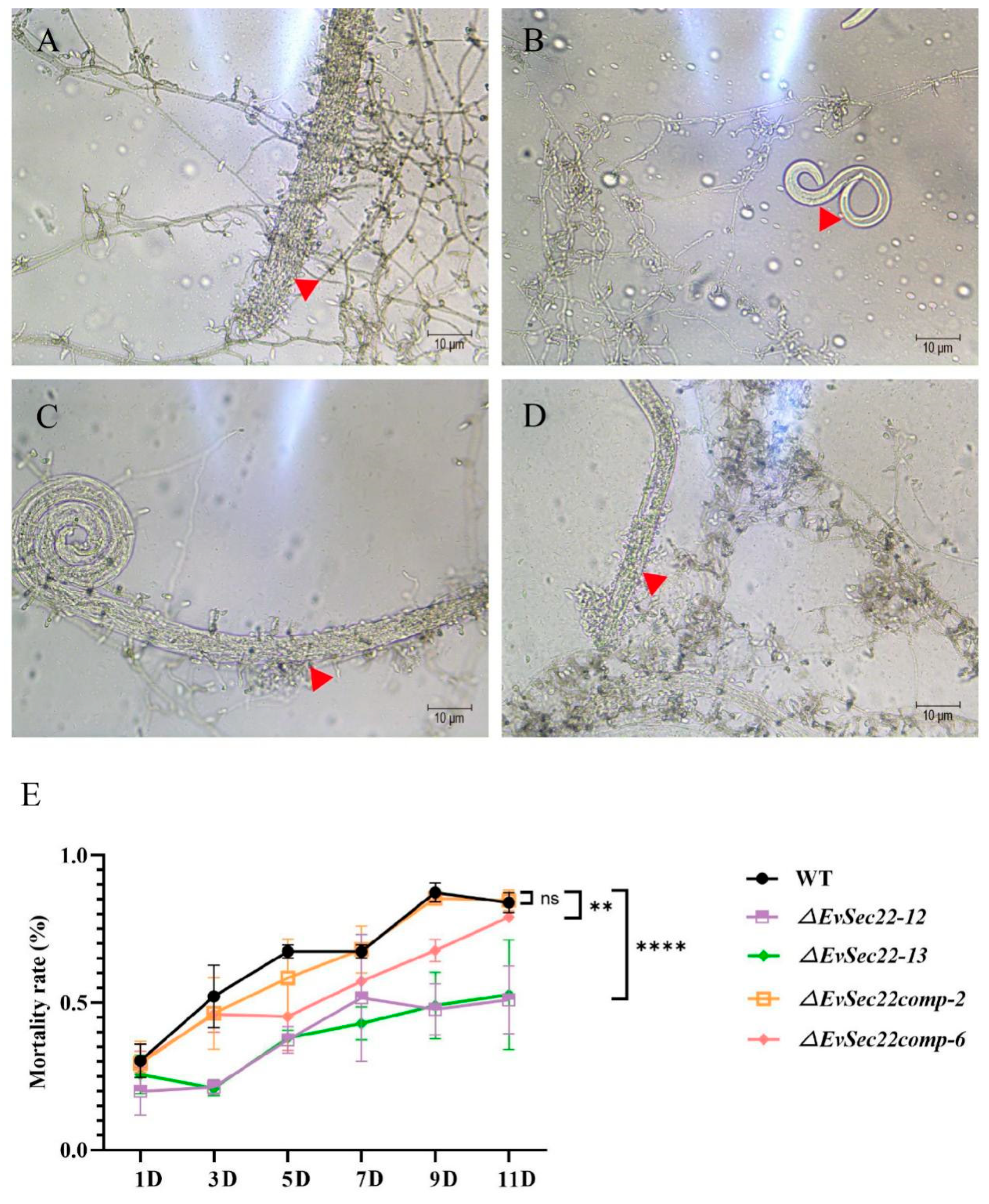
3.3. EvSec22 Mutants Impaired the Infectivity of E. vermicola Against B. xylophilus

To evaluate the infectivity of E. vermicola CBS115803 toward B. xylophilus, conidia of E. vermicola were first cultured on water agar, followed by the introduction of approximately 400 B. xylophilus nematodes. After two days, nematode mortality was significantly higher in wild-type and complemented strains (Figure 3A, C, D), while nematodes in the ΔEvSec22 group remained active (Figure 3B). Infection assays revealed that the ΔEvSec22 strain exhibited a significantly reduced ability to infect B. xylophilus. After 11 days of co-culture, the nematode mortality rate of ΔEvSec22 -12 and ΔEvSec22 -13 was 51.84% and 52.67%, respectively, compared to 84% in the wild-type group. Complementation of EvSec22 restored infectivity to near wild-type levels, with mortality rates of 84.67% and 79%, respectively. These results confirm that EvSec22 is crucial for E. vermicola infectivity against B. xylophilus.

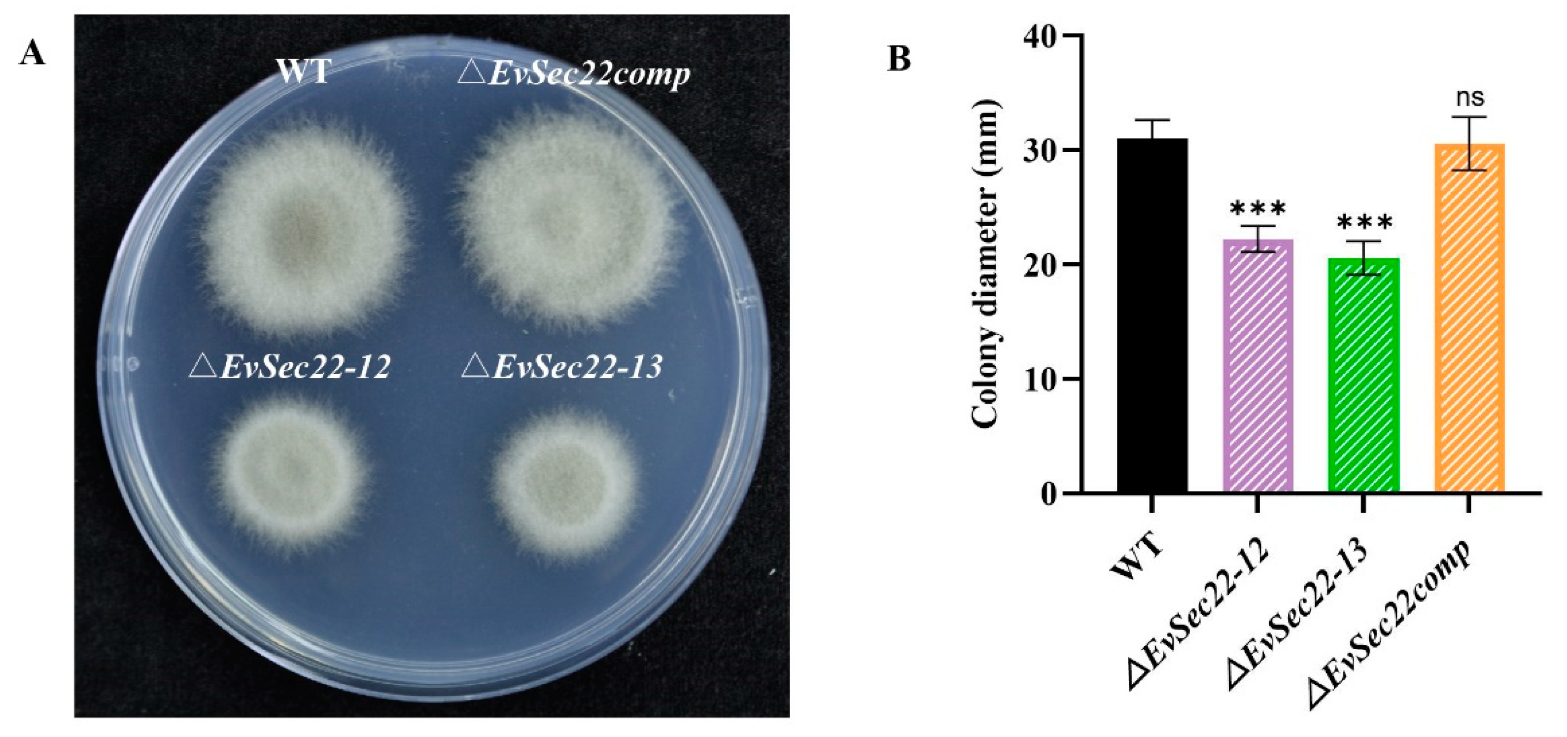
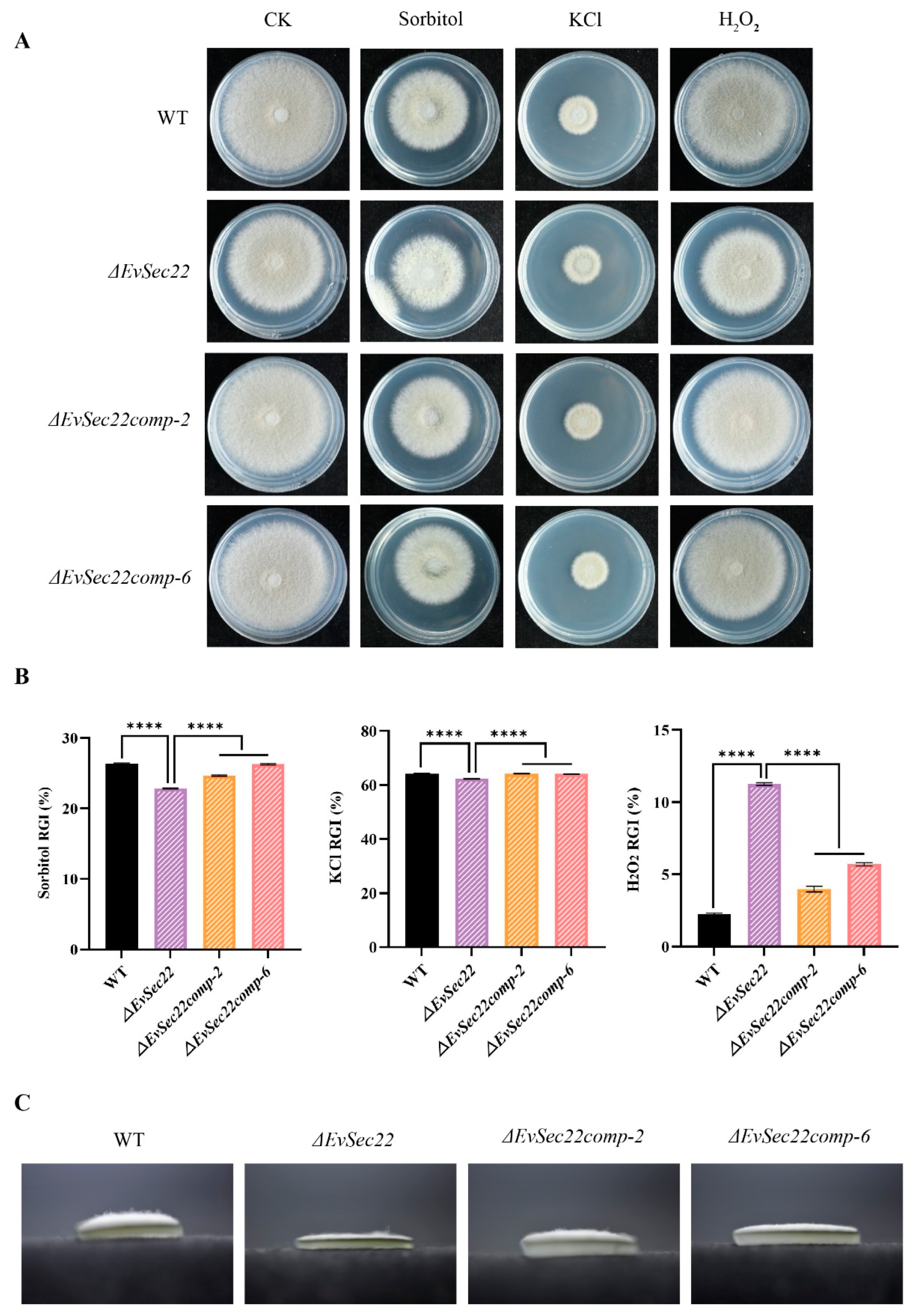
3.4. Loss of EvSec22 Leads to Slower Hyphal Growth and Hyphal Septal Spacing in E. vermicola

On PDA medium, the colony morphology and color of WT, ΔEvSec22 mutant, and complementation strains exhibited no significant differences, with all colonies displaying a grayish-white appearance and smooth margins. However, distinct variations were observed in growth rate and colony size. Deletion of EvSec22 significantly reduced the growth rate of E. vermicola, whereas reintroduction of EvSec22 into the mutant strain fully restored its growth capacity (Figure 4A,B).
We further compared the hyphal septal spacing among the different strains. The results revealed that the ΔEvSec22 mutant exhibited a significantly reduced septal distance. However, reintroduction of EvSec22 restored the septal spacing to WT levels in the complemented strain (Figure S2).

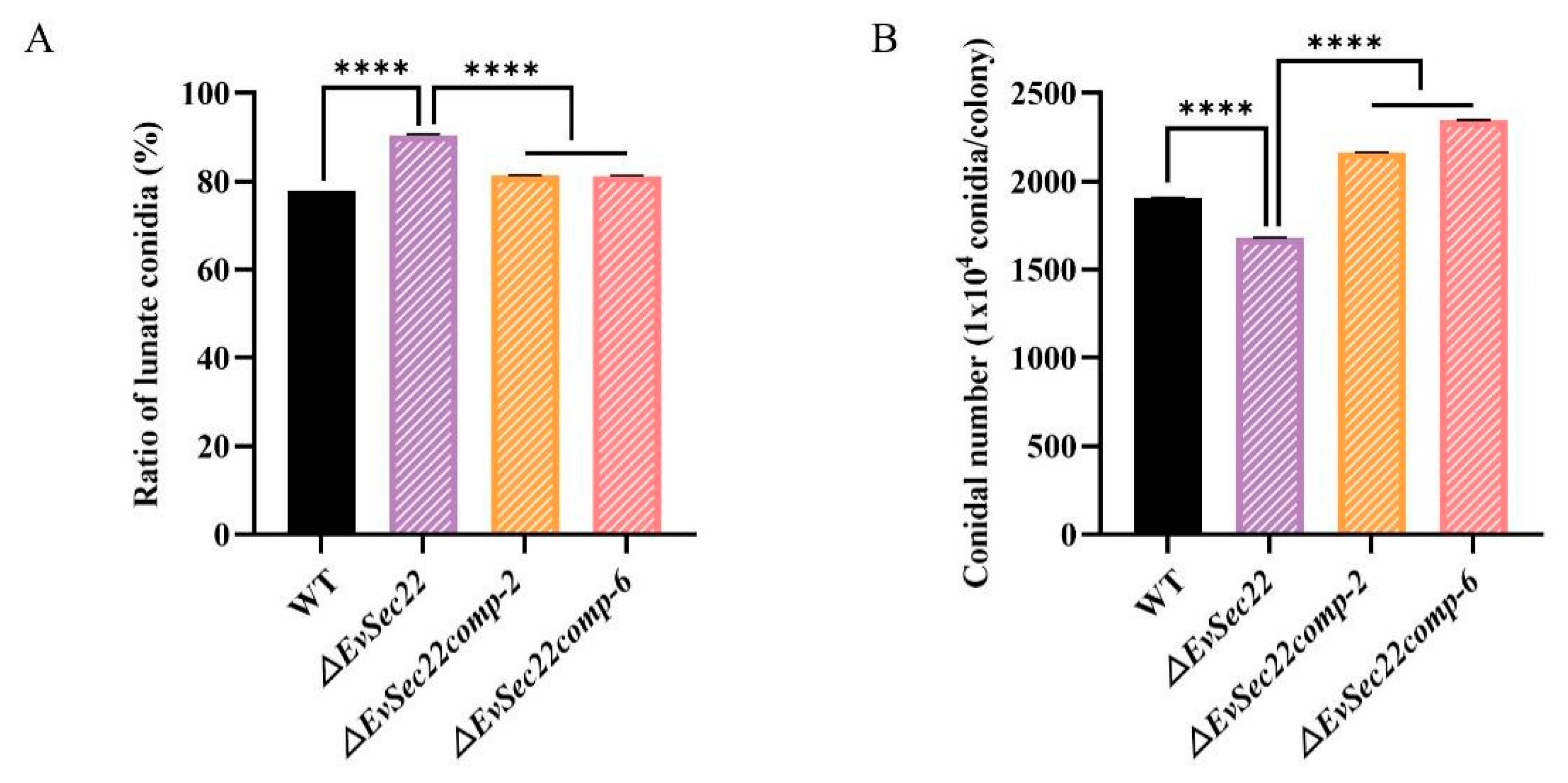
3.5. EvSec22 Deletion Reduces Total Conidia Count but Increases the Proportion of Lunate-Shaped Conidia

Conidia play a critical role in the infection process of E. vermicola against B. xylophilus. Compared to WT, the total conidia count was significantly reduced in the ΔEvSec22 mutant. However, reintroduction of EvSec22 into the mutant strain significantly increased the total conidia count (Figure 5A). Surprisingly, the proportion of lunate-shaped conidia in the ΔEvSec22 mutant was higher than that in WT, while the ΔEvSec22 complemented strain exhibited a lower proportion of lunate-shaped conidia, though it remained significantly higher than that of WT strain (Figure 5B).

3.6. Deletion of EvSec22 Affects E. vermicola's Tolerance to Abiotic Stress

To evaluate the role of EvSec22 in abiotic stress tolerance in E. vermicola, we examined the sensitivity of four strains (WT, ΔEvSec22, ΔEvSec22comp-2, and ΔEvSec22comp-6) under various abiotic stress conditions, including sorbitol, potassium chloride, and hydrogen peroxide. All strains exhibited inhibited growth under these stress conditions (Figure 6A).
To quantify stress tolerance, we calculated the relative growth inhibition (RGI) based on colony diameter measurements. The ΔEvSec22 mutant displayed enhanced tolerance to sorbitol and potassium chloride but showed extreme sensitivity to hydrogen peroxide. Reintroduction of EvSec22 partially restored hydrogen peroxide tolerance (Figure 6B). Notably, the ΔEvSec22 mutant formed sparse aerial mycelia on sorbitol-supplemented PDA medium, indicating that the mutant is, in fact, highly sensitive to sorbitol. Although the relative growth inhibition (RGI) values based on colony diameter showed that the ΔEvSec22 mutant had a stronger tolerance to sorbitol (Figure 6C). These findings suggest that EvSec22 plays a critical role in regulating hyphal growth and stress tolerance in E. vermicola.

4. Discussion

During the genetic transformation of E. vermicola CBS115803, we encountered challenges in obtaining single colonies due to the rapid growth of filamentous fungal hyphae. To address this, clethodim, a herbicide known to inhibit hyphal growth in filamentous fungi, was added to the medium at a concentration of 0.02%. This significantly improved the efficiency of single-colony isolation and enhanced transformation efficiency. Additionally, sodium deoxycholate was incorporated into the medium to further inhibit hyphal growth. However, we observed that its effect on E. vermicola was less pronounced compared to clethodim (unpublished).
The deletion of Sec22 has been shown to impair the ability of pathogenic fungi to infect plants. In our study, the ΔEvSec22 mutant exhibited a 30% reduction in lethality against B. xylophilus compared to the wild-type E. vermicola CBS115803. Previous research has demonstrated that VdSec22 not only regulates protein secretion but also influences conidial production[25]. Consistent with this, we observed a reduction in the total conidia count in the ΔEvSec22 mutant, alongside an increased proportion of lunate-shaped infective conidia. Under low-nutrient conditions, E. vermicola CBS115803 produces a higher proportion of lunate-shaped conidia[15]. Given that Sec22 is involved in the secretion of extracellular degradative enzymes such as cellulases, pectinases, and xylanases[25], we hypothesize that the ΔEvSec22 mutation disrupts the secretion of nutrient-absorbing proteins in E. vermicola. This disruption likely induces an intracellular low-nutrient state, promoting the formation of lunate-shaped conidia.
Our findings also revealed that the ΔEvSec22 mutant exhibited significantly reduced hyphal growth and decreased hyphal septal spacing, which may contribute to the overall decline in conidia production. Studies on other fungi have similarly demonstrated that Sec22 is critical for hyphal germination and apical growth. For instance, deletion of FgSec22 in Fusarium graminearum resulted in conidial morphological defects, including altered septation[36]. Similarly, knockout of Sec22 in Arthrobotrys oligospora and Sordaria macrospora led to reduced sporulation, lower conidia germination rates, abnormal conidia morphology, and thinner, sparser aerial hyphae[37,38]. Collectively, these results indicate that deletion of EvSec22 impairs the growth rate, conidia composition, sporulation, and stress tolerance of E. vermicola, which may also explain its reduced virulence.
In response to environmental stresses and infections, organisms generate reactive oxygen species (ROS) to bolster their defenses while producing enzymes to scavenge free radicals and minimize cellular damage[39,40]. The ΔEvSec22 mutant exhibited heightened sensitivity to hydrogen peroxide stress, suggesting an impaired ability to secrete catalase, which likely hindered its growth. When E. vermicola infects B. xylophilus, the nematode produces ROS as a defense mechanism. Efficient secretion of ROS-scavenging enzymes by E. vermicola could suppress this defense. The increased sensitivity of ΔEvSec22 to hydrogen peroxide indicates a reduced capacity for ROS detoxification, potentially explaining its diminished infectivity against B. xylophilus.
Although relative growth inhibition (RGI) analysis based on colony diameters suggested that ΔEvSec22 exhibited greater tolerance to sorbitol, we observed significantly reduced hyphal density in the mutant, indicating severe growth impairment. Thus, in reality, ΔEvSec22 is highly sensitive to sorbitol. Similar findings have been reported in other studies, where high sorbitol concentrations induce osmotic stress, leading to reduced colony diameters and restricted hyphal growth[41].
In summary, our results demonstrate that the SNARE protein Sec22 in E. vermicola CBS115803 plays a critical role in vegetative growth, abiotic stress tolerance, and the infection of B. xylophilus. These findings provide valuable insights into the biological control mechanisms of E. vermicola CBS115803 against B. xylophilus. Future research will focus on analyzing differences in extracellular secreted proteins following EvSec22 knockout in E. vermicola CBS115803.

Supplementary Materials

The following supporting information can be downloaded at the website of this paper posted on Preprints.org. Figure S1. Schematic representation of the knockout mutation screening strategy. Figure S2. Quantitative assessment of hyphal septal spacing in E. vermicola strains. Table S1. All the primers used in this study. Table S2. Fungal names and sequence numbers in the phylogenetic tree. Supplementary protein and genome sequence. Supplementary Methods.

Author Contributions

Conceptualization, JJ.Y. and CJ.X.; methodology, JJ.Y. and R.Z.; software, X.P. and YL.W.; investigation, JJ.Y. and ZW.C.; resources, TQ.Y. and CJ.X; writing—original draft preparation, JJ.Y. and CJ.X.; visualization, LH.H; supervision and funding acquisition, CJ.X. All authors have read and agreed to the published version of the manuscript.

Funding

This research was funded by the National Natural Science Foundation of China [32271898] and the Natural Science Foundation of Chongqing [cstc2021jcyj-msxmX0098].

Institutional Review Board Statement

Not applicable.

Informed Consent Statement

Not applicable.

Conflicts of Interest

The authors declare no conflict of interest.

References

  1. Nickle, W.R.; Golden, A.M.; Mamiya, Y.; Wergin, W.P. On the Taxonomy and Morphology of the Pine Wood Nematode, Bursaphelenchus xylophilus (Steiner &Buhrer 1934) Nickle 1970. J Nematol. 1981, 13, 385-92.
  2. Mamiya, Y. Pine wood nematode, Bursaphelenchus lignicolus Mamiya and Kiyohara, as a causal agent of pine wilting disease. Environmental Science, Biology, Agricultural and Food Sciences. 1972.
  3. Jones J.T.; Haegeman, A.; Danchin, E.G.; Gaur, H.S.; Helder, J.; Jones, M.G.; Kikuchi, T.; Manzanilla-López, R.; Palomares-Rius, J.E.; Wesemael, W.M.; Perry, R.N. Top 10 plant-parasitic nematodes in molecular plant pathology. Mol Plant Pathol. 2013, 14, 946-61. [CrossRef]
  4. Hunt, D., Pine wilt disease: a worldwide threat to forest ecosystems. Nematology, 2009. [CrossRef]
  5. Robertson, L.; Arcos, S.C.; Escuer, M.; Merino, R.S.; Espárrago, G.; Abelleira, A.; Navas, A. Incidence of the pinewood nematode Bursaphelenchus xylophlius Steiner & Buhrer, 1934 (Nickle, 1970) in Spain. Nematology. 2011, 13, 755-757.
  6. Zhao, L.L.; Mota, M.; Vieira, P.; Butcher, R.A.; Sun, J.H. Interspecific communication between pinewood nematode, its insect vector, and associated microbes. Trends Parasitol. 2014, 30, 299-308. [CrossRef]
  7. Chen, J.X.; Li, Q.X.; Song, B.A. Chemical Nematicides: Recent Research Progress and Outlook. J Agric Food Chem. 2020, 68, 12175-12188. [CrossRef]
  8. Liang, W.L.; Yu, A.X.; Wang, G.D.; Zheng, F.; Jia, J.L.; Xu, H.H. Chitosan-based nanoparticles of avermectin to control pine wood nematodes. Int J Biol Macromol. 2018, 112, 258-263. [CrossRef]
  9. Affokpon, A.; Coyne, D.L.; Htay, C.C.; Agbèdè, R.D.; Lawouin, L.; Coosemans, J. Biocontrol potential of native Trichoderma isolates against root-knot nematodes in West African vegetable production systems. Soil Biology & Biochemistry, 2011, 43, 600-608. [CrossRef]
  10. Kerry, B.R. Rhizosphere Interactions and the Exploitation of Microbial Agents for the Biological Control of Plant-Parasitic Nematodes. Annu Rev Phytopathol. 2000, 38:423-441. [CrossRef]
  11. Liou, J.Y.; Shih, J.Y.; Tzean, S.S. Esteya, a new nematophagous genus from Taiwan, attacking the pinewood nematode (Bursaphelenchus xylophilus). Mycological Research, 1999, 103, 242-248. [CrossRef]
  12. Wang, C.Y.; Fang, Z.M.; Sun, B.S.; Gu, L.J.; Zhang, K.Q.; Sung, C.K. High infectivity of an endoparasitic fungus strain, Esteya vermicola, against nematodes. J Microbiol. 2008, 46, 380-9. [CrossRef]
  13. Lin, F.; Ye, J.L.; Wang, H.G.; Zhang, A.J.; Zhao, B.G. Host deception: predaceous fungus, Esteya vermicola, entices pine wood nematode by mimicking the scent of pine tree for nutrient. PLoS One. 2013, 8, e71676. [CrossRef]
  14. Wang, C.Y.; Fang, Z.M.; Wang, Z.; Zhang, D.L.; Gu, L.J.; Lee, M.R.; Liu, L.; Sung, C.K. Biological control of the pinewood nematode Bursaphelenchus xylophilus by application of the endoparasitic fungus Esteya vermicola. J BioControl. 2011. 56, 91-100. [CrossRef]
  15. Wang, X.; Wang, T.T.; Wang, J.C.; Guan, T.L.; Li, H.M. Morphological, molecular and biological characterization of Esteya vermicola, a nematophagous fungus isolated from intercepted wood packing materials exported from Brazil. J Mycoscience. 2014, 55, 367-377. [CrossRef]
  16. Bonifacino, J.S.; Glick, B.S. The mechanisms of vesicle budding and fusion. Cell. 2004, 116, 153-66. [CrossRef]
  17. Zhang, Y.; Yang, J. Securing SNAREs for assembly. J Biol Chem. 2020, 295, 10136-10137. [CrossRef]
  18. Jahn, R.; Scheller, R.H. SNAREs--engines for membrane fusion. Nat Rev Mol Cell Biol. 2006, 7, 631-43. [CrossRef]
  19. Fasshauer, D.; Sutton, R.B.; Brunger, A.T.; Jahn, R. Conserved structural features of the synaptic fusion complex: SNARE proteins reclassified as Q- and R-SNAREs. Proc Natl Acad Sci U S A. 1998, 95, 15781-6. [CrossRef]
  20. Shao, K.K.; Li, F.; Yang, Y.; Wang, N.; Gao, X.D.; Nakanishi, H. Characteristics of SNARE proteins are defined by distinctive properties of SNARE motifs. Biochim Biophys Acta Gen Subj. 2020, 1864, 129658. [CrossRef]
  21. Aalto, M.K.; Ronne, H.; Keränen, S. Yeast syntaxins Sso1p and Sso2p belong to a family of related membrane proteins that function in vesicular transport. EMBO J. 1993, 12, 4095-104. [CrossRef]
  22. Adnan, M.; Islam, W.; Waheed, A.; Hussain, Q.; Shen, L.; Wang, J.; Liu, G. SNARE Protein Snc1 Is Essential for Vesicle Trafficking, Membrane Fusion and Protein Secretion in Fungi. Cells. 2023, 12, 1547. [CrossRef]
  23. Ballensiefen, W.; Ossipov, D.; Schmitt, H.D. Recycling of the yeast v-SNARE Sec22p involves COPI-proteins and the ER transmembrane proteins Ufe1p and Sec20p. J Cell Sci. 1998, 111, 1507-20. [CrossRef]
  24. Chen, L.; Lau, M.S.Y.; Banfield, D.K. Multiple ER-Golgi SNARE transmembrane domains are dispensable for trafficking but required for SNARE recycling. Mol Biol Cell. 2016, 27, 2633-41. [CrossRef]
  25. Wang, J.; Tian, L.; Zhang, D.D.; Short, D.P.G.; Zhou, L.; Song, S.S.; Liu, Y.; Wang, D.; Kong, Z.Q.; Cui, W.Y.; Ma, X.F.; Klosterman, S.J.; Subbarao, K.V.; Chen, J.Y.; Dai, X.F. SNARE-Encoding Genes VdSec22 and VdSso1 Mediate Protein Secretion Required for Full Virulence in Verticillium dahliae. Mol Plant Microbe Interact. 2018, 31, 651-664. [CrossRef]
  26. Irieda, H.; Maeda, H.; Akiyama, K.; Hagiwara, A.; Saitoh, H.; Uemura, A.; Terauchi, R.; Takano, Y. Colletotrichum orbiculare Secretes Virulence Effectors to a Biotrophic Interface at the Primary Hyphal Neck via Exocytosis Coupled with SEC22-Mediated Traffic. Plant Cell. 2014, 26, 2265-2281. [CrossRef]
  27. Song, W.W.; Dou, X.Y.; Qi, Z.Q.; Wang, Q.; Zhang, X.; Zhang, H.F.; Guo, M.; Dong, S.M.; Zhang, Z.G.; Wang, P.; Zheng X.B. R-SNARE homolog MoSec22 is required for conidiogenesis, cell wall integrity, and pathogenesis of Magnaporthe oryzae. PLoS One. 2010, 5, e13193. [CrossRef]
  28. Zhou, S.Z.; Liu, S.; Guo, C.C.; Wei, H.W.; He, Z.H.; Liu, Z.Q.; Li, X.Y. The C2H2 Transcription Factor Con7 Regulates Vegetative Growth, Cell Wall Integrity, Oxidative Stress, Asexual Sporulation, Appressorium and Hyphopodium Formation, and Pathogenicity in Colletotrichum graminicola and Colletotrichum siamense. J Fungi (Basel). 2024, 10, 495. [CrossRef]
  29. Scott, B.; Eaton, C.J. Role of reactive oxygen species in fungal cellular differentiations. Curr Opin Microbiol. 2008, 11, 488-93. [CrossRef]
  30. Xie, C.J.; Li, Q.L.; Yang, X.Y. Characterization of VdASP F2 Secretory Factor from Verticillium dahliae by a Fast and Easy Gene Knockout System. Mol Plant Microbe Interact. 2017, 30, 444-454. [CrossRef]
  31. Wang, H.H.; Wang, Y.B.; Yin, C.; Gao, J.; Tao, R.; Sun, Y.L.; Wang, C.Y.; Wang, Z., Li, Y.X.; Sung, C.K. In vivo infection of Bursaphelenchus xylophilus by the fungus Esteya vermicola. Pest Manag Sci. 2020, 76, 2854-2864. [CrossRef]
  32. Chen, C.; Hu, Z.J.; Zheng, X.Y.; Yuan, J.J.; Zou, R.; Wang, Y.L.; Peng, X.; Xie C.J. The essential role of arginine biosynthetic genes in lunate conidia formation, conidiation, mycelial growth, and virulence of nematophagous fungus, Esteya vermicola CBS115803. Pest Manag Sci. 2024, 80, 786-796. [CrossRef]
  33. Li, N., Wang, B.J.; Cui, X.Y.; Hou, J.; Zhang, N. Biocontrol activities of grey mould of grapes with the volatile organic compounds generated by yeast HXMG-1 isolated from grapes. Journal of Plant Diseases and Protection, 2024, 131, 1387-1397. [CrossRef]
  34. Guo, C.M.; Yang, X.; Shi, H.L.; Chen, C.; Hu, Z.J.; Zheng, X.Y.; Yang, X.Y.; Xie, C.J. Identification of VdASP F2-interacting protein as a regulator of microsclerotial formation in Verticillium dahliae. Microb Biotechnol. 2022, 15, 2040-2054. [CrossRef]
  35. Liu, J.; Wang, Z.K.; Sun, H.H.; Ying, S.H.; Feng, M.G. Characterization of the Hog1 MAPK pathway in the entomopathogenic fungus Beauveria bassiana. Environ Microbiol. 2017, 19,1808-1821. [CrossRef]
  36. Adnan, M.; Fang, W.; Sun, P.; Zheng, Y.; Abubakar, Y.S.; Zhang, J.; Lou, Y.; Zheng, W.; Lu, G.D. R-SNARE FgSec22 is essential for growth, pathogenicity and DON production of Fusarium graminearum. Curr Genet. 2020, 66, 421-435. [CrossRef]
  37. Zhu, Y.M.; Zhou, D.X.; Bai, N., Liu, Q.Q.; Zhao, N, Yang, J.K.;. SNARE Protein AoSec22 Orchestrates Mycelial Growth, Vacuole Assembly, Trap Formation, Stress Response, and Secondary Metabolism in Arthrobotrys oligospora. J Fungi (Basel). 2023, 9, 75. [CrossRef]
  38. Traeger, S.; Nowrousian, M. Functional Analysis of Developmentally Regulated Genes chs7 and sec22 in the Ascomycete Sordaria macrospora. G3 (Bethesda). 2015, 5, 1233-45. [CrossRef]
  39. Angelova, M.B.; Pashova, S.B.; Spasova, B.K.; Vassilev, S.V.; Slokoska, L.S. Oxidative stress response of filamentous fungi induced by hydrogen peroxide and paraquat. Mycol Res. 2005, 109, 150-8. [CrossRef]
  40. Ferrigo, D.; Scarpino, V.; Vanara, F.; Causin, R.; Raiola, A.; Blandino, M. Influence of H2O2-Induced Oxidative Stress on In Vitro Growth and Moniliformin and Fumonisins Accumulation by Fusarium proliferatum and Fusarium subglutinans. Toxins (Basel). 2021 ,13, 653. [CrossRef]
  41. Liu, Y.W.; Gong, X.D.; Li, M.X.; Si, H.L.; Zhou, Q.H.; Liu, X.C.; Fan, Y.; Zhang, X.Y.; Han, J.M.; Gu, S.Q.; Dong, J.G. Effect of Osmotic Stress on the Growth, Development and Pathogenicity of Setosphaeria turcica. Front Microbiol. 2021, 12, 706349. [CrossRef]
Figure 1. Homology comparison and phylogenetic analysis of EvSec22 amino acid sequences. (A) Multiple sequence alignment of EvSec22 with homologous amino acid sequences. The intensity of the black shading reflects the level of amino acid similarity at each position, including sequences from Saccharomyces cerevisiae (DAA09582.1), Magnaporthe oryzae (KLU89933.1), Verticillium dahliae (XP_009658406.1), and Colletotrichum orbiculare (BAO27797.1). (B) Phylogenetic tree of Sec22 in E. vermicola and their homologs from the annotated NCBI protein database in other fungi. The phylogenetic tree was constructed with the maximum likelihood method with MEGA X software. The protein evolutionary model was analyzed using the “find best protein model”, resulting in LG + G, and bootstrap values were based on 1000 replicates. The names of the fungi and serial number involved in the construction of the evolutionary tree are listed in Table S2.
Preprints 147786 g001
Figure 2. Optimization of transformation methods and mutant and gene-complementation strain construction in E. vermicola. (A-C) The effect of clethodim on single-colony formation in E. vermicola CBS115803. A-C: Clethodim concentrations (v/v) of 0%, 0.01%, and 0.02%, respectively. The photographs were taken on the 10th day after conidia plating and cultivation. (D) PCR amplification of the E. vermicola CBS115803 genome (control) and EvSec22 knockout mutants (red box indicates delayed amplification bands). Among them, bands N5 and N6 were the correct knockout transformants, named ΔEvSec22-12 and ΔEvSec22-13, respectively. (E) No expression of the EvSec22 gene was detected in the mutants, while EvSec22 gene expression was observed in both the wild-type and gene-complemented strains. β-Tubulin served as the internal reference gene.
Preprints 147786 g002
Figure 3. Infection assay of E. vermicola against B. xylophilus. Infection of B. xylophilus by the wild-type WT (A), ΔEvSec22-12 (B), ΔEvSec22comp-2 (C), and ΔEvSec22comp-6 (D) strains. Photographs were taken two days post-inoculation. (E) Mortality rate of nematodes infected by the four strains. Compared to WT, two ΔEvSec22 mutants exhibited significantly reduced infectivity, whereas the complemented strains restored infection capability. The x-axis shows the number of days, reflecting nematode mortality over time. The experiment was repeated six times. Values represent mean ± standard deviation (SD) of six independent replications. "ns" denotes no significant difference (P > 0.05); "**" and "****" denote a highly significant difference (P < 0.01) and an extremely significant difference (P < 0.0001), respectively.
Preprints 147786 g003
Figure 4. Colony Diameter Analysis. (A) Comparison of colony morphology of E. vermicola CBS115803, the mutant strain, and the complementation strain on PDA medium. (B) Comparison of colony diameters among E. vermicola CBS115803, the mutant strain, and the complementation strain. Since differences in growth are more visible in the same medium, WT, ΔEvSec22-12, ΔEvSec22-13 and ΔEvSec22comp-2 were chosen for this experiment. Colonies were cultured at 26°C for 8 days before imaging, and the experiment was repeated three times. Values represent mean ± SD. "ns" indicates no significant difference (P>0.05), while "***" denote significant differences at P<0.001.
Preprints 147786 g004
Figure 5. Analysis of Total Conidia Count (A) and Lunate-Shaped Conidia Proportion (B) in PDA Medium. The WT, ΔEvSec22, ΔEvSec22comp-2, and ΔEvSec22comp-6 strains were cultured at 26°C for 14 days. The experiment was performed in triplicate. Values represent mean ± SD. "ns" indicates no significant difference (P > 0.05), whereas "****" denotes a significant difference at P < 0.0001.
Preprints 147786 g005
Figure 6. The effects of abiotic stress on the growth of different strains. (A) Abiotic stress conditions were simulated by supplementing PDA medium with sorbitol (1 M), potassium chloride (0.7 M), and hydrogen peroxide (1 mM). (B) Relative growth inhibition (RGI) was calculated based on colony diameter measurements for the WT, ΔEvSec22, ΔEvSec22comp-2, and ΔEvSec22comp-6 strains. The experiment was performed in triplicate. Values represent mean ± SD. "****" indicates a significant difference at P < 0.0001. (C) Aerial hyphal growth of strains on sorbitol-supplemented PDA medium. Colonies were cultured at 26°C for 10 days prior to imaging and diameter measurement.
Preprints 147786 g006
Disclaimer/Publisher’s Note: The statements, opinions and data contained in all publications are solely those of the individual author(s) and contributor(s) and not of MDPI and/or the editor(s). MDPI and/or the editor(s) disclaim responsibility for any injury to people or property resulting from any ideas, methods, instructions or products referred to in the content.
Copyright: This open access article is published under a Creative Commons CC BY 4.0 license, which permit the free download, distribution, and reuse, provided that the author and preprint are cited in any reuse.
Prerpints.org logo

Preprints.org is a free preprint server supported by MDPI in Basel, Switzerland.

Subscribe

Disclaimer

Terms of Use

Privacy Policy

Privacy Settings

© 2025 MDPI (Basel, Switzerland) unless otherwise stated