Preprint
Article

This version is not peer-reviewed.

Different Isometric Exercise Modalities Induce Divergent Acute Effects on Myocardial Work in Trained Hypertensive Patients with Ischemic Heart Disease

A peer-reviewed article of this preprint also exists.

Submitted:

20 January 2025

Posted:

21 January 2025

You are already at the latest version

Abstract

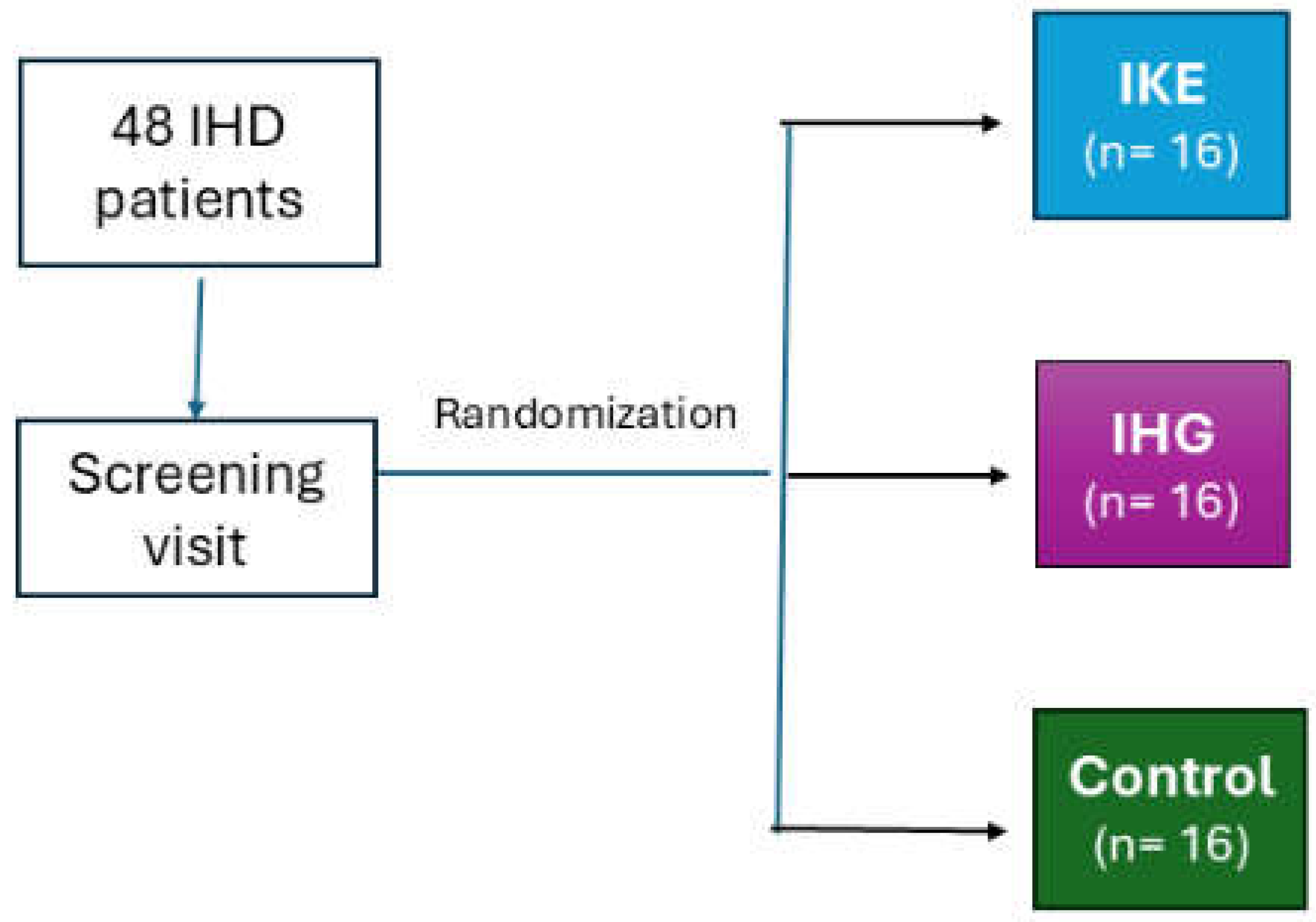
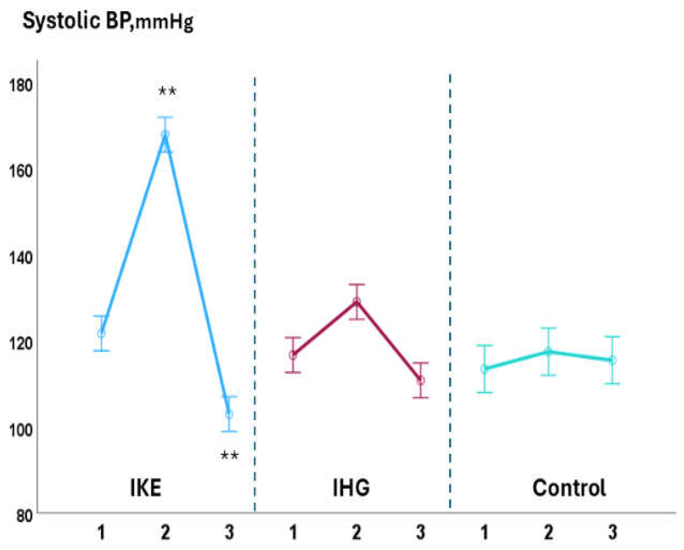
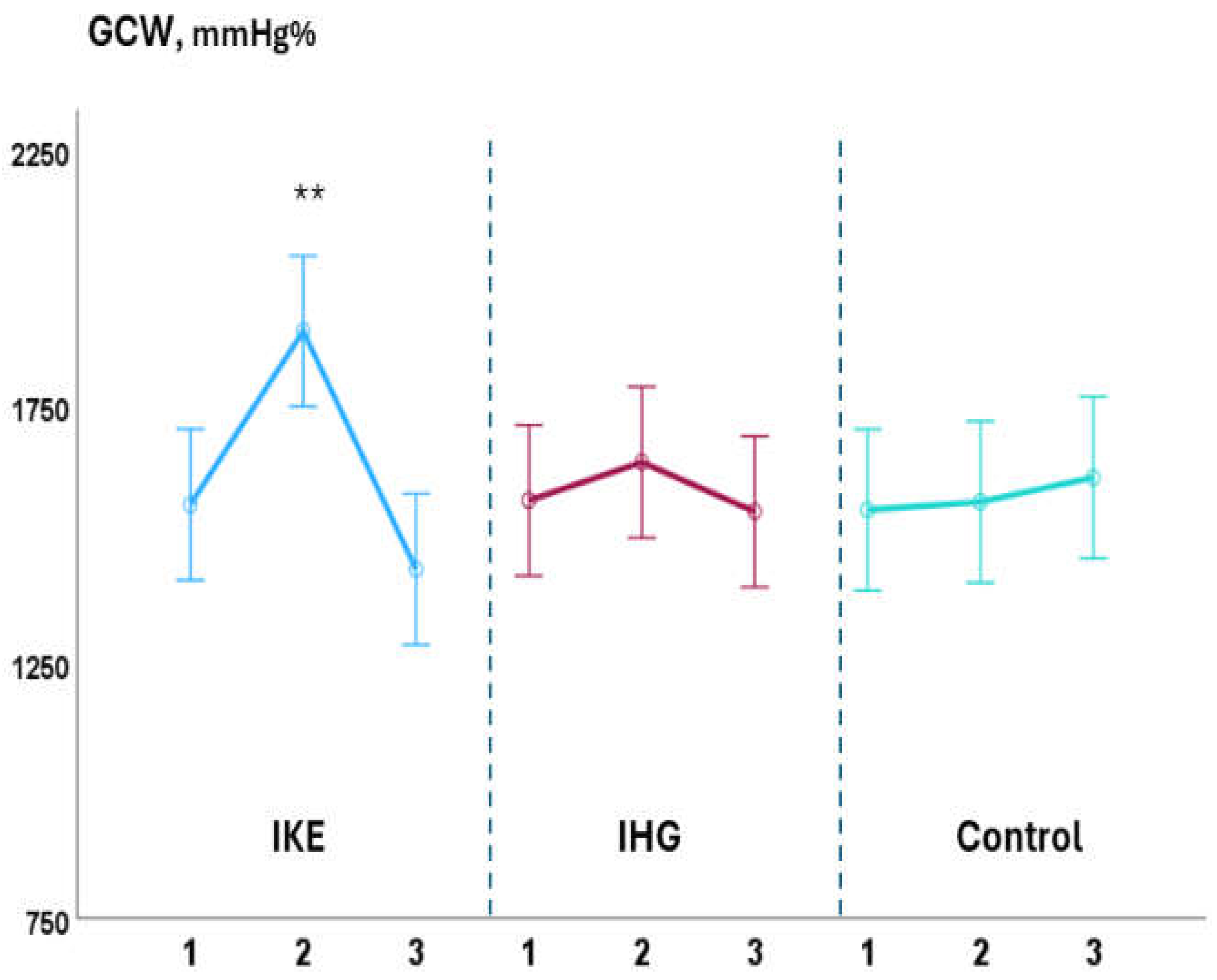
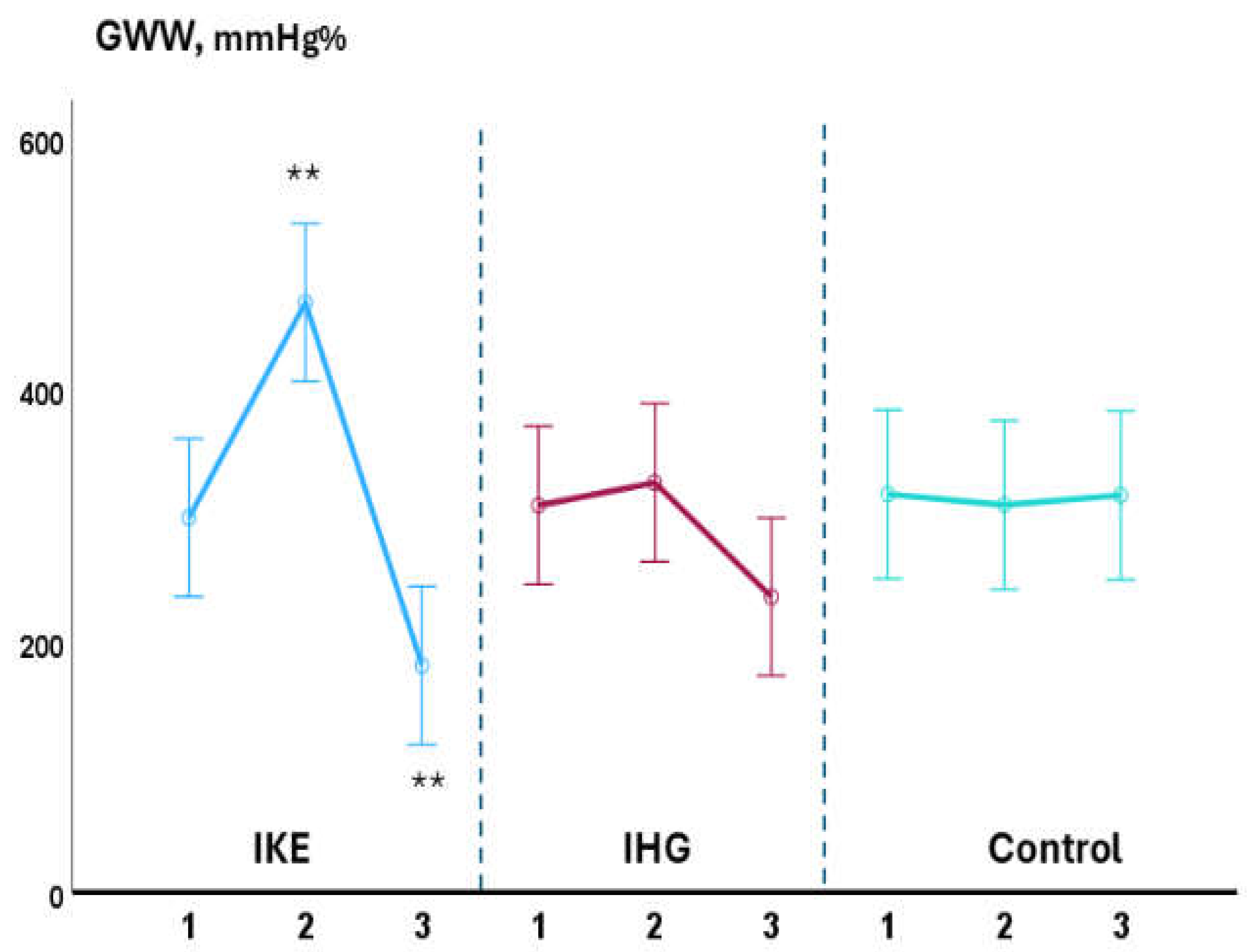
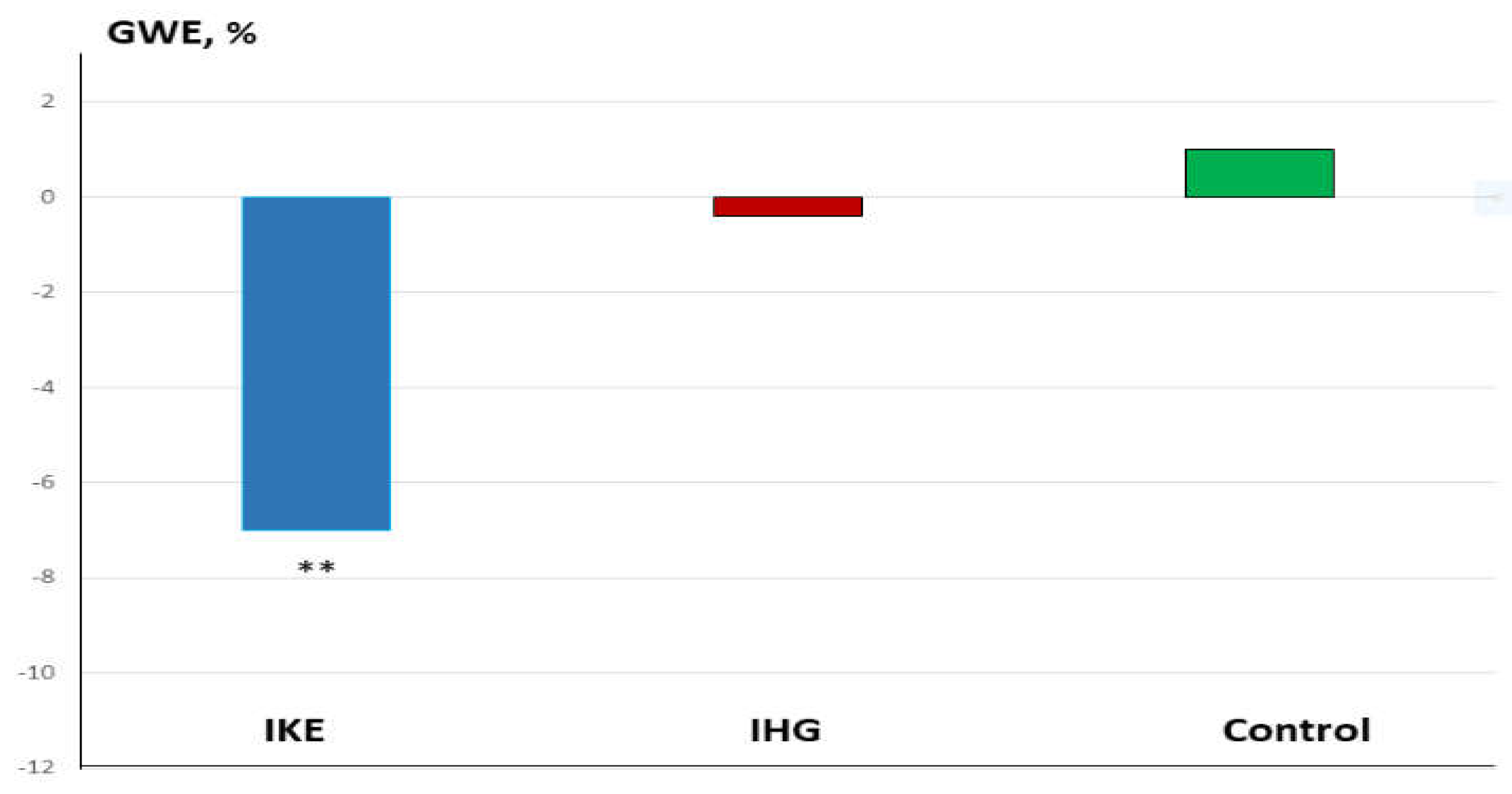
Background: Isometric exercise effectively reduces blood pressure (BP) but its effects on myocardial work have been poorly studied. In the present study we compared acute changes in myocardial work during two different isometric exercise, bilateral knee extension and handgrip in patients with hypertension and underlying ischemic heart disease (IHD). Methods: 48 stable, trained patients with hypertension and IHD were enrolled. They were randomly assigned to perform a single session of bilateral knee extension (IKE) or handgrip (IHG) or no exercise (control) with a 1:1:1 ratio. Both exercises were performed at 30% of maximal voluntary contraction and lasted three minutes. Echocardiography and BP measurements were performed at rest, during the exercise and after ten minutes of recovery. Results: both exercises were well tolerated, and no side effects occurred. During the exercise: systolic BP increased significantly in the IKE group compared to IHG and control (ANOVA p <0.001). LVGLS decreased significantly in IKE (-21%) compared IHG and control (ANOVA p 0.002). The global work index increased significantly in KE (+28%) compared to HG and control (ANOVA p 0.034). Global constructive work and wasted work increased significantly in the IKE compared to HG and control (ANOVA p 0.009 and <0.001 respectively). Global work efficiency decreased significantly in the IKE group (-8%) while remained unchanged in the IHG and controls (ANOVA p 0.002). Systolic BP increased significantly in the KE group and was unchanged in HG and control. Conclusion: myocardial work efficiency was impaired during isometric bilateral knee extension but not during handgrip. Handgrip evoked a limited hemodynamic response.

Keywords: 
;  ;  ;  ;  

1. Introduction

Exercise training is an effective non-pharmacological intervention for the prevention and treatment of hypertension [1]. Performing daily physical exercise is a recommended with level of evidence 1A by European and American guidelines on hypertension [2]. Among different exercise modalities, isometric exercise (IE) has attracted the attention of researchers since it has showed potential advantages beyond its effectiveness in reducing blood pressure (BP) in normotensive and hypertensive subjects [3]. In particular, IE presents a convenient time-efficiency profile since short bouts of IE, lasting 11–20 min, resulted as effective as longer sessions of aerobic or high-intensity interval training in reducing BP [4,5]. Furthermore, some types of isometric exercises, such as wall squat or handgrip, can comfortably be done at home without expenses or with a very low financial burden for the patients [6]. Therefore, the use IE appears to be particularly attractive in the long-term management of hypertensive patients alone or in addition to their anti-hypertensive drugs therapy. Reducing BP is one of the mechanisms through which exercise, as part of cardiac rehabilitation programs, contributes to reducing cardiovascular risk in patients with already established ischemic heart disease (IHD) [7]. Regarding the use of IE in such complex and high risk patients, an important aspect to take into consideration is the hemodynamic tolerability of this type of exercise, since an excessive increase in afterload during the effort can induce unfavourable changes in left ventricular (LV) filling pressure with a consequent increase in myocardial wall tension and myocardial oxygen consumption [8]. Therefore, the preliminary assessment of the hemodynamic response to IE could help to identify which is the most tolerated IE modality and intensity. While in the past the measurement of hemodynamic parameters was limited to the experimental setting by the need to use invasive procedures [], the utilization of speckle tracking echocardiography permits today to considerably expand hemodynamic assessment into clinical practice. Through this ultrasound technique is possible to perform a comprehensive evaluation of LV and left atrial (LA) function [9,10,11], also allowing to assess the changes induced by the exercise in different scenarios [12,13,14,15]. In addition, the assessment of myocardial work makes it possible to reconstruct in a non-invasive way the LV pressure–volume loops [16], giving the opportunity to study relationships among preload, afterload, and myocardial contractility [17]. The purpose of the present study was to compare acute changes in myocardial work and LA function, occurring during two different isometric exercise modalities, isometric knee extension and handgrip, in hypertensive patients with IHD.

2. Materials and Methods

2.1. Population

We enrolled 40 patients of both genders who were attending secondary prevention/rehabilitation programs at San Raffaele IRCCS of Rome. We used the following inclusion criteria: age over 50 years old; established diagnosis of hypertension; previous diagnosis of IHD: the following diagnostic criteria of IHD were adopted: previous acute coronary syndrome, including ST-elevation myocardial infarction (STEMI), non-ST-elevation myocardial infarction (NSTEMI), unstable angina; previous percutaneous coronary intervention (PCI) or coronary artery bypass grafting (CABG) or both; stable clinical conditions: patients must not have been hospitalized in the previous six months and their pharmacological therapy must have remained unchanged for at least three months before the enrolment; being physically active: we enrolled patients who declared to perform moderate-intensity aerobic exercise for at least 150 minutes/week (walking, cycling, swimming) [18].We adopted the following exclusion criteria: signs and/or symptoms of myocardial ischemia or threatening arrhythmias during the resting assessment or during the ergometric test; permanent atrial fibrillation or history of recurrent episodes of atrial fibrillation; baseline BP levels at rest over 160/100 mmHg despite the current drugs therapy. Subjects carrying severe heart valve diseases; with diagnosis of hypertrophic cardiomyopathy, with previous diagnosis of chronic heart failure during the screening visit were also excluded. The following extracardiac conditions were considered among exclusion criteria: low levels of haemglobin (below 10.5 g/dl); diagnosis of advanced chronic pulmonary disease (GOLD stage III-IV); diagnosis of symptomatic peripheral artery disease (Leriche-Fontaine stage II-IV). The study complied with the Declaration of Helsinki and was approved by the local Ethics Committee of San Raffaele IRCCS (protocol number 26/2023). All patients gave written informed consent before entering the study.

2.2. Study Design

The study design is summarized in Figure 1. This research was conceived as a three arms randomized study in which patients were alternately allocated to one of the following groups, with a ratio of 1:1:1. 1) isometric knee extension (IKE); 2) isometric handgrip (IHG); 3) control, no exercise. All patients were evaluated during a preliminary screening that included the following examinations: clinical history collection; measurement of anthropometric parameters, resting heart rate (HR), resting systolic and diastolic BP; carrying out a symptom-limited ergometric test. Those patients who fit the inclusion/exclusion criteria were proposed to participate to the study. Patients were then summoned for a trial session during which they were made to try out the devices and exercises that were the object of the study in order to familiarize with the experimental protocol and the devices’ use. For each patient the experimental session was performed within ten days form the screening visit.

2.3. Echocardiography

Transthoracic echocardiography: For the echocardiographic examinations, the Vivid E95® cardiovascular ultrasound (GE Healthcare, Chicago, IL, USA) with a 4.0 MHz transducer was used for the entire duration of the study. Each examination was performed with one-lead electrocardiographic monitoring and the imaging windows with the respective measurements were obtained according to the current guidelines of the European Association of Cardiovascular Imaging [19]. All acquired echocardiographic images were digitally archived and their analysis was performed offline. During the review process, an experienced technician performed strain measurements using proprietary software (version 10.8, EchoPAC; GE Vingmed Ultrasound, Horten, Norway). Left ventricular diastolic function was assessed using the E/A ratio, defined as the ratio of the E-wave (corresponding to the peak left ventricular filling velocity in early diastole) to the A-wave (corresponding to the peak flow velocity in late diastole). Colour tissue Doppler tracings were performed in the 4-chamber view and the range gate was positioned at the lateral mitral annular segments. The E/e’ ratio was defined as the ratio of the E-wave velocity to the average of the septal and lateral e’-wave velocities of the left ventricle. The volume of the left atrium (LA) was obtained from apical four-chamber and two-chamber views at the end of systole, prior to the adoption of Simpson’s biplanar disc method and prior to the opening of the mitral valve. The LA volume index (LAVI) was obtained by dividing the LA volume by the body surface area of the study subjects. Measurements of left ventricular end-diastolic volume (LVEDV) and left ventricular end-systolic volume (LVESV) were calculated from the apical windows of two and four chambers; subsequently, LVEF was calculated using Simpson’s modified method. Stroke volume (SV) was then calculated as EDV - ESV, cardiac output (CO) as heart rate (HR) x SV and ejection fraction (EF) as EF = (EDV - ESV)/EDV. Measurements of global longitudinal LV strain (LVGLS) were obtained from four-chamber, three-chamber and two-chamber views. The software measured the maximum negative value of the deformation during systole and this value was considered as the maximum contractility for each segment. The software is able to automatically detect the endocardial LV boundaries; however, whenever deemed appropriate, images can be modified to conform to the displayed LV boundaries. Therefore, LVGLS was calculated by considering the mean values of each segment. LA deformation was assessed through four-chamber and two-chamber views. The software automatically plotted the endocardial and epicardial contours of the LA, using R-R gating, where the R-wave represented the starting point. Again, manual adjustments were made if necessary. A series of control points was automatically placed on the central curve of the myocardial wall in the reference phase, based on the endocardial and epicardial contours plotted. The software program generated longitudinal deformation curves for each segment and calculated the average curve for each segment. LA reservoir strain, conduit strain and contractile strain were obtained by splitting the longitudinal strain measurements. The MW was assessed from closure to opening of the mitral valve. PACS was defined as a positive peak during the onset of left ventricular diastole, prior to the onset of the atrial systolic phase; whereas PALS was defined as a positive peak during left ventricular systole, at the end of the atrial diastolic phase. A 17-segment bull’s eye was obtained with the segmental and global work index (GWI) corresponding to the area within the total work curve from mitral valve closure to opening. Global constructive work (GCW) was defined as the work performed during shortening in systole and the negative work performed during lengthening in isovolumetric relaxation. Global wasted work (GWW) corresponds to the work performed during shortening in isovolumetric relaxation plus the negative work performed during lengthening in systole. Global work efficiency (GWE) was defined as the constructive work divided by the sum of constructive work and wasted work. Using pulsed wave Doppler recordings at mitral and aortic valve level, the timing of valve events was identified. Confirmation of the valve events was performed by 2D evaluation of the long axis and apical view [20].

2.4. Experimental Sessions:

All experimental sessions were performed during the morning between 9.00 and 11.30 am. Patients were asked to avoid drinking coffee and alcohol for at least 24/h before the session. A light breakfast was allowed at least two hours before the experimental session. Bilateral knee extension: the experiments were conducted on a knee flex/extension dynamometer (Technogym Wellness System, Technogym, Cesena, Italy). Patients seated on the dynamometer with their backs reclined at 120 degrees from the horizontal plane and with their knees bent at 90 degrees from the trunk. The seat was individually regulated so that the axis of rotation around the dynamometer shaft was adjacent to the lateral femoral condoyle of the subject’s right leg. Patients positioned their legs under the knee extension/flexion attachment arm of the dynamometer. Both arms were positioned along the trunk. On the left side of the examined subjects was positioned a sonographer responsible for the acquisition of echocardiographic data. Patients had a manual sphygmomanometer cuff placed on their right arm. Handgrip: experiment were conducted with patients lying on a bench with their backs reclined at 120 degrees from the horizontal plane. The sonographer was placed on their left side and a sphygmomanometer cuff was placed on the arm contralateral to the dominant one that was used for the handgrip (GIMA, S.P.A., Gessate (MI), Italy). For both bilateral knee extension and handgrip the determination of the maximal voluntary contraction (MVC) consisted of 3 maximal contractions, each one lasting 3–5 s, with 1 min of rest between contractions. For both exercises, the intensity was set at 30 of the patients’ MVC. The exercise phase lasted 3 minutes, and during this time the patients had to exert constant force- During the exercise patients were instructed to breathe at a normal rhythm and depth in order to avoid Valsalva manoeuvres. The echocardiography examination started after the first minute of exercise. Control group: patients were lying on a bench with their backs reclined at 120 degrees from the horizontal plane. The sonographer was placed on their left side and a sphygmomanometer cuff placed on their right arm. For patients of the IKE and IHG groups, echocardiography acquisitions and BP measurements were made: 1) at rest; 2) during the exercise phase, starting one minute from the begin of the isometric effort; 3) ten minutes after the end of the isometric effort. For patients in the control group, echocardiographic acquisitions and BP measurements were made with the same timing as the other two groups but the patients of this group remained at rest during the entire procedure.

2.5. Statistical Analysis

This research was conceived as a pilot study no formal a priori power analysis was performedand Sample size was determined by feasibility and patients’ availability [21]. Data were expressed as mean ± SD. The assumption of normality was checked using the Shapiro–Wilk hypothesis test. Comparisons of changes occurring in different variables during the three different stages of the experimental session, atrest, during exercise, and at recovery, were compared by using repeated-measures two-way ANOVA with Bonferroni corrections for post hoc testing. The level of significance was set at p < 0.05. Categorical variables are expressed as absolute and percentages values and were compared with the chi-square testThe statistical program, IBM SPSS Statistics v26.0, was used for the processing, presentation, and statistical analysis of the data.

3. Results

Baseline anthropometric, clinical and echocardiography data are reported in Table 1. At baseline there were not significant differences in clinical characteristics and drug therapy between the three study groups. Thirty-six (75%) out of 48 patients had a previous STEMI. Twenty-three (64%) out of 36, had an anterior STEMI and 13 (36%) had an inferior STEMI. LVEF ranged between 38% and 57%. Thirty-seven (77%) out of 48 had a diagnosis of multivessel coronary disease. All patients were treated with anti-platelets agents, statins and betablockers. Overall they were taking on average 2.4±1.3 anti-hypertensive drugs. Every patient included in the study completed the protocol. Both exercises sessions were well tolerated, and no side effects occurred.

3.1. Intra-Groups Changes

During the exercise phase, compared to baseline values, systolic BP significantly increased in the IKE group (+36.0±1.7 mmHg) while it remained unchanged in the IHG (+3.5±1.7mmHg) and control groups (-2.5±0.8 mmHg). LVGLS decreased significantly in IKE (-21%) while was unchanged in the IHG (-3%) and control(+2%). The IKE group presented a significant increase in GWI (+28%, p 0.003); while no significant changes in GWI occurred in IHG and control. GCW and GWW increased significantly in the IKE (+30%, and +56% respectively); no significant changes in GCW and GWW were observed in IHG (+4%, and+11% respectively) and control (+2% and -3% respectively). GWE decreased -7% significantly in the IKE group and remained unchanged in the IHG (-0.4%) and control (+1%). PALS was unchanged in the IKE (-4%), IHG (-3%) and control (-1%). No significant changes in diastolic BP, E/e’, PALS, LAVI, LVEF, SV and CO, occurred during exercise in comparison to baseline values in the two active groups and control. During the recovery phase: compared to baseline values, systolic BP decreased significantly in IKE compared to IHG and control. GWW decreased significantly in the IKE and and IHG and was unchanged in control.

3.2. Between-Groups Changes

The increase in systolic BP observed in the IKE was significantly greater than IHG and control. (ANOVA: F= 7.13; p <0.001) (Figure 2). The reduction in GLS in the IKE was significantly greater than IHG and control (ANOVA: F= 3.8; p 0.002). The increase in GWI (ANOVA: F= 2.7; p 0.034), GCW (ANOVA: F= 3.81; p 0.009) and GWW (ANOVA: F=6.20. p<0.001) in the IKE resulted significantly greater compared to IHG and control (Figure 3). The IKE group presented a significant decrease in GWE compared to IHG and control (Figure 4). No between-groups changes occurred regarding diastolic BP, E/e’, PALS, LVEF, SV and CO. During the recovery phase, the decrease in systolic-BP in IKE was significantly greater than IHG and control (ANOVA: F=2.4. p 0.032) (Figure 2).

4. Discussion

The utilization of IE in the context of the rehabilitation of patients with IHD has, thus far, been prevented by the concern that it may result in an excessive increase in myocardial oxygen consumption. However, it remains an attractive exercise modality in the long-term management of hypertension in these patients since, besides its anti-hypertensive efficacy, it presents a valuable time-efficiency and cost-sparing profile [6]. Investigating the hemodynamic response to different types of isometric exercise can help in identifying which is the best tolerated modality and in choosing the more appropriate intensity and duration of the exercise itself. In the present study the hemodynamic response observed during IE was very different between bilateral knee extension and handgrip. The IKE group presented a significant increase in systolic BP that was coupled with a decrease in GLS and a rise in myocardial work, with a prevalence of GWW over GCW and a consequent reduction in GWE. These changes indicate overall a maladaptive LV contractile response during IKE. This kind of response is similar to that previously observed by our group in a similar population [22] and by Beaumont et al [23] in healthy subjects by performing bilateral knee extension respectively at 30% and 40% of MV. However, compared to our previous study [22] there are some differences that should be underlined: the rate of GWE loss during exercise in the present study appears to be lower when compared to that observed in the previous one (- 7% vs -18%); moreover, contrary to what happened in the previous research [22], changes in E/e’and PALS in the IKE group, during the exercise, did not reach the statistical significance. We believe that these results may depend on the difference in the fitness level of the patients enrolled in the two studies, since the previous one enrolled only sedentary patients while, in the present research, being physically active was one of the inclusion criteria. This hypothesis is supported by recent findings in the literature: a four-weeks exercise training was effective in improving GWE in hypertensive patients [24]. In a study conducted in healthy subjects, Rovithis et al [25] demonstrated that active men did not show a statistically significant change in the E/e’ ratio during isometric handgrip; conversely, the inactive participants’ E/e’ ratio was higher, during isometric handgrip exercise. Considering that both E/e’ ratio, and PALS are considered non-invasive metrics of LV filling pressure [26,27] we can hypothesize that IE performed with bilateral knee extension had a lower impact on diastolic function in our patients because of their active physical status. However, we cannot rule out that these results could be affected by the small sample size and they need to be confirmed in further larger trials. Moreover, direct comparisons of the hemodynamic response to IE between physically active versus sedentary IHD patients are required. The IHG group showed only a slight increase in systolic BP, no changes in LV filling pressure metrics, and mild, non-significant, increase in myocardial work that was balanced between GCW and GWW, so as not to observe changes in contractile efficiency. This response of the IHG group was statistically not different from that observed in the control group, indicating that isometric handgrip performed at 30% of MVC had neutral effects on central hemodynamic parameters. This result agrees with previous research investigating the BP response to isometric handgrip, overall showing that, when performed at low intensities, it does not result in a cardiovascular overload [28,29]. Since the timing of the exercise and the percent of MVC utilized were the same between IKE and IHG, our data suggest that the amount of muscle mass involved in the IE was the main determinant of the BP response during the exercise phase. This result appears also to be in line with the current literature [30]. Kounoupis et al [31] showed that, when using small-muscle mass, isometric and dynamic resistance exercises evoked equal increases in BP. We observed a greater drop in systolic BP ten minutes after the end of the exercise in the IKE group compared to IHG. This result complies with previous reports showing that isometric exercise involving larger muscle mass is associated with a significantly greater post-exercise hypotensive response in the first minutes of the recovery compared to smaller muscle mass, as those involved in the isometric handgrip exercise [32]. The effectiveness of isometric handgrip in eliciting acute reductions in BP has recently been questioned by a meta-analysis demonstrating that while a single session of isometric handgrip did not produce significant BP reductions, isometric handgrip training decreased SBP and DBP by 6.7 mmHg and 4.5 mmHg, respectively [33]. Another aspect to consider is the complexity of pharmacological treatment already taken by the patients that might have interfered with the expected BP reduction induced by IE. While a single bout of isometric handgrip, performed at low intensity, was effective in lowering BP in healthy and pre-hypertensive individuals [34], the same exercise did not induce significant reductions in BP in patients with coronary artery disease [35]. Moreover, our results on post-exercise BP could be influenced by the fact that they were obtained after single prolonged contractions (three minutes for both exercise); it is possible that shorter repeated contractions, as is generally the case during an exercise session, might have produced different results as showed by Souza et al, [36] in elderly hypertensive patients. Considering its mild impact on systolic BP and its neutral effects on diastolic LV metrics and GWE, low-intensity isometric training using handgrip appears to have a safer profile in patients with IHD. These considerations make this type of exercise a suitable non-pharmacological intervention for treating hypertension in IHD patients. However, the lack of significant BP reduction in the post-exercise phase that we observed in our study requires further research on this topic: Future studies should assess the safety and effectiveness of moderate and high intensities isometric handgrip in IHD patients and investigate whether BP reductions achievable with moderate-high intensities isometric handgrip are comparable to that of bilateral isometric knee extension. We think that this study adds useful informations regarding the choice of the exercise modality aimed at non-pharmacological management of hypertension in patients with IHD and underline the potential advantages for non-invasively assessing the hemodynamic response before starting an IE protocol.
Limitations: Since this research was conceived as a pilot study, it included a small sample size; therefore, caution is needed in drawing general conclusion by our data. The study enrolled only 3 female patients in the active arms; we think thatwe cannot extend our findings on female and further studies focusing on female gender are needed. Bilateral isometric knee extension and handgrip were both performed at 30% of MVC; we cannot rule out that IE performed at different intensities could evoke a different myocardial response.

5. Conclusions

Our data suggests that in hypertensive patients with underlying IHD, performing IE with handgrip seems a viable choice in terms of hemodynamic tolerability. Future studies should clarify whether handgrip IE is also effective in lowering BP in these patients..

Author Contributions

M.A.P., F.I. and G.C. contributed to the conceptualization of the paper; M.V. (Matteo Vitarelli). S.V., S.F., G.M., B.S. and S.F. prepared the initial draft after acquisition, analysis, and interpretation of the results; V.M. (Valentina Morsella), M.V. (Maurizio Volterrani), and V.M. (Vincenzo Manzi) substantively revised it. All authors have read and agreed to the published version of the manuscript.

Funding

This work was supported by funding of the Italian Ministry of Health [Ricerca corrente].

Institutional Review Board Statement

The study was conducted in accordance with the Declaration of Helsinki and approved by the Institutional Ethics Committee of San Raffaele IRCCS of Rome (protocol code 26/2023, approval date: September, 21th 2023). Informed Consent Statement: Informed consent was obtained from all subjects involved in the study. Written informed consent has been also obtained from the patients to publish this paper.

Informed Consent Statement

Informed consent was obtained from all subjects involved in the study.

Data Availability Statement

The data presented in this study are available upon request from the corresponding authors.

References

  1. Pescatello, L.S.; Buchner, D.M.; Jakicic, J.M.; Powell, K.E.; Kraus, W.E.; Bloodgood, B.; Campbell, W.W.; Dietz, S.; Dipietro, L.; George, S.M.; Macko, R.F.; McTiernan, A.; Pate, R.R.; Piercy, K.L. Physical Activity to Prevent and Treat Hypertension: A Systematic Review. Med Sci Sports Exerc. 2019, 51, 1314–1323. [Google Scholar] [CrossRef] [PubMed]
  2. McEvoy, J.W.; McCarthy, C.P.; Bruno, R.M.; Brouwers, S.; Canavan, M.D.; Ceconi, C.; Christodorescu, R.M.; Daskalopoulou, S.S.; Ferro, C.J.; Gerdts, E.; Hanssen, H.; Harris, J.; Lauder, L.; McManus, R.J.; Molloy, G.J.; Rahimi, K.; Regitz-Zagrosek, V.; Rossi, G.P.; Sandset, E.C.; Scheenaerts, B.; Staessen, J.A.; Uchmanowicz, I.; Volterrani, M.; Touyz, R.M. ESC Scientific Document Group. 2024 ESC Guidelines for the management of elevated blood pressure and hypertension. Eur Heart J. 2024, 45, 3912–4018. [Google Scholar] [CrossRef] [PubMed]
  3. Baffour-Awuah, B.; Pearson, M.J.; Dieberg, G.; Smart, N.A. Isometric Resistance Training to Manage Hypertension: Systematic Review and Meta-analysis. Curr Hypertens Rep. 2023, 25, 35–49. [Google Scholar] [CrossRef]
  4. Edwards, J.; De Caux, A.; Donaldson, J.; Wiles, J.; O’Driscoll, J. Isometric exercise versus high-intensity interval training for the management of blood pressure: a systematic review and meta-analysis. Br J Sports Med. 2022, 56, 506–514. [Google Scholar] [CrossRef] [PubMed]
  5. Jae, S.Y.; Yoon, E.S.; Kim, H.J.; Cho, M.J.; Choo, J.; Kim, J.Y.; Kunutsor, S.K. Isometric handgrip versus aerobic exercise: a randomized trial evaluating central and ambulatory blood pressure outcomes in older hypertensive participants. J Hypertens. 2025, 43, 351–358. [Google Scholar] [CrossRef] [PubMed]
  6. Miura, S. Evidence for exercise therapies including isometric handgrip training for hypertensive patients. Hypertens Res. 2024. [Google Scholar] [CrossRef]
  7. Alves, A.J.; Viana, J.L.; Cavalcante, S.L.; Oliveira, N.L.; Duarte, J.A.; Mota, J.; Oliveira, J.; Ribeiro, F. Physical activity in primary and secondary prevention of cardiovascular disease: Overview updated. World J Cardiol. 2016, 8, 575–583. [Google Scholar] [CrossRef]
  8. Suga, H.; Hayashi, T.; Shirahata, M. Ventricular systolic pressure-volume area as predictor of cardiac oxygen consumption. Am J Physiol. 1981, 240, H39–H44. [Google Scholar] [CrossRef]
  9. Hanson, P.; Nagle, F. Isometric exercise: cardiovascular responses in normal and cardiac populations. Cardiol Clin. 1987, 5, 157–170. [Google Scholar] [CrossRef]
  10. Kleijn, S.A.; Aly, M.F.; Terwee, C.B.; van Rossum, A.C.; Kamp, O. Three-dimensional speckle tracking echocardiography for automatic assessment of global and regional left ventricular function based on area strain. J Am Soc Echocardiogr. 2011, 24, 314–321. [Google Scholar] [CrossRef]
  11. Luis, S.A.; Yamada, A.; Khandheria, B.K.; Speranza, V.; Benjamin, A.; Ischenko, M.; Platts, D.G.; Hamilton-Craig, C.R.; Haseler, L.; Burstow, D.; Chan, J. Use of three-dimensional speckle-tracking echocardiography for quantitative assessment of global left ventricular function: a comparative study to three-dimensional echocardiography. J Am Soc Echocardiogr. 2014, 27, 285–291. [Google Scholar] [CrossRef] [PubMed]
  12. Cacciapuoti, F.; Paoli, V.D.; Scognamiglio, A.; Caturano, M.; Cacciapuoti, F. Left Atrial Longitudinal Speckle Tracking Echocardiography in Healthy Aging Heart. J Cardiovasc Echogr. 2015, 25, 40–45. [Google Scholar] [CrossRef] [PubMed]
  13. Mandraffino, G.; Imbalzano, E.; Lo Gullo, A.; Zito, C.; Morace, C.; Cinquegrani, M.; Savarino, F.; Oreto, L.; Giuffrida, C.; Carerj, S.; Squadrito, G. Abnormal left ventricular global strain during exercise-test in young healthy smokers. Sci Rep. 2020, 10, 5700. [Google Scholar] [CrossRef] [PubMed]
  14. Forsythe, L.; George, K.; Oxborough, D. Speckle Tracking Echocardiography for the Assessment of the Athlete’s Heart: Is It Ready for Daily Practice? Curr Treat Options Cardiovasc Med. 2018, 20, 83. [Google Scholar] [CrossRef] [PubMed]
  15. Sugimoto, T.; Bandera, F.; Generati, G.; Alfonzetti, E.; Bussadori, C.; Guazzi, M. Left Atrial Function Dynamics During Exercise in Heart Failure: Pathophysiological Implications on the Right Heart and Exercise Ventilation Inefficiency. JACC Cardiovasc Imaging. 2017, 10 Pt B, 1253–1264. [Google Scholar] [CrossRef]
  16. Perrone, M.A. : Iellamo, F.; D’Antoni, V.; Gismondi, A.; Di Biasio, D.; Vadalà, S.; Marazzi, G.; Morsella, V.; Volterrani, M.; Caminiti, G. Acute Changes on Left Atrial Function during Incremental Exercise in Patients with Heart Failure with Mildly Reduced Ejection Fraction: A Case-Control Study. J Pers Med. 2023, 13, 1272. [Google Scholar]
  17. Trimarchi, G.; Carerj, S.; Di Bella, G.; Manganaro, R.; Pizzino, F.; Restelli, D.; Pelaggi, G.; Lofrumento, F.; Licordari, R.; Taverna, G.; Paradossi, U.; de Gregorio, C.; Micari, A.; Di Giannuario, G.; Zito, C. Clinical Applications of Myocardial Work in Echocardiography: A Comprehensive Review. J Cardiovasc Echogr. 2024, 34, 99–113. [Google Scholar] [CrossRef]
  18. Virani, S.S.; Newby, L.K.; Arnold, S.V.; Bittner, V.; Brewer, L.C.; Demeter, S.H.; Dixon, D.L.; Fearon, W.F.; Hess, B.; Johnson, H.M.; Kazi, D.S.; Kolte, D.; Kumbhani, D.J.; LoFaso, J.; Mahtta, D.; Mark, D.B.; Minissian, M.; Navar, A.M.; Patel, A.R.; Piano, M.R.; Rodriguez, F.; Talbot, A.W.; Taqueti, V.R.; Thomas, R.J.; van Diepen, S.; Wiggins, B.; Williams, M.S. Peer Review Committee Members. 2023 AHA/ACC/ACCP/ASPC/NLA/PCNA Guideline for the Management of Patients With Chronic Coronary Disease: A Report of the American Heart Association/American College of Cardiology Joint Committee on Clinical Practice Guidelines. Circulation. 2023, 148, e9–e119. [Google Scholar] [CrossRef]
  19. Nagueh, S.F.; Smiseth, O.A.; Appleton, C.P.; Byrd, B.F. 3rd; Dokainish, H.; Edvardsen, T.; Flachskampf, F.A.; Gillebert, T.C.; Klein, A.L.; Lancellotti, P.; Marino, P.; Oh, J.K.; Popescu, B.A.; Waggoner, A.D. Recommendations for the Evaluation of Left Ventricular Diastolic Function by Echocardiography: An Update from the American Society of Echocardiography and the European Association of Cardiovascular Imaging. Eur Heart J Cardiovasc Imaging. 2016, 17, 1321–1360. [Google Scholar] [CrossRef]
  20. Papadopoulos, K.; Özden Tok, Ö.; Mitrousi, K.; Ikonomidis, I. Myocardial Work: Methodology and Clinical Applications. Diagnostics. 2021, 11, 573. [Google Scholar] [CrossRef]
  21. Flight, L.; Julious, S.A. Practical guide to sample size calculations: an introduction. Pharm Stat. 2016, 15, 68–74. [Google Scholar] [CrossRef] [PubMed]
  22. Caminiti, G.; Volterrani, M.; Iellamo, F.; Marazzi, G.; D’Antoni, V.; Calandri, C.; Vadalà, S.; Catena, M.; Di Biasio, D.; Manzi, V.; Morsella, V.; Perrone, M.A. Acute Changes in Myocardial Work during Isometric Exercise in Hypertensive Patients with Ischemic Heart Disease: A Case-Control Study. J Clin Med. 2024, 13, 5955. [Google Scholar] [CrossRef] [PubMed]
  23. Beaumont, A.; Sculthorpe, N.; Hough, J.; Unnithan, V.; Richards, J. Global and regional left ventricular circumferential strain during incremental cycling and isometric knee extension exercise. Echocardiography. 2018, 35, 1149–1156. [Google Scholar] [CrossRef] [PubMed]
  24. O’Driscoll, J.M.; Edwards, J.J.; Wiles, J.D.; Taylor, K.A.; Leeson, P.; Sharma, R. Myocardial work and left ventricular mechanical adaptations following isometric exercise training in hypertensive patients. Eur J Appl Physiol. 2022, 122, 727–734. [Google Scholar] [CrossRef]
  25. Rovithis, D.; Anifanti, M.; Koutlianos, N.; Teloudi, A.; Kouidi, E.; Deligiannis, A. Left Ventricular Diastolic Response to Isometric Handgrip Exercise in Physically Active and Sedentary Individuals. J Cardiovasc Dev Dis. 2022, 9, 389. [Google Scholar] [CrossRef]
  26. Mitter, S.S.; Shah, S.J.; Thomas, J.D. A Test in Context: E/A and E/e’ to Assess Diastolic Dysfunction and LV Filling Pressure. J Am Coll Cardiol. 2017, 69, 1451–1464. [Google Scholar] [CrossRef]
  27. Carluccio, E.; Cameli, M.; Rossi, A.; Dini, F.L.; Biagioli, P.; Mengoni, A.; Jacoangeli, F.; Mandoli, G.E.; Pastore, M.C.; Maffeis, C.; Ambrosio, G. Left Atrial Strain in the Assessment of Diastolic Function in Heart Failure: A Machine Learning Approach. Circ Cardiovasc Imaging. 2023, 16, e014605. [Google Scholar] [CrossRef]
  28. Olher, R. dosR.; Bocalini, D.S.; Bacurau, R.F.; Rodriguez, D.; Figueira, A., Jr.; Pontes, F.L., Jr.; Navarro, F.; Simões, H.G.; Araujo, R.C.; Moraes, M.R. Isometric handgrip does not elicit cardiovascular overload or post-exercise hypotension in hypertensive older women. Clin Interv Aging. 2013, 8, 649–655. [Google Scholar]
  29. Coneglian, J.C.; Barcelos, G.T.; Bandeira, A.C.N.; Carvalho, A.C.A.; Correia, M.A.; Farah, B.Q.; Ritti-Dias, R.M.; Gerage, A.M. Acute Blood Pressure Response to Different Types of Isometric Exercise: A Systematic Review with Meta-Analysis. Rev Cardiovasc Med 2023, 24, 60. [Google Scholar] [CrossRef]
  30. Lin, S.; Sun, P.; Huang, L.; Hernandez, M.; Yu, H.; Jan, Y.K. Effects of the intensity, duration and muscle mass factors of isometric exercise on acute local muscle hemodynamic responses and systematic blood pressure regulation. Front Bioeng Biotechnol. 2024, 12, 1444598. [Google Scholar] [CrossRef]
  31. Kounoupis, A.; Papadopoulos, S.; Galanis, N.; Dipla, K.; Zafeiridis, A. Are Blood Pressure and Cardiovascular Stress Greater in Isometric or in Dynamic Resistance Exercise? Sports. 2020, 8, 41. [Google Scholar] [CrossRef] [PubMed]
  32. Swift, H.T.; O’Driscoll, J.M.; Coleman, D.D.; Caux, A.; Wiles, J.D. Acute cardiac autonomic and haemodynamic responses to leg and arm isometric exercise. Eur J Appl Physiol. 2022, 122, 975–985. [Google Scholar] [CrossRef] [PubMed]
  33. Oliveira, P.C.; Silva, M.R.; Lehnen, A.M.; Waclawovsky, G. Isometric handgrip training, but not a single session, reduces blood pressure in individuals with hypertension: a systematic review and meta-analysis. J Hum Hypertens. 2023, 37, 844–853. [Google Scholar] [CrossRef] [PubMed]
  34. van Assche, T.; Buys, R.; de Jaeger, M.; Coeckelberghs, E.; Cornelissen, V.A. One single bout of low-intensity isometric handgrip exercise reduces blood pressure in healthy pre- and hypertensive individuals. J Sports Med Phys Fitness. 2017, 57, 469–475. [Google Scholar] [CrossRef]
  35. Goessler, K.; Buys, R.; Cornelissen, V.A. Low-intensity isometric handgrip exercise has no transient effect on blood pressure in patients with coronary artery disease. J Am Soc Hypertens. 2016, 10, 633–639. [Google Scholar] [CrossRef]
  36. Souza, L.R.; Vicente, J.B.; Melo, G.R.; Moraes, V.C.; Olher, R.R.; Sousa, I.C.; Peruchi, L.H.; Neves, R.V.; Rosa, T.S.; Ferreira, A.P.; Moraes, M.R. Acute Hypotension After Moderate-Intensity Handgrip Exercise in Hypertensive Elderly People. J Strength Cond Res. 2018, 32, 2971–2977. [Google Scholar] [CrossRef]
Figure 1. Study flow-chart.
Preprints 146627 g001
Figure 2. Changes in systolic BP during the three phases of exercise (1= baseline; 2= exercise; 3= recovery) in the IKE. IHG and Control groups. **= p<0.05 versus active group and control.
Preprints 146627 g002
Figure 3. Changes in GCW during the three phases of exercise (1= baseline; 2= exercise; 3= recovery) in the IKE. IHG and Control groups. **= p<0.05 versus active group and control.
Preprints 146627 g003
Figure 4. Changes in GWW during the three phases of exercise (1= baseline; 2= exercise; 3= recovery) in the IKE. IHG and Control groups. **= p<0.05 versus active group and control.
Preprints 146627 g004
Figure 5. Percent canges in GWE during the three phases of exercise (1= baseline; 2= exercise; 3= recovery) in the IKE. IHG and Control groups. **= p<0.05 versus active group and control.
Preprints 146627 g005
Table 1.
IKE
(n= 16)
IHG
(n= 16)
Control
(n= 16)
Age, years 64.9±14.7 63.4±16.1 64.2±13.5
Male/female, n 14/2 14/1 7/1
BMI, kg/m2 27.1±5.5 28.2±8.0 27.5±7.3
BSA, m2 1.88±1.1 1.93±0.8 1.91±1.3
Previous STEMI 13 (81) 11 (69) 12 (75)
PCI, n (%) 11 (69) 10 (62) 10(62)
CABG, n (%) 8 (50) 7 (44) 9(56)
Physical activity (min/week) 174 178 173
HR, bpm 66.8±13.6 63.5±11.4 65.2±8.2
SBP, mmHg 128.9±27.5 135.2±33.8 136.7±32.1
DBP, mmHg 81.6±9.2 80.9±12.4 81.8±1.3
Comorbidities
Diabetes, n(%) 3 (19) 2 (12) 3 (19)
Hypercholesterolemia, n(%) 14 (87) 15 (94) 14 (87)
GFR < 60 ml/min/1.73 m2 3 (19) 4 (25) 2 (25)
Previous smoke habit, n (%) 8 (50) 10 (62) 8 (50)
Echocardiography
LVEDV, ml 140.4 ± 33,4 136.3 ± 32.4 137± 32.4
LVESV, ml 70.5 ± 11.7 67.0 ± 26.3 69.4±19.4
LVEF, % 50.3 ± 7.4 49.2± 8.2 50.3±11.4
LVGLS, % -12.4 ± 3.8 -13.1 ± 3.5 -12.9± 3.1
GWI, % 1150.5 ± 412.6 ì284.9 ± 492.0 1198±331.6
GCW, % 1601.3 ± 491.8 1678 ± 558.3 1523±341.9
GWW, % 338.7 ± 127.7 345.2 ± 198.9 344.3± 166.5
GWE, % 82.5± 11.7 82.8 ± 9.7 81.5±13.1
DT, ms 208.7 ± 60.4 197.4 ± 37.5 211.7±44.5
E, cm/s 48.1 ± 9.0 50.6± 13.0 52.3± 12.3
A, cm/s 66.4 ± 16.1 67.3 ± 13.9 66.9 ± 14.1
E/A ratio 0.75 ± 0.16 0.75 ± 0.22 0.78± 0.13
e’, cm/s 5.9± 1.5 6.2±2.2 6.3±2.2
E/e’ ratio 8.1± 1.9 8.1 ± 2.6 8.3± 2.3
TRV, m/s 1.9± 0.4 2.1± 0.1 1.7 ± 0.4
PALS, % 19.7 ± 8.5 21.4 ± 6.0 19.2± 6.0
PACS, % -13.9± 3.5 -15.4 ± 3.5 -15 7±4.3
LAVI, ml/m2 31.6 ± 7.2 34.2 ± 10.3 32.72 ±9.1
SV, ml 70.3 ± 14.3 72.0± 14.4 69.3± 16.4
CO, l/min 4.7 ± 1.1 4.6± 1.0 4.5± 1.6
Treatment
Anti-platelets agents, n (%) 16 (100) 16 (100) 16 (100)
ACE-Is/ARBs, n (%) 15 (94) 16 (100) 15 (94)
Betablockers, n (%) 16 (100) 16 (100) 8 (100)
Tiazidics, n (%) 4 (25) 5 (31) 5 (31)
CCAs, n (%) 5 (31) 6 (37) 7 (44)
Ranolazine, n (%) 3 (19) 2 (12) 2 (12)
Furosemide, n (%) 1(6) 2 (12) -
Statins, n (%) 16 (100) 16 (100) 16 (100)
Ezetimibe, n (%) 13 (81) 12 (75) 13 (81)
BMI= body mass index; BSA= body surface area; STEMI= ST-elevation myocardial infarction ; PCI=percutaneous coronary intervention;CABG=coronary artery bypass grafting; HR= heart rate; SBP= systolic blood pressure; DBP= diastolic blood pressure; DP= double product; CWI= global work index; GCW= global costructive work; GWW= global waste work, GWE= global work efficiency; DT= deceleration time; TRV= tricuspid regurgitation velocity; PALS= peak atrial longitudinal strain; PACS= Peak atrial contraction strain; SV= stroke volume; CO= cardiac output; ACE-Is= angiotensin-converting-enzyme inhibitors; ARBs= angiotensin receptor blockers; CCAs= Calcium-channel antagonists.
Disclaimer/Publisher’s Note: The statements, opinions and data contained in all publications are solely those of the individual author(s) and contributor(s) and not of MDPI and/or the editor(s). MDPI and/or the editor(s) disclaim responsibility for any injury to people or property resulting from any ideas, methods, instructions or products referred to in the content.
Copyright: This open access article is published under a Creative Commons CC BY 4.0 license, which permit the free download, distribution, and reuse, provided that the author and preprint are cited in any reuse.
Prerpints.org logo

Preprints.org is a free preprint server supported by MDPI in Basel, Switzerland.

Subscribe

Disclaimer

Terms of Use

Privacy Policy

Privacy Settings

© 2025 MDPI (Basel, Switzerland) unless otherwise stated